

Submitted:
26 June 2026
Posted:
30 June 2026
You are already at the latest version
Abstract
Fragrant pear canker, caused by Cytospora pyri, is a major branch disease that reduces the productivity and longevity of Korla fragrant pear orchards in Xinjiang, China. During infection and lesion expansion in woody tissues, C. pyri must breach the host cell wall barrier, in which pectin degradation plays a central role by weakening intercellular adhesion and disrupting tissue integrity. Members of glycoside hydrolase family 28 (GH28), represented mainly by polygalacturonases and other pectin-degrading enzymes, are closely involved in fungal invasion and colonization. However, the composition and virulence-related functions of this gene family in C. pyri remain unclear. In this study, GH28 genes were systematically identified and comparatively analyzed in C. pyri and closely related Cytospora species. Infection-stage expression profiling and functional validation were then performed to assess their roles in pathogenicity. A total of 73 GH28 genes were identified across five Cytospora species, including 15 in C. pyri. Most C. pyri GH28 proteins were predicted to be acidic, hydrophilic, extracellularly secreted proteins carrying conserved motifs. Phylogenetic analysis showed that C. pyri GH28 members were closely related to homologs from C. mali. Genomic distribution analysis revealed that these genes were dispersed across multiple scaffolds, with no obvious tandem duplication events. RT-qPCR analysis showed that all seven candidate GH28 genes were induced during infection of fragrant pear branches, with VP1G_08835 and VP1G_03209 exhibiting strong expression responses at 6 dpi. Functional validation further showed that deletion of VP1G_08835 impaired vegetative growth and reduced lesion length on detached pear branches by 26.62% compared with the wild type, whereas complementation restored these phenotypes. These findings demonstrate that GH28 genes participate in C. pyri infection and identify VP1G_08835 as an important GH28 member required for normal growth and full virulence.
Keywords:
Pear Valsa canker
; Cytospora pyri
; GH28 gene family
; gene deletion
1. Introduction
Korla fragrant pear (Pyrus sinkiangensis) is an economically important and distinctive fruit tree resource in China, playing a vital role in the fruit industry of Xinjiang and in regional agricultural economic development. In recent years, pear canker caused by Cytospora pyri has become widespread in major production areas, resulting in branch and trunk necrosis, reduced tree vigor, and even tree mortality, thereby severely affecting fruit yield and the sustainable productivity of pear orchards[1]. Therefore, elucidating the pathogenicity-related genes of C. pyri and their underlying regulatory mechanisms is essential for understanding the occurrence and progression of Korla fragrant pear canker and for developing effective disease management strategies[2].
The plant cell wall represents the first physical barrier that pathogenic fungi must overcome during host infection, with pectin serving as a major component of the middle lamella and primary cell wall. To breach this barrier, pathogenic fungi secrete a wide range of cell wall-degrading enzymes that remodel host cell wall architecture, thereby facilitating penetration, colonization, and lesion expansion[3]. Glycoside hydrolase family 28 (GH28) primarily comprises pectin-degrading enzymes, including polygalacturonases, which hydrolyze α-1,4-glycosidic bonds in the pectin backbone and play important roles in fungal pathogenesis[4]. Unlike general degradative enzymes, GH28 family members have dual biological significance in pathogen–host interactions. On the one hand, their enzymatic activity directly compromises host cell wall integrity, creating favorable conditions for pathogen invasion and lesion development. On the other hand, pectin-derived fragments may be perceived by the host as damage-associated molecular patterns, thereby activating defense responses[5,6]. In addition, plants can restrict pathogen-derived polygalacturonase activity through defense-related proteins, such as polygalacturonase-inhibiting proteins (PGIPs)[7,8]. Thus, the GH28 enzyme system not only functions as a cell wall-degrading machinery during infection but also represents a key component linking pathogen virulence and host immune recognition[9].Previous studies have shown that GH28 members in phytopathogenic fungi, such as Botrytis cinerea and Fusarium spp., are involved not only in host cell wall degradation but also in virulence and host adaptation. For example, several endopolygalacturonase genes in B. cinerea show host-dependent expression patterns during infection[10], and deletion of Bcpg1 significantly reduces the pathogen’s capacity to colonize and expand within various host tissues[11]. Moreover, GH28 polygalacturonases secreted by Fusarium species can be recognized or inhibited by plant defense-related proteins. further highlighting the biological importance of this enzyme family in pathogen–host interactions[7,12].
Although the GH28 family has been extensively studied in several model phytopathogenic fungi, the member composition, structural characteristics, evolutionary relationships, and infection-related roles of this family remain poorly understood in C. pyri, the causal agent of Korla fragrant pear canker. In particular, it remains unclear whether C. pyri harbors GH28 candidate genes closely associated with pear branch infection and whether these genes contribute to fungal growth, colonization, and virulence. Identification of GH28 members based solely on genome annotation is insufficient to determine their functions during host infection. Therefore, in this study, we systematically identified and characterized GH28 gene family members in C. pyri and closely related Cytospora species. We further examined the expression patterns of candidate GH28 genes during infection of Korla fragrant pear branches using RT-qPCR and selected VP1G_08835, which exhibited a strong transcriptional response, for functional validation to determine its roles in the growth and pathogenicity of C. pyri. This study aimed to clarify the relationship between the GH28 enzyme system and the pathogenesis of Korla fragrant pear canker at both the gene family and single-gene functional levels, thereby providing a theoretical foundation for understanding the infection mechanisms of C. pyri and identifying potential targets for disease control.
2. Materials and Methods
2.1. Experimental Materials
Cytospora pyri strain L48, the causal agent of Korla fragrant pear canker, was isolated from the margin between diseased and healthy tissues of infected Korla fragrant pear branches collected in Alar, Xinjiang, China, and maintained in our laboratory. Pear branches were collected from healthy, high-quality plants at the Horticultural Experimental Station of Tarim University.
2.2. Identification of GH28 Family Members
Whole-genome data for C. pyri (Assembly ID: ASM81338v1), C. mali (Assembly ID: ASM81815v1), C. chrysosperma (Assembly ID: ASM379527v1), C. schulzeri (Assembly ID: ASM379531v1), and C. paraplurivora (Assembly ID: ASM2127294v2) were downloaded from the NCBI database. The hidden Markov model (HMM) profile of the GH28 family, corresponding to protein domain PF00295, was obtained from the Pfam database. HMMER searches were performed in TBtools using the GH28 HMM profile against the whole-genome protein sequences of the five species to identify candidate GH28 proteins. The candidate proteins were subsequently verified using the CDD database, and those containing a complete GH28 domain (PF00295) were retained as the final GH28 family members.
2.3. Protein Sequence Analysis and Structural Characterization
The physicochemical properties of C. pyri GH28 proteins, including amino acid length, molecular weight, theoretical isoelectric point (pI), instability index, aliphatic index, and grand average of hydropathicity (GRAVY), were analyzed using the ExPASy ProtParam tool. Subcellular localization was predicted using CELLO v2.5. Conserved motifs were identified using MEME Suite, with the maximum number of motifs set to 10 and the maximum motif width set to 50 amino acids[13]. Gene structures, including exon–intron organization, were analyzed using TBtools based on genome annotation files. The conserved motif and gene structure results were subsequently integrated and visualized in TBtools.
2.4. Phylogenetic Analysis of the GH28 Gene Family
A total of 73 GH28 protein sequences were collected from five species, including C. pyri, C. mali, C. chrysosperma, C. schulzeri, and C. paraplurivora. Redundant sequences were removed using CD-HIT v4.8.1 with a sequence identity threshold of 90%, resulting in 69 retained sequences. These sequences were further verified against the Pfam HMM profile, and only those containing a complete PF00295 domain were selected for subsequent analysis. Orthologous clustering was performed using OrthoFinder v2.5.4 with an MCL inflation parameter of 1.5[14]. For each gene cluster, the longest sequence or the sequence with the most complete annotation was selected as the representative sequence for phylogenetic tree construction.
A maximum likelihood (ML) phylogenetic tree was constructed using IQ-TREE 2 v2.2.0. The best-fit substitution model, LG+G, was determined using the built-in ModelFinder module[15]. Node support was assessed using 1,000 ultrafast bootstrap replicates and 1,000 SH-aLRT tests with the following command: iqtree2 -s example.phy -m LG + G -B 1000 -alrt 1000 -T 2. Only nodes with support values of ≥70% were displayed. The final consensus tree (.contree) was visualized using FigTree v1.4.4. In addition, an independent ML tree was constructed using MEGA 12 with 1,000 bootstrap replicates to verify the consistency of the phylogenetic results[16].
2.5. Analysis of Promoter Cis-Acting Elements
To predict cis-acting elements in the promoter regions of C. pyri GH28 genes, 2,000-bp upstream sequences were extracted using TBtools based on the C. pyri genome sequence and annotation files. Cis-acting elements in these promoter sequences were predicted using the PlantCARE database[17]. Non-specific elements, such as TATA-box-derived elements without functional annotation, were removed, and elements with similar annotations were merged. Only elements with clearly defined biological functions were retained for subsequent analysis and visualization.
2.6. Sequence Distribution and Intraspecific Collinearity Analysis
Based on the C. pyri genome data, intraspecific collinearity analysis of GH28 gene family members was performed using TBtools with the MCScanX algorithm to detect locally collinear blocks (LCBs) and potential tandem duplication or large-scale fragment rearrangement events. The distribution of GH28 genes on scaffolds was also analyzed to investigate the expansion patterns of this gene family[18].
2.7. Collection of Infection Samples and RT-qPCR Expression Analysis
The mycelial plug inoculation method was used to analyze the expression patterns of C. pyri GH28 candidate genes during infection of Korla fragrant pear branches. Mycelial plugs with a diameter of 5 mm were excised from the margins of actively growing 3 d C. pyri colonies and inoculated onto artificially wounded sites on surface-sterilized healthy pear branches. For the control treatment, sterile PDA plugs of the same diameter were placed onto wounded branches. After inoculation, the branches were placed in moist chambers lined with wet filter paper and incubated at 25 °C. Tissues from the margin between diseased and healthy areas were collected at 0 h, 12 h, 2 d, 4 d, 6 d, and 8 d post-inoculation, immediately frozen in liquid nitrogen, and stored at −80 °C. Three biological replicates were included for each time point.
Total RNA was extracted using the TRNzol Universal Total RNA Extraction Kit, and contaminating genomic DNA was removed. RT-qPCR was performed using the Universal SYBR Green Master Mix on a LightCycler 480 II Real-Time PCR System. The Actin gene of C. pyri was used as the internal reference, and the relative expression levels of candidate GH28 genes were calculated using the 2^−ΔΔCt method[19]. All primers were designed using SnapGene, and the primer sequences are listed in Table 1. Amplification specificity was confirmed by melting curve analysis. Statistical significance was assessed using one-way ANOVA followed by Tukey’s multiple comparison test.
Based on the RT-qPCR results, VP1G_08835, which showed a strong transcriptional response during infection and was predicted to encode a secreted GH28 protein, was selected for subsequent functional validation.
2.8. Generation of Gene Deletion Mutants, Complemented Strains, and Overexpression Strains
To generate deletion mutants, a gene knockout cassette was constructed using double-joint PCR. Approximately 1,500-bp upstream and downstream flanking regions of VP1G_08835 were fused with the G418 resistance marker to generate the knockout cassette. The cassette was introduced into C. pyri protoplasts by PEG-mediated transformation. Transformants were selected on TB3 medium containing 100 μg·mL⁻¹ G418, and target gene deletion mutants were identified by PCR using four pairs of primers (Supplementary Figure S2A).
To obtain complemented strains, a fragment containing the full-length VP1G_08835 gene and its 1.5-kb native promoter region was amplified using the CF/R primer pair and cloned into the PDL2 vector by gap-repair cloning. The resulting construct was introduced into protoplasts of the deletion mutant strain. Transformants were selected on medium containing 100 μg·mL⁻¹ hygromycin, and complemented strains were identified by PCR using the VP1G_08835-1F/4R primer pair. The primers used in this study are listed in Supplementary Table S1.
To construct overexpression strains, the complete coding sequence of VP1G_08835 without its native promoter and introns was amplified. The PCR product was cloned into the PDL2 vector using the ClonExpress II One Step Cloning Kit (Vazyme, China). After sequence verification, the recombinant plasmid was introduced into wild-type C. pyri protoplasts. Transformants were selected on TB3 plates containing 100 μg·mL⁻¹ hygromycin, and stable overexpression strains were obtained after confirmation of VP1G_08835 copy number by qPCR and transcript levels by RT-qPCR.
2.9. Colony Growth and Virulence Assays on Detached Branches
The wild-type strain, ΔVP1G_08835 mutant, complemented strain, and overexpression strains were placed at the center of PDA plates and incubated at 25 °C in the dark for 3 d. Colony diameters were measured daily, and three biological replicates were included for each strain. Healthy Korla fragrant pear branches were selected, surface-sterilized, and uniformly wounded using a sterile cork borer. Mycelial plugs with a diameter of 5 mm were placed onto the wounded sites, and sterile PDA plugs were used as negative controls. After inoculation, the branches were placed in moist chambers and incubated at 25 °C for 5 d, after which lesion lengths were measured. At least three biological replicates were included for each strain.
2.10. Statistical Analysis
Data were analyzed using one-way analysis of variance (ANOVA) followed by Tukey’s post hoc test, with the significance threshold set at P < 0.05. All statistical analyses were performed using SPSS 20.0 (IBM), and graphs were generated using GraphPad Prism 9. Lesion lengths were measured using ImageJ software (National Institutes of Health, Bethesda, MD, USA).
3. Results
3.1. Identification and Physicochemical Characterization of the GH28 Gene Family in Cytospora pyri
A total of 73 GH28 family members were identified from five Cytospora species, including 15 in C. pyri, 16 in C. mali, 15 in C. chrysosperma, 14 in C. schulzeri, and 13 in C. paraplurivora. The amino acid lengths of the C. pyri GH28 proteins ranged from 327 to 758 aa, with most proteins concentrated between 300 and 500 aa. Their molecular weights varied from 35,573.51 to 82,574.07 Da, and their theoretical pI values ranged from 4.11 to 5.22, indicating that these proteins are generally acidic. This acidic property may be associated with their solubility and stability in acidic microenvironments, potentially reflecting adaptation to the localized acidification of host cell walls during infection. The instability indices ranged from 23.61 to 44.12, with values below 40 indicating relatively stable proteins, whereas the aliphatic indices ranged from 67.83 to 88.46. GRAVY values varied from −0.596 to 0.087. Except for VP1G_08494 (0.087) and VP1G_03614 (0.003), which showed slightly positive values, the remaining 13 members had negative GRAVY values, suggesting that most C. pyri GH28 proteins are hydrophilic. This feature is consistent with their putative roles as secreted cell wall-degrading enzymes functioning in extracellular aqueous environments. Subcellular localization analysis showed that VP1G_03071 was predicted to localize to the cytoplasm, whereas the other 14 members were predicted to be extracellular secretory proteins (Table 1).
3.2. Conserved Motif and Gene Structure Analyses
A total of 10 conserved motifs were identified by MEME analysis (Figure 1). Motifs 1, 4, and 5 were highly conserved in most GH28 members and were predicted to correspond to core functional regions of the β-helical catalytic domain of GH28 proteins[4,20]. In contrast, VP1G_05880 contained only two motifs, whereas VP1G_03817 and VP1G_03723 contained all 10 motifs, suggesting structural divergence among family members. Gene structure analysis revealed substantial differences in exon–intron number and organization. VP1G_03723 and VP1G_05365 exhibited single-exon, intronless structures, whereas both VP1G_03209 and VP1G_02270 contained eight exons and seven introns. This structural diversity may be closely associated with functional differentiation among GH28 family members.
3.3. Phylogenetic Analysis
The ML phylogenetic tree constructed using 69 non-redundant GH28 protein sequences revealed the evolutionary relationships of the GH28 family within the genus Cytospora (Figure 2). In terms of tree topology, homologous genes from C. pyri and C. mali, represented by the VP1G and VM1G prefixes, respectively, frequently clustered as sister pairs, such as VP1G_06282/VM1G_08430 and VP1G_03817/VM1G_07780, with relatively short branch lengths. This pattern indicates a close evolutionary relationship between these two species and suggests that many GH28 family members have retained strong orthologous relationships. Similarly, genes from C. chrysosperma and C. schulzeri, represented by the VSDG and VMCG prefixes, respectively, were frequently clustered adjacent to each other, suggesting a close evolutionary relationship between these species. In contrast, genes from C. paraplurivora, represented by the SLS53 prefix, were widely distributed across the major branches of the tree but were generally positioned outside the VP1G/VM1G and VSDG/VMCG pairs, indicating that C. paraplurivora diverged earlier than the other four species.
Differences in branch length indicated substantial heterogeneity in evolutionary rates within the GH28 family. The branch containing VSDG_09395 was exceptionally long, suggesting that this gene may have undergone rapid sequence evolution or functional specialization and may represent a functionally diverged paralog. The GH28 family showed evidence of expansion in the examined species and formed multiple evolutionary subfamilies, with major gene duplication events likely occurring before species divergence. This expansion pattern may reflect the functional diversification of GH28 enzymes required for pathogen adaptation to different hosts or for the utilization of diverse pectin substrates[4].
3.4. Analysis of Promoter Cis-Acting Elements
Cis-acting elements were predicted in the 2,000-bp upstream regions relative to the start codons of C. pyri GH28 genes (Figure 3). Several categories of regulatory elements were identified, including hormone-responsive elements associated with gibberellin, auxin, abscisic acid (ABA), and methyl jasmonate (MeJA) responses[21,22,23,24], as well as light-responsive, low-temperature-responsive, defense/stress-responsive, and wound-responsive elements[25]. The presence of these elements suggests that the expression of C. pyri GH28 genes may be coordinately regulated by multiple external signals, allowing flexible modulation of enzymatic activity during infection in response to host defense status and environmental conditions. The presence of low-temperature-responsive elements may also be associated with the overwintering adaptation of this pathogen in Xinjiang[26,27].
3.5. Genomic Distribution and Intraspecific Collinearity Analysis of GH28 Genes
Intraspecific collinearity analysis performed using TBtools with the MCScanX algorithm showed that the 15 GH28 genes of C. pyri were dispersed across 14 scaffolds (Figure 4). Only one scaffold contained two GH28 genes, whereas each of the remaining scaffolds contained a single GH28 gene. No obvious positive correlation was observed between scaffold length and GH28 gene number. Collinearity analysis did not detect any collinear blocks among GH28 genes, indicating that tandem duplication may not have contributed substantially to the expansion of the GH28 gene family in C. pyri. Instead, this family may have expanded mainly through dispersed duplication mechanisms[18,28], which may help reduce functional redundancy and promote the differentiation and diversification of substrate specificity among family members.
3.6. Expression Analysis of GH28 Genes During Infection of Korla Fragrant Pear Branches
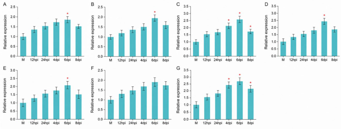
To further identify GH28 candidate genes associated with C. pyri infection, seven GH28 genes were selected based on their predicted relevance to pathogenicity, and their expression patterns at different time points during infection of Korla fragrant pear branches were examined by RT-qPCR. The results showed that all seven candidate GH28 genes were upregulated to varying degrees after inoculation, suggesting that these GH28 members may be involved in the infection of Korla fragrant pear branches by C. pyri[10,29].
Based on the intensity of transcriptional responses and the timing of peak expression, the tested genes were classified into three groups. The first group comprised strongly responsive genes, including VP1G_08835 and VP1G_03209. VP1G_08835 showed a continuous increase in expression after inoculation and reached its highest expression level at 6 days post-inoculation (dpi), with an expression level approximately 2.77-fold higher than that of the control. This gene exhibited the strongest response among all tested genes. VP1G_03209 was also significantly upregulated during infection and peaked at 6 dpi, with an expression level approximately 2.57-fold higher than that of the control. These results indicate that VP1G_08835 and VP1G_03209 may represent important candidate genes closely associated with the infection process among the GH28 family members of C. pyri. The second group consisted of moderately responsive genes, including VP1G_05365 and VP1G_05880. These two genes were continuously upregulated after inoculation, with peak expression also occurring around 6 dpi, reaching approximately 2.44- and 2.15-fold higher levels than those of the control, respectively. This expression pattern suggests that they may be involved in host cell wall degradation or lesion expansion during the middle to late stages of infection. The third group included mildly responsive genes, namely VP1G_00675, VP1G_03071, and VP1G_08384. These genes also showed increased expression after inoculation, but the magnitude of induction was relatively low, suggesting that they may play auxiliary roles during infection.
Taken together, the protein structure prediction, subcellular localization analysis, and expression profiling results indicated that both VP1G_08835 and VP1G_03209 exhibited strong features of pathogenicity-related candidate genes. Among them, VP1G_08835 was predicted to encode an extracellular protein, contained a complete GH28 conserved domain, and showed the strongest transcriptional response during infection. Therefore, VP1G_08835 was selected as a representative candidate gene for further gene deletion, complementation, and overexpression analyses to validate its role in the pathogenicity of C. pyri.
Figure 5.
Relative expression levels of C. pyri GH28 candidate genes at different time points during infection of Korla fragrant pear branches.(A) VP1G_00675; (B) VP1G_03071; (C) VP1G_03209; (D) VP1G_05365; (E) VP1G_05880; (F) VP1G_08384; and (G) VP1G_08835. Asterisks indicate significant differences: *, P < 0.05; **, P < 0.01; ***, P < 0.001.
Figure 5.
Relative expression levels of C. pyri GH28 candidate genes at different time points during infection of Korla fragrant pear branches.(A) VP1G_00675; (B) VP1G_03071; (C) VP1G_03209; (D) VP1G_05365; (E) VP1G_05880; (F) VP1G_08384; and (G) VP1G_08835. Asterisks indicate significant differences: *, P < 0.05; **, P < 0.01; ***, P < 0.001.
3.7. VP1G_08835 Is an Important GH28 Candidate Gene Required for Full Virulence in C. pyri
To preliminarily characterize the structural features of the protein encoded by VP1G_08835, its amino acid sequence was subjected to bioinformatic analysis. The results showed that the protein contains an N-terminal signal peptide with a predicted cleavage site[26]. The central region harbors a conserved domain and also contains low-complexity and intrinsically disordered regions (Figure 6A). Further analysis indicated that the VP1G_08835 protein lacks obvious transmembrane helices and is likely secreted extracellularly via the signal peptide-mediated secretory pathway (Figure 6B).
To investigate the function of VP1G_08835 in C. pyri, the gene was deleted using double-joint PCR and polyethylene glycol (PEG)-mediated protoplast transformation. Two deletion mutants, ΔVP1G_08835-85 and ΔVP1G_08835-107, were obtained. The full-length VP1G_08835 sequence was then cloned into the PDL2 plasmid vector and introduced into the protoplasts of the ΔVP1G_08835-85 mutant, yielding the complemented strains ΔVP1G_08835-C-1 and ΔVP1G_08835-C-10. In parallel, the construct was introduced into wild-type C. pyri protoplasts, generating the overexpression strains VP1G_08835-OE-1 and VP1G_08835-OE-22.
Mycelial growth assays showed that deletion of VP1G_08835 affected colony growth of C. pyri to a certain extent, as the colony diameter of ΔVP1G_08835 differed significantly from that of the wild type (WT). In contrast, the complemented strain ΔVP1G_08835-C partially restored the growth phenotype and did not differ significantly from WT. These results indicate that VP1G_08835 contributes to the normal vegetative growth of C. pyri (Figure 6C).
Pathogenicity assays on detached twigs using the WT, ΔVP1G_08835, ΔVP1G_08835-C, and VP1G_08835-OE strains showed that the virulence of ΔVP1G_08835 was reduced. The mean lesion length on twigs was 47.32 mm, representing a 26.62% reduction relative to the WT strain (Figure 6D). Inoculation with ΔVP1G_08835-C restored the WT phenotype, further confirming the crucial role of VP1G_08835 in C.pyri infection.
4. Discussion and Conclusions
In this study, the GH28 gene family members of the pear rot pathogen C.pyri were systematically identified. By integrating analyses of protein physicochemical properties, conserved motifs, gene structure, phylogenetic relationships, promoter cis-acting elements, genomic distribution, and expression patterns during infection, we preliminarily characterized the composition of this family in C.pyri and its potential association with pathogenicity. The results showed that most C.pyri GH28 family members were predicted to encode secreted hydrophilic proteins and possessed relatively conserved functional motifs, suggesting that they may be involved in the degradation of host cell wall pectin components[12,13]. In addition, the observed differences in gene structure, motif composition, and genomic distribution among family members further indicate that, while retaining the core catalytic function, this gene family has undergone a certain degree of structural and functional divergence[22,23].
Infection-induced expression analysis showed that multiple GH28 genes were upregulated to varying degrees during the infection of pear twigs by C.pyri. Among them, VP1G_08835 and VP1G_03209 exhibited a strong transcriptional response during infection, indicating that they may be candidate genes closely associated with the pathogenic process of C. pyri[33]. The presence of hormone-responsive, wound-responsive, defense/stress-responsive, and low-temperature-responsive cis-elements in the promoter regions also suggests that GH28 gene expression may be influenced by host wounding, defense signaling, and external environmental cues[28,29]; however, the underlying regulatory mechanisms remain to be further validated.
On this basis, VP1G_08835 was further functionally validated in the present study. The results showed that the protein encoded by VP1G_08835 possesses typical secreted protein features and may function as an extracellular effector involved in pathogen–host interactions[30,31]. After deletion of VP1G_08835, both colony growth of C.pyri and pathogenicity on detached pear twigs were affected, whereas the complemented strain restored the corresponding phenotypes, indicating that VP1G_08835 plays an important role in normal growth and full virulence of C.pyri[5,32]. These findings further suggest that not all members of the GH28 family merely function redundantly in pectin degradation; instead, some members may play key roles during pathogen infection[12,33].
Overall, this study, from both the gene family level and single-gene functional validation, preliminarily revealed the relationship between the C.pyri GH28 gene family and the pathogenic process of pear rot, providing a molecular basis for understanding how the pathogen breaches the host cell wall barrier, colonizes host tissues, and causes lesion development[27,28]. In particular, VP1G_08835 may serve as an important candidate gene for subsequent studies on the pathogenic mechanism of C.pyri and the identification of potential control targets. Future work may further combine enzyme activity assays, analysis of pectin degradation products, detection of host immune responses, and multi-omics approaches to clarify the specific roles of the GH28 enzyme system in the development of pear rot, thereby providing a theoretical basis and candidate targets for the green management of pear rot in Korla pear.
Supplementary Materials
The following supporting information can be downloaded at the website of this paper posted on Preprints.org.
Author Contributions
Conceptualization, Z.X and Z.W.; methodology, Z.X. and L.W.; software, Z.X. and Y.D.; validation, S.P.; formal analysis, Z.X. and S.P.; investigation, Z.X., C.Y. and Z.W.; resources, Z.X., Z.W. and L.W.; writing—original draft preparation, Z.X.; writing—review and editing, S.P., Z.L. and C.Y.; visualization, Y.D.; supervision, Z.W.; funding acquisition, Z.W. and L.W. All authors have read and agreed to the published version of the manuscript.
Funding
This research was funded by the Guiding Science and Technology Program of Xinjiang Production and Construction Corps (Grant No. 2024ZD078), the 2024 “Tianchi Talent” Young Doctoral Talent Introduction Program of Xinjiang Uygur Autonomous Region, and the President’s Fund for Doctoral Talents of Tarim University (Grant No. TDZKBS202404).
Institutional Review Board Statement
Not applicable.
Informed Consent Statement
Not applicable.
Data Availability Statement
The data in this study are available on request from the corresponding author/first author.
Conflicts of Interest
The authors declare no conflict of interest.
References
- Du, Q.; Kong, L.; Zhao, S. F.; et al. Pathogen identification of pear tree rot in Korla pear from Xinjiang. Xinjiang Agric. Sci. 2013, 50(12), 2258–2265. [Google Scholar]
- Kubicek, C. P.; Starr, T. L.; Glass, N. L. Plant cell wall-degrading enzymes and their secretion in plant-pathogenic fungi[J]. Annu. Rev. Phytopathol. 2014, 52(1), 427–451. [Google Scholar] [PubMed]
- Kars, I.; Krooshof, G.H.; Wagemakers, L.; Joosten, R.; Benen, J.A.E.; van Kan, J.A.L. Necrotizing activity of five Botrytis cinerea endopolygalacturonases produced in Pichia pastoris[J]. Plant J. 2005, 43(2), 213–225. [Google Scholar] [PubMed]
- Blanco-Ulate, B.; Morales-Cruz, A.; Amrine, K. C. H.; et al. Genome-wide transcriptional profiling of Botrytis cinerea genes targeting plant cell walls during infections of different hosts[J]. Front. Plant Sci. 2014, 5, 435. [Google Scholar] [PubMed]
- ten Have, A.; Mulder, W.; Visser, J.; et al. The endopolygalacturonase gene Bcpg1 is required for full virulence of Botrytis cinerea[J]. Mol. Plant-Microbe Interact. 1998, 11(10), 1009–1016. [Google Scholar] [PubMed]
- Xiao, Y.; Sun, G.; Yu, Q.; et al. A plant mechanism of hijacking pathogen virulence factors to trigger innate immunity[J]. Science 2024, 383(6684), 732–739. [Google Scholar] [PubMed]
- Bailey, T. L.; Boden, M.; Buske, F. A.; et al. MEME SUITE: tools for motif discovery and searching[J]. Nucleic Acids Res. 2009, 37(Web Server issue), W202–W208. [Google Scholar] [CrossRef]
- Kumar, S.; Stecher, G.; Tamura, K. MEGA7: Molecular evolutionary genetics analysis version 7.0 for bigger datasets[J]. Mol. Biol. Evol. 2016, 33(7), 1870–1874. [Google Scholar] [PubMed]
- Lescot, M.; Déhais, P.; Thijs, G.; et al. PlantCARE, a database of plant cis-acting regulatory elements and a portal to tools for in silico analysis of promoter sequences[J]. Nucleic Acids Res. 2002, 30(1), 325–327. [Google Scholar] [PubMed]
- Chen, C.; Chen, H.; Zhang, Y.; et al. TBtools: An integrative toolkit developed for interactive analyses of big biological data[J]. Mol. Plant 2020, 13(8), 1194–1202. [Google Scholar] [PubMed]
- Livak, K. J.; Schmittgen, T. D. Analysis of relative gene expression data using real-time quantitative PCR and the 2-ΔΔCT method[J]. Methods 2001, 25(4), 402–408. [Google Scholar] [PubMed]
- Villarreal, F.; Stocchi, N.; ten Have, A. Functional classification and characterization of the fungal glycoside hydrolase 28 protein family[J]. J. Fungi 2022, 8(3), 217. [Google Scholar] [CrossRef] [PubMed]
- van Santen, Y.; Benen, J. A. E.; Schröter, K.-H.; et al. 1.68-Å crystal structure of endopolygalacturonase II from Aspergillus niger and identification of active site residues by site-directed mutagenesis[J]. J. Biol. Chem. 1999, 274(43), 30474–30480. [Google Scholar] [PubMed]
- Zhang, L.; Zhang, F.; Melotto, M.; Yao, J.; He, S.Y. Jasmonate signaling and manipulation by pathogens and insects[J]. J. Exp. Bot. 2017, 68(6), 1371–1385. [Google Scholar] [PubMed]
- Liu, C.; Yu, H.; Rao, X.; Li, L.; Dixon, R.A. Abscisic acid regulates secondary cell-wall formation and lignin deposition in Arabidopsis thaliana through phosphorylation of NST1[J]. Proceedings of the National Academy of Sciences of the United States of America 2021, 118(5), e2010911118. [Google Scholar] [PubMed]
- Sivakumaran, A.; Akinyemi, A.; Mandon, J.; et al. ABA suppresses Botrytis cinerea elicited NO production in tomato to influence H2O2 generation and increase host susceptibility[J]. Front. Plant Sci. 2016, 7, 709. [Google Scholar] [PubMed]
- Lievens, L.; Pollier, J.; Goossens, A.; et al. Abscisic acid as pathogen effector and immune regulator[J]. Front. Plant Sci. 2017, 8, 587. [Google Scholar] [CrossRef] [PubMed]
- Berrocal-Lobo, M.; Molina, A. Ethylene response factor 1 mediates Arabidopsis resistance to the soilborne fungus Fusarium oxysporum[J]. Mol. Plant-Microbe Interact. 2004, 17(7), 763–770. [Google Scholar] [CrossRef] [PubMed]
- Takahashi, H.; Kanayama, Y.; Zheng, M. S.; et al. Antagonistic interactions between the SA and JA signaling pathways in Arabidopsis modulate expression of defense genes and gene-for-gene resistance to cucumber mosaic virus[J]. Plant Cell Physiol. 2004, 45(6), 803–809. [Google Scholar] [PubMed]
- Ma, K.-W.; Ma, W. Phytohormone pathways as targets of pathogens to facilitate infection[J]. Plant Mol. Biol. 2016, 91(6), 713–725. [Google Scholar] [CrossRef] [PubMed]
- Gladyshev, E. Repeat-induced point mutation and other genome defense mechanisms in fungi[J]. Microbiol. Spectr. 2017, 5(4), FUNK-0042-2017. [Google Scholar]
- Aspeborg, H.; Coutinho, P. M.; Wang, Y.; et al. Evolution, substrate specificity and subfamily classification of glycoside hydrolase family 5 (GH5)[J]. BMC Evol. Biol. 2012, 12(1), 186. [Google Scholar] [PubMed]
- Viborg, A. H.; Terrapon, N.; Lombard, V.; et al. A subfamily roadmap of the evolutionarily diverse glycoside hydrolase family 16 (GH16)[J]. J. Biol. Chem. 2019, 294(44), 15973–15986. [Google Scholar] [PubMed]
- Emms, D. M.; Kelly, S. OrthoFinder: phylogenetic orthology inference for comparative genomics[J]. Genome Biol. 2019, 20(1), 238. [Google Scholar] [CrossRef] [PubMed]
- Minh, B. Q.; Schmidt, H. A.; Chernomor, O.; et al. IQ-TREE 2: New models and efficient methods for phylogenetic inference in the genomic era[J]. Mol. Biol. Evol. 2020, 37(5), 1530–1534. [Google Scholar] [PubMed]
- Teufel, F.; Almagro Armenteros, J. J.; Johansen, A. R.; et al. SignalP 6.0 predicts all five types of signal peptides using protein language models[J]. Nat. Biotechnol. 2022, 40(7), 1023–1025. [Google Scholar] [PubMed]
- Kalunke, R. M.; Tundo, S.; Benedetti, M.; et al. An update on polygalacturonase-inhibiting protein (PGIP), a leucine-rich repeat protein that protects crop plants against pathogens[J]. Front. Plant Sci. 2015, 6, 146. [Google Scholar] [PubMed]
- Ferrari, S.; Savatin, D. V.; Sicilia, F.; et al. Oligogalacturonides: plant damage-associated molecular patterns and regulators of growth and development[J]. Front. Plant Sci. 2013, 4, 49. [Google Scholar] [PubMed]
- Pontiggia, D.; Benedetti, M.; Costantini, S.; et al. Dampening the DAMPs: How plants maintain the homeostasis of cell wall molecular patterns and avoid hyper-immunity[J]. Front. Plant Sci. 2020, 11, 613259. [Google Scholar] [PubMed]
- Bradley, E. J.; Ökmen, B.; Doehlemann, G.; et al. Secreted glycoside hydrolase proteins as effectors and invasion patterns of plant-associated fungi and oomycetes[J]. Front. Plant Sci. 2022, 13, 853106. [Google Scholar] [PubMed]
- Borras-Hidalgo, O.; Caprari, C.; Hernandez-Estevez, I.; et al. A gene for plant protection: expression of a bean polygalacturonase inhibitor in tobacco confers a strong resistance against Rhizoctonia solani and two oomycetes[J]. Front. Plant Sci. 2012, 3, 268. [Google Scholar] [PubMed]
- Feng, H.; Wang, C.; He, Y.; et al. Apple Valsa canker: insights into pathogenesis and disease control[J]. Phytopathol. Res. 2023, 5: 45. [Google Scholar]
- Xu, Z.; Luo, Z.; Xiong, D.; et al. A glycoside hydrolase 12 protein from Cytospora chrysosperma triggers plant immunity but is not essential to virulence[J]. Phytopathol. Res. 2023, 5: 32. [Google Scholar]
Figure 1.
Phylogenetic relationships, conserved motifs, and gene structures of C. pyri GH28 genes.
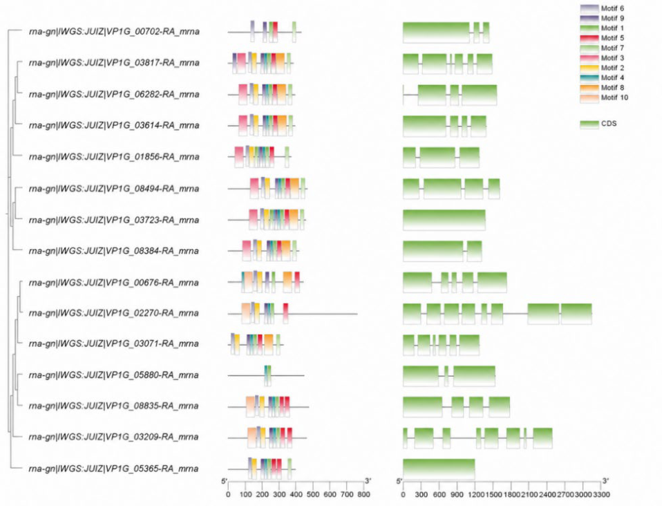
Figure 2.
Phylogenetic tree of the GH28 gene family in five Cytospora species.
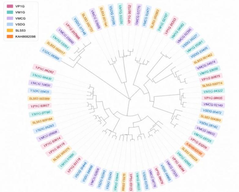
Figure 3.
Distribution of cis-acting elements in the promoters of C. pyri GH28 genes.
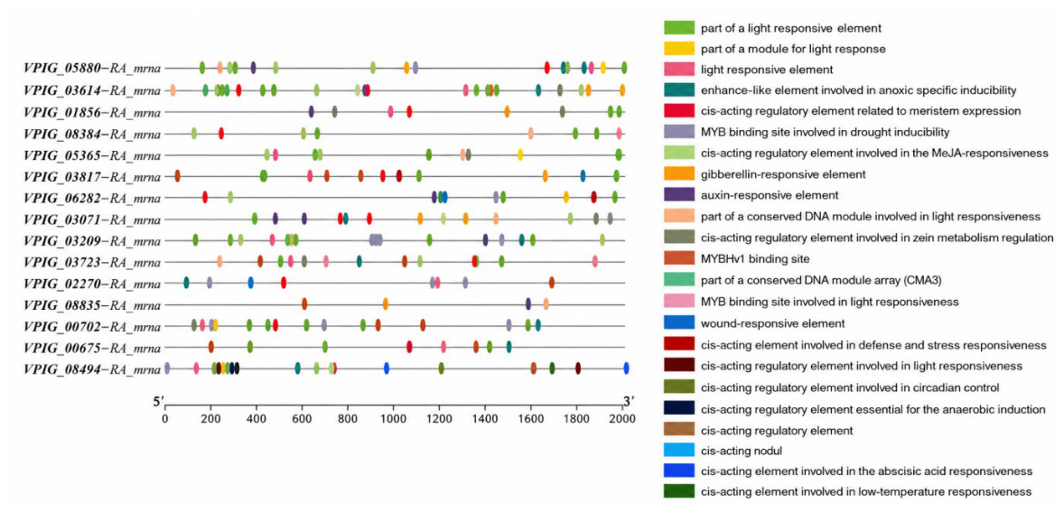
Figure 4.
Distribution of C. pyri GH28 genes and inter-scaffold collinearity relationships.
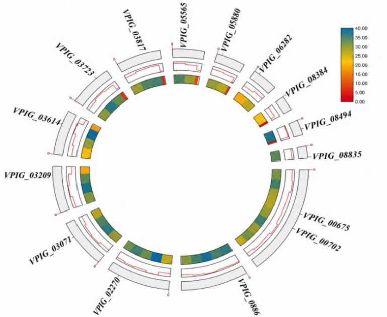
Figure 6.
Structural prediction of VP1G_08835 and phenotypic analysis of the related strains.(A) Domain organization and signal peptide prediction of the VP1G_08835-encoded protein.(B) Prediction of the secretion characteristics and subcellular localization of VP1G_08835.(C) Colony morphology and statistical analysis of colony diameter for different strains grown on PDA.(D) Lesion phenotypes and lesion length measurements on detached pear twigs inoculated with different strains.
Figure 6.
Structural prediction of VP1G_08835 and phenotypic analysis of the related strains.(A) Domain organization and signal peptide prediction of the VP1G_08835-encoded protein.(B) Prediction of the secretion characteristics and subcellular localization of VP1G_08835.(C) Colony morphology and statistical analysis of colony diameter for different strains grown on PDA.(D) Lesion phenotypes and lesion length measurements on detached pear twigs inoculated with different strains.
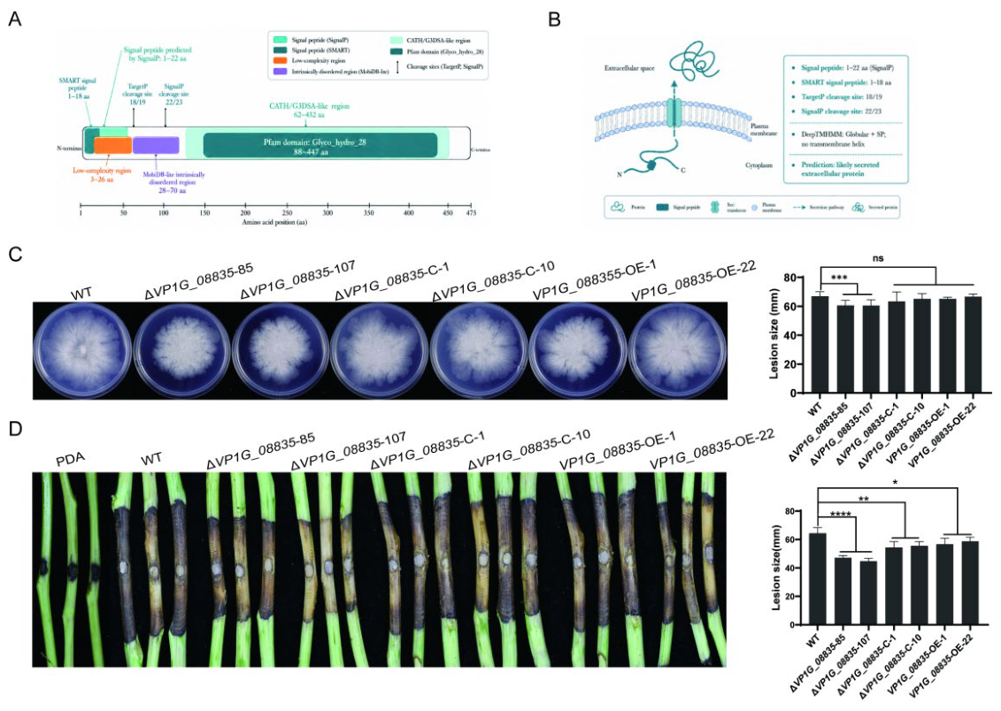

Table 1.
Structural information of C. pyri GH28 genes.
| Sequence ID | Number of Amino Acid | Molecular Weight | Theoretical pI | Instability Index | Aliphatic Index | Grand Average of Hydropathicity | Cell localization |
|---|---|---|---|---|---|---|---|
| VP1G_08384 | 414 | 41974.22 | 4.32 | 27.20 | 78.26 | 0.021 | Extracell |
| VP1G_00702 | 430 | 47050.52 | 4.60 | 23.61 | 75.42 | -0.062 | Extracell |
| VP1G_08835 | 475 | 51563.56 | 5.09 | 33.99 | 76.19 | -0.156 | Extracell |
| VP1G_03209 | 461 | 50326.97 | 4.52 | 39.38 | 79.28 | -0.216 | Extracell |
| VP1G_01856 | 371 | 38820.05 | 4.48 | 29.72 | 88.46 | -0.083 | Extracell |
| VP1G_02270 | 758 | 82574.07 | 4.82 | 44.12 | 73.21 | -0.596 | Extracell |
| VP1G_05880 | 445 | 47503.72 | 4.47 | 25.17 | 73.24 | -0.048 | Extracell |
| VP1G_06282 | 391 | 40267.62 | 4.76 | 23.80 | 75.58 | -0.132 | Extracell |
| VP1G_03723 | 456 | 46796.17 | 4.40 | 34.23 | 69.30 | -0.103 | Extracell |
| VP1G_00675 | 440 | 48014.64 | 5.22 | 29.39 | 78.43 | -0.275 | Extracell |
| VP1G_08494 | 462 | 47171.11 | 4.25 | 30.57 | 78.14 | 0.087 | Extracell |
| VP1G_05365 | 397 | 40875.91 | 4.50 | 28.97 | 67.83 | -0.143 | Extracell |
| VP1G_03817 | 383 | 39581.31 | 4.11 | 27.71 | 75.64 | -0.163 | Extracell |
| VP1G_03614 | 394 | 40422.68 | 4.18 | 31.94 | 82.87 | 0.003 | Extracell |
| VP1G_03071 | 327 | 35573.51 | 4.50 | 42.35 | 74.25 | -0.238 | Cytosol |
Disclaimer/Publisher’s Note: The statements, opinions and data contained in all publications are solely those of the individual author(s) and contributor(s) and not of MDPI and/or the editor(s). MDPI and/or the editor(s) disclaim responsibility for any injury to people or property resulting from any ideas, methods, instructions or products referred to in the content. |
© 2026 by the authors. Licensee MDPI, Basel, Switzerland. This article is an open access article distributed under the terms and conditions of the Creative Commons Attribution (CC BY) license (http://creativecommons.org/licenses/by/4.0/).
Copyright: This open access article is published under a Creative Commons CC BY 4.0 license, which permit the free download, distribution, and reuse, provided that the author and preprint are cited in any reuse.


