Submitted:
26 June 2026
Posted:
29 June 2026
You are already at the latest version
Abstract
Background: Endothelial dysfunction is observed in some disease states, which is characterized by a decrease in nitric oxide NO and an increase in superoxide anion (O2-). Therapeutic approaches seek to restore the balance between NO and superoxide, highlighting the potential of herbal medicines such as Opuntia ficus-indica and Rosa canina due to their antioxidant properties. Aims: This study aimed evaluate the effects of combining extracts from Opuntia ficus-indica and Rosa canina in preventing or reversing endothelial dysfunction induced by bacterial lipopolysaccharide (LPS). Methods: A vascular reactivity experiment was conducted by using aortic rings from Wistar rats. Endothelium dysfunction was induced by LPS, and endothelial dependent vasodilation was performed to acetylcholine. Incubation with extracts was performed after (reverse) or before (prevent) LPS incubation. Results: The combination O. ficus-indica and R. canina was able to completely reverse and partially prevent endothelial function induced by LPS. Treatment with R. canina or O. ficus-indica alone did not improve relaxation. Additionally, treatment of aortic rings or HUVEC cells with these plant extracts, alone or in combination, reduced oxidative stress. Conclusion: Our results indicate that combining standardized extracts from O. ficus-indica and R. canina can reverse endothelial dysfunction induced by LPS through a mechanism dependent on decreased reactive oxygen species.
Keywords:
herbal medicines
; inflammation
; vascular reactivity
; Opuntia ficus-indica and Rosa canina
1. Introduction
Endothelial cells, which line the interior surface of the entire vascular system, are a complex, specialized, and metabolically active organ essential for maintaining vascular health and homeostasis [1]. Under physiological conditions, their functions are modulated by the release of various factors, including major vasodilators, antithrombotic agents, and antiatherogenic factors, with nitric oxide (NO) being the most characterized and produced by constitutive endothelial NO synthase (eNOS) [2,3]. In disease states, including those involving cardiovascular risk factors, endothelial cells lose their protective functions due to alterations in the release of these factors. This condition, known as ‘endothelial dysfunction’, is primarily characterized by impaired NO bioavailability [1,3].
Nitric oxide is a gas with a short half-life in the biological environment. During endothelial dysfunction, its synthesis by NOS may be reduced, or its breakdown by the radical superoxide (O2-) may be increased [2,4]. Elevated levels of O2- can modulate vascular tone, promote vasoconstriction, and/or reduce vasodilation by interacting with endogenous mediators such as NO. The reaction between O2 and NO forms peroxynitrite (ONOO-) [5,6]. Chronically elevated O2- levels and decreased NO levels contribute to the pathophysiology of cardiovascular diseases, correlating with a loss of redox control [6].
Activation of the toll-like receptor 4 (TLR4) stimulates the formation of reactive oxygen species within the intracellular environment by activating transcription factors NF-κB, AP-1, and IRF-5, leading to the expression of pro-inflammatory cytokines [7,8,9]. Toll-like receptors (TLRs) serve as the first line of defense against microbes, recognizing invading pathogens and endogenous danger molecules released from dying cells and damaged tissues. They play a crucial role in linking innate and adaptive immunity [10]. Activation of TLR4 by lipopolysaccharides (LPS) can induce endothelial dysfunction, a method frequently used to induce endothelial dysfunction in experimental models [7,9].
Given the imbalance between NO and superoxide observed in endothelial dysfunction, pharmacological strategies to improve this imbalance could be beneficial for cardiovascular diseases. Thus, the potential of medicinal plants is noteworthy, as numerous studies have identified their efficacy in treating several diseases induced by endothelial dysfunction, particularly in animal models [3].
Considering the synergistic actions between compounds from the same species and associations between different species, isolated compounds often demonstrate lower therapeutic activity compared to the original crude extracts. In traditional medicine, most herbal remedies are based on a mixture of plants, where the interaction between different constituents amplifies the therapeutic potential. This synergistic effect can be fundamental in the design of effective therapeutic agents.
Species of the genus Opuntia, especially Opuntia ficus-indica, have been used for centuries both as food resources and in traditional folk medicine due to their nutritional properties and benefits in managing chronic diseases, including diabetes, obesity, cardiovascular diseases, and cancer. These species are widely distributed across America, Africa, and the Mediterranean basin. Different parts of the plant, including the cladodes, exhibit beneficial properties attributed to their high content of antioxidants (such as flavonoids and ascorbate), pigments (carotenoids and betalains), and phenolic acids. Additional phytochemical components, including biopeptides and soluble fibers, also contribute to their medicinal properties [11].
Rosa canina (rose hips) is a medicinal herb known for its extensive benefits, which include antioxidant, antimicrobial, anticancer, antidiabetic, anti-inflammatory, and immunomodulatory effects. These benefits are primarily attributed to its high content of vitamin C, minerals, carotenoids, flavonoids, and other phenolic compounds present in its fruits [12,13].
This study evaluated the effects of combining extracts from Opuntia ficus-indica and Rosa canina, as well as the effects of extracts isolated from these species, in preventing endothelial dysfunction induced by bacterial lipopolysaccharide (LPS).
2. Methods
2.1. Experimental Animals
This work was approved by the Animal Care and Use Committee of the Federal University of São Carlos (CEUA nº 7936070618) which follows the follows the provisions of Law No. 11,794, of October 8, 2008, and other standards applicable to the use of animals in teaching and/or research, especially the Normative Resolutions of the National Council for the Control of Animal Experimentation - CONCEA in Brazil. Twenty male Wistar rats with 250 g to 300 g were used. Animals were maintained on a light-dark cycle with free access to both food (standard rat chow) and water. The animals were sacrificed by decapitation and some tissues were collected for further analysis.
2.2. Vascular Reactivity Studies
Rats were anesthetized with isoflurane, euthanized by decapitation and the thoracic aortas were isolated. Aortic rings, 3 mm in length, were placed in bath chambers (5 mL) for isolated organs containing Krebs solution at 37˚C, continuously bubbled with 95% O2 and 5% CO2, pH 7.4, in an isometric myograph (Mulvany-Halpern-model 610 DMT-USA, Marietta, GA) and recorded by a PowerLab8/SP data acquisition system (ADInstruments Pty Ltd., Colorado Springs, CO).
The aortic rings were submitted to a tension of 1.5 g, which was readjusted every 15 min throughout a 60-min equilibration period before the addition of the given drug. Endothelial integrity was assessed by the degree of relaxation induced by 1 μM of acetylcholine after contraction of the aortic ring by phenylephrine (0.1 μM) and if the relaxation with acetylcholine was less than 80%, the ring was discarded. Endothelial dysfunction was induced by incubating aortic rings with LPS (50 µg/mL) for 60 min [14]. We carry out protocols to evaluate whether reversive (therapeutic) or preventive treatment with standardized plant extracts from O. ficus-indica, R. canina and association O. ficus-indica + R. canina improves endothelial function in isolated aortic rings. Incubation with standardized plant extracts were performed after (reverse) or before (prevent) LPS incubation. Treatments were carried out for 30 minutes with 1 mg/mL of O. ficus-indica or 1 mg/mL of R. canina or 0.5 mg/mL of O. ficus-indica + 0.5 mg/mL of R. canina (Drenow C®). The incubation concentration (1 mg/mL total extract) was selected based on concentrations commonly used in in vitro studies with medicinal plant extracts, in which this concentration has been shown to be non-cytotoxic and to enhance the antioxidant status of endothelial cells [24]. Furthermore, the experimental design was intended to investigate a possible synergistic interaction between O. ficus-indica and R. canina while maintaining the same total extract concentration (1 mg/mL) among experimental groups; therefore, isolated extracts were tested at 1 mg/mL, whereas the combination group received 0.5 mg/mL of each extract (1:1, total concentration of 1 mg/mL).
After treatments, aortic rings were contracted with a vasoconstrictor (phenylephrine) and then concentration effect curves were performed for acetylcholine, to obtain endothelium-dependent vasodilation and evaluates the endothelial function. Concentration effect curves were fitted with a sigmoidal dose-response, and the maximum relaxant effect (ME) were obtained.
2.3. Aorta Lipid Peroxidation (Ferrous Oxidation-Xylenol Orange – FOX)
After vascular reactivity studies, aortic rings were frozen until quantification. We carried out protocols to evaluate whether reversive (therapeutic) treatment with standardized plant extracts is able to improves lipid peroxidation induced by LPS. 30 µg of aortic rings homogenates (previously treated with 10% TCA) were incubated for 30 minutes at room temperature with 900 µL of reactive mixture containing 100 µM xylene orange, 250 µM FeSO4 (ammonium ferrous sulfate), 25 mM of H2SO4 and 4 mM of hydroxytoluene butylate (BHT) in 90% (v/v) metal. The Fe3+ formed in the presence of hydroperoxides reacts with the orange xylenol forming a colored compound with an absorption peak at 560 nm. The results were expressed as nmol of hydroperoxide per mg of protein [15].
2.4. Cell Culture
Immortalized human umbilical endothelial cells (HUVEC) were grown in DMEM (Inlab) supplemented with 10% of fetal calf serum, antibiotics, and antimycotics. The HUVECs were maintained at 37 ± 2 ºC in a 5% CO2 atmosphere. The cells in the confluence of 80 to 90% were trypsinized, centrifuged at 1200 rpm for 5 minutes, and plated in 96-well plates.
After inducing endothelial dysfunction with LPS (50 µg/mL) for 60 min, HUVECs cells were treated with herbal extracts. Treatments were carried out for 30 minutes with 1 mg/mL of O. ficus-indica or 1 mg/mL of R. canina or 0.5 mg/mL of O. ficus-indica + 0.5 mg/mL of R. canina (Drenow C®). The evaluation of intracellular reactive oxygen species (ROS) was performed by lucigenin dye (5 µM), after incubation for 15 minutes. The increase in fluorescence intensity was measured by fluorescence microplate reader (SpectraMaxGeminiXS, Molecular Devices) at 510 nm and 595 excitation wavelength pair.
2.5. Standardized Plant Extracts of Opuntia Ficus-indica and Rosa canina
The standardized plant extracts of O. ficus-indica, R. canina or O. ficus-indica + R. canina association (1:1) were kindly provided by Florien Fitoativos LTDA (SM Empreendimentos Farmaceuticos Ltda). These standardized plant extracts association make up the formulation of a product registered under the name Drenow C®. This product has a triple standardization of 10% betalains, 0.06% indicaxanthin and 30% vitamin C. The isolated extract from O. ficus-indica is standardized in 10% betalains and 0.06% indicaxanthin. The isolated extract from R. canina is standardized in 30% vitamin C. The extracts were solubilized in water, just before perform experiments.
2.6. Statistical Analysis
Statistical analysis of the results was performed using GraphPad Prism version 3.0. Statistical significance was tested by ANOVA one way (post hoc test: Newman–Keuls). Data are expressed as mean ± S.E.M. In each set of experiments, n indicates the number of rats studied. Values of p < 0.05 were considered significant.
3. Results
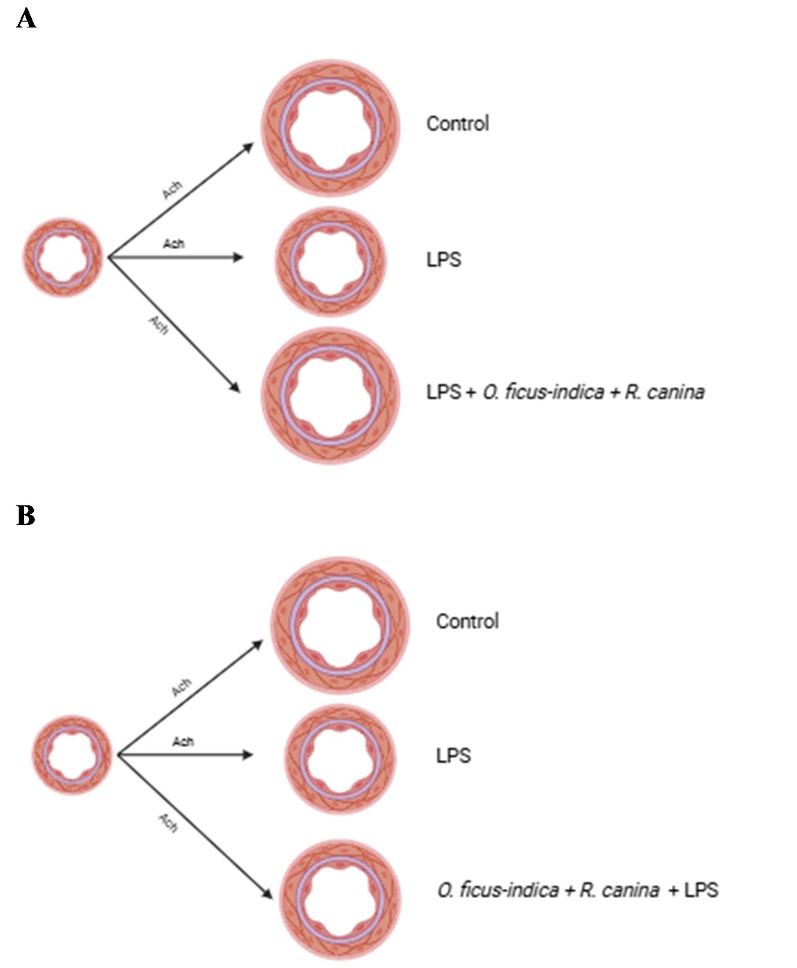
We perform experiments to evaluate the potential of standardized plant extracts from O. ficus-indica, R. canina, and the association of O. ficus-indica + R. canina in reverse or prevent endothelial function induced by LPS. Figure 1 and Figure 2 are presented the results of reversal, and Figure 3 and Figure 4 are presented the results of prevention.
Aortic ring treatment with LPS was able to induce endothelial dysfunction, impairing the relaxation endothelium-dependent to acetylcholine (Control ME: 97.90 ± 0.49 %, n = 6; LPS ME: 44.18 ± 3.30%, n = 6, p < 0.001 – Figure 1). After inducing endothelial dysfunction with LPS, the aortic rings were treated with herbal extracts. Treatment with R. canina or O. ficus-indica improved the relaxation endothelium-dependent (LPS + R. canina ME: 63.91 ± 4.40%, n = 6; LPS + O. ficus-indica ME: 60.28 ± 5.92%, n = 6), compared to LPS (LPS ME: 44.18 ± 3.30%, n = 6, p < 0.001), however, the association of standardized extract from O. ficus-indica + R. canina was able to revert endothelial function to normal (LPS + O. fícus-indica + R. canina ME: 93.96 ± 0.89%, n = 6); as can be seen at Figure 1 and Figure 2.
Before inducing endothelial dysfunction with LPS, the rings were treated with herbal extracts. Treatment with R. canina or O. ficus-indica was not able to improve the relaxation endothelium-dependent (LPS ME: 45.21 ± 2.25%, n = 6; LPS + R. canina ME: 46.47 ± 2.88%, n = 6; LPS + O. ficus-indica ME: 51.85 ± 4.18%, n = 6), however, the association of standardized extract from O. ficus-indica + R. canina was able to improve endothelial function but was not able to revert to normal (LPS + O. ficus-indica + R. canina ME: 63.916 ± 4.41, n = 6; LPS ME: 45.21 ± 2.25%, n = 6, p < 0.01).
Levels of lipid peroxidation in aortic rings increased by LPS treatment (13.51 ± 0.41 nM CHP, n=6) compared to control (2.95 ± 0.11 nM CHP, n = 6; p < 0.001). After inducing endothelial dysfunction with LPS, the rings were treated with herbal extracts. Treatment with O. ficus-indica, R. canina, or R. canina + O. ficus-indica decreased the lipid peroxidation in aortic rings (LPS: 13.51 ± 0.41 nM CHP, n = 6 > LPS + R. canina: 5.25 ± 0.38 nM CHP, n = 6; LPS + O. ficus-indica: 9.52 ± 0.34 nM CHP, n = 6; LPS + R. canina + O. ficus-indica: 5.58 ± 0.15 nM CHP, n = 6, p < 0.001) (Figure 5).
Intracellular reactive oxygen species (ROS) detection was performed by lucigenin dye, in isolated endothelial cells. After inducing endothelial dysfunction with LPS, the rings were treated with herbal extracts. Treatment with LPS induced an increase in intracellular ROS concentration (LPS: 164.00 ± 1.79 AU, n = 3; Control: 138.75 ± 5.84 AU, n = 3, p < 0.05). Treatment with O. ficus-indica + R. canina decreased the intracellular ROS (LPS + O. ficus-indica + R. canina: 143.98 ± 3.44 AU, n = 3, p < 0.05), compared to LPS. The treatment with Tempol also decreased the intracellular ROS concentration (LPS + Tempol: 146.01 ± 1.89 AU, n = 3, p < 0.001), compared to LPS. Tempol was used as a positive control (Figure 6).
4. Discussion
Aortic rings and HUVEC cells treated with LPS exhibited characteristics of endothelial dysfunction, identified by impaired endothelium-dependent vasodilation induced by acetylcholine and elevation of intracellular reactive oxygen species (ROS), respectively.
In this paper, experiments performed showed that LPS was able to increase ROS in both aortic rings and HUVEC cells. These effects of LPS treatment have been similarly observed in isolated aortic rings and animal cell cultures. Studies in healthy volunteers have shown that LPS infusion can induce endothelial dysfunction, as evidenced by reduced endothelium-dependent vasodilation in response to acetylcholine in forearm blood flow [16]. LPS induces endothelial dysfunction by activating inflammatory pathways, leading to cytokine expression in the endothelium and increased ROS production in the vascular wall [16]. Additionally, the treatment of animals and isolated endothelial cells with LPS was able to induce endothelial dysfunction and accumulation of intracellular ROS, respectively [9].
Our results indicate that standardized plant extracts of Opuntia ficus-indica and Rosa canina can improve endothelial function after LPS-induced dysfunction, as shown in Figure 1 and Figure 2 and Central figure. The combination of these extracts in equal proportions demonstrated the highest efficacy, completely reversing the endothelial dysfunction induced by LPS. Although both extracts administered individually partially improved endothelium-dependent relaxation, their combination at equal proportions completely restored endothelial function, indicating a synergistic effect between these plants. Importantly, this superior efficacy cannot be attributed to a greater amount of plant material, since the total extract concentration was kept constant (1 mg/mL) across all experimental groups. In traditional medicine, combining medicinal plants to amplify therapeutic potential is common, leveraging the interaction of bioactive present in these plants, which can be crucial in designing effective therapeutic agents [17].
High levels of superoxide radicals can influence blood vessel tone control, resulting in vasoconstriction and reduced dilation due to decreased nitric oxide (NO) availability [5,6]. Antioxidants such as superoxide dismutase (SOD), vitamin C, vitamin E, and tempol can improve endothelium-dependent vasodilation by inactivating superoxide radicals, highlighting the importance of ROS control for vascular health [18,19,20]. The improvement in endothelial function observed with the tested plant extracts may be due to their antioxidant bioactive present in O. ficus-indica (flavonoids, ascorbate, carotenoids, betalains, and phenolic acids) [11] and R. canina (vitamin C, minerals, carotenoids, flavonoids, and other phenolic compounds) [12,13]. The standardized extracts of O. ficus-indica and R. canina contain 10% betalains, 0.06% indicaxanthin, and 30% vitamin C, and the isolated O. ficus-indica extract is standardized to 10% betalains and 0.06% indicaxanthin, while the R. canina extract is standardized to 30% vitamin C. The combination of these bioactives likely explains the synergistic effect observed in reversing endothelial dysfunction. We acknowledge that the lack of direct mechanistic measurements (e.g., NO bioavailability, inflammatory cytokines or eNOS signaling) represents a limitation of the study. Therefore, our conclusions were carefully restricted to the demonstration that the association improves endothelial function and reduces oxidative stress, without claiming the precise molecular pathway involved.
Treatment of aortic rings or HUVEC cells with these standardized plant extracts, alone or in combination, reduced oxidative stress. The antioxidant effects of O. ficus-indica and R. canina have been previously documented. Opuntia ficus-indica is rich in antioxidants (flavonoids and ascorbic acid), pigments (carotenoids and betalains), and phenolic acids [11].
Rosa canina has demonstrated significant antioxidant properties, primarily by inactivating superoxide radicals and preventing lipid peroxidation [21,22,24]. Its effects are attributed to its high vitamin C content and other bioactives, as shown in experiments with vitamin C-depleted extracts containing proanthocyanidins and flavonoids [23,24].
5. Conclusions
Our results indicated that the association of standardized extracts from O. ficus-indica and R. canina can revert endothelial dysfunction induced by LPS, by a mechanism dependent on a decrease in reactive oxygen species.
Acknowledgments
This work was supported by grants from São Paulo Research Foundation (FAPESP grants: 2022/01093-1).
Conflicts of Interest
The authors declare that there is no conflict of interest.
References
- Lüscher, T.F. Endothelial control of vascular tone and growth. Clin. Exp. Hypertens. Part A Theory Pract. 1990, 12, 897–902. [Google Scholar] [CrossRef]
- Vanhoutte, P.M. Endothelium and control of vascular function. State of the Art lecture. Hypertension 1989, 13, 658–67. [Google Scholar] [CrossRef] [PubMed]
- Versari, D.; Daghini, E.; Virdis, A.; Ghiadoni, L.; Taddei, S. Endothelium-dependent contractions and endothelial dysfunction in human hypertension. Br. J. Pharmacol. 2009, 157, 527–36. [Google Scholar] [PubMed]
- Hamilton, C.A.; Brosnan, M.J.; McIntyre, M.; Graham, D.; Dominiczak, A.F. Superoxide excess in hypertension and aging: A common cause of endothelial dysfunction. Hypertension 2001, 37, 529–34. [Google Scholar] [PubMed]
- Gao, Y.J.; Takemori, K.; Su, L.Y.; An, W.S.; Lu, C.; Sharma, A.M.; Lee, R.M. Perivascular adipose tissue promotes vasoconstriction: The role of superoxide anion. Cardiovasc. Res. 2006, 71, 363–73. [Google Scholar] [CrossRef] [PubMed]
- Obradovic, M.; Essack, M.; Zafirovic, S.; Sudar-Milovanovic, E.; Bajic, V.P.; Van Neste, C.; Trpkovic, A.; Stanimirovic, J.; Bajic, V.B.; Isenovic, E.R. Redox control of vascular biology. Biofactors 2020, 46, 246–62. [Google Scholar] [PubMed]
- Joshi, A.D.; Dimitropoulou, C.; Thangjam, G.; Snead, C.; Feldman, S.; Barabutis, N.; Fulton, D.; Hou, Y.; Kumar, S.; Patel, V.; Gorshkov, B.; Verin, A.D.; Black, S.M.; Catravas, J.D. Heat shock protein 90 inhibitors prevent LPS-induced endothelial barrier dysfunction by disrupting RhoA signaling. Am. J. Respir. Cell Mol. Biol. 2014, 50, 170–9. [Google Scholar] [PubMed]
- Lu, Y.C.; Yeh, W.C.; Ohashi, P.S. LPS/TLR4 signal transduction pathway. Cytokine 2008, 42, 145–51. [Google Scholar] [CrossRef] [PubMed]
- Toral, M.; Romero, M.; Jiménez, R.; Robles-Vera, I.; Tamargo, J.; Martínez, M.C.; Pérez-Vizcaíno, F.; Duarte, J. Role of UCP2 in the protective effects of PPARβ/δ activation on lipopolysaccharide-induced endothelial dysfunction. Biochem. Pharmacol. 2016, 110-11, 25–36. [Google Scholar]
- El-Zayat, S.R.; Sibaii, H.; Mannaa, F.A. Toll-like receptors activation, signaling, and targeting: An overview. Bull. Natl. Res. Cent. 2019, 43, 187. [Google Scholar] [CrossRef]
- Del Socorro Santos Díaz, M.; Barba de la Rosa, A.P.; Héliès-Toussaint, C.; Guéraud, F.; Nègre-Salvayre, A. Opuntia spp.: Characterization and benefits in chronic diseases. Oxidative Med. Cell. Longev. 2017, 2017, 8634249. [Google Scholar] [CrossRef]
- Nasrolahi, A.; Hosseini, L.; Farokhi-Sisakht, F.; Mahmoudi, J.; Karimi, P.; Badalzadeh, R.; Erfani, M. Cardioprotective effect of Rosa canina L. methanolic extract on heat shock induced cardiomyocyte injury: An experimental study. J. Cardiovasc. Thorac. Res. 2020, 12, 286–93. [Google Scholar] [PubMed]
- Peña, F.; Valencia, S.; Tereucán, G.; Nahuelcura, J.; Jiménez-Aspee, F.; Cornejo, P.; Ruiz, A. Bioactive compounds and antioxidant activity in the fruit of rosehip (Rosa canina L. and Rosa rubiginosa L.). Molecules 2023, 28, 3544. [Google Scholar] [CrossRef] [PubMed]
- Huang, L.; Li, Y.; Cheng, Z.; Lv, Z.; Luo, S.; Xia, Y. PCSK9 Promotes endothelial dysfunction during sepsis via the TLR4/MyD88/NF-κB and NLRP3 pathways. Inflammation 2023, 46, 115–28. [Google Scholar] [PubMed]
- Jiang, Z.Y.; Woollard, A.C.; Wolff, S.P. Lipid hydroperoxide measurement by oxidation of Fe2+ in the presence of xylenol orange. Comparison with the TBA assay and an iodometric method. Lipids 1991, 26, 853–56. [Google Scholar] [CrossRef] [PubMed]
- Pleiner, J.; Schaller, G.; Mittermayer, F.; Zorn, S.; Marsik, C.; Polterauer, S.; Kapiotis, S.; Wolzt, M. Simvastatin prevents vascular hypo-reactivity during inflammation. Circulation 2004, 110, 3349–54. [Google Scholar] [PubMed]
- Rajčević, N.; Bukvički, D.; Dodoš, T.; Marin, P.D. Interactions between natural products – A review. Metabolites 2022, 12, 1256. [Google Scholar] [PubMed]
- Nakamura, T.; Igarashi, R.; Kurashina, T.; Saito, Y.; Hoshino, J.; Sumino, H.; Sakamoto, H.; Nagai, R. Lecithinized superoxide dismutase induces vasodilation; evidence of direct contribution of superoxide anions to modulating vascular tone. Life Sci. 1999, 64, 65–70. [Google Scholar]
- Chen, X.; Touyz, R.M.; Park, J.B.; Schiffrin, E.L. Antioxidant effects of vitamins C and E are associated with altered activation of vascular NADPH oxidase and superoxide dismutase in stroke-prone SHR. Hypertension 2001, 38, 606–11. [Google Scholar] [CrossRef] [PubMed]
- Rodrigues, G.J.; Lunardi, C.N.; Lima, R.G.; Santos, C.X.; Laurindo, F.R.; da Silva, R.S.; Bendhack, L.M. Vitamin C improves the effect of a new nitric oxide donor on the vascular smooth muscle from renal hypertensive rats. Nitric Oxide 2008, 18, 176–83. [Google Scholar] [PubMed]
- Costantino, L.; Rastelli, G.; Rossi, T.; Bertoldi, M.; Albasini, A. Composition, superoxide radicals scavenging and antilipoperoxidant activity of some edible fruits. Fitoterapia 1994, 65, 44–7. [Google Scholar]
- Kähkönen, M.P.; Hopia, A.I.; Vuorela, H.J.; Rauha, J.P.; Pihlaja, K.; Kujala, T.S.; Heinonen, M. Antioxidant activity of plant extracts containing phenolic compounds. J. Agric. Food Chem. 1999, 47, 3954–62. [Google Scholar] [CrossRef] [PubMed]
- Daels-Rakotoarison, D.A.; Gressier, B.; Trotin, F.; Brunet, C.; Luyckx, M.; Dine, T.; Bailleul, F.; Cazin, M.; Cazin, J.C. Effects of Rosa canina fruit extract on neutrophil respiratory burst. Phyther. Res. 2002, 16, 157–61. [Google Scholar] [CrossRef]
- Kerasioti, E.; Apostolou, A.; Kafantaris, I.; Chronis, K.; Kokka, E.; Dimitriadou, C.; et al. Polyphenolic composition of Rosa canina, Rosa sempervivens and Pyrocantha coccinea extracts and assessment of their antioxidant activity in human endothelial cells. Antioxidants 2019, 8, 92. [Google Scholar] [CrossRef] [PubMed]
Figure 1.
Study of reversal effect of standardized plant extracts in LPS-induced endothelial dysfunction. Cumulative concentration-effect curves performed for acetylcholine in aortas pre-contracted with phenylephrine. Each point represents the mean and the Mean Standard Error (SEM) of the data obtained in independent determinations. * Indicates the difference (p < 0.001) in ME values between Control and LPS + O. ficus-indica + R. canina vs LPS, LPS + Rosa canina and LPS + O. ficus-indica.

Figure 2.
Study of reversal effect of standardized plant extracts in LPS-induced endothelial dysfunction. Cumulative concentration-effect curves performed for acetylcholine in aortas pre-contracted with phenylephrine. Each point represents the mean and the Mean Standard Error (SEM) of the data obtained in independent determinations, in Maximum relaxant effect (ME). * Indicates the difference (p < 0.001) in ME values between Control and LPS + O. ficus-indica + R. canina vs LPS, LPS + R. canina and LPS + O. ficus-indica.

Figure 3.
Study of preventive effect of standardized plant extracts in LPS-induced endothelial dysfunction. Cumulative concentration-effect curves performed for acetylcholine in aortas pre-contracted with phenylephrine. Each point represents the mean and the Mean Standard Error (SEM) of the data obtained in independent determinations. * Indicates the difference (p < 0.001) in ME values between Control vs LPS, ** Indicates the difference (p < 0.01) between LPS vs O. ficus-indica + R. canina.

Figure 4.
Study of preventive effect of standardized plant extracts in LPS-induced endothelial dysfunction. Cumulative concentration-effect curves performed for Acetylcholine in aortas pre-contracted with phenylephrine. Each point represents the mean and the Mean Standard Error (SEM) of the data obtained in independent determinations. * Indicates the difference (p < 0.001) in ME values between Control vs LPS, ** Indicates the difference (p < 0.01) between LPS vs O. ficus-indica + R. canina.

Figure 5.
Lipid peroxidation measurement in aortic rings from rats, after vascular reactivity studies from reversal effect from standardized plant extracts in LPS-induced endothelial dysfunction. CHP: cumene hydroperoxide. * Indicates the difference (p < 0.001) values between Control vs LPS, LPS vs LPS + R. canina, LPS vs LPS + O. ficus-indica, LPS vs LPS + O. ficus-indica + R. canina.

Figure 6.
Intracellular reactive oxygen species (ROS) detection in HUVECs cells by lucigenin dye. * Indicates the difference (p < 0.05) values between Control vs LPS, LPS vs LPS + O. ficus-indica + R. canina, ** Indicates the difference (p < 0.001) values between LPS vs LPS + Tempol.
Figure 6.
Intracellular reactive oxygen species (ROS) detection in HUVECs cells by lucigenin dye. * Indicates the difference (p < 0.05) values between Control vs LPS, LPS vs LPS + O. ficus-indica + R. canina, ** Indicates the difference (p < 0.001) values between LPS vs LPS + Tempol.

Disclaimer/Publisher’s Note: The statements, opinions and data contained in all publications are solely those of the individual author(s) and contributor(s) and not of MDPI and/or the editor(s). MDPI and/or the editor(s) disclaim responsibility for any injury to people or property resulting from any ideas, methods, instructions or products referred to in the content. |
© 2026 by the authors. Licensee MDPI, Basel, Switzerland. This article is an open access article distributed under the terms and conditions of the Creative Commons Attribution (CC BY) license (http://creativecommons.org/licenses/by/4.0/).
Copyright: This open access article is published under a Creative Commons CC BY 4.0 license, which permit the free download, distribution, and reuse, provided that the author and preprint are cited in any reuse.


