

Submitted:
28 June 2026
Posted:
29 June 2026
You are already at the latest version
Abstract
Despite transformative advances in genome sequencing, a substantial proportion of patients with neurodevelopmental disorders remain undiagnosed. A central reason is that pathogenic variants rarely act in isolation: their effects depend on developmental timing, cell type, epigenomic context and the buffering capacity of the regulatory networks in which they operate. Artificial intelligence (AI) is increasingly applied across the neurogenomic workflow, supporting phenotyping, facial gestalt analysis, variant prioritisation, splicing prediction, DNA methylation episignature classification, protein modelling and multi-omic integration, yet most tools operate as independent classifiers rather than components of a unified biological model. In this review, we argue that the critical unmet need is a shift from isolated diagnostic classifiers toward interpretable models of how genomic and epigenomic perturbations propagate through regulatory networks to destabilise cell state, developmental trajectory and circuit function. We present a systems neurogenomics framework built on four constructs: regulatory load, network capacity, developmental buffering and regulatory network instability, and use chromatinopathies and DNA methylation episignatures as concrete models of convergent neurodevelopmental pathology. We propose five testable predictions arising from this framework, outline a practical AI-enabled workflow for resolving nondiagnostic sequencing cases and discuss the concept of Instability Twins as patient-specific digital neurodevelopmental simulations for forecasting developmental divergence and evaluating therapeutic windows. We address current limitations, including ancestry imbalance, overfitting, mechanistic opacity and the need for prospective validation, and argue that the next generation of AI tools in neurogenomics must be not merely accurate, but explainable, equitable and mechanistic.
Keywords:
artificial intelligence
; systems neurogenomics
; neurodevelopmental disorders
; chromatinopathies
; DNA methylation episignatures
; regulatory network instability
; developmental buffering
; instability twins
; digital neurodevelopmental twins
; single-cell transcriptomics
; multimodal AI
; precision neurogenomics
1. Introduction
Neurodevelopmental disorders (NDDs), including intellectual disability, autism spectrum disorder, developmental epilepsies, and complex neuropsychiatric conditions, collectively represent one of the most significant challenges in genomic medicine. Despite major advances in sequencing technology over the past decade [1,2,3,4,5,6,7,8,9,10,11,12,13,14], a substantial proportion of patients remain undiagnosed. Even when pathogenic variants are identified, considerable uncertainty often remains regarding disease mechanisms, phenotypic variability, prognosis, and treatment responsiveness [15].
A major reason for this interpretive gap is that neurodevelopment is fundamentally a systems-level process. Brain development requires tightly coordinated interactions among chromatin regulators, transcription factors, non-coding RNAs, signalling pathways, RNA-processing machinery, neuronal migration programs, synaptic networks, and activity-dependent plasticity mechanisms [15,16,17]. Disruption of any component within this hierarchy may alter developmental trajectories. Yet, the resulting phenotype often reflects the collective behaviour of interconnected regulatory systems rather than the isolated effect of a single gene. Consequently, diverse molecular disorders frequently converge on overlapping clinical presentations, while pathogenic variants in the same gene may produce markedly different phenotypes [16,17,18,19]. Traditional approaches to clinical genomics have largely been gene-centric, focusing on identifying pathogenic variants and assigning molecular diagnoses. Although this paradigm has transformed rare disease diagnostics, it is increasingly challenged by variable expressivity, incomplete penetrance, locus heterogeneity, and the complex regulatory architecture of the human brain. Understanding neurodevelopmental disease, therefore, requires more than identifying which gene is broken. It requires models that explain how a molecular change spreads through developmental networks to produce disease, across multiple biological scales [20,21,22]. This calls for a systems-level framework that extends gene-centric interpretation rather than replacing it. AI has emerged as a powerful tool for addressing this challenge. Machine learning, deep learning and foundation-model architectures now support computational phenotyping, facial analysis, variant prioritisation, splicing prediction, protein structure modelling, episignature classification, single-cell transcriptomics and multimodal data integration [23,24,25,26,27,28,29,30,31]. Importantly, the value of AI extends beyond automation and prediction. By integrating diverse biological datasets, AI offers an opportunity to reconstruct regulatory relationships that are inaccessible to conventional analytical approaches and to generate mechanistic hypotheses regarding disease pathogenesis. Figure 1 presents the systems neurogenomics framework, in which genomic variation, epigenomic regulation, transcriptomic output, cellular state, neural circuitry and clinical phenotype are interpreted as connected layers rather than independent data streams.
Table 1 summarises the core conceptual vocabulary of the systems neurogenomics framework, with working definitions and their relevance to neurodevelopmental disorders.
The central argument of this review can be stated: many neurodevelopmental disorders are best understood not as broken genes, but as regulatory networks pushed past the point where they can absorb stress. This review differs from existing surveys of AI in neurogenomics in four respects. First, rather than cataloguing tools layer by layer, we propose a unifying mechanistic framework, regulatory load, network capacity, developmental buffering and regulatory network instability, that explains how diverse molecular perturbations converge on shared neurodevelopmental phenotypes. Second, we derive five falsifiable predictions from this framework, establishing a standard against which it can be tested rather than merely asserted. Third, we introduce the concept of Instability Twins, composable, patient-specific simulations of regulatory network dynamics, as a forward-looking architecture for predictive neurogenomics. Fourth, we translate the framework into a practical, auditable clinical workflow for cases that remain undiagnosed after standard sequencing. Together, these reposition AI in neurogenomics from a collection of classifiers toward an integrated, mechanism-oriented and clinically actionable discipline.
2. Systems Neurogenomics: A Conceptual Framework
2.1. From Gene-Centric to Network-Centric Disease Models
Clinical genetics has long been organised around a gene-centric model in which disease is attributed to dysfunction of a single gene. This framework has been extraordinarily productive for Mendelian disorders, enabling the discovery of thousands of disease genes and establishing the foundation of genomic medicine. Its explanatory power, however, is challenged by observations that are routine in clinical neurogenetics: variable expressivity, incomplete penetrance, phenotypic convergence across unrelated genes, and divergent outcomes from variants in the same gene. These are not exceptions to the gene-centric model; they are features of the underlying biology that the model was not designed to capture.
Neurodevelopment is governed not by isolated genes but by interconnected regulatory systems operating across multiple biological scales. Chromatin remodellers control transcriptional accessibility; transcription factors coordinate gene-expression programmes; RNA-processing factors determine transcript structure and abundance; signalling pathways integrate cellular responses; and synaptic proteins shape circuit formation and plasticity. The functional consequence of any molecular perturbation, therefore, depends not only on the affected gene but on the network in which that gene operates, and on the developmental state of the cells in which it is expressed.
This network dependence creates an interpretive problem that genomic data alone cannot resolve. A single patient may generate millions of variant calls, genome-wide methylation measurements, bulk and single-cell RNA sequencing data, neuroimaging findings and longitudinal phenotypic observations. Each layer captures a partial view of the underlying biology, and disease mechanisms typically emerge from interactions between layers rather than from any single data type [32,39]. Whole-genome sequencing identifies variation across billions of base pairs; methylation arrays quantify CpG state at hundreds of thousands of sites; RNA sequencing captures expression, splicing, allele-specific expression and outlier events simultaneously; single-cell assays resolve thousands of molecular features across diverse and transitional cell states. Integrating these into a coherent biological interpretation requires more than parallelising individual analyses. Temporal regulation compounds the challenge. The pathogenic effect of a variant depends not only on what it disrupts but on when that disruption occurs [40]. A chromatin remodeller critical during progenitor proliferation may be dispensable after terminal differentiation; a synaptic scaffolding protein may become essential only during circuit maturation. Variants acting at different neurodevelopmental stages, neural induction, cortical patterning, interneuron migration, synaptogenesis, pruning or postnatal plasticity, can produce distinct phenotypic outcomes even when they converge on the same molecular target. Static genomic annotation cannot represent this developmental contingency.
Cell-type specificity adds a further layer. The same gene may serve distinct regulatory roles in radial glia, intermediate progenitors, excitatory neurons, inhibitory interneurons, astrocytes, oligodendrocytes and microglia, differences arising from cell-type-specific chromatin states, transcription factor occupancy and local regulatory dependencies [15,23].
Bulk tissue datasets average across these populations, diluting pathogenic signals that are strong in one lineage but weak in others. The practical implication is that understanding why a broadly expressed gene produces a selective neurodevelopmental phenotype requires cell-type-resolved data rather than tissue averages. Systems neurogenomics addresses these limitations by shifting the central question from “is this variant damaging to this protein?” to “how does this perturbation alter the stability and behaviour of the developmental network as a whole?” This reframing is not merely conceptual; it determines which data types are collected, how they are integrated, and what a satisfactory clinical explanation looks like. Importantly, systems neurogenomics does not replace molecular genetics; rather, it extends it. Variants remain the initiating events, but their biological consequences are determined by how perturbations propagate through interconnected regulatory networks. AI becomes essential because these interactions are often nonlinear, distributed across multiple molecular layers, and impossible to reconstruct through conventional reductionist approaches alone.
2.2. Regulatory Load, Network Capacity and Developmental Buffering
To explain how molecular perturbations translate into neurodevelopmental disease, we propose two complementary constructs: regulatory load and network capacity.
Regulatory load is the cumulative burden imposed on a developmental system by a molecular perturbation. It depends on the magnitude of the molecular defect, the breadth of downstream targets affected, the developmental timing and persistence of the perturbation, and the topological position of the affected node within the regulatory network. This last factor matters disproportionately: a perturbation in a highly connected chromatin regulator or master transcription factor imposes greater regulatory load than a larger defect in a peripheral gene with limited downstream reach.
Network capacity is the countervailing property: the ability of a developmental system to absorb perturbation without losing functional fidelity. Regulatory redundancy, compensatory transcriptional programmes, feedback architecture, epigenetic plasticity and circuit-level adaptation confer capacity. Critically, network capacity is neither fixed nor uniform. It varies among individuals, cell types, developmental stages and tissues, which is why the same variant can produce a severe phenotype in one person and a mild or subclinical one in another.
Developmental buffering is what network capacity enables: the active maintenance of cellular identity and developmental trajectory despite molecular stress. When regulatory load remains within capacity, buffering preserves normal development. When load exceeds capacity, buffering fails — and the consequences are not merely quantitative but qualitative. Network behaviour can shift abruptly rather than gradually, producing threshold-dependent transitions in cellular identity, transcriptional precision and circuit development.
This framework provides a mechanistic explanation for several phenomena that are difficult to reconcile within gene-centric models. Variable expressivity reflects variation in network capacity across individuals with the same variant. Incomplete penetrance reflects cases where network capacity is sufficient to buffer the perturbation entirely. Phenotypic convergence across molecularly distinct disorders reflects shared downstream network vulnerabilities rather than shared molecular mechanisms. And the nonlinear relationship between variant severity and clinical outcome reflects threshold behaviour, small differences in regulatory load or network capacity can determine whether development remains compensated or decompensates.
2.3. Regulatory Network Instability
Regulatory network instability is the state that arises when developmental buffering fails. It is not synonymous with gene dysfunction; a gene can be substantially disrupted while the network remains stable but rather reflects failure of the regulatory system to maintain developmental fidelity under accumulated perturbation.
Instability progresses through recognisable states. Initially, perturbations are absorbed within normal buffering capacity and development proceeds without detectable deviation. As regulatory load accumulates, buffering mechanisms become increasingly strained and transcriptional precision begins to decline. Once a critical threshold is crossed, network behaviour changes qualitatively: cellular identity becomes less stable, transcriptional noise rises, and developmental trajectories diverge. The clinical phenotype emerges from this downstream cascade rather than directly from the molecular defect.
This threshold model predicts that disorders arising from distinct molecular mechanisms will converge on common phenotypes if they destabilise the same developmental systems. Chromatinopathies, spliceosomopathies, transcriptional disorders and synaptopathies may therefore represent different molecular routes to shared network-level failure, which is precisely what is observed clinically. It also predicts that the relationship between molecular perturbation and clinical severity will be nonlinear, with disproportionate phenotypic consequences once instability thresholds are crossed.
2.4. Transcriptional Noise as a Readout of Instability
A measurable downstream consequence of regulatory network instability is increased transcriptional noise, excessive cell-to-cell variability in gene expression within otherwise homogeneous cell populations.
Neurons are particularly vulnerable to transcriptional imprecision. Their long lifespan, complex morphology, high metabolic demands and dependence on tightly coordinated gene-expression programmes leave little tolerance for variability. Small deviations in expression precision can disrupt dendritic development, alter synaptic scaffolding stoichiometry, disturb ion-channel balance and destabilise the excitatory-inhibitory ratio on which circuit function depends. In this sense, transcriptional noise is not merely a molecular observation but a mechanistic bridge between regulatory perturbation and circuit dysfunction.
Single-cell transcriptomics makes transcriptional noise directly measurable, providing an opportunity to quantify regulatory instability and identify the developmental stages and cell populations in which it first emerges. Disentanglement frameworks such as TarDis address a prerequisite challenge: separating genuine disease-associated transcriptional variability from confounding variability introduced by batch effects, age, cell-type composition and technical platform [22]. In rare disease datasets, where small cohort sizes create conditions under which models readily learn confounders rather than mechanisms, this separation is essential before noise can be interpreted as a biological signal.
2.5. Testable Predictions of the Framework
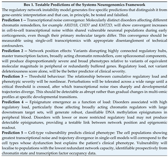
A useful framework should generate predictions that can be tested and, in principle, falsified. The five testable predictions generated by the regulatory network instability framework are set out in Box 1.
2.6. Core AI Methodologies
The AI methods applied in neurogenomics span from classical supervised classifiers to large-scale foundation models. Understanding their strengths and failure modes is necessary for evaluating what any given tool can and cannot contribute to systems-level interpretation.
Supervised learning trains models on labelled examples to classify new observations. In clinical neurogenomics, supervised approaches underpin variant pathogenicity classification, DNA methylation episignature prediction, disease-group assignment and facial phenotype recognition. Common architectures include support vector machines (SVMs), random forests, gradient boosting and feedforward neural networks. Their central limitation is dependence on training labels: models trained on biased, incomplete or ancestrally restricted cohorts inherit those limitations directly, and performance degrades when applied to populations or variant classes underrepresented in training data.
Unsupervised and self-supervised learning identify structure without predefined labels, making it well-suited to exploratory analysis of high-dimensional biological data. Principal component analysis (PCA), multidimensional scaling (MDS), t-distributed stochastic neighbour embedding (t-SNE) and uniform manifold approximation and projection (UMAP) are widely used to visualise methylation and transcriptomic datasets, identify molecular subgroups and map trajectory structure [37,38,39]. A critical interpretive caution applies that visual separation in a reduced-dimensional projection does not establish diagnostic validity. Clusters must be anchored to biological comparators, external validation cohorts and clinically meaningful boundaries before they can support diagnostic inference.
Transformer architectures capture long-range dependencies in sequence and multi-omic data that earlier architectures could not represent. In neurogenomics, transformers are applied to DNA regulatory sequences, protein sequences, splicing prediction and multi-omic feature integration. Attention weights offer a partial window into which features drive model outputs, though this should not be mistaken for a mechanistic explanation.
Foundation models learn general biological representations from very large datasets and transfer those representations to specific tasks. Protein language models learn evolutionary constraints in amino-acid sequence space; genomic foundation models infer regulatory syntax from DNA sequence; single-cell foundation models learn representations of cellular state across diverse tissues [40,41,42]. Their principal appeal for rare disease research is transferability model pre-trained on broad biological data can, in principle, be fine-tuned on small disease-specific cohorts. Their principal limitation is domain shift: representations learned from population-scale data may not transfer cleanly to rare constitutional variants where cohorts are small, biology is specific and miscalibration carries significant clinical consequences. The principal AI methodologies applied across neurogenomics, together with their typical inputs, outputs and representative tools, are summarised in Table 2.
Key takeaway: AI tools differ in what they can and cannot tell you; match the method to the question.
3. AI-Assisted Genomic Variant Interpretation
3.1. Variant Prioritisation
AI-assisted variant interpretation aims to move beyond manual filtering toward probabilistic prioritisation of candidates most likely to affect biological function — from individual amino acid substitutions to genome-wide regulatory architecture. The goal is not a single score but a mechanism-aware inference that links a variant to gene function, tissue context, developmental timing and clinical phenotype. Foundational scoring tools remain central to clinical practice. REVEL integrates multiple predictive features into a calibrated estimate of missense variant pathogenicity and is widely used in clinical pipelines. CADD provides a deleteriousness score across variant classes, extending prioritisation to synonymous, intronic and noncoding candidates. Constraint metrics, principally pLI and LOEUF, use population variation to identify genes intolerant of loss-of-function; highly constrained genes are substantially enriched for neurodevelopmental phenotypes, making constraint an important prior when evaluating candidate variants [33,34,35,36,37,70,71,72,73].
AlphaMissense extended missense prediction by integrating evolutionary and structural information through protein language modelling, providing proteome-wide pathogenicity estimates with improved calibration [51,52,53,54,55]. Its clinical use requires interpretive discipline. A high score confirms that a missense change is likely damaging to the protein, but it does not specify how — the same score is consistent with loss of function, gain of function, dominant-negative effect, altered subcellular localisation, impaired post-translational modification or disrupted protein interaction. These mechanistic distinctions matter clinically: they determine how a variant is classified, how recurrence risk is framed and, increasingly, which therapeutic strategy is relevant. popEVE addresses a related problem by combining evolutionary constraint with human population data to rank missense variants on a proteome-wide severity scale [55,74,75]. This is particularly useful when evaluating de novo missense variants across a child’s exome, where the question is not only whether a variant damages its protein but whether that damage is severe enough, in a gene sufficiently relevant to neural development, to plausibly explain the phenotype.
Regulatory variant interpretation is becoming increasingly important as clinical sequencing moves beyond coding regions. AlphaGenome predicts a broad range of functional genomic outputs, gene expression, chromatin accessibility, histone modifications, transcription factor binding, chromatin contacts and splicing signals, across long sequence contexts from primary sequence alone [49,50,76,77]. Many causal variants in NDDs act through enhancers, promoters, insulators, untranslated regions or splice-regulatory elements rather than protein-coding sequence, and models like AlphaGenome provide the only systematic means of evaluating them. Their practical clinical utility remains limited by the difficulty of validating predictions in patient-specific contexts and by incomplete representation of brain-relevant cell types in training data. Representative tools and approaches spanning each layer of the interpretive workflow, from variant prioritisation to therapeutic target discovery, are listed in Table 3.
3.2. AI and RNA/Splicing Prediction
Splicing disruption is one of the most consequential and systematically underdetected mechanisms in neurogenetic disease. Many pathogenic variants affecting splicing lie outside canonical donor and acceptor sites, are classified as synonymous or intronic by standard annotation, and are missed by conventional filtering pipelines. AI-based splice prediction has substantially expanded the detectable fraction of splicing-relevant variation by learning sequence determinants of splicing directly from large transcriptomic datasets.
SpliceAI uses deep learning applied to primary DNA sequence to predict splice-altering variants genome-wide, and has become a standard tool for identifying cryptic splice site activation, pseudoexon inclusion and disruption of canonical splice signals [25,28]. Pangolin extends this approach by modelling tissue-specific splice-site usage and sequence context, improving sensitivity for variants whose splicing consequences are cell-type or isoform dependent [78]. AbSplice and related frameworks incorporate tissue-specific transcriptomic data to further refine predictions by estimating whether a predicted splice change is likely to be expressed in the relevant tissue [79]. FexSplice addresses a narrower but diagnostically important problem: machine-learning prediction of variants affecting the first nucleotide of exons, a position where standard tools have historically underperformed [27].
These tools are particularly relevant in neurogenomics because the brain has among the most complex alternative splicing programmes of any tissue. Developmental isoform switching, neuron-specific microexon regulation and lineage-restricted splice factor expression mean that a variant appearing synonymous or weakly scored by traditional tools may nonetheless alter transcript structure, shift isoform ratios, trigger nonsense-mediated decay or disrupt a developmentally critical protein domain but only in a specific neural cell type or at a specific developmental stage [29]. RNA sequencing provides the critical empirical anchor for splice prediction. Where a candidate splice-altering variant is identified, RNA sequencing from an accessible tissue can directly test whether the predicted transcript consequence occurs, quantify its magnitude and determine whether the resulting transcript is degraded or produces an altered protein. A persistent limitation applies: a splicing change detected in blood-derived RNA may not reflect the relevant neuronal isoform, and a variant predicted to alter a brain-specific exon may produce no detectable effect in peripheral tissue. This mismatch between the tissue of clinical accessibility and the tissue of biological relevance strengthens the case for patient-derived iPSC models, cerebral organoids and high-resolution reference brain transcriptomic atlases as complementary validation substrates. Splice prediction should be understood not as a standalone diagnostic output but as a hypothesis about transcript consequence that requires tissue-appropriate confirmation [28,80].
3.3. Structural Neurogenomics
Protein structure prediction has become a practical tool in missense variant interpretation, shifting structural analysis from a specialist resource requiring experimental data to a routinely applicable computational step. AlphaFold established this transition by achieving near-experimental accuracy in structure prediction from sequence alone, enabling variant interpretation to consider folding consequences, domain organisation, active site integrity and protein-protein interaction interfaces at the proteome scale. ColabFold extended accessibility, making structure prediction practical for laboratories without dedicated computational infrastructure [81,82,83,84,85,86,87]. The expanding AlphaFold ecosystem has produced tools addressing specific interpretive needs. AlphaFind supports structural similarity searches across the predicted proteome, allowing a protein of interest to be positioned within a structural neighbourhood that may illuminate functional relationships not apparent from sequence alone. Missense3D maps predicted missense consequences onto three-dimensional structures in relation to post-translational modification sites, phosphorylation, ubiquitination, acetylation and others, whose disruption may be pathogenic independently of gross structural change [82]. Protein language models complement structure prediction by learning evolutionary sequence constraints that reflect functional importance, providing pathogenicity signals even for regions where structural prediction is uncertain [82,83,88].
Structural interpretation is particularly informative for neurodevelopmental genes where domain-specific effects are biologically meaningful. A missense variant disrupting a bromodomain’s histone-binding pocket may phenocopy a loss-of-function allele, whereas a variant in a structurally peripheral region of the same protein may be tolerated or act through an entirely different mechanism. Variants in ion-channel pore-forming regions, DNA-binding domains, chromatin-reader domains, ATPase cassettes, zinc fingers or kinase activation loops carry different mechanistic implications from variants elsewhere in the same protein, and structural context is often the only way to make that distinction from sequence data alone.
A critical interpretive limit applies throughout. Structural predictions constrain the hypothesis space but cannot establish disease mechanism. A predicted destabilising effect is consistent with loss of function but cannot distinguish haploinsufficiency from dominant-negative action. A predicted interface disruption suggests impaired protein interaction but does not establish whether the relevant complex is stoichiometrically sensitive, scaffolding-dependent or gain-of-function. Structural evidence should direct functionally testing, not replace it.
3.4. Challenges in Variant Interpretation
Neurodevelopmental variant interpretation remains difficult even with high-quality AI tools because the underlying biology resists the assumptions most models are built on. Many neurodevelopmental genes show direction-sensitive pathogenic mechanisms: loss-of-function and gain-of-function variants in the same gene produce disease through distinct and sometimes opposite mechanisms [37,51]. A variant may be deleterious only within a specific developmental window or cell lineage, producing no detectable consequence in the tissues and timepoints accessible to clinical testing. Chromatin regulators, transcription factors, ion channels and signalling pathway genes are particularly prone to these context-dependent effects because their functional consequences depend on the regulatory state of the cell rather than on intrinsic protein properties alone. Penetrance prediction is an emerging and largely unsolved challenge. Machine-learning approaches using population-scale and clinical datasets are beginning to estimate variant penetrance, offering the prospect of distinguishing highly penetrant alleles from those whose pathogenicity is modifier-dependent or context-specific [116]. These models remain limited by ascertainment bias in clinical datasets, systematic underdiagnosis of mild or atypical presentations, and incomplete phenotyping in the population cohorts used for training. A model trained predominantly on severely affected probands will systematically underestimate penetrance variability and may misclassify attenuated or conditionally pathogenic variants [116]. These limitations define the appropriate role of AI in clinical variant interpretation. AI predictions are structured evidence components that narrow the hypothesis space, surface non-obvious candidate mechanisms and direct confirmatory testing, not standalone diagnostic outputs. Clinical interpretation continues to require phenotype correlation, segregation analysis, functional data, transcriptomic evidence, methylation analysis, population constraint, inheritance pattern and expert synthesis. The irreplaceable role of the clinician is to ask whether the convergent outputs of multiple AI tools constitute a biologically coherent explanation for this patient; a question that requires biological judgement, not score aggregation [117]. The integration of structural, splicing, phenotypic and multi-omic evidence into a single mechanism-aware interpretation is illustrated in Figure 2.
Key takeaway: A pathogenicity score tells you a variant is probably abnormal, not how it causes disease; treat AI outputs as evidence that narrows the question, not as the answer.
4. AI-Assisted Phenomics and Rare Disease Diagnosis
4.1. Human Phenotype Ontology and Computational Phenotyping
The Human Phenotype Ontology (HPO) provides a structured, computable vocabulary for clinical features and enables systematic comparison between patients, genes and disease profiles [56,57,58,59,66,69]. HPO-driven prioritisation tools improve candidate variant ranking by matching a patient’s phenotype to known gene-disease associations, a particularly valuable function when sequencing identifies multiple plausible candidates and the phenotype must arbitrate between them. The diagnostic yield of sequencing is therefore not independent of phenotype quality: a precisely coded HPO profile improves prioritisation, while a coarse or incomplete one dilutes it [57,58,60,63,65,66,67]. Automated HPO extraction is becoming operationally important as genomic medicine scales beyond what manual annotation can sustain. FastHPOCR, PhenoBERT, PhenoTagger and PhenoRerank represent different approaches to phenotype concept recognition, structured annotation and candidate re-ranking from clinical text [61,62,64,66,68]. These systems can reduce annotation burden and support cohort-level phenotype discovery, but they must handle the linguistic complexity of clinical documentation accurately. Negation, uncertainty, temporality and differential diagnosis framing each require specific handling; the distinction between “no regression” and regression, or between “query seizures” and confirmed epilepsy, is clinically critical and must be preserved rather than collapsed by an annotation model [61,62,64,66,68].
Phenotype ontologies can also be linked to other biological knowledge frameworks. HPO2GO connects phenotype terms to gene ontology associations, enabling inference from clinical features to molecular function and cellular process [69]. MitoPhen demonstrates disease-specific HPO application for identifying mitochondrial DNA disease patterns from phenotypic signatures [95]. These connections are relevant to systems neurogenomics because they provide a structured path from bedside observation to pathway, cellular mechanism and regulatory network, the direction of inference that systems-level interpretation requires.
4.2. Facial AI and Dysmorphology and the FaceMatch Platform
Facial AI systems use deep learning to identify syndrome-associated craniofacial patterns from photographs, providing a phenotypic signal that is orthogonal to genomic and metabolic data. GestaltMatcher demonstrated that facial phenotype descriptors can support rare disease matching even for ultra-rare conditions, by embedding faces in a shared phenotypic space that allows similarity-based retrieval across disorders [44,45,46,47,48]. Face2Gene and DeepGestalt operate on related principles. The biological rationale is not incidental: craniofacial and neurodevelopmental systems share early developmental programmes, neural crest biology, chromatin regulation and transcriptional control, such that a syndrome affecting brain development frequently leaves a structural signature in facial morphology. Facial AI can therefore serve as a hypothesis generator for the underlying molecular mechanism, not merely a pattern-matching tool [44,45,46,47,48].
Its limitations are substantial and must be explicitly recognised. Facial morphology varies with age, ancestry, sex, image quality and ascertainment context. The same molecular disorder may present with subtle, absent or age-dependent craniofacial features, and some NDDs have no consistent facial gestalt. Validation studies demonstrate diagnostic utility but consistently identify the need for ancestry-diverse training datasets, age-aware modelling and prospective real-world evaluation [44,45,48,96]. Facial AI should be understood as phenotype support that generates structured hypotheses requiring independent confirmation — not as a diagnostic classifier operating independently of other evidence. The FaceMatch platform (facematch.org.au) was developed to address several of these limitations directly [97,118]. As a family-facing initiative, FaceMatch collects longitudinal serial facial imaging paired with curated genomic and clinical data from individuals with diagnosed and undiagnosed syndromic intellectual disability. Its defining feature is longitudinal depth: rather than relying on photographs from a single time point, FaceMatch captures how craniofacial features evolve across the age spectrum. This matters because many chromatinopathies and regulatory disorders present with subtle facial features in infancy that become clinically recognisable only in adolescence or adulthood. By building an internationally representative, multi-ancestral cohort, FaceMatch supports reverse phenotyping, grouping geographically separated undiagnosed patients on the basis of shared machine-identified facial features, and provides the training data needed for age-aware, ancestry-calibrated computer vision models. Its long-term objective is to close the gap between facial AI performance in research settings and its reliability in real-world clinical use across diverse populations [97,118].
4.3. Multimodal Diagnosis
The diagnostic potential of rare disease genomics will be most fully realised when genomic data are integrated with structural neuroimaging, neurophysiology, developmental trajectories and structured clinical language, not as parallel data streams, but as components of a unified biological model. MRI captures structural brain development, regional volumetric vulnerability and white matter integrity; EEG reflects circuit excitability and network synchrony; longitudinal developmental phenotyping captures trajectory rather than cross-sectional status; and clinical language models can extract structured phenotypic data from unstructured clinical records at scale. The critical opportunity is not data aggregation but biologically coherent multimodal modelling, in which each input constrains the interpretation of the others [60,90].
Prenatal deep phenotyping represents an important and underserved extension of this framework. Fetal anomalies detected in the first or second trimester are frequently difficult to interpret because many phenotypic features, brain morphology, growth trajectory, and neurodevelopmental function, evolve substantially later in gestation or postnatally. Structured prenatal HPO terminology and prenatal deep phenotyping initiatives directly address this gap by providing computable frameworks for fetal phenotype that can interface with genomic interpretation. AI-assisted prenatal phenomics could eventually integrate fetal ultrasound, fetal MRI and genomic findings into a unified interpretive model, but clinical implementation requires particularly careful validation: prenatal predictions carry major and often irreversible counselling implications, and model uncertainty must be explicitly preserved and communicated at every step.
A general principle applies across all multimodal diagnostic models: uncertainty must be preserved, not averaged away. A model integrating MRI, EEG, facial analysis and genotype may generate a high-confidence candidate diagnosis while each contributing input carries its own error structure and representational limitation. Clinical reporting from such models should explicitly distinguish observed features, inferred features, absent features and model-derived probabilities, and should communicate what the model does not know as clearly as what it asserts [119,120].
4.4. Deep Phenotyping in Neurodevelopmental Disorders
Deep phenotyping moves beyond broad diagnostic labels (autism, epilepsy, developmental delay) to capture the structured dimensions that carry biological meaning: onset timing, developmental trajectory, regression, seizure semiology, seizure type and frequency, epilepsy syndrome classification, ictal and interictal EEG features, pharmacological treatment response and pharmacoresistance, motor pattern, sleep architecture, behavioural profile, growth parameters, autonomic features, neuroimaging findings and neurophysiology [7,46,116,121,122]. AI model performance in genomic interpretation is directly dependent on phenotype quality. Coarse phenotyping produces coarse prioritisation regardless of model sophistication: the genomic interpretation is only as precise as the phenotypic input that guides it. Epilepsy phenotyping deserves particular emphasis. Seizure onset age, semiology, syndrome classification and EEG signature carry diagnostic and mechanistic information that broad labels obscure. Infantile epileptic spasms syndrome, Dravet syndrome, KCNQ2-related neonatal epilepsy and glucose transporter type 1 deficiency syndrome each have characteristic electroclinical signatures that, when structured and computable, direct genomic interpretation toward specific gene classes, molecular mechanisms and treatment pathways. The distinction between a sodium channel gain-of-function epilepsy (responsive to low-dose sodium channel blockade) and a GABAergic interneuron disorder requiring a fundamentally different approach is not recoverable from the label “epileptic encephalopathy” alone. It requires structured electroclinical phenotyping integrated with genomic and functional data.
Deep phenotyping is also the primary tool for understanding variable expressivity. Patients carrying variants in the same gene may differ substantially in epilepsy severity, seizure type, treatment response, movement phenotype, language trajectory, autonomic dysfunction and psychiatric features. Without structured longitudinal phenotype data, these differences collapse into the same diagnostic label and their biological determinants, modifier variants, epigenomic context, sex, and stochastic developmental variation become invisible to analysis. Structured longitudinal phenotypes allow AI systems to learn developmental trajectories rather than static cross-sectional states, which is the appropriate unit of analysis for neurodevelopmental disease. Early hypotonia followed by epilepsy onset and cognitive regression carries different biological implications from stable developmental delay without seizures, or from epilepsy with preserved cognition and spontaneous remission, even when all three presentations receive the same broad diagnostic classification. Capturing this trajectory structure is not a refinement of phenotyping practice; it is a prerequisite for mechanistic interpretation.
Key takeaway: Phenotype quality sets the ceiling on everything downstream; a precisely coded, longitudinal HPO profile does more for diagnostic yield than any single algorithm.
5. AI in Epigenomics and Chromatin Disorders
5.1. Epigenomic Regulation in Neurodevelopment
Epigenomic regulation is not a peripheral layer of neurodevelopmental biology but its organisational substrate. DNA methylation, histone modification, chromatin accessibility, enhancer-promoter communication and three-dimensional genome organisation collectively establish and maintain the gene-expression programmes required for neural progenitor specification, neuronal differentiation, synaptic maturation and circuit formation. Disruption of these systems produces a broad and clinically heterogeneous group of NDDs, most prominently the chromatinopathies, conditions caused by variants in chromatin remodellers, writers, erasers and readers that alter the epigenomic landscape at a systems level rather than at a single gene [98,99,100]
The clinical relevance of chromatin biology extends well beyond the chromatinopathies. Epigenomic mechanisms govern genomic imprinting, X-chromosome dosage compensation, developmental enhancer activation and silencing, alternative promoter usage, activity-dependent transcription in mature neurons and stress-response pathway regulation. A variant whose primary effect is regulatory rather than protein-coding may therefore leave no interpretable signal in standard variant annotation while producing a coherent and diagnostically informative perturbation of the epigenomic landscape. This is the biological basis for epigenomic data as functional evidence in genomic diagnosis, not a supplement to sequence interpretation but an independent and sometimes decisive layer [98,99,100]. AI is well-suited to epigenomic analysis because disease-associated methylation signatures are distributed, high-dimensional and pattern-based. A pathogenic episignature may involve hundreds or thousands of CpGs distributed across the genome, with no individual site carrying sufficient signal to be diagnostically informative in isolation. The diagnostic content resides in the profile rather than in any individual position. Machine learning is structurally adapted to detect this kind of distributed pattern, but only when models are trained on appropriately controlled, biologically validated and ancestrally representative reference sets. The quality of the reference cohort is as important as the architecture of the classifier [98,99,100,101].
5.2. AI-Assisted DNA Methylation Episignatures Diagnostics
DNA methylation episignatures are disorder-specific, genome-wide methylation profiles that serve as molecular fingerprints of underlying regulatory dysfunction [102,103,118]. Their diagnostic utility operates on two levels. First, they can support or establish a diagnosis in genomically unresolved cases where sequencing has not identified a pathogenic variant. Second, they can provide functional evidence for the pathogenicity of a variant of uncertain significance (VUS) in a chromatin-related gene, not by reporting on the variant directly, but by showing whether the variant has produced the regulatory perturbation expected of a pathogenic allele. Analytical pipelines for episignature classification typically proceed through selection of the most informative CpG sites, dimensionality reduction for visualisation and outlier detection, hierarchical clustering against reference cohorts, and supervised classification using SVMs or related methods. Methylation variant pathogenicity (MVP) scores provide calibrated probabilistic estimates of whether a patient’s methylation profile is consistent with a known disorder, and can be integrated into variant classification frameworks alongside sequence-based evidence [24,41,99,104,105,116].
The value of this approach is clearest in chromatinopathies, where the pathogenic effect is regulatory and distributed, precisely the scenario where sequence-based interpretation alone is insufficient. A missense variant in a chromatin regulator that is difficult to classify from genomic evidence may receive a concordant episignature demonstrating that it perturbs the expected regulatory programme in a manner consistent with pathogenic loss or gain of function [106,107]. The episignature does not prove the variant is pathogenic, but it substantially shifts the prior and can be the decisive evidence in a variant classification decision. Episignature results must be interpreted carefully. Classifier output alone is not sufficient — results must be evaluated against appropriately matched controls, molecularly confirmed reference cases and phenotypically overlapping disorders with distinct signatures. Batch effects, blood cell composition variability, patient age, tissue of origin, cohort size and classifier training domain all influence interpretation and must be explicitly considered. A negative episignature result does not exclude pathogenicity: the disorder may lack a characterised blood-accessible signature, the variant mechanism may not produce the expected methylation perturbation, or the patient may fall outside the validated domain of the classifier. Sensitivity, specificity and domain boundaries should be reported alongside episignature results as standard practice, not as supplementary information but as a required component of the clinical report. The episignature classification workflow, from informative CpG selection through dimensionality reduction to supervised classification and clinical interpretation, is depicted in Figure 3.
5.3. Chromatinopathies as Systems Disorders
Chromatinopathies are best understood as systemic disorders rather than single-gene diseases. A pathogenic variant in a chromatin regulator does not disrupt one gene’s expression, it perturbs the regulatory architecture governing many downstream targets, altering developmental timing, enhancer activity, transcriptional stability, cell identity maintenance and neuronal maturation across multiple lineages and time points. The shared neurodevelopmental phenotypes observed across mechanistically distinct chromatinopathies, ID, ASD, epilepsy, behavioural dysregulation and growth abnormalities, reflect convergence at the level of regulatory network architecture rather than molecular identity.
Direction-sensitive phenotypes are a characteristic feature of chromatin disorders that the regulatory load framework helps explain. Opposing biochemical perturbations, gain and loss of the same histone modification, can produce mirrored somatic growth effects while converging on overlapping neurological outcomes [38]. This pattern suggests that neurodevelopmental systems are vulnerable not primarily to the direction of regulatory change but to the magnitude of departure from an optimal regulatory equilibrium. Cognitive development, synaptic maturation and circuit stability may be impaired by both insufficient and excessive chromatin regulatory activity if either state exceeds buffering capacity and destabilises transcriptional precision.
Variable expressivity in chromatinopathies follows from the same logic. When chromatin load is modest (a variant with partial functional impact, in a gene with redundant regulatory partners, acting during a developmental window with high compensatory capacity), development may remain within a buffered range and the clinical phenotype may be mild or subclinical. When load is broad, early, persistent or concentrated in regulatory hubs with limited redundancy, buffering fails and phenotypic severity increases. This framework makes variable expressivity mechanistically interpretable rather than simply observed, and it defines the parameters that AI models should aim to estimate: load, timing, network position, and residual capacity [38].
Variants in ARID1B, a core subunit of the BAF (SWI/SNF) chromatin remodelling complex, illustrate how regulatory network instability emerges in practice [98,118]. ARID1B haploinsufficiency affects a broad set of downstream transcriptional targets, generating high regulatory load distributed across neurodevelopmental pathways. Episignature studies demonstrate consistent genome-wide methylation perturbation, indicating system-level regulatory disruption rather than gene-specific effects. Disease severity in ARID1B-related disorder reflects the interaction between the magnitude of transcriptional perturbation and residual network capacity, shaped by genetic background and developmental timing. The clinical phenotype, ID, ASD and characteristic dysmorphology of Coffin-Siris syndrome, emerges when buffering fails to maintain transcriptional precision across neuronal lineages.
5.4. AI and Chromatin-State Modelling
AI offers a means of modelling chromatin-state dysregulation that integrates methylation, histone modification, chromatin accessibility, regulatory topology and transcriptional output into a unified representation of regulatory perturbation. EPInformer and related frameworks integrate promoter-enhancer sequence with multimodal epigenomic data to predict cell-type-specific gene expression from regulatory architecture [90]. AlphaGenome extends the sequence-to-function model further, predicting multiple functional genomic outputs, chromatin accessibility, histone modification state, transcription factor binding occupancy, chromatin contacts and splicing signals, across long sequence contexts from primary sequence alone [49,50,76,77]. Together, these tools create a bridge between noncoding variation and developmental phenotype: a variant in a regulatory element may alter enhancer activity in a specific progenitor population, disrupt chromatin looping to a target promoter, redirect promoter usage or modify splicing of a developmentally critical transcript.
The interpretive strength of this approach lies in convergence. Any individual predicted regulatory consequence is uncertain in isolation; regulatory models are imperfectly calibrated, training data are incomplete and cell-type coverage is uneven. When predictions from multiple epigenomic layers converge on a coherent perturbation of the same regulatory axis, the cumulative evidence can support a mechanistic hypothesis that no single layer could establish alone. This is what regulatory network instability looks like in practice from an AI perspective: not a single abnormal track, but a pattern of convergent dysregulation across chromatin, transcriptional and cellular layers that exceeds what developmental buffering can absorb.
Key takeaway: In chromatin disorders the diagnostic signal is distributed, not local; an episignature reads the whole regulatory disturbance, which is exactly what a single variant score cannot.
AI-assisted chromatin modelling should therefore aim not only to classify whether a variant is likely regulatory, but to estimate how the resulting perturbation propagates through developmental networks, which downstream targets are affected, at what developmental stage, in which cell lineages, and whether the cumulative effect approaches or crosses the instability threshold. This is the modelling objective that distinguishes systems neurogenomics from layer-specific classification, and the standard against which current chromatin AI tools should be evaluated.
6. AI in Transcriptomics and Single-Cell Neurogenomics
6.1. Single-Cell Transcriptomics and Brain Cell Atlases
When gene expression is measured from bulk brain tissue, the signal is an average across millions of mixed cell types. This average obscure the biology that matters most in NDDs, a gene critical in one rare progenitor population may appear unimportant when its expression is diluted across everything else. Single-cell transcriptomics resolves this by measuring gene expression in individual cells, and AI-based clustering and trajectory inference allow these cells to be mapped into a coordinate system of cell types, developmental stages and lineage relationships [15,117]. The result is a reference framework within which disease-associated perturbations can be precisely positioned, not just “this gene is expressed in the brain” but “this gene is active in this cell type, at this stage of development, and its loss affects this lineage.”
When a candidate variant is identified, these atlases allow a direct question: in which cell type is this gene most active, and when during development does that activity peak? A gene expressed primarily in cortical radial glia during progenitor expansion points toward malformation or progenitor phenotypes; one enriched in mature parvalbumin-positive inhibitory interneurons points toward epilepsy and circuit dysfunction. This cell-type-to-phenotype inference is not decorative; it generates testable hypotheses, directs experimental validation toward the right cellular substrate and helps explain why the same variant produces different phenotypes in different individuals depending on which cell populations are most affected [17]. Large-scale brain cell atlases put this framework into practice. STAB2 and ZEBRA provide detailed spatiotemporal maps of gene expression across human and model-organism brain cell states, covering the progression from early progenitors through neuronal differentiation to mature circuit components [108,112]. Single-cell foundation models such as Geneformer take this further by learning general representations of cellular state from large-scale datasets, potentially allowing prediction of how a specific cell lineage responds to a genetic perturbation. The clinical potential is real but translation remains early. Current models depend heavily on the completeness of reference atlases, and the cell states most likely to be pathologically relevant in NDDs, rare transitional progenitors, early postmitotic neurons, lineage-restricted interneuron subtypes, are often the least represented in training data. This is not a minor caveat; it is a central limitation that needs to be addressed before single-cell foundation models can reliably support clinical interpretation.
6.2. Spatial Transcriptomics
Single-cell transcriptomics tells us what is happening in each cell type but when tissue is dissociated to isolate individual cells, the information about where each cell lived in the brain is lost. Spatial transcriptomics restores that information by measuring gene expression while preserving the physical location of each cell within a tissue section [123]. This matters because brain disorders are almost always anatomically specific. Vulnerability is not distributed evenly across the brain; it concentrates in particular cortical layers, subcortical nuclei, projection pathways, or local circuit components. Standard transcriptomic approaches cannot explain why. Spatial transcriptomics can, by mapping molecular perturbations onto the anatomical structures where clinical vulnerability actually resides. AI-assisted spatial analysis using frameworks such as SpaDiT enables these perturbations to be linked to cortical layers, circuit projections and cell-cell interaction networks in ways that neither genomic nor dissociated single-cell data can achieve [123,124].
The central interpretive puzzle that spatial neurogenomics addresses is this: if a chromatin regulator or transcription factor is expressed throughout the brain, why does losing it only damage specific regions or cell types? The answer lies in the regulatory context: the local chromatin state, transcription factor occupancy and enhancer landscape that differ between anatomical locations. Some cell populations are specifically vulnerable because of how their regulatory networks are wired, not simply because the gene is expressed there. Spatial transcriptomics makes that differential vulnerability visible and mechanistically interpretable, providing the link between a molecular perturbation and the anatomical specificity of the clinical phenotype.
6.3. AI and Developmental Trajectory Modelling
Brain development is not a snapshot but a journey. Neural induction, progenitor expansion, neuronal differentiation, migration, synaptogenesis, circuit pruning, and activity-dependent maturation proceed in sequence, each stage imposing different regulatory demands. A variant that is well tolerated during one stage may be catastrophic during another, when new regulatory dependencies emerge, or compensatory mechanisms become unavailable. Developmental trajectory modelling uses AI to ask: at what point in this journey does a patient’s molecular profile begin to diverge from the expected path, and what does that divergence tell us about mechanism and timing [2,38].
Patient-derived iPSC models and cerebral organoids provide experimental systems in which trajectory divergence hypotheses generated by AI models can be directly tested, by comparing individual molecular trajectories against reference neurodevelopmental maps derived from brain cell atlases. RNA sequencing is the practical clinical bridge between DNA variation and cellular consequence in this context. It can identify aberrant splicing, allele-specific expression, cryptic exon inclusion, intron retention, and nonsense-mediated decay, each of which may be the primary pathogenic mechanism of a variant that looks benign at the sequence level [25,26,27,28,29,30,31]. Where a variant produces an abnormal protein isoform rather than simple transcript loss, the distinction between a change in transcript abundance and a change in protein structure carries direct implications for mechanism and therapeutic strategy.
A critical biological property must be built into trajectory models: development is nonlinear. A regulatory system can absorb perturbation and appear normal across a wide range of conditions, then tip abruptly when a threshold is crossed, analogous to a structure that tolerates cumulative stress until it suddenly fails. Molecular differences that are compensated during progenitor expansion may become clinically manifest during synaptogenesis, synaptic pruning or activity-dependent maturation, when new regulatory demands reduce the redundancy available for buffering [20]. AI trajectory models that assume gradual linear divergence from a reference state will systematically miss these transitions. Model architecture needs to explicitly accommodate nonlinear and bifurcating dynamics, and validation datasets should include longitudinal measurements taken at the developmental transitions where threshold behaviour is most likely to emerge.
Key takeaway: Cell type, location and timing matter as much as the variant itself; single-cell and spatial data tell you where and when a perturbation actually bites.
7. Multi-Omics Integration and Systems Neurogenomics
7.1. Integrating Genome, Epigenome and Phenome
Each diagnostic modality in neurogenomics captures a different slice of the underlying biology, and each has its own error structure. Genomic data are stable and comprehensive but frequently ambiguous: a variant may be rare, constrained, and computationally predicted to be damaging without any of those properties individually establishing pathogenicity. RNA data provide direct functional evidence but are tissue- and stage-dependent, and may not reflect the relevant biological context when derived from peripheral tissues. Methylation data capture regulatory consequences at the epigenomic scale but are classifier-dependent, tissue-specific and sensitive to technical covariates. Phenotype data anchor molecular findings to clinical reality but are vulnerable to incomplete ascertainment, variable depth and evolving presentation over developmental time. The diagnostic value of integration arises precisely because these error structures are different and partially complementary: what one modality cannot resolve, another sometimes can [118,125]. A concrete example illustrates what integration looks like in practice. Consider a de novo missense variant in a chromatin regulator identified through exome sequencing. Variant-level prediction tools flag it as likely damaging but cannot establish a mechanism. RNA sequencing reveals reduced expression of downstream targets. DNA methylation profiling shows a concordant episignature consistent with known pathogenic variants in the same gene. Single-cell reference data localise the gene’s expression to cortical progenitors during a critical window of neurogenesis. The patient’s HPO-coded phenotype aligns with the expected clinical spectrum. None of these findings is individually conclusive but together they converge on a single coherent explanation: a regulatory perturbation affecting progenitor cell states at a critical developmental window. The diagnosis emerges from the coherence of evidence across layers, not from any individual dataset. This is the difference between score aggregation and mechanism-based inference [118]. A systems model that integrates these layers should combine their complementary strengths while explicitly preserving and propagating the uncertainty each contributes. The goal is not a single confidence score that averages uncertainty away, it is a structured explanation that makes clear which evidence is strong, which is inferential and which remains unresolved.
7.2. Regulatory Network Instability in Neurodevelopment
The network instability framework developed in Section 2 has a specific practical implication for how multi-omic data should be interpreted. A gene-centric model asks whether a variant is damaging to its immediate molecular target. A network instability model asks something different: does the pattern of evidence across genomic, epigenomic, transcriptomic and phenotypic layers indicate that a developmental regulatory system has been destabilised, and if so, which system, at what stage, and in which cell type?
This reframing changes what counts as a satisfying clinical explanation. In a gene-centric model, identifying a pathogenic variant and matching it to a known disease gene is sufficient. In a network instability model, a satisfying explanation specifies the mechanism: which regulatory system has failed, why buffering was insufficient, and what the downstream consequences were for cell identity, transcriptional precision, and circuit development. AI tools contribute to this explanation not by producing a final answer but by generating the structured evidence that makes such an explanation possible. Agentic systems point to how this may be operationalised: DeepRare, for example, decomposes diagnosis across specialised agents for phenotype extraction, variant prioritisation and evidence retrieval, then integrates their outputs into ranked hypotheses with explicit reasoning chains [113]. This architecture mirrors the systems neurogenomics goal of assembling convergent evidence into a coherent mechanistic explanation rather than a single score.
The framework also explains phenotypic convergence across molecularly diverse disorders. Chromatin regulators, transcriptional machinery components, RNA-processing factors, synaptic proteins, and signalling effectors converge on shared neurodevelopmental phenotypes (ID, ASD, epilepsy, movement disorder) not because they affect the same gene or pathway, but because they destabilise the same downstream developmental systems through different entry points. Cataloguing this convergence at the phenotypic level is descriptive. Modelling it at the network level is mechanistic, and that is what systems neurogenomics aims to achieve.
7.3. Regulatory Load and Network Capacity in Practice
Regulatory load and network capacity are not merely theoretical constructs; they have practical implications for how variants are interpreted and why patients with the same variant can present so differently.
Regulatory load is a composite. A variant in a highly connected chromatin remodeller that acts early in cortical development, affects a broad set of downstream targets and has no redundant regulatory partners imposes a high regulatory load. A variant in a peripheral signalling component that acts late, has a narrow downstream effect and operates in a pathway with multiple compensatory routes imposes low regulatory load. The magnitude of the molecular defect matters, but so does its position in the network, its timing and its persistence. Two variants with identical predicted deleteriousness scores can impose very different regulatory loads; this is one reason why pathogenicity scores alone are insufficient for predicting clinical severity.
Network capacity explains the rest. Two individuals with the same high-load variant may have very different clinical outcomes if their network capacities differ because of modifier variants in redundant regulatory pathways, differences in epigenomic context, sex-specific differences in regulatory architecture, or simply the stochastic variation in gene expression that shapes how developmental systems are wired in each individual. Variable expressivity is therefore not unexplained noise; it is a predictable consequence of variation in network capacity, and it is potentially measurable given sufficient multi-omic data.
SCN2A illustrates how opposing molecular effects can converge on shared neurodevelopmental vulnerability through this framework. Gain-of-function SCN2A variants increase neuronal excitability and are associated with early-onset epilepsy; loss-of-function variants reduce functional output and are more commonly associated with ASD and later developmental phenotypes. Both impose regulatory load on neuronal networks, but through different mechanisms, one through hyperexcitability and network instability, the other through reduced functional output and impaired circuit maturation. The resulting phenotypes reflect how different forms of perturbation interact with developmental timing and network capacity, rather than the direction of molecular change alone. This is why the load-capacity framework is more explanatory than a simple gain-versus-loss-of-function classification.
7.4. Transcriptional Noise and Neuronal Vulnerability
When regulatory buffering begins to fail, one of the earliest measurable consequences is increased transcriptional noise, greater cell-to-cell variability in gene expression within what should be a homogeneous cell population. Neurons are particularly vulnerable to this kind of imprecision. Their long lifespan, high metabolic demand, complex morphology and dependence on tightly coordinated gene-expression programmes leave little tolerance for variability. Small deviations in expression precision can disrupt dendritic arbour development, alter synaptic scaffolding stoichiometry, disturb ion-channel subunit balance and destabilise the excitatory-inhibitory ratio on which circuit function depends. Transcriptional noise is therefore not merely a molecular observation; it is a direct mechanistic route from regulatory perturbation to circuit dysfunction, and it may be detectable before overt clinical phenotypes appear.
AI-assisted modelling can help identify when and where transcriptional variability becomes pathogenic, which cell populations show elevated noise, at which developmental stage the increase becomes detectable and whether it precedes or follows phenotypic divergence. Single-cell transcriptomics makes this measurable in principle, but a prerequisite challenge must be addressed first: separating genuine disease-associated transcriptional variability from the confounding variability introduced by batch effects, age, cell-type composition and technical platform. Disentanglement frameworks such as TarDis use deep learning to perform this separation, isolating true biological signal from technical noise in single-cell datasets [22]. In rare disease cohorts, where small sample sizes create conditions under which models readily learn confounders rather than mechanisms, this step is not optional, it is the foundation on which any noise analysis must rest.
7.5. Convergent Neurodevelopmental Phenotypes
Phenotypic convergence is one of the most diagnostically challenging features of NDDs. Variants in chromatin regulators, transcription factors, splicing factors, synaptic scaffold proteins and signalling components all converge on ID, ASD, epilepsy, and movement disorder, not because they share a molecular mechanism, but because they destabilise the same downstream developmental systems through different entry points. Understanding this convergence mechanistically, rather than simply cataloguing it phenotypically, is one of the core objectives of systems neurogenomics.
The practical diagnostic implication is a shift from score stacking to evidence synthesis. Score stacking treats independent tool outputs (a pathogenicity score, a splice prediction, a phenotype similarity score, a methylation result) as additive evidence and combines them into a cumulative probability. Evidence synthesis asks a different question: do these outputs, taken together, point toward one coherent biological mechanism? A candidate variant becomes progressively more convincing as independent evidence layers converge on the same explanation; structural prediction implicates a functional domain; splice prediction identifies a transcript consequence; constraint metrics establish population intolerance; phenotype matching aligns the presentation with the gene’s known spectrum; methylation analysis confirms a concordant epigenomic perturbation; RNA sequencing demonstrates an expression or splicing effect; and network biology positions the gene at a relevant developmental hub. Conversely, when evidence layers are discordant (for example, when predicted deleteriousness is not reflected in the methylation profile, or when the phenotype does not match the expected spectrum), that discordance should prompt caution and targeted functional investigation, not score averaging.
7.6. Making the Framework Practical
Making systems neurogenomics practically implementable requires moving from a conceptual framework to an auditable clinical workflow. A concrete implementation would construct a patient-specific evidence graph in which nodes represent variants, genes, regulatory elements, transcripts, cell types, pathways and structured phenotypic terms, and edges represent known or predicted relationships: enhancer-promoter linkage, protein-protein interaction, co-expression, HPO semantic similarity and network co-regulation. AI models operating on this graph would estimate not only whether a candidate diagnosis is probable, but whether the observed evidence forms a mechanistically coherent explanation for how the molecular perturbation produces the patient’s phenotype.
The output should specify a mechanism, not merely a candidate. A clinically useful report would state, for example, that a de novo variant in a highly constrained gene is predicted to disrupt a canonical splice site; that the affected transcript is enriched in cortical progenitors during neurogenesis; that the patient’s methylation profile is concordant with a known chromatinopathy episignature; and that the structured HPO phenotype aligns with the expected clinical spectrum, and would then identify the confirmatory assay most likely to resolve residual uncertainty. This mechanistic specificity is what makes an AI-assisted output clinically actionable rather than probabilistically suggestive.
Minimum reporting standards are a prerequisite for this framework to be clinically auditable. Reports should specify input data types and quality metrics, model version and training domain, comparator cohorts and their ancestry and age distribution, uncertainty estimates for each evidence layer, external validation status and an explicit statement of whether each output is clinically validated or research-stage. These standards are what transform AI-guided neurogenomics from opaque prediction into transparent, reproducible and mechanistically grounded clinical inference. The progression from buffered perturbation to threshold-dependent network instability, and its convergence on shared neurodevelopmental phenotypes, is illustrated in Figure 4.
7.7. Practical Value for Clinicians and Bioinformaticians
The systems neurogenomics framework has different but complementary practical value for clinicians and bioinformaticians, and it is worth being explicit about both.
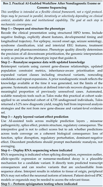
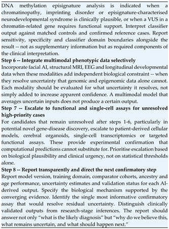
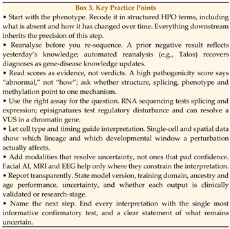
For the clinician, the framework reframes what a satisfying diagnostic outcome looks like. A pathogenicity score or a variant classification is a starting point, not an answer. The question that matters is whether the available evidence, genomic, epigenomic, transcriptomic, and phenotypic, tells a coherent biological story about what has gone wrong in this patient’s developing nervous system. The practical tools for pursuing that story are already available: RNA sequencing, episignature testing, single-cell reference atlases, facial AI platforms, HPO-structured phenotyping and multimodal variant interpretation pipelines. What the framework provides is a logic for combining them, deciding which tool to deploy next, interpreting discordant results, knowing when the evidence is sufficient and knowing when to escalate to functional testing. A practical, step-by-step AI-enabled workflow for cases that remain undiagnosed after exome or genome sequencing is presented in Box 2. The immediate clinical benefit is a higher diagnostic yield in cases that remain unresolved after standard exome or genome sequencing, the patients who currently leave the clinic without an answer.
Key takeaway: Synthesise toward a mechanism, do not stack scores; when evidence layers converge they support a diagnosis, and when they conflict they tell you where to look next.
For the bioinformatician, the framework defines a modelling objective that goes beyond classification accuracy. The question is not only whether a model correctly predicts variant pathogenicity or episignature assignment in a held-out test set, but whether its outputs can be integrated with other biological layers to produce a mechanistically coherent explanation. This sets concrete requirements: models need to produce interpretable intermediate representations, not just final scores; they need to be calibrated for uncertainty, not just optimised for accuracy; they need to be validated against the disorders and populations they will actually encounter in clinical use, not just the training distribution; and they need to expose their reasoning in a form that a clinician can interrogate and challenge. The practical benefit is a clearer design target: AI systems that explain as well as predict, and that improve iteratively as clinical feedback is incorporated.
For both audiences, the most immediate practical implication is the same: multi-omic evidence should be synthesised toward a mechanism, not aggregated toward a score. When the evidence converges, it supports a diagnosis and directs the next confirmatory step. When it diverges, it signals that the explanation is incomplete and that further investigation is warranted. That principle, convergence toward mechanism, transparency about uncertainty, is what distinguishes clinically useful AI from technically impressive AI.
8. Current Challenges and Limitations
8.1. Ancestry Imbalance and Diagnostic Inequity
AI models are only as representative as the data they are trained on, and the systematic underrepresentation of non-European populations in genomic reference datasets, methylation cohorts, facial image libraries and clinical phenotype registries creates a compounding equity problem, one that does not merely reduce accuracy but actively denies diagnostic precision to the patients who already have the least access to specialist care.
The consequences are specific and pervasive. Population allele frequency databases used to assess variant rarity are ancestrally skewed: pathogenic variants enriched in underrepresented populations are misclassified as likely benign on the basis of apparent frequency, while genuinely benign population-specific variants are misprioritised as rare candidates. A child from a West African, South Asian or Indigenous Australian family may therefore receive a less accurate variant interpretation than a child from a European family, not because their variant is harder to classify, but because the tools were not built with their population in mind. Methylation episignature classifiers trained predominantly on European-ancestry cohorts may produce miscalibrated scores for patients whose baseline methylation landscape differs systematically. Facial AI systems trained on ancestrally restricted image sets show reduced recognition accuracy for underrepresented groups, with performance degradation that is rarely reported transparently in validation studies [44,48].
Population constraint metrics, pLI, LOEUF and related scores, are derived from cohorts with substantial European ancestry bias, limiting their reliability precisely in the genes where constraint estimates are most uncertain.
Addressing this requires structural investment, not incremental adjustment. Diverse reference genome panels, ancestrally representative methylation and transcriptomic controls, ancestry-aware allele frequency interpretation frameworks, inclusive and age-stratified facial image datasets, and transparent validation reporting stratified by ancestry and demographic group are not optional enhancements; they are prerequisites for equitable clinical AI. Without them, AI in neurogenomics risks automating and scaling existing diagnostic inequities while appearing technically sophisticated.
8.2. Overfitting in Rare Disease Datasets
Rare disease cohorts are small by definition, and small training datasets create conditions under which AI models readily learn features specific to the training sample rather than generalisable to the disorder. The most familiar form of this is statistical overfitting, the model learns noise, batch effects, family-specific variants or platform artefacts as if they were disease signal, and performs well on held-out samples from the same cohort while failing on independent data.
Less familiar but equally dangerous is conceptual overfitting. A model can correctly separate known cases from controls in a validation set while failing systematically when it encounters the patients who matter most: those with attenuated phenotypes, atypical molecular mechanisms, somatic mosaicism, dual diagnoses or ancestral backgrounds underrepresented in training. These are not edge cases; they are precisely the patients most likely to reach a specialist genomic service after standard approaches have failed. A model that performs well on typical cases and fails on atypical ones is a model that widens the diagnostic gap rather than narrowing it.
Episignature classifiers, facial AI systems and ultra-rare syndrome classifiers are particularly vulnerable because their training cohorts are the smallest and their comparator spaces the most complex. Independent external validation, replication in geographically and ancestrally distinct cohorts, and careful comparator selection, ensuring models are tested against the disorders they will actually need to distinguish in clinical practice, are essential quality standards. Diagnostic models should report training cohort size, ancestry composition, validation strategy, known failure modes and the clinical contexts in which performance has not been established. A confidence score without these contextual parameters is not clinically interpretable, it is a number without a denominator.
8.3. Explainability and Mechanistic Opacity
Predictive accuracy and mechanistic explanation are not the same thing, and in clinical neurogenomics, the difference matters enormously. A model that classifies a variant as likely pathogenic with high confidence but provides no account of how that variant disrupts neurodevelopmental biology offers limited clinical utility. The clinician cannot determine which confirmatory assay to pursue. The biologist cannot identify the regulatory system at risk. The family cannot be given a coherent account of what has gone wrong and why. Mechanistic opacity converts a potentially powerful tool into an opaque score that must be accepted or rejected without the ability to interrogate it.
This problem is most acute in counselling contexts. Whether a result supports loss of function, gain of function, dominant-negative action, altered splicing, regulatory disruption or an uncertain combination of mechanisms determines how the variant is classified, how the phenotype is explained and what the recurrence risk implies. A pathogenicity score without a biological explanation cannot answer any of these questions. Pathway-guided and interpretable AI architectures that link model predictions to biological processes, regulatory modules and causal hypotheses represent a partial solution, but interpretability methods vary substantially in their biological validity and should not be equated with mechanistic understanding [41]. A model that highlights which input features drove a prediction is not the same as a model that explains why a variant disrupts a developmental regulatory system.
The standard for clinical AI in neurogenomics should be not only that a model predicts correctly, but that its predictions are accompanied by a biologically coherent explanation that can be evaluated, challenged and refined by the clinician using it. The field should resist accepting high accuracy as sufficient justification for clinical deployment when the mechanism supporting that accuracy remains opaque.
8.4. Clinical Validation, Updateability and Reporting Standards
Moving from a well-performing research model to a clinically deployable tool requires reproducibility across laboratories and platforms, standardisation of input data quality, prospective validation in independent clinical cohorts and integration with established diagnostic classification frameworks. Most published AI tools in neurogenomics have not met these requirements. Research performance metrics (typically derived from retrospective case-control datasets with favourable ascertainment) should not be assumed to reflect prospective clinical performance, where presentations are more heterogeneous, phenotyping is more variable and the prior probability of any specific diagnosis is lower.
Tool-specific validation requirements apply across the workflow. For variant interpretation tools, predictions must be mapped to established clinical classification frameworks such as ACMG/AMP criteria, with clear guidance on how AI-derived evidence codes interact with existing evidence categories. For methylation episignature testing, laboratories should define and report classifier thresholds, comparator disorder panels, quality control metrics and standardised reporting language. For phenotyping tools, the distinction between observed, inferred and model-derived features must be preserved in clinical outputs and not collapsed into a single diagnostic suggestion.
Updateability is an underappreciated but operationally critical challenge that deserves more attention than it typically receives. Models trained on current gene-disease knowledge, transcript annotations, population databases and disease cohorts become progressively outdated as these resources expand and outdated models may perpetuate superseded classifications or miss newly characterised disease genes. A model that was state-of-the-art two years ago may now misclassify variants in genes that have since been robustly associated with disease. Clinical workflows should incorporate systematic model versioning, defined reanalysis triggers and mechanisms for updating prior interpretations when model or knowledge base changes are clinically significant. The discipline of treating AI models as living clinical tools that require maintenance not static software that is deployed once and forgotten, has not yet been established in most genomic medicine services. Automated reanalysis platforms such as Talos, which draw on continuously updated gene-disease and variant databases to re-examine previously undiagnosed cases, demonstrate that treating interpretation as a recurring, updatable process rather than a one-off event yields additional diagnoses over time [126].
The TRIPOD+AI reporting framework provides a useful standard for model development, validation and clinical transparency [125]. It should be considered a minimum expectation for AI tools seeking clinical deployment in diagnostic genomics, not a ceiling, but a floor.
8.5. Ethical Considerations
The ethical challenges of AI in clinical neurogenomics are not peripheral to implementation but intrinsic to it. They intensify as the modalities integrated become more personal, the populations studied become more vulnerable and the decisions informed become more consequential.
Privacy risks are heightened when AI systems combine facial images, longitudinal clinical records, paediatric genomic data and prenatal findings, data types that may have been collected and consented under different frameworks and for different purposes. The combination of modalities can reconstitute identifiability from data that were individually considered adequately de-identified, and governance frameworks designed for single-modality research may not adequately address multimodal integration. This is not a hypothetical concern; it is a concrete risk that applies to any platform combining genomic, phenotypic and imaging data, including the episignature and facial AI workflows described in this review.
Algorithmic bias is an ethical issue as well as a technical one. When AI tools perform less well for underrepresented populations and this limitation is not transparently communicated, the result is not merely reduced accuracy but differential access to diagnostic precision: a form of structural inequity embedded in the clinical tool. The ethical obligation is not only to build better models but to be transparent about current limitations, so that clinicians can appropriately discount AI outputs for patients whose populations are underrepresented in training data.
Incidental findings require explicit governance. AI systems operating across multiple biological layers may detect epigenomic patterns suggestive of an unrelated disorder, transcriptomic signals with prognostic implications beyond the diagnostic question, or methylation signatures associated with conditions the patient was not referred for. What is reportable, to whom, under what consent structure and with what clinical follow-up pathway needs to be defined before these systems are deployed, not improvised after an unexpected finding has already been generated.
Clinical AI systems must be auditable as a non-negotiable operational standard. Reports should document model version, input data types and quality, confidence estimates, known limitations, ancestry and age performance characteristics and an explicit statement of whether each output is clinically validated or research-stage. Human oversight is not an optional safeguard but a structural requirement, particularly when AI-derived results inform reproductive decision-making, paediatric diagnosis, treatment selection or rare disease classification with limited prior evidence. The goal of auditability is not regulatory compliance but clinical accountability: ensuring that when an AI-assisted diagnosis is made, a responsible clinician can explain, defend and if necessary revise it.
Key takeaway: An AI result is only clinically usable if it is auditable and equitable; report training domain, ancestry performance and uncertainty as standard, not as an afterthought.
9. Future Directions
9.1. Mechanistic AI
The most important near-term advance in AI-guided neurogenomics will not be a more accurate classifier, it will be a shift in what AI models are designed to do. Current tools predominantly answer the question of whether something is abnormal. The next generation needs to answer how: which regulatory system is disrupted, in which cell type, at which developmental stage, through which downstream pathway and with what consequence for cellular identity and circuit function. This shift from classification to causal modelling is what the systems neurogenomics framework requires, and it defines the design objective for the tools that come next.
The computational foundation for this shift is already emerging. AlphaGenome connects DNA sequence to chromatin accessibility, histone modification, transcription factor binding, gene expression, chromatin contacts and splicing across long sequence contexts: a unified sequence-to-function model that covers much of the regulatory machinery relevant to NDDs [59,60,61,62]. The next step is to align these predictions with developmental brain cell states, lineage-specific regulatory programmes and structured clinical phenotypes, creating a continuous model from variant to molecular consequence to cellular vulnerability to clinical presentation. This will require not only better models but better training targets: developmental time-series data, cell-type-resolved chromatin and expression profiles, and longitudinal clinical phenotypes linked to molecular measurements across multiple biological scales. These data do not yet exist at the scale needed; building them is as important as building the models that will use them [49,50,76,77].
9.2. Predictive Developmental Modelling
If trajectory modelling matures as described in Section 6, its clinical implications go well beyond diagnosis. A model that can identify when and where a patient’s developmental trajectory is beginning to diverge from expected paths could support earlier intervention, not waiting for overt clinical features to emerge but acting during the window when regulatory systems are still partially compensated and the trajectory is not yet fixed.
Realising this potential requires longitudinal molecular data linked to developmental phenotype, single-cell atlases with sufficient temporal resolution to capture transient progenitor states and patient-derived cellular models that can be used to test trajectory predictions experimentally. No single centre or cohort can provide this; it will require coordinated international data sharing with appropriate governance frameworks that protect patient privacy while enabling the scale of data integration these models need.
These models must be explicitly probabilistic. Neurodevelopment is shaped by genetic background, epigenomic context, environmental exposure, medical comorbidities and therapeutic history in ways that produce irreducible individual variability. A trajectory model can identify risk windows and narrow probability estimates as new data become available, but it cannot predict a fixed developmental outcome, and it should not be designed or communicated as if it can. Telling a family that their child’s development is predicted to follow a specific course carries profound psychological and practical consequences. Clinical deployment of predictive developmental models should be preceded by careful study of how probabilistic predictions are understood and acted upon by families and clinicians, not only by how accurately the models perform in held-out test sets.
9.3. AI-Guided Precision Therapeutics
Mechanistic diagnosis opens a direct path to therapeutic strategy that symptom-based diagnosis cannot provide. When we understand which regulatory system has failed, in which cell type and at which developmental stage, we can ask a much more specific question: what intervention could restore regulatory fidelity, and when would it need to be delivered to matter?
AI has a practical role at multiple points in this pathway. Splice prediction tools can identify variants potentially amenable to antisense oligonucleotide-mediated splice correction, a therapeutic modality of growing clinical relevance in rare neurogenetic disease. Transcriptomic and epigenomic profiling can identify dysregulated downstream pathways that represent therapeutic entry points, not necessarily the primary molecular target of the variant but downstream consequences that may be more pharmacologically accessible. Machine-learning-assisted drug design frameworks may accelerate target identification, lead optimisation and toxicity prediction for disorders where conventional development pipelines are economically unviable given cohort size.
Developmental timing is the critical constraint on all of these strategies. A molecular correction delivered after critical windows of synaptic maturation, circuit refinement or activity-dependent plasticity have closed may have limited effect on established circuit architecture even if the primary molecular defect is successfully addressed. Conversely, therapies targeting ongoing pathophysiology (excitability dysregulation, synaptic maintenance, RNA processing errors, or metabolic stress) may remain relevant across a broader developmental window and represent more tractable near-term targets. AI-guided therapeutic modelling should explicitly incorporate developmental stage, target tissue accessibility and the distinction between correcting the molecular cause and mitigating the functional consequence, because these are not the same intervention and they do not have the same window [115].
9.4. Digital Neurodevelopmental Twins
To understand what a digital neurodevelopmental twin is, it helps to start with what it is not. It is not a simulation of the brain. It is not a predictive model of cognition or behaviour. It is not a complete computational replica of a patient. A digital neurodevelopmental twin, as we define it here, is a patient-specific computational model of regulatory network dynamics -- one that integrates a patient’s multi-omic data with reference developmental maps to simulate how that patient’s specific combination of genetic, epigenomic and transcriptomic perturbations interacts with developmental timing and network capacity, and what the downstream consequences are likely to be for cell state, developmental trajectory and clinical phenotype.
The core idea is straightforward. Every patient with a neurodevelopmental disorder has a unique combination of regulatory load -- the molecular perturbations acting on their developmental system -- and network capacity -- the buffering resources available to absorb those perturbations. These two quantities interact across developmental time to determine whether buffering holds, when it begins to fail and what the clinical consequences are. A standard diagnostic model takes a snapshot: it classifies a variant, scores a methylation profile, matches a phenotype. A digital twin asks a dynamic question: given this patient’s specific load-capacity balance, when and where is the developmental system likely to cross the instability threshold?
We use the term Instability Twin to describe a specific implementation of this concept -- a composable, patient-specific computational simulation designed to model load-capacity dynamics, forecast developmental divergence windows and support in silico evaluation of therapeutic strategies. The word ‘instability’ is deliberate: it anchors the model to the regulatory network instability framework developed in Section 2, and distinguishes it from broader digital twin concepts that model anatomy, physiology or behaviour rather than regulatory network dynamics. The word ‘composable’ is equally deliberate: an Instability Twin is not a single monolithic model but a set of modular components -- a regulatory layer, a cellular trajectory layer, a circuit layer and a therapeutic layer -- that can be built, validated and deployed independently and then integrated as data and validation allow.
What would an Instability Twin look like in practice? At the regulatory layer, it integrates the patient’s genomic variants with sequence-to-function predictions from models like AlphaGenome to estimate how the variant alters enhancer activity, transcription factor binding, chromatin accessibility and gene expression across the patient’s relevant cell types. At the cellular layer, it positions the patient’s transcriptomic data within reference brain developmental atlases to identify which cell lineages show trajectory divergence and at which developmental stage that divergence becomes detectable. At the circuit layer, it integrates neuroimaging and EEG data to connect molecular and cellular perturbations to network-level consequences -- the excitatory-inhibitory imbalance, the circuit desynchronisation, the regional vulnerability that produces the clinical phenotype. At the therapeutic layer, it uses these integrated representations to evaluate in silico whether a candidate intervention -- an antisense oligonucleotide correcting a splice defect, a small molecule targeting a downstream pathway, an epigenetic therapy modulating chromatin state -- would restore regulatory fidelity, at what developmental stage it would need to be delivered and whether the window for effective intervention remains open. Table 4 specifies the data inputs, simulation targets, modelling approaches and clinical utilities for each component layer.
Early precedents in adjacent fields demonstrate that patient-specific multi-scale modelling is technically feasible. Microbiome digital twins have been used to predict metabolic and neurodevelopmental trajectories in preterm infants from early-life data. The Virtual Brain framework models macro-scale neural circuit dynamics and seizure propagation using patient-specific structural connectivity data. Neither is directly analogous to an Instability Twin -- one operates at the metabolic scale, the other at the circuit scale -- but both demonstrate that patient-specific simulation of biological trajectories from limited data is achievable and clinically informative.
The honest assessment of where Instability Twins stand today is this: the concept is well-grounded, the component technologies exist in early forms, but no fully integrated patient-specific Instability Twin has been built, validated or deployed in clinical practice. Near-term progress is therefore incremental. A patient-specific regulatory model for a candidate noncoding variant -- connecting the variant to predicted enhancer activity changes, affected target genes and relevant developmental cell states -- is achievable now. An organoid-based single-cell trajectory model for a chromatinopathy, benchmarked against reference atlases and used to identify the developmental window and cell population most affected, is achievable in a research setting. These narrower models have defined inputs, testable predictions and interpretable outputs, and they can be prospectively validated in ways that a fully integrated twin cannot. Building and validating these composable sub-models is the realistic near-term roadmap -- not waiting for the complete twin but assembling it incrementally from validated components.
The clinical value of any patient-specific model depends on biological grounding and prospective validation, not on architectural complexity. A model that aggregates risk scores across layers without mechanistic connection between them is a weighted classifier with additional inputs -- not a patient-specific model in any meaningful sense. A genuinely useful Instability Twin must connect variant mechanism to developmental timing, cellular vulnerability and measurable biomarkers through a biologically coherent causal structure, and must demonstrate prospective predictive validity before it influences clinical management. The aspiration is worth pursuing. The discipline required to pursue it responsibly is what the field currently needs to develop.
9.5. Explainable Systems Neurogenomics
Explainability in clinical AI is sometimes framed as a desirable property to be balanced against predictive performance, as if the two were in tension and accuracy should win when they conflict. In clinical neurogenomics, this framing is wrong. Explainability is a clinical requirement, not a technical preference. An unexplainable prediction cannot be appropriately acted upon, challenged, refined or communicated to a patient and their family. A clinician presented with a high-confidence pathogenicity score without a biological explanation cannot determine whether it reflects genuine mechanistic insight or a training data artefact, cannot identify the confirmatory assay most likely to resolve uncertainty and cannot give a family a coherent account of their child’s diagnosis.
The standard that explainable systems neurogenomics sets is higher than feature importance or attention weights. AI outputs should specify the mechanism they support, the biological evidence on which that mechanism rests, the model’s training domain and known limitations and the confirmatory steps most likely to strengthen or refute the interpretation. A mature implementation might report that a variant disrupts a conserved chromatin-reader domain; that the affected gene is expressed selectively in cortical progenitors during neurogenesis; that the patient’s genome-wide methylation profile is concordant with a known episignature for this gene class; and that the structured HPO phenotype aligns with the expected neurodevelopmental spectrum, and would conclude by identifying the functional assay or RNA sequencing approach most likely to confirm the consequence. This kind of layered, mechanism-anchored output is categorically more useful than a numerical score, and it is the standard toward which AI development in clinical neurogenomics should be directed. Early real-world systems illustrate that this standard is achievable. DeepRare, a large language model-based agentic system for rare disease diagnosis, generates ranked diagnostic hypotheses in which each is accompanied by a transparent chain of reasoning that links intermediate analytic steps to verifiable medical evidence, integrating more than 40 specialised tools and continuously updated knowledge sources within a self-reflective diagnostic loop [113]. Although developed for rare disease diagnosis generally rather than for neurodevelopmental disorders specifically, it demonstrates how traceable, evidence-anchored reasoning can be built into an integrative AI system rather than retrofitted as post hoc explanation.
Achieving this will require deliberate architectural choices (models designed to produce biologically interpretable intermediate representations, not merely accurate final outputs) and deliberate validation choices, testing not only whether predictions are correct but whether the explanations accompanying them are biologically coherent and clinically actionable. High accuracy is not sufficient justification for clinical deployment when the mechanism supporting that accuracy remains opaque. The field needs to hold the line on this, even when opaque models perform impressively on benchmark datasets.
Key takeaway: Accuracy is not enough; a model that cannot explain its reasoning cannot be safely acted on, challenged, or communicated to a family.
10. Conclusions
AI is transforming neurogenomics, but the transformation that matters most is not the one measured by benchmark accuracy. It is the shift from asking whether a variant is damaging to asking how molecular perturbation propagates through a developing regulatory system to produce a clinical phenotype. That shift, from classification to mechanism, from isolated tools to integrated evidence, from score to explanation, is what this review has argued for, and it is what the next phase of precision neurogenomics requires.
The framework we have developed (regulatory load, network capacity, developmental buffering, and regulatory network instability) provides a vocabulary for this shift. These are not abstract concepts. They are operationalisable parameters that explain why the same variant produces different phenotypes in different individuals, why mechanistically distinct disorders converge on the same clinical outcomes, and why buffering can hold for years and then fail abruptly at a critical developmental transition. They define what a satisfying clinical explanation looks like in systems neurogenomics, and they set the standard against which AI tools in this space should be evaluated.
The practical tools to begin implementing this framework already exist. RNA sequencing, DNA methylation episignature testing, single-cell transcriptomics, spatial transcriptomics, AI-assisted variant interpretation, HPO-structured deep phenotyping, facial AI platforms and multimodal integration pipelines are available now, in research settings and increasingly in clinical practice. What has been missing is a unifying logic for combining them: a way of deciding which tool to deploy next, how to interpret convergent and discordant evidence, and what a mechanism-based clinical explanation should look like. Box 2 provides that logic as a practical workflow and Box 3 distils it into key practice points. Section 2 through 7 provide the biological and computational foundations that make the workflow coherent rather than algorithmic.
The limitations are real and should not be minimised. Ancestry imbalance, small rare-disease cohorts, conceptual overfitting, mechanistic opacity, the absence of prospective validation and the lack of updateability frameworks are not peripheral technical problems; they are central obstacles that determine whether AI in neurogenomics widens or narrows the diagnostic gap, and whether its benefits are distributed equitably or concentrated in already well-served populations. Addressing them requires investment in diverse and deeply phenotyped cohorts, international data sharing infrastructure, reporting standards that make model limitations transparent and a culture of prospective validation that does not mistake retrospective performance for clinical readiness.
The patients who will benefit most from this framework are the ones who currently leave the clinic without an answer, after exome sequencing, after genome sequencing, after standard reanalysis. For these patients, the diagnostic gap is not a technical failure of sequencing but an interpretive failure: the evidence is there, distributed across genomic, epigenomic, transcriptomic and phenotypic layers, but no integrated model has yet connected it into a coherent explanation. AI-guided systems neurogenomics is the framework for building that model. The next phase of precision neurogenomics will be defined not by how much data we can generate, but by how well we can explain what it means.
Key Clinical Takeaways
- Recode your undiagnosed cases using structured HPO terms before reanalysis. Structured, detailed HPO phenotyping directly improves variant prioritisation performance — a precisely coded phenotype improves candidate ranking, while a coarse one dilutes it.
- Set up automated periodic reanalysis for stored genomic data. Open-source tools such as Talos can automate this process, querying updated gene-disease and variant databases monthly at a variant burden of approximately one actionable candidate per 200 cases. In undiagnosed cohorts this has yielded diagnoses in around 5% of cases that prior analysis missed.
- When a VUS falls in a chromatin-related gene, request episignature testing. A concordant methylation profile can provide the functional evidence needed to substantially shift evidence toward pathogenicity, supporting reclassification.
- Use RNA sequencing when a splicing or expression outlier mechanism is plausible. AI splice prediction tools (SpliceAI, Pangolin) can identify candidates; RNA sequencing from accessible tissue directly tests whether predicted consequences are real.
- Ask a network question, not just a variant question. When reviewing an unresolved case, shift from “is this variant damaging?” to “does the overall pattern of evidence across genomic, epigenomic, transcriptomic and phenotypic layers suggest that a specific developmental regulatory system has been destabilised?” That reframing changes which data you collect and what counts as a satisfying answer.
Author Contributions
Conceptualisation, H.G.; the original draft preparation, H.G.; writing: review and editing, H.G., T.D.-B., BK. All authors have read and agreed to the published version of the manuscript.
Funding
This research received no external funding.
Institutional Review Board Statement
Not applicable.
Informed Consent Statement
Not applicable.
Data Availability Statement
No new data were generated or analysed in this study.
Acknowledgments
The authors acknowledge ongoing clinical and scientific discussions in neurogenetics, epigenomics and rare disease diagnostics that informed the conceptual development of this review.
Conflicts of Interest
The authors declare no conflict of interest.
GenAI use Statement
During the preparation of this manuscript, the author used ChatGPT (OpenAI) solely for figure development. ChatGPT was not used for text drafting, text revision, language editing, literature search, scientific interpretation, or the construction of any Table. All scientific content, mechanistic interpretation, figure concepts, and conclusions were developed, verified, and approved by the author, who takes full responsibility for the accuracy and integrity of the work.
References
- Rinkwitz, S.; Mourrain, P.; Becker, T.S. Zebrafish: an integrative system for neurogenomics and neurosciences. Prog. Neurobiol. 2011, 93, 231–243. [Google Scholar] [CrossRef] [PubMed]
- Nowakowski, T.J.; Salama, S.R. Cerebral Organoids as an Experimental Platform for Human Neurogenomics. Cells 2022, 11. [Google Scholar] [CrossRef] [PubMed]
- Lim, E.T.; Chan, Y. Editorial for the Neurogenetics and Neurogenomics special issue. Hum. Genet 2023, 142, 997–999. [Google Scholar] [CrossRef] [PubMed]
- Karikari, T.K.; Aleksic, J. Neurogenomics: An opportunity to integrate neuroscience, genomics and bioinformatics research in Africa. Appl. Transl. Genom. 2015, 5, 3–10. [Google Scholar] [CrossRef] [PubMed]
- Heimel, J.A.; Overall, R.W.; Williams, R.W. Workshop report: INCF short course on neuroinformatics, neurogenomics, and brain disease, 14-21 September 2013. Front Neurosci. 2015, 9, 31. [Google Scholar] [CrossRef] [PubMed]
- Deriziotis, P.; Fisher, S.E. Neurogenomics of speech and language disorders: the road ahead. Genome Biol. 2013, 14, 204. [Google Scholar] [CrossRef] [PubMed]
- Yaneva, A.; Levkova, M.; Stoyanova, M.; Hachmeriyan, M.; Angelova, L.; Pancheva, R. Diagnostic Yield and Genotype-Phenotype Overlap in Pediatric Autism Spectrum Disorder Patients Using Whole-Exome Sequencing and Phenotype-Driven Variant Interpretation: A Single-Center Cohort Study. Children 2026, 13. [Google Scholar] [CrossRef] [PubMed]
- Wang, B.; Li, M.; Zhou, W.; Chen, H.; Xiang, Y.; Shi, G.; You, G.; Chen, L.; Zhang, Y.; Zhang, X.; et al. Identification of novel genes involved in pediatric congenital heart disease using trio-based whole genome sequencing. Sci. Bull. 2026. [Google Scholar] [CrossRef] [PubMed]
- Miller, A.R.; Anderson, J.J.; Gonzalez, M.E.H.; Venkata, L.P.R.; Stonerock, E.; Mashburn-Warren, L.; Daley, A.; Leonard, J.; Pindrik, J.; Shaikhouni, A.; et al. Optical genome mapping identifies clinically relevant somatic structural variation in epilepsy-affected brain tissue. medRxiv 2026. [Google Scholar] [CrossRef] [PubMed]
- Kamlungkuea, T.; Traisrisilp, K.; Luewan, S.; Klangjorhor, J.; Wattanasirichaigoon, D.; Tongprasert, F. Prenatal Whole-Genome Sequencing for Fetal Anomalies: Diagnostic Performance, Challenges, and Clinical Implications. Int. J. Mol. Sci. 2026, 27. [Google Scholar] [CrossRef] [PubMed]
- Brazilian Rare Genomes Project, C.; Campos Coelho, A.V.; Sales de Albuquerque, R.; Gomes, C.D.S.; Bandeira do Nascimento Junior, J.; Santos de Oliveira, G.; Silva Moura, L.M.; Mofatto, L.S.; Muniz Guedes, R.L.; Sequeira Barreiro, R.A.; et al. Genome Sequencing for the Diagnosis of Rare Disorders: The Brazilian Rare Genomes Project. HGG Adv. 2026, 100624. [Google Scholar] [CrossRef] [PubMed]
- Srivastava, S.; Lewis, S.A.; Cohen, J.S.; Zhang, B.; Aravamuthan, B.R.; Chopra, M.; Sahin, M.; Kruer, M.C.; Poduri, A. Molecular Diagnostic Yield of Exome Sequencing and Chromosomal Microarray in Cerebral Palsy: A Systematic Review and Meta-analysis. JAMA Neurol. 2022, 79, 1287–1295. [Google Scholar] [CrossRef] [PubMed]
- Wright, C.F.; Eberhardt, R.Y.; Constantinou, P.; Hurles, M.E.; FitzPatrick, D.R.; Firth, H.V.; Study, D.D.D. Evaluating variants classified as pathogenic in ClinVar in the DDD Study. Genet Med. 2021, 23, 571–575. [Google Scholar] [CrossRef] [PubMed]
- Danecek, P.; Gardner, E.J.; Fitzgerald, T.W.; Gallone, G.; Kaplanis, J.; Eberhardt, R.Y.; Wright, C.F.; Firth, H.V.; Hurles, M.E. Detection and characterization of copy-number variants from exome sequencing in the DDD study. Genet Med. Open 2024, 2, 101818. [Google Scholar] [CrossRef] [PubMed]
- Chen, X.; Huang, Y.; Huang, L.; Huang, Z.; Hao, Z.Z.; Xu, L.; Xu, N.; Li, Z.; Mou, Y.; Ye, M.; et al. A brain cell atlas integrating single-cell transcriptomes across human brain regions. Nat. Med. 2024, 30, 2679–2691. [Google Scholar] [CrossRef] [PubMed]
- Pattie, E.A.; Iffland, P.H., 2nd. Shared Disease Mechanisms in Neurodevelopmental Disorders: A Cellular and Molecular Biology Perspective. Brain Sci. 2025, 16. [Google Scholar] [CrossRef] [PubMed]
- Fernandez Garcia, M.; Retallick-Townsley, K.; Pruitt, A.; Davidson, E.A.; Balafkan, N.; Warrell, J.; Huang, T.C.; Kibowen, A.; Chu, Z.; Dai, Y.; et al. Transcriptomic and phenotypic convergence of neurodevelopmental disorder risk genes in vitro and in vivo. Nat. Neurosci. 2026, 29, 1079–1094. [Google Scholar] [CrossRef] [PubMed]
- Gao, S.; Shan, C.; Zhang, R.; Wang, T. Genetic advances in neurodevelopmental disorders. Med. Rev. (2021) 2025, 5, 139–151. [Google Scholar] [CrossRef] [PubMed]
- Sullivan, J.M.; De Rubeis, S.; Schaefer, A. Convergence of spectrums: neuronal gene network states in autism spectrum disorder. Curr. Opin. Neurobiol. 2019, 59, 102–111. [Google Scholar] [CrossRef] [PubMed]
- Shen, X.; Wang, C.; Zhou, X.; Zhou, W.; Hornburg, D.; Wu, S.; Snyder, M.P. Nonlinear dynamics of multi-omics profiles during human aging. Nat. Aging 2024, 4, 1619–1634. [Google Scholar] [CrossRef] [PubMed]
- Jha, N.K.; Chen, W.C.; Kumar, S.; Dubey, R.; Tsai, L.W.; Kar, R.; Jha, S.K.; Gupta, P.K.; Sharma, A.; Gundamaraju, R.; et al. Molecular mechanisms of developmental pathways in neurological disorders: a pharmacological and therapeutic review. Open Biol. 2022, 12, 210289. [Google Scholar] [CrossRef] [PubMed]
- Inecik, K.; Kara, A.; Rose, A.; Haniffa, M.; Theis, F.J. TarDis: Achieving robust and structured disentanglement of multiple covariates. Cell Syst. 2026, 101573. [Google Scholar] [CrossRef] [PubMed]
- Loher, P.; Karathanasis, N. Machine Learning Approaches Identify Genes Containing Spatial Information From Single-Cell Transcriptomics Data. Front Genet 2020, 11, 612840. [Google Scholar] [CrossRef] [PubMed]
- Aref-Eshghi, E.; Abadi, A.B.; Farhadieh, M.E.; Hooshmand, A.; Ghasemi, F.; Youssefian, L.; Vahidnezhad, H.; Kerrins, T.M.; Zhao, X.; Akbarzadeh, M.; et al. DNA methylation and machine learning: challenges and perspective toward enhanced clinical diagnostics. Clin. Epigenet. 2025, 17, 170. [Google Scholar] [CrossRef] [PubMed]
- Liu, Z.; Duan, X.; Peymani, F.; Wang, J.; Bao, C.; Xu, C.; Zou, Y.; Zhang, Z.; Zhang, Y.; Li, T.; et al. RNA Sequencing Resolves Cryptic Pathogenic Variants in Mitochondrial Disease. Ann. Clin. Transl. Neurol. 2026. [Google Scholar] [CrossRef] [PubMed]
- Halperin, R.F.; Hegde, A.; Lang, J.D.; Raupach, E.A.; Group, C.R.R.; Legendre, C.; Liang, W.S.; LoRusso, P.M.; Sekulic, A.; Sosman, J.A.; et al. Improved methods for RNAseq-based alternative splicing analysis. Sci. Rep. 2021, 11, 10740. [Google Scholar] [CrossRef] [PubMed]
- Joudaki, A.; Takeda, J.I.; Masuda, A.; Ode, R.; Fujiwara, K.; Ohno, K. FexSplice: A LightGBM-Based Model for Predicting the Splicing Effect of a Single Nucleotide Variant Affecting the First Nucleotide G of an Exon. Genes 2023, 14. [Google Scholar] [CrossRef] [PubMed]
- Utsuno, Y.; Hamanaka, K.; Sakamoto, M.; Tsuchida, N.; Uchiyama, Y.; Koshimizu, E.; Fujita, A.; Miyatake, S.; Mizuguchi, T.; Matsumoto, N. A practical framework for predicting splicing single nucleotide variants in exome sequencing. NAR Genom. Bioinform. 2025, 7, lqaf180. [Google Scholar] [CrossRef] [PubMed]
- Song, Y.; Zhang, C.; Omenn, G.S.; O’Meara, M.J.; Welch, J.D. Predicting the structural impact of human alternative splicing. Genome Biol. 2025, 26, 283. [Google Scholar] [CrossRef] [PubMed]
- Shin, J.; Fredericks, A.M.; Armstead, B.E.; Ayala, A.; Cohen, M.; Fairbrother, W.G.; Levy, M.M.; Lillard, K.K.; Raggi, E.; Nau, G.J.; Monaghan, S.F. Predicting nonsense-mediated mRNA decay from splicing events in sepsis using RNA-sequencing data. Life Sci. Alliance 2025, 8. [Google Scholar] [CrossRef] [PubMed]
- Jaganathan, K.; Kyriazopoulou Panagiotopoulou, S.; McRae, J.F.; Darbandi, S.F.; Knowles, D.; Li, Y.I.; Kosmicki, J.A.; Arbelaez, J.; Cui, W.; Schwartz, G.B.; et al. Predicting Splicing from Primary Sequence with Deep Learning. Cell 2019, 176, 535–548 e524. [Google Scholar] [CrossRef] [PubMed]
- Zheng, X.; Li, J.; Jin, X. Functional Neurogenomics to Dissect Disease Mechanisms Across Models. Annu Rev. Genom. Hum. Genet 2025, 26, 189–216. [Google Scholar] [CrossRef] [PubMed]
- Yang, Y.; Shim, Y.K.; Miyake, N.; Takada, S.; Silva, S.; Peters-Foitzick, A.; Gupta, A.R.; Neuhaus, E.; Bradley, C.; Taylor, C.; et al. Loss-of-function variants in MARK2 cause neurodevelopmental disorder. HGG Adv. 2026, 7, 100600. [Google Scholar] [CrossRef] [PubMed]
- Tenywa, J.F.; Lamouche, J.B.; Baer, S.; Nicaise, S.; Le Bechec, A.; Piton, A.; Muller, J. Genome region aware CADD thresholds for noncoding variant prioritization. NAR Genom. Bioinform. 2025, 7, lqaf157. [Google Scholar] [CrossRef] [PubMed]
- Rentzsch, P.; Witten, D.; Cooper, G.M.; Shendure, J.; Kircher, M. CADD: predicting the deleteriousness of variants throughout the human genome. Nucleic Acids Res. 2019, 47, D886–D894. [Google Scholar] [CrossRef] [PubMed]
- Mather, C.A.; Mooney, S.D.; Salipante, S.J.; Scroggins, S.; Wu, D.; Pritchard, C.C.; Shirts, B.H. CADD score has limited clinical validity for the identification of pathogenic variants in noncoding regions in a hereditary cancer panel. Genet Med. 2016, 18, 1269–1275. [Google Scholar] [CrossRef] [PubMed]
- Hopkins, J.J.; Wakeling, M.N.; Johnson, M.B.; Flanagan, S.E.; Laver, T.W. REVEL Is Better at Predicting Pathogenicity of Loss-of-Function than Gain-of-Function Variants. Hum. Mutat. 2023, 2023, 8857940. [Google Scholar] [CrossRef] [PubMed]
- Khodosevich, K.; Sellgren, C.M. Neurodevelopmental disorders-high-resolution rethinking of disease modeling. Mol. Psychiatry 2023, 28, 34–43. [Google Scholar] [CrossRef] [PubMed]
- Perezcano, C.; Perez-Coria, M. An integrative neurogenomics workflow for precision medicine in neurodegenerative disorders. Front Dement 2026, 5, 1745504. [Google Scholar] [CrossRef] [PubMed]
- Subramanian, L.; Calcagnotto, M.E.; Paredes, M.F. Cortical Malformations: Lessons in Human Brain Development. Front Cell Neurosci. 2019, 13, 576. [Google Scholar] [CrossRef] [PubMed]
- Zhou, Q.; Madala, N.S.; Huang, C. Pathway-guided architectures for interpretable AI in biological research. Comput Struct. Biotechnol. J. 2025, 27, 4779–4791. [Google Scholar] [CrossRef] [PubMed]
- Qi, Y.; Chen, Y.; Wu, Y.; Guo, Y.; Gao, M.; Zhang, F.; Liao, X.; Shang, X. CREATE: a novel attention-based framework for efficient classification of transposable elements. Brief. Bioinform. 2025, 26. [Google Scholar] [CrossRef] [PubMed]
- Sainburg, T.; McInnes, L.; Gentner, T.Q. Parametric UMAP Embeddings for Representation and Semisupervised Learning. Neural Comput 2021, 33, 2881–2907. [Google Scholar] [CrossRef]
- Reiter, A.M.V.; Pantel, J.T.; Danyel, M.; Horn, D.; Ott, C.E.; Mensah, M.A. Validation of 3 Computer-Aided Facial Phenotyping Tools (DeepGestalt, GestaltMatcher, and D-Score): Comparative Diagnostic Accuracy Study. J. Med. Internet Res. 2024, 26, e42904. [Google Scholar] [CrossRef] [PubMed]
- Lesmann, H.; Hustinx, A.; Moosa, S.; Klinkhammer, H.; Marchi, E.; Caro, P.; Abdelrazek, I.M.; Pantel, J.T.; Hagen, M.T.; Thong, M.K.; et al. GestaltMatcher Database - A global reference for facial phenotypic variability in rare human diseases. Res. Sq. 2024. [Google Scholar] [CrossRef] [PubMed]
- Kusikova, K.; Hsieh, T.C.; Pfeifer, M.; Fauth, C.; Murakami, Y.; Laccone, F.; Karall, D.; Bonfig, W.; Stewart, H.; Weis, D. Two novel cases with PIGQ-CDG: expansion of the genotype-phenotype spectrum and evaluation of GestaltMatcher as a diagnostic tool. Front Genet 2025, 16, 1598602. [Google Scholar] [CrossRef] [PubMed]
- Hsieh, T.C.; Bar-Haim, A.; Moosa, S.; Ehmke, N.; Gripp, K.W.; Pantel, J.T.; Danyel, M.; Mensah, M.A.; Horn, D.; Rosnev, S.; et al. GestaltMatcher facilitates rare disease matching using facial phenotype descriptors. Nat. Genet 2022, 54, 349–357. [Google Scholar] [CrossRef] [PubMed]
- Gupta, S.; Bawa, P.; Kumari, A.; Panigrahi, I.; Srivastava, P.; Kaur, A. Real-world performance of Face2Gene and GestaltMatcher for facial image analysis in a large Indian ethnic cohort. Eur. J. Med. Genet 2025, 78, 105063. [Google Scholar] [CrossRef] [PubMed]
- Gankin, D.; Beltrao, P. The AlphaGenome deep learning model predicts effects of non-coding variants. Nat. Struct. Mol. Biol. 2026, 33, 373–374. [Google Scholar] [CrossRef] [PubMed]
- Avsec, Z.; Latysheva, N.; Cheng, J.; Novati, G.; Taylor, K.R.; Ward, T.; Bycroft, C.; Nicolaisen, L.; Arvaniti, E.; Pan, J.; et al. Advancing regulatory variant effect prediction with AlphaGenome. Nature 2026, 649, 1206–1218. [Google Scholar] [CrossRef] [PubMed]
- Pillai, J.; Sridhar, A.; Sung, K.; Shi, L.; Wu, C. Deciphering gain-of-function from loss-of-function variants with AlphaMissense: A case study with the mechanosensitive PIEZO1 ion channel protein. Biochem Biophys. Rep. 2026, 45, 102480. [Google Scholar] [CrossRef] [PubMed]
- Ljungdahl, A.; Kohani, S.; Page, N.F.; Wells, E.S.; Wigdor, E.M.; Dong, S.; Sanders, S.J. AlphaMissense is better correlated with functional assays of missense impact than earlier prediction algorithms. bioRxiv 2023. [Google Scholar] [CrossRef] [PubMed]
- Lindquist, M.; Darrah, S.; Stafie, S.T.; Mustafi, D. Benchmarking AlphaMissense against ClinVar for Diagnostic Interpretation of Missense Variants in Inherited Retinal Diseases. Ophthalmol. Sci. 2026, 6, 100997. [Google Scholar] [CrossRef] [PubMed]
- Cheng, J.; Novati, G.; Pan, J.; Bycroft, C.; Zemgulyte, A.; Applebaum, T.; Pritzel, A.; Wong, L.H.; Zielinski, M.; Sargeant, T.; et al. Accurate proteome-wide missense variant effect prediction with AlphaMissense. Science 2023, 381, eadg7492. [Google Scholar] [CrossRef] [PubMed]
- Chen, Y.; Butler-Laporte, G.; Liang, K.Y.H.; Ilboudo, Y.; Yasmeen, S.; Sasako, T.; Langenberg, C.; Greenwood, C.M.T.; Richards, J.B. The performance of AlphaMissense to identify genes influencing disease. HGG Adv. 2024, 5, 100344. [Google Scholar] [CrossRef] [PubMed]
- Thai, B.D.; Arens, S.; Reinhard, T.; Bohringer, D. Automated phenotyping of ophthalmologic diseases from routine medical records using small language models and the human phenotype ontology (HPO). Sci. Rep. 2026, 16. [Google Scholar] [CrossRef] [PubMed]
- Slavotinek, A.; Prasad, H.; Yip, T.; Rego, S.; Hoban, H.; Kvale, M. Predicting genes from phenotypes using human phenotype ontology (HPO) terms. Hum. Genet 2022, 141, 1749–1760. [Google Scholar] [CrossRef] [PubMed]
- Nixon, A.; Fang, L.; Havrilla, J.M.; Wang, K. Termviewer - A Web Application for Streamlined Human Phenotype Ontology (HPO) Tagging and Document Annotation. Chem. Biodivers. 2022, 19, e202200805. [Google Scholar] [CrossRef] [PubMed]
- Kohler, S.; Carmody, L.; Vasilevsky, N.; Jacobsen, J.O.B.; Danis, D.; Gourdine, J.P.; Gargano, M.; Harris, N.L.; Matentzoglu, N.; McMurry, J.A.; et al. Expansion of the Human Phenotype Ontology (HPO) knowledge base and resources. Nucleic Acids Res. 2019, 47, D1018–D1027. [Google Scholar] [CrossRef] [PubMed]
- Zhong, W.; Yan, Y.; Yang, K.; Liu, Y.; Fu, X.; Yao, Z.; Yin, C. [Development and validation of PhenoRAG: A visualization tool for automated human phenotype ontology term annotation based on large language models and retrieval-augmented generation technology]. Zhonghua Yi Xue Yi Chuan Xue Za Zhi 2026, 43, 36–43. [Google Scholar] [CrossRef] [PubMed]
- Zhong, W.; Sun, M.; Yao, S.; Liu, Y.; Peng, D.; Liu, Y.; Yang, K.; Gao, H.; Yan, H.; Hao, W.; et al. Enhancing the Accuracy of Human Phenotype Ontology Identification: Comparative Evaluation of Multimodal Large Language Models. J. Med. Internet Res. 2025, 27, e73233. [Google Scholar] [CrossRef] [PubMed]
- Yan, S.; Luo, L.; Lai, P.T.; Veltri, D.; Oler, A.J.; Xirasagar, S.; Ghosh, R.; Similuk, M.; Robinson, P.N.; Lu, Z. PhenoRerank: A re-ranking model for phenotypic concept recognition pre-trained on human phenotype ontology. J. BioMed Inf. 2022, 129, 104059. [Google Scholar] [CrossRef] [PubMed]
- Nematollahi, S.; Hamdy, R.C.; van Bosse, H.; Li, J.; Blanshay-Goldberg, D.; de Vries, J.I.P.; Dieterich, K.; Filges, I.; Bedard, T.; Haendel, M.; et al. Human Phenotype Ontology Annotations for Rare Congenital Conditions: Application to Arthrogryposis Multiplex Congenita. Am. J. Med. Genet A 2025, 197, e64067. [Google Scholar] [CrossRef] [PubMed]
- Luo, L.; Yan, S.; Lai, P.T.; Veltri, D.; Oler, A.; Xirasagar, S.; Ghosh, R.; Similuk, M.; Robinson, P.N.; Lu, Z. PhenoTagger: a hybrid method for phenotype concept recognition using human phenotype ontology. Bioinformatics 2021, 37, 1884–1890. [Google Scholar] [CrossRef] [PubMed]
- Le, D.H.; Dao, L.T.M. Annotating Diseases Using Human Phenotype Ontology Improves Prediction of Disease-Associated Long Non-coding RNAs. J. Mol. Biol. 2018, 430, 2219–2230. [Google Scholar] [CrossRef] [PubMed]
- Groza, T.; Gration, D.; Baynam, G.; Robinson, P.N. FastHPOCR: pragmatic, fast, and accurate concept recognition using the human phenotype ontology. Bioinformatics 2024, 40. [Google Scholar] [CrossRef] [PubMed]
- Gargano, M.A.; Matentzoglu, N.; Coleman, B.; Addo-Lartey, E.B.; Anagnostopoulos, A.V.; Anderton, J.; Avillach, P.; Bagley, A.M.; Bakstein, E.; Balhoff, J.P.; et al. The Human Phenotype Ontology in 2024: phenotypes around the world. Nucleic Acids Res. 2024, 52, D1333–D1346. [Google Scholar] [CrossRef] [PubMed]
- Feng, Y.; Qi, L.; Tian, W. PhenoBERT: A Combined Deep Learning Method for Automated Recognition of Human Phenotype Ontology. IEEE/ACM Trans. Comput Biol. Bioinform. 2023, 20, 1269–1277. [Google Scholar] [CrossRef] [PubMed]
- Dogan, T. HPO2GO: prediction of human phenotype ontology term associations for proteins using cross ontology annotation co-occurrences. PeerJ 2018, 6, e5298. [Google Scholar] [CrossRef] [PubMed]
- Fiorini, M.R.; Dilliott, A.A.; Farhan, S.M.K. Evaluating the Utility of REVEL and CADD for Interpreting Variants in Amyotrophic Lateral Sclerosis Genes. Hum. Mutat. 2023, 2023, 8620557. [Google Scholar] [CrossRef] [PubMed]
- Ioannidis, N.M.; Rothstein, J.H.; Pejaver, V.; Middha, S.; McDonnell, S.K.; Baheti, S.; Musolf, A.; Li, Q.; Holzinger, E.; Karyadi, D.; et al. REVEL: An Ensemble Method for Predicting the Pathogenicity of Rare Missense Variants. Am. J. Hum. Genet 2016, 99, 877–885. [Google Scholar] [CrossRef] [PubMed]
- Tao, W.; Ying, Y.; Sun, J.; Wu, Y.; Jiang, X.; Zhang, J.; Zhou, J. Heterozygous loss-of-function variant in METTL5 is associated with intellectual disability. Hum. Mol. Genet 2026, 35. [Google Scholar] [CrossRef] [PubMed]
- Acharya, A.; Jarvela, I.; Hernandez, A.; Rajendran, Y.; Bharadwaj, T.; Goodloe, D.H.; Hiatt, S.M.; Morrison, J.; Wheeler, P.G.; Hunter, J.M.; et al. Heterozygous CECR2 variants support a distinct neurodevelopmental syndrome with features overlapping cat eye syndrome. HGG Adv. 2026, 7, 100613. [Google Scholar] [CrossRef] [PubMed]
- Orenbuch, R.; Shearer, C.A.; Kollasch, A.W.; Spinner, A.D.; Hopf, T.; van Niekerk, L.; Franceschi, D.; Dias, M.; Frazer, J.; Marks, D.S. Proteome-wide model for human disease genetics. Nat. Genet 2025, 57, 3165–3174. [Google Scholar] [CrossRef] [PubMed]
- Orenbuch, R.; Kollasch, A.W.; Spinner, H.D.; Shearer, C.A.; Hopf, T.A.; Franceschi, D.; Dias, M.; Frazer, J.; Marks, D.S. Deep generative modeling of the human proteome reveals over a hundred novel genes involved in rare genetic disorders. Res. Sq. 2024. [Google Scholar] [CrossRef] [PubMed]
- Shen, L. AlphaGenome Enhances Personal Gene Expression Prediction but Retains Key Limitations. bioRxiv 2026. [Google Scholar] [CrossRef] [PubMed]
- Garcia-Gonzalez, J.; Gogolewski, K. AlphaGenome, a Swiss-army knife for exploring non-coding DNA. Trends Genet 2026, 42, 4–6. [Google Scholar] [CrossRef] [PubMed]
- Zeng, T.; Li, Y.I. Predicting RNA splicing from DNA sequence using Pangolin. Genome Biol. 2022, 23, 103. [Google Scholar] [CrossRef] [PubMed]
- Wagner, N.; Celik, M.H.; Holzlwimmer, F.R.; Mertes, C.; Prokisch, H.; Yepez, V.A.; Gagneur, J. Aberrant splicing prediction across human tissues. Nat. Genet 2023, 55, 861–870. [Google Scholar] [CrossRef] [PubMed]
- Chen, K.; Lu, Y.; Zhao, H.; Yang, Y. Predicting the change of exon splicing caused by genetic variant using support vector regression. Hum. Mutat. 2019, 40, 1235–1242. [Google Scholar] [CrossRef] [PubMed]
- Jumper, J.; Evans, R.; Pritzel, A.; Green, T.; Figurnov, M.; Ronneberger, O.; Tunyasuvunakool, K.; Bates, R.; Zidek, A.; Potapenko, A.; et al. Highly accurate protein structure prediction with AlphaFold. Nature 2021, 596, 583–589. [Google Scholar] [CrossRef] [PubMed]
- Zhao, H.; Pye, R.; Walker, G.; Tran, W.; Simmonds, O.; Tsitsa, I.; Islam, S.; Hanna, G.; David, A. Missense3D-PTMdb: A Web Tool for Visualising and Exploring Human Genetic Variants and Post-translational Modification Sites Using AlphaFold Models. J. Mol. Biol. 2025, 169595. [Google Scholar] [CrossRef] [PubMed]
- Lang, B.; Meszaros, B.; Sejdiu, B.I.; Patel, J.; Babu, M.M. AlphaSync is an enhanced AlphaFold structure database synchronized with UniProt. Nat. Struct. Mol. Biol. 2025, 32, 2628–2632. [Google Scholar] [CrossRef] [PubMed]
- Grimmler, M.; Reinhart, M.; Alers, S.; Peter, C. Spliceosomal Sm core assembly: AlphaFold 3 predicted structure and phosphorylation-dependent regulation of the human 6S complex. Comput Struct. Biotechnol. J. 2026, 31, 51–60. [Google Scholar] [CrossRef] [PubMed]
- Davison, H.R.; Bohme, U.; Mesdaghi, S.; Wilkinson, P.A.; Roos, D.S.; Jones, A.R.; Rigden, D.J. The promise of AlphaFold for gene structure annotation. Nucleic Acids Res. 2026, 54. [Google Scholar] [CrossRef] [PubMed]
- Nji, E. AlphaFold can help African researchers to do cutting-edge structural biology. Nature 2026, 649, 555. [Google Scholar] [CrossRef] [PubMed]
- Ryan-Phillips, F.; Henehan, L.; Ramdas, S.; Palace, J.; Beeson, D.; Dong, Y.Y. Assessing the Utility of ColabFold and AlphaMissense in Determining Missense Variant Pathogenicity for Congenital Myasthenic Syndromes. Biomedicines 2024, 12. [Google Scholar] [CrossRef] [PubMed]
- Slaninakova, T.; Rosinec, A.; Cillik, J.; Krenek, A.; Gresova, K.; Porubska, J.; Marsalkova, E.; Olha, J.; Prochazka, D.; Hejtmanek, L.; et al. AlphaFind v2: similarity search in AlphaFold DB and TED domains across structural contexts. Nucleic Acids Res. 2026. [Google Scholar] [CrossRef] [PubMed]
- Kwon, T.; Kim, H.; Kim, S.U.; Kim, S.K. AlphaGenome: a framework for integrated regulatory variant interpretation. Int. J. Biol. Sci. 2026, 22, 4144–4147. [Google Scholar] [CrossRef] [PubMed]
- Lin, J.; Luo, R.; Pinello, L. EPInformer: a scalable deep learning framework for gene expression prediction by integrating promoter-enhancer sequences with multimodal epigenomic data. bioRxiv 2024. [Google Scholar] [CrossRef] [PubMed]
- Cooperstein, I.B.; Marwaha, S.; Ward, A.; Kobren, S.N.; Carter, J.N.; Undiagnosed Diseases, N.; Wheeler, M.T.; Marth, G.T. An optimized variant prioritization process for rare disease diagnostics: recommendations for Exomiser and Genomiser. Genome Med. 2025, 17, 127. [Google Scholar] [CrossRef] [PubMed]
- Smedley, D.; Jacobsen, J.O.; Jager, M.; Kohler, S.; Holtgrewe, M.; Schubach, M.; Siragusa, E.; Zemojtel, T.; Buske, O.J.; Washington, N.L.; et al. Next-generation diagnostics and disease-gene discovery with the Exomiser. Nat. Protoc. 2015, 10, 2004–2015. [Google Scholar] [CrossRef] [PubMed]
- Vestito, L.; Jacobsen, J.O.B.; Walker, S.; Cipriani, V.; Harris, N.L.; Haendel, M.A.; Mungall, C.J.; Robinson, P.; Smedley, D. Efficient reinterpretation of rare disease cases using Exomiser. npj Genom. Med. 2024, 9, 65. [Google Scholar] [CrossRef] [PubMed]
- Kohler, S.; Schulz, M.H.; Krawitz, P.; Bauer, S.; Dolken, S.; Ott, C.E.; Mundlos, C.; Horn, D.; Mundlos, S.; Robinson, P.N. Clinical diagnostics in human genetics with semantic similarity searches in ontologies. Am. J. Hum. Genet 2009, 85, 457–464. [Google Scholar] [CrossRef] [PubMed]
- Ratnaike, T.E.; Greene, D.; Wei, W.; Sanchis-Juan, A.; Schon, K.R.; van den Ameele, J.; Raymond, L.; Horvath, R.; Turro, E.; Chinnery, P.F. MitoPhen database: a human phenotype ontology-based approach to identify mitochondrial DNA diseases. Nucleic Acids Res. 2021, 49, 9686–9695. [Google Scholar] [CrossRef] [PubMed]
- Fu, W.; Quan, X.; Bai, S.; Zhang, H. Image2Gene: A Minimalist and Weakly-Supervised Framework for Morphology-Aligned Gene Expression Prediction From Histology Images. IEEE J. BioMed Health Inf. 2026, PP. [Google Scholar] [CrossRef] [PubMed]
- Dudding-Byth, T.; Baxter, A.; Holliday, E.G.; Hackett, A.; O’Donnell, S.; White, S.M.; Attia, J.; Brunner, H.; de Vries, B.; Koolen, D.; et al. Computer face-matching technology using two-dimensional photographs accurately matches the facial gestalt of unrelated individuals with the same syndromic form of intellectual disability. BMC Biotechnol. 2017, 17, 90. [Google Scholar] [CrossRef] [PubMed]
- Aref-Eshghi, E.; Bend, E.G.; Hood, R.L.; Schenkel, L.C.; Carere, D.A.; Chakrabarti, R.; Nagamani, S.C.S.; Cheung, S.W.; Campeau, P.M.; Prasad, C.; et al. BAFopathies’ DNA methylation epi-signatures demonstrate diagnostic utility and functional continuum of Coffin-Siris and Nicolaides-Baraitser syndromes. Nat. Commun. 2018, 9, 4885. [Google Scholar] [CrossRef] [PubMed]
- Aref-Eshghi, E.; Rodenhiser, D.I.; Schenkel, L.C.; Lin, H.; Skinner, C.; Ainsworth, P.; Pare, G.; Hood, R.L.; Bulman, D.E.; Kernohan, K.D.; et al. Genomic DNA Methylation Signatures Enable Concurrent Diagnosis and Clinical Genetic Variant Classification in Neurodevelopmental Syndromes. Am. J. Hum. Genet 2018, 102, 156–174. [Google Scholar] [CrossRef] [PubMed]
- Frasca, F.; Matteucci, M.; Leone, M.; Morelli, M.J.; Masseroli, M. Accurate and highly interpretable prediction of gene expression from histone modifications. BMC Bioinform. 2022, 23, 151. [Google Scholar] [CrossRef] [PubMed]
- Smits, D.J.; Debuy, C.; Brooks, A.S.; Schot, R.; Ferraro, F.; Rots, D.; Bouman, A.; Verhoeven, V.J.M.; Donker Kaat, L.; Kant, S.G.; et al. Clinical utility of DNA-methylation signatures in routine diagnostics for neurodevelopmental disorders. Eur. J. Hum. Genet 2025, 33, 1281–1289. [Google Scholar] [CrossRef] [PubMed]
- Aref-Eshghi, E.; Kerkhof, J.; Pedro, V.P.; France, G.D.; Barat-Houari, M.; Ruiz-Pallares, N.; Andrau, J.C.; Lacombe, D.; Van-Gils, J.; Fergelot, P.; et al. Evaluation of DNA Methylation Episignatures for Diagnosis and Phenotype Correlations in 42 Mendelian Neurodevelopmental Disorders. Am. J. Hum. Genet 2021, 108, 1161–1163. [Google Scholar] [CrossRef] [PubMed]
- Tkemladze, T.; Campbell, C.; Bregvadze, K.; Kvaratskhelia, E.; Abzianidze, E.; Demain, L.; Jenkinson, S.; Hilton, S.; Levy, M.; Kerkhof, J.; et al. Evaluating DNA methylation episignatures as a first-tier diagnostic test in individuals with suspected genetic disorders. Eur. J. Hum. Genet 2026, 34, 296–299. [Google Scholar] [CrossRef] [PubMed]
- ElKarami, B.; Alkhateeb, A.; Qattous, H.; Alshomali, L.; Shahrrava, B. Multi-omics Data Integration Model Based on UMAP Embedding and Convolutional Neural Network. Cancer Inf. 2022, 21, 11769351221124205. [Google Scholar] [CrossRef] [PubMed]
- Li, T.; Zou, Y.; Li, X.; Wong, T.K.F.; Rodrigo, A.G. Mugen-UMAP: UMAP visualization and clustering of mutated genes in single-cell DNA sequencing data. BMC Bioinform. 2024, 25, 308. [Google Scholar] [CrossRef] [PubMed]
- McConkey, H.; White-Brown, A.; Kerkhof, J.; Dyment, D.; Sadikovic, B. Genetically unresolved case of Rauch-Steindl syndrome diagnosed by its wolf-hirschhorn associated DNA methylation episignature. Front Cell Dev. Biol. 2022, 10, 1022683. [Google Scholar] [CrossRef] [PubMed]
- Dias, K.R.; Shrestha, R.; Schofield, D.; Evans, C.A.; O’Heir, E.; Zhu, Y.; Zhang, F.; Standen, K.; Weisburd, B.; Stenton, S.L.; et al. Narrowing the diagnostic gap: Genomes, episignatures, long-read sequencing, and health economic analyses in an exome-negative intellectual disability cohort. Genet Med. 2024, 26, 101076. [Google Scholar] [CrossRef] [PubMed]
- Yang, Y.T.; Gan, Z.; Zhang, J.; Zhao, X.; Yang, Y.; Han, S.; Wu, W.; Zhao, X.M. STAB2: an updated spatio-temporal cell atlas of the human and mouse brain. Nucleic Acids Res. 2024, 52, D1033–D1041. [Google Scholar] [CrossRef] [PubMed]
- Ito, K.; Hirakawa, T.; Shigenobu, S.; Fujiyoshi, H.; Yamashita, T. Mouse-Geneformer: A deep learning model for mouse single-cell transcriptome and its cross-species utility. PLoS Genet 2025, 21, e1011420. [Google Scholar] [CrossRef] [PubMed]
- Zhang, Y.; Venkatesh, M.S.; Theodoris, C.V. Discovery of candidate therapeutic targets with Geneformer. Nat. Protoc. 2026. [Google Scholar] [CrossRef] [PubMed]
- Zheng, Y.; Gao, G.F. Geneformer: a deep learning model for exploring gene networks. Sci. China Life Sci. 2023, 66, 2952–2954. [Google Scholar] [CrossRef] [PubMed]
- Flotho, M.; Amand, J.; Hirsch, P.; Grandke, F.; Wyss-Coray, T.; Keller, A.; Kern, F. ZEBRA: a hierarchically integrated gene expression atlas of the murine and human brain at single-cell resolution. Nucleic Acids Res. 2024, 52, D1089–D1096. [Google Scholar] [CrossRef] [PubMed]
- Zhao, W.; Wu, C.; Fan, Y.; Qiu, P.; Zhang, X.; Sun, Y.; Zhou, X.; Zhang, S.; Peng, Y.; Wang, Y.; et al. An agentic system for rare disease diagnosis with traceable reasoning. Nature 2026, 651, 775–784. [Google Scholar] [CrossRef] [PubMed]
- Badarala, L. Q-CaDD: accelerating in silico methodologies with quantum computation and machine learning for Epidermal growth factor receptor. Sci. Rep. 2026, 16. [Google Scholar] [CrossRef] [PubMed]
- Siddiqui, B.; Yadav, C.S.; Akil, M.; Faiyyaz, M.; Khan, A.R.; Ahmad, N.; Hassan, F.; Azad, M.I.; Owais, M.; Nasibullah, M.; Azad, I. Artificial Intelligence in Computer-Aided Drug Design (CADD) Tools for the Finding of Potent Biologically Active Small Molecules: Traditional to Modern Approach. Comb. Chem. High Throughput Screen 2025. [Google Scholar] [CrossRef] [PubMed]
- Forrest, I.S.; Vy, H.M.T.; Rocheleau, G.; Jordan, D.M.; Petrazzini, B.O.; Nadkarni, G.N.; Cho, J.H.; Ganapathi, M.; Huang, K.L.; Chung, W.K.; Do, R. Machine learning-based penetrance of genetic variants. Science 2025, 389, eadm7066. [Google Scholar] [CrossRef] [PubMed]
- Palastea, E.A.; Matache, I.M.; Radu, E.; Henegariu, O.; Bucur, O. AI-Based Prediction of Gene Expression in Single-Cell and Multiscale Genomics and Transcriptomics. Int. J. Mol. Sci. 2026, 27. [Google Scholar] [CrossRef] [PubMed]
- Forwood, C.; Ashton, K.; Zhu, Y.; Zhang, F.; Dias, K.R.; Standen, K.; Evans, C.A.; Carey, L.; Cardamone, M.; Shalhoub, C.; et al. Integration of EpiSign, facial phenotyping, and likelihood ratio interpretation of clinical abnormalities in the re-classification of an ARID1B missense variant. Am. J. Med. Genet C Semin Med. Genet 2023, 193, e32056. [Google Scholar] [CrossRef] [PubMed]
- Sanchez Barbero, A.I.; Rodriguez de Alba Freiria, M.; Trujillo-Tiebas, M.J.; Avila Fernandez, A.; Blanco-Kelly, F.; Lopez Grondona, F.; Swafiri, S.; Mirea, A.M.; Salgado Barbado, E.; Sanchez Jimeno, C.; et al. Prenatal Deep Phenotyping in Genetic Syndromes Diagnosed in the First Trimester of Pregnancy. Prenat. Diagn. 2026, 46, 556–572. [Google Scholar] [CrossRef] [PubMed]
- Dhombres, F.; Morgan, P.; Chaudhari, B.P.; Filges, I.; Sparks, T.N.; Lapunzina, P.; Roscioli, T.; Agarwal, U.; Aggarwal, S.; Beneteau, C.; et al. Prenatal phenotyping: A community effort to enhance the Human Phenotype Ontology. Am. J. Med. Genet C Semin Med. Genet 2022, 190, 231–242. [Google Scholar] [CrossRef] [PubMed]
- Jay, K.L.; Gogate, N.; Hall, P.I.; Ezell, K.M.; Andrews, J.C.; Jangam, S.V.; Pan, H.; Pham, K.; German, R.; Gomez, V.; et al. Resolving SLC6A1 variable expressivity with deep clinical phenotyping and Drosophila models. HGG Adv. 2026, 7, 100541. [Google Scholar] [CrossRef] [PubMed]
- Hupalo, D.; McCauley, J.L.; Gomez, L.; Griswold, A.J.; Hoher, G.; Konidari, I.; Lorenzo, J.; Parker, G.S.; Pascual, J.; Sandford, A.R.; et al. The NeuroBioBank whole-genome catalogue of human brain donors with central nervous system disorders. Brain 2026. [Google Scholar] [CrossRef] [PubMed]
- Yao, S.; Schroeder, A.; Jiang, S.; Im, S.; Park, J.H.; Dumoulin, B.; Hwang, T.H.; Susztak, K.; Li, M. Pixel2Gene enables histology-guided reconstruction and prediction of spatial gene expression. bioRxiv 2026. [Google Scholar] [CrossRef] [PubMed]
- Li, X.; Zhu, F.; Min, W. SpaDiT: diffusion transformer for spatial gene expression prediction using scRNA-seq. Brief. Bioinform. 2024, 25. [Google Scholar] [CrossRef] [PubMed]
- Collins, G.S.; Moons, K.G.M.; Dhiman, P.; Riley, R.D.; Beam, A.L.; Van Calster, B.; Ghassemi, M.; Liu, X.; Reitsma, J.B.; van Smeden, M.; et al. TRIPOD+AI statement: updated guidance for reporting clinical prediction models that use regression or machine learning methods. BMJ 2024, 385, e078378. [Google Scholar] [CrossRef] [PubMed]
- Welland, M.J.; Ahlquist, K.D.; De Fazio, P.; Austin-Tse, C.; Pais, L.; Wedd, L.; Bryen, S.; Rius, R.; Franklin, M.; Morrison, C.; et al. Scalable automated reanalysis of genomic data in research and clinical rare disease cohorts. medRxiv 2025. [Google Scholar] [CrossRef] [PubMed]
Figure 1.
AI-guided systems neurogenomics framework. Genomic variation, epigenomic regulation, transcriptomic output, cell state, neural circuitry and clinical phenotype are interpreted as connected layers. AI adds value when it models how chromatin load interacts with network capacity, developmental buffering and instability thresholds, rather than when it functions only as a diagnostic classifier.
Figure 1.
AI-guided systems neurogenomics framework. Genomic variation, epigenomic regulation, transcriptomic output, cell state, neural circuitry and clinical phenotype are interpreted as connected layers. AI adds value when it models how chromatin load interacts with network capacity, developmental buffering and instability thresholds, rather than when it functions only as a diagnostic classifier.
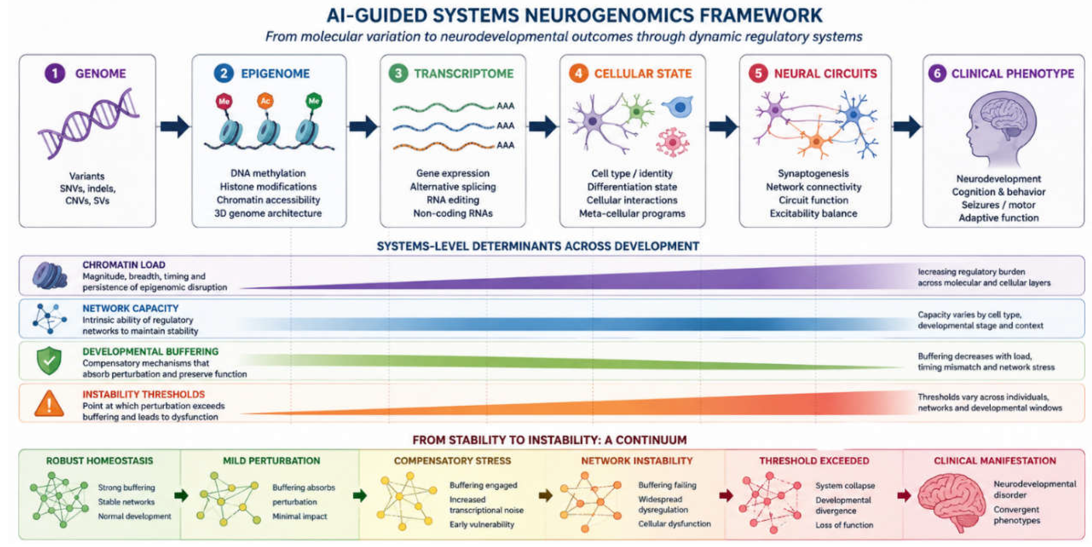
Figure 2.
AI-assisted variant interpretation pipeline. Six-stage AI-supported workflow— (1) variant annotation, (2) structural prediction, (3) splicing prediction, (4) phenotype matching, (5) multi-omic integration, (6) clinical classification—illustrated with the pathogenic variant KAT6A c.1928A>G p.(Asn643Ser) (Arboleda-Tham syndrome). WES, whole-exome sequencing; WGS, whole-genome sequencing.
Figure 2.
AI-assisted variant interpretation pipeline. Six-stage AI-supported workflow— (1) variant annotation, (2) structural prediction, (3) splicing prediction, (4) phenotype matching, (5) multi-omic integration, (6) clinical classification—illustrated with the pathogenic variant KAT6A c.1928A>G p.(Asn643Ser) (Arboleda-Tham syndrome). WES, whole-exome sequencing; WGS, whole-genome sequencing.
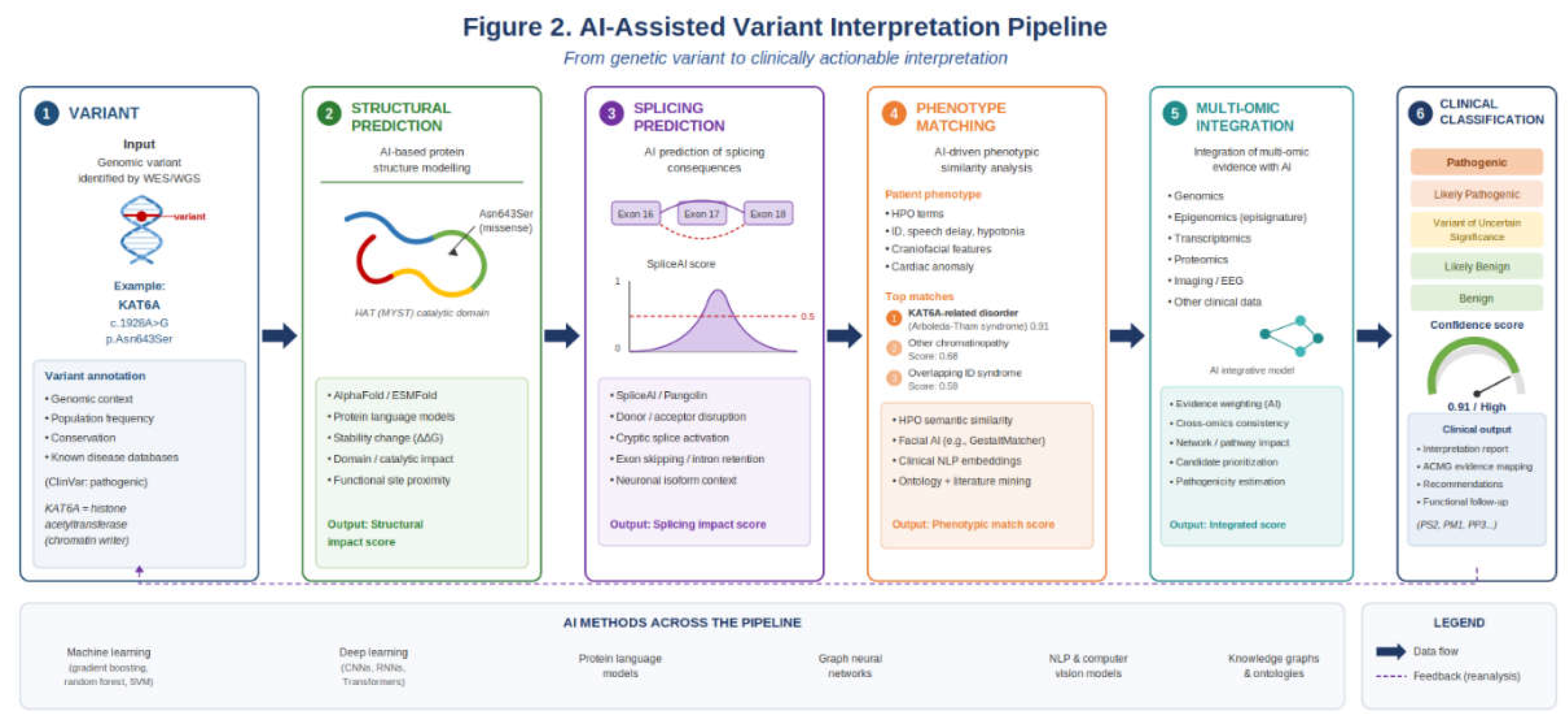
Figure 3.
AI-assisted DNA methylation episignature workflow. Informative CpGs are selected from genome-wide methylation data, visualised by dimensionality-reduction methods and interpreted using clustering and supervised classifiers. MVP-type scores can support diagnosis and VUS interpretation, but must be read with appropriate controls, comparator disorders, batch assessment and clinical context.
Figure 3.
AI-assisted DNA methylation episignature workflow. Informative CpGs are selected from genome-wide methylation data, visualised by dimensionality-reduction methods and interpreted using clustering and supervised classifiers. MVP-type scores can support diagnosis and VUS interpretation, but must be read with appropriate controls, comparator disorders, batch assessment and clinical context.
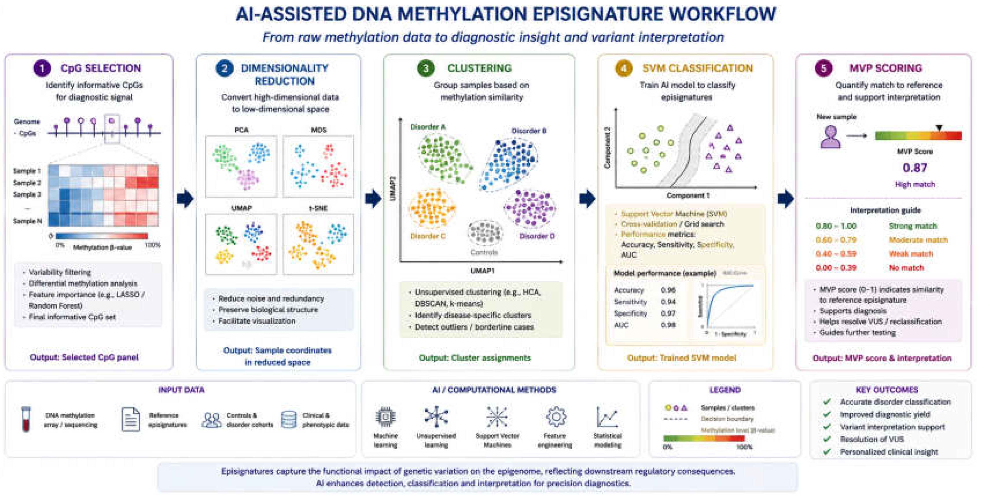
Figure 4.
Regulatory network instability model in neurodevelopment. Molecular perturbations may initially be buffered, but increasing regulatory load can raise transcriptional noise and reduce developmental fidelity. Disease emerges when instability thresholds are crossed, allowing diverse molecular mechanisms to converge on shared phenotypes such as intellectual disability, autism, epilepsy, movement disorders and psychiatric vulnerability.
Figure 4.
Regulatory network instability model in neurodevelopment. Molecular perturbations may initially be buffered, but increasing regulatory load can raise transcriptional noise and reduce developmental fidelity. Disease emerges when instability thresholds are crossed, allowing diverse molecular mechanisms to converge on shared phenotypes such as intellectual disability, autism, epilepsy, movement disorders and psychiatric vulnerability.
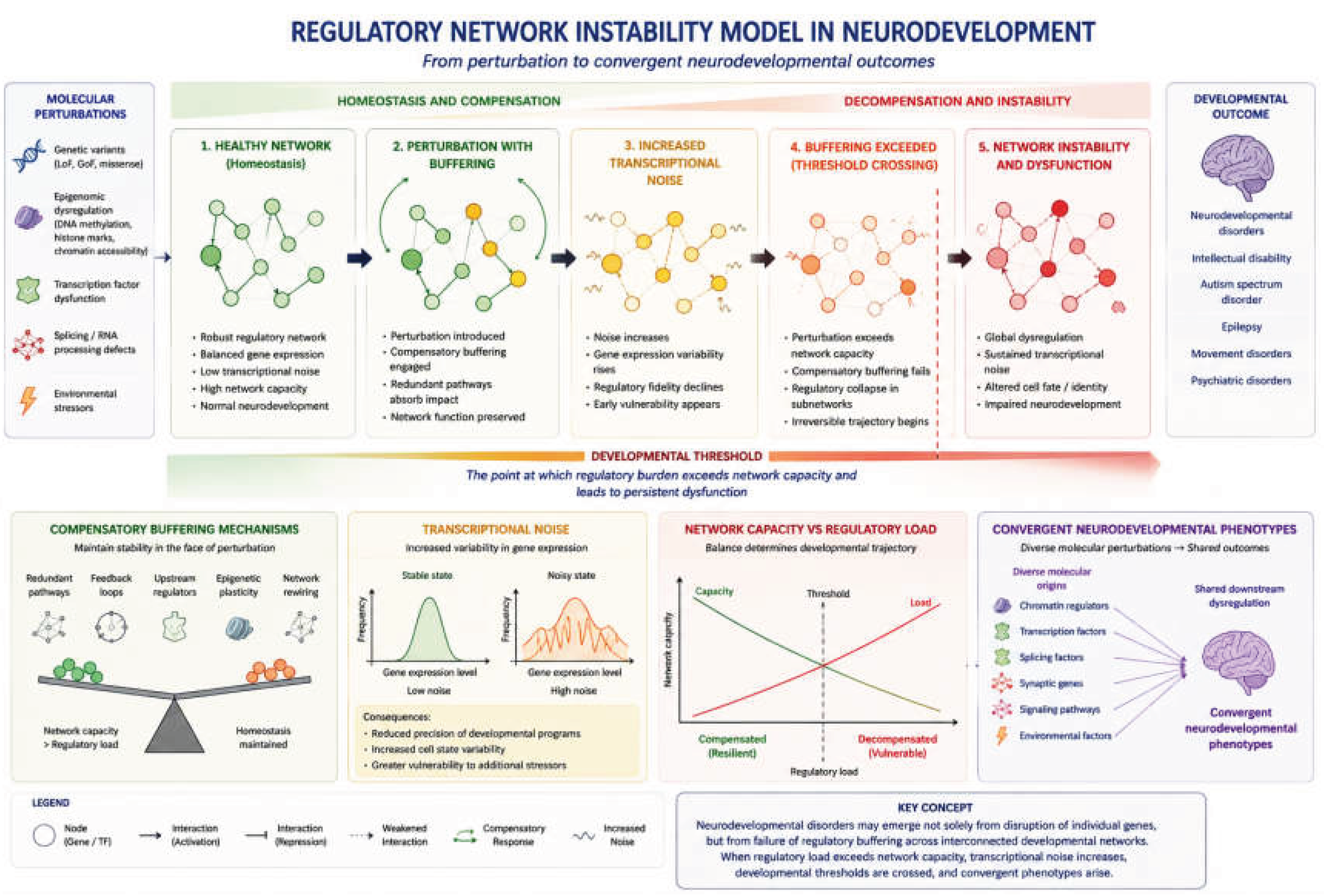

Table 1.
Conceptual vocabulary for AI-guided systems neurogenomics.
| Concept | Working Definition | Relevance to Neurodevelopmental Disorders | Key References |
| Regulatory network | A connected set of chromatin, transcriptional, RNA-processing and signalling mechanisms that collectively control cell identity and developmental trajectory across neural lineages. | Explains why variants in functionally distinct genes converge on shared neurodevelopmental phenotypes, and why single-gene models are insufficient for many disorders. | [13,14,15,16,17] |
| Developmental timing | The stage-specific vulnerability of neural progenitors, neuronal lineages and maturing circuits to regulatory perturbation, arising from the sequential and non-redundant nature of developmental programmes. | A variant may be deleterious only within a defined developmental window; outside it, the same perturbation may be tolerated or compensated, confounding genotype-phenotype prediction. | [18,32] |
| Cell-type specificity | Differential variant effect across progenitors, excitatory neurons, interneurons, astrocytes, oligodendrocytes and microglia, arising from cell-type-specific chromatin states, transcription factor occupancy and regulatory dependencies. | Motivates single-cell and spatial transcriptomic approaches to identify the vulnerable cell populations that drive clinical phenotype, rather than inferring effects from bulk tissue. | [13,15,21] |
| Regulatory load | The cumulative magnitude, breadth, developmental timing, persistence and topological network position of a genomic, epigenomic or transcriptional perturbation acting on a developmental regulatory system. | Explains variable expressivity and nonlinear genotype-phenotype relationships -- two individuals with the same variant may differ substantially if their regulatory loads differ due to genetic background or epigenomic context. | [18,33,34,35,36,37] |
| Network capacity | The ability of a developmental regulatory system to absorb perturbation without loss of functional fidelity, conferred by regulatory redundancy, feedback architecture, compensatory effectors, developmental plasticity and alternative cellular lineages. | Provides a mechanistic account of incomplete penetrance and variable expressivity, individuals with higher network capacity may buffer the same variant that causes overt disease in others. | [18,33,34,35,36,37] |
| Developmental buffering | The active maintenance of cellular identity and developmental trajectory despite genetic, epigenomic or environmental stress, sustained while regulatory load remains within network capacity. | Explains why molecular perturbations can be present without producing a clinical phenotype, and why phenotypic thresholds exist rather than linear dose-response relationships. | [18] |
| Regulatory network instability | The threshold-dependent failure of buffering across interconnected chromatin, transcriptional, RNA-processing and cellular systems when cumulative regulatory load exceeds network capacity, manifesting as rising transcriptional noise, impaired cell-state fidelity and convergent neurodevelopmental phenotypes. | Provides a unifying mechanistic explanation for phenotypic convergence across molecularly diverse NDDs and for the nonlinear relationship between molecular perturbation and clinical severity. | [18,33,34,35,36,37] |
| Transcriptional noise | Increased cell-to-cell variability or reduced precision of gene-expression programmes resulting from regulatory perturbation. | Destabilises neuronal identity, impairs synaptic maturation and disrupts activity-dependent plasticity; may be a shared downstream consequence of diverse upstream chromatin and transcriptional disorders, measurable by single-cell transcriptomics. | [18,20] |
| Instability threshold | The point at which cumulative regulatory load exceeds network capacity, causing buffering failure and overt phenotypic dysfunction. | Provides a systems-level explanation for nonlinear genotype-phenotype relationships, variable expressivity and the phenotypic gap between molecularly similar patients. | [18,33,34,35,36,37] |
| Instability Twin | A composable, patient-specific computational simulation of multi-scale regulatory network dynamics, designed to model load-capacity thresholds, forecast developmental divergence windows and support in silico therapeutic evaluation. | Converts static multi-omic diagnostic data into a dynamic predictive model of developmental trajectory and therapeutic window; the operational implementation of the systems neurogenomics framework at the patient level. | [2,38] |
Table 2.
AI approaches commonly used in neurogenomics.
| AI Approach | Typical Input | Output | Representative Tools | Key References |
| Classical supervised learning | Labelled variants, methylation profiles, phenotype descriptors | Pathogenicity score, disease-class probability, episignature assignment | REVEL, ClinPred, SVM-based episignature classifiers | [33,34,36,37,40,41,42] |
| Unsupervised and self-supervised learning | Methylation, transcriptomic or single-cell feature matrices | Clusters, low-dimensional embeddings, molecular subgroups, trajectory structure | PCA, t-SNE, UMAP; episignature cohort subgroup discovery | [40,41,42] |
| Deep learning (sequence and image models) | DNA/RNA sequence, protein sequence, clinical images, spatial transcriptomic data | Predicted splice effect, missense impact, regulatory consequence, image-derived phenotype score | SpliceAI, AlphaMissense, AlphaGenome, GestaltMatcher | [23,26,44,45,46,47,48,49,50,51,52,53,54,55] |
| Foundation models (pre-trained, transferable) | Large-scale sequence, protein or cell-state corpora | Biological embeddings; zero-shot or fine-tuned task predictions | ESM-2, Nucleotide Transformer, Geneformer | [41,42,43] |
| Knowledge graphs and ontologies | HPO terms, gene-disease relationships, pathway annotations | Semantic similarity scores, phenotype-driven gene rankings, pathway connections | Phenomizer, Exomiser, HPO-driven prioritisation pipelines | [56,57,58,59,60,61,62,63,64,65,66,67,68,69] |
HPO, Human Phenotype Ontology; PCA, principal component analysis; SVM, support vector machine; t-SNE, t-distributed stochastic neighbour embedding; UMAP, uniform manifold approximation and projection.
Table 3.
Representative tools and concepts across the AI-assisted neurogenomics workflow.
| Layer | Representative Tools and Approaches | Clinical or Biological Question Addressed | Key References |
| Variant prioritisation | REVEL, CADD, AlphaMissense, popEVE, pLI/LOEUF constraint metrics, TALOS(automated reanalysis) | Does this coding variant likely disrupt a disease-relevant gene, and how severe is the predicted effect relative to other candidates? | [33,35,36,37,51,52,53,54,55,70,71,72,73,74,75] ref for talos |
| Splicing and RNA processing | SpliceAI, Pangolin, AbSplice, FexSplice, RNA-seq with outlier detection | Does the variant alter exon inclusion, activate a cryptic splice site, induce pseudoexon inclusion, trigger nonsense-mediated decay or affect transcript stability? | [25,27,28,78,79,80] |
| Protein structure and function | AlphaFold, ColabFold, Missense3D, protein language models (ESM-2) | Does the variant disrupt protein folding, domain function, active site integrity, interaction interface or post-translational modification context? | [81,82,83,84,85,86,87,88] |
| Regulatory genomics | AlphaGenome, EPInformer, transcription factor binding models | Does the variant alter chromatin accessibility, histone modification state, transcription factor occupancy, gene expression or enhancer-promoter regulatory topology? | [49,50,76,77,89,90] |
| Clinical phenomics | Exomiser, Phenomizer, PhenoBERT, PhenoTagger, PhenoRerank, HPO2GO, MitoPhen | Does the patient phenotype match the gene, syndrome or biological pathway, and how precisely does the HPO-coded presentation align with the known disease spectrum? | [56,57,58,59,60,62,63,64,65,66,67,68,69,91,92,93,94,95] |
| Facial and dysmorphology phenomics | GestaltMatcher, DeepGestalt, Face2Gene, FaceMatch, Image2Gene | Is there a recognisable craniofacial or image-derived syndromic pattern consistent with the candidate diagnosis, across age and ancestry? | [44,45,46,47,48,96,97] |
| Episignature diagnostics | Disorder-specific SVM classifiers, MVP scoring pipelines, reference methylation cohorts | Does the patient’s genome-wide methylation profile match a known episignature and provide functional support for a VUS in a chromatin-related gene? | [22,24,98,99,100,101,102,103,104,105,106,107] |
| Single-cell and spatial biology | Brain cell atlases, STAB2, ZEBRA, Geneformer, spatial transcriptomic models | Which cell types, developmental lineages, brain regions or circuit components are most vulnerable to the identified perturbation, and at what developmental stage? | [15,91,108,109,110,111,112] |
| Developmental trajectory modelling | iPSC and organoid models, RNA velocity, TarDis disentanglement frameworks | At what point does the patient’s molecular profile diverge from expected paths, and does the perturbation approach or cross the instability threshold? | [2,19,20,22,38] |
| Integrative diagnostic reasoning | Agentic LLM systems (e.g., DeepRare), multi-agent evidence integration, traceable reasoning chains | Can heterogeneous inputs (free text, HPO terms, genetic results) be integrated into ranked, mechanism-aware diagnostic hypotheses with transparent, verifiable reasoning? | [113] |
| Therapeutic target discovery | Pathway-guided network models, drug-target interaction predictors, ASO design tools, perturbation screens | Does mechanistic modelling identify druggable nodes, splice correction candidates, pathway entry points or repurposing opportunities relevant to the disorder? | [114,115] |
The layer ordering reflects the clinical interpretive workflow from molecular variant to mechanism to therapeutic implication. Tools vary substantially in clinical maturity: some are in routine diagnostic use, others are research-stage implementations included to illustrate how AI methods map onto the systems neurogenomics framework. HPO: Human Phenotype Ontology; MDS: multidimensional scaling; MVP: methylation variant pathogenicity; PCA: principal component analysis; SVM: support vector machine.
Disclaimer/Publisher’s Note: The statements, opinions and data contained in all publications are solely those of the individual author(s) and contributor(s) and not of MDPI and/or the editor(s). MDPI and/or the editor(s) disclaim responsibility for any injury to people or property resulting from any ideas, methods, instructions or products referred to in the content. |
© 2026 by the authors. Licensee MDPI, Basel, Switzerland. This article is an open access article distributed under the terms and conditions of the Creative Commons Attribution (CC BY) license (http://creativecommons.org/licenses/by/4.0/).
Copyright: This open access article is published under a Creative Commons CC BY 4.0 license, which permit the free download, distribution, and reuse, provided that the author and preprint are cited in any reuse.


