

Submitted:
26 June 2026
Posted:
29 June 2026
You are already at the latest version
Abstract
Background: Skin health is increasingly recognized as a key determinant of overall well-being. Hydroxytyrosol (HT), a phenolic compound abundant in olives and olive-derived products, has attracted growing interest because of its antioxidant and anti-inflammatory properties and its potential role in promoting skin health and healthy ageing. Objective: To critically review the evidence supporting oral HT and HT-rich nutraceuticals as strategies for improving skin health and healthy ageing. Methods: This narrative review summarizes mechanistic, preclinical, and clinical studies investigating the effects of oral HT and HT-rich formulations on biological pathways involved in skin homeostasis, ageing, and inflammatory skin disorders. Results: HT exhibits high oral bioavailability and modulates antioxidant defenses, inflammatory signaling, autophagy, apoptosis, and metabolic homeostasis. Preclinical studies consistently demonstrate protective effects against oxidative stress, extracellular matrix degradation, cellular senescence, photoaging, impaired wound healing, and UV-induced skin damage. Experimental evidence also indicates antiproliferative and pro-apoptotic effects in melanoma models through modulation of oncogenic signaling pathways, suggesting a potential role in skin cancer prevention. Oral HT administration improves glycative skin ageing, psoriasis, intestinal barrier integrity, and systemic inflammation in animal models. Clinical studies support systemic antioxidant, anti-inflammatory, and cardiometabolic effects of purified HT and HT-rich formulations, while preliminary dermatological evidence suggests benefits in psoriasis, melasma, and wound healing. Conclusions: HT is a promising bioactive compound targeting multiple mechanisms involved in skin ageing and homeostasis. Although mechanistic and preclinical evidence is robust, clinical evidence remains limited and heterogeneous. Therefore, randomized controlled trials with validated dermatological endpoints are needed to confirm the clinical efficacy of HT.
Keywords:
hydroxytyrosol
; olive polyphenols
; skin health
; skin ageing
; cancer
; nutraceuticals
; oxidative stress
1. Introduction
The skin is a dynamic, multifunctional organ essential for maintaining homeostasis through its roles in barrier function, thermoregulation, and immune surveillance, which contribute to overall health [Zhu et al., 2025]. Skin health is closely associated with the integrity of the skin barrier, which provides protection against ultraviolet (UV) radiation, pollutants, and pathogens [Mohania and Chandel, 2017; Fitoussi et al., 2022; Elias, 2008]. Beyond barrier integrity, healthy skin is characterized by adequate hydration, preservation of extracellular matrix (ECM) components such as collagen and elastin, efficient wound healing, and protection against oxidative damage and systemic inflammation. Together, these features contribute to skin elasticity, resilience, and overall appearance. Aging progressively compromises these functions, leading to reduced hydration, impaired repair capacity, and increased skin pH, promoting cutaneous and systemic inflammation via elevated circulating cytokines [Wang et al., 2020; Hu and Mauro, 2017]. Skin ageing is driven by a complex interplay of intrinsic and extrinsic factors, with oxidative stress and chronic low-grade inflammation representing the principal mechanisms underlying progressive structural and functional skin decline [Rinnerthaler et al., 2015; Kammeyer and Luiten, 2015]. Poor nutrition, dehydration, sleep deprivation and hormonal changes, particularly estrogen decline during ageing, may further exacerbate these alterations [Fitzmaurice et al., 2011; Raine-Fenning et al., 2003]. In addition, growing interest has focused on the contribution of microbial ecosystems to skin physiology and ageing. Both the skin microbiome and the gut-skin axis have been increasingly recognized as important components of cutaneous homeostasis and immune regulation [Lunjani et al., 2021; Liu et al., 2023; Millman et al., 2024; Jimenez-Sanchez et al., 2025]. Emerging evidence suggests that alterations in these interconnected systems may influence skin function and contribute to age-related changes and inflammatory skin conditions, highlighting their potential relevance as targets for preventive and therapeutic strategies [Lunjani et al., 2021; Liu et al., 2023; Millman et al., 2024; Jimenez-Sanchez et al., 2025].
These mechanisms have stimulated growing interest in systemic and dietary strategies aimed at supporting skin health and healthy ageing [Jiang et al., 2025]. Among naturally occurring bioactive compounds, hydroxytyrosol (HT), a phenolic compound abundant in olives and olive-derived products, has attracted considerable attention because of its potent antioxidant and anti-inflammatory properties [D’Angelo et al., 2020; Echeverria et al., 2017]. Oral HT supplementation has been associated with improvements in oxidative balance, inflammatory status, and metabolic health, all of which are relevant to skin homeostasis and ageing processes [Frumuzachi et al., 2024; Moratilla-Rivera et al., 2025; Colica et al., 2017].
This review focuses primarily on the evidence supporting oral HT and HT-rich nutraceuticals as potential strategies to promote skin health and healthy ageing. Mechanistic, preclinical, and clinical evidence are critically examined to provide an integrated overview of the biological rationale underlying HT supplementation and its potential implications for skin function. Particular attention is given to animal and human studies evaluating oral HT administration, while current limitations, knowledge gaps, and future research priorities are also discussed.
2. Methods
The literature search was conducted in PubMed/MEDLINE, Scopus, and Google Scholar using combinations of keywords related to hydroxytyrosol (HT), skin health, skin ageing, and the biological pathways involved in skin homeostasis. The primary search terms included: “hydroxytyrosol” AND “oral supplementation”, “hydroxytyrosol” AND “oral administration”, “hydroxytyrosol” AND “dietary supplementation”, “hydroxytyrosol” AND “nutraceutical”, “hydroxytyrosol” AND “skin health”, “hydroxytyrosol” AND “skin ageing”, “hydroxytyrosol” AND “skin aging”, “hydroxytyrosol” AND “photoaging”, “hydroxytyrosol” AND “photoprotection”, “hydroxytyrosol” AND “skin homeostasis”, “hydroxytyrosol” AND “wound healing”, “hydroxytyrosol” AND “skin regeneration”, “hydroxytyrosol” AND “extracellular matrix”, “hydroxytyrosol” AND “collagen”, “hydroxytyrosol” AND “oxidative stress”, “hydroxytyrosol” AND “reactive oxygen species”, “hydroxytyrosol” AND “inflammation”, “hydroxytyrosol” AND “cellular senescence”, “hydroxytyrosol” AND “advanced glycation end products”, “hydroxytyrosol” AND “AGEs”, “hydroxytyrosol” AND “gut-skin axis”, and “hydroxytyrosol” AND “microbiome”. Additional searches were performed using terms related to olive-derived bioactive compounds and nutraceutical formulations, including “olive polyphenols”, “olive oil polyphenols”, “olive phenolics”, “olive-derived polyphenols”, “extra virgin olive oil”, “olive mill wastewater”, “OMWW polyphenols”, and “hydroxytyrosol-rich nutraceuticals”, combined with keywords such as “skin health”, “skin ageing”, “skin aging”, “healthy ageing”, “photoaging”, “skin inflammation”, “oxidative stress”, “wound healing”, “microbiome”, and “gut-skin axis”. Reference lists of relevant original articles and review papers were manually screened to identify additional studies of interest. Studies published in English up to June 2026 were considered. Original articles, clinical studies, animal studies and relevant reviews were included according to their relevance to the scope of the review.
3. Skin Ageing and Alterations in Skin Homeostasis
Clinically, skin ageing is characterized by wrinkles, pigmentation changes, xerosis, epidermal thinning, actinic keratoses, and reduced dermal elasticity. Importantly, the deterioration of skin structure and function extends beyond cosmetic concerns, affecting barrier integrity, immune competence, and susceptibility to disease [Yang and Man, 2023]. Skin ageing is a multifactorial process driven by both intrinsic (chronological) and extrinsic factors, including environmental and lifestyle influences, collectively referred to as the exposome [Thompson et al., 2022]. The exposome plays a major role in accelerating skin ageing, highlighting the need for preventive and targeted interventions [Krutmann et al., 2017]. The skin is highly vulnerable to environmental stressors such as ionizing radiation, pollution, and UV exposure, as well as to lifestyle factors including alcohol consumption and poor diet [Rittié and Fisher, 2015; Farage et al., 2013; Wong and Chew, 2021]. In particular, UV radiation damages the dermal ECM and keratinocyte DNA, leading to structural and cellular alterations that manifest clinically [Rittié and Fisher, 2015]. Intrinsic ageing, by contrast, is an inevitable process driven by genetic and metabolic factors [Zhang and Duan, 2018].
Ageing is also associated with alterations in the skin microbiome and immune function, which may contribute to barrier dysfunction, chronic inflammation, and impaired tissue repair [Yang et al., 2024]. In women, menopause further accelerates skin ageing through estrogen decline, resulting in reduced collagen synthesis, impaired elasticity, decreased hydration, thinning of the epidermis and dermis, and delayed wound healing [Viscomi et al., 2025]. Both extrinsic and intrinsic factors contribute to ageing by increasing reactive oxygen species (ROS) production and activate inflammatory pathways [Vierkötter et al., 2010]. ROS accumulation promotes ECM degradation, collagen breakdown, cellular senescence, and progressive loss of skin function, contributing to the visible signs of ageing. Moreover, skin barrier dysfunction is increasingly recognized as a potential driver of systemic inflammaging, a chronic low-grade inflammatory state associated with systemic consequences [Agrawal et al., 2023; Zhang et al., 2025]. This process involves glycation, reduced autophagy, mitochondrial dysfunction, telomere shortening, immune dysregulation, and diminished regenerative capacity [Agrawal et al., 2023; Buckingham and Klingelhutz, 2011; He et al., 2024; Ge et al., 2020].
Beyond its role in physiological ageing, oxidative stress also contributes to the pathogenesis of inflammatory skin disorders. In psoriasis, for example, redox imbalance promotes keratinocyte hyperproliferation and immune activation, establishing a self-perpetuating cycle between oxidative stress and inflammation [Dobrică et al., 2022]. UV radiation and other environmental stressors further amplify oxidative damage through lipid peroxidation, protein oxidation, and DNA injury in keratinocytes and fibroblasts [Rinnerthaler et al., 2015; Kammeyer and Luiten, 2015]. These alterations impair cellular function, accelerate cellular senescence, and increase matrix metalloproteinase (MMP) expression, ultimately promoting ECM degradation [Gilchrest et al., 1999; Yaar et al., 2007]. Sustained activation of inflammatory pathways also results in overproduction of pro-inflammatory cytokines, including TNF-α, IL-1β, and IL-6, further exacerbating tissue damage [Pillai et al., 2005; Quan and Fisher, 2015]. In parallel, ageing of the skin immune system reduces pathogen defense, impairs clearance of senescent cells, and promotes dysregulated inflammatory responses, thereby increasing susceptibility to infections, inflammatory disorders, and delayed wound healing [He et al., 2024].
Among the intrinsic drivers of ageing, advanced glycation end products (AGEs) have attracted considerable attention. AGEs are formed through non-enzymatic reactions between reducing sugars and macromolecules and originate from both endogenous metabolism and dietary sources [Singh et al., 2001; Scheijen et al., 2016]. In the skin, AGEs accumulate in collagen and elastin fibers, increasing stiffness, reducing elasticity, and disrupting ECM turnover [Fan et al., 2025; Gkogkolou and Böhm, 2012; Chen et al., 2022]. They also promote fibroblast apoptosis, reduce collagen content, and enhance dermal fragmentation [Chen and Guo, 2021]. Furthermore, AGEs contribute to oxidative stress and inflammation by increasing ROS production and inducing mitochondrial dysfunction [Twarda-Clapa et al., 2022]. Their biological effects are mediated through protein crosslinking, ROS generation, and activation of receptor-mediated pathways, particularly the receptor for advanced glycation end products (RAGE), which triggers NF-κB-dependent inflammatory signaling and cytokine production [Chrysanthou et al., 2022; Twarda-Clapa et al., 2022]. Although autophagy normally contributes to AGE clearance, excessive glycation impairs autophagic function, creating a vicious cycle that promotes further AGE accumulation and cellular dysfunction [Takahashi et al., 2017; Gómez et al., 2021]. Conversely, activation of autophagy has been shown to facilitate AGE removal and improve cellular homeostasis [Laughlin et al., 2020].
The gut-skin axis is increasingly recognized as a key contributor to skin ageing and inflammatory skin disorders through the interplay between the gut microbiota, immune responses, and circulating metabolites [Jimenez-Sanchez et al., 2025]. Among the mechanisms underlying this connection, oxidative stress appears to play a central role. Increased ROS levels can impair intestinal barrier integrity, promoting microbial translocation and systemic inflammatory responses. In turn, gut dysbiosis may further enhance oxidative stress through immune dysregulation and the production of pro-inflammatory metabolites, creating a self-perpetuating cycle that contributes to chronic inflammation, disruption of tissue homeostasis, and progression of inflammatory skin conditions [Pleńkowska et al., 2020; Ni and Zhang, 2022].
Increasing evidence highlights the importance of preserving skin barrier integrity, reducing oxidative stress, and modulating inflammation not only to maintain skin health but also to mitigate systemic ageing processes [Haykal et al., 2026]. Consequently, plant-derived phytochemicals are increasingly being explored as therapeutic and preventive strategies for inflammatory skin conditions, both as topical agents and nutritional supplements. These compounds may modulate inflammatory pathways, provide photoprotective effects, and support skin barrier function with fewer adverse effects than conventional pharmacological approaches [Nisar and Jagtap, 2023; Chaiprasongsuk and Panich, 2022; Assaf and Kelly, 2024; Albini et al., 2025]. Among these bioactive compounds, HT has emerged as a particularly promising candidate due to its combined antioxidant, anti-inflammatory, and metabolic effects, which target several of the biological pathways involved in skin ageing and skin homeostasis.
4. Hydroxytyrosol: Bioavailability and Mechanistic Pathways
4.1. HT Bioavailability
HT is a small, highly polar phenolic alcohol naturally present in olives, olive oil, and olive-derived products [D’Angelo et al., 2020; Echeverria et al., 2017]. It can also be generated endogenously through the metabolism of oleuropein (OLE), the major secoiridoid found in olive leaves and unripe olive fruits. Structurally, HT contains a catechol moiety that underlies its potent radical-scavenging activity and high reactivity toward ROS [Omar, 2010]. One of the most relevant characteristics of HT is its excellent oral bioavailability. Following ingestion, HT is efficiently absorbed in the small intestine and rapidly metabolized into glucuronide and sulphate conjugates that retain significant antioxidant and anti-inflammatory activity [Soldevila-Domenech et al., 2019; Feng et al., 2024]. Peak plasma concentrations are generally reached within 30-60 minutes after administration, supporting the rapid onset of systemic biological effects observed in acute intervention studies [Muriana et al., 2017]. HT bioavailability is influenced by several factors, including formulation, lipid matrix, dose, timing of administration, and the presence of other polyphenols [Silva et al., 2020; Di Renzo et al., 2023]. These characteristics make HT particularly suitable for oral supplementation strategies aimed at targeting systemic pathways involved in oxidative stress, inflammation, and tissue homeostasis.
4.2. HT Mechanistic Pathways
At the mechanistic level, HT exerts its biological activity through a combination of antioxidant, anti-inflammatory, metabolic, and microbiota-modulating effects [Moratilla-Rivera et al., 2026; Gao et al., 2025]. Rather than acting solely as a direct scavenger of reactive oxygen species, HT primarily functions as a regulator of endogenous cellular defense systems. A central mechanism involves activation of the nuclear factor erythroid 2-related factor 2 (Nrf2) pathway, which promotes the expression of antioxidant enzymes and enhances cellular resilience to oxidative stress [Tebay et al., 2015].
In parallel, HT modulates inflammatory responses through inhibition of key signaling pathways, including NF-κB and cyclooxygenase-2 (COX-2). These effects result in reduced production of pro-inflammatory mediators such as C-reactive protein (CRP), tumor necrosis factor-alpha (TNF-α), interleukin (IL)-1β, and IL-6, together with increased expression of anti-inflammatory mediators including IL-10 [Alblihed, 2021; Yonezawa et al., 2019; Fuccelli et al., 2018]. Clinical studies further support the broad anti-inflammatory activity of HT [Richard et al., 2013].
Growing evidence also indicates that HT influences cellular quality-control mechanisms involved in ageing and tissue homeostasis. In particular, HT has been shown to regulate apoptosis, autophagy, and immune responses while interacting with lipid metabolism and epigenetic signaling pathways [Moratilla-Rivera et al., 2026]. In primary rat chondrocytes exposed to oxidative and inflammatory stress, HT enhanced autophagic activity through a Sirtuin 1 (SIRT1)-dependent mechanism, increasing the expression of key autophagy-related proteins, including LC3-II and ATG5/7, while reducing p62 accumulation. These effects were accompanied by reductions in ROS production, NADPH oxidase (NOX)2 and NOX4 expression, and inflammatory mediators including IL-6, TNF-α, and MMP-13, ultimately improving cellular viability under stress conditions [Sun et al., 2019]. Importantly, inhibition of either autophagy or SIRT1 abolished these protective effects, confirming the central role of this pathway.
Beyond intracellular signaling, HT exerts beneficial systemic effects that are highly relevant to ageing-related processes. Cardiovascular protection represents one of the most extensively documented areas of research. HT improves endothelial function, enhances nitric oxide bioavailability, reduces the level of low-density lipoprotein (LDL) oxidation, and attenuates inflammatory signaling, thereby contributing to improved vascular homeostasis and reduced cardiovascular risk [Noguera-Navarro et al., 2023].
Emerging evidence also suggests that HT may influence microbiota composition and gut barrier function, contributing to modulation of the gut-skin axis and systemic inflammatory responses [Gao et al., 2025]. These activities are particularly relevant in the context of skin ageing, where oxidative stress, chronic inflammation, metabolic dysfunction, and microbiome alterations interact to drive progressive deterioration of skin structure and function.
Overall, HT displays a favorable safety profile and appears particularly effective under conditions characterized by increased oxidative stress and chronic inflammation [Wang et al., 2025]. Through the coordinated modulation of antioxidant defenses, inflammatory pathways, cellular quality-control mechanisms, and systemic metabolic processes, HT targets several of the biological mechanisms implicated in skin ageing and impaired skin homeostasis.
4.3. Systemic Effects of Hydroxytyrosol-Rich Formulations and Purified Hydroxytyrosol
Olive oil contains a variety of bioactive constituents that can be broadly classified into two fractions: the unsaponifiable (non-polar) fraction, which includes squalene, triterpenes, sterols, tocopherols, and pigments, and the polar fraction, which comprises phenolic compounds, including HT [Covas et al., 2015]. In addition to olive oil, HT and other phenolic compounds are highly concentrated in olive mill wastewater (OMWW), the aqueous by-product generated during extra virgin olive oil production. Despite being biodegradable, OMWW poses significant environmental challenges. Its high phenolic content and acidic pH contribute to phytotoxic effects, while its lipid-rich composition can reduce dissolved oxygen levels, adversely affecting aquatic ecosystems [Sciubba et al., 2020]. Advances in extraction and purification technologies have enabled the recovery of polyphenol-rich fractions from OMWW, transforming an agricultural waste stream into a valuable and sustainable source of nutraceutical compounds within a circular economy framework [Albini et al., 2023]. One example is OliPhenolia®, a phytocomplex obtained from OMWW through concentration, reverse osmosis, and mechanical filtration processes. Besides HT, OliPhenolia® contains verbascoside, tyrosol, p-coumaroyl secoiridoids, and other minor polyphenols, suggesting that its biological activity may result from the combined action of multiple compounds rather than HT alone [Cuffaro et al., 2023]. The process of preparation of OliPhenolia® is depicted in Figure 1.
4.3.1. Evidence from HT-Rich Formulations
The first human studies investigating OMWW-derived formulations primarily focused on bioavailability and antioxidant activity. In a pilot crossover study involving healthy volunteers, Bender et al. demonstrated that HT-rich formulations derived from OMWW (OliPhenolia®) were rapidly absorbed after oral administration, as evidenced by the urinary excretion of HT. In parallel, in vitro experiments showed significant antioxidant activity, including reductions in reactive oxygen species, lipid peroxidation, and advanced glycation end-product formation, together with stimulation of endogenous antioxidant defenses [Bender et al., 2021].
The bioavailability of OliPhenolia® was confirmed in a randomized, double-blind, placebo-controlled study conducted in healthy recreationally active adults. Following ingestion of a single dose, plasma HT concentrations increased rapidly and remained elevated for several hours. During a subsequent exercise challenge designed to induce oxidative stress, supplementation resulted in modest but significant antioxidant effects, including increased glutathione levels and reduced superoxide dismutase activity following exercise, suggesting improved redox balance and recovery capacity. However, no significant effects were observed for several other oxidative stress biomarkers, indicating that the physiological impact was relatively modest [Roberts et al., 2022].
A more detailed pharmacokinetic characterization of olive-derived HT formulations (OliPhenolia®) was subsequently provided by Bender et al. in a randomized, controlled, blinded crossover study in 12 healthy volunteers. Using serial blood and urine sampling over 12 h after a single dose, the authors tracked both free HT and its major metabolites in plasma and urine, showing rapid absorption, a peak plasma concentration at approximately 30 min, dose-dependent bioavailability, and extensive phase II/metabolic conversion. By integrating plasma kinetics with urinary excretion data, this study provided a mechanistic basis for subsequent efficacy analyses in the same experimental setting [Bender, Strassman et al., 2023]. Additional evidence for short-term antioxidant activity was provided by the analysis of biomarkers of lipid oxidation, including plasma oxidized LDL (oxLDL) and urinary F2-isoprostanes, which showed a reduction following supplementation. Interestingly, these effects appeared more pronounced in participants with higher baseline oxidative stress, suggesting that the benefits of supplementation may be greater under conditions of increased oxidative burden [Bender et al., 2023]. Although the study was designed around cardiovascular markers rather than dermatological endpoints, these findings remain of interest in a beauty-from-within context because F2-isoprostanes are robust markers of in vivo lipid peroxidation [Roberts and Morrow, 2000; Milatovic et al., 2011], while oxidative stress-related lipid damage has also been documented in skin under oxidative challenge and is implicated in skin aging and age-related tissue damage [Rinnerthaler et al., 2015]
Beyond antioxidant activity, HT-rich formulations have shown promising effects on metabolic health. In a pilot longitudinal intervention study involving 29 adults presenting characteristics associated with metabolic syndrome, supplementation with OliPhenolia® (25 mL twice daily for 30 days) was associated with favorable changes in several cardiometabolic parameters, including reductions in blood pressure, fasting glucose, insulin levels, and LDL cholesterol. Improvements were also observed in hydration status, while metabolomic analyses suggested modulation of pathways related to vitamin D metabolism, homocysteine metabolism, and inflammation. Importantly, no clinically relevant adverse effects were reported [Aiello et al., 2024]. A secondary analysis of the same cohort further explored body composition and muscle-related outcomes. Supplementation was associated with modest increases in skeletal muscle mass and muscle mass percentage, reductions in fat mass, and improvements in hydration status. These changes were accompanied by increased antioxidant capacity, reflected by higher protein thiol concentrations and Trolox equivalent antioxidant capacity, suggesting a potential role for OMWW-derived polyphenols in supporting muscle health and counteracting early sarcopenic changes in individuals at metabolic risk [Morelli et al., 2026].
Evidence supporting broader cardiometabolic benefits also comes from studies using HT-enriched foods. In a 12-week randomized dietary intervention including 60 adults with overweight/obesity and type 2 diabetes, daily consumption of HT-enriched whole wheat bread within an energy-restricted Mediterranean diet produced greater reductions in fasting glucose, glycated hemoglobin (HbA1c), insulin levels, LDL cholesterol, body fat, and inflammatory markers, particularly TNF-α, compared with conventional whole wheat bread [Binou et al., 2023].
Similarly, a 6-month pilot intervention study conducted in 11 healthy older adults evaluated the effects of olive oils differing in polyphenol content. Participants consuming high-polyphenol extra virgin olive oil, characterized by higher concentrations of HT and tyrosol, showed reductions in arterial inflammation and atherosclerotic microcalcification assessed by positron emission tomography/computed tomography (PET/CT) imaging. In contrast, standard extra virgin olive oil produced no significant changes, while refined olive oil was associated with worsening microcalcification markers. These findings suggest a potential vascular protective effect of olive polyphenols in ageing populations [Zoubdane et al., 2024].
4.3.2. Evidence from Purified Hydroxytyrosol
Compared with HT-rich formulations, clinical evidence specifically evaluating purified HT remains more limited. Nevertheless, available studies generally support the biological effects observed with complex formulations. The strongest evidence currently derives from a randomized, double-blind, placebo-controlled trial involving 49 adults aged 40–70 years with overweight and prediabetes. Participants received either 15 mg/day of purified HT or placebo for 16 weeks. HT supplementation significantly reduced several biomarkers of oxidative stress and inflammation, including oxidized LDL, protein carbonyls, 8-hydroxy-2′-deoxyguanosine (8-OHdG), and interleukin-6, while preserving antioxidant capacity and glutathione peroxidase activity. These findings indicate that HT may be particularly effective under conditions characterized by increased oxidative stress and low-grade inflammation [Moratilla-Rivera et al., 2025].
Overall, current human evidence consistently supports antioxidant, anti-inflammatory, and cardiometabolic effects of both HT-rich formulations and purified HT. However, most studies have evaluated systemic biomarkers and metabolic outcomes rather than dermatological endpoints. Therefore, while these findings provide a strong biological rationale for a potential role of oral HT supplementation in skin health, direct clinical evidence linking HT intake to improvements in skin ageing, skin function, or other dermatological outcomes remains limited.
5. Hydroxytyrosol and Skin Biology: Mechanistic Rationale and Implications for Topical and Oral Supplementation
The biological effects of HT on skin health are supported by a broad body of mechanistic evidence demonstrating its ability to modulate key pathways involved in skin ageing, tissue homeostasis, and repair. These effects encompass oxidative stress regulation, inflammation control, ECM preservation, photoprotection, and tissue regeneration, collectively providing a biological rationale for its potential application in skin health and healthy ageing [Fan et al., 2025]. HT exerts potent antioxidant activity by directly scavenging ROS and by activating endogenous antioxidant defense systems, including Nrf2-dependent pathways. Through these mechanisms, HT helps maintain redox homeostasis and protects skin cells from oxidative injury induced by ageing, environmental stressors, and UV radiation [Fan et al., 2025].
In parallel, HT exhibits significant anti-inflammatory activity. Experimental studies have demonstrated modulation of several signaling pathways involved in cutaneous inflammation, particularly NF-κB-dependent pathways, resulting in reduced production of pro-inflammatory cytokines and attenuation of chronic low-grade inflammation. This activity may be particularly relevant in the context of inflammaging, which contributes substantially to age-related skin deterioration [Aparicio-Soto et al., 2019; Fan et al., 2025].
The integrity of the ECM is a major determinant of skin structure, elasticity, and mechanical resilience. HT contributes to ECM homeostasis by limiting collagen degradation, reducing MMP activity, and supporting collagen preservation. Through these effects, HT may help maintain skin firmness and counteract structural alterations associated with chronological ageing and photoageing [Jeon and Choi, 2018; Menicacci et al., 2017].
These mechanisms are particularly relevant under conditions of UV exposure, where oxidative stress and inflammation accelerate matrix degradation and skin ageing. By reducing oxidative injury and inflammatory responses induced by UV radiation, HT exerts photoprotective effects that may contribute to preserving skin architecture and function under environmental stress [Zwane et al., 2012].
Beyond its anti-ageing properties, HT appears to support tissue repair and regenerative processes. Experimental studies indicate that HT promotes fibroblast proliferation and migration, enhances re-epithelialisation, stimulates ECM remodeling, and supports wound closure. In addition, HT modulates epithelial-mesenchymal transition (EMT) and inflammatory responses involved in tissue repair, potentially facilitating effective healing while limiting excessive fibrosis and pathological remodeling [Batarfi et al., 2023; González-Acedo et al., 2023]. Evidence also suggests that HT activates cytoprotective pathways such as heme oxygenase-1 (HO-1)/Nrf2 signaling, further enhancing cellular resilience and repair capacity under conditions of oxidative stress [Zrelli et al., 2015].
Collectively, these mechanisms provide the biological rationale for both topical and oral administration of HT. Topically applied HT may support skin homeostasis by enhancing epidermal barrier function, promoting tissue regeneration, and reducing cutaneous inflammation through the modulation of pro-inflammatory cytokines [Smeriglio et al., 2019]. On the other side, oral supplementation may contribute indirectly to skin health through systemic antioxidant and anti-inflammatory effects [Fan et al., 2025]. Importantly, although mechanistic and preclinical evidence consistently supports a protective role of HT in skin biology, the strength of evidence varies across outcomes. Direct clinical evidence demonstrating improvements in skin ageing, skin function, or dermatological conditions following oral HT supplementation remains limited and should therefore be interpreted cautiously. Nevertheless, the available mechanistic data strongly support further investigation of HT as a potential strategy for maintaining skin homeostasis, promoting tissue repair, and mitigating age-related skin changes.
5.1. Studies Relevant to Skin Biology
The mechanistic rationale supporting the role of HT in skin health is supported by a growing body of experimental, preclinical, and clinical evidence. Studies conducted in cellular and animal models have consistently demonstrated that HT modulates biological processes directly involved in skin ageing and tissue homeostasis, including oxidative stress, inflammation, ECM remodeling, wound healing, and photoprotection. These findings are complemented by a limited number of clinical studies investigating the effects of HT-containing formulations in selected dermatological conditions. The available evidence is heterogeneous with respect to study design, HT source, and outcomes evaluated. Some studies have investigated purified HT, whereas others have examined HT-rich extracts or complex olive-derived formulations containing additional bioactive polyphenols. Therefore, the results should be interpreted considering the specific intervention tested. To facilitate interpretation, the evidence is presented according to the level of investigation, moving from in vitro studies to animal models and, finally, clinical studies evaluating skin-related outcomes.
5.1.1. In Vitro Evidence
A substantial body of in vitro evidence supports the role of HT in protecting skin cells against oxidative stress, inflammation, ECM degradation, and cellular senescence.
Several studies have investigated the anti-photoaging properties of HT. Using a UVA-induced photoaging model in human dermal fibroblasts (HDFs), Jeon and Choi demonstrated that purified HT (>90% purity) was non-cytotoxic up to 30 μM and significantly attenuated cellular senescence, as evidenced by reduced senescence-associated β-galactosidase activity. HT also downregulated MMP-1 and MMP-3, key mediators of collagen degradation, while reducing the expression of the pro-inflammatory cytokines IL-1β, IL-6, and IL-8 in a dose-dependent manner [Jeon and Choi, 2018].
Additional evidence supporting anti-aging activity was provided by Li et al., who investigated purified HT and OLE. Both compounds exhibited moderate inhibitory activity against elastase and collagenase, with HT showing inhibition ranging from 12.6-31.0% and 11.6-31.9%, respectively. When combined at a 1:1 ratio, OLE and HT exerted synergistic elastase inhibition, confirmed by combination index analysis, enzyme kinetics, and molecular docking studies. Furthermore, HT reduced H2O2-induced cytotoxicity and intracellular ROS production in human dermal fibroblasts, while OLE–HT combinations produced synergistic reductions in ROS levels, suggesting enhanced cytoprotective activity [Li et al., 2022].
The anti-senescence activity of HT has also been demonstrated in chronically treated human fibroblasts. Menicacci et al. evaluated HT and OLE in pre-senescent lung (MRC5) and dermal (NHDF) fibroblasts and observed reductions in senescence-associated β-galactosidase activity, p16 expression, IL-6 production, COX-2 expression, NF-κB activation, and MMP activity. These findings suggest attenuation of both the senescence-associated secretory phenotype (SASP) and ECM degradation [Menicacci et al., 2017].
The anti-inflammatory properties of HT have been extensively investigated in keratinocyte models. Aparicio-Soto et al. demonstrated that HT and HT acetate significantly reduced the expression of thymic stromal lymphopoietin (TSLP), IL-6, IL-8, and TNF-α in primary human keratinocytes stimulated with IL-1β or toll-like receptor (TLR)3 ligands. Mechanistically, these effects were mediated through inhibition of NF-κB activation, preventing IκB degradation and NF-κB nuclear translocation [Aparicio-Soto et al., 2019].
Similarly, Chen et al. employed a psoriasis-like model using HaCaT keratinocytes stimulated with an M5 cytokine cocktail (TNF-α, IL-17A, IL-1α, IL-22, and oncostatin M). Treatment with purified HT significantly reduced the expression of IL-6, IL-8, and TNF-α, suppressed antimicrobial proteins, and inhibited keratinocyte proliferation, supporting a potential role for HT in inflammatory skin disorders such as psoriasis [Chen et al., 2023].
A growing body of evidence also supports a role for HT in wound healing and tissue regeneration. Zrelli et al. demonstrated that HT upregulates HO-1 expression in vascular endothelial cells through activation of phosphoinositide 3-kinase (PI3K)/protein kinase B (Akt) and extracellular signal-regulated kinase 1/2 (ERK1/2) signaling pathways. This effect was associated with Nrf2 activation, enhanced antioxidant defenses, and accelerated wound closure, highlighting a potential pro-repair mechanism [Zrelli et al., 2015].
Consistent with these findings, Batarfi et al. reported that HT modulates EMT in human dermal fibroblasts by increasing E-cadherin expression and reducing vimentin expression, thereby limiting excessive fibroblast transformation and fibrosis. HT also promoted fibroblast proliferation and migration while attenuating NF-κB-mediated inflammation, resulting in accelerated wound healing [Batarfi et al., 2023].
Finally, González-Acedo et al. demonstrated that HT significantly enhanced proliferation and migration of CCD-1064Sk human skin fibroblasts. HT increased fibronectin and α-actin expression without altering cell-cycle progression, supporting its role in ECM formation, tissue repair, and skin regeneration [González-Acedo et al., 2023].
5.1.2. In Vivo Evidence
Animal studies provide important translational evidence supporting the biological activity of HT in skin health. Compared with in vitro models, animal studies allow the evaluation of complex interactions between oxidative stress, inflammation, immune responses, tissue remodeling, and systemic metabolic pathways that collectively influence skin structure and function. Fan et al. employed a mouse model of skin ageing induced by a high-AGE diet, which resulted in impaired skin structure, reduced hydration, oxidative stress, systemic inflammation, and disruption of intestinal barrier function. Oral supplementation with purified HT (25-50 mg/kg/day for 16 weeks) improved skin hydration, dermal and epidermal thickness, and hydroxyproline content, while reducing inflammatory cytokines and oxidative stress markers. HT also enhanced intestinal barrier integrity through increased expression of the tight-junction proteins ZO-1 and occludin, suggesting a potential role of the gut-skin axis in mediating its protective effects against glycative skin ageing [Fan et al., 2025].
The anti-psoriatic potential of HT was investigated by Liu et al. using both imiquimod-induced psoriasis-like dermatitis in BALB/c mice and M5-stimulated HaCaT keratinocytes. Oral HT administration (10-50 mg/kg/day) significantly improved psoriasis-like lesions, reducing Psoriasis Area and Severity Index (PASI) scores, epidermal hyperplasia, inflammatory infiltration, and keratinocyte proliferation. HT also decreased local and systemic levels of TNF-α, IL-1β, IL-6, IL-17A, IL-22, and IL-23 while inhibiting ERK and NF-κB signaling pathways. These findings support HT as a promising candidate for the management of psoriasis-associated inflammation [Liu et al., 2025].
5.1.3. Clinical Evidence
While the mechanistic rationale and preclinical evidence supporting the effects of HT on skin biology are substantial, clinical studies specifically evaluating dermatological outcomes remain relatively limited. Most available human investigations have been exploratory in nature, involving small populations and heterogeneous interventions, ranging from purified HT to olive-derived polyphenol-rich formulations. Although direct clinical evidence linking HT supplementation to skin outcomes remains limited, the existing studies provide preliminary evidence suggesting potential benefits in inflammatory skin disorders, pigmentation abnormalities, and wound healing processes, thereby supporting further clinical investigation.
The strongest evidence currently comes from inflammatory skin disorders. In a pilot randomized, double-blind, placebo-controlled trial involving 30 patients with mild-to-moderate psoriasis, supplementation with an HT-enriched olive polyphenol nutraceutical (Alyvium®) for 12 weeks significantly reduced PASI scores and affected body surface area compared with placebo, suggesting potential adjunctive benefits in psoriasis management [Acosta and Suárez-Pérez, 2016].
A randomized, double-blind, placebo-controlled pilot study involving 42 women with melasma evaluated both oral and topical HT administration over 90 days. Oral supplementation resulted in significant reductions in melasma severity scores (mMASI) and melanin index after 60 days, whereas topical treatment produced more modest improvements. Although between-group differences were not statistically significant, oral HT showed the most consistent depigmenting and anti-inflammatory effects [de Toledo Bagatin et al., 2020].
Evidence supporting wound repair has also been reported. Duarte et al. investigated the effects of extra virgin olive oil and HT in diabetic wound healing models combining animal and cellular experiments. HT accelerated wound closure, enhanced collagen deposition, reduced oxidative stress and inflammation, and promoted a shift toward an anti-inflammatory microenvironment characterized by increased IL-10 and anti-inflammatory macrophages. In vitro, HT further enhanced fibroblast migration and contraction, supporting tissue repair processes [Duarte et al., 2024].
A summary of the studies is presented in Table 1 and a schematic representation of HT and HT-enriched supplements beneficial effects on skin health and ageing are depicted in Figure 2 and Figure 3.
Collectively, available evidence indicates that HT exerts antioxidant, anti-inflammatory, anti-photoaging, and regenerative effects across multiple experimental models. However, direct clinical evidence remains limited and is largely restricted to small pilot studies and specific dermatological conditions. Further well-designed clinical trials are required to determine whether the promising mechanistic and preclinical findings translate into meaningful benefits for skin ageing and skin health in humans.
Beyond skin ageing and inflammatory skin disorders, growing evidence indicates that the antioxidant and anti-inflammatory mechanisms underlying the beneficial effects of HT may also contribute to protection against UV-induced photodamage and skin carcinogenesis. The following section summarizes the emerging preclinical evidence supporting the role of HT in photoprotection and skin cancer prevention.
6. Hydroxytyrosol: Emerging Evidence on Photoprotection and Skin Cancer Prevention
UV radiation is the principal environmental factor responsible for skin photoaging and a major contributor to skin carcinogenesis. Both UVA and UVB induce excessive production of ROS, leading to oxidative stress, DNA damage, chronic inflammation, activation of mitogen-activated protein kinase (MAPK), NF-κB, and activator protein-1 (AP-1) signaling pathways, extracellular matrix degradation through increased MMP expression, and dysregulation of cell survival and apoptotic pathways. Together, these molecular events promote photodamage, accelerate skin aging, and contribute to skin cancer development [Wei et al., 2024]. Consequently, compounds capable of limiting UV-induced cellular damage may also contribute to skin cancer prevention. HT, the major phenolic compound of olive oil, has attracted considerable attention because, besides its antioxidant and anti-inflammatory properties, it exerts photoprotective activities and direct anticancer effects by inducing apoptosis and cell cycle arrest, suppressing angiogenesis and metastasis, and modulating oncogenic signaling pathways, including PI3K/Akt/mTOR, MAPK, NF-κB, and vascular endothelial growth factor (VEGF)/VEGF receptor (VEGFR). These mechanisms provide the biological rationale for investigating HT as a multifunctional agent for both photoprotection and skin cancer prevention [Guo et al., 2010; Kumar et al., 2024].
6.1. Photoprotective Effects
Owing to its potent antioxidant and anti-inflammatory properties, HT has been investigated as a potential photoprotective agent in a range of preclinical models evaluating its ability to attenuate UV-induced oxidative damage and photoaging.
Guo et al. subsequently investigated the protective effects of purified HT in an in vitro study using UVB-exposed HaCaT human keratinocytes. HT significantly reduced UVB-induced DNA strand breaks, intracellular ROS production, oxidative DNA damage (8-OHdG), and the expression of the stress-related proteins p53 and NF-κB in a concentration-dependent manner, supporting its antioxidant and photoprotective activity against UVB-induced cellular damage [Guo et al., 2010].
Photoprotective effects have also been demonstrated using HT derivatives. Zwane et al. evaluated a biocatalytically synthesized HT dimer in UVA-exposed HaCaT keratinocytes. Compared with HT alone, the dimer exhibited greater antioxidant activity in DPPH and FRAP assays and enhanced resistance to UV-induced apoptosis through increased Bcl-2 and reduced Bax expression, indicating superior protection against oxidative damage [Zwane et al., 2012].
More recently, Wang et al. investigated the photoprotective effects of a combination of purified HT, oleuropein (OLE), and verbascoside (Verb) in in vitro models of UVB-induced photoaging using human dermal fibroblasts, HaCaT keratinocytes, and a keratinocyte-fibroblast co-culture system. The combined treatment significantly reduced oxidative stress, inflammation, apoptosis, cellular senescence, and collagen degradation induced by UVB exposure. These protective effects were associated with inhibition of the MAPK/NF-κB pathway and activation of the Nrf2 antioxidant pathway, with the combination demonstrating greater efficacy than the individual polyphenols [Wang J et al., 2025].
Evidence from in vivo studies remains limited but encouraging. Wang et al. evaluated the anti-photoaging potential of purified HT delivered through hyaluronic acid-based soluble microneedles using both in vitro and in vivo models. Human skin fibroblasts exposed to UVA radiation and mice subjected to combined UVA/UVB irradiation were treated with HT-loaded microneedles. The transdermal delivery system improved cell viability, reduced ROS production and cellular senescence in fibroblasts, and significantly enhanced skin hydration, elasticity, and collagen content in irradiated mice while decreasing oxidative stress markers and matrix metalloproteinase-1 expression. These findings indicate that purified HT, when delivered through soluble microneedles, effectively protects against UV-induced photoaging through antioxidant, anti-senescent, and extracellular matrix-preserving mechanisms [Wang E et al., 2026].
6.2. Skin Cancer Prevention
Evidence supporting the potential role of HT in skin cancer prevention has primarily been generated in in vitro melanoma models.
One of the earliest studies investigating the anti-melanoma activity of HT was conducted by D’Angelo et al., who evaluated purified HT (DOPET) in an in vitro model of UVA-exposed M14 human melanoma cells. HT significantly reduced ROS generation, lipid peroxidation, and protein oxidation in a dose-dependent manner, demonstrating protection against UVA-induced oxidative damage. At higher concentrations, HT also inhibited melanoma cell proliferation and promoted apoptosis through caspase-3 activation, suggesting concentration-dependent photoprotective and antiproliferative effects [D’Angelo et al., 2005].
Schlupp et al. evaluated a phenol-enriched purified OMWW containing HT together with other olive polyphenols in HaCaT keratinocytes, normal human epidermal keratinocytes (NHEK), A375 melanoma cells, and a three-dimensional melanoma skin model. Besides reducing ROS production and inflammatory responses in keratinocytes, the extract reduced the size and growth of A375 melanoma nodules without impairing normal keratinocyte viability, suggesting that HT-enriched olive polyphenol extracts may exert both photoprotective and chemopreventive effects [Schlupp et al., 2019].
Costantini et al. investigated the antitumor activity of purified HT in an in vitro study using metastatic human melanoma cell lines. HT reduced melanoma cell viability in a dose- and time-dependent manner by inducing apoptosis, increasing intracellular ROS, upregulating p53 and γH2AX expression, downregulating Akt signaling, and suppressing colony formation. These findings indicate that purified HT inhibits melanoma growth by promoting oxidative stress-mediated apoptosis and interfering with survival pathways [Costantini et al., 2020].
Brito et al. evaluated purified HT, together with oleic acid and homovanillyl alcohol, in an in vitro study using A375 and MNT1 human melanoma cell lines. Among the tested compounds, only HT significantly reduced the viability of the glycolytic A375 cells, whereas it had little effect on the more oxidative MNT1 cells. HT also promoted metabolic reprogramming by enhancing detoxification pathways, stimulating the use of alternative energy sources, and inhibiting c-Jun N-terminal kinase (JNK) and ERK signaling, suggesting that its anticancer activity depends on the metabolic phenotype of melanoma cells [Brito et al., 2021].
More recently, Tovar-Parra and Mangion investigated the effects of purified HT using three-dimensional spheroid models of human melanoma (C32) and non-tumorigenic melanocytes (HEMa). HT selectively reduced melanoma spheroid growth, viability, migration, and invasiveness while inducing cell cycle arrest and apoptosis with minimal toxicity toward non-tumorigenic cells. Proteomic analysis further demonstrated suppression of several oncogenic pathways, including erythroblastic leukemia viral oncogene homolog (ERBB)2/3/4, VEGFR-2, PI3K/Akt, and MAPK/ERK, supporting a multitarget mechanism underlying the antitumor activity of HT [Tovar-Parra and Mangion, 2025].
Finally, Gervasi et al. comprehensively reviewed the evidence regarding purified HT and oleuropein as potential anticancer agents. The authors concluded that HT exerts antiproliferative, pro-apoptotic, antioxidant, and anti-inflammatory effects across several cancer types, including melanoma. However, they also emphasized that most of the reported anticancer effects have been observed at concentrations exceeding those achievable through dietary intake, highlighting bioavailability, metabolism, and the dual antioxidant/pro-oxidant properties of HT as major challenges for its translation into clinical practice [Gervasi et al., 2024].
The evidence for HT photoprotective and anti-cancer effects are summarized in Table 2.
7. Translational Challenges and Research Gaps
Despite the promising mechanistic and preclinical evidence, several challenges currently limit the translation of HT into evidence-based dermatological applications. Key knowledge gaps include the identification of optimal dosing regimens, the influence of formulation and bioavailability on biological efficacy, and the lack of standardized biomarkers to objectively assess skin-related outcomes. Furthermore, most clinical studies have focused on systemic endpoints rather than validated dermatological measures, limiting the ability to establish clear cause-effect relationships. Future research should prioritize well-designed randomized controlled trials incorporating standardized HT preparations, pharmacokinetic assessments, objective skin biomarkers, microbiome-related parameters, and clinically relevant dermatological endpoints, including skin hydration, elasticity, firmness, transepidermal water loss, wrinkle morphology, dermal thickness, collagen integrity, and wound-healing outcomes. Such studies will be essential to better define the therapeutic potential of HT for skin health and healthy ageing and to clarify the relationship between its systemic biological effects and clinically meaningful dermatological benefits.
8. Future Directions: Emerging Mechanisms Beyond Antioxidant and Anti-Inflammatory Effects
Beyond its established antioxidant, anti-inflammatory, and regenerative properties, HT may exert additional biological activities that could be relevant to skin health and healthy ageing. However, these emerging mechanisms remain less extensively investigated and should currently be considered exploratory.
One area of growing interest concerns the potential interaction between HT and estrogen-related pathways. Due to structural similarities with estradiol, HT has been proposed to exhibit phytoestrogen-like activity, potentially influencing estrogen receptor signaling [Boss et al., 2016; Imperatrice et al., 2024]. This hypothesis is particularly intriguing in the context of menopause, where estrogen decline contributes to reduced collagen synthesis, impaired skin barrier function, loss of elasticity, and accelerated skin ageing. In a randomized, double-blind, placebo-controlled clinical trial involving 60 postmenopausal women, supplementation with olive leaf extract significantly improved menopause-related quality of life, increased bone mineral density, and improved selected cardiometabolic parameters [Imperatrice et al., 2024]. However, because the intervention consisted of a complex olive-derived extract rather than purified HT and skin-related outcomes were not specifically evaluated, further studies are required before drawing conclusions regarding a potential role of HT in menopause-associated skin ageing.
Another emerging field concerns the possible contribution of HT to skin and gut microbiome homeostasis. A growing body of preclinical evidence suggests that HT and HT-derived compounds possess antimicrobial and anti-biofilm properties against a variety of bacterial species. In vitro and in silico studies have shown that HT extracted from olive leaves exhibits antibacterial activity against both standard and extended-spectrum β-lactamase (ESBL)-producing bacteria through interactions with essential bacterial proteins involved in DNA replication and cell wall synthesis [Ben Hassena et al., 2025]. Similarly, HT-based polymers have demonstrated antioxidant, antibacterial, and anti-adhesive activity against Staphylococcus epidermidis, reducing bacterial adhesion and biofilm formation in vitro [Crisante et al., 2016]. HT acetate has also shown antibacterial activity against Vibrio species through mechanisms involving increased membrane permeability and disruption of bacterial DNA function [Wei et al., 2018].
Although these findings suggest that HT may influence microbial ecosystems relevant to both skin and gut health, current evidence remains largely limited to experimental models. Whether such antimicrobial and microbiome-modulating activities translate into clinically meaningful effects on skin homeostasis, skin ageing, inflammatory skin diseases, or the gut-skin axis remains unknown. Future studies should therefore investigate the impact of HT on microbiome composition and function in humans, as well as its potential implications for skin health and disease prevention.
Overall, these emerging areas highlight the broad biological potential of HT while underscoring the need for dedicated mechanistic and clinical studies to determine their relevance in dermatology and healthy ageing.
9. Discussion
This review summarizes the current evidence regarding the potential role of HT and HT-rich olive-derived formulations in skin health and healthy ageing. Overall, the available literature suggests that HT exerts multiple biological activities that are highly relevant to skin homeostasis, including antioxidant, anti-inflammatory, photoprotective, and regenerative effects. These mechanisms target several key drivers of skin ageing, such as oxidative stress, chronic low-grade inflammation, ECM degradation, cellular senescence, and impaired tissue repair [Agrawal et al., 2023; Yang and Man, 2023; Fan et al., 2025].
One of the major strengths of the current evidence base is the consistency observed across mechanistic and preclinical studies. In vitro investigations have repeatedly demonstrated that HT reduces reactive oxygen species production, attenuates inflammatory signaling, limits MMP activity, preserves collagen homeostasis, and promotes wound-healing processes [Jeon and Choi, 2018; Menicacci et al., 2017; Aparicio-Soto et al., 2019; Li et al., 2022; González-Acedo et al., 2023]. Furthermore, experimental studies have shown that HT modulates key pathways involved in cellular ageing and tissue repair, including NF-κB, Nrf2, PI3K/Akt, and ERK signaling pathways, supporting its role in maintaining skin resilience under conditions of environmental and metabolic stress [Zrelli et al., 2015; Fan et al., 2025; Liu et al., 2025]. More recent evidence also suggests that HT may protect against UV-induced skin damage and contribute to skin cancer prevention. Experimental studies demonstrated that purified HT and HT-rich formulations reduce oxidative DNA damage, inflammation, and cellular senescence following UV exposure, while inhibiting melanoma cell proliferation, migration, and survival through modulation of oncogenic pathways, including PI3K/Akt, MAPK/ERK, and VEGF signaling [Guo et al., 2010; Schlupp et al., 2019; Costantini et al., 2020; Brito et al., 2021; Tovar-Parra and Mangion, 2025]. Although these findings are currently limited to preclinical models, they further support the multifunctional biological activity of HT in skin protection.
The evidence generated in animal models further supports these observations. Oral HT supplementation has been associated with improvements in skin hydration, dermal structure, collagen content, inflammatory status, and oxidative stress markers in models of glycative skin ageing [Fan et al., 2025]. Similarly, HT has demonstrated anti-inflammatory effects in experimental models of psoriasis, reducing epidermal hyperplasia, inflammatory infiltration, and cytokine production through inhibition of NF-κB and ERK signaling pathways [Liu et al., 2025]. Together, these findings provide a coherent biological framework supporting the potential relevance of HT for skin health.
In addition to its direct effects on cutaneous biology, HT has been extensively investigated for its systemic activity. Clinical studies evaluating purified HT and HT-rich formulations consistently report reductions in biomarkers of oxidative stress and inflammation, together with improvements in selected cardiometabolic parameters [Bender et al., 2021; Roberts et al., 2022; Bender et al., 2023; Binou et al., 2023; Aiello et al., 2024; Moratilla-Rivera et al., 2025]. These findings may be particularly relevant because systemic oxidative stress, chronic inflammation, metabolic dysfunction, and vascular impairment are increasingly recognized as important contributors to skin ageing and impaired skin function [Agrawal et al., 2023; Yang and Man, 2023]. Moreover, emerging evidence suggests that HT may influence pathways associated with microbiome homeostasis and the gut-skin axis, although the clinical significance of these observations remains to be established [Pleńkowska et al., 2020; Ni and Zhang, 2022].
Despite the strong mechanistic rationale and encouraging preclinical evidence, direct clinical evidence linking HT supplementation to skin outcomes remains limited. Only a small number of clinical studies have specifically evaluated dermatological endpoints. Preliminary evidence suggests potential benefits in inflammatory skin disorders such as psoriasis [Acosta and Suárez-Pérez, 2016] and pigmentary conditions such as melasma [de Toledo Bagatin et al., 2020]. However, these studies were characterized by relatively small sample sizes, heterogeneous interventions, and short follow-up periods, limiting the strength and generalizability of their findings.
An additional challenge in interpreting the available evidence is the heterogeneity of the interventions investigated. While some studies have evaluated purified HT [Moratilla-Rivera et al., 2025], many clinical investigations have used complex olive-derived formulations containing multiple bioactive compounds, including tyrosol, verbascoside, secoiridoids, and other phenolic constituents [Cuffaro et al., 2023; Albini et al., 2023]. Consequently, the biological and clinical effects observed in these studies may reflect synergistic interactions among different polyphenols rather than the activity of HT alone. Besides this, most clinical evidence regarding OMWW-derived formulations as sources of HT currently derives from a limited number of research groups and requires independent confirmation.
Several emerging research areas also deserve further attention. Preliminary evidence suggests potential roles for HT in wound repair, microbiome modulation, and the management of inflammation-related dermatological conditions [González-Acedo et al., 2023; Duarte et al., 2024; Ben Hassena et al., 2025]. In addition, the proposed phytoestrogen-like activity of HT may have implications for skin ageing in postmenopausal women, although current evidence remains indirect and largely derived from studies evaluating olive-derived extracts rather than purified HT [Boss et al., 2016; Imperatrice et al., 2024]. These hypotheses warrant further mechanistic investigation and dedicated clinical evaluation.
Overall, the available evidence supports HT as a promising bioactive compound capable of influencing multiple pathways involved in skin health and ageing. Nevertheless, the transition from biological plausibility to clinical applicability requires additional well-designed human studies specifically assessing dermatological outcomes.
10. Conclusions
HT is a biologically active olive-derived polyphenol with well-documented antioxidant and anti-inflammatory properties and a growing body of evidence supporting its potential relevance for skin health [Covas et al., 2015; Moratilla-Rivera et al., 2025]. Mechanistic and preclinical studies consistently demonstrate beneficial effects on oxidative stress regulation, inflammation, ECM preservation, photoprotection, cellular senescence, and tissue repair, all of which are highly relevant to skin ageing and skin homeostasis [Jeon and Choi, 2018; Menicacci et al., 2017; Aparicio-Soto et al., 2019; Fan et al., 2025; Liu et al., 2025].
Human studies evaluating purified HT and HT-rich nutraceutical formulations further support its systemic antioxidant and anti-inflammatory activity and suggest potential benefits in selected dermatological conditions [Roberts et al., 2022; Binou et al., 2023; Aiello et al., 2024; Acosta and Suárez-Pérez, 2016; de Toledo Bagatin et al., 2020]. However, direct clinical evidence supporting the use of oral HT supplementation for skin ageing prevention or skin health promotion remains limited. Most available clinical studies have focused primarily on systemic biomarkers and cardiometabolic outcomes, whereas relatively few have incorporated validated dermatological endpoints.
At present, HT should be considered a promising candidate rather than an established intervention for skin health. Future randomized controlled trials should evaluate standardized HT preparations, clearly distinguish between purified HT and HT-rich formulations, include objective measures of skin structure and function, and investigate the relationship between systemic biological effects and clinically meaningful dermatological outcomes. Such studies will be essential to determine whether the encouraging mechanistic and preclinical findings translate into tangible benefits for skin ageing, skin resilience, and overall skin health in humans.
Author Contributions
Manuscript drafting: All. Literature search: All. Coordination: AA. Manuscript editing and illustration: All. Approval to submit: All.
Funding
This work has been supported through a donation from “Fattoria La Vialla di Gianni, Antonio e Bandino Lo Franco—SAS” (Castiglion Fibocchi, Arezzo, Italy) for the project entitled “Studi sulle proprietà degli estratti di acque di vegetazione dell’olio di oliva. Approfondimenti di prevenzione e nutraceutica” to the IEO-MONZINO Foundation FIEO and IRCCS IEO (A.A.). The study was also supported by the Italian Ministry of Health Ricerca Corrente–IRCCS IEO and IRCCS MultiMedica (AA, PC).
Institutional Review Board Statement
Not applicable.
Informed Consent Statement
Not applicable.
Data availability statement
No new data were created or analyzed in this study. Data sharing is not applicable to this article.
Acknowledgments
The authors acknowledge Francesca Albini and Lara Vecchi for editorial assistance.
Conflicts of interest
The authors declare that FIEO received a charitable donation from Fattoria La Vialla di Gianni, Antonio e Bandino Lofranco SAS. The funder was not involved in the study design, collection of literature evidence, analysis, interpretation of data, the writing of this article or the decision to submit it for publication.
References
- Fan, R.; Ma, Y.; Sun, M.; Zhang, H.; Han, Y.; Wang, J.; Zhu, W.; Zhang, Z. Improvement Effect and Mechanism of Hydroxytyrosol on Skin Aging Induced Advanced Glycation End Products. Nutrients 2025, 17(17), 2810. [Google Scholar] [CrossRef] [PubMed] [PubMed Central]
- Liu, M.; Fan, R.; Chen, L.; Zhang, D.; Chen, C. Hydroxytyrosol ameliorates imiquimod-induced psoriasis-like dermatitis by modulating ERK and NF-κB signaling pathways in mice. Sci Rep. 2025, 15, 30125. [Google Scholar] [CrossRef] [PubMed] [PubMed Central]
- González-Acedo, A.; Ramos-Torrecillas, J.; Illescas-Montes, R.; Costela-Ruiz, V.J.; Ruiz, C.; Melguizo-Rodríguez, L.; García-Martínez, O. The Benefits of Olive Oil for Skin Health: Study on the Effect of Hydroxytyrosol, Tyrosol, and Oleocanthal on Human Fibroblasts. Nutrients 2023, 15(9), 2077. [Google Scholar] [CrossRef] [PubMed] [PubMed Central]
- Li, H.; He, H.; Liu, C.; Akanji, T.; Gutkowski, J.; Li, R.; Ma, H.; Wan, Y.; Wu, P.; Li, D.; Seeram, N.P.; Ma, H. Dietary polyphenol oleuropein and its metabolite hydroxytyrosol are moderate skin permeable elastase and collagenase inhibitors with synergistic cellular antioxidant effects in human skin fibroblasts. Int J Food Sci Nutr. 2022, 73(4), 460–470. [Google Scholar] [CrossRef] [PubMed]
- Zwane, R.E.; Parker, A.; Kudanga, T.; Burton, S.G.; Beukes, D.R.; Botes, A.; Manley, M.; de Kock, M.; van de Venter, M.; Frost, C.L. Hydroxytyrosol dimer protects against UV-induced cell death in human keratinocytes. J Agric Food Chem. 2012, 60, 11509–11517. [Google Scholar] [CrossRef]
- Batarfi, W.A.; Mohd Yunus, M.H.; Hamid, A.A. Hydroxytyrosol in Epithelial-Mesenchymal Transition during Wound Healing. Molecules 2023, 28(6), 2652. [Google Scholar] [CrossRef] [PubMed]
- D’Angelo, C.; Franceschelli, S.; Quiles, J.L.; Speranza, L. Wide Biological Role of Hydroxytyrosol: Possible Therapeutic and Preventive Properties in Cardiovascular Diseases. Cells 2020, 9(9), 1932. [Google Scholar] [CrossRef] [PubMed]
- Echeverría, F.; Ortiz, M.; Valenzuela, R.; Videla, L.A. Hydroxytyrosol and Cytoprotection: A Projection for Clinical Interventions. Int J Mol Sci 2017, 18(5), 930. [Google Scholar] [CrossRef] [PubMed]
- Soldevila-Domenech, N.; Boronat, A.; Mateus, J.; Diaz-Pellicer, P.; Matilla, I.; Pérez, M.; Tresserra-Rimbau, A.; Pérez-Fernández, S.; de la Torre, R.; Lamuela-Raventós, R.M. Generation of Hydroxytyrosol from Tyrosol in Humans. Nutrients 2019, 11(9), 2241. [Google Scholar] [CrossRef] [PubMed]
- Feng, M.; Yu, Y.; Li, X.; Song, S.; Zhang, W.; Xu, W.; Shao, X.; Yu, X. Hydroxytyrosol Permeability and Stability Strategies Using Lipid-Based Carriers. Int J Pharm. 2024, 661, 124434. [Google Scholar] [CrossRef]
- Silva, A.F.R.; Resende, D.; Monteiro, M.; Coimbra, M.A.; Silva, A.M.S.; Cardoso, S.M. Application of Hydroxytyrosol in the Functional Foods Field: From Ingredient to Dietary Supplements. Antioxidants (Basel) 2020, 9(12), 1246. [Google Scholar] [CrossRef] [PubMed]
- Di Renzo, L.; Cioccoloni, G.; Bernardini, S.; Abenavoli, L.; Aiello, V.; Marchetti, M. A Randomized Controlled Trial Evaluating the Effects of a Hydroxytyrosol-Based Pharmaceutical Formulation. Pharmaceutics 2023, 15(3), 743. [Google Scholar] [CrossRef] [PubMed]
- Rinnerthaler, M.; Bischof, J.; Streubel, M.K.; Trost, A.; Richter, K. Oxidative stress in aging human skin. Biomolecules 2015, 5(2), 545–589. [Google Scholar] [CrossRef] [PubMed]
- Kammeyer, A.; Luiten, R.M. Oxidation events and skin aging. Ageing Res Rev 2015, 21, 16–29. [Google Scholar] [CrossRef] [PubMed]
- Pillai, S.; Oresajo, C.; Hayward, J. Ultraviolet radiation and skin aging: roles of reactive oxygen species, inflammation and protease activation. Int J Cosmet Sci. 2005, 27(1), 17–34. [Google Scholar] [CrossRef] [PubMed]
- Quan, T.; Fisher, G.J. Role of Age-Associated Alterations of the Dermal Extracellular Matrix Microenvironment in Human Skin Aging. Gerontology 2015, 61(5), 427–34. [Google Scholar] [CrossRef] [PubMed]
- Gkogkolou, P.; Böhm, M. Advanced glycation end products: Key players in skin aging? Dermatoendocrinol 2012, 4(3), 259–70. [Google Scholar] [CrossRef] [PubMed]
- Vierkötter, A.; Schikowski, T.; Ranft, U. Airborne particle exposure and extrinsic skin aging. J Invest Dermatol. 2010, 130(12), 2719–26. [Google Scholar] [CrossRef] [PubMed]
- Gilchrest, B.A.; Eller, M.S.; Geller, A.C.; Yaar, M. The pathogenesis of melanoma induced by ultraviolet radiation. N Engl J Med 1999, 340(17), 1341–8. [Google Scholar] [CrossRef] [PubMed]
- Yaar, M.; Gilchrest, B.A. Photoageing: mechanism, prevention and therapy. Br J Dermatol 2007, 157(5), 874–87. [Google Scholar] [CrossRef] [PubMed]
- Fitzmaurice, S.D.; Sivamani, R.K.; Isseroff, R.R. Antioxidant therapies for wound healing: a clinical guide to currently commercially available products. Skin Pharmacol Physiol. 2011, 24(3), 113–26. [Google Scholar] [CrossRef] [PubMed]
- Frumuzachi, O.; Păduraru, O.; Khan, F.; Mocan, A.; Moldovan, L. Systemic Health Effects of Oleuropein and Hydroxytyrosol Supplementation: A Systematic Review of Randomized Controlled Trials. Antioxidants (Basel) 2024, 13(9), 1040. [Google Scholar] [CrossRef] [PubMed]
- Moratilla-Rivera, I.; Pérez-Ortiz, A.F.; García-Martínez, O.; Villalón-Mir, M.; Sánchez-Sánchez, M.A.; Marhuenda, J.; López-Román, F.J. Hydroxytyrosol supplementation modulates metabolic and cardiovascular risk factors in overweight/obese prediabetic subjects: a randomized, double-blind, placebo-controlled clinical trial. Clin Nutr. 2025, 52, 17–26. [Google Scholar] [CrossRef] [PubMed]
- Colica, C.; Di Renzo, L.; Longo, R.; Merra, G.; Botta, R.; Ferraro, S. Antioxidant Effects of a Hydroxytyrosol-Based Pharmaceutical Formulation on Body Composition, Metabolic State, and Gene Expression: A Randomized, Double-Blinded, Placebo-Controlled Crossover Trial. Oxid Med Cell Longev. 2017, 2017, 2473495. [Google Scholar] [CrossRef] [PubMed]
- Zoubdane, N.; Abdo, R.A.; Nguyen, M.; Bentourkia, M.; Turcotte, E.E.; Berrougui, H.; Fulop, T.; Khalil, A. High Tyrosol and Hydroxytyrosol Intake Reduces Arterial Inflammation and Atherosclerotic Lesion Microcalcification in Healthy Older Populations. Antioxidants (Basel) 2024, 13(1), 130. [Google Scholar] [CrossRef] [PubMed] [PubMed Central]
- Covas, M.I.; Fitó, Montserrat; de la Torre, Rafael. Minor Bioactive Olive Oil Components and Health: Key Data for Their Role in Providing Health Benefits in Humans. 2015. [Google Scholar] [CrossRef]
- Richard, C.; Couture, P.; Desroches, S.; Charest, A.; Lamarche, B. Effect of the Mediterranean diet with and without weight loss on markers of inflammation in men with metabolic syndrome. Obesity (Silver Spring) 2013, 21(1), 51–7. [Google Scholar] [CrossRef] [PubMed]
- Muriana, F.J.G.; Montserrat-De La Paz, S.; Lucas, R.; Bermudez, B.; Jaramillo, S.; Morales, J.C.; Abia, R.; Lopez, S. Tyrosol and its metabolites as antioxidative and anti-inflammatory molecules in human endothelial cells. Food Funct. 2017, 8, 2905–2914. [Google Scholar] [CrossRef] [PubMed]
- Albini, A.; Corradino, P.; Morelli, D.; Albini, F.; Noonan, D. Cosmeceutical and Dermatological Potential of Olive Mill Wastewater: A Sustainable and Eco-Friendly Source of Natural Ingredients. Cosmetics 2025, 12(4), 142. [Google Scholar] [CrossRef]
- Mohania, D.; Chandel, S.; Kumar, P.; Verma, V.; Digvijay, K.; Tripathi, D.; Choudhury, K.; Mitten, S.K.; Shah, D. Ultraviolet Radiations: Skin Defense-Damage Mechanism. Adv Exp Med Biol. 2017, 996, 71–87. [Google Scholar] [CrossRef] [PubMed]
- Fitoussi, R.; Faure, M.O.; Beauchef, G.; Achard, S. Human skin responses to environmental pollutants: A review of current scientific models. Environ Pollut 2022, 306, 119316. [Google Scholar] [CrossRef] [PubMed]
- Elias, P.M. Skin barrier function. Curr Allergy Asthma Rep 2008, 8(4), 299–305. [Google Scholar] [CrossRef] [PubMed] [PubMed Central]
- Wang, Z.; Man, M.Q.; Li, T.; Elias, P.M.; Mauro, T.M. Aging-associated alterations in epidermal function and their clinical significance. Aging (Albany NY) 2020, 12(6), 5551–5565. [Google Scholar] [CrossRef] [PubMed] [PubMed Central]
- Hu, L.; Mauro, T.M.; Dang, E.; Man, G.; Zhang, J.; Lee, D.; Wang, G.; Feingold, K.R.; Elias, P.M.; Man, M.Q. Epidermal Dysfunction Leads to an Age-Associated Increase in Levels of Serum Inflammatory Cytokines. J Invest Dermatol. 2017, 137(6), 1277–1285. [Google Scholar] [CrossRef] [PubMed] [PubMed Central]
- Yang, B.; Man, M.Q. Improvement in Cutaneous Conditions Can Benefit Some Health Conditions in the Elderly. Clin Interv Aging 2023, 18, 2031–2040. [Google Scholar] [CrossRef] [PubMed] [PubMed Central]
- Lunjani, N.; Ahearn-Ford, S.; Dube, F.S.; Hlela, C.; O’Mahony, L. Mechanisms of microbe-immune system dialogue within the skin. Genes Immun. 2021, 22(5-6), 276–288. [Google Scholar] [CrossRef] [PubMed] [PubMed Central]
- Thompson, E.L.; Pitcher, L.E.; Niedernhofer, L.J.; Robbins, P.D. Targeting Cellular Senescence with Senotherapeutics: Development of New Approaches for Skin Care. Plast Reconstr Surg 2022, 150, 12S–19S. [Google Scholar] [CrossRef] [PubMed] [PubMed Central]
- Krutmann, J.; Bouloc, A.; Sore, G.; Bernard, B.A.; Passeron, T. The skin aging exposome. J Dermatol Sci. 2017, 85(3), 152–161. [Google Scholar] [CrossRef] [PubMed]
- Rittié, L.; Fisher, G.J. Natural and sun-induced aging of human skin. Cold Spring Harb Perspect Med 2015, 5(1), a015370. [Google Scholar] [CrossRef] [PubMed] [PubMed Central]
- Farage, M.A.; Miller, K.W.; Elsner, P.; Maibach, H.I. Characteristics of the Aging Skin. Adv Wound Care (New Rochelle) 2013, 2(1), 5–10. [Google Scholar] [CrossRef] [PubMed] [PubMed Central]
- Wong, Q.Y.A.; Chew, F.T. Defining skin aging and its risk factors: a systematic review and meta-analysis. Sci Rep. 2021, 11(1), 22075. [Google Scholar] [CrossRef] [PubMed] [PubMed Central]
- Zhang, S.; Duan, E. Fighting against Skin Aging: The Way from Bench to Bedside. Cell Transplant 2018, 27(5), 729–738. [Google Scholar] [CrossRef] [PubMed] [PubMed Central]
- Viscomi, B.; Muniz, M.; Sattler, S. Managing Menopausal Skin Changes: A Narrative Review of Skin Quality Changes, Their Aesthetic Impact, and the Actual Role of Hormone Replacement Therapy in Improvement. J Cosmet Dermatol. 2025, 24 Suppl 4(Suppl 4), e70393. [Google Scholar] [CrossRef] [PubMed] [PubMed Central]
- Yang, Y.; Huang, J.; Zeng, A.; Long, X.; Yu, N.; Wang, X. The role of the skin microbiome in wound healing. Burns Trauma 2024, 12, tkad059. [Google Scholar] [CrossRef] [PubMed] [PubMed Central]
- Agrawal, R.; Hu, A.; Bollag, W.B. The Skin and Inflamm-Aging. Biology (Basel) 2023, 12(11), 1396. [Google Scholar] [CrossRef] [PubMed] [PubMed Central]
- Buckingham, E.M.; Klingelhutz, A.J. The role of telomeres in the ageing of human skin. Exp Dermatol 2011, 20(4), 297–302. [Google Scholar] [CrossRef] [PubMed] [PubMed Central]
- He, X.; Gao, X.; Xie, W. Research Progress in Skin Aging and Immunity. Int J Mol Sci 2024, 25(7), 4101. [Google Scholar] [CrossRef] [PubMed] [PubMed Central]
- Ge, Y.; Miao, Y.; Gur-Cohen, S.; Gomez, N.; Yang, H.; Nikolova, M.; Polak, L.; Hu, Y.; Verma, A.; Elemento, O.; Krueger, J.G.; Fuchs, E. The aging skin microenvironment dictates stem cell behavior. Proc Natl Acad Sci U S A 2020, 117(10), 5339–5350. [Google Scholar] [CrossRef] [PubMed] [PubMed Central]
- Jimenez-Sanchez, M.; Celiberto, L.S.; Yang, H.; Sham, H.P.; Vallance, B.A. The gut-skin axis: a bi-directional, microbiota-driven relationship with therapeutic potential. Gut Microbes 2025, 17(1), 2473524. [Google Scholar] [CrossRef] [PubMed] [PubMed Central]
- Singh, R.; Barden, A.; Mori, T.; Beilin, L. Advanced glycation end-products: a review. Diabetologia 2001, 44(2), 129–46. [Google Scholar] [CrossRef] [PubMed]
- Scheijen, J.L.J.M.; Clevers, E.; Engelen, L.; Dagnelie, P.C.; Brouns, F.; Stehouwer, C.D.A.; Schalkwijk, C.G. Analysis of advanced glycation endproducts in selected food items by ultra-performance liquid chromatography tandem mass spectrometry: Presentation of a dietary AGE database. Food Chem. 2016, 190, 1145–1150. [Google Scholar] [CrossRef] [PubMed]
- Chen, C.Y.; Zhang, J.Q.; Li, L.; Guo, M.M.; He, Y.F.; Dong, Y.M.; Meng, H.; Yi, F. Advanced Glycation End Products in the Skin: Molecular Mechanisms, Methods of Measurement, and Inhibitory Pathways. Front Med (Lausanne) 2022, 9, 837222. [Google Scholar] [CrossRef] [PubMed] [PubMed Central]
- Chen, Y.; Guo, T.L. Dietary advanced glycation end-products elicit toxicological effects by disrupting gut microbiome and immune homeostasis. J Immunotoxicol. 2021, 18(1), 93–104. [Google Scholar] [CrossRef] [PubMed] [PubMed Central]
- Twarda-Clapa, A.; Olczak, A.; Białkowska, A.M.; Koziołkiewicz, M. Advanced Glycation End-Products (AGEs): Formation, Chemistry, Classification, Receptors, and Diseases Related to AGEs. Cells 2022, 11(8), 1312. [Google Scholar] [CrossRef] [PubMed] [PubMed Central]
- Chrysanthou, M.; Miro Estruch, I.; Rietjens, I.M.C.M.; Wichers, H.J.; Hoppenbrouwers, T. In Vitro Methodologies to Study the Role of Advanced Glycation End Products (AGEs) in Neurodegeneration. Nutrients 2022, 14(2), 363. [Google Scholar] [CrossRef] [PubMed] [PubMed Central]
- Takahashi, A.; Takabatake, Y.; Kimura, T.; Maejima, I.; Namba, T.; Yamamoto, T.; Matsuda, J.; Minami, S.; Kaimori, J.Y.; Matsui, I.; Matsusaka, T.; Niimura, F.; Yoshimori, T.; Isaka, Y. Autophagy Inhibits the Accumulation of Advanced Glycation End Products by Promoting Lysosomal Biogenesis and Function in the Kidney Proximal Tubules. Diabetes 2017, 66(5), 1359–1372. [Google Scholar] [CrossRef] [PubMed]
- Gómez, O.; Perini-Villanueva, G.; Yuste, A.; Rodríguez-Navarro, J.A.; Poch, E.; Bejarano, E. Autophagy and Glycative Stress: A Bittersweet Relationship in Neurodegeneration. Front Cell Dev Biol. 2021, 9, 790479. [Google Scholar] [CrossRef] [PubMed] [PubMed Central]
- Laughlin, T.; Tan, Y.; Jarrold, B.; Chen, J.; Li, L.; Fang, B.; Zhao, W.; Tamura, M.; Matsubara, A.; Deng, G.; Wang, X.; Hakozaki, T. Autophagy activators stimulate the removal of advanced glycation end products in human keratinocytes. J Eur Acad Dermatol Venereol. 2020, 34 Suppl 3, 12–18. [Google Scholar] [CrossRef] [PubMed]
- Haykal, D.; Flament, F.; Balooch, G. Integrative Dermatology for Longevity: The Synergy of Topical and Internal Approaches; Dermatol Ther (Heidelb), 2026. [Google Scholar] [CrossRef] [PubMed]
- Pleńkowska, J.; Gabig-Cimińska, M.; Mozolewski, P. Oxidative Stress as an Important Contributor to the Pathogenesis of Psoriasis. Int J Mol Sci 2020, 21(17), 6206. [Google Scholar] [CrossRef] [PubMed] [PubMed Central]
- Ni, Q.; Zhang, P.; Li, Q.; Han, Z. Oxidative Stress and Gut Microbiome in Inflammatory Skin Diseases. Front Cell Dev Biol. 2022, 10, 849985. [Google Scholar] [CrossRef] [PubMed] [PubMed Central]
- Nisar, A.; Jagtap, S.; Vyavahare, S.; Deshpande, M.; Harsulkar, A.; Ranjekar, P.; Prakash, O. Phytochemicals in the treatment of inflammation-associated diseases: the journey from preclinical trials to clinical practice. Front Pharmacol. 2023, 14, 1177050. [Google Scholar] [CrossRef] [PubMed] [PubMed Central]
- Chaiprasongsuk, A.; Panich, U. Role of Phytochemicals in Skin Photoprotection via Regulation of Nrf2. Front Pharmacol 2022, 13, 823881. [Google Scholar] [CrossRef] [PubMed] [PubMed Central]
- Assaf, S.; Kelly, O. Nutritional Dermatology: Optimizing Dietary Choices for Skin Health. Nutrients 2024, 17(1), 60. [Google Scholar] [CrossRef] [PubMed] [PubMed Central]
- Millman, J.F.; Kondrashina, A.; Walsh, C.; Busca, K.; Karawugodage, A.; Park, J.; Sirisena, S.; Martin, F.P.; Felice, V.D.; Lane, J.A. Biotics as novel therapeutics in targeting signs of skin ageing via the gut-skin axis. Ageing Res Rev 2024, 102, 102518. [Google Scholar] [CrossRef] [PubMed]
- Raine-Fenning, N.J.; Brincat, M.P.; Muscat-Baron, Y. Skin aging and menopause: implications for treatment. Am J Clin Dermatol. 2003, 4(6), 371–8. [Google Scholar] [CrossRef] [PubMed]
- Jiang, N.; Quan, T.; Li, R.; Chen, Y.; Gao, T. Role of Nutritional Elements in Skin Homeostasis: A Review. Biomolecules 2025, 15(6), 808. [Google Scholar] [CrossRef] [PubMed] [PubMed Central]
- Boss, A.; Bishop, K.S.; Marlow, G.; Barnett, M.P.; Ferguson, L.R. Evidence to Support the Anti-Cancer Effect of Olive Leaf Extract and Future Directions. Nutrients 2016, 8(8), 513. [Google Scholar] [CrossRef] [PubMed] [PubMed Central]
- Imperatrice, M.; Lasfar, A.; van Kalkeren, C.A.J.; Troost, F. Olive Leaf Extract Supplementation Improves Postmenopausal Symptoms: A Randomized, Double-Blind, Placebo-Controlled Parallel Study on Postmenopausal Women. Nutrients 2024, 16(22), 3879. [Google Scholar] [CrossRef] [PubMed] [PubMed Central]
- Omar, S.H. Oleuropein in olive and its pharmacological effects. Sci Pharm. 2010, 78(2), 133–54. [Google Scholar] [CrossRef] [PubMed] [PubMed Central]
- Gao, Y.; Liu, W.; Pan, S.; Li, J.; Wang, J.; Chen, L.; Ma, X.; Leng, H. Hydroxytyrosol: biological activities and potential application in livestock production. Front Vet Sci 2025, 12, 1632596. [Google Scholar] [CrossRef] [PubMed] [PubMed Central]
- Tebay, L.E.; Robertson, H.; Durant, S.T.; Vitale, S.R.; Penning, T.M.; Dinkova-Kostova, A.T.; Hayes, J.D. Mechanisms of activation of the transcription factor Nrf2 by redox stressors, nutrient cues, and energy status and the pathways through which it attenuates degenerative disease. Free Radic Biol Med. 2015, 88 Pt B, 108–146. [Google Scholar] [CrossRef] [PubMed] [PubMed Central]
- Alblihed, M.A. Hydroxytyrosol ameliorates oxidative challenge and inflammatory response associated with lipopolysaccharide-mediated sepsis in mice. Hum Exp Toxicol 2021, 40(2), 342–354. [Google Scholar] [CrossRef] [PubMed]
- Yonezawa, Y.; Kihara, T.; Ibi, K.; Senshu, M.; Nejishima, H.; Takeda, Y.; Imai, K.; Ogawa, H. Olive-Derived Hydroxytyrosol Shows Anti-inflammatory Effect without Gastric Damage in Rats. Biol Pharm Bull. 2019, 42(7), 1120–1127. [Google Scholar] [CrossRef] [PubMed]
- Moratilla-Rivera, Ignacio; Pérez-Jiménez, Jara; Ángeles Martín, María; Mateos, Raquel. Does hydroxytyrosol deserve a health claim? Effects of its supplementation on aging mechanism from preclinical to human intervention studies. Food Research International, Volume 231, Part 1 2026, 118578. Available online: https://www.sciencedirect.com/science/article/pii/S096399692600253X. [CrossRef]
- Noguera-Navarro, C.; Montoro-García, S.; Orenes-Piñero, E. Hydroxytyrosol: Its role in the prevention of cardiovascular diseases. Heliyon 2023, 9(1), e12963. [Google Scholar] [CrossRef] [PubMed] [PubMed Central]
- Fuccelli, R.; Fabiani, R.; Rosignoli, P. Hydroxytyrosol Exerts Anti-Inflammatory and Anti-Oxidant Activities in a Mouse Model of Systemic Inflammation. Molecules 2018, 23(12), 3212. [Google Scholar] [CrossRef] [PubMed] [PubMed Central]
- Wang, Z.; Lei, Z.; Zhang, H.; Liu, Z.; Chen, W.; Jia, Y.; Shi, R.; Wang, C. From Biosynthesis to Legislation: A Review of Hydroxytyrosol’s Biological Functions and Safety. Int J Mol Sci 2025, 26(10), 4470. [Google Scholar] [CrossRef] [PubMed] [PubMed Central]
- Bender, C.; Candi, I.; Rogel, E. Efficacy of Hydroxytyrosol-Rich Food Supplements on Reducing Lipid Oxidation in Humans. Int J Mol Sci. 2023, 24(6), 5521. [Google Scholar] [CrossRef] [PubMed] [PubMed Central]
- Sun, T.; Chen, Q.; Zhu, S.Y.; Wu, Q.; Liao, C.R.; Wang, Z.; Wu, X.H.; Wu, H.T.; Chen, J.T. Hydroxytyrosol promotes autophagy by regulating SIRT1 against advanced oxidation protein product-induced NADPH oxidase and inflammatory response. Int J Mol Med. 2019, 44(4), 1531–1540. [Google Scholar] [CrossRef] [PubMed]
- Chen, Q.; Sun, T.; Wang, J.; Jia, J.; Yi, Y.H.; Chen, Y.X.; Miao, Y.; Hu, Z.Q. Hydroxytyrosol prevents dermal papilla cells inflammation under oxidative stress by inducing autophagy. J Biochem Mol Toxicol. 2019, 33(9), e22377. [Google Scholar] [CrossRef] [PubMed]
- Bender, C.; Straßmann, S.; Heidrich, P. Cellular Antioxidant Effects and Bioavailability of Food Supplements Rich in Hydroxytyrosol. Appl. Sci. 2021, 11, 4763. [Google Scholar] [CrossRef]
- Jeon, S.; Choi, M. Anti-inflammatory and anti-aging effects of hydroxytyrosol on human dermal fibroblasts (HDFs) Biomed. Dermatol. 2018, 2, 21. [Google Scholar] [CrossRef]
- Menicacci, B.; Cipriani, C.; Margheri, F.; Mocali, A.; Giovannelli, L. Modulation of the Senescence-Associated Inflammatory Phenotype in Human Fibroblasts by Olive Phenols. Int J Mol Sci 2017, 18(11), 2275. [Google Scholar] [CrossRef] [PubMed] [PubMed Central]
- Aparicio-Soto, M.; Redhu, D.; Sánchez-Hidalgo, M.; Fernández-Bolaños, J.G.; Alarcón-de-la-Lastra, C.; Worm, M.; Babina, M. Olive-Oil-Derived Polyphenols Effectively Attenuate Inflammatory Responses of Human Keratinocytes by Interfering with the NF-κB Pathway. Mol Nutr Food Res 2019, 63(21), e1900019. [Google Scholar] [CrossRef] [PubMed]
- Acosta, Eric; Suárez-Pérez, Jorge; Arjona, J.Aguilera; Visioli, F. An olive polyphenol-based nutraceutical improves cutaneous manifestations of psoriasis in humans. Pharma Nutrition 2016, 4. [Google Scholar] [CrossRef]
- de Toledo Bagatin, J.; Bagatin, E.; Campos, P.M.B.G.M. A pilot clinical study to evaluate the effectiveness of olive extract containing hydroxytyrosol for oral and topical treatment of melasma. Biomed Biopharm Res. 2020, 17, 48–62. [Google Scholar] [CrossRef]
- Ben Hassena, A.; Abidi, J.; Miled, N.; Kulinowski, Ł.; Skalicka-Woźniak, K.; Bouaziz, M. New insights into the antibacterial activity of hydroxytyrosol extracted from olive leaves: molecular docking simulations of its antibacterial mechanisms. Chem Biodivers. 2025, 22, e202401714. [Google Scholar] [CrossRef] [PubMed]
- Crisante, F.; Taresco, V.; Donelli, G.; Vuotto, C.; Martinelli, A. Dnt, antibacterial, and anti-adhesive activity against Stant hydroxytyrosol-based polyacrylate with antimicrobial and antiadhesive activity versus Staphylococcus epidermidis. Adv Exp Med Biol. 2016, 901, 25–36. [Google Scholar] [CrossRef] [PubMed]
- Wei, J.; Wang, S.; Pei, D.; Qu, L.; Li, Y.; Chen, J. Antibacterial activity of hydroxytyrosol acetate from olive leaves (Olea europaea L.). Nat Prod Res. 2018, 32(16), 1967–1970. [Google Scholar] [CrossRef] [PubMed]
- Duarte, M.S.; Fuhro, V.M.; de Souza Nogueira, J.; Romana-Souza, B. Polyphenol hydroxytyrosol present olive oil improves skin wound healing of diabetic mice. Wound Repair Regen 2024, 32(6), 904–915. [Google Scholar] [CrossRef] [PubMed]
- Zrelli, H.; Kusunoki, M.; Miyazaki, H. Role of Hydroxytyrosol-dependent Regulation of HO-1 Expression in Promoting Wound Healing of Vascular Endothelial Cells via Nrf2 De Novo Synthesis and Stabilization. Phytother Res 2015, 29(7), 1011–8. [Google Scholar] [CrossRef] [PubMed]
- Dobrică, E.C.; Cozma, M.A.; Găman, M.A.; Voiculescu, V.M.; Găman, A.M. The Involvement of Oxidative Stress in Psoriasis: A Systematic Review. Antioxidants (Basel) 2022, 11(2), 282. [Google Scholar] [CrossRef] [PubMed] [PubMed Central]
- Morelli, D.; Nofri, S.; Corradino, P.; Pellegrini-Giampietro, D.E.; Caruso, C.; Aiello, A.; Albini, A. A Polyphenol-Rich Olive Oil Byproduct-Derived Nutraceutical Preserves Muscle Health in Adults at Metabolic Risk: A Secondary Analysis of a Pilot Study. Nutrients 2026, 18(10), 1551. [Google Scholar] [CrossRef] [PubMed] [PubMed Central]
- Aiello, A.; Calabrone, L.; Noonan, D.M.; Corradino, P.; Nofri, S.; Cristoni, S.; Accardi, G.; Candore, G.; Caruso, C.; Zinellu, A.; Albini, A. Effect of a Phytochemical-Rich Olive-Derived Extract on Anthropometric, Hematological, and Metabolic Parameters. Nutrients 2024, 16(18), 3068. [Google Scholar] [CrossRef] [PubMed] [PubMed Central]
- Chen, C.; Chen, L.; Zhou, J.; Cai, R.; Ye, Z.; Zhang, D. Anti-psoriasis activities of hydroxytyrosol on HaCaT cells under psoriatic inflammation in vitro. Immunopharmacol Immunotoxicol 2023, 45(3), 328–333. [Google Scholar] [CrossRef] [PubMed]
- Cuffaro, D.; Bertolini, A.; Bertini, S.; Ricci, C.; Cascone, M.G.; Danti, S.; Saba, A.; Macchia, M.; Digiacomo, M. Olive Mill Wastewater as Source of Polyphenols with Nutraceutical Properties. Nutrients 2023, 15(17), 3746. [Google Scholar] [CrossRef] [PubMed] [PubMed Central]
- Zhu, T.; Fang, F.; Li, H.; Lei, D.; Man, M.Q. Skin Care Supports Overall Well-Being. Clin Cosmet Investig Dermatol 2025, 18, 2013–2023. [Google Scholar] [CrossRef] [PubMed] [PubMed Central]
- Sciubba, F.; Chronopoulou, L.; Pizzichini, D.; Lionetti, V.; Fontana, C.; Aromolo, R.; Socciarelli, S.; Gambelli, L.; Bartolacci, B.; Finotti, E.; Benedetti, A.; Miccheli, A.; Neri, U.; Palocci, C.; Bellincampi, D. Olive Mill Wastes: A Source of Bioactive Molecules for Plant Growth and Protection against Pathogens. Biology (Basel) 2020, 9(12), 450. [Google Scholar] [CrossRef] [PubMed] [PubMed Central]
- Smeriglio, A.; Denaro, M.; Mastracci, L.; Grillo, F.; Cornara, L.; Shirooie, S.; Nabavi, S.M.; Trombetta, D. Safety and efficacy of hydroxytyrosol-based formulation on skin inflammation: in vitro evaluation on reconstructed human epidermis model. Daru 2019, 27(1), 283–293. [Google Scholar] [CrossRef] [PubMed] [PubMed Central]
- Wang, J.; Yuan, M.; Li, Q.; Shen, C.; Zhang, X.; Zhu, C.; Cen, Q. Combined protection against UVB-induced photoaging by oleuropein, hydroxytyrosol, and verbascoside through modulation of inflammation, oxidative stress, and collagen homeostasis. Sci Rep. 2025, 15(1), 41008. [Google Scholar] [CrossRef] [PubMed] [PubMed Central]
- Wang, E.; Wang, Y.; Xu, Z.; Zhang, Z.; Wang, J.; Jiang, Y.; Zhao, C. Hydroxytyrosol Improves UV-Induced Skin Photoaging via PI3K-AKT/MAPK/JAK-STAT and NF-κB Pathways: Insights From Keratinocytes and In Vivo Validation. Phytother Res 2026. [Google Scholar] [CrossRef] [PubMed]
- Tovar-Parra, D.; Mangion, M.Z. Hydroxytyrosol Reprograms the Tumor Microenvironment in 3D Melanoma Models by Suppressing ERBB Family and Kinase Pathways. Int J Mol Sci 2025, 26(14), 6957. [Google Scholar] [CrossRef] [PubMed] [PubMed Central]
- Brito, C.; Tomás, A.; Silva, S.; Bronze, M.R.; Serra, A.T.; Pojo, M. The Impact of Olive Oil Compounds on the Metabolic Reprogramming of Cutaneous Melanoma Cell Models. Molecules 2021, 26(2), 289. [Google Scholar] [CrossRef] [PubMed] [PubMed Central]
- Costantini, F.; Di Sano, C.; Barbieri, G. The Hydroxytyrosol Induces the Death for Apoptosis of Human Melanoma Cells. Int J Mol Sci 2020, 21(21), 8074. [Google Scholar] [CrossRef] [PubMed] [PubMed Central]
- Gervasi, F.; Pojero, F. Use of Oleuropein and Hydroxytyrosol for Cancer Prevention and Treatment: Considerations about How Bioavailability and Metabolism Impact Their Adoption in Clinical Routine. Biomedicines 2024, 12(3), 502. [Google Scholar] [CrossRef] [PubMed] [PubMed Central]
- Kumar, A.; Singh, B.; Paul, K.; Bakshi, P.; Bajaj, P.; Kumar, M.; Dhiman, S.; Jasrotia, S.; Kumar, P.; Dutta, R. Hydroxytyrosol in Cancer Research: Recent and Historical Insights on Discoveries and Mechanisms of Action. Future J. Pharm. Sci. 2024, 10, 129. [Google Scholar] [CrossRef]
- Guo, W.; An, Y.; Jiang, L.; Geng, C.; Zhong, L. The protective effects of hydroxytyrosol against UVB-induced DNA damage in HaCaT cells. Phytother Res 2010, 24(3), 352–9. [Google Scholar] [CrossRef] [PubMed]
- Schlupp, P.; Schmidts, T.M.; Pössl, A.; Wildenhain, S.; Lo Franco, G.; Lo Franco, A.; Lo Franco, B. Effects of a Phenol-Enriched Purified Extract from Olive Mill Wastewater on Skin Cells. Cosmetics 2019, 6(2), 30. [Google Scholar] [CrossRef]
- D’Angelo, S.; Ingrosso, D.; Migliardi, V.; Sorrentino, A.; Donnarumma, G.; Baroni, A.; Masella, L.; Tufano, M.A.; Zappia, M.; Galletti, P. Hydroxytyrosol, a natural antioxidant from olive oil, prevents protein damage induced by long-wave ultraviolet radiation in melanoma cells. Free Radic Biol Med 2005, 38(7), 908–19. [Google Scholar] [CrossRef] [PubMed]
- Wei, M.; He, X.; Liu, N.; Deng, H. Role of reactive oxygen species in ultraviolet-induced photodamage of the skin. Cell Div. 2024, 19(1), 1. [Google Scholar] [CrossRef] [PubMed] [PubMed Central]
- Milatovic, D.; Montine, T.J.; Aschner, M. Measurement of isoprostanes as markers of oxidative stress. Methods Mol Biol. 2011, 758, 195–204. [Google Scholar] [CrossRef] [PubMed] [PubMed Central]
- Bender, C.; Strassmann, S.; Golz, C. Oral Bioavailability and Metabolism of Hydroxytyrosol from Food Supplements. Nutrients 2023, 15(2), 325. [Google Scholar] [CrossRef] [PubMed] [PubMed Central]
- Zhang, J.; Xia, B.; Wakefield, J.S.; Elias, P.M.; Wang, X. The Role and Implications of Epidermal Dysfunction in the Pathogenesis of Inflammaging. J Invest Dermatol. 2025, 145(7), 1580–1588. [Google Scholar] [CrossRef] [PubMed]
Figure 1.
Recovery of hydroxytyrosol-rich fractions from olive mill wastewater (OMWW) through a circular economy approach. (A) During extra virgin olive oil production, olives undergo washing, crushing, malaxation, and centrifugation, generating both olive oil and OMWW as a by-product. (B) OMWW is subsequently processed through a series of purification steps, including microfiltration, reverse osmosis, concentration, and pasteurization, resulting in the recovery of a polyphenol-rich extract (e.g., OliPhenolia®). (C) This process exemplifies a circular economy and green chemistry strategy, whereby a potentially polluting agricultural waste stream is transformed into a valuable source of bioactive compounds with nutraceutical applications.
Figure 1.
Recovery of hydroxytyrosol-rich fractions from olive mill wastewater (OMWW) through a circular economy approach. (A) During extra virgin olive oil production, olives undergo washing, crushing, malaxation, and centrifugation, generating both olive oil and OMWW as a by-product. (B) OMWW is subsequently processed through a series of purification steps, including microfiltration, reverse osmosis, concentration, and pasteurization, resulting in the recovery of a polyphenol-rich extract (e.g., OliPhenolia®). (C) This process exemplifies a circular economy and green chemistry strategy, whereby a potentially polluting agricultural waste stream is transformed into a valuable source of bioactive compounds with nutraceutical applications.
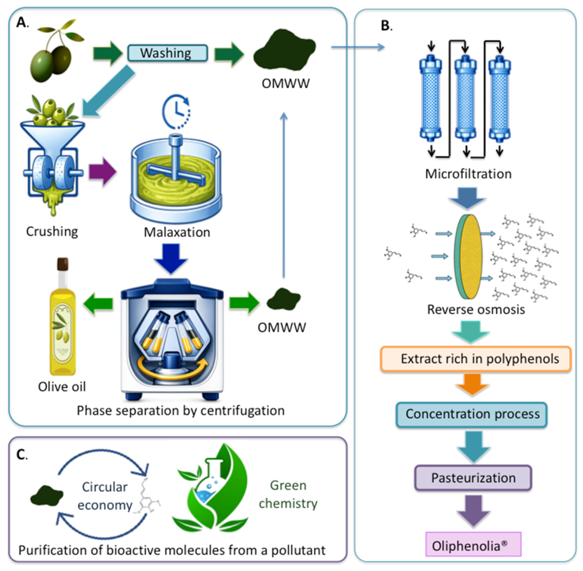
Figure 2.
Schematic representation of the proposed effects of hydroxytyrosol (HT) on skin health. HT contributes to the maintenance of skin homeostasis by supporting epidermal hydration, preserving the integrity of extracellular matrix components, including collagen and elastin fibers, and promoting tissue repair and wound healing.
Figure 2.
Schematic representation of the proposed effects of hydroxytyrosol (HT) on skin health. HT contributes to the maintenance of skin homeostasis by supporting epidermal hydration, preserving the integrity of extracellular matrix components, including collagen and elastin fibers, and promoting tissue repair and wound healing.
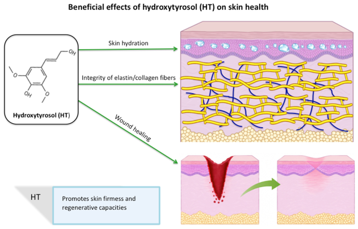
Figure 3.
Proposed mechanisms by which hydroxytyrosol (HT) may counteract skin ageing and inflammatory skin disorders. HT modulates multiple biological pathways involved in skin homeostasis, including the reduction of oxidative stress and reactive oxygen species (ROS) generation, attenuation of pro-inflammatory signaling pathways (e.g., NF-κB) and cytokine production (e.g., IL-1β, IL-6, and TNF-α), and enhancement of endogenous antioxidant defenses through activation of Nrf2-dependent pathways. HT may also preserve extracellular matrix integrity by limiting collagen degradation, promote autophagy-mediated clearance of advanced glycation end products (AGEs), and support mitochondrial function. In addition, emerging evidence suggests that HT may influence the gut-skin axis by improving intestinal barrier function and modulating microbiota-related inflammatory responses, thereby contributing to the maintenance of skin homeostasis and reducing age-related skin deterioration and inflammation.
Figure 3.
Proposed mechanisms by which hydroxytyrosol (HT) may counteract skin ageing and inflammatory skin disorders. HT modulates multiple biological pathways involved in skin homeostasis, including the reduction of oxidative stress and reactive oxygen species (ROS) generation, attenuation of pro-inflammatory signaling pathways (e.g., NF-κB) and cytokine production (e.g., IL-1β, IL-6, and TNF-α), and enhancement of endogenous antioxidant defenses through activation of Nrf2-dependent pathways. HT may also preserve extracellular matrix integrity by limiting collagen degradation, promote autophagy-mediated clearance of advanced glycation end products (AGEs), and support mitochondrial function. In addition, emerging evidence suggests that HT may influence the gut-skin axis by improving intestinal barrier function and modulating microbiota-related inflammatory responses, thereby contributing to the maintenance of skin homeostasis and reducing age-related skin deterioration and inflammation.
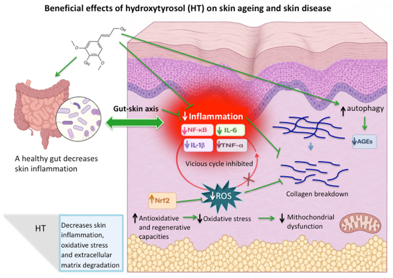

Table 1.
Preclinical and clinical evidence supporting the protective effects of hydroxytyrosol on skin homeostasis and aging-related pathways.
Table 1.
Preclinical and clinical evidence supporting the protective effects of hydroxytyrosol on skin homeostasis and aging-related pathways.
| Type of study and intervention | Model / Population | Main findings |
|---|---|---|
|
-In vitro -Purified HT (>90%) |
UVA-induced photoaging model in human dermal fibroblasts (HDFs) | Reduced cellular senescence (↓ SA-β-gal), decreased MMP-1 and MMP-3 expression, and reduced IL-1β, IL-6, and IL-8 production, supporting anti-photoaging and anti-inflammatory effects [Jeon & Choi, 2018] |
| -In vitro -Purified HT, oleuropein (OLE), and HT/OLE combination |
Human dermal fibroblasts exposed to H2O2 | Moderate anti-elastase and anti-collagenase activity. HT reduced ROS production and cytotoxicity. HT/OLE combinations showed synergistic elastase inhibition and enhanced cytoprotective activity [Li et al., 2022] . |
| -In vitro -HT and OLE |
Pre-senescent MRC5 lung fibroblasts and NHDF dermal fibroblasts | Reduced senescence markers (SA-β-gal, p16), inflammatory mediators (IL-6, COX-2, NF-κB), and MMP activity, suggesting attenuation of SASP and ECM degradation [Menicacci et al., 2017] |
| -In vitro -HT and HT acetate |
Primary human keratinocytes stimulated with IL-1β or TLR3 ligands | Reduced TSLP, IL-6, IL-8, and TNF-α expression through inhibition of NF-κB activation and nuclear translocation [Aparicio-Soto et al., 2019]. |
| -In vitro -Purified HT |
M5-stimulated HaCaT keratinocytes (psoriasis-like model) | Reduced IL-6, IL-8, and TNF-α expression, suppressed antimicrobial proteins, and inhibited keratinocyte proliferation [Chen et al., 2023]. . |
| -In vitro -HT dimer |
UVA-exposed HaCaT keratinocytes | Enhanced antioxidant activity (DPPH, FRAP assays) and resistance to UV-induced apoptosis through ↑ Bcl-2 and ↓ Bax expression [Zwane et al., 2012]. |
| -In vitro -HT |
Vascular endothelial cells | Increased HO-1 expression via PI3K/Akt and ERK1/2 pathways, activated Nrf2, enhanced antioxidant defenses, and accelerated wound closure [Zrelli et al., 2015]. |
| -In vitro -HT |
Human dermal fibroblasts | Modulated EMT (↑ E-cadherin, ↓ vimentin), promoted fibroblast proliferation and migration, reduced NF-κB-mediated inflammation, and accelerated wound healing [Batarfi et al., 2023]. |
| -In vitro -HT |
CCD-1064Sk human skin fibroblasts | Increased fibroblast proliferation and migration, enhanced fibronectin and α-actin expression, supporting tissue repair and ECM formation [González-Acedo et al., 2023]. |
| -In vivo -Oral purified HT (25–50 mg/kg/day, 16 weeks) |
Mouse model of skin ageing induced by high-AGE diet | Improved skin hydration, dermal and epidermal thickness, and hydroxyproline content; reduced oxidative stress and inflammatory cytokines; enhanced intestinal barrier integrity (↑ ZO-1, occludin) [Fan et al., 2025]. |
| -In vivo -Oral HT (10–50 mg/kg/day) |
IMQ-induced psoriasis-like dermatitis in BALB/c mice; M5-stimulated HaCaT cells | Improved psoriasis-like lesions, reduced PASI scores, epidermal hyperplasia, inflammatory infiltration, and cytokines (TNF-α, IL-1β, IL-6, IL-17A, IL-22, IL-23); inhibited ERK and NF-κB signaling [Liu et al., 2025]. |
| -In vitro and in vivo -Extra virgin olive oil and HT |
Diabetic wound-healing models and fibroblast cultures | Accelerated wound closure, increased collagen deposition, reduced oxidative stress and inflammation, promoted anti-inflammatory macrophage polarization (↑ IL-10), and enhanced fibroblast migration and contraction [Duarte et al., 2024]. |
| -Clinical (pilot RCT) -HT-enriched olive polyphenol nutraceutical (Alyvium®), 12 weeks |
30 patients with mild-to-moderate psoriasis | Significant reduction in PASI score and affected body surface area versus placebo [Acosta & Suárez-Pérez, 2016]. |
| -Clinical (randomized, double-blind, placebo-controlled pilot study) -Oral and topical HT, 90 days |
42 women with melasma | Oral HT significantly reduced mMASI and melanin index; topical treatment showed more modest improvements [de Toledo Bagatin et al., 2020]. |
AGEs, advanced glycation end products; Akt, protein kinase B; Bax, Bcl-2-associated X protein; Bcl-2, B-cell lymphoma 2; CCD-1064Sk, human skin fibroblast cell line; COX-2, cyclooxygenase-2; DPPH, 2,2-diphenyl-1-picrylhydrazyl; ECM, extracellular matrix; EMT, epithelial-to-mesenchymal transition; ERK1/2, extracellular signal-regulated kinases 1 and 2; FRAP, ferric reducing antioxidant power; HaCaT, human adult low-calcium high-temperature keratinocyte cell line; HDFs, human dermal fibroblasts; HO-1, heme oxygenase-1; HT, hydroxytyrosol; H2O2, hydrogen peroxide; IL, interleukin; IMQ, imiquimod; M5, cytokine cocktail composed of TNF-α, IL-17A, IL-22, IL-1α, and oncostatin M; mMASI, modified Melasma Area and Severity Index; MMP, matrix metalloproteinase; MRC-5, human fetal lung fibroblast cell line; NF-κB, nuclear factor kappa B; NHDF, normal human dermal fibroblasts; Nrf2, nuclear factor erythroid 2–related factor 2; OLE, oleuropein; PASI, Psoriasis Area and Severity Index; PI3K, phosphoinositide 3-kinase; RCT, randomized controlled trial; ROS, reactive oxygen species; SA-β-gal, senescence-associated β-galactosidase; SASP, senescence-associated secretory phenotype; TLR3, Toll-like receptor 3; TNF-α, tumor necrosis factor alpha; TSLP, thymic stromal lymphopoietin.
Table 2.
Preclinical evidence supporting the photoprotective and skin cancer preventive effects of hydroxytyrosol (HT) and HT-rich formulations.
Table 2.
Preclinical evidence supporting the photoprotective and skin cancer preventive effects of hydroxytyrosol (HT) and HT-rich formulations.
| Type of study and intervention | Model | Main findings |
|---|---|---|
|
-In vitro -Purified HT (DOPET) |
UVA-exposed M14 human melanoma cells | Reduced ROS generation, lipid peroxidation, and protein oxidation in a dose-dependent manner. Higher HT concentrations inhibited melanoma cell proliferation and induced caspase-3-mediated apoptosis, demonstrating both photoprotective and antiproliferative effects [D’Angelo et al., 2005]. |
|
-In vitro -Purified HT |
UVB-exposed HaCaT human keratinocytes | Reduced UVB-induced DNA strand breaks, intracellular ROS production, oxidative DNA damage (8-OHdG), and p53 and NF-κB expression, supporting antioxidant and photoprotective activity [Guo et al., 2010]. |
|
-In vitro -Biocatalytically synthesized HT dimer |
UVA-exposed HaCaT keratinocytes | Demonstrated greater antioxidant activity than HT alone (DPPH and FRAP assays) and enhanced resistance to UV-induced apoptosis through increased Bcl-2 and reduced Bax expression [Zwane et al., 2012]. |
|
-In vitro -Phenol-enriched purified olive mill wastewater (OMWW) extract containing HT |
HaCaT keratinocytes, normal human epidermal keratinocytes (NHEK), A375 melanoma cells, and a 3D melanoma skin model | Reduced ROS production and inflammatory responses in keratinocytes and inhibited growth of A375 melanoma nodules while preserving normal keratinocyte viability, suggesting photoprotective and chemopreventive effects [Schlupp et al., 2019]. |
|
-In vitro -Purified HT |
Metastatic human melanoma cell lines | Reduced melanoma cell viability by inducing apoptosis, increasing intracellular ROS, upregulating p53 and γH2AX, downregulating Akt signaling, and suppressing colony formation [Costantini et al., 2020]. |
|
-In vitro -Purified HT |
Human melanoma cell lines (A375 and MNT1) | Selectively reduced viability of glycolytic A375 cells, promoted metabolic reprogramming, enhanced detoxification pathways, and inhibited JNK and ERK signaling, indicating metabolism-dependent anticancer activity [Brito et al., 2021]. |
|
-In vitro -Purified HT + oleuropein + verbascoside |
Human dermal fibroblasts, HaCaT keratinocytes, and keratinocyte–fibroblast co-culture exposed to UVB | Reduced oxidative stress, inflammation, apoptosis, cellular senescence, and collagen degradation through inhibition of MAPK/NF-κB signaling and activation of Nrf2. The combination was more effective than the individual polyphenols [Wang J et al., 2025]. |
|
-In vitro -Purified HT |
Three-dimensional C32 melanoma spheroids and HEMa melanocytes | Selectively inhibited melanoma spheroid growth, migration, and invasiveness while inducing cell cycle arrest and apoptosis. Suppressed ERBB2/3/4, VEGFR-2, PI3K/Akt, and MAPK/ERK signaling pathways [Tovar-Parra and Mangion, 2025]. |
|
-In vitro and in vivo -Purified HT delivered through hyaluronic acid-based soluble microneedles |
UVA-exposed human dermal fibroblasts and UVA/UVB-induced mouse model of photoaging | Reduced ROS production and cellular senescence, improved skin hydration, elasticity, and collagen content, and decreased matrix metalloproteinase-1 expression, supporting anti-photoaging and extracellular matrix-preserving effects [Wang E et al., 2026]. |
|
-Review -Purified HT and oleuropein |
Review of in vitro and in vivo cancer studies | HT exerts antiproliferative, pro-apoptotic, antioxidant, and anti-inflammatory effects across multiple cancers, including melanoma. Clinical translation remains limited by bioavailability and the high concentrations required to achieve anticancer effects [Gervasi et al., 2024]. |
3D, three-dimensional; 8-OHdG, 8-hydroxy-2′-deoxyguanosine; Akt, protein kinase B; Bax, Bcl-2-associated X protein; Bcl-2, B-cell lymphoma 2; DOPET, 3,4-dihydroxyphenylethanol (hydroxytyrosol); DPPH, 2,2-diphenyl-1-picrylhydrazyl; ERBB, erythroblastic leukemia viral oncogene homolog; ERK, extracellular signal-regulated kinase; FRAP, ferric reducing antioxidant power; HaCaT, human adult low-calcium high-temperature keratinocytes; HEMa, human epidermal melanocytes; HT, hydroxytyrosol; JNK, c-Jun N-terminal kinase; MAPK, mitogen-activated protein kinase; MMP-1, matrix metalloproteinase-1; NF-κB, nuclear factor kappa B; NHEK, normal human epidermal keratinocytes; Nrf2, nuclear factor erythroid 2-related factor 2; OMWW, olive mill wastewater; PI3K, phosphoinositide 3-kinase; ROS, reactive oxygen species; UVA, ultraviolet A; UVB, ultraviolet B; VEGFR-2, vascular endothelial growth factor receptor 2.
Disclaimer/Publisher’s Note: The statements, opinions and data contained in all publications are solely those of the individual author(s) and contributor(s) and not of MDPI and/or the editor(s). MDPI and/or the editor(s) disclaim responsibility for any injury to people or property resulting from any ideas, methods, instructions or products referred to in the content. |
© 2026 by the authors. Licensee MDPI, Basel, Switzerland. This article is an open access article distributed under the terms and conditions of the Creative Commons Attribution (CC BY) license (http://creativecommons.org/licenses/by/4.0/).
Copyright: This open access article is published under a Creative Commons CC BY 4.0 license, which permit the free download, distribution, and reuse, provided that the author and preprint are cited in any reuse.


