

Submitted:
25 June 2026
Posted:
26 June 2026
You are already at the latest version
Abstract
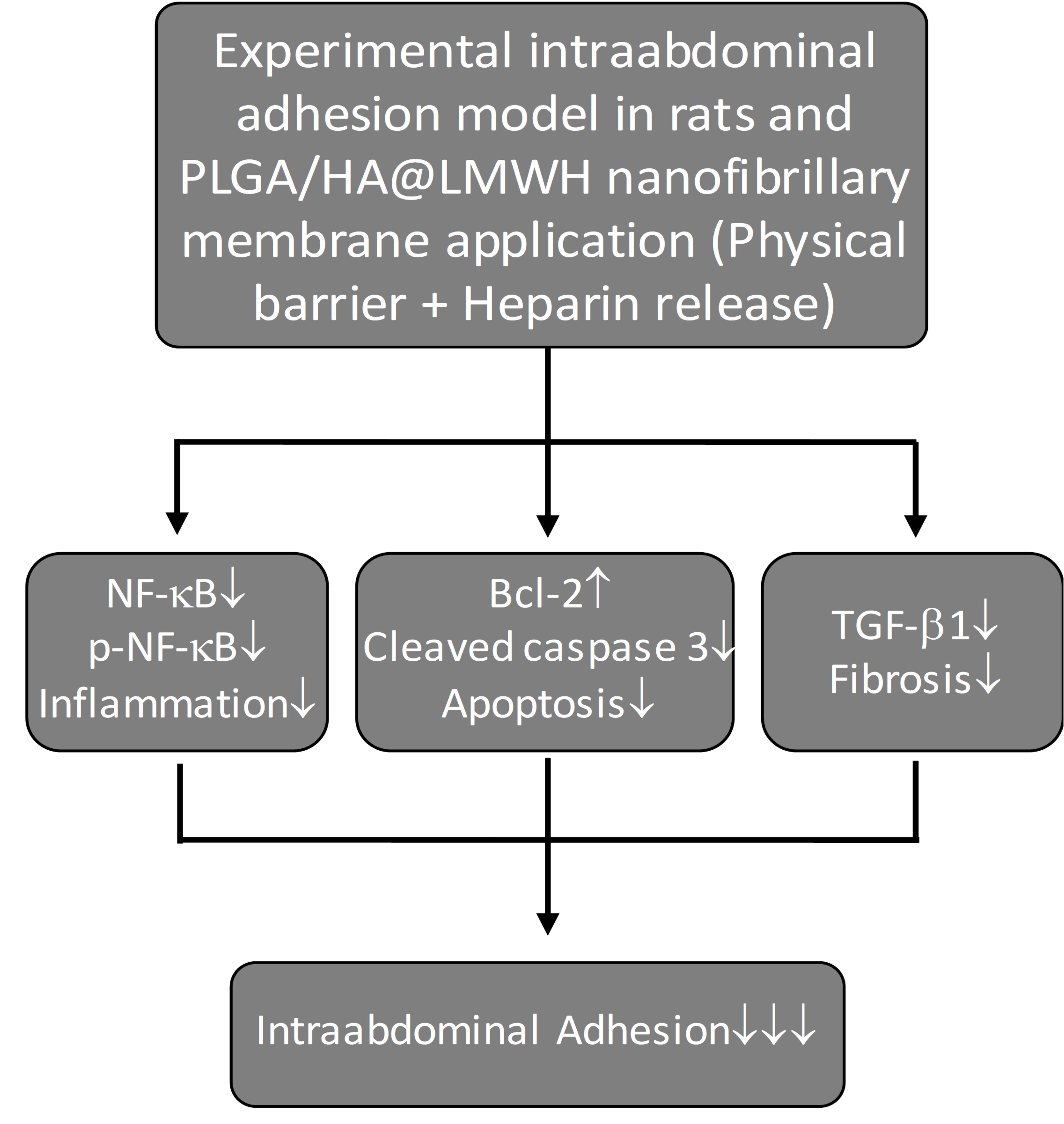
Objective: This study aimed to produce and characterize nanofiber membranes (PLGA/HA@LMWH) containing Poly(lactic-co-glycolic acid)/Hyaluronic Acid (PLGA/HA) immobilized with low molecular weight heparin (LMWH), and to investigate their physical and chemical effects in preventing intraabdominal adhesions in a rat model using multidisciplinary methods. Materials and Methods: Forty female Wistar Albino rats were divided into four groups (n=10). Cecal abrasions were induced in three groups: treated with PLGA/HA membrane (NFM), PLGA/HA@LMWH membrane (NFM+H), and 0.9% NaCl (SS). The fourth group underwent laparotomy only. On day 21, rats were sacrificed and evaluated macroscopically, histopathologically, immunohistochemically, and biochemically. Results: Macroscopic adhesion, inflammation, fibrosis, neovascularization, collagen scores, and TGF-β1 expression were found to be high in the SS and NFM groups; while a significant decrease (p < 0.05–0.01) was detected in all of these values in the NFM+H group. NF-κB activation was significantly decreased in the NFM and especially the NFM+H groups compared to the SS group (p < 0.05). In addition, apoptosis was suppressed in the NFM+H group with increased Bcl-2 and decreased cleaved caspase-3. Conclusion: PLGA/HA@LMWH membrane reduced peritoneal adhesions by suppressing inflammation, fibrosis, neovascularization, and collagen deposition, lowering TGF-β1 expression and NF-κB and p-NF-κB levels, and suppressing apoptosis. However, large-scale studies are needed for clinical use.
Keywords:
intraabdominal adhesion
; electrospinning
; PLGA/HA nanofiber membrane
; heparin
; drug delivery
1. Introduction
Intra-abdominal adhesions are pathological attachments of organ surfaces or peritoneal layers to each other following abdominal or pelvic surgery, inflammatory processes, radiation, or trauma [1,2,3]. Despite the development of various barrier biomaterials and active pharmacological agents, peritoneal adhesions continue to represent a significant clinical challenge. Indeed, even with modern surgical techniques, adhesion formation is reported in over 90% of patients undergoing major abdominal surgery [4,5,6,7].
Although peritoneal adhesions are generally asymptomatic, they can lead to serious problems such as small bowel obstruction, chronic pain, infertility, and surgical difficulties [3,4,7,8,9,10]. While increasing operation time and intraoperative risks, mortality in bowel obstruction reaches 7.2% [8]. In the US, the annual economic cost exceeds $1.3 billion [4], and in addition to the billions of dollars it places on healthcare systems, it also causes significant legal liabilities [11,12].
Mesothelial cells in the peritoneum provide lubrication between surfaces (4). Under physiological conditions, these cells prevent fibrin accumulation. In cases such as surgical trauma, ischemia, or infection, peritoneal injury triggers inflammation and coagulation, leading to the formation of fibrin-rich exudate. Peritoneal mesothelial cells are thought to play a significant role in regulating peritoneal fibrinolytic activity by producing the majority of tissue plasminogen activator (tPA) and plasminogen activator inhibitor-1 (PAI-1) [7,8,13].
Adhesion formation begins with inflammation following surgical trauma, increased vascular permeability, and activation of the coagulation cascade [14]. The resulting fibrous exudate establishes a fibrin network between the damaged surfaces [15,16,17]. Increased PAI-1 and decreased t-PA activity inhibit fibrin degradation, leading to the persistence of the fibrin matrix; this matrix transforms into fibrotic adhesions through fibroblast proliferation and collagen deposition [8,10,11,15].
In current practice, adhesion prevention relies on surgical technique optimization and the use of barrier agents [18]. Although minimally invasive surgery, careful hemostasis, reduction of tissue trauma and prevention of ischemia limit adhesions, they are not sufficient on their own, therefore the use of barrier agents is gaining importance [9,18,19]. The first 5-7 days are critical for adhesion formation, and the anti adhesive barriers prevent fibrinous adhesions by separating damaged surfaces during this period. The most common methods are physical barriers such as membranes, gels, and liquids [20,21,22]. However, the success rates of commonly used membranes (Seprafilm®, Interceed®) are low due to the difficulty and fragility of their application in laparoscopy, and liquid barriers are also low due to their short duration of action [1,19,20,22,23].
The effectiveness of existing barriers is limited, and innovative strategies combining structural and pharmacological properties are needed to prevent adhesion. Electrospinning nanofiber membranes offer local solutions depending on the polymer type; with their large surface area and porous structure, they are widely used as physical barriers to prevent adhesion [24,25]. The use of these membranes as barriers requires them to be biocompatible, have high mechanical strength, be easy to apply, and have a wettable surface. Furthermore, electrospinning-produced structures must be biodegradable and support tissue regeneration. Synthetic biopolymers [e.g., Poly-lactic-co-glycolic acid (PLGA)] are frequently preferred in tissue engineering due to their biocompatibility, tunable biodegradability, and mechanical performance. Natural glycosaminoglycan hyaluronic acid (HA), on the other hand, supports tissue regeneration and modulates inflammation [21,26].
Current adhesion prevention strategies focus on the functionalization of barriers with pharmacological agents. In this context, Low Molecular Weight Heparin (LMWH) stands out with its anticoagulant, anti-inflammatory, anti-fibrotic, and angiogenesis-modulating effects. LMWH prevents adhesion formation by inhibiting coagulation and suppressing fibrin deposition and inflammation [27]. It also modulates the activity of profibrotic mediators such as TGF-β. This multifaceted biological activity makes heparin an effective candidate for the functionalization of adhesion prevention barriers [28]. LMWH inhibits thrombin formation with antithrombin III and supports the fibrinolytic process [12]. Although free heparin carries a risk of bleeding, local and controlled release from nanofiber membranes minimizes these side effects [29].
Our hypothesis is that LMWH-loaded PLGA/HA nanofiber membranes will significantly reduce adhesion formation in a rat intraperitoneal adhesion model, and that this effect will arise from the synergistic interaction between the membrane’s physical barrier function and the anticoagulant and anti-inflammatory properties of heparin. Furthermore, it is anticipated that the findings obtained from this study will elucidate the potential of nanotechnology-based, pharmacologically functionalized biomaterials in the prevention of intra-abdominal adhesions and provide a scientific basis for future clinical investigations.
The aim of this study is to characterize PLGA/HA nanofiber membranes produced by electrospinning, to functionalize these membranes with LMWH to create a PLGA/HA@LMWH formulation, and to investigate the intraabdominal adhesion-preventing efficacy of the membranes in a rat model.
2. Materials and Methods
2.1. Preliminary Study
2.1.1. Membrane Production and Characterization
In this study, biodegradable Poly-lactic-co-glycolic acid (PLGA) and Hyaluronic Acid (HA) polymers were used to produce nanofiber membranes by electrospinning. In optimization studies, in vitro experiments were conducted with at least two nanofiber membranes with different PLGA and HA formulation contents. Biocompatibility studies were investigated using L-929 mouse (Mus musculus) fibroblast cells. In in vitro experiments, a preliminary study was conducted on rats with intra-abdominal adhesions created using two different membranes that were tissue-compatible and biodegraded within one week. These two different membranes were tested on 18 Wistar Albino female rats with intra-abdominal adhesions. The nanofiber membrane used in our study demonstrated the ability to easily adhere to abrasive surfaces, and it also exhibited no fragility (Figure 1). Each membrane was applied to 9 rats; 3 rats from each group were sacrificed on days 7, 14, and 21. In this preliminary study, the in vivo antiadhesive efficacy and biodegradability of membranes were tested in a rat-induced intraabdominal adhesion model. Based on the results of the preliminary study, it was decided to use the PLGA/HA nanofiber membrane containing 0.6 g PLGA and 0.24 g HA, which had the best in vivo antiadhesive efficacy and biodegradability within one week, in the main study. Figure 2 and Figure 3 show images the in vivo nanofiber membrane biodegredation in rat abdomens 7 and 14 days after application of a nanofiber membrane consisting of a combination of 0.6 g PLGA and 0.24 g HA. Macroscopic images taken after 7 and 14 days show that the nanofiber membranes have biodegraded and disappeared (Figure 2 and Figure 3).
2.2. Main Study
2.2.1. Materials
All chemicals used in this study were of analytical purity and, unless otherwise indicated, were used without further purification. Poly(lactic-co-glycolic acid) (PLGA) (mean Mn: 25,000), hyaluronic acid sodium salt (high molecular weight, >1 MDa), and 1,1,1,3,3,3-hexafluoro-2-propanol (HFIP, ≥99%) were supplied by Sigma-Aldrich. Dimethylformamide (DMF, ≥99.8%) and chloroform (CHCl₃, 99.8%) were sourced from ISOLAB (Wertheim, Germany). Low molecular weight heparin (LMWH) (Enoxaparin sodium – OKSAPAR 6000 anti-Xa IU/0.6 mL) was supplied by Koçak Farma İlaç ve Kimya Sanayi A.Ş., Türkiye.
2.2.2. Production of PLGA/HA and PLGA/HA@LMWH Nanofiber Membranes
In this study, biocompatible and biodegradable nanofiber membranes were developed to prevent postoperative adhesions. By adding LMWH to the polymer solution, membranes with dual-effect (chemical and physical) barrier properties were produced. In accordance with the literature, anti-adhesion PLGA/HA membranes with different porosities were produced by electrospinning method [ 30]. For membrane fabrication, PLGA and HA (0.6 g / 0.24 g) were dissolved in HFIP and mixed using a magnetic stirrer (Labron HS1-M3) at 600 rpm for 12 h at room temperature to obtain a homogeneous solution. Subsequently, the solution was subjected to ultrasonication using an ultrasonic processor (Bandelin Sonopuls UW 200, Germany) at 25 °C for 1 h to improve polymer dispersion and eliminate air bubbles. Following this step, PLGA/HA nanofiber membranes (2 × 2 cm dimensions) were produced using the electrospinning method shown in Figure 4, which is a widely used technique for fabricating nanofibrous structures with controllable morphology and functional properties [31].
To 1. units of LMWH (250 units of LMWH per cm2 of the nanofiber membrane) were added to the PLGA/HA solution before the electrospinning process and homogenized, resulting in PLGA/HA@LMWH nanofiber membranes. It has been stated in the literature that LMWH regulates the biological response by reducing thrombosis in nanofiber systems [32]. The amount of LMWH was determined according to the appropriate concentration that would optimize process stability, fiber morphology and biological activity. The PLGA/HA@LMWH solution was taken into a 2.5 mL syringe with a 0.80 × 38 mm needle and electrospinned at a flow rate of 0.6 mL/hour, 22 kV voltage and a distance of 15 cm. All electrospinning experiments were carried out under controlled ambient conditions at a temperature of 25 ± 1 °C and a relative humidity of 50–55%. The fabrication process of PLGA/HA@LMWH nanofiber membranes is shown in Figure 5.
2.2.3. Characterization Techniques
The structural and morphological properties of the nanofibers were characterized for comparison. Fourier transform infrared spectroscopy (FT-IR) spectra were recorded using a Perkin-Elmer spectrophotometer in the wavelength range of 400–4000 cm⁻¹. The surface morphology of the samples was examined using scanning electron microscopy (SEM).
2.2.4. Cell Viability
0.1 g was taken from each electrospun fiber and placed in 1 mL of high-glucose DMEM containing 10% FBS and 1% Pen-Strep. Samples were incubated at 37°C in 5% CO₂ for 3 days; then the fibers were removed and the conditioned supernatant was collected. L-929 cells were seeded into 96-well plates (10,000 cells/well) and allowed to adhere; then the medium was changed and 100 µL of conditioned DMEM was added to each well. Cells were incubated for 24 hours; their morphology was examined under an inverted microscope prior to MTT. The medium was removed and DMEM containing 10% MTT was added. An MTT solution prepared in PBS at a concentration of 5 mg/mL and adjusted to pH 7.4 was used, and the plates were incubated for 4 hours. At the end of the incubation period, the medium containing MTT was carefully aspirated. To dissolve the formed formazan crystals, 100 µL of DMSO was added to each well; the absorbance values of the samples were measured spectrophotometrically at a wavelength of 540 nm.
2.3. Experimental Animals and Ethical Approval
The study was initiated after approval from the İnönü University Faculty of Medicine Local Ethics Committee for Experimental Animals (HAYDEK, 2023/7-1; HAYBİS No:18167). Experimental animals were housed at a temperature of 22–24 °C, 50–60% humidity, and a 12-hour light/dark cycle, with ad libitum access to food and water. A total of 58 Wistar Albino female rats, aged 8–12 weeks and weighing 150–250 g, were used in the study (18 rats for preliminary study, 40 rats for main study).
2.4. Experimental Groups (Main Study)
2.4.1. Power Analysis Report and Groups
For alpha=0.05, power=0.8 and effect size=0.6, at least 9 samples per group (total 36) are required to detect significant differences using one-way ANOVA in independent samples. Calculations were performed using WSSPAS (Web-Based Sample Size & Power Analysis Software) [33]. The number of animals was increased from 36 to 40 to account for potential losses. The main study used 40 Wistar Albino female rats, divided into four groups of 10 animals each.
Nanofiber Membrane Group (NFM): PLGA/HA membrane applied.
Nanofiber Membrane + Heparin Group (NFM+H): PLGA/HA@LMWH membrane applied.
Saline Solution Group (SS): 0.9% NaCl solution applied.
Laparotomy Group (LO): Laparotomy performed only.
2.5. Creating an Intraabdominal Adhesion Model
2.5.1. Anesthesia and Postoperative Analgesia
2.5.2. Surgical Technique
All rats underwent midline laparotomy (3 cm) after abdominal wall shaving and disinfection under ketamine-xylazine anesthesia. In the first three groups, abrasion was performed on the antimesenteric surface of the cecum in a 1.5 x 1.5 cm area using a sterile toothbrush until petechial bleeding occurred. In the NFM group, a PLGA/HA nanofiber membrane was applied to the abrasion area; in the NFM+H group, a PLGA/HA@LMWH nanofiber membrane (2x2 cm dimentions) was applied; and in the SS group, 5 mL of 0.9% NaCl was applied. Laparotomy was performed only in the LO group. The anterior abdominal wall was closed separately with 3/0 prolene and the skin with 3/0 silk. The animals were sacrificed on day 21 with a high dose of anesthesia (four times the dose of ketamine and xylazine anesthetics).
2.6. Evaluation Parameters
2.6.1. Macroscopic Adhesion Scoring
2.6.2. Histopathological Evaluation
Sections obtained from paraffinized tissues were stained using the Masson-trichrome staining method and histopathological examinations were performed. Adhesions and inflammation, fibrosis, and vascular proliferation in surrounding tissues were evaluated. Cecum samples were fixed with 10% buffered formalin, cut from paraffin blocks, and stained with H&E, Masson trichrome, and immunohistochemically. Evaluation was performed on a scale of 0–3 over 5 randomly selected fields under light microscopy according to the scoring system of Giusto et al. [37] (Table 2). Collagen density in the adhesion area was semi-quantitatively evaluated using Masson trichrome (Bio-Optica, Milan, Italy; Cat No: 04-010802) staining. Briefly, it was scored as follows: 0: no collagen, 1: less than 5% collagen in the adhesion area, 2: between 6-20% collagen, 3: more than 21% collagen.
2.6.3. Immunohistochemical Evaluation
Sections were stained using the Avidin-Biotin Complex (ABC) method with TGF-beta1 antibodies (Invitrogen) [38]; the manufacturer's (Thermo Fischer Scientific) protocol was followed in the procedures. Tissues on positively charged slides were deparaffinized after being kept at 65 °C for 1 hour, passed through graduated alcohol series and brought to distilled water. To recover antigens, sections were microwaved (600W, 15 min) in citrate buffer, cooled to room temperature, and washed with PBS. To eliminate endogenous peroxidase activity, tissues were treated with 3% H2O2 prepared with methanol for 10 minutes, and after PBS washing, stained according to a commercial kit protocol. After non-specific binding was inhibited with protein block (Ultra V Block) serum (10 min), sections were incubated overnight at +4 °C with TGF-beta1 primary antibody (1/200). Non-immune serum was used as a negative control. Subsequently, biotin-containing secondary antibody (30 min) and HRP (Horse Radish Peroxidase)-streptavidin (20 min) were applied to the sections. Following PBS washing (15 min), sections were incubated with 3-amino-9-ethylcarbosol (AEC) (Thermo Fischer Scientific, USA) in a controlled manner and washed with distilled water. For counterstaining, sections were held in Gill’s hematoxylin solution for 45-60 seconds. Sections sealed with water-based adhesive (Lerner Laboratories, USA) were examined under a light microscope. For immunohistochemical scoring, positive cells in 5 randomly selected fields at 40X magnification were graded as 0 (none), 1 (<10), 2 (11-20), and 3 (>21).
2.6.4. Biochemical Evaluation
Protein levels associated with inflammatory and apoptotic pathways (Bcl-2, Cleaved Caspases-3, NFκB, p-NFκB p65) in tissues obtained from abrasion sites in rats were evaluated using the western blot method. Additionally, levels of hydroxyproline and some oxidant-antioxidant parameters in the tissues were measured.
2.7. Statistical Analyses
The Shapiro-Wilk normality test was applied to evaluate the distribution characteristics of the research data set. Since the data did not show a normal distribution, the non-parametric Kruskal-Wallis test was used to examine the data set, and the Conover post hoc test was used for intergroup comparisons. Statistical results are presented as median (minimum-maximum) values. p < 0.05 was considered statistically significant.
3. Results
3.1. Produced Antiadhesive Materials
3.1.1. Characterization results
FT-IR spectra showing the intermolecular interactions of PLGA/HA and PLGA/HA@LMWH nanofibers are presented in Figure 6. The spectra of pure LMWH, PLGA/HA, and PLGA/HA@LMWH nanofiber membranes were evaluated comparatively. In the pure LMWH spectrum, the C–O–S vibrations with asymmetric S=O at 1220–1260 cm⁻¹ and symmetric S=O at 1030–1080 cm⁻¹ indicate the sulfated structure of heparin. In the PLGA/HA nanofiber spectrum, PLGA's ~1755 cm⁻¹ ester C=O and HA's 1600–1650 cm⁻¹ amide bands were observed, while sulfate bands were absent. Upon addition of LMWH, the characteristic PLGA and HA bands were preserved in the PLGA/HA@LMWH spectrum, and LMWH bands of 1220–1260 and 1030–1080 cm⁻¹ became prominent. The sulfate ester bands in the range of 840–850 cm⁻¹ demonstrate that LMWH was incorporated into the nanofiber matrix while preserving its chemical structure [39,40,41].
Figure 7 presents SEM images and fiber diameter distributions of PLGA/HA and PLGA/HA@LMWH membranes. The randomly oriented, bead-free, and homogeneous fiber morphology in Figure 7A demonstrates that the electrospinning process has been successfully optimized. The fiber diameter histogram in Figure 7B shows that PLGA/HA nanofibers exhibit a near-normal distribution, with an average diameter of 290 ± 5 nm. This narrow distribution demonstrates the favorable rheological properties of the polymer solution and is consistent with the findings on PLGA-based nanofibers in the literature [42,43]. The SEM image in Figure 7C shows that the PLGA/HA@LMWH nanofibers maintain their homogeneous and bead-free structure despite the addition of heparin. According to Figure 7D, the average fiber diameter increased to 320 ± 5 nm with the addition of heparin. This increase can be attributed to the increase in solution viscosity and conductivity, consistent with the literature [39,40]. The nanofibers exhibited a predominantly bead-free morphology, even in the presence of heparin, indicating successful incorporation. However, based on SEM observations, the fibers were found to be randomly oriented with a relatively heterogeneous distribution. Consequently, both membranes demonstrated morphological features suitable for barrier applications, such as high surface area-to-volume ratio.
3.1.2. Results of in vitro heparin releasing from PLGA/HA@LMWH nanofibers
Figure 8 presents the colorimetric evaluation of LMWH release from PLGA/HA@LMWH nanofiber membranes over time. As clearly observed, a progressive change in color intensity occurs with increasing time, indicating continuous release of LMWH. Notably, from day four onwards, the colorimetric response suggests an almost complete release of LMWH. This behavior is attributed to the rapid degradation of the polymer matrix, which leads to a loss of structural integrity and facilitates the diffusion of LMWH into the surrounding medium.
3.1.3. Results Related to Cell Viability
According to the MTT test results, PLGA/HA (86.72 ± 3.83%) and PLGA/HA@LMWH (91.67 ± 3.73%) scaffolds exhibited high cell viability (Figure 9). These rates, which are above the 70% threshold value in the ISO 10993-5 standard, prove that both materials are non-cytotoxic and show good biocompatibility [44]. PLGA/HA@LMWH scaffolds exhibited higher cell viability compared to PLGA/HA alone; this was attributed to LMWH's increased surface hydrophilicity, promoting cell adhesion and proliferation. Optical microscope images in Figure 10 confirm the homogeneous distribution of cells after 24 hours of culture, their strong adhesion to the surface, and the preservation of their natural morphology. These findings, which do not show cytotoxic effects, prove that PLGA/HA@LMWH scaffolds are a suitable matrix for tissue engineering applications.
3.2. Macroscopic Adhesion Scoring
No morbidity or mortality was observed during the study; rats were sacrificed on day 21. Macroscopic adhesions in rats with opened abdominal regions were evaluated by an independent surgeon according to the classification of Nair et al. [36] (Figure 11)
According to Figure 12, macroscopic adhesion scores were highest in the SS group. The scores of the NFM+H group were significantly lower than those of the NFM and SS groups (p < 0.05, p < 0.01). The SS group exhibited significantly higher adhesion compared to NFM (p < 0.01) and LO (p < 0.001).
3.3. Histopathological Evaluation
Histopathological examination revealed a normal serosa in the LO group, while the NFM, NFM+H, and SS groups showed adhesions characterized by varying degrees of inflammatory cell infiltration, fibrosis, and neovascularization (Figure 13).
Staining with Masson trichrome revealed collagen fibers stained blue to varying degrees in the adhesion area (Figure 14 A, B, C). In some cases, they appeared as a small number of independent fibers (Figure 14 A), while in others they were fused together (Figure 14 B) or formed bundles (Figure 14 C).
Immunohistochemical staining with TGF beta 1 (Transforming Growth Factor beta 1) monoclonal antibodies revealed intracytoplasmically positive stained cells in the adhesion area (Figure 14 D, E, F).
The highest inflammatory infiltration (Figure 15a) and fibrosis (Figure 15b) scores were observed in the NFM and SS groups. In the NFM+H group, both parameters were significantly lower compared to NFM and SS (p < 0.05). The difference between NFM and LO was more significant in inflammation (p < 0.01), while the difference between SS and LO was more significant in fibrosis (p < 0.001). Neovascularization (Figure 15c) and collagen deposition (Figure 15d) were most pronounced in the SS and NFM groups. In the NFM+H group, both parameters were significantly lower compared to NFM (p < 0.05) and SS (p < 0.01). The difference between SS and LO in neovascularization is highly significant (p < 0.001); collagen deposition is significantly higher in the SS group compared to LO (p < 0.01). These findings confirm heparin's suppressive effect on inflammatory infiltration, fibrosis, neovascularization, and collagen deposition.
3.4. Immunohistochemical Evaluation
Intracytoplasmic positivity was observed in cells in the adhesion areas in TGF-β1 monoclonal antibody staining (Figure 14 D-F). According to the TGF-β1 analysis in Figure 16, the highest expression was observed in the SS group, followed by the NFM group. In the NFM+H group, a significant decrease was detected compared to NFM (p < 0.05) and SS (p < 0.01); the difference between SS and LO was highly significant (p < 0.001). These findings also confirm the TGF-β1 suppressive effect of heparin.
3.5. Biochemical Evaluations
On the 21st day after abrasion, tissue samples taken from NFM, NFM+H, SS, and LO group rats were analyzed for NF-κB, p-NF-κB, Bcl-2, and cleaved caspase 3 levels using Western blot. According to Western blot analysis (Figure 17), increased NF-κB and cleaved caspase-3 levels in the SS group were significantly decreased in the NFM and NFM+H groups (p < 0.05). In the NFM+H group, Bcl-2 levels increased while cleaved caspase-3 levels decreased compared to the SS group (Figure 17 d-e). Significant changes in p-NF-κB and Bcl-2 levels were observed in both groups, but these changes were not statistically significant.
These findings indicate that heparin inhibits cell death by suppressing the apoptotic pathway. A decrease in NF-κB indicates a transition to the resolution phase of inflammation, a decrease in cleaved caspase-3 suggests a slowing of cell death and tissue stabilization, while an increase in Bcl-2 indicates support for cell survival and successful continuation of tissue repair.
Western blot analysis of NF-κB, p-NF-κB, Bcl-2, and beta-actin (Figure 17a), relative expression levels of NF-κB, p-NF-κB, Bcl-2, and cleaved caspase 3 proteins and statistical analysis (n = 3 per group) (Figure 17b-e) are given in Figure 17.
GSH levels were found to be statistically similar in all groups (Figure 18a). This can be attributed to the completion of wound healing and antioxidant system compensation on day 21. Hydroxyproline levels were found to be significantly higher (p < 0.05) in the NFM, NFM+H, and SS groups compared to the LO group (Figure 18b), however, no statistically significant difference was observed in terms of fibrotic response among these three groups.
4. Discussion
Postoperative intraabdominal adhesions are a significant problem that reduces quality of life, developing with inflammation, fibrin deposition, and fibroblast proliferation. The ideal approach is to prevent fibrotic bands without disrupting physiological healing. Therefore, biocompatible and biodegradable barrier membranes are an effective strategy [4,45,46].
This study investigated the effectiveness of electrospinning-produced PLGA/HA nanofiber membranes and PLGA/HA@LMWH membranes obtained by adding low molecular weight heparin in preventing intraabdominal adhesions in a rat model. The findings showed that the PLGA/HA@LMWH combination was superior to other groups in terms of inflammation, fibrosis, neovascularization, collagen deposition, TGF-β1 expression, and apoptotic pathways.
4.1. Design and Characterization of Nanofiber Membranes
Electrospinning is widely used in tissue engineering by producing nanofiber structures with high surface area/volume ratio, controlled porosity and extracellular matrix (ECM)-like morphology [43,47]. In our study, the homogeneous morphology of nanofiber membranes produced with PLGA/HA was confirmed by SEM.
The mechanical strength of PLGA and the hydrophilic structure and biocompatibility of HA make the membrane ideal for adhesion barriers [42]. HA's transient ECM function and modulation of inflammation contribute to the biological performance of PLGA/HA membranes [48,49].
In the PLGA/HA@LMWH spectrum, the characteristic bands of PLGA and HA are preserved, while the LMWH-specific bands at 1220–1260 and 1030–1080 cm⁻¹ and the sulfate ester band at 840–850 cm⁻¹ become more prominent. This situation, similar to previous studies, shows that LMWH was successfully integrated into the nanofiber matrix while maintaining its chemical structure during electrospinning [39,40,41]. With the addition of LMWH, the average fiber diameter increased from 290 ± 5 nm to 320 ± 5 nm, accompanied by a slight broadening of the distribution. This increase can be attributed to changes in solution properties, particularly viscosity and conductivity. It has also been reported in the literature that the incorporation of bioactive molecules can lead to a slight increase in fiber diameter [39,41]. The preservation of the homogeneous and bead-free morphology of the nanofibers despite the addition of heparin demonstrates the successful integration of LMWH into the polymeric matrix. Both membranes retained properties critical for the adhesion barrier, such as a high surface area/volume ratio and controlled porosity.
MTT analysis revealed that PLGA/HA and PLGA/HA@LMWH nanofibers showed high biocompatibility in L-929 fibroblast cells. The higher cell viability exhibited by PLGA/HA@LMWH indicates that LMWH supports early cell proliferation by enhancing cell adhesion, proliferation, and surface hydrophilicity. These findings support the idea that the PLGA/HA@LMWH scaffold could be a suitable matrix for tissue engineering applications.
4.2. Macroscopic Adhesion Evaulation
The most important finding of the study is that macroscopic adhesion scores are significantly lower in the NFM+H group compared to the NFM and SS groups (p < 0.05, P < 0,01). This finding indicates that the nanofiber membrane alone is insufficient and that the anti-adhesion effect is significantly increased with the addition of heparin. The highest scores being observed in the SS group (p < 0.01) confirms the validity of the experimental model.
Fibrin clots formed after surgery become permanent and form a matrix for fibrosis if the fibrinolytic system is suppressed [50]. The anti-adhesion effect of heparin is based on its anticoagulant and anti-inflammatory properties; it supports fibrinolytic activity by binding to antithrombin III, suppressing inflammatory cell adhesion, angiogenesis and fibroblast proliferation [12,51,52].
Although the anti-adhesion effect of heparin derivatives is known in the literature, studies using local and controlled release strategies with nanofiber membranes are limited [27,53,54]. In our study, loading LMWH onto the nanofiber membrane minimized systemic side effects by providing locally controlled heparin release. While the nanofiber membrane acted as a physical barrier, the version with added LMWH acquired both physical and chemical barrier properties.
In an experimental intraabdominal adhesion model conducted by Toyama et al. using the commercial product Seprafilm, macroscopic adhesion scores in the Seprafilm group were found to be highly significantly lower than in the control group (P < 0.01) [55]. In our study, similar to the findings of Toyama et al., the macroscopic adhesion scores in the NFM+H group were statistically significantly lower than those in the NFM group (p < 0.05) and highly lower than those in the SS group (p < 0.01).
The literature also includes studies in which nanofiber membranes have been functionalized with different materials. In the study by Jamshidi-Adegani et al., the anti-adhesion efficacy of PLGA-ibuprofen (PLGA-IB) nanofiber networks was investigated in a mouse model. The results showed that adhesion was at the least level in the PLGA-IB nanofiber membrane group compared to other groups [56]. Zhu et al. produced Flurbiprofen axetil-loaded PVP/PLGA composite nanofibers by coaxial electrospinning. It was determined that the drug-loaded membrane prevented adhesion and significantly reduced drug release [57].
4.3. Evaluation of Histopathological Findings
Histopathological examinations showed that inflammation was highest in the NFM group, while it was significantly lower in the NFM+H group (p < 0.05) (Figure 15a). This suggests that the nanofiber membrane may cause mild inflammation, but this response is suppressed by the addition of heparin. Acidic products formed during the degradation of PLGA may contribute to mild inflammation [58]. Heparin suppresses inflammation by inhibiting neutrophil adhesion, pro-inflammatory cytokine release, and inflammatory cell activation [59].
Fibrosis scores were found to be significantly higher in the SS and NFM groups compared to the NFM+H group (p < 0.05). This can be explained by heparin inhibiting fibroblast proliferation and collagen synthesis, modulating factors such as transforming growth factor-beta (TGF-β) and fibroblast growth factor (FGF) [12,27,28]. Furthermore, the high fibrosis scores in the NFM group indicate that the physical barrier alone is insufficient to prevent fibrotic response and that pharmacological support is necessary.
Neovascularization was highest in the SS group, while it was significantly lower in the NFM+H group (p < 0.01). Excessive vascularization during the adhesion process contributes to the persistence of adhesions. The effect of heparin on angiogenesis is biphasic; at low concentrations it can inhibit angiogenesis by modulating the activity of angiogenic factors (VEGF, bFGF), while at high concentrations it can exhibit a pro-angiogenic effect [27,60,61]. It is thought that the LMWH concentration used in our study suppressed neovascularization in the NFM+H group by inhibiting angiogenesis.
Collagen accumulation was found to be high in the SS and NFM groups, while it was low in the NFM+H group (p < 0.05). This finding indicates that heparin suppresses collagen synthesis through TGF-β1 inhibition or fibroblast modulation [28].
4.4. Immunohistochemical Findings: TGF-β1 Expression
In our study, TGF-β1 expression was highest in the SS group and significantly lower in the NFM+H group (p < 0.01). This result shows that NFM+H application inhibits adhesion formation by suppressing TGF-β1 signaling. TGF-β1 plays a central role in adhesion pathogenesis; it induces fibroblast proliferation, ECM synthesis, and myofibroblast differentiation [62]. The higher TGF-β1 in the NFM group compared to NFM+H indicates that the maximum effect is achieved with the heparin combination.
4.5. Biochemical Findings: Inflammatory and Apoptotic Pathways
NF-κB and p-NF-κB levels were found to be significantly lower in the NFM and NFM+H groups compared to the SS group (p < 0.05). Suppression of NF-κB activity indicates that the inflammatory response is controlled. NF-κB, activated after surgical trauma, plays a critical role in initiating the inflammatory cascade that triggers adhesion formation. Nuclear factor kappa B (NF-κB) is central to the inflammatory response by regulating the expression of pro-inflammatory mediators [63,64].
In the NFM+H group, anti-apoptotic Bcl-2 levels were significantly increased, while pro-apoptotic cleaved caspase-3 levels were decreased (p < 0.05). Bcl-2 supports cell survival by inhibiting the mitochondrial apoptotic pathway, while cleaved caspase-3 is an effector caspase that is activated in the final stages of apoptosis and makes cell death irreversible [65,66]. Post-surgical trauma, ischemia, inflammation, and oxidative stress induce apoptosis in peritoneal mesothelial cells, creating a predisposition to adhesion (67). Suppression of apoptosis supports mesothelial regeneration. This observed in the NFM+H group demonstrates that heparin protects tissue integrity by suppressing apoptotic cell death.
The similarity of GSH levels in all groups can be explained by the regression of the acute oxidative stress response on day 21. Although hydroxyproline levels were significantly higher (p < 0.05) in the NFM, NFM+H, and SS groups compared to the LO group, no statistically significant difference was observed in terms of fibrotic response among these three groups. This indicates that a fibrotic response developed in all abrasion groups. Hydroxyproline is a specific amino acid found in the structure of collagen and is a marker of tissue collagen content [68]. While lower hydroxyproline levels were expected in the NFM+H group, the lack of a statistically significant difference may be related to the sample size or the timing of the evaluation.
4.6. Clinical Applicability and Strengths of the Study
This study highlights the importance of a multidisciplinary approach in adhesion prevention. While the nanofibrillar membrane forms a physical barrier, controlled-release heparin exhibits local anti-inflammatory, anti-fibrotic, and anticoagulant effects. This synergistic approach provides more effective protection by targeting different stages of adhesion formation.
One of the key strengths of our study is the inclusion of a well-defined biomaterial design supported by comprehensive characterization studies. FT-IR and SEM analyses revealed the chemical structure and morphological features of the membranes in detail, demonstrating the success of heparin integration. Furthermore, the combined use of macroscopic, histopathological, immunohistochemical, and biochemical parameters in adhesion assessment allowed for a multidimensional interpretation of the results. In particular, the analysis of molecular markers such as NF-κB, p-NF-κB, Bcl-2, and cleaved caspase-3 provided important clues regarding the underlying mechanisms of adhesion pathogenesis.
4.7. Limitations of the Study and Future Perspectives
One limitation of this study is that the rat model may not fully reflect human surgical conditions. Peritoneal healing is faster in rats, and adhesion dynamics may differ.
Secondly, adhesion assessment was performed at a single time point (day 21). Since adhesion is a dynamic process, assessments at different time points may provide more detailed information.
Thirdly, the dose-response relationship of LMWH has not been investigated. The effectiveness of different heparin concentrations should be determined through optimization studies. In addition, in vivo characterization of membrane heparin release kinetics will provide a better understanding of the system's performance.
Fourthly, a comparison of the anti-adhesive materials used in our study with commercial products (Seprafilm® etc.) will be carried out in the next stage of this study; furthermore, the lack of such a comparison can be considered one of the limitations of our study.
Future studies could investigate the efficacy of NFM+H membranes in different adhesion patterns (ischemic, infectious). Combinations with other anti-adhesive agents may lead to more effective strategies.
5. Conclusion
This study demonstrated that composite membranes obtained by adding low molecular weight heparin to electrospinning-produced PLGA/HA nanofiber membranes were effective in preventing intraabdominal adhesions in a rat model. This approach, combining physical barrier and pharmacological modulation, is promising in preventing postoperative adhesions. However, more comprehensive preclinical and clinical studies are needed for clinical application.
Author Contributions
Conceptualization, T.K., Y.T.and B.A.; Methodology, T.K., Y.T., B.A., K.K.G., T.D. and A.U. ; Software, T.K., Y.T. and N.T.; Validation, T.K., Y.T., B.A. and N.T.; Formal Analysis, Y.T., N.G., K.K.G, T.D. and D.B.T.; Investigation, D.B.T., N.G., K.K.G., B.K., A.K., M.A.T., T.D. and M.B.; Resources, T.K., D.B.T., A.U., T.D. and K.K.G.; Data Curation, E.B.S., M.K and T.K.; Writing – Original Draft Preparation, A.U., B.A., K.K.G., T.D., N.T. and N.G.; Writing – Review & Editing, T.K., Y.T., M.K. and E.B.S.; Visualization, A.U., T.D., K.K.G., N.T. and B.K.; Supervision, T.K. and Y.T.; Project Administration, T.K. and Y.T; Funding Acquisition, T.K., All authors have read and agreed to the published version of the manuscript.
Funding
Financial support for this study was provided by the İnönü University BAP (Scientific Research Project) unit with the project numbered TOA-2024-3420.
Institutional Review Board Statement
This study was approved by the İnönü University Faculty of Medicine Local Ethics Committee for Experimental Animals (HAYDEK, 2023/7-1; HAYBİS No:18167; Approval date: 03.08.2023).
Informed Consent Statement
Not applicable.
Data Availability Statement
All data generated or analyzed during this study are included in this published article.
Conflicts of Interest
The authors declare no conflicts of interest.
References
- Kim, Y.I.; Lee, M.; Kim, S.I.; Seol, A.; Lee, E.J.; Kim, H.S.; Song, Y.S. A Randomized Controlled Trial of Thermo-Sensitive Sol-Gel Anti-Adhesion Agent after Gynecologic Surgery. J. Clin. Med. 2020, 9, 2261. [Google Scholar] [PubMed]
- Tabibian, N.; Swehli, E.; Boyd, A.; Umbreen, A.; Tabibian, J.H. Abdominal adhesions: A practical review of an often overlooked entity. Ann. Med. Surg. (Lond) 2017, 15, 9–13. [Google Scholar] [CrossRef] [PubMed]
- Bai, J.; Liu, H.; Li, D.; Cui, L.; Wu, X. Duodenum clamping trauma induces significant postoperative intraperitoneal adhesions on a rat model. PLoS ONE 2012, 7, e49673. [Google Scholar] [CrossRef] [PubMed]
- Brochhausen, C.; Schmitt, V.H.; Planck, C.N.; Rajab, T.K.; Hollemann, D.; Tapprich, C.; Krämer, B.; Wallwiener, C.; et al. Current strategies and future perspectives for intraperitoneal adhesion prevention. J. Gastrointest. Surg. 2012, 16, 1256–1274. [Google Scholar] [CrossRef] [PubMed]
- Wu, F.; Liu, W.; Feng, H.; Long, L.; Hou, L.; Hou, C. Application of Traditional Chinese Medicines in Postoperative Abdominal Adhesion. Evid. Based Complement. Altern. Med. 2020, 2020, 8073467. [Google Scholar] [CrossRef]
- Kaya, F.; Kismet, K.; Ozer, H.; Soylu, V.G.; Duymus, M.E.; Akgun, Y.A.; Demir, A.; Celepli, P.; et al. Can platelet-rich plasma be used safely in intra-abdominal operations? Bratisl. Lek. Listy 2016, 117, 525–529. [Google Scholar] [PubMed]
- Liakakos, T.; Thomakos, N.; Fine, P.M.; Dervenis, C.; Young, R.L. Peritoneal adhesions: etiology, pathophysiology, and clinical significance. Recent advances in prevention and management. Dig. Surg. 2001, 18, 260–273. [Google Scholar] [CrossRef] [PubMed]
- Christensen, R.R.; Wright, K.E.; Ives, J.K.; Minnick, C.E.; Chen, Q.; Girgis, M.D.; Ko, C.; Gibbons, M.M.; et al. A Developmental Roadmap Toward Abdominal Adhesions Prevention Technologies. Ann. Surg. Open 2025, 6, e637. [Google Scholar] [CrossRef] [PubMed]
- Neagoe, O.C.; Ionica, M.; Mazilu, O. Use of methylene blue in the prevention of recurrent intra-abdominal postoperative adhesions. J. Int. Med. Res. 2018, 46, 504–510. [Google Scholar] [PubMed]
- Türkoğlu, A.; Gül, M.; Yuksel, H.K.; Alabalik, U.; Ülger, B.V.; Uslukaya, O.; Avci, Y. Effect of intraperitoneal curcumin instillation on postoperative peritoneal adhesions. Med. Princ. Pract. 2015, 24, 153–158. [Google Scholar] [PubMed]
- Arung, W.; Meurisse, M.; Detry, O. Pathophysiology and prevention of postoperative peritoneal adhesions. World J. Gastroenterol. 2011, 17, 4545–4553. [Google Scholar] [CrossRef] [PubMed]
- Arikan, S.; Adas, G.; Barut, G.; Toklu, A.S.; Kocakusak, A.; Uzun, H.; Kemik, O.; Daduk, Y.; et al. An evaluation of low molecular weight heparin and hyperbaric oxygen treatment in the prevention of intra-abdominal adhesions and wound healing. Am. J. Surg. 2005, 189, 155–160. [Google Scholar] [CrossRef] [PubMed]
- Chu, D.I.; Lim, R.; Heydrick, S.; Gainsbury, M.L.; Abdou, R.; D'Addese, L.; Reed, K.L.; Stucchi, A.F.; et al. N-acetyl-l-cysteine decreases intra-abdominal adhesion formation through the upregulation of peritoneal fibrinolytic activity and antioxidant defenses. Surgery 2011, 149, 801–812. [Google Scholar] [PubMed]
- Yang, Y.L.; Lee, M.G.; Lee, C.C.; Su, P.I.; Chi, C.Y.; Liu, C.H.; Wu, M.C.; Yen, Z.S.; et al. Pentoxifylline decreases post-operative intra-abdominal adhesion formation in an animal model. PeerJ 2018, 6, e5434. [Google Scholar] [PubMed]
- Imudia, A.N.; Kumar, S.; Saed, G.M.; Diamond, M.P. Pathogenesis of Intra-abdominal and pelvic adhesion development. Semin. Reprod. Med. 2008, 26, 289–297. [Google Scholar] [CrossRef] [PubMed]
- Fang, C.C.; Chou, T.H.; Huang, J.W.; Lee, C.C.; Chen, S.C. The Small Molecule Inhibitor QLT-0267 Decreases the Production of Fibrin-Induced Inflammatory Cytokines and Prevents Post-Surgical Peritoneal Adhesions. Sci. Rep. 2018, 8, 9481. [Google Scholar] [PubMed]
- Alpay, Z.; Saed, G.M.; Diamond, M.P. Postoperative adhesions: from formation to prevention. Semin. Reprod. Med. 2008, 26, 313–321. [Google Scholar] [CrossRef] [PubMed]
- Risberg, B. Adhesions: preventive strategies. Eur. J. Surg. Suppl. 1997, 577, 32–39. [Google Scholar]
- Takagi, K.; Araki, M.; Fukuoka, H.; Takeshita, H.; Hidaka, S.; Nanashima, A.; Sawai, T.; Nagayasu, T.; et al. Novel powdered anti-adhesion material: preventing postoperative intra-abdominal adhesions in a rat model. Int. J. Med. Sci. 2013, 10, 467–474. [Google Scholar] [PubMed]
- Allègre, L.; Le Teuff, I.; Leprince, S.; Warembourg, S.; Taillades, H.; Garric, X.; Letouzey, V.; Huberlant, S. A new bioabsorbable polymer film to prevent peritoneal adhesions validated in a post-surgical animal model. PLoS ONE 2018, 13, e0202285. [Google Scholar] [CrossRef] [PubMed]
- Li, J.; Feng, X.; Liu, B.; Yu, Y.; Sun, L.; Liu, T.; Wang, Y.; Ding, J.; et al. Polymer materials for prevention of postoperative adhesion. Acta Biomater. 2017, 61, 21–40. [Google Scholar] [CrossRef] [PubMed]
- Fatehi Hassanabad, A.; Zarzycki, A.N.; Jeon, K.; Dundas, J.A.; Vasanthan, V.; Deniset, J.F.; Fedak, P.W.M. Prevention of Post-Operative Adhesions: A Comprehensive Review of Present and Emerging Strategies. Biomolecules 2021, 11, 1027. [Google Scholar] [CrossRef] [PubMed]
- Song, Z.; Zhang, Y.; Shao, H.; Ying, Y.; Chen, X.; Mei, L.; Ma, X.; Chen, L.; et al. Effect of xanthan gum on the prevention of intra-abdominal adhesion in rats. Int. J. Biol. Macromol. 2019, 126, 531–538. [Google Scholar] [CrossRef] [PubMed]
- Li, J.; Zhu, J.; He, T.; Li, W.; Zhao, Y.; Chen, Z.; Zhang, J.; Wan, H.; et al. Prevention of intra-abdominal adhesion using electrospun PEG/PLGA nanofibrous membranes. Mater. Sci. Eng. C Mater. Biol. Appl. 2017, 78, 988–997. [Google Scholar] [PubMed]
- Yan, B.; Zhang, Y.; Li, Z.; Zhou, P.; Mao, Y. Electrospun nanofibrous membrane for biomedical application. SN Appl. Sci. 2022, 4, 172. [Google Scholar] [CrossRef] [PubMed]
- Yeo, Y.; Highley, C.B.; Bellas, E.; Ito, T.; Marini, R.; Langer, R.; Kohane, D.S. In situ cross-linkable hyaluronic acid hydrogels prevent post-operative abdominal adhesions in a rabbit model. Biomaterials 2006, 27, 4698–4705. [Google Scholar] [PubMed]
- Kim, Y.I. Comparative study for preventive effects of intra-abdominal adhesion using cyclo-oxygenase-2 enzyme (COX-2) inhibitor, low molecular weight heparin (LMWH), and synthetic barrier. Yonsei Med. J. 2013, 54, 1491–1497. [Google Scholar] [PubMed]
- Lee, M.J. Heparin inhibits activation of latent transforming growth factor-β1. Pharmacology 2013, 92, 238–244. [Google Scholar] [CrossRef] [PubMed]
- Mutlu, A.E.; Boztosun, A.; Sümer, D.; Yanık, A. Comparison of the effectiveness of clexane "enoxaparin sodium" and methylene blue in the prevention of adhesions after cesarean section in pregnant rats. J. Turk. Soc. Obstet. Gynecol. 2012, 9, 216–223. [Google Scholar]
- Pekdemir, S.S.; Karadaş Gedik, K.; Kuruçay, A.; Onay, H.; Ulu, A.; Pekdemir, M.E.; et al. Nature-derived otolith loaded polyethylene glycol-polycaprolactone electrospun composite nanofiber membranes: preparation, characterization, and biocompatibility evaluation. Iran. Polym. J. 2025, 34, 1865–1878. [Google Scholar]
- Song, W.; Tang, Y.; Qian, C.; Kim, B.J.; Liao, Y.; Yu, D.G. Electrospinning spinneret: A bridge between the visible world and the invisible nanostructures. The Innovation 2023, 4, 100381. [Google Scholar] [CrossRef] [PubMed]
- Luong-Van, E.; Grøndahl, L.; Chua, K.N.; Leong, K.W.; Nurcombe, V.; Cool, S.M. Controlled release of heparin from poly (ε-caprolactone) electrospun fibers. Biomaterials 2006, 27, 2042–2050. [Google Scholar] [PubMed]
- Arslan, A.K.; Yasar, S.; Colak, C.; Yologlu, S. WSSPAS: An Interactive Web Application for Sample Size and Power Analysis with R Using Shiny. Turk. Klin. J. Biostat. 2018, 10, 224–246. [Google Scholar] [CrossRef]
- Karataş, T.; Kanlioz, M.; Göktürk, N.; Çevirgen, F.; Turkoz, Y.; Yıldız, A.; Arslan, A.K.; et al. Comparison of 'primary repair' and 'placing a drain without repair' methods in duodenum perforations. Ulus. Travma Acil Cerrahi Derg. 2023, 29, 647–654. [Google Scholar] [CrossRef] [PubMed]
- Foley, P.L.; Kendall, L.V.; Turner, P.V. Clinical Management of Pain in Rodents. Comp. Med. 2019, 69, 468–489. [Google Scholar] [CrossRef] [PubMed]
- Nair, S.K.; Bhat, I.K.; Aurora, A.L. Role of proteolytic enzyme in the prevention of postoperative intraperitoneal adhesions. Arch. Surg. 1974, 108, 849–853. [Google Scholar] [CrossRef] [PubMed]
- Giusto, G.; Vercelli, C.; Iussich, S.; Audisio, A.; Morello, E.; Odore, R.; Gandini, M. A pectin-honey hydrogel prevents postoperative intraperitoneal adhesions in a rat model. BMC Vet. Res. 2017, 13, 55. [Google Scholar] [PubMed]
- Hsu, S.M.; Raine, L.; Fanger, H. The use of antiavidin antibody and avidin-biotin-peroxidase complex in immunoperoxidase technics. Am. J. Clin. Pathol. 1981, 75, 816–821. [Google Scholar] [PubMed]
- Cestari, M.; Muller, V.; Rodrigues, J.H.; Nakamura, C.V.; Rubira, A.F.; Muniz, E.C. Preparing silk fibroin nanofibers through electrospinning: further heparin immobilization toward hemocompatibility improvement. Biomacromolecules 2014, 15, 1762–1767. [Google Scholar] [CrossRef] [PubMed]
- Kara, F.; Aksoy, E.A.; Calamak, S.; Hasirci, N.; Aksoy, S. Immobilization of heparin on chitosan-grafted polyurethane films to enhance anti-adhesive and antibacterial properties. J. Bioact. Compat. Polym. 2016, 31, 72–90. [Google Scholar]
- Sepehri, R.; Zahedi, P.; Kabiri, M.; Nojavan, C. Heparin-coated poly (ethylene terephthalate)/graphene oxide nanofibers used for vascular tissue engineering application. Fibers Polym. 2022, 23, 3012–3021. [Google Scholar] [CrossRef]
- Li, D.; Xia, Y. Electrospinning of nanofibers: reinventing the wheel? Adv. Mater. 2004, 16, 1151–1170. [Google Scholar] [CrossRef]
- Bhardwaj, N.; Kundu, S.C. Electrospinning: a fascinating fiber fabrication technique. Biotechnol. Adv. 2010, 28, 325–347. [Google Scholar] [CrossRef] [PubMed]
- Jhamb, S.K.; Goyal, A.; Pandey, A.; et al. A review of cytotoxicity testing methods and in vitro study of biodegradable Mg-1%Sn-2%HA composite by elution method. J. Mater. Sci. Mater. Med. 2025, 36, 92. [Google Scholar] [PubMed]
- Ergul, E.; Korukluoglu, B. Peritoneal adhesions: facing the enemy. Int. J. Surg. 2008, 6, 253–260. [Google Scholar] [CrossRef] [PubMed]
- Ward, B.C.; Panitch, A. Abdominal adhesions: current and novel therapies. J. Surg. Res. 2011, 165, 91–111. [Google Scholar] [CrossRef] [PubMed]
- Sill, T.J.; von Recum, H.A. Electrospinning: applications in drug delivery and tissue engineering. Biomaterials 2008, 29, 1989–2006. [Google Scholar] [CrossRef] [PubMed]
- Laurent, T.C.; Fraser, J.R. Hyaluronan. FASEB J. 1992, 6, 2397–2404. [Google Scholar] [CrossRef] [PubMed]
- Chen, W.Y.; Abatangelo, G. Functions of hyaluronan in wound repair. Wound Repair Regen. 1999, 7, 79–89. [Google Scholar] [CrossRef] [PubMed]
- Holmdahl, L.; Ivarsson, M.L. The role of cytokines, coagulation, and fibrinolysis in peritoneal tissue repair. Eur. J. Surg. 1999, 165, 1012–1019. [Google Scholar] [CrossRef] [PubMed]
- Casu, B.; Naggi, A.; Torri, G. Heparin-derived heparan sulfate mimics to modulate heparan sulfate-protein interaction in inflammation and cancer. Matrix Biol. 2010, 29, 442–452. [Google Scholar] [CrossRef] [PubMed]
- Lever, R.; Page, C.P. Novel drug development opportunities for heparin. Nat. Rev. Drug Discov. 2002, 1, 140–148. [Google Scholar] [CrossRef] [PubMed]
- Sağol, S.; Ozşener, S.; Dinçer, O.; Yilmaz, H.; Karadadas, N. The effect of medroxyprogesterone acetate and heparin in the prevention of postsurgical adhesion formation in the rat uterine model. J. Obstet. Gynaecol. Res. 1999, 25, 287–293. [Google Scholar] [CrossRef] [PubMed]
- Kutlay, J.; Ozer, Y.; Isik, B.; Kargici, H. Comparative effectiveness of several agents for preventing postoperative adhesions. World J. Surg. 2004, 28, 662–665. [Google Scholar] [CrossRef] [PubMed]
- Toyama, K.; Takahashi, K.; Funayama, S.; Yoshioka, K. Effects of Novel Cross-Linked Chondroitin Sulfate (SI-449) as a Postoperative Anti-Adhesion Barrier. J. Surg. Res. 2024, 304, 162–172. [Google Scholar] [PubMed]
- Jamshidi-Adegani, F.; Seyedjafari, E.; Gheibi, N.; Soleimani, M.; Sahmani, M. Prevention of adhesion bands by ibuprofen-loaded PLGA nanofibers. Biotechnol. Prog. 2016, 32, 990–997. [Google Scholar] [PubMed]
- Zhu, T.; Chen, S.; Li, W.; Lou, J.; Wang, J. Flurbiprofen axetil loaded coaxial electrospun poly(vinyl pyrrolidone)-nanopoly(lactic-co-glycolic acid) core-shell composite nanofibers: Preparation, characterization, and anti-adhesion activity. J. Appl. Polym. Sci. 2015, 132, 41982. [Google Scholar]
- Shive, M.S.; Anderson, J.M. Biodegradation and biocompatibility of PLA and PLGA microspheres. Adv. Drug Deliv. Rev. 1997, 28, 5–24. [Google Scholar] [CrossRef] [PubMed]
- Kement, M.; Censur, Z.; Oncel, M.; Buyukokuroglu, M.E.; Gezen, F.C. Heparin for adhesion prevention: comparison of three different dosages with Seprafilm in a murine model. Int. J. Surg. 2011, 9, 225–228. [Google Scholar] [CrossRef] [PubMed]
- Presta, M.; Dell'Era, P.; Mitola, S.; Moroni, E.; Ronca, R.; Rusnati, M. Fibroblast growth factor/fibroblast growth factor receptor system in angiogenesis. Cytokine Growth Factor Rev. 2005, 16, 159–178. [Google Scholar] [CrossRef] [PubMed]
- Folkman, J.; Shing, Y. Angiogenesis. J. Biol. Chem. 1992, 267, 10931–10934. [Google Scholar] [CrossRef] [PubMed]
- Chegini, N. TGF-beta system: the principal profibrotic mediator of peritoneal adhesion formation. Semin. Reprod. Med. 2008, 26, 298–312. [Google Scholar] [PubMed]
- Mao, H.; Zhao, X.; Sun, S.C. NF-κB in inflammation and cancer. Cell. Mol. Immunol. 2025, 22, 811–839. [Google Scholar] [PubMed]
- Liu, T.; Zhang, L.; Joo, D.; Sun, S.C. NF-κB signaling in inflammation. Signal Transduct. Target Ther. 2017, 2, 17023. [Google Scholar] [PubMed]
- Cory, S.; Adams, J.M. The Bcl2 family: regulators of the cellular life-or-death switch. Nat. Rev. Cancer 2002, 2, 647–656. [Google Scholar] [PubMed]
- Porter, A.G.; Jänicke, R.U. Emerging roles of caspase-3 in apoptosis. Cell Death Differ. 1999, 6, 99–104. [Google Scholar] [CrossRef] [PubMed]
- Ma, Y.; Sun, T.; Ren, K.; Ma, R.; Min, T.; Wang, X.; et al. Plasma-activated solutions prevent peritoneal adhesion formation by regulating eNOS expression in mesothelial cells. J. Adv. Res. 2025, 78, 555–571. [Google Scholar] [PubMed]
- Srivastava, A.K.; Khare, P.; Nagar, H.K.; Raghuwanshi, N.; Srivastava, R. Hydroxyproline: A Potential Biochemical Marker and Its Role in the Pathogenesis of Different Diseases. Curr. Protein Pept. Sci. 2016, 17, 596–602. [Google Scholar] [CrossRef] [PubMed]
Figure 1.
Application of nanofiber membrane to the abraded area in the cecum.
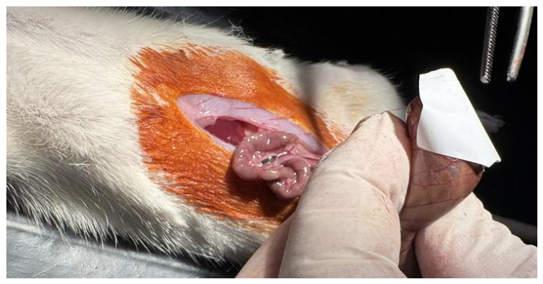
Figure 2.
Intra-abdominal image 7 days after application of a nanofiber membrane.
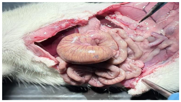
Figure 3.
Intra-abdominal image 14 days after application of a nanofiber membrane.
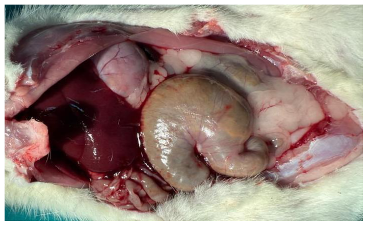
Figure 4.
Preparation of PLGA/HA nanofiber membrane using electrospinning method.
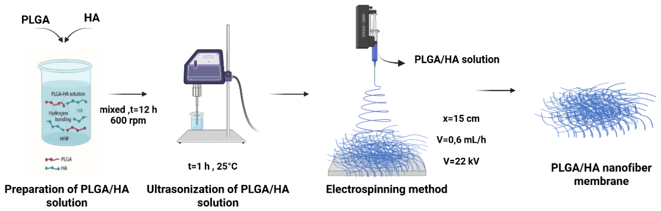
Figure 5.
a) Preparation steps of the PLGA/HA@LMWH nanofiber membrane using the electrospinning method. b) Application of the prepared PLGA/HA@LMWH nanofiber membrane in a rat model to prevent intra-abdominal adhesions.
Figure 5.
a) Preparation steps of the PLGA/HA@LMWH nanofiber membrane using the electrospinning method. b) Application of the prepared PLGA/HA@LMWH nanofiber membrane in a rat model to prevent intra-abdominal adhesions.
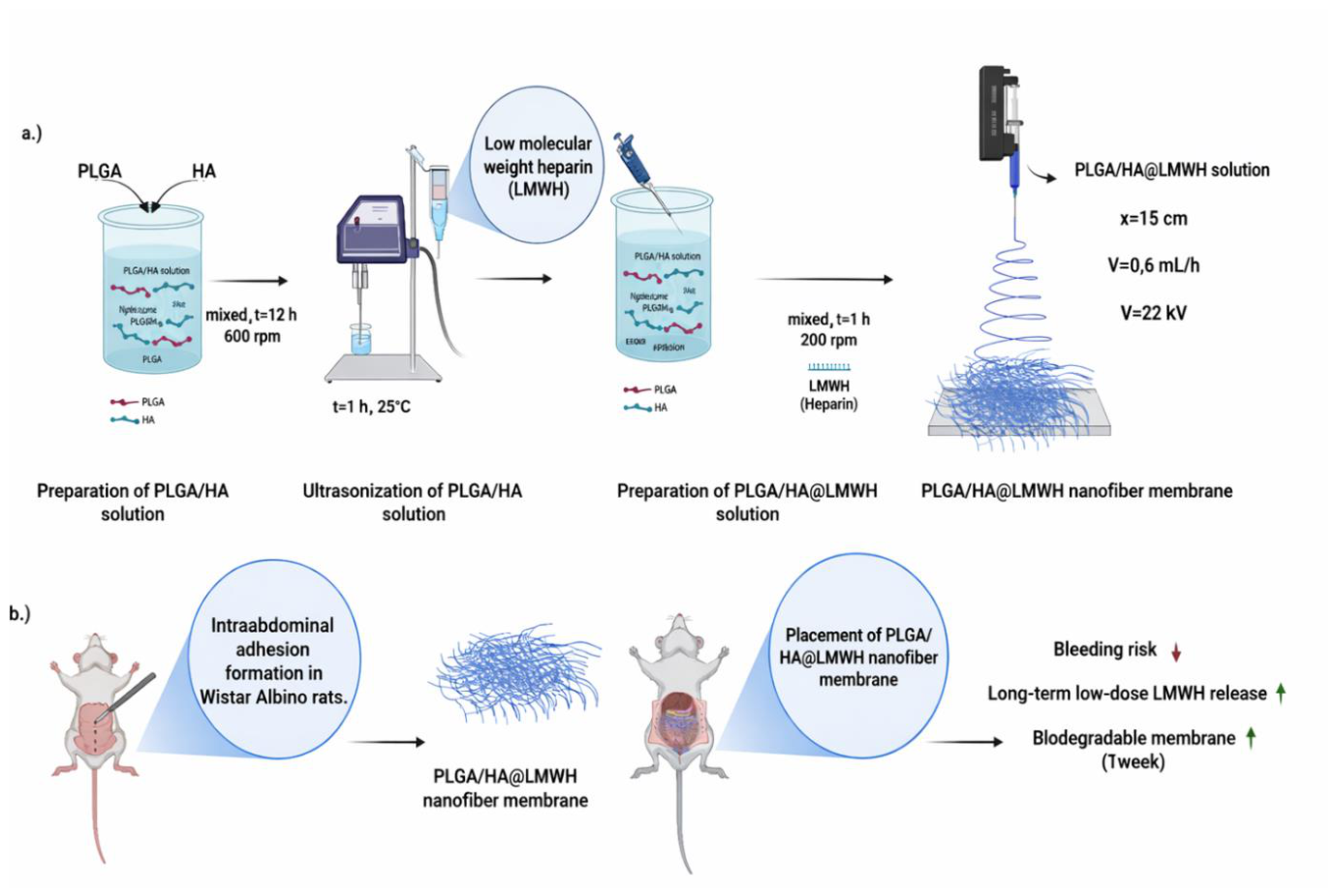
Figure 6.
FT-IR spectra of LMWH, PLGA/HA, and PLGA/HA@LMWH nanofibers.
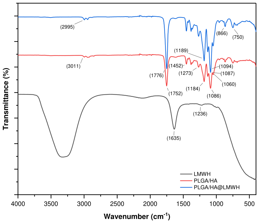
Figure 7.
(A, C) SEM images of PLGA/HA and PLGA/HA@LMWH nanofibers, respectively. (B, D) Corresponding fiber diameter distribution histograms for PLGA/HA and PLGA/HA@LMWH nanofibers.
Figure 7.
(A, C) SEM images of PLGA/HA and PLGA/HA@LMWH nanofibers, respectively. (B, D) Corresponding fiber diameter distribution histograms for PLGA/HA and PLGA/HA@LMWH nanofibers.
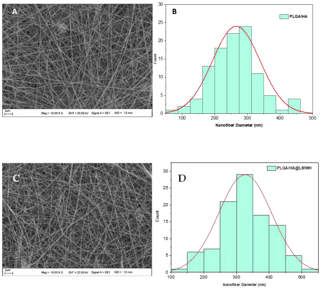
Figure 8.
Colorimetric evaluation of LMWH release from PLGA/HA@LMWH nanofiber membranes over time using toluidine blue. Control samples and release medium aliquots were collected on a daily basis (Day 1–Day 7). A progressive change in color intensity is observed, indicating continuous LMWH release. Notably, from day 3 onwards, partial degradation of the nanofiber membrane becomes evident, accompanied by a distinct color shift from deep blue to purple, suggesting increased LMWH release. From day 4 onwards, the colorimetric response indicates an almost complete release of LMWH due to rapid matrix degradation and loss of structural integrity. [Each control sample contains PLGA/HA nanofiber membrane PBS buffer solution (100 microliters) + toluidine blue dye (500 microliters). Each release medium aliquat sample contains PLGA/HA@LMWH nanofiber membrane PBS buffer solution (100 microliters) + toluidine blue dye (500 microliters).].
Figure 8.
Colorimetric evaluation of LMWH release from PLGA/HA@LMWH nanofiber membranes over time using toluidine blue. Control samples and release medium aliquots were collected on a daily basis (Day 1–Day 7). A progressive change in color intensity is observed, indicating continuous LMWH release. Notably, from day 3 onwards, partial degradation of the nanofiber membrane becomes evident, accompanied by a distinct color shift from deep blue to purple, suggesting increased LMWH release. From day 4 onwards, the colorimetric response indicates an almost complete release of LMWH due to rapid matrix degradation and loss of structural integrity. [Each control sample contains PLGA/HA nanofiber membrane PBS buffer solution (100 microliters) + toluidine blue dye (500 microliters). Each release medium aliquat sample contains PLGA/HA@LMWH nanofiber membrane PBS buffer solution (100 microliters) + toluidine blue dye (500 microliters).].
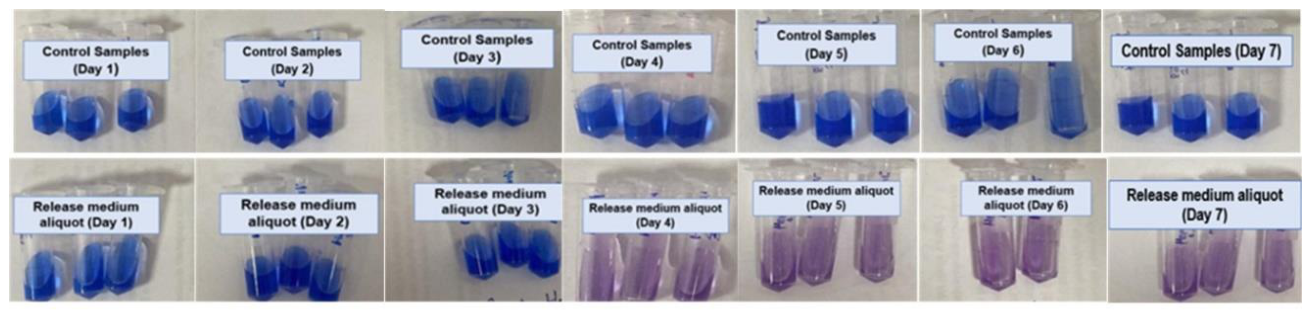
Figure 9.
Cell viability of PLGA/HA and PLGA/HA@LMWH nanofibers.
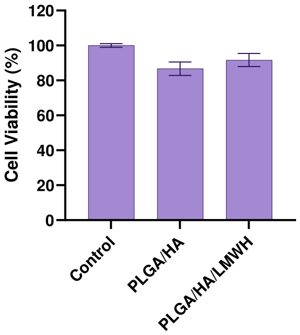
Figure 10.
Optical microscope images of L929 cells after 24 hours.
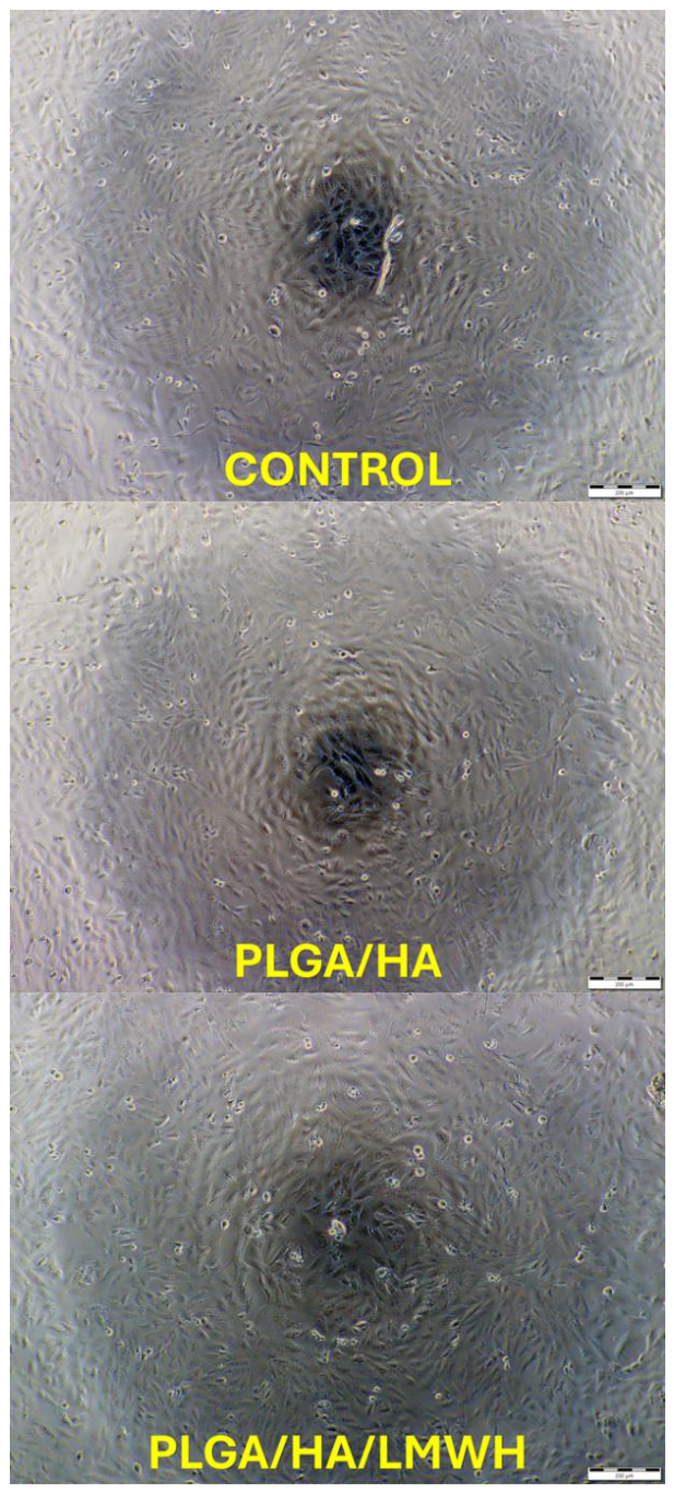
Figure 11.
Images of intraabdominal adhesions in our study (according to the classification described by Nair et al.).
Figure 11.
Images of intraabdominal adhesions in our study (according to the classification described by Nair et al.).
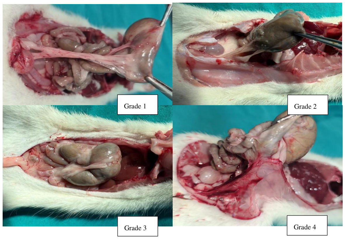
Figure 12.
Macroscopic adhesion scores of the groups (Because the data did not show a normal distribution, the Kruskal-Wallis test was used to determine differences between groups, and Dunn's multiple comparison test was used for pairwise comparisons. *, **, *** mean statistically significant (p < 0.05), highly significant (p < 0.01), and extremely significant (p < 0.001) respectively. NFM: Nanofibrillar Membrane, NFM+H: Nanofibrillar Membrane + Heparin, SS: Saline Solution and LO: Laparotomy Only.).
Figure 12.
Macroscopic adhesion scores of the groups (Because the data did not show a normal distribution, the Kruskal-Wallis test was used to determine differences between groups, and Dunn's multiple comparison test was used for pairwise comparisons. *, **, *** mean statistically significant (p < 0.05), highly significant (p < 0.01), and extremely significant (p < 0.001) respectively. NFM: Nanofibrillar Membrane, NFM+H: Nanofibrillar Membrane + Heparin, SS: Saline Solution and LO: Laparotomy Only.).
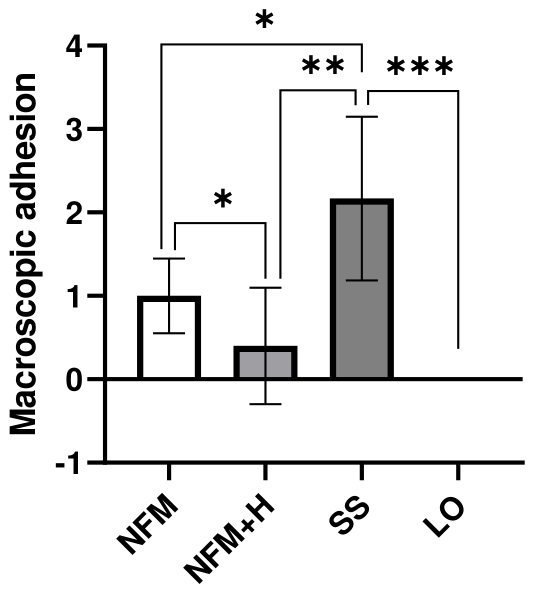
Figure 13.
Fibrosis, neovascularization, and inflammatory cell infiltrations in the serosa of the cases (Mu: mucosa, Sm: submucosa, Ms: muscularis layer, *: adhesion area).
Figure 13.
Fibrosis, neovascularization, and inflammatory cell infiltrations in the serosa of the cases (Mu: mucosa, Sm: submucosa, Ms: muscularis layer, *: adhesion area).
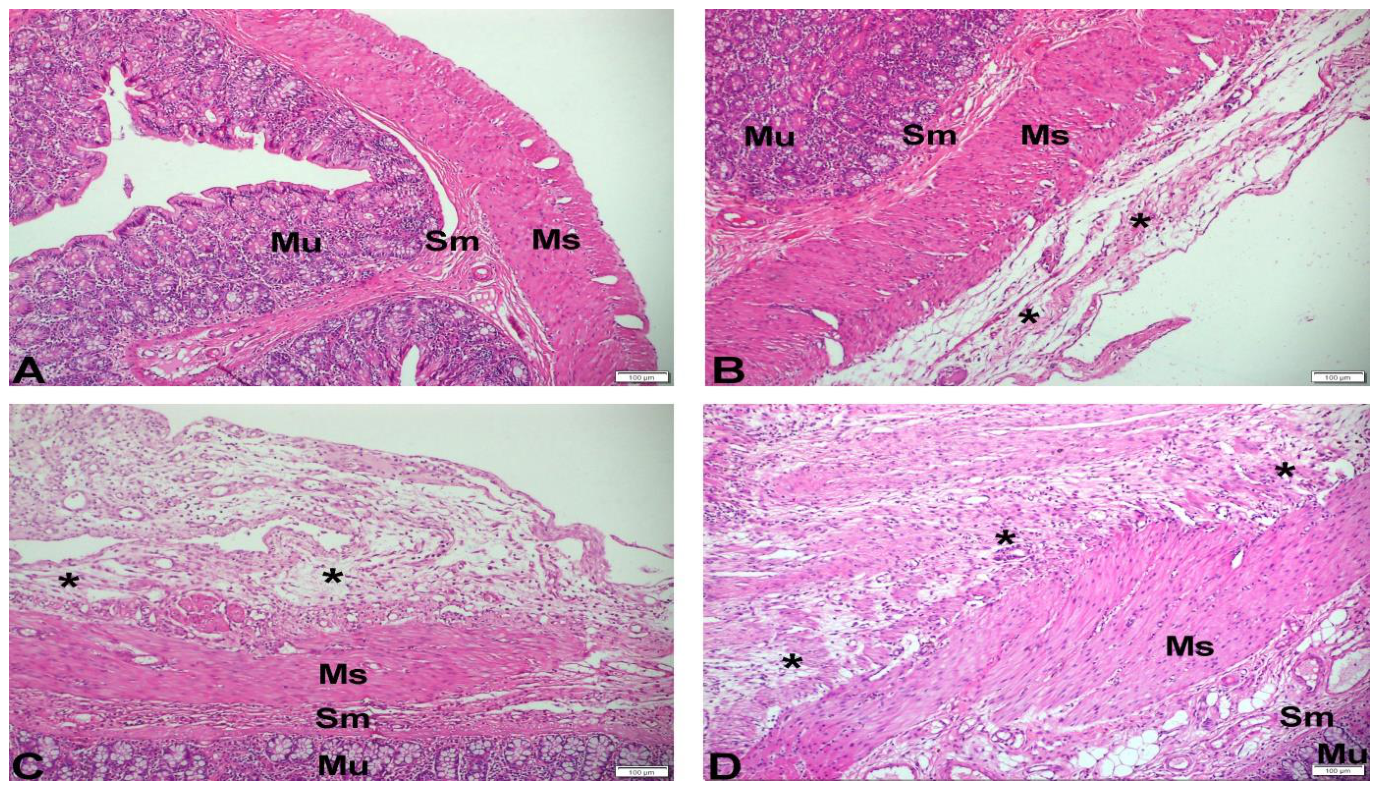
Figure 14.
In the adhesion area, the arrows in Figure 14 A, B, C show collagen fibers and the arrows in Figure 14 D, E, F show intracytoplasmically positively stained cells. (Mu: mucosa, Sm: submucosa, Ms: muscularis layer, *: adhesion area).
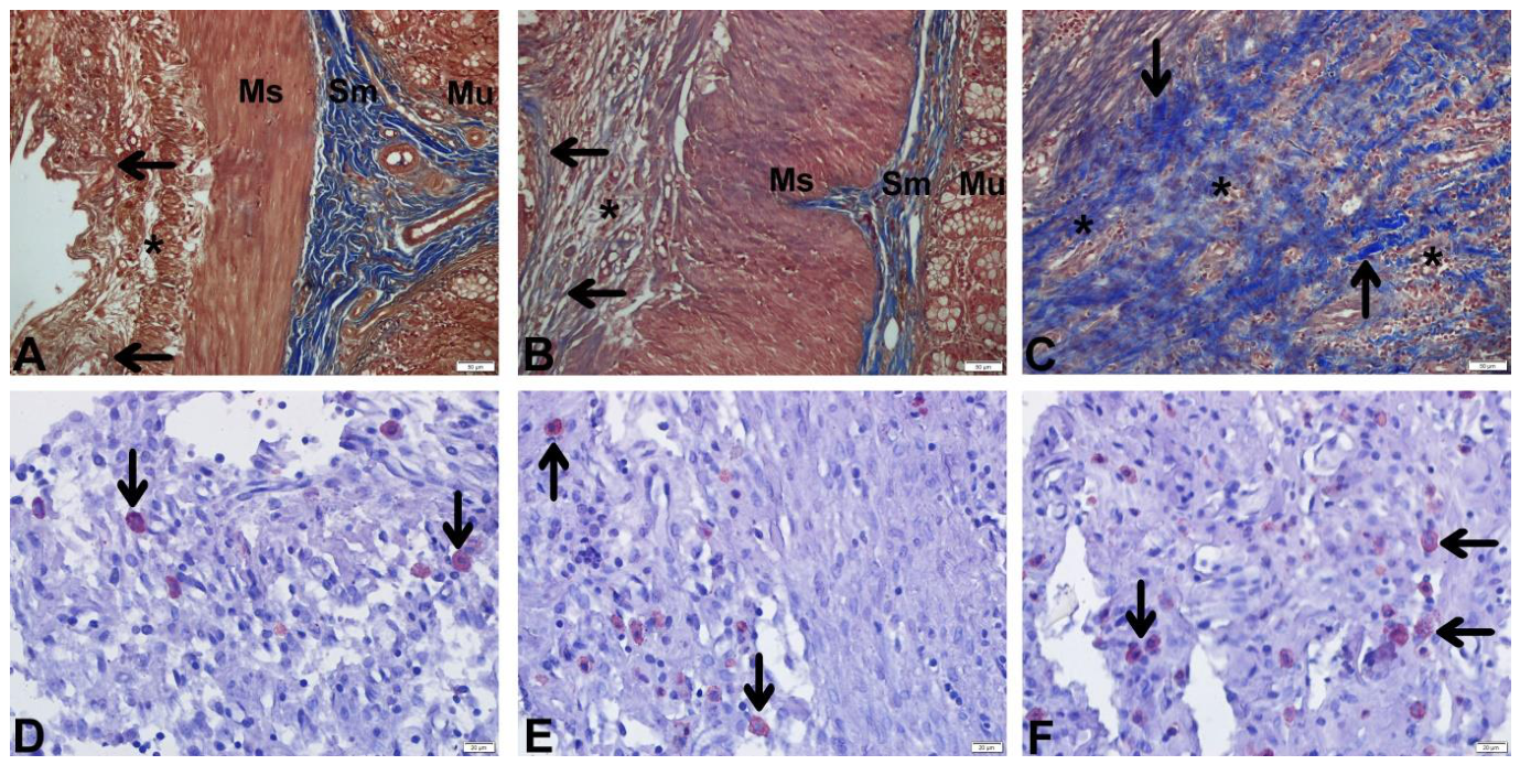
Figure 15.
Comparison of histopathological findings (Because the data did not show a normal distribution, the Kruskal-Wallis test was used to determine differences between groups, and Dunn's multiple comparison test was used for pairwise comparisons. *, **, *** mean statistically significant (p < 0.05), highly significant (p < 0.01), and extremely significant (p < 0.001) respectively. NFM: Nanofibrillar Membrane, NFM+H: Nanofibrillar Membrane + Heparin, SS: Saline Solution and LO: Laparotomy Only.).
Figure 15.
Comparison of histopathological findings (Because the data did not show a normal distribution, the Kruskal-Wallis test was used to determine differences between groups, and Dunn's multiple comparison test was used for pairwise comparisons. *, **, *** mean statistically significant (p < 0.05), highly significant (p < 0.01), and extremely significant (p < 0.001) respectively. NFM: Nanofibrillar Membrane, NFM+H: Nanofibrillar Membrane + Heparin, SS: Saline Solution and LO: Laparotomy Only.).
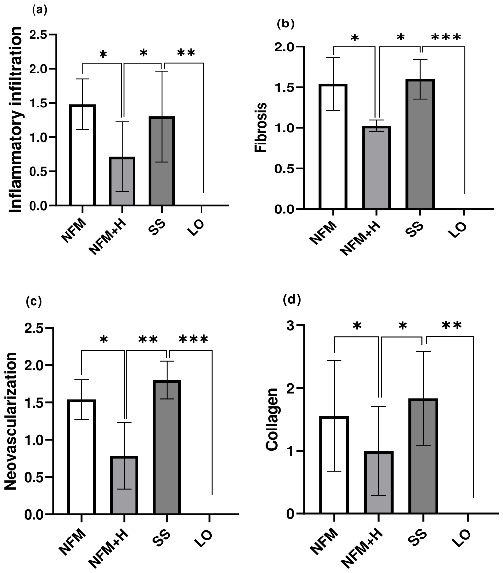
Figure 16.
Comparison of TGF-β1 levels (Because the data did not show a normal distribution, the Kruskal-Wallis test was used to determine differences between groups, and Dunn's multiple comparison test was used for pairwise comparisons. *, **, *** mean statistically significant (p < 0.05), highly significant (p < 0.01), and extremely significant (p < 0.001) respectively. NFM: Nanofibrillar Membrane, NFM+H: Nanofibrillar Membrane + Heparin, SS: Saline Solution and LO: Laparotomy Only.) .
Figure 16.
Comparison of TGF-β1 levels (Because the data did not show a normal distribution, the Kruskal-Wallis test was used to determine differences between groups, and Dunn's multiple comparison test was used for pairwise comparisons. *, **, *** mean statistically significant (p < 0.05), highly significant (p < 0.01), and extremely significant (p < 0.001) respectively. NFM: Nanofibrillar Membrane, NFM+H: Nanofibrillar Membrane + Heparin, SS: Saline Solution and LO: Laparotomy Only.) .
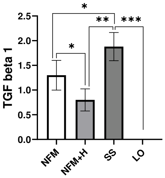
Figure 17.
Western blot analysis results of NF-κB, p-NF-κB, Bcl-2, and Cleaved caspase 3. (a) NF-κB, p-NF-κB, Bcl-2, and Cleaved caspase 3 protein bands analyzed by Western blot. (b–e) The relative expression levels and statistical analysis of NF-κB, p-NF-κB, Bcl-2, and Cleaved caspase 3 proteins. All experiments were independently repeated at least three times (n=3 per group). (Data are expressed as median ± minimum-maximum deviations; (p < 0.05) is considered statistically significant. * means statistically significant (p < 0.05). NFM: Nanofibrillar Membrane, NFM+H: Nanofibrillar Membrane + Heparin, SS: Saline Solution and LO: Laparotomy Only.).
Figure 17.
Western blot analysis results of NF-κB, p-NF-κB, Bcl-2, and Cleaved caspase 3. (a) NF-κB, p-NF-κB, Bcl-2, and Cleaved caspase 3 protein bands analyzed by Western blot. (b–e) The relative expression levels and statistical analysis of NF-κB, p-NF-κB, Bcl-2, and Cleaved caspase 3 proteins. All experiments were independently repeated at least three times (n=3 per group). (Data are expressed as median ± minimum-maximum deviations; (p < 0.05) is considered statistically significant. * means statistically significant (p < 0.05). NFM: Nanofibrillar Membrane, NFM+H: Nanofibrillar Membrane + Heparin, SS: Saline Solution and LO: Laparotomy Only.).
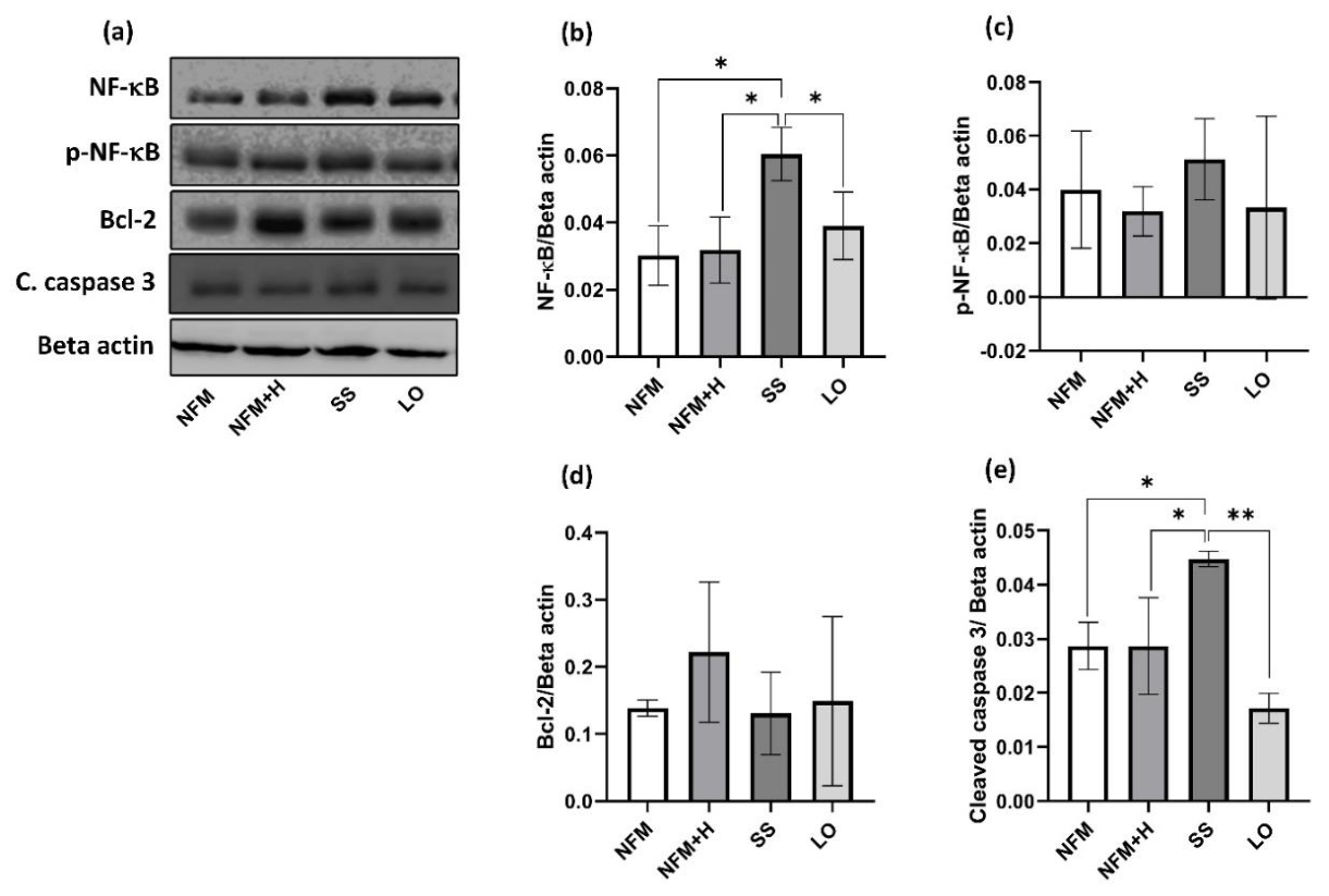
Figure 18.
Comparison of glutathione and hydroxyproline values in the groups.
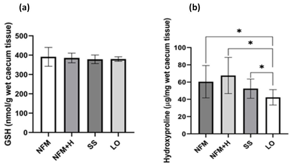

Table 1.
Nair Adhesion Scoring System.
| Score | Definition |
|---|---|
| 0 | No adhesions |
| 1 | Single band adhesion (between organs or between an organ and the abdominal wall) |
| 2 | Two band adhesions |
| 3 | More than two bands or mass adhesions between bowel loops, but no spread to the abdominal wall |
| 4 | Widespread, dense adhesions; bowel loops adhering to the abdominal wall |
Table 2.
Histopathological scoring system of Giusto et al.
| Grade | Inflammatory cell infiltration | Fibrosis | Vascularization |
|---|---|---|---|
| 0 | None | None | None |
| 1 | Mild | Mild | One or two vessels |
| 2 | Moderate | Moderate | Three to nine vessels |
| 3 | Severe | Intense | Ten or more vessels |
Disclaimer/Publisher’s Note: The statements, opinions and data contained in all publications are solely those of the individual author(s) and contributor(s) and not of MDPI and/or the editor(s). MDPI and/or the editor(s) disclaim responsibility for any injury to people or property resulting from any ideas, methods, instructions or products referred to in the content. |
© 2026 by the authors. Licensee MDPI, Basel, Switzerland. This article is an open access article distributed under the terms and conditions of the Creative Commons Attribution (CC BY) license (http://creativecommons.org/licenses/by/4.0/).
Copyright: This open access article is published under a Creative Commons CC BY 4.0 license, which permit the free download, distribution, and reuse, provided that the author and preprint are cited in any reuse.


