Preprint
Article

This version is not peer-reviewed.

Structure-Guided Discovery of a Cold-Responsive Antifreeze-Like Protein from Antarctic Flavobacterium sp. PL002

A peer-reviewed article of this preprint also exists.

Submitted:

25 June 2026

Posted:

25 June 2026

You are already at the latest version

Abstract

Antarctic microorganisms experience persistent subzero temperatures and repeated freeze–thaw cycles that require specialized mechanisms for survival. Although transcriptomic studies have identified numerous cold-responsive genes, many remain annotated as hypothetical proteins with unknown functions. In this study, we investigated PL002-1792, a strongly upregulated hypothetical protein from Antarctic Flavobacterium sp. PL002 identified under severe cold stress (−20 °C versus −6 °C; log₂ fold change = 5.61, adjusted p = 1.89 × 10⁻¹³⁸). Sequence analysis revealed a 402-amino-acid protein containing a predicted Sec/SPII lipoprotein signal peptide. AlphaFold3 prediction generated a high-confidence structural model (pTM = 0.96) with an elongated β-sheet-rich architecture resembling bacterial ice-binding proteins. Comparative analysis with the ice-binding protein from Flavobacterium frigoris (FfIBP) identified conserved glycine-rich and TXT-like motifs associated with putative ice-binding surfaces. Recombinant PL002-1792 was expressed in Escherichia coli, recovered from inclusion bodies, and successfully refolded into a predominantly β-sheet-rich conformation as confirmed by circular dichroism spectroscopy. Functional assays demonstrated moderate ice recrystallisation inhibition activity, reducing relative ice crystal mean grain size to approximately 90% of the control, and significantly enhanced freeze–thaw survival, with recombinant cells retaining 67% viability after three freeze–thaw cycles compared with 27% for the empty-vector control. These findings identify PL002-1792 as a novel antifreeze-like protein and highlight the utility of structure-guided approaches for uncovering previously uncharacterized cold-adaptation mechanisms in polar microorganisms.

Keywords: 
;  ;  ;  ;  ;  ;  

1. Introduction

Antarctic environments represent some of the most extreme ecosystems on Earth, characterized by persistent subzero temperatures, repeated freeze–thaw cycles, high salinity in sea-ice habitats, and limited nutrient availability [1]. These conditions impose severe physiological stress on microorganisms, requiring specialized adaptations to maintain cellular integrity and metabolic activity. One of the most significant challenges associated with subzero environments is the formation and recrystallisation of ice. Ice formation around microbial cells can restrict oxygen diffusion, impair nutrient transport, concentrate solutes, and cause mechanical damage to cellular structures through ice crystal growth [2,3]. Consequently, microorganisms inhabiting polar environments have evolved a diverse array of cold-adaptation strategies, including membrane remodeling, cryoprotectant production, cold-shock proteins, and antifreeze proteins (AFPs), also known as ice-binding proteins (IBPs) [4,5,6,7].
AFPs and IBPs are specialised proteins that interact directly with ice crystals, thereby modifying ice growth and recrystallisation [8]. These proteins typically function by adsorbing onto ice crystal surfaces, restricting crystal expansion and reducing ice-induced cellular damage [9]. Their activities are commonly manifested as thermal hysteresis and ice recrystallisation inhibition (IRI), both of which contribute to enhanced survival under freezing conditions [10,11]. Although AFPs have been extensively studied in fish, insects, plants, fungi, and several bacterial species, relatively few have been characterised from Antarctic bacteria [10,12,13,14]. Furthermore, the structural diversity and physiological roles of bacterial AFPs remain incompletely understood compared with their eukaryotic counterparts [10].
Members of the genus Flavobacterium are widespread in marine, freshwater, glacier, and polar ecosystems and are recognised for their remarkable capacity to thrive under low-temperature conditions [15]. Several Antarctic Flavobacterium species have been reported to possess diverse metabolic and stress-response capabilities that facilitate survival in cold environments [16,17,18]. Nevertheless, despite their ecological importance and abundance in polar habitats, the molecular mechanisms underlying subzero adaptation in Flavobacterium remain poorly understood [19]. In particular, little is known about the proteins that enable these bacteria to withstand repeated freezing and thawing events that frequently occur in Antarctic environments.
Recent advances in transcriptomics have revealed that a substantial proportion of genes induced during cold stress encode hypothetical proteins with unknown functions [20,21,22,23]. Similar observations have been reported in several psychrophilic and psychrotolerant microorganisms, suggesting that many important cold-adaptation mechanisms remain undiscovered [20,22]. Traditionally, the functional characterization of hypothetical proteins has been hindered by their low sequence similarity to proteins with known functions. However, recent breakthroughs in protein structure prediction, particularly the development of AlphaFold, have enabled reliable functional inference based on tertiary structure rather than sequence homology alone [24,25,26]. Structure-guided approaches therefore provide a powerful strategy for uncovering previously unrecognized proteins involved in environmental adaptation.
Despite increasing recognition of the importance of hypothetical proteins in microbial stress responses, few studies have combined transcriptomic discovery, structural prediction, and experimental validation to investigate their roles in cold adaptation. Moreover, no antifreeze-like proteins have been experimentally characterized from Antarctic Flavobacterium species using an integrated structure-guided workflow. This represents a significant knowledge gap in our understanding of bacterial adaptation to subzero environments and limits our ability to identify novel cryoprotective mechanisms employed by polar microorganisms.
To address this gap, we investigated a highly upregulated hypothetical protein, PL002-1792, identified from the Antarctic bacterium Flavobacterium sp. PL002 during growth under subzero conditions. Based on its strong induction under severe cold stress, predicted β-sheet-rich architecture, and the presence of sequence motifs associated with bacterial ice-binding proteins, we hypothesized that PL002-1792 functions as an antifreeze-like protein that contributes to cellular protection during freezing stress. We further hypothesized that structural features characteristic of ice-binding proteins would be reflected in measurable ice recrystallisation inhibition activity and enhanced tolerance to repeated freeze–thaw cycles.
Accordingly, the objectives of this study were to (i) identify and prioritize a strongly cold-responsive hypothetical protein from Antarctic Flavobacterium sp. PL002, (ii) characterize its structural and physicochemical properties using bioinformatic and structural prediction approaches, (iii) express and experimentally validate the folding of the recombinant protein, and (iv) determine its potential antifreeze and cryoprotective functions through ice recrystallisation inhibition and freeze–thaw survival assays. By integrating transcriptomic analysis, structure-guided prediction, and functional validation, this study provides new insights into the roles of previously uncharacterized proteins in microbial adaptation to extreme cold environments.

2. Materials and Methods

2.1. Bacterial strain and Identification

Flavobacterium sp. PL002 was isolated from the macroalgae Porphyra collected from Ardley Island, Fildes Bay, Antarctica (62°12′30.92″S, 58°56′26.65″W). Taxonomic identification was performed using 16S rRNA gene sequencing and phylogenetic analysis. Genome sequencing and annotation revealed a complete genome of 4.47 Mb with a GC content of 33% and 4,291 predicted genes.

2.2. Identification of a Cold-Responsive Candidate Protein

The amino acid sequence of PL002-1792 was retrieved from the annotated genome and analysed using ExPASy ProtParam. Protein length, molecular weight, theoretical isoelectric point (pI), instability index, aliphatic index, and grand average hydropathicity (GRAVY) were calculated [27,28]. These parameters were used to assess the physicochemical characteristics and potential stability of the protein.

2.3. Signal Peptide and Motif Analysis

Signal peptide prediction was performed using SignalP 6.0 to determine the presence and type of N-terminal secretion signals [29]. Conserved sequence motifs were analysed by manual inspection and sequence alignment with known bacterial ice-binding proteins. Particular attention was given to glycine-rich regions and TXT-like motifs previously associated with ice-binding activity [30].

2.4. Homology Search Against Known Proteins

Sequence homology analysis was performed to identify potential similarities between the candidate protein PL002-1792 and previously characterised proteins. BLASTp searches were conducted against the NCBI non-redundant (nr) protein database and the Protein Data Bank (PDB) to identify related sequences and structural homologs [31,32]. In addition, HHpred was used to detect remote homologies and structural relationships using hidden Markov model comparisons, thereby allowing the identification of potential structural templates even at low sequence identity [33]. Structural similarity searches were further performed using Foldseek against the PDB database to compare the predicted three-dimensional structure with experimentally determined protein structures [34]. These analyses were carried out to assess the degree of similarity between PL002-1792 and known proteins and to determine whether the protein represents a structurally distinct antifreeze-like protein with low sequence similarity to currently available PDB structures.

2.5. Structural prediction

The three-dimensional structure of the candidate protein PL002-1792 was predicted using AlphaFold3 (https://alphafoldserver.com/) [24]. The resulting structural model was evaluated based on the predicted local distance difference test (pLDDT) score, which provides residue-level confidence in the predicted structure, and the predicted template modeling score (pTM), which estimates the overall accuracy of the predicted fold. Residues with pLDDT values above 70 were considered to have reliable structural predictions. The predicted protein structure was subsequently visualised and analysed using PyMOL and ChimeraX to examine the overall fold, secondary structure organisation, and surface features potentially associated with antifreeze-like activity [35].

2.6. Structure validation

The quality and reliability of the predicted three-dimensional structure of PL002-1792 were evaluated using multiple structural validation tools. PROCHECK was used to assess stereochemical quality by generating Ramachandran plots and determining the distribution of residues in favoured, allowed, and disallowed regions [36]. MolProbity was employed to evaluate overall model geometry, including side-chain conformations and steric clashes [37]. In addition, Verify3D was used to assess the compatibility between the predicted three-dimensional structure and its amino acid sequence by evaluating the environmental profile of each residue [38]. Ramachandran plot statistics and other structural quality metrics were used to determine the reliability and overall structural integrity of the predicted protein model.

2.7. Structural Comparison with Known Antifreeze Proteins

Structural comparison was performed to evaluate the similarity between the predicted structure of PL002-1792 and previously characterised bacterial antifreeze proteins. The predicted model was compared with representative bacterial ice-binding proteins (IBPs), including the well-characterised F. frigoris ice-binding protein (FfIBP). Structural alignments were performed using TM-align to assess global structural similarity and calculate the root-mean-square deviation (RMSD) and template modelling score (TM-score). In addition, structural similarity searches were conducted using the DALI server to identify proteins with related three-dimensional folds within the Protein Data Bank (PDB) [39]. The resulting RMSD and TM-score values were used to evaluate structural conservation between PL002-1792 and known antifreeze proteins, providing insights into potential functional similarities despite low sequence identity.

2.8. Identification of Putative Ice-Binding Surface

The predicted structure of PL002-1792 was analysed to identify potential ice-binding surfaces commonly observed in bacterial antifreeze and ice-binding proteins. Structural features characteristic of ice-interacting regions, including flat β-sheet surfaces, regularly spaced threonine or serine residues, and repetitive TXT-like motifs, were examined in the predicted model [30,40]. These features are known to contribute to ordered water interactions and ice crystal binding in antifreeze proteins [41]. Structural visualisation and surface analysis were performed using PyMOL and UCSF Chimera to map the spatial arrangement of these residues and to identify candidate ice-binding surfaces on the protein structure. The presence and organisation of these motifs were used to infer potential regions involved in ice-binding activity.

2.9. Recombinant expression

The gene encoding PL002-1792 was synthesised and cloned into the pET-28a(+) expression vector to enable recombinant protein production with an N-terminal 6×His-tag for purification. The recombinant plasmid was transformed into Escherichia coli BL21(DE3) competent cells for protein expression. Expression conditions were optimised by varying induction temperature (8, 10, 12, 20, and 25 °C), IPTG concentration (0.01–0.5 mM), induction duration (16–48 h), and induction optical density (OD600 0.5–0.6). Protein expression and solubility were evaluated by SDS-PAGE analysis of soluble and insoluble fractions following cell lysis. These conditions were selected to enhance soluble expression and minimise inclusion body formation during recombinant protein production.

2.10. Protein refolding and structural validation

PL002-1792 was predominantly recovered in the insoluble fraction, where the inclusion bodies were isolated by centrifugation and washed with buffer containing 0.5% Triton X-100 to remove contaminating proteins and membrane components. Purified inclusion bodies were solubilised in 8 M urea containing dithiothreitol (DTT) and incubated at 37 °C for 1 h. Insoluble debris was removed by centrifugation. Protein refolding was achieved by stepwise dialysis at 4 °C over six days with gradual reduction of denaturant concentration and daily buffer replacement. Refolding was performed under continuous stirring to minimise aggregation.

2.11. SDS-PAGE and Circular Dichroism Spectroscopy

Refolded PL002-1792 was analysed by SDS-PAGE under reducing and non-reducing conditions to assess protein integrity and aggregation state. Secondary structure content was evaluated using far-UV circular dichroism (CD) spectroscopy. Spectra were recorded between 200 and 290 nm and used to assess the overall folding state of the recombinant protein [42]. The resulting spectra were compared with structural predictions generated by AlphaFold3. Secondary-structure composition was estimated from the far-UV CD spectrum using the BeStSel server, which deconvolutes CD spectra to quantify the relative proportions of α-helices, β-sheets, turns, and other structural elements.

2.12. Ice Recrystallisation Inhibition Assay

Ice recrystallisation inhibition (IRI) activity was assessed using a modified splat-cooling assay based on a previously described method [43]. Protein samples (1, 5, and 10 mg mL⁻¹) were prepared in Tris-HCl buffer. A 10 μL droplet was released from a height of approximately 1.2 m onto a liquid nitrogen-cooled glass coverslip to generate a thin ice wafer. The frozen sample was transferred to a Linkam TMHS6000 cold stage (Linkam Scientific Instruments) and annealed at −6 °C for 30 min. Ice crystals were visualized under polarized light microscopy using an Olympus BX51 microscope, and the resulting images were captured with an Olympus DP71 CCD camera. Bovine serum albumin (BSA) 1 mg/mL was used as a negative protein control. Mean grain size (MGS) was quantified using image analysis software and normalised against the BSA control.

2.13. Freeze–Thaw Protection Assay

The freeze–thaw assay was adapted from [44] with modifications. The protective effect of PL002-1792 was evaluated using recombinant E. coli expressing the target protein. Cultures harboring pET28a-PL002-1792 and the empty pET28a vector were induced and cultivated at either 12 °C or 20 °C for 24 h. Following induction, cells were harvested, washed, and resuspended in sterile 1× phosphate-buffered saline (PBS). The cell density of each culture was adjusted to an OD₆₀₀ of 1.0, corresponding to approximately 5 × 10⁸ CFU mL⁻¹ based on a previously established calibration curve. The standardized cell suspensions were subjected to three freeze–thaw cycles. Each cycle consisted of freezing the samples at −80 °C without cryoprotectant for 24 h, followed by thawing at room temperature for 30 min. After each thawing step, the optical density at 600 nm (OD₆₀₀) was measured and converted to estimated cell concentrations using the calibration curve. The samples were subsequently re-frozen and subjected to the same procedure for a total of three freeze–thaw cycles. The relative cell concentration after each freeze–thaw cycle was calculated as a percentage of the initial cell concentration before freezing.

3. Results

3.1. Genome Features and Identification of a Cold-Responsive Candidate Protein

Phylogenetic analysis based on the 16S rRNA gene confirmed that strain PL002 belongs to the genus Flavobacterium [45]. The isolate clustered within the Flavobacterium clade and showed close phylogenetic relationships with several cold-adapted Flavobacterium species, including F. frigoris and F. frigidarium, supporting its taxonomic placement within this genus. These findings are consistent with the ecological origin of PL002 from Antarctic marine environments, where members of the genus are commonly associated with cold-adapted microbial communities.
Genome sequencing and annotation revealed that Flavobacterium sp. PL002 possesses a complete genome of approximately 4.47 Mb with a GC content of 33%. A total of 4,291 protein-coding sequences (CDSs) were predicted, encompassing genes associated with central metabolism, environmental sensing, nutrient acquisition, and stress adaptation. Notably, a substantial proportion of the predicted CDSs were annotated as hypothetical proteins, indicating the presence of numerous uncharacterized genes that may contribute to adaptation in extreme Antarctic environments [45]. To identify genes potentially involved in subzero adaptation, comparative transcriptomic analyses were performed under different low-temperature conditions. Transcriptomic screening identified PL002-1792 as a highly cold-induced hypothetical protein. When comparing cultures grown at −20 °C and −6 °C, PL002-1792 exhibited a log₂ fold change of 5.61 with an adjusted p-value of 1.89 × 10⁻¹³⁸ (Table 1). Similarly, comparison between −20 °C and 15 °C showed significant upregulation of PL002-1792, with a log₂ fold change of 4.89 and an adjusted p-value of 1.10 × 10⁻⁵⁹.
Despite its strong induction under cold stress, PL002-1792 was annotated as a hypothetical protein with no experimentally validated function. Domain analysis identified the presence of an LIPL48-related domain, while BLAST searches revealed similarity to hypothetical proteins from other Flavobacterium species, suggesting that the protein belongs to a poorly characterised family of bacterial proteins. Given its exceptionally high level of induction under subzero conditions and lack of functional annotation, PL002-1792 was selected for further structural and functional characterisation as a potential contributor to cold adaptation in Antarctic Flavobacterium sp. PL002.

3.2. Sequence Analysis and Physicochemical Properties of PL002-1792

The PL002-1792 gene encodes a protein consisting of 402 amino acid residues with a predicted molecular weight of 41.3 kDa. Physicochemical analysis using the ExPASy ProtParam server predicted a theoretical isoelectric point (pI) of 4.70, indicating that the protein is acidic in nature [27]. The instability index was calculated to be 12.66, classifying the protein as stable according to ProtParam criteria. In addition, the protein exhibited an aliphatic index of 75.02 and a grand average of hydropathicity (GRAVY) value of −0.044. The calculated physicochemical parameters indicate that PL002-1792 is a moderately sized acidic protein with predicted stability and a slightly hydrophilic character. A summary of the physicochemical properties of PL002-1792 is presented in Table 2.

3.3. Signal Peptide Prediction and Conserved Motif Analysis of PL002-1792

Signal peptide analysis using SignalP 6.0 predicted that PL002-1792 contains an N-terminal lipoprotein signal peptide (Table 3). The protein was classified as a Sec/SPII substrate with a prediction probability of 0.997. The most probable signal peptide cleavage site was identified between amino acid residues 21 and 22, indicating that the mature protein is generated following removal of the N-terminal signal peptide. The presence of a Sec/SPII signal peptide suggests that PL002-1792 is processed through the bacterial Sec secretion pathway and may be associated with the cell envelope following lipid modification. No alternative signal peptide types, including Sec/SPI, Tat/SPI, Tat/SPII, or Sec/SPIII pathways, were predicted. Sequence analysis further identified several glycine-rich regions and multiple TXT-like motifs distributed throughout the protein sequence. These motifs have previously been reported in bacterial ice-binding proteins and were therefore selected for further structural analysis. The predicted signal peptide and conserved sequence motifs indicate that PL002-1792 possesses sequence features commonly associated with extracellular or surface-associated proteins. To further investigate whether these conserved sequence features are associated with ice-binding function, the three-dimensional structure of PL002-1792 was predicted and analyzed using AlphaFold3.

3.4. Structural Prediction and Comparative Analysis of PL002-1792

The three-dimensional structure of PL002-1792 was predicted using AlphaFold3 to gain insight into its potential function. The resulting model exhibited a high predicted template modeling (pTM) score of 0.96, indicating a highly reliable overall fold prediction [53] (Figure 1).
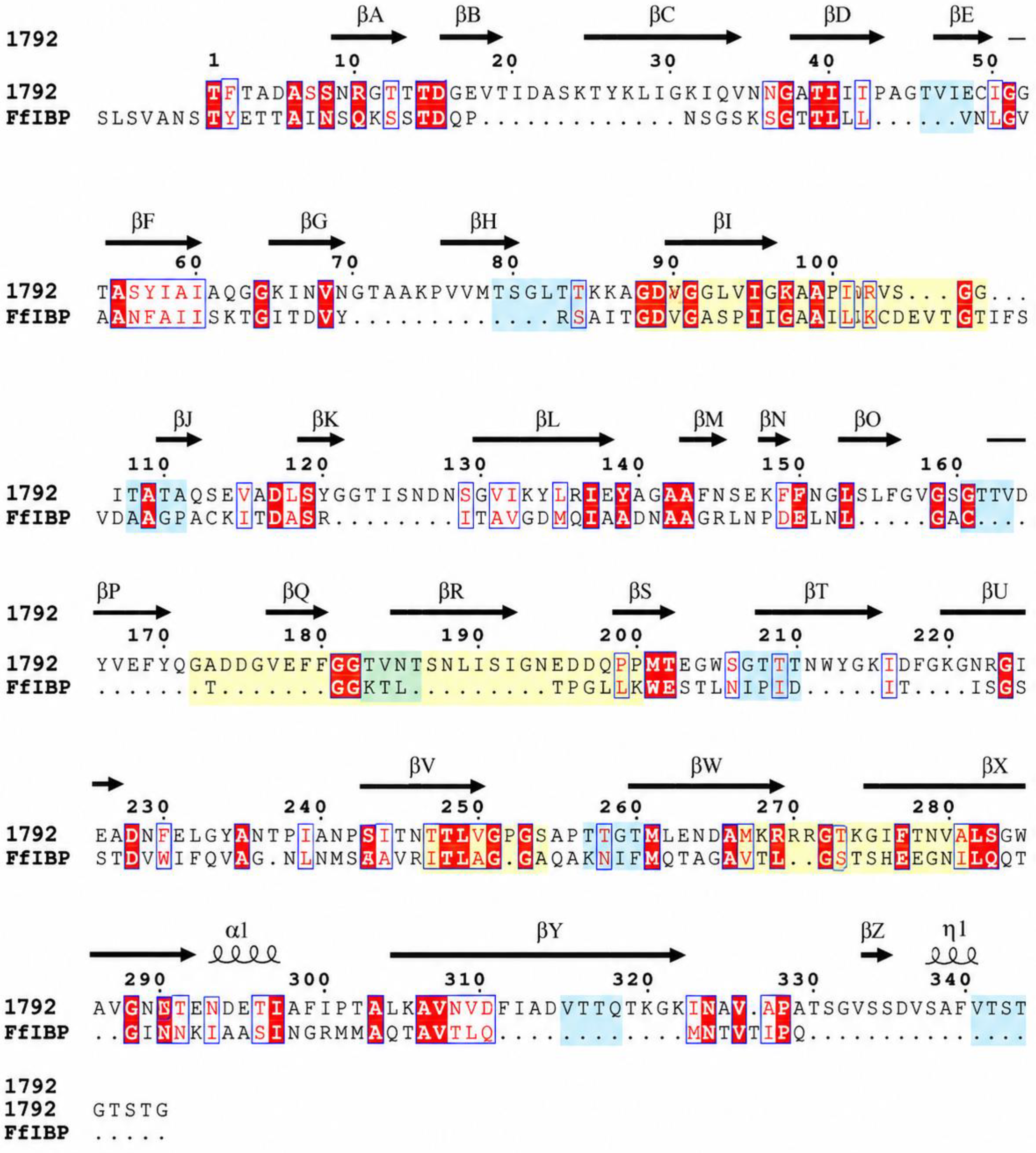
Visualization of the predicted structure revealed an elongated architecture dominated by β-strands interconnected by short loop regions. The protein adopted a β-sheet-rich fold extending throughout the length of the molecule, producing an overall topology characteristic of several previously described bacterial ice-binding proteins. Examination of the structure from different orientations demonstrated the presence of stacked β-sheet surfaces forming an elongated β-solenoid-like scaffold (Figure 2A,B). The N- and C-terminal regions were positioned at opposite ends of the molecule, resulting in an extended structure with relatively flat surface regions. The predominance of β-sheet secondary structure observed in the predicted model was consistent with the sequence-derived characteristics of PL002-1792 and was subsequently evaluated experimentally using circular dichroism spectroscopy. To investigate structural similarity with known bacterial ice-binding proteins, PL002-1792 was compared with the ice-binding protein from F. frigoris (FfIBP). Multiple sequence alignment revealed several conserved residues located within predicted β-strand regions despite relatively low overall sequence identity (Figure 3).
In addition, multiple glycine-rich segments and TXT-like motifs were identified throughout the sequence. These motifs were distributed within regions corresponding to the predicted β-sheet-rich scaffold and were retained in both PL002-1792 and FfIBP. Comparison of the aligned sequences further showed conservation of several residues associated with repetitive β-strand architecture characteristic of bacterial ice-binding proteins. Although extensive sequence divergence was observed across much of the protein sequence, the overall arrangement of conserved motifs and β-strand-associated residues remained evident. The preservation of these sequence features, together with the highly similar β-sheet-rich structural organisation predicted by AlphaFold3, indicated structural conservation between PL002-1792 and previously characterised bacterial ice-binding proteins. Collectively, the structural prediction and comparative sequence analyses demonstrated that PL002-1792 possesses an elongated β-sheet-rich architecture, conserved glycine-rich regions, and multiple TXT-like motifs commonly observed in bacterial ice-binding proteins. These features provided the basis for subsequent experimental characterisation of the protein.

3.5. Recombinant Expression of PL002-1792

Recombinant expression of PL002-1792 was evaluated under multiple induction conditions to optimise protein production and solubility. Expression was performed in E. coli BL21(DE3) using a pET-28a(+) expression system, with induction temperatures ranging from 8 to 25 °C, induction periods of 16–48 h, and IPTG concentrations between 0.01 and 0.5 mM.
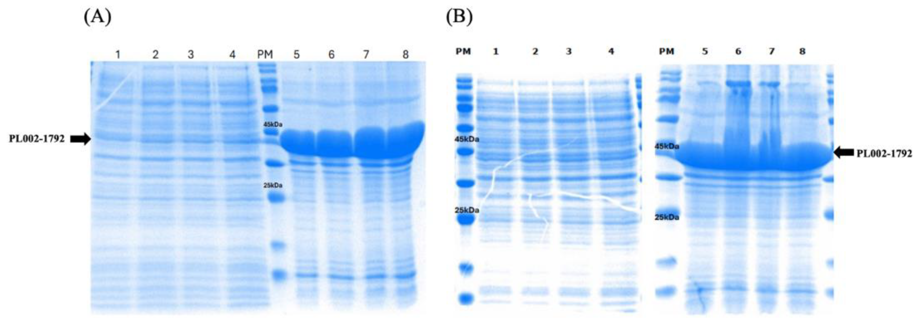
SDS-PAGE analysis consistently revealed the production of a prominent protein band at approximately 41 kDa, corresponding to the predicted molecular weight of PL002-1792 (Figure 4). Initially, protein expression was tested at 20C, induced at 0.5mM IPTG at different OD600, but overexpression was found in pellet fractions (Figure A,B). Next, recombinant protein expression was tested and detected under all induction conditions tested, confirming successful heterologous production of PL002-1792 in E. coli (Figures S1–S4). Comparison of soluble and insoluble fractions showed that the majority of the recombinant protein was recovered in the pellet fraction. This pattern was observed at all induction temperatures tested, including 25, 20, 12, 10, and 8 °C. Extending the induction period from 16 h to 48 h at 20 °C did not substantially alter the distribution of the protein between soluble and insoluble fractions. Similarly, reducing the induction temperature and IPTG concentration did not result in a noticeable increase in soluble protein recovery. Across all expression conditions examined, only trace amounts of PL002-1792 were detected in the soluble fraction, whereas a strong protein band corresponding to approximately 41 kDa was consistently observed in the pellet fraction. These results indicate that recombinant PL002-1792 was predominantly produced as insoluble protein during heterologous expression.

3.6. Recovery and Refolding of Recombinant PL002-1792

Since recombinant PL002-1792 was predominantly recovered in the insoluble fraction, inclusion bodies were isolated for subsequent protein recovery. Following cell lysis and centrifugation, the insoluble pellet containing PL002-1792 was collected and washed to remove contaminating cellular proteins and membrane components. SDS-PAGE analysis confirmed enrichment of the target protein in the purified inclusion body fraction. The isolated inclusion bodies were subsequently solubilized in denaturing buffer containing 8 M urea and dithiothreitol (DTT), resulting in complete dissolution of the protein. Insoluble debris was removed by centrifugation, and the denatured protein solution was subjected to stepwise dialysis-based refolding at 4 °C. Refolding was performed over six days with gradual removal of the denaturant through sequential buffer exchanges. The refolding procedure yielded soluble recombinant PL002-1792 suitable for downstream structural and functional analyses. The recovered protein was subsequently evaluated by SDS-PAGE and circular dichroism spectroscopy to assess its structural integrity and folding status.

3.7. Structural Validation of Refolded PL002-1792

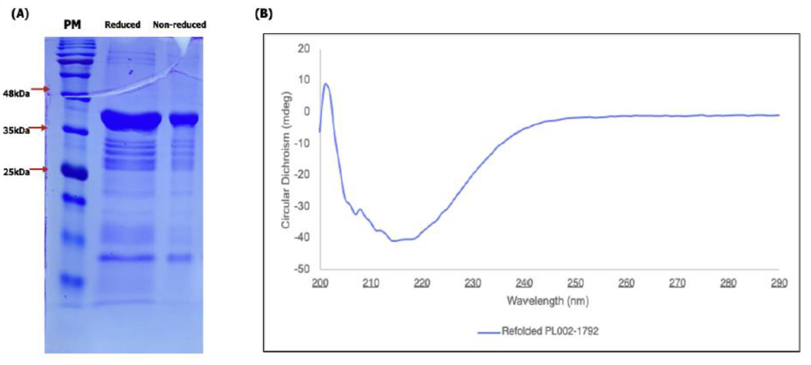
The structural integrity of refolded PL002-1792 was evaluated using SDS-PAGE and circular dichroism (CD) spectroscopy. SDS-PAGE analysis under both reducing and non-reducing conditions revealed a prominent protein band at approximately 41 kDa, corresponding closely to the predicted molecular weight of PL002-1792 (Figure 5A). Similar migration patterns were observed under both conditions, indicating that the protein remained predominantly monomeric following refolding. No substantial high-molecular-weight species or visible protein aggregates were detected, suggesting successful recovery of a soluble protein suitable for downstream functional analyses. The secondary structure of refolded PL002-1792 was further examined using far-UV CD spectroscopy. The CD spectrum exhibited a pronounced negative ellipticity minimum centred between 216 and 218 nm (Figure 5B), a characteristic feature of β-sheet-rich proteins. Quantitative secondary-structure analysis using the BeStSel server estimated that PL002-1792 contains 38.4% β-sheet structure, comprising 17.9% relaxed antiparallel β-sheet, 19.8% left-twisted antiparallel β-sheet, and 0.7% parallel β-sheet, together with 16.1% turns and only 1.6% α-helical content (Table 4). The predominance of β-sheet structures is consistent with the elongated β-sheet-rich architecture predicted by AlphaFold3. Together, the CD spectrum and deconvolution analysis provide experimental evidence that the refolded protein adopts the β-sheet-dominated conformation predicted in silico.

3.8. Ice Recrystallisation Inhibition Activity of PL002-1792

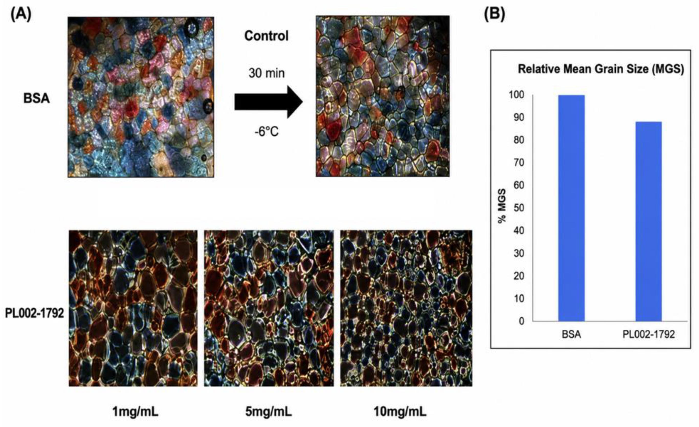
The ice recrystallisation inhibition (IRI) activity of PL002-1792 was evaluated using a splat-cooling assay followed by annealing at −6 °C for 30 min. Ice crystal morphology was examined under polarised light microscopy and compared with bovine serum albumin (BSA), which served as a negative protein control (Figure 6A). Following annealing, the BSA control exhibited relatively large and well-developed ice crystals characteristic of extensive ice recrystallisation. In contrast, samples containing PL002-1792 displayed visibly smaller ice crystals, particularly at higher protein concentrations. The reduction in ice crystal size was most evident at a protein concentration of 10 mg mL⁻¹, where the formation of large recrystallised ice grains was noticeably reduced compared with the control. Quantitative analysis of relative Mean Grain Size (MGS) supported these observations (Figure 6B). When normalised against the BSA control, PL002-1792 reduced ice crystal grain size to approximately 90% of the control value. The reduction in MGS was observed across the tested protein concentrations and was most pronounced at 10 mg mL⁻¹. These results demonstrate that PL002-1792 possesses measurable ice recrystallisation inhibition activity and is capable of reducing ice crystal growth during annealing under freezing conditions.

3.9. PL002-1792 Enhances Freeze–Thaw Tolerance

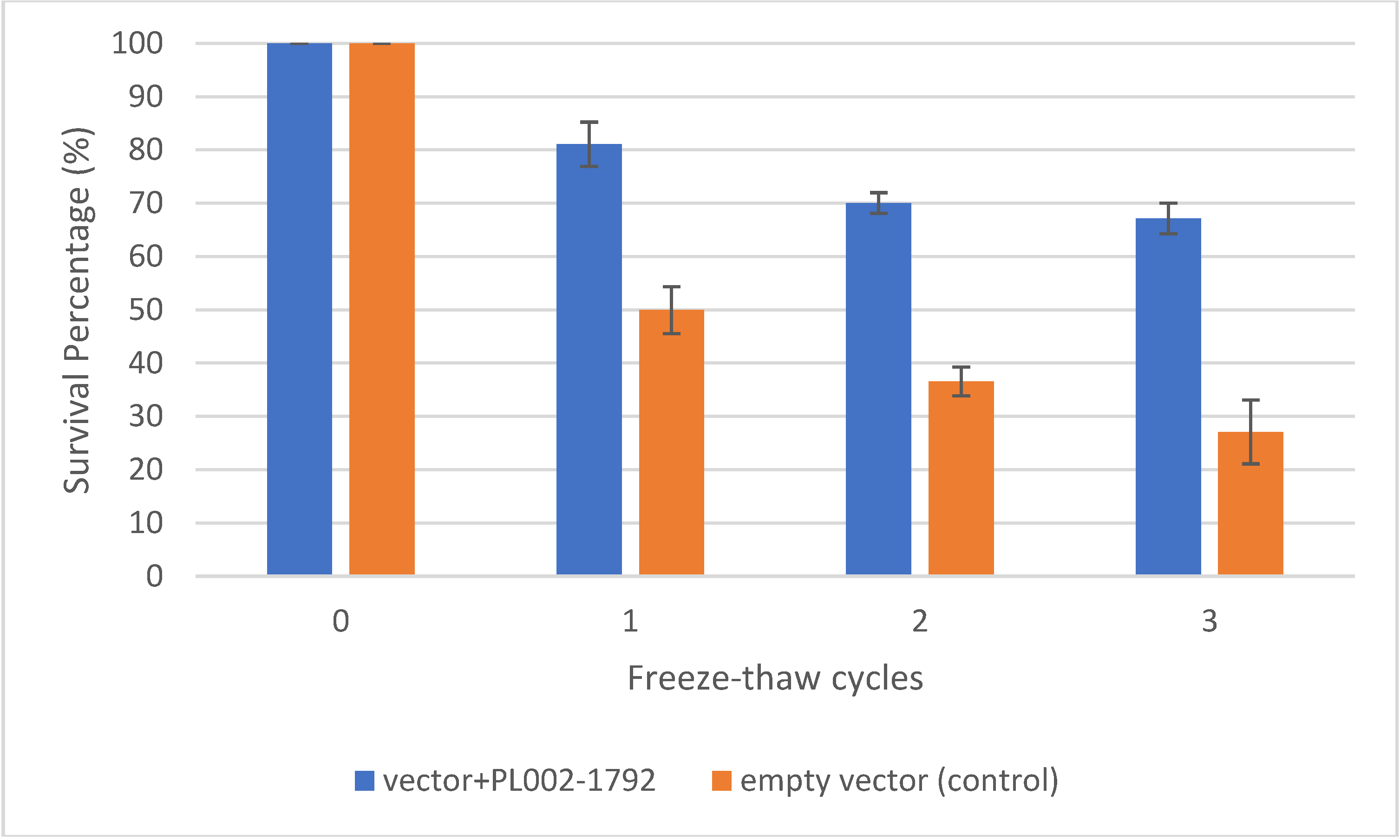
To evaluate the protective effect of PL002-1792 against freezing stress, recombinant E. coli expressing PL002-1792 and empty-vector control cells were subjected to repeated freeze–thaw cycles. Cell survival was determined after each cycle and expressed as a percentage relative to the initial cell density before freezing (Figure 7). Both strains exhibited a progressive decline in survival with increasing numbers of freeze–thaw cycles. However, cells expressing PL002-1792 consistently maintained significantly higher survival rates than the empty-vector control throughout the experiment. After the first freeze–thaw cycle, PL002-1792-expressing cells retained approximately 70% survival, whereas the empty-vector control retained only 36%. Following the second cycle, survival remained high in the PL002-1792-expressing strain (67%), compared with 27% in the control. The difference became even more apparent after the third freeze–thaw cycle, where PL002-1792-expressing cells retained 67% survival, while the empty-vector control retained only 27%. The enhanced survival observed in cells expressing PL002-1792 demonstrates that the protein confers substantial protection against freeze–thaw-induced cellular damage. The ability of PL002-1792 to maintain cell viability during repeated freezing and thawing is consistent with the proposed function of the protein as a cryoprotective or ice-binding protein and supports its potential role in improving cellular tolerance to freezing stress.

4. Discussion

The ability to survive prolonged freezing and repeated freeze–thaw events is a fundamental requirement for microbial persistence in Antarctic environments [46,47,48]. Such conditions expose cells to ice crystal formation, osmotic stress, membrane damage, and macromolecular destabilisation [12]. In the present study, we applied a structure-guided discovery approach to investigate a strongly cold-responsive hypothetical protein, PL002-1792, from Antarctic Flavobacterium sp. PL002. By integrating transcriptomic analysis, structural prediction, recombinant expression, and functional assays, we provide evidence that PL002-1792 represents a previously uncharacterized antifreeze-like protein involved in cold adaptation. The phylogenetic analysis confirmed that strain PL002 belongs to the genus Flavobacterium and clusters closely with psychrophilic species such as Flavobacterium frigoris and Flavobacterium frigidarium (Figure 1). These species are commonly associated with polar and glacial environments and are known to possess specialised mechanisms for growth at low temperatures. The close phylogenetic relationship suggests that strain PL002 may share similar cold-adaptive traits, providing ecological support for the identification of cold-responsive genes involved in subzero survival. Transcriptomic screening identified PL002-1792 as one of the most strongly induced genes under severe cold stress (Table 1). The observed log₂ fold changes of 5.61 and 4.89 under −20 °C conditions indicate that expression of PL002-1792 is highly responsive to freezing temperatures. Such dramatic transcriptional induction is consistent with genes that perform important physiological functions during environmental stress. Interestingly, PL002-1792 was annotated only as a hypothetical protein despite its strong regulation. Similar observations have been reported in psychrophilic microorganisms, where many of the most highly induced cold-responsive genes lack functional annotation [20]. This highlights the limitations of sequence-based annotation and emphasises the importance of combining transcriptomics with structural prediction to uncover previously unrecognised stress-response proteins.
The physicochemical properties of PL002-1792 (Table 2) further support a role in cold adaptation. The protein possesses a relatively low theoretical pI (4.70), indicating an acidic character. Acidic proteins are frequently enriched in psychrophilic organisms because increased surface charge can enhance hydration and maintain conformational flexibility at low temperatures [49,50,51]. In addition, the low instability index suggests that PL002-1792 is intrinsically stable despite its predicted flexibility. The slightly negative GRAVY value indicates a predominantly hydrophilic nature, a characteristic commonly observed among extracellular or surface-associated proteins that interact with aqueous environments [28]. Together, these properties are consistent with proteins adapted for function under cold and ice-associated conditions. Signal peptide analysis revealed that PL002-1792 contains a Sec/SPII lipoprotein signal peptide with a highly confident prediction score (Table 3) [29]. This finding suggests that the mature protein is exported through the Sec secretion pathway and subsequently anchored to the cell envelope through lipid modification [52]. Many bacterial ice-binding proteins are secreted or cell-surface associated, enabling them to interact directly with extracellular ice crystals [12,30,53]. Notably, the ice-binding protein from F. frigoris (FfIBP) has been shown to function as a membrane-anchored protein that promotes the formation of unfrozen brine pockets around the cell during freezing, thereby reducing local salt concentration and enhancing microbial survival under freezing conditions [35]. Given the presence of a similar Sec/SPII lipoprotein signal peptide, PL002-1792 may employ a comparable membrane-associated survival strategy, allowing direct interaction with extracellular ice and modulation of the local physicochemical environment surrounding the cell. The predicted lipoprotein nature of PL002-1792 therefore provides an important functional clue linking the protein to extracellular cryoprotection and freeze–thaw tolerance.
One of the most striking findings of this study was the structural similarity between PL002-1792 and known bacterial ice-binding proteins. AlphaFold3 prediction generated a highly confident model with a pTM score of 0.96, indicating strong reliability of the predicted fold (Figure 3) [24,53]. The model revealed an elongated architecture dominated by β-sheets arranged into a β-solenoid-like scaffold. This structural organisation closely resembles that reported for several bacterial ice-binding proteins, including the well-characterised ice-binding protein from F. frigoris (FfIBP) [30,40,41]. Importantly, structural conservation was observed despite relatively low sequence similarity, illustrating how protein function may be retained through preservation of three-dimensional architecture rather than primary sequence identity [54]. Such observations highlight the value of modern structure prediction tools for identifying proteins that would otherwise remain functionally uncharacterized [26].
The sequence comparison with FfIBP provided additional evidence supporting an antifreeze-like function (Figure 4). Multiple glycine-rich regions and TXT-like motifs were identified within PL002-1792. Similar motifs have previously been associated with ice-binding surfaces in bacterial antifreeze proteins, where regularly spaced threonine residues facilitate ordered interactions with water molecules and ice lattices [40,41,55]. Although extensive sequence divergence was observed between PL002-1792 and FfIBP, the conservation of motif organisation and β-strand architecture suggests evolutionary preservation of structural features important for ice interaction [30,54]. The combination of motif conservation and structural similarity strongly supports the hypothesis that PL002-1792 belongs to the broader family of bacterial ice-binding proteins [12].
Recombinant expression studies revealed that PL002-1792 was produced efficiently in E. coli but accumulated predominantly as inclusion bodies regardless of induction temperature, IPTG concentration, or induction duration (Figure 5). This behaviour is not uncommon among β-sheet-rich proteins and has been reported for several recombinant antifreeze proteins expressed in heterologous hosts [56]. The extensive β-sheet architecture predicted for PL002-1792 may promote intermolecular interactions during overexpression, leading to aggregation and inclusion body formation [57]. While insolubility complicated protein recovery, successful refolding from inclusion bodies enabled subsequent structural and functional characterisation [56]. The ability to recover soluble protein after denaturation also suggests that the protein possesses an intrinsically stable fold capable of re-establishing its native conformation following refolding.
Experimental validation of the refolded protein provided strong support for the AlphaFold3 structural prediction. SDS-PAGE analysis confirmed recovery of a monomeric protein with the expected molecular mass of approximately 41 kDa, while circular dichroism (CD) spectroscopy revealed a pronounced minimum at 216–218 nm characteristic of β-sheet-rich proteins (Figure 6) [42]. Quantitative secondary-structure analysis of the CD spectrum using the BeStSel server estimated that PL002-1792 contains 38.4% β-sheet content, comprising predominantly antiparallel β-sheets, whereas α-helical content accounted for only 1.6% (Table 4). These results are consistent with the elongated β-sheet-rich architecture predicted by AlphaFold3 and further support the proposed β-solenoid-like fold of the protein [53]. The close agreement between the experimental CD data and the predicted structural model strengthens confidence in the inferred architecture of PL002-1792. This agreement is particularly important because structural prediction alone cannot confirm whether a recombinant protein adopts the predicted fold in solution [26]. Therefore, the CD spectrum and secondary-structure deconvolution provide critical experimental validation of the predicted structure and support the structural basis of the proposed antifreeze-like function.
Functional analysis demonstrated that PL002-1792 possesses measurable ice recrystallisation inhibition activity (Figure 7). Although the reduction in relative mean grain size was modest, with ice crystal size decreasing to approximately 90% of the BSA control, the observed effect was consistent across protein concentrations and supported by microscopic observations of smaller ice crystals. Compared with highly active antifreeze proteins from insects or fish, the IRI activity of PL002-1792 may be considered moderate [40,58]. However, bacterial antifreeze proteins frequently exhibit lower activity than their eukaryotic counterparts while still providing substantial physiological benefits [30,48]. In natural environments, moderate IRI activity may be sufficient when combined with complementary cold-adaptation mechanisms such as compatible solute accumulation, membrane remodeling, and extracellular polymeric substance production [12]. Therefore, the relatively modest IRI activity observed here should not be interpreted as evidence of limited biological relevance.
The freeze–thaw protection assay provided the strongest functional evidence supporting a cryoprotective role for PL002-1792. Recombinant E. coli expressing PL002-1792 consistently exhibited higher survival than empty-vector controls throughout repeated freeze–thaw cycles (Figure 8). Following three cycles, PL002-1792-expressing cells retained approximately 67% viability compared with only 27% for control cells. This represents an approximately 2.48-fold improvement in survival and demonstrates that expression of PL002-1792 confers a measurable cellular advantage under freezing stress. The magnitude of protection exceeded that observed in the IRI assay alone, suggesting that the biological function of PL002-1792 may extend beyond simple inhibition of ice recrystallisation [56,58]. Potential mechanisms include stabilisation of extracellular ice interfaces, protection of membrane integrity, modulation of local ice crystal morphology, or interactions with other cellular stress-response systems [40,59]. Similar discrepancies between in vitro IRI activity and in vivo cryoprotective performance have been reported for other microbial antifreeze proteins, indicating that multiple mechanisms likely contribute to cellular protection [60,61].
Collectively, the results support a model in which PL002-1792 functions as a cold-induced, surface-associated antifreeze-like protein that contributes to freezing tolerance in Antarctic Flavobacterium sp. PL002. The strong transcriptional induction under subzero conditions, the presence of a lipoprotein signal peptide, the β-sheet-rich ice-binding protein-like structure, measurable ice recrystallisation inhibition activity, and enhanced freeze–thaw survival all converge toward the same functional interpretation. More broadly, this work demonstrates the effectiveness of integrating transcriptomics, structural prediction, and experimental validation for the discovery of previously uncharacterized cold-adaptation proteins. As a substantial proportion of polar microbial genomes remains populated by hypothetical proteins, similar structure-guided approaches may reveal additional mechanisms that enable microbial survival in extreme cryogenic environments.

5. Conclusions

PL002-1792 was identified as a highly cold-induced hypothetical protein from Antarctic Flavobacterium sp. PL002 through transcriptomic analysis. Structural prediction revealed a β-sheet-rich architecture, a lipoprotein signal peptide, and conserved motifs commonly associated with bacterial ice-binding proteins. Experimental validation confirmed successful refolding of the recombinant protein into a predominantly β-sheet-rich structure. Functional assays demonstrated moderate ice recrystallisation inhibition activity and significantly enhanced freeze–thaw survival of recombinant Escherichia coli expressing PL002-1792. These findings indicate that PL002-1792 functions as a novel antifreeze-like protein that contributes to cellular protection under freezing stress. Overall, this study highlights the effectiveness of integrating transcriptomics, structural prediction, and experimental validation to uncover the functions of previously uncharacterized proteins and advances our understanding of microbial cold adaptation in Antarctic environments.

Supplementary Materials

The following supporting information can be downloaded at the website of this paper posted on Preprints.org. Figure S1. Optimisation of recombinant PL002-1792 expression in Escherichia coli BL21(DE3) under different induction conditions.

Author Contributions

Conceptualization, N.A.Y.; Methodology, J.C.L., H.D., H.T.N.T., and N.A.Y.; Software, J.C.L., H.T.N.T., and H.D.; Validation, N.A.Y.; Formal Analysis, J.C.L., H.D., H.T.N.T.; Investigation, J.C.L., H.D., H.T.N.T. and N.A.Y.; Resources, N.A.Y.; Data Curation, N.A.Y.; Writing—Original Draft Preparation, N.A.Y.; Writing—Review & Editing, J.C.L., H.Y.F., H.D., P.L., H.T.N.T. and M.F.A.B.; Visualization, J.C.L., N.A.Y.; Supervision, N.A.Y., and H.Y.F.; Project Administration, N.A.Y., and H.Y.F.; Funding Acquisition, H.Y.F. All authors have read and agreed to the published version of the manuscript.

Funding

The funding support from the Yayasan Penyelidikan Antartika Sultan Mizan (YPASM), grant number ‘LPS2309’, is gratefully acknowledged.

Institutional Review Board Statement

Not applicable.

Data Availability Statement

The RNA sequencing datasets generated in this study have been deposited in the NCBI database under PRJNA1337565 and SRA accession SRR35731852. The genome sequence of Flavobacterium sp. strain PL002 is available under accession GCF_054165825.1. All additional data generated or analysed during this study are included in this article and its Supplementary Materials.

Acknowledgments

The Korea Polar Research Institute (KOPRI), through the KOPRI Asia Fellowship Program, is acknowledged for providing laboratory facilities, consumables, and technical assistance, which contributed to part of the experimental work in this study. Universiti Malaysia Sabah (UMS) is acknowledged for providing research facilities and institutional support essential to this study.

Conflicts of Interest

The authors declare no conflicts of interest.

Abbreviations

The following abbreviations are used in this manuscript:
MDPI Multidisciplinary Digital Publishing Institute
DOAJ Directory of open access journals
TLA Three letter acronym
LD Linear dichroism

References

  1. Yoon, J.; Lee, H.; Han, Y.; et al. Environmental gradients shape microbial community structure and ecosystem processes in Antarctic lakes on King George Island. Sci. Rep. 2025, 15, 37519. [Google Scholar] [CrossRef] [PubMed]
  2. Jiang, P.; Li, Q.; Liu, B.; Liang, W. Effect of cryoprotectant-induced intracellular ice formation and crystallinity on bacteria during cryopreservation. Cryobiology 2023, 113, 104786. [Google Scholar] [CrossRef] [PubMed]
  3. Li, X.; Qian, S.; Song, Y.; Guo, Y.; Huang, F.; Han, D.; Zhang, C.; Blecker, C. New insights into the mechanism of freeze-induced damage based on ice crystal morphology and exudate proteomics. Food Res. Int. 2022, 161, 111757. [Google Scholar] [CrossRef] [PubMed]
  4. Choudhary, P.; Bhatt, S.; Chatterjee, S. From freezing to functioning: Cellular strategies of cold-adapted bacteria for surviving in extreme environments. Arch. Microbiol. 2024, 206, 329. [Google Scholar] [CrossRef] [PubMed]
  5. Furhan, J. Adaptation, production, and biotechnological potential of cold-adapted proteases from psychrophiles and psychrotrophs: Recent overview. J. Genet. Eng. Biotechnol. 2020, 18, 36. [Google Scholar] [CrossRef] [PubMed]
  6. Liu, Q.; Liu, H.-C.; Zhou, Y.-G.; Xin, Y.-H. Microevolution and adaptive strategy of psychrophilic species Flavobacterium bomense sp. nov. isolated from glaciers. Front. Microbiol. 2019, 10, 1069. [Google Scholar] [CrossRef] [PubMed]
  7. Montuori, E.; Saggiomo, M.; Lauritano, C. Microalgae from cold environments and their possible biotechnological applications. Mar. Drugs 2023, 21, 292. [Google Scholar] [CrossRef] [PubMed]
  8. Wu, X.; Yao, F.; Zhang, H.; Li, J. Antifreeze proteins and their biomimetics for cell cryopreservation: Mechanism, function and application—A review. Int. J. Biol. Macromol. 2021, 192, 1276–1291. [Google Scholar] [CrossRef] [PubMed]
  9. Arai, T.; Yamauchi, A.; Yang, Y.; et al. Adsorption of ice-binding proteins onto whole ice crystal surfaces does not necessarily confer a high thermal hysteresis activity. Sci. Rep. 2022, 12, 15443. [Google Scholar] [CrossRef] [PubMed]
  10. Ghalamara, S.; Silva, S.; Brazinha, C.; et al. Structural diversity of marine anti-freezing proteins, properties and potential applications: A review. Bioresour. Bioprocess. 2022, 9, 5. [Google Scholar] [CrossRef] [PubMed]
  11. Rahman, A.T.; Arai, T.; Yamauchi, A.; et al. Ice recrystallization is strongly inhibited when antifreeze proteins bind to multiple ice planes. Sci. Rep. 2019, 9, 2212. [Google Scholar] [CrossRef] [PubMed]
  12. Lopes, J.C.; Kinasz, C.T.; Luiz, A.M.C.; Kreusch, M.G.; Duarte, R.T.D. Frost fighters: Unveiling the potential of microbial antifreeze proteins in biotech innovation. J. Appl. Microbiol. 2024, 135, lxae140. [Google Scholar] [CrossRef] [PubMed]
  13. Tomalty, H.E.; Graham, L.A.; Eves, R.; Gruneberg, A.K.; Davies, P.L. Laboratory-scale isolation of insect antifreeze protein for cryobiology. Biomolecules 2019, 9, 180. [Google Scholar] [CrossRef] [PubMed]
  14. Zhang, Z.; Liu, W.; Huang, Y.; Li, P. Research progress on plant anti-freeze proteins. Phyton 2024, 93, 1263–1274. [Google Scholar] [CrossRef]
  15. Górniak, D.; Świątecki, A.; Kowalik, J.; et al. High antagonistic activity and antibiotic resistance of flavobacteria of polar microbial freshwater mats on King George Island in maritime Antarctica. Sci. Rep. 2025, 15, 13615. [Google Scholar] [CrossRef] [PubMed]
  16. Králová, S.; et al. Flavobacterium flabelliforme sp. nov. and Flavobacterium geliluteum sp. nov., two multidrug-resistant psychrotrophic species isolated from Antarctica. Front. Microbiol. 2021, 12, 729977. [Google Scholar] [CrossRef] [PubMed]
  17. Králová, S.; et al. Flavobacterium chryseum sp. nov. and Flavobacterium psychroterrae sp. nov., novel environmental bacteria isolated from Antarctica. Int. J. Syst. Evol. Microbiol. 2018, 68, 2952–2959. [Google Scholar] [CrossRef] [PubMed]
  18. Xu, M.; et al. Flavobacterium sinopsychrotolerans sp. nov., isolated from a glacier. Int. J. Syst. Evol. Microbiol. 2011, 61, 2038–2042. [Google Scholar] [CrossRef] [PubMed]
  19. Liu, Q.; Liu, H.-C.; Zhou, Y.-G.; Xin, Y.-H. Microevolution and adaptive strategy of psychrophilic species Flavobacterium bomense sp. nov. isolated from glaciers. Front. Microbiol. 2019, 10, 1069. [Google Scholar] [CrossRef] [PubMed]
  20. Ijaq, J.; Chandra, D.; Ray, M.K.; Jagannadham, M.V. Investigating the functional role of hypothetical proteins from an Antarctic bacterium Pseudomonas sp. Lz4W: Emphasis on identifying proteins involved in cold adaptation. Front. Genet. 2022, 13, 825269. [Google Scholar] [CrossRef] [PubMed]
  21. Liu, W.; Cong, B.; Lin, J.; et al. Taxonomic identification and temperature stress tolerance mechanisms of Aequorivita marisscotiae sp. nov. Commun. Biol. 2023, 6, 1186. [Google Scholar] [CrossRef] [PubMed]
  22. Masnoddin, M.; Ling, C.M.W.V.; Yusof, N.A. Functional analysis of conserved hypothetical proteins from the Antarctic bacterium Pedobacter cryoconitis strain BG5 reveals protein cold adaptation and thermal tolerance strategies. Microorganisms 2022, 10, 1654. [Google Scholar] [CrossRef] [PubMed]
  23. Vincent, A.T. Bacterial hypothetical proteins may be of functional interest. Front. Bacteriol. 2024, 3, 1334712. [Google Scholar] [CrossRef]
  24. Jumper, J.; Evans, R.; Pritzel, A.; et al. Highly accurate protein structure prediction with AlphaFold. Nature 2021, 596, 583–589. [Google Scholar] [CrossRef] [PubMed]
  25. Pan, H.; Wu, Z.; Liu, W.; Zhang, G. AlphaFun: Structural-alignment-based proteome annotation reveals why the functionally unknown proteins (uPE1) are so understudied. J. Proteome Res. 2024, 23, 1593–1602. [Google Scholar] [CrossRef] [PubMed]
  26. Zhang, J.; Wang, S.; Gao, L.; Li, Q. Deep learning revolutionizes protein research: Advances in structure prediction, functional annotation, and engineered design. J. Biotechnol. 2026, 414, 91–110. [Google Scholar] [CrossRef] [PubMed]
  27. Duvaud, S.; Gabella, C.; Lisacek, F.; Stockinger, H.; Ioannidis, V.; Durinx, C. Expasy, the Swiss Bioinformatics Resource Portal, as designed by its users. Nucleic Acids Res. 2021, 49, W216–W227. [Google Scholar] [CrossRef] [PubMed]
  28. Gasteiger, E.; Hoogland, C.; Gattiker, A.; Duvaud, S.; Wilkins, M.R.; Appel, R.D.; Bairoch, A. Protein identification and analysis tools on the ExPASy server. In The Proteomics Protocols Handbook; Walker, J.M., Ed.; Humana Press: Totowa, NJ, USA, 2005; pp. 571–607. [Google Scholar] [CrossRef]
  29. Teufel, F.; Almagro Armenteros, J.J.; Johansen, A.R.; Gíslason, M.H.; Pihl, S.I.; Tsirigos, K.D.; Winther, O.; Brunak, S.; von Heijne, G.; Nielsen, H. SignalP 6.0 predicts all five types of signal peptides using protein language models. Nat. Biotechnol. 2022, 40, 1023–1025. [Google Scholar] [CrossRef] [PubMed]
  30. Guo, S.; Garnham, C.P.; Whitney, J.C.; Graham, L.A.; Davies, P.L. Re-evaluation of a bacterial antifreeze protein as an adhesin with ice-binding activity. PLoS ONE 2012, 7, e48805. [Google Scholar] [CrossRef] [PubMed]
  31. Altschul, S.F.; Gish, W.; Miller, W.; Myers, E.W.; Lipman, D.J. Basic local alignment search tool. J. Mol. Biol. 1990, 215, 403–410. [Google Scholar] [CrossRef] [PubMed]
  32. Sayers, E.W.; Beck, J.; Bolton, E.E.; Brister, J.R.; Chan, J.; Connor, R.; Feldgarden, M.; Fine, A.M.; Funk, K.; Hoffman, J.; et al. Database resources of the National Center for Biotechnology Information in 2025. Nucleic Acids Res. 2025, 53, D20–D29. [Google Scholar] [CrossRef] [PubMed]
  33. Gabler, F.; Nam, S.-Z.; Till, S.; Mirdita, M.; Steinegger, M.; Söding, J.; Lupas, A.N.; Alva, V. Protein sequence analysis using the MPI bioinformatics toolkit. Curr. Protoc. Bioinform. 2020, 72, e108. [Google Scholar] [CrossRef] [PubMed]
  34. van Kempen, M.; Kim, S.S.; Tumescheit, C.; et al. Fast and accurate protein structure search with Foldseek. Nat. Biotechnol. 2024, 42, 243–246. [Google Scholar] [CrossRef] [PubMed]
  35. Pettersen, E.F.; Goddard, T.D.; Huang, C.C.; Meng, E.C.; Couch, G.S.; Croll, T.I.; Morris, J.H.; Ferrin, T.E. UCSF ChimeraX: Structure visualization for researchers, educators, and developers. Protein Sci. 2021, 30, 70–82. [Google Scholar] [CrossRef] [PubMed]
  36. Wlodawer, A. Stereochemistry and validation of macromolecular structures. In Methods in Molecular Biology; Humana Press: New York, NY, USA, 2017; Vol. 1607, pp. 595–610. [Google Scholar] [CrossRef] [PubMed]
  37. Williams, C.J.; Headd, J.J.; Moriarty, N.W.; Prisant, M.G.; Videau, L.L.; Deis, L.N.; Verma, V.; Keedy, D.A.; Hintze, B.J.; Chen, V.B.; et al. MolProbity: More and better reference data for improved all-atom structure validation. Protein Sci. 2018, 27, 293–315. [Google Scholar] [CrossRef] [PubMed]
  38. Bhattacharya, D.; Cheng, J. 3Drefine: Consistent protein structure refinement by optimizing hydrogen bonding network and atomic-level energy minimization. Proteins 2013, 81, 119–131. [Google Scholar] [CrossRef] [PubMed]
  39. Holm, L. DALI and the persistence of protein shape. Protein Sci. 2020, 29, 128–140. [Google Scholar] [CrossRef] [PubMed]
  40. Davies, P.L. Ice-binding proteins: A remarkable diversity of structures for stopping and starting ice growth. Trends Biochem. Sci. 2014, 39, 548–555. [Google Scholar] [CrossRef] [PubMed]
  41. Garnham, C.P.; Campbell, R.L.; Davies, P.L. Anchored clathrate waters bind antifreeze proteins to ice. Proc. Natl. Acad. Sci. USA 2011, 108, 7363–7367. [Google Scholar] [CrossRef] [PubMed]
  42. Greenfield, N.J. Using circular dichroism spectra to estimate protein secondary structure. Nat. Protoc. 2006, 1, 2876–2890. [Google Scholar] [CrossRef] [PubMed]
  43. Hoang, T.; Nguyen, D.L.; Kim, B.; Choi, W.; Cho, S.M.; Kim, H.-W.; Han, S.J.; Kim, K.; Lee, J.H.; Do, H. Ice affinity purification system for recombinant proteins using a DUF3494 ice-binding protein. Int. J. Biol. Macromol. 2025, 315, 144378. [Google Scholar] [CrossRef] [PubMed]
  44. Hwang, J.; Kim, B.; Lee, M.J.; Kim, E.J.; Cho, S.M.; Lee, S.G.; Han, S.J.; Kim, K.; Lee, J.H.; Do, H. Importance of rigidity of ice-binding protein (FfIBP) for hyperthermal hysteresis activity and microbial survival. Int. J. Biol. Macromol. 2022, 204, 485–499. [Google Scholar] [CrossRef] [PubMed]
  45. Labo, J.C.; Lavin, P.; Fan, H.Y.; Abu Bakar, M.F.; Yusof, N.A. Complete genome dataset of Flavobacterium sp. strain PL002 isolated from Antarctic Porphyra algae. Data Brief. 2026, 66, 112684. [Google Scholar] [CrossRef] [PubMed]
  46. Cavicchioli, R. Cold-adapted archaea. Nat. Rev. Microbiol. 2006, 4, 331–343. [Google Scholar] [CrossRef] [PubMed]
  47. D’Amico, S.; Collins, T.; Marx, J.-C.; Feller, G.; Gerday, C. Psychrophilic microorganisms: Challenges for life. EMBO Rep. 2006, 7, 385–389. [Google Scholar] [CrossRef] [PubMed]
  48. De Maayer, P.; Anderson, D.; Cary, C.; Cowan, D.A. Some like it cold: Understanding the survival strategies of psychrophiles. EMBO Rep. 2014, 15, 508–517. [Google Scholar] [CrossRef] [PubMed]
  49. D’Amico, S.; Marx, J.-C.; Gerday, C.; Feller, G. Activity-stability relationships in extremophilic enzymes. J. Biol. Chem. 2003, 278, 7891–7896. [Google Scholar] [CrossRef] [PubMed]
  50. Mavromatis, K.; Tsigos, I.; Tzanodaskalaki, M.; Kokkinidis, M.; Bouriotis, V. Exploring the role of a glycine cluster in cold adaptation of an alkaline phosphatase. Eur. J. Biochem. 2002, 269, 2330–2335. [Google Scholar] [CrossRef] [PubMed]
  51. Siddiqui, K.S.; Cavicchioli, R. Cold-adapted enzymes. Annu. Rev. Biochem. 2006, 75, 403–433. [Google Scholar] [CrossRef] [PubMed]
  52. Hutchings, M.I.; Palmer, T.; Harrington, D.J.; Sutcliffe, I.C. Lipoprotein biogenesis in Gram-positive bacteria: Knowing when to hold ’em, knowing when to fold ’em. Trends Microbiol. 2009, 17, 13–21. [Google Scholar] [CrossRef] [PubMed]
  53. Abramson, J.; Adler, J.; Dunger, J.; Evans, R.; Green, T.; Pritzel, A.; Ronneberger, O.; Willmore, L.; Ballard, A.J.; Bambrick, J.; et al. Accurate structure prediction of biomolecular interactions with AlphaFold 3. Nature 2024, 630, 493–500. [Google Scholar] [CrossRef] [PubMed]
  54. Illergård, K.; Ardell, D.H.; Elofsson, A. Structure is three to ten times more conserved than sequence—A study of structural response in protein cores. Proteins 2009, 77, 499–508. [Google Scholar] [CrossRef] [PubMed]
  55. Garnham, C.P.; Gilbert, J.A.; Hartman, C.P.; Campbell, R.L.; Laybourn-Parry, J.; Davies, P.L. A Ca²⁺-dependent bacterial antifreeze protein domain has a novel β-helical ice-binding fold. Biochem. J. 2008, 411, 171–180. [Google Scholar] [CrossRef] [PubMed]
  56. Hashim, N.H.F.; Sulaiman, S.; Abu Bakar, F.D.; et al. Molecular cloning, expression and characterisation of Afp4, an antifreeze protein from Glaciozyma antarctica. Polar Biol. 2014, 37, 1495–1505. [Google Scholar] [CrossRef]
  57. Singh, A.; Upadhyay, V.; Upadhyay, A.K.; Singh, S.M.; Panda, A.K. Protein recovery from inclusion bodies of Escherichia coli using mild solubilization process. Microb. Cell Fact. 2015, 14, 41. [Google Scholar] [CrossRef] [PubMed]
  58. Bar Dolev, M.; Braslavsky, I.; Davies, P.L. Ice-binding proteins and their function. Annu. Rev. Biochem. 2016, 85, 515–542. [Google Scholar] [CrossRef] [PubMed]
  59. Ewart, K.V.; Lin, Q.; Hew, C.L. Structure, function and evolution of antifreeze proteins. Cell. Mol. Life Sci. 1999, 55, 271–283. [Google Scholar] [CrossRef] [PubMed]
  60. Gruneberg, A.K.; Graham, L.A.; Eves, R.; Agrawal, P.; Oleschuk, R.D.; Davies, P.L. Ice recrystallization inhibition activity varies with ice-binding protein type and does not correlate with thermal hysteresis. Cryobiology 2021, 99, 28–39. [Google Scholar] [CrossRef] [PubMed]
  61. Sun, Y.; Maltseva, D.; Liu, J.; Hooker, T., II; Mailänder, V.; Ramløv, H.; DeVries, A.L.; Bonn, M.; Meister, K. Ice recrystallization inhibition is insufficient to explain cryopreservation abilities of antifreeze proteins. Biomacromolecules 2022, 23, 1214–1220. [Google Scholar] [CrossRef] [PubMed]
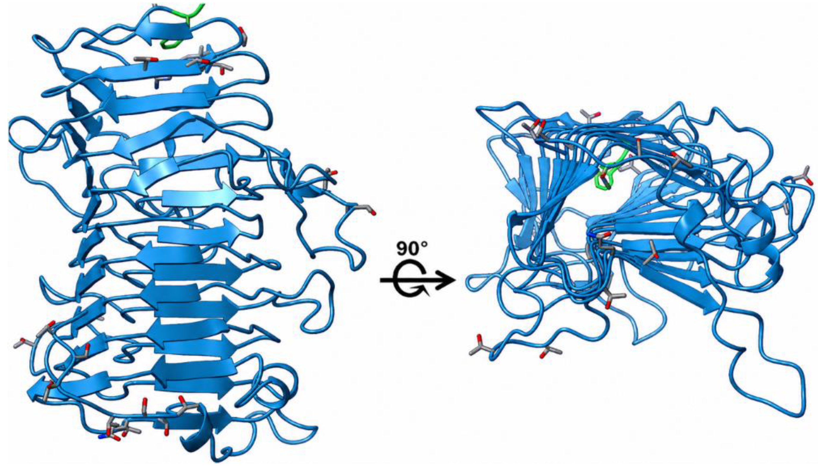
Figure 1. AlphaFold3-predicted structure of PL002-1792. (A) Ribbon representation showing the overall fold of the protein. (B) Structure rotated by 90° to illustrate the elongated β-sheet-rich architecture. The predicted model exhibited a high-confidence pTM score of 0.96 and was dominated by stacked β-strands interconnected by short loop regions.
Preprints 220151 g001
Figure 2. Sequence alignment of PL002-1792 with the experimentally characterised ice-binding protein from F. frigoris (FfIBP). Conserved residues are highlighted in red. Blue and yellow boxes indicate putative TXT-like motifs and glycine-rich regions, respectively. Predicted β-strand regions (βA–βZ) are shown above the alignment. Despite low overall sequence identity, PL002-1792 retains structural features characteristic of bacterial ice-binding and antifreeze proteins, including TXT-like motifs, glycine-rich regions, and a β-strand-rich architecture.
Preprints 220151 g002
Figure 3. Optimisation of recombinant expression of PL002-1792 in E. coli BL21(DE3). SDS-PAGE analysis of recombinant PL002-1792 expression following IPTG induction under different conditions. (A) Induction with 0.5 mM IPTG at 20 °C for 16 h; (B) induction with 0.5 mM IPTG at 20 °C for 48 h. For each condition, total cell lysate, soluble fraction (supernatant), and insoluble fraction (pellet) were analysed to evaluate protein expression and solubility. Lanes 1–4 correspond to soluble fractions, whereas lanes 5–8 correspond to insoluble pellet fractions. The expected molecular mass of PL002-1792 is approximately 41 kDa. PM, broad-range protein molecular weight marker (SMOBIO, Taiwan).
Preprints 220151 g003
Figure 4. Structural validation of refolded PL002-1792. (A) SDS-PAGE analysis of refolded protein under reducing (R) and non-reducing (NR) conditions showing a predominant protein band at approximately 41 kDa. (B) Far-UV circular dichroism spectrum of refolded PL002-1792 displaying a pronounced negative ellipticity minimum at 216–218 nm, indicative of a β-sheet-rich secondary structure. The experimental CD profile was consistent with the β-sheet-rich architecture predicted by AlphaFold3.
Preprints 220151 g004
Figure 5. Ice recrystallization inhibition activity of PL002-1792. (A) Representative polarized light micrographs of ice crystals following annealing at −6 °C for 30 min in the presence of bovine serum albumin (BSA) or PL002-1792 at different protein concentrations. Samples containing PL002-1792 exhibited smaller ice crystals compared with the BSA control. (B) Relative mean grain size (MGS) of ice crystals normalised to the BSA control (1 mg/mL). PL002-1792 reduced ice crystal grain size to approximately 90% of the control, indicating moderate ice recrystallisation inhibition activity.
Preprints 220151 g005
Figure 6. Freeze–thaw tolerance of recombinant E. coli expressing PL002-1792. Cultures harboring pET28a-PL002-1792 (vector_PL002-1792) or the empty pET28a vector were subjected to four successive freeze–thaw cycles. Cell survival was expressed as a percentage of the initial cell density before freezing. PL002-1792-expressing cells consistently exhibited higher survival than the empty-vector control, retaining approximately 67% viability after the fourth freeze–thaw cycle compared with 27% for the control. Values represent the mean ± SD of three independent biological replicates.
Preprints 220151 g006
Table 1. Differential expression and annotation characteristics of the candidate cold-responsive gene PL002-1792 identified from Flavobacterium sp. PL002 under subzero growth conditions.
Gene ID DEG Temperature (°C) DEG Pfam/InterPro hits BLASTp top hits /
% identity
Log2fold change Adjusted
P-value
1792 -20 vs -6 5.61 1.89E-138 LIPL48
(domain, Protein)
87.87%_ hypothetical protein [Flavobacterium muglaense].
-20 vs 15 4.89 1.10E-59
Table 2. Physicochemical properties of the candidate protein PL002-1792 predicted using the ExPASy ProtParam server.
Property Value
Amino acid length 402 aa
Molecular weight 41.3 kDa
Theoretical pI 4.70
Instability index 12.66
Aliphatic index 75.02
GRAVY −0.044
Table 3. Signal peptide prediction of PL002-1792 generated using SignalP 6.0.
Protein type Other Signal Peptide (Sec/SPI) Lipoprotein signal peptide (Sec/SPII) TAT Signal (Tat/SPI) TAT Lipoprotein signal peptide (Tat/SPII) Pilin-like signal peptide (Sec/SPII)
Likelihood 0 0 1 0 0 0
Table 4. Secondary structure composition of PL002-1792 estimated from far-UV circular dichroism spectra using the BeStSel server.
Secondary structure
Content (%)
Antiparallel β-sheet (relaxed) 17.9
Antiparallel β-sheet (left-twisted) 19.8
Parallel β-sheet 0.7
Turn 16.1
Helix 1.6
Other 44.0
Disclaimer/Publisher’s Note: The statements, opinions and data contained in all publications are solely those of the individual author(s) and contributor(s) and not of MDPI and/or the editor(s). MDPI and/or the editor(s) disclaim responsibility for any injury to people or property resulting from any ideas, methods, instructions or products referred to in the content.
Copyright: This open access article is published under a Creative Commons CC BY 4.0 license, which permit the free download, distribution, and reuse, provided that the author and preprint are cited in any reuse.
Prerpints.org logo

Preprints.org is a free preprint server supported by MDPI in Basel, Switzerland.

Subscribe

© 2026 MDPI (Basel, Switzerland) unless otherwise stated

Accessibility

Disclaimer

Terms of Use

Privacy Policy

Privacy Settings