

Submitted:
23 June 2026
Posted:
25 June 2026
You are already at the latest version
Abstract
The liver ranks among the peripheral organs exhibiting the most robust circadian rhythmicity, with glucose homeostasis, lipid metabolism, and bile acid turnover governed by tightly phased diurnal oscillations. Mitochondria are the primary executors of these metabolic programs, their functional output temporally coordinated with the circadian state of the hepatocyte. Accumulating evidence indicates that the mitochondrial network undergoes dynamic remodeling throughout the 24-hour cycle, encompassing oscillatory changes in bioenergetics, fusion–fission balance, and quality control. This clock–mitochondria interplay is bidirectional: core clock components drive rhythmic remodeling via CDK1/MAPK-dependent phosphorylation of DRP1 and the NAD⁺–SIRT1/SIRT3 axis, while retrograde mitochondrial signals -including ROS flux and acetyl-CoA availability-modulate clock amplitude and entrainment. Disruption of this temporal architecture promotes metabolic dysfunction underlying MASLD onset and progression to MASH and HCC. As the clock–mitochondria axis operates upstream of these pathogenic cascades, its chronopharmacological restoration constitutes a mechanistically grounded therapeutic strategy. Interventions including time-restricted feeding, NAD⁺ precursor supplementation, PPAR agonists, and ACC inhibitors converge on clock-regulated mitochondrial pathways and may yield enhanced efficacy when delivered in a circadian-informed manner. This review delineates the molecular architecture of hepatic clock–mitochondria coupling, its breakdown across the MASLD/MASH continuum, and the translational rationale for mitochondria-centered chronotherapy.
Keywords:
circadian clock
; hepatic mitochondria
; mitochondrial dynamics
; NAD+–sirtuin axis
; MASLD
; MASH
; chronotherapy
; chronopharmacology
1. Introduction
The circadian clock constitutes an evolutionarily conserved molecular timing system that aligns physiological processes with the 24-hour light-dark cycle. In mammals, this system is organized hierarchically: a central pacemaker residing in the suprachiasmatic nucleus (SCN) coordinates peripheral oscillators via neuroendocrine and metabolic cues [1,2], whereas the hepatic oscillator is predominantly entrained by feeding-fasting cycles rather than photic signals [4,5]. Multi-omics investigations have established that circadian regulation operates across multiple layers of gene expression, encompassing the transcriptome and metabolome, and governs hepatic glucose homeostasis, lipogenesis, bile acid biosynthesis, and xenobiotic detoxification [7,8].
Mitochondria are indispensable effectors of hepatic metabolic function, and a growing body of evidence demonstrates that they are subject to pervasive circadian regulation. The dynamin-related GTPase DRP1 undergoes rhythmic phosphorylation at Ser-637, driving cyclic transitions between mitochondrial fission and fusion states that are temporally coupled to diurnal oscillations in oxidative phosphorylation (OXPHOS) capacity [20]. Hepatocyte-specific deletion of Bmal1 abolishes circadian cycling of DRP1 phosphorylation, mitochondrial network dynamics, and respiratory chain activity [21]; furthermore, Drp1 expression is itself subject to rhythmic post-transcriptional regulation by CLOCK [22]. Critically, this relationship is bidirectional: pharmacological inhibition of DRP1 lengthens circadian period and attenuates amplitude of core clock oscillations [20], establishing a closed regulatory loop rather than a unidirectional hierarchy.
The pathophysiological relevance of this axis is most apparent in the context of metabolic liver disease. MASLD,redefined by the 2023 multi-society consensus [43], affects up to 38% of the adult population [44,45] and progresses through MASH, fibrosis, and HCC. Mitochondrial dysfunction represents a hallmark feature across all disease stages [46,47], and circadian disruption has been shown to exacerbate hepatic pathology in rodent models and to correlate with disease severity in human cohorts [34,48,49]. Rhythmic oscillation of NAD+ bioavailability and the consequent circadian deacetylation of mitochondrial protein complexes by SIRT1/SIRT3 constitute an additional mechanistic layer within this regulatory axis [58,59].
Despite the mechanistic evidence linking circadian dysregulation to hepatic disease, chronotherapeutic strategies remain profoundly underexplored in hepatology: fewer than 0.16% of registered clinical trials incorporate time-of-day as an experimental variable, and no trial to date has targeted a liver-specific indication [61]. Nevertheless, several agents with established or emerging efficacy in MASH including time-restricted feeding, NAD+ precursors, PPAR agonists, ACC inhibitors, MSDC-0602K, resmetirom, and FGF21-based therapies, exert their effects through rhythmically regulated molecular pathways [60], raising the prospect of chronopharmacological optimization. In this review, we systematically examine the circadian mechanisms governing hepatic mitochondrial function and delineate how their disruption contributes to metabolic liver disease progression.
2. The Biological Clock in Liver
Mammalian timing system is hierarchical. Central pacemaker, SCN, receiving light input through the retinohypothalamic tract, synchronizes peripheral oscillators, such as liver, using neuronal, endocrine, and metabolic outputs [1,2]. However, machinery of circadian clock is the same for all cells – TTFLs producing sustained rhythms of ∼24-hour period [1,3]. In canonical TTFL, CLOCK (or its paralog NPAS2) forms a heterodimer with BMAL1 and binds E-box motifs in Per1, Per2, Cry1, and Cry2 gene promoters. PER and CRY accumulate in the cytoplasm and return to the nucleus once their concentration rises high enough. In the nucleus, PER and CRY inhibit the activity of CLOCK: BMAL1 complex, shutting off their own transcription. Finally, degradation of PER and CRY by F-box protein-dependent proteasome leads to activation of the TTFL and restarting the cycle [3]. Another stabilizing loop is based on competition between two families of nuclear receptors: REV-ERBα/β (NR1D1, NR1D2) and RORα/β/γ. These factors compete for the binding sites within Bmal1 promoter and suppress or activate it, respectively [3]. In addition to functioning as clock components, some of core TTFL constituents participate in cell-cycle regulation and autophagy [3].
The hepatic circadian clock exhibits a distinctive characteristic in that feeding cues, rather than the light–dark cycle, serve as its primary zeitgeber. Experimental studies in nocturnal rodents have demonstrated that restricting food availability to the inactive (light) phase induces an approximately 12-hour phase shift in the liver clock relative to the suprachiasmatic nucleus (SCN), accompanied by a corresponding shift in the peak expression of most clock genes and clock-controlled transcripts [4,5]. Remarkably, even in Cry1−/−;Cry2−/− mice, which lack a functional molecular circadian oscillator, the imposition of a feeding rhythm is sufficient to generate rhythmic patterns of hepatic gene expression, highlighting the dominant role of nutrient timing in regulating liver transcriptional programs [5].
However, compared to the liver, the impact of similar feeding manipulations on circadian rhythmicity in other peripheral organs such as kidney, heart, and lung is relatively minimal, indicating that the liver’s circadian pacemaker is highly responsive to metabolic factors. [4]. Furthermore, studies employing hepatocyte-specific restoration of clock function have revealed that hepatocytes possess the capacity to synchronize their oscillatory activity independently of other peripheral clocks, with feeding schedules further strengthening this intrahepatic synchronization [6]. Collectively, these findings indicate that individual tissues utilize distinct entrainment pathways that connect external stimuli to the core molecular clock, reflecting their specialized physiological and metabolic functions [4,7]
Twenty years of omics studies have resulted in a fairly comprehensive view of how ubiquitous the hepatic clock is. Rhythms have been described at the transcriptomic, cistromic, proteomic, phosphoproteomic, acetylproteomic, metabolomic, and lipidomic levels [7,8]. A few metabolic sensors link the cellular energy status to the clock via positive and negative feedback. The NAD+-dependent deacetylase SIRT1 displays circadian rhythms as a response to NAD+ oscillations due to oscillations in the NAD+ production, NAD+ being produced via NAMPT which regulates NAD+ biosynthesis in a circadian fashion; SIRT1 then deacetylates PER2 and BMAL1 to modulate the period and amplitude of the hepatic clock [9]. AMPK, activated by low ATP/AMP ratio, phosphorylates and degrades CRY1 thus affecting the speed of the clock [10]. The coactivator of mitochondrial gene expression PGC-1α also acts on clock genes [11]. They are not merely the downstream effectors of the clock: they are embedded in regulatory feedback loops ensuring the robustness and metabolic responsiveness of the clock [7,8].
Downstream, the hepatic clock regulates glucose metabolism, lipogenesis, bile acid synthesis, and xenobiotic metabolism. In hepatocytes, Bmal1 deletion results in fasting hypoglycemia and loss of glucose transporter 2 rhythmicity, demonstrating how essential the role of the liver clock is in glucose regulation under post-absorptive conditions [12]. Recent proteomic studies have also demonstrated that disruptions of the circadian system via external factors lead to widespread remodeling of the proteome in the liver, and thus lipid and glucose metabolism is also altered in ways that are more complex than merely the change at the transcript level [13]. As a result of circadian disruption caused by shift work, chronic jet lag, high-fat diet, and knockout of key circadian proteins, the development of a group of diseases occurs including obesity, type 2 diabetes, MASLD/MASH, and HCC [7,8,14].
3. Circadian Control of Mitochondrial Dynamics
The mitochondrion is an active organelle. Its reticulum is continuously remodeled by the actions of fusion and fission, and the dynamics of these processes define the structure and functionality of this organelle. The processes of mitochondrial fusion are mediated by the mitofusin MFN1/2 proteins located in the outer mitochondrial membrane and by the OPA1 protein in the inner mitochondrial membrane. These processes result in the formation of long, tubular mitochondria with high oxidative phosphorylation and ATP production capacity. Mitochondrial fission is mediated by DRP1GTPase, which targets the constrictions. [15,16]. Strong evidence for the circadian regulation of mitochondrial morphology was provided by Schmitt et al., who employed confocal microscopy to demonstrate a robust 24-hour oscillation in mitochondrial network architecture in synchronized human skin fibroblasts [20]. Sixteen hours following synchronization, mitochondria exhibited a highly interconnected and tubular morphology, coinciding with maximal ATP production and respiratory activity. By contrast, 28 hours after synchronization, the network became fragmented and bioenergetic output reached its lowest level [20]. Similar rhythmic changes have been observed in vivo. At circadian time (CT) 0, corresponding to the onset of the subjective rest phase, mitochondria display a predominantly tubular morphology in both mouse hippocampal CA1 neurons and hepatocytes. In contrast, at CT12, marking the beginning of the active phase, the mitochondrial network becomes fragmented. Notably, in mPer1/mPer2 double-knockout mice, mitochondria remain constitutively fragmented throughout the circadian cycle, indicating a critical role for PERIOD proteins in maintaining rhythmic mitochondrial remodeling [20].
Significantly, these cycles do not correlate with cell cycle phases either. Similar patterns of cycling have been detected in post-mitotic cells [20], and also in AraC-treated fibroblasts, proving that circadian modulation of mitochondrial morphology operates independently of cell division [20].
Several independent studies have subsequently reinforced these observations, collectively demonstrating a critical role for the circadian clock in regulating mitochondrial dynamics through multiple molecular mechanisms. Jacobi et al. reported that hepatocyte-specific deletion of Bmal1 abolishes rhythmic mitochondrial remodeling and impairs respiratory chain function in vivo, accompanied by reduced expression of key genes involved in mitochondrial fission and quality control, including Drp1, Fis1, Pink1, and Bnip3 [21]. Building upon these findings, Xu et al. identified a post-transcriptional mechanism through which CLOCK regulates mitochondrial dynamics. In mouse liver, CLOCK interacts with the RNA-binding protein PUF60 to promote Drp1 mRNA degradation. Consequently, ClockΔ19 mutant mice exhibit excessive mitochondrial fragmentation, elevated reactive oxygen species (ROS) production, impaired aerobic metabolism, and a phenotype resembling non-alcoholic fatty liver disease (NAFLD), all of which can be ameliorated by treatment with the DRP1 inhibitor Mdivi-1 [22].
Further evidence for the importance of DRP1-mediated mitochondrial remodeling was provided by Steffen et al., who demonstrated that hepatocyte-specific deletion of Drp1 exacerbates NASH progression and activates the mitochondrial integrated stress response, indicating that appropriate mitochondrial fission is required to maintain hepatic homeostasis and limit disease progression [23]. In addition, recent reviews have consolidated these findings and highlighted that proteins involved in mitochondrial fission and mitophagy exhibit pronounced diurnal oscillations in wild-type mouse liver, whereas these rhythms are markedly attenuated in Bmal1-deficient animals [24,25]. Moreover, reduced Bmal1 expression directly compromises BNIP3-mediated mitophagy, thereby contributing to mitochondrial dysfunction and cellular stress.
Collectively, these studies indicate that the circadian clock regulates both the structural organization and turnover of the mitochondrial network through multiple complementary mechanisms, including transcriptional control of genes involved in mitochondrial fission and mitophagy, post-transcriptional regulation of Drp1 mRNA stability, and post-translational modification of DRP1 activity.
Among these regulatory mechanisms, the post-translational control of DRP1 has been particularly well characterized. Although total DRP1 protein abundance remains relatively constant throughout the day, phosphorylation of DRP1 at Ser637 exhibits a robust circadian rhythm in synchronized fibroblasts and mouse brain tissue [26,27]. Phosphorylation at this residue suppresses DRP1 GTPase activity by disrupting the interaction between its GTPase and GED domains, thereby favoring mitochondrial fusion and the maintenance of an interconnected mitochondrial network [26,27]. Notably, this rhythmic phosphorylation pattern is completely abolished in mPer1/mPer2 double-knockout mice [20].
The phosphorylation status of Ser637 is determined by the balance between protein kinase A (PKA) and calcineurin. PKA phosphorylates and inhibits DRP1, promoting mitochondrial fusion, whereas calcineurin, which is itself subject to circadian regulation, dephosphorylates DRP1 and stimulates mitochondrial fission [28,29]. Consistent with this model, pharmacological inhibition of calcineurin using FK506 eliminates both the circadian rhythm of DRP1 Ser637 phosphorylation and the associated oscillations in mitochondrial network morphology [20]. These findings highlight the central role of post-translational DRP1 regulation in mediating circadian control of mitochondrial dynamics.
4. Circadian Control of Mitochondrial Bioenergetics
Beyond its role in regulating mitochondrial morphology, circadian fission–fusion cycling plays a central role in generating rhythmic hepatic bioenergetics.. Metabolomic analyses of synchronized human U2OS cells have demonstrated that approximately 29% of detected metabolites exhibit circadian oscillations, including tricarboxylic acid (TCA) cycle intermediates, the GSH/GSSG redox couple, and key energy metabolites such as ATP and NAD+/NADH [16]. These findings are consistent with earlier studies in both rodents and humans, highlighting the widespread temporal organization of cellular metabolism [21,22].
Notably, rhythmic ATP production is dependent on an intact circadian clock. Mouse embryonic fibroblasts lacking Per1 and Per2 display persistently low and arrhythmic ATP levels, indicating that ATP oscillations are under clock control [16]. Furthermore, these oscillations originate primarily from oxidative phosphorylation (OXPHOS) rather than glycolysis, as they are abolished by the ATP synthase inhibitor oligomycin but remain unaffected by the glycolytic inhibitor 2-deoxyglucose [16]. Importantly, pharmacological inhibition of DRP1 using P110 or Mdivi-1, as well as genetic deletion of Drp1, eliminates circadian ATP oscillations both in vitro and in vivo, demonstrating that DRP1-dependent mitochondrial fission–fusion dynamics are essential for maintaining rhythmic bioenergetic output [16]. These findings indicate that mitochondrial dynamics are not merely a downstream consequence of circadian regulation but rather a fundamental component of the circadian metabolic program.
The circadian clock regulates mitochondrial dynamics through several well-established metabolic sensors. Pharmacological inhibition of AMPK, SIRT1, or SIRT3 individually abolishes the circadian rhythm of DRP1 Ser637 phosphorylation, positioning these molecules as key intermediates linking the molecular clock to the mitochondrial fission machinery [16]. These sensors are themselves integrated into clock regulatory networks through mechanisms involving CRY phosphorylation and NAD+-dependent deacetylation of both clock proteins and mitochondrial targets [8,23,24].
Importantly, the relationship between the circadian clock and mitochondrial dynamics is bidirectional. Disruption of mitochondrial remodeling through DRP1 inhibition or calcineurin blockade lengthens the circadian period in Bmal1::luciferase reporter fibroblasts, while Drp1-deficient mouse embryonic fibroblasts exhibit dampened oscillations of Bmal1, Per1, and Per2 expression [16]. These observations suggest that while the circadian clock governs mitochondrial morphology through DRP1-dependent mechanisms, mitochondrial dynamics reciprocally reinforce clock function through ATP- and NAD+-dependent signaling pathways involving AMPK and SIRT1.
In the liver, BMAL1 has been shown to coordinate rhythmic mitochondrial remodeling and support metabolic fitness, further emphasizing the importance of the clock–mitochondria axis in hepatic physiology [25]. Collectively, these findings support a model in which reciprocal interactions between circadian regulators and mitochondrial dynamics enable hepatocytes to synchronize cellular bioenergetics with daily metabolic demands and the broader temporal organization of the organism.
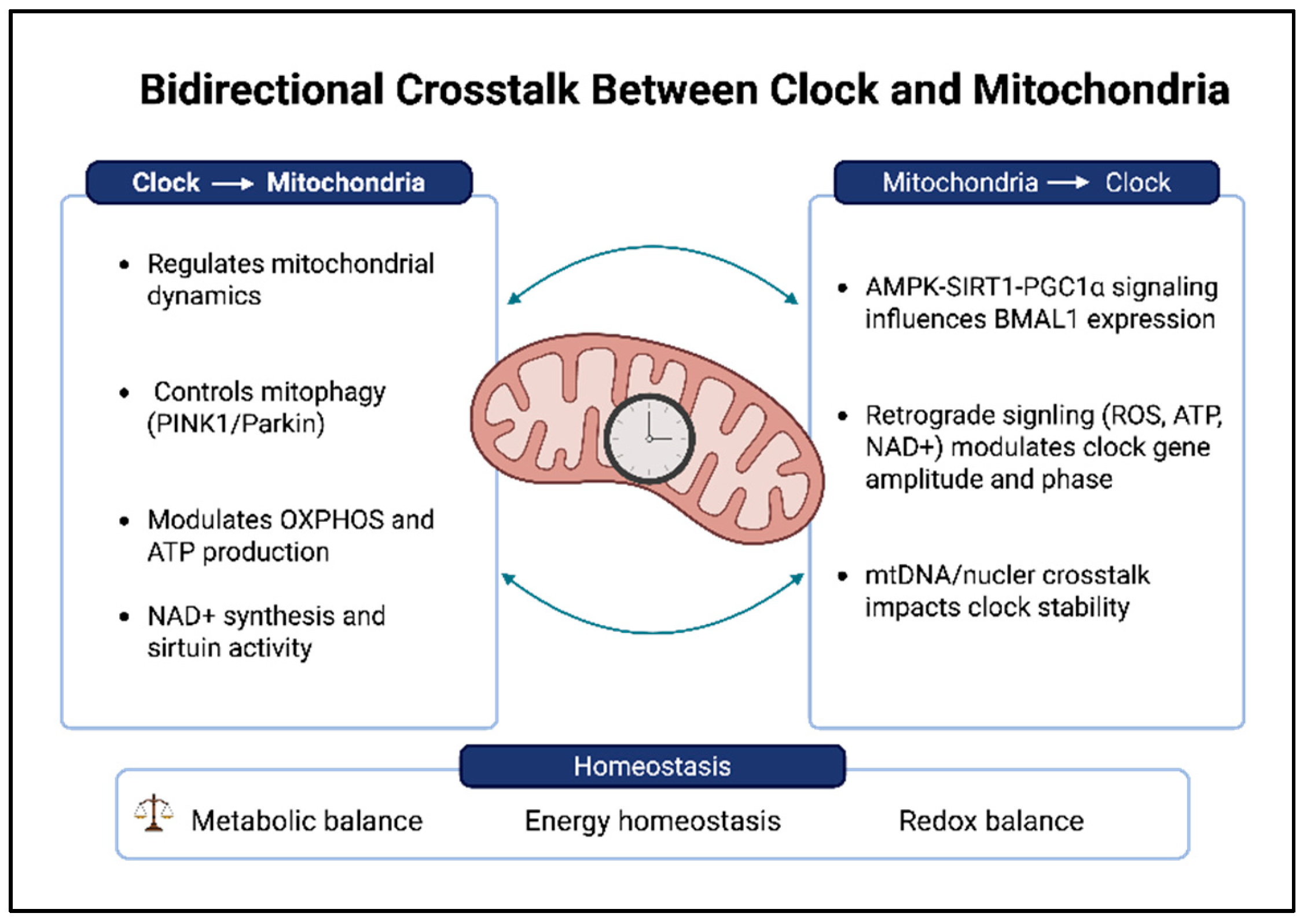
Figure 1.
Schematic Representation of bidirectional crosstalk between the circadian clock circuitry and mitochondria. Created by Biorender.com.
Figure 1.
Schematic Representation of bidirectional crosstalk between the circadian clock circuitry and mitochondria. Created by Biorender.com.
5. Disruption of the Clock–Mitochondria Axis in Metabolic Liver Disease
Given the close functional interplay between the hepatic circadian clock and the mitochondrial network, it is unsurprising that disruption of either system adversely affects liver metabolism.. Over the past decade, substantial evidence from both animal models and human studies has established a strong link between circadian dysregulation and the development of metabolic disorders [7,14,34,35]. In mice, both genetic and environmental disruption of the circadian oscillator have been shown to promote insulin resistance, hepatic steatosis, and hepatocarcinogenesis [36,37,38]. Notably, ClockΔ19 mutant mice spontaneously develop steatosis and hyperinsulinemia and exhibit impaired glucose tolerance even when maintained on a standard diet [37]. Similarly, hepatocyte-specific deletion of Bmal1 disrupts rhythmic gluconeogenesis and exacerbates diet-induced liver injury, highlighting the critical role of the hepatic clock in maintaining metabolic homeostasis [12,33,38].
In humans, genetic variants of CLOCK have been associated with increased susceptibility to NAFLD, whereas polymorphisms in PER and CRY genes have been linked to obesity and type 2 diabetes mellitus [39,40,41]. Furthermore, large-scale observational studies have identified significant associations between hepatic steatosis and behaviors that promote circadian misalignment, including shift work, chronic sleep restriction, and recurrent jet lag [14,34,42]. Importantly, these associations remain significant after adjustment for potential confounding factors such as dietary habits, body mass index, and physical activity, supporting the notion that circadian disruption itself contributes directly to metabolic disease pathogenesis rather than simply reflecting unhealthy lifestyle behaviors [14,34].
In June 2023, an international multisociety Delphi consensus introduced a revised nomenclature for fatty liver disease, replacing non-alcoholic fatty liver disease (NAFLD) with metabolic dysfunction-associated steatotic liver disease (MASLD) and non-alcoholic steatohepatitis (NASH) with metabolic dysfunction-associated steatohepatitis (MASH) [43]. This reclassification was intended to emphasize the underlying metabolic pathophysiology of the disease rather than defining it by the absence of significant alcohol consumption, while also addressing concerns regarding disease-related stigma. Throughout this review, the terms MASLD/MASH and NAFLD/NASH are used interchangeably when discussing earlier studies, as the underlying biological mechanisms remain unchanged.
MASLD is currently the most prevalent chronic liver disease worldwide, affecting approximately 38% of the adult population, with substantially higher prevalence rates among individuals with obesity and type 2 diabetes mellitus [44,45]. Mitochondrial dysfunction represents a hallmark feature throughout the entire disease spectrum. During the early stage of simple steatosis, chronic lipid excess exceeds the β-oxidation capacity of the mitochondrial network, leading to the accumulation of triglycerides, diacylglycerols, and ceramides. In response, mitochondrial mass increases as a compensatory mechanism to maintain respiratory function; however, this adaptation eventually becomes maladaptive, resulting in enhanced electron leakage and excessive production of reactive oxygen species (ROS) [46,47].
Notably, similar metabolic alterations are observed under conditions of circadian disruption. Impairment of the core molecular clock leads to persistent mitochondrial fragmentation and loss of rhythmic ATP production [20]. Likewise, hepatocyte-specific deletion of Bmal1 abolishes rhythmic mitochondrial remodeling, compromises respiratory chain efficiency, and impairs overall metabolic fitness [21,33]. Under physiological conditions, the daily alternation between fused and fragmented mitochondrial states enables hepatocytes to adapt their metabolic activity to feeding–fasting cycles. Consequently, disruption of this rhythmic remodeling promotes a state of chronic bioenergetic imbalance characterized by impaired fatty acid oxidation and increased oxidative stress, both of which are central features of MASLD pathogenesis [15,16,20,46,47]. Consistent with these observations, expression of core clock genes, including BMAL1, PER2, and REV-ERBα, is reduced or phase-shifted in liver biopsies from patients with MASLD, and the degree of circadian disruption correlates with histopathological disease severity [34,48,49].
Progression from MASLD to MASH is accompanied by a further deterioration of mitochondrial function, at which point mitochondrial dysfunction becomes a major driver of hepatic inflammation. In addition to the ultrastructural abnormalities observed in MASLD, including paracrystalline inclusions, loss of cristae integrity, and impaired assembly of electron transport chain complexes, MASH is characterized by reduced respiratory capacity, diminished ATP synthesis, and increased oxidative damage affecting mitochondrial DNA, proteins, and lipids [46,47]. Importantly, several cellular stress-response and immune pathways implicated in MASH progression, including autophagy, mitophagy, the unfolded protein response, and antioxidant defense mechanisms, are themselves under circadian regulation [34,50,51,52].
Once circadian regulation is disrupted, the temporal coordination of these cellular programs is lost, leading to impaired mitochondrial quality control and the transition from tightly regulated adaptive responses to chronic and dysregulated activation. Mitophagy represents a critical component of this process. Indeed, PINK1-, Parkin-, BNIP3-, and FUNDC1-mediated mitophagy pathways are consistently downregulated in both MASLD/MASH liver tissue and high-fat diet-induced murine models, resulting in the accumulation of damaged mitochondria, persistent release of mitochondrial damage-associated molecular patterns (mtDAMPs), and activation of pro-inflammatory and pro-fibrotic signaling pathways [52,53]. Mitochondria-derived DAMPs, including mtDNA, mtRNA, ATP, and cardiolipin, activate Kupffer cells through multiple innate immune pathways, including TLR9, cGAS–STING signaling, and the NLRP3 inflammasome, ultimately promoting sterile hepatic inflammation [46,54,55].
Under physiological conditions, these responses are tightly regulated through circadian control of autophagy and antioxidant defense mechanisms. However, circadian desynchronization disrupts the rhythmicity of these protective pathways, resulting in sustained activation of innate immune signaling, recruitment of hepatic stellate cells, and progressive fibrogenesis [34,46,52]. Consistent with these observations, patients with MASLD/MASH exhibit elevated circulating levels of mtDNA, which correlate positively with the severity of hepatic inflammation and fibrosis [55,56].
A central mediator linking circadian regulation and mitochondrial function is the NAD+–sirtuin axis. Circadian oscillations in NAD+ levels are driven by rhythmic expression of NAMPT [18] and support the activity of both the nuclear deacetylase SIRT1 and the mitochondrial deacetylase SIRT3. Together, these enzymes regulate circadian clock amplitude and maintain mitochondrial metabolic function through the deacetylation of key enzymes involved in energy metabolism [32,57]. During both ageing and MASLD progression, hepatic NAD+ levels progressively decline, resulting in attenuation of NAD+ rhythmicity and impaired SIRT3-dependent regulation of the mitochondrial acetylome [58,59]. Consequently, components of the oxidative phosphorylation (OXPHOS) system, tricarboxylic acid (TCA) cycle enzymes, and proteins involved in long-chain fatty acid oxidation become hyperacetylated. These alterations are associated with reduced respiratory efficiency, accumulation of acyl-carnitines, and increased production of reactive oxygen species (ROS), all of which have been documented in MASH liver tissue [46,47,58].
These events establish a self-perpetuating cycle in which circadian disruption impairs mitochondrial function, while mitochondrial dysfunction further weakens circadian regulation through reduced ATP- and NAD+-dependent signaling to AMPK and SIRT1 [8,20]. This reciprocal deterioration may explain why circadian and metabolic disorders frequently exacerbate one another over time and highlights the NAD+–sirtuin axis as a promising therapeutic target [58,59].
The clock–mitochondria axis also contributes to the broader metabolic complications associated with MASLD. Hepatic insulin resistance, a defining feature of MASLD and a major driver of disease progression, is exacerbated by circadian disruption. Liver-specific deletion of Bmal1 disrupts rhythmic gluconeogenesis and alters glucose homeostasis [12], whereas ClockΔ19 mutant mice develop hyperinsulinemia and glucose intolerance despite maintaining normal feeding behavior [37]. In addition, mitochondrial dysfunction directly contributes to insulin resistance through the accumulation of diacylglycerols and ceramides, which activate PKCε and promote inhibitory serine phosphorylation of the insulin receptor and IRS1, thereby impairing downstream insulin signaling [46,47].
At the advanced stage of disease progression, chronic circadian disruption has also been implicated in the development of hepatocellular carcinoma (HCC). ClockΔ19 mutant mice develop HCC spontaneously [38], while epidemiological studies have reported an increased incidence of HCC among shift workers, even after adjustment for dietary and lifestyle-related confounding factors [14,42]. Furthermore, experimental models of chronic jet lag have demonstrated that circadian disruption alone is sufficient to promote progression from NAFLD to HCC through mechanisms involving bile acid dysregulation and activation of the constitutive androstane receptor (CAR) [38]. Additional mechanisms linking circadian dysfunction to hepatocarcinogenesis include impaired clock-dependent DNA damage repair and sustained mitochondrial ROS production, both of which favor the accumulation of oncogenic mutations [38,46].
Interestingly, serum alanine aminotransferase (ALT), a widely used marker of liver injury, also exhibits a pronounced diurnal rhythm in patients with chronic liver disease, with significantly higher levels observed during the afternoon compared with the morning [60]. This observation further supports the concept that hepatic injury and disease progression are closely linked to the temporal organization of liver metabolism.
Collectively, these findings demonstrate that the clock–mitochondria axis is not merely associated with MASLD progression but actively contributes to disease development across the entire spectrum, from simple steatosis and fibrosis to hepatocellular carcinoma. Consequently, this regulatory network represents a mechanistically relevant and potentially tractable target for therapeutic intervention.
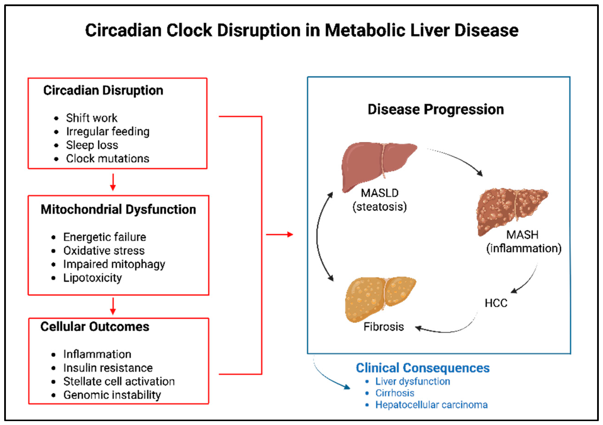
Figure 2.
Schematic Representation of Circadian Clock Disruption in metabolic liver disease.
6. Clock-Regulated Pathways as Targets for Chronotherapy in Metabolic Liver Disease
The identification of circadian regulation as a key determinant of hepatic mitochondrial function, together with evidence demonstrating that this temporal organization is disrupted in MASLD/MASH, provides a strong rationale for chronotherapeutic interventions. Chronotherapy refers to the strategic timing of nutritional, behavioral, or pharmacological interventions to align with endogenous biological rhythms. While this approach has been successfully implemented in several areas of medicine, particularly cardiology and oncology, where it has demonstrated improvements in therapeutic efficacy, its application in hepatology remains largely unexplored [60].
A systematic analysis of more than 200,000 clinical trials registered on ClinicalTrials.gov revealed that only 0.16% incorporated a circadian-informed intervention, with none focusing on liver diseases [61]. This observation raises the possibility that some emerging therapies for MASLD may not achieve their full therapeutic potential because treatment administration is not synchronized with the circadian biology of their molecular targets. Although this hypothesis has not yet been directly evaluated in clinical trials of MASH, growing evidence supporting extensive circadian regulation of hepatic metabolism and mitochondrial function suggests that treatment timing may represent an important, yet largely overlooked, determinant of therapeutic response.
Accordingly, the following sections discuss several mitochondria-targeted or mitochondria-related interventions through the perspective of the clock–mitochondria axis. It should be noted, however, that although the circadian regulation of many of the molecular targets involved is well established, the clinical benefits of optimizing treatment timing remain to be rigorously evaluated in dedicated chronotherapeutic studies.
Time-restricted eating (TRE), referred to as time-restricted feeding (TRF) in animal studies, represents one of the simplest and most accessible chronotherapeutic interventions. This approach involves restricting food intake to a defined daily window of approximately 8–12 hours without reducing total caloric consumption. In murine models of diet-induced obesity, TRF has been shown to prevent hepatic steatosis, restore the rhythmic expression of clock-regulated metabolic genes, remodel hepatic transcriptomic and metabolomic profiles, and improve systemic insulin sensitivity, even when caloric intake is equivalent to that of ad libitum-fed controls [62,63].
At the mechanistic level, TRF restores the amplitude of hepatic NAD+ oscillations, reactivates SIRT1- and SIRT3-dependent signaling pathways, and re-establishes regulation of the mitochondrial acetylome. In addition, TRF promotes the recovery of rhythmic autophagy and mitophagy, facilitating the clearance of dysfunctional mitochondria and reducing inflammation driven by mitochondrial damage-associated molecular patterns (mtDAMPs) [50,63]. Collectively, these effects contribute to improved mitochondrial quality control and metabolic homeostasis.
Clinical studies are increasingly supporting the beneficial effects of TRE in patients with metabolic liver disease. The TREATY-FLD trial demonstrated that TRE significantly reduces hepatic fat content in individuals with NAFLD, with efficacy comparable to that achieved through caloric restriction [64]. More recently, a larger clinical study involving 337 patients with MASLD reported a 25.8% reduction in hepatic steatosis following 16 weeks of TRE, accompanied by significant decreases in body weight and waist circumference comparable to those observed with conventional caloric restriction strategies [65].
Importantly, the timing of food intake relative to the circadian cycle appears to influence therapeutic outcomes. Proof-of-concept studies in men with prediabetes demonstrated that early TRE, in which food consumption is concentrated during the morning and early afternoon, produces greater metabolic benefits than late TRE [66]. These findings are consistent with the circadian regulation of insulin sensitivity and mitochondrial oxidative phosphorylation (OXPHOS), both of which typically peak during the first half of the day. Therefore, TRE provides a compelling behavioral example of how restoring temporal alignment between nutrient intake and endogenous metabolic rhythms can improve mitochondrial function and metabolic health. Nevertheless, long-term adherence, as well as the optimal timing and duration of the feeding window across different populations, remains to be determined.
Hepatic NAD+ levels are reduced in MASLD and decline further with ageing, and this depletion contributes to impaired SIRT3-dependent mitochondrial quality control.
Pharmacological restoration of NAD+ homeostasis has emerged as a promising therapeutic strategy for MASLD, supported by substantial preclinical evidence. Supplementation with NAD+ precursors, particularly nicotinamide riboside (NR) and nicotinamide mononucleotide (NMN), has been shown to restore hepatic NAD+ levels, reactivate SIRT3-dependent pathways, reduce protein hyperacetylation, and improve mitochondrial oxidative phosphorylation (OXPHOS) efficiency and fatty acid β-oxidation in experimental models of MASLD [58,59]. Early clinical studies in obese individuals demonstrated that NR is well tolerated and effectively increases circulating NAD+ concentrations [67].
More disease-specific evidence has subsequently emerged from clinical investigations in patients with NAFLD. A double-blind, placebo-controlled trial evaluating a combination of NR and pterostilbene (NRPT) reported reductions in markers of hepatic inflammation [68]. Similarly, a phase II study investigating a combined metabolic activator formulation containing NR demonstrated an approximately 10% reduction in hepatic fat content in patients with NAFLD [69]. Although these findings are encouraging, the available clinical evidence remains preliminary, and larger randomized studies incorporating histological endpoints are required before NAD+ supplementation can be considered an established therapeutic approach.
An additional consideration is the circadian regulation of NAD+ metabolism itself. Endogenous NAD+ levels exhibit robust daily oscillations and typically reach peak concentrations during the active phase under the control of rhythmically expressed NAMPT [18]. Consequently, the timing of NAD+ precursor administration may influence therapeutic efficacy by altering interactions with endogenous NAD+ rhythms. For example, morning administration may reinforce the physiological active-phase NAD+ peak, whereas evening supplementation could potentially interfere with the declining phase of the rhythm. While this concept remains hypothetical, it represents an important area for future chronopharmacological investigation.
More broadly, many pharmacological approaches currently under development for MASLD target metabolic pathways that are themselves under circadian regulation. Among these, peroxisome proliferator-activated receptors (PPARs) represent key regulators of hepatic lipid metabolism. Specifically, PPARα and PPARδ serve as major transcriptional drivers of fatty acid β-oxidation through the induction of genes encoding carnitine palmitoyltransferase 1 (CPT1), acyl-CoA dehydrogenases, and other enzymes involved in mitochondrial fatty acid catabolism. In addition, PPAR activation promotes mitochondrial biogenesis through PGC-1α signaling [11,60].
Importantly, all three PPAR isoforms exhibit circadian patterns of expression in the liver and are regulated by core clock components, including CLOCK, BMAL1, and REV-ERBα [60,70,71]. Furthermore, genetic deletion of PPARα disrupts the rhythmic expression of clock genes and impairs circadian adaptation to feeding schedules [60,72]. Evidence from preclinical studies also indicates that the biological activity of PPAR agonists is time dependent. For instance, nocturnal administration of bezafibrate induces hepatic FGF21 expression, whereas administration during the morning fails to produce a comparable response [73].
These observations suggest that pharmacological agents currently under investigation for MASH, including the dual PPARα/δ agonist elafibranor and the pan-PPAR agonist lanifibranor, may exhibit enhanced efficacy when administered during periods of increased hepatic fatty acid flux and β-oxidation demand, corresponding to the active phase of the circadian cycle [60,74]. Whether such chronopharmacological optimization translates into clinically meaningful benefits remains to be determined.
Another promising therapeutic strategy involves inhibition of acetyl-CoA carboxylase (ACC), a key enzyme responsible to produce malonyl-CoA, a potent endogenous inhibitor of CPT1. By reducing malonyl-CoA availability, ACC inhibition enhances mitochondrial fatty acid transport and β-oxidation. Firsocostat, a dual ACC1/ACC2 inhibitor, has demonstrated efficacy in reducing hepatic fat accumulation and improving fibrosis-associated biomarkers in clinical trials involving patients with NASH [75].
Acetyl-CoA carboxylase (ACC) is also subject to circadian regulation, with hepatic ACC expression exhibiting daily rhythmicity that is partially controlled by the clock-regulated histone deacetylase HDAC3 [76]. Furthermore, numerous plasma lipid species display circadian oscillations in humans, reflecting the temporal organization of lipid metabolism [77]. Interestingly, one of the majors on-target adverse effects associated with ACC inhibition-elevated plasma triglyceride levels resulting from reduced PPARα activity and enhanced SREBP1c-mediated lipogenesis-also involves pathways that are under circadian control [78]. Consequently, optimizing the timing of ACC inhibitor administration may represent a potential strategy to minimize this adverse effect. However, this possibility remains speculative, as no clinical studies have yet evaluated the impact of dosing time on the efficacy or safety of ACC inhibitors.
Among the therapeutic candidates currently under investigation for MASH, MSDC-0602K possesses one of the most direct mitochondria-targeted mechanisms of action. This compound modulates the mitochondrial pyruvate carrier (MPC), thereby reducing pyruvate transport into the mitochondrial matrix and redirecting glucose-derived carbon away from mitochondrial oxidation toward alternative metabolic pathways. Improved insulin sensitivity represents the primary therapeutic outcome associated with this mechanism [60,79]. Results from a phase IIb clinical trial demonstrated improvements in insulin resistance, hepatic glucose production, and markers of liver injury, without the fluid retention and weight gain commonly associated with full PPARγ agonists [79].
Given that the MPC occupies a critical position at the interface between glycolysis and the tricarboxylic acid (TCA) cycle and considering that several key enzymes involved in pyruvate metabolism exhibit PER1/PER2-dependent circadian oscillations [19], the efficacy of MSDC-0602K may depend on the temporal state of hepatic metabolic pathways. Consequently, chronopharmacological optimization represents a potentially valuable approach for enhancing therapeutic efficacy; however, this concept has not yet been investigated experimentally or clinically.
A major milestone in the treatment of MASH was achieved in March 2024 with the FDA approval of resmetirom (Rezdiffra), the first therapy specifically approved for MASH based on the results of the MAESTRO-NASH phase III clinical trial [80,81]. Resmetirom is a liver-targeted, oral thyroid hormone receptor-β (THR-β) selective agonist that promotes hepatic fatty acid oxidation and reduces lipotoxicity. Activation of THR-β signaling stimulates mitochondrial biogenesis, enhances β-oxidation, and promotes mitophagy, thereby improving mitochondrial function and metabolic homeostasis.
Importantly, the hypothalamic–pituitary–thyroid (HPT) axis is itself under circadian regulation, with thyroid-stimulating hormone (TSH) and thyroid hormone concentrations displaying pronounced diurnal fluctuations [60]. Clinical studies have shown that evening administration of thyroxine in hypothyroid patients results in higher circulating thyroid hormone levels and lower TSH concentrations compared with morning dosing [60]. Therefore, part of the pharmacodynamic variability observed among patients receiving resmetirom may reflect differences in the timing of drug administration. Whether chronopharmacological optimization of THR-β agonist therapy could enhance efficacy or reduce interindividual variability remains an important unanswered question.
Another promising target at the intersection of circadian and mitochondrial biology is fibroblast growth factor 21 (FGF21). FGF21 functions as a hepatokine that is induced by mitochondrial stress, fasting, and oxidative stress, and subsequently promotes fatty acid oxidation, mitochondrial biogenesis through PGC-1α, and systemic metabolic adaptation in peripheral tissues and the central nervous system [11,60,82]. In humans, circulating FGF21 concentrations exhibit a pronounced circadian rhythm characterized by a peak during the early morning hours followed by a marked decline throughout the day. Notably, this rhythmic pattern is attenuated in obesity and metabolic disorders [60,83].
Beyond its peripheral metabolic functions, FGF21 can cross the blood–brain barrier and modulating circadian behavior through actions on β-Klotho-expressing neurons within the suprachiasmatic nucleus (SCN) [84]. This observation highlights a bidirectional communication pathway through which mitochondrial status in peripheral tissues can influence central circadian regulation. The FGF21 analogue pegbelfermin has demonstrated efficacy in reducing hepatic fat accumulation in patients with MASH when administered as a weekly subcutaneous injection. However, this treatment results in sustained elevations of circulating FGF21 that differ substantially from its physiological rhythmic pattern [60,85].
It has been proposed that chronic elevation of FGF21 may promote receptor desensitization and potentially impair adaptive mitochondrial responses [60,83]. Consequently, therapeutic strategies that restore physiological FGF21 rhythmicity rather than continuously elevating hormone levels may provide superior long-term metabolic benefits. Comparative studies directly evaluating these approaches would therefore be of considerable interest.
Evidence supporting chronotherapy also comes from several other medical disciplines. For example, short-acting statins are routinely administered in the evening to coincide with the nocturnal peak in HMG-CoA reductase activity, thereby maximizing their inhibitory effects on cholesterol biosynthesis while also influencing the production of mitochondrial ubiquinone [86]. Similarly, evening administration of certain cytotoxic chemotherapeutic agents has been associated with improved outcomes in pediatric acute lymphoblastic leukemia, partly due to circadian variations in enzymes involved in mitochondrial DNA replication and DNA repair pathways [87,88]. Furthermore, formal guidelines for chronopharmacology-based clinical trials have already been established in the field of hypertension management [89].
Taken together, these examples demonstrate the feasibility and potential clinical value of aligning therapeutic interventions with endogenous biological rhythms. Given the extensive interplay between circadian regulation, mitochondrial function, and hepatic metabolism, mitochondria-centered chronotherapy represents a promising strategy for the management of MASLD/MASH and warrants further evaluation in dedicated clinical studies.
7. Outstanding Questions and Future Perspectives
The clock–mitochondria axis in the liver remains a relatively young field of investigation, and several clinically important questions remain unresolved. Although substantial progress has been made in understanding the molecular interplay between circadian regulation and mitochondrial function, significant gaps in knowledge continue to limit translational applications. One of the most important challenges is the lack of direct evidence demonstrating circadian regulation of mitochondrial dynamics and mitophagy in human hepatocytes. Most mechanistic insights have been generated from murine models and cultured fibroblasts, leaving it unclear whether processes such as DRP1 phosphorylation, mitochondrial network remodeling, and PINK1/BNIP3-dependent mitophagy exhibit robust 24-hour oscillations in the human liver and whether these rhythms are disrupted in MASLD/MASH. Time-of-day-stratified liver biopsy studies could provide critical evidence to establish the translational relevance of these findings.
Another important area of investigation concerns the extent to which clock–mitochondria coupling is conserved between rodents and humans. While rodents are nocturnal and humans are diurnal, resulting in opposite phases of many circadian processes, it remains uncertain whether the underlying molecular mechanisms are fully conserved or whether species-specific regulatory features exist. Comparative chronobiological studies, supported by ex vivo analyses of human liver tissue, will be necessary to clarify the similarities and differences between these systems and to improve the interpretation of findings derived from animal models.
At the same time, the development of reliable biomarkers of circadian function represents a major prerequisite for the implementation of chronotherapy in clinical practice. Without accurate methods for assessing circadian disruption in individual patients, the evaluation of chronotherapeutic interventions in MASH will remain challenging. Potential biomarkers include dim-light melatonin onset (DLMO), diurnal cortisol profiles, time-resolved serum metabolomic and lipidomic signatures, and rhythmic expression of clock genes in peripheral blood cells. However, whether these surrogate markers accurately reflect hepatic circadian activity and whether their normalization predicts therapeutic responses remain important unanswered questions.
The rapid expansion of the therapeutic landscape for MASH further highlights the need to integrate chronobiological considerations into clinical trial design. The recent approval of resmetirom, the positive results obtained with semaglutide in the phase III ESSENCE trial, the continued development of lanifibranor, and the increasing likelihood of combination therapies all raise important questions regarding the optimal timing of treatment administration. Because many of these agents target pathways that are under circadian regulation, their efficacy and safety profiles may vary according to dosing time. Clinical studies that incorporate treatment timing as a predefined variable, ideally in conjunction with chronotype-based stratification, will be essential for determining whether current dosing strategies can be further optimized.
An additional area that remains largely unexplored is the interaction between circadian regulation and the spatial organization of hepatic metabolism. Hepatocytes exhibit considerable metabolic heterogeneity across the hepatic lobule, yet little is known about how mitochondrial function and circadian programs differ among distinct hepatocyte populations. The integration of temporal information into single-cell and spatial omics approaches may provide valuable insights into the existence of region-specific circadian mitochondrial phenotypes and help determine whether MASLD progression is associated with disruption of both temporal and spatial metabolic organization.
Although these challenges represent only a subset of the unanswered questions in the field, they collectively define a research framework that integrates chronobiology, mitochondrial biology, hepatology, and pharmacology. Addressing these knowledge gaps has the potential to substantially
advance our understanding of liver disease pathogenesis and facilitate the development of more effective, mechanism-based therapeutic strategies in the coming years.
8. Conclusions
The hepatic circadian clock and the mitochondrial network function as a highly integrated regulatory system rather than as independent cellular components. Within this framework, the circadian clock orchestrates daily fluctuations in mitochondrial morphology, bioenergetic activity, and quality control mechanisms through processes involving rhythmic DRP1 phosphorylation and regulation of the NAD+–sirtuin axis. In turn, mitochondrial metabolism provides feedback signals that influence both the amplitude and periodicity of the molecular clock. This bidirectional interaction enables hepatocytes to anticipate recurring feeding–fasting cycles and to synchronize mitochondrial energy production with predictable metabolic demands.
Disruption of this coordinated network has profound consequences for liver physiology and contributes to disease progression across the entire spectrum of metabolic liver disorders, ranging from simple steatosis to MASLD/MASH, fibrosis, and ultimately hepatocellular carcinoma (HCC). Increasing evidence indicates that dysfunction of the clock–mitochondria axis is not merely associated with these conditions but actively participates in their pathogenesis through alterations in mitochondrial dynamics, oxidative metabolism, mitophagy, and inflammatory signaling pathways.
Importantly, the therapeutic implications of these findings are becoming increasingly apparent. Several interventions currently used or under development for MASH, including time-restricted eating, NAD+ repletion strategies, PPAR agonists, ACC inhibitors, mitochondrial pyruvate carrier modulators, the THR-β agonist resmetirom, and FGF21-based therapies, target pathways that are subject to significant circadian regulation. Consequently, these approaches offer an opportunity to exploit the temporal organization of hepatic metabolism and mitochondrial function for therapeutic benefit.
Given the pronounced rhythmicity of many pathways targeted by current MASH therapies, the limited consideration of treatment timing may represent an overlooked factor contributing to the modest and sometimes variable efficacy observed in clinical trials of otherwise promising compounds. Future clinical studies should therefore consider incorporating dosing schedules aligned with endogenous mitochondrial and metabolic rhythms, treating treatment timing as a predefined experimental variable rather than a secondary consideration.
More broadly, the integration of chronobiological principles into hepatology-from basic mechanistic research to clinical practice and trial design-has the potential to fundamentally advance our understanding of metabolic liver disease and improve therapeutic outcomes. As knowledge of the clock–mitochondria axis continues to expand, mitochondria-centered chronotherapy may emerge as an important component of precision medicine strategies for the prevention and treatment of MASLD/MASH.
Author Contributions
Conceptualization, G.M, E.M, T.C,V.M.; writing original draft preparation, V.M, E.M, A.S, U.S, ; writing—review and editing, V.M, E.M, A.S, U.S, G.M. T.C, M.S, R.V.; visualization, G.M, T.C,G.S.; supervision, G.M. T.C, G.S.; project administrating, T.C.,; funding acquisition, G.M, T.C. All authors have read and agreed to the published version of the manuscript.
Funding
T.C. is supported by the European Union (EU) within the Italian Ministry of Health [PNRR-MCNT1-2023-12378252].
Institutional Review Board Statement
Not applicable.
Informed Consent Statement
Not applicable.
Data Availability Statement
No new data were created or analyzed in this study. Data sharing is not applicable to this article.:
Acknowledgments
T.C. was a recipient of a fellowship from Associazione Italiana Ricerca sul Cancro (#19548) and of a fellowship from Fondazione Umberto Veronesi.
Conflicts of Interest
The authors declare no conflicts of interest.
Abbreviations
SCN, suprachiasmatic nucleus; TTFL, transcription–translation feedback loop; CLOCK, circadian locomotor output cycles kaput; NPAS2, neuronal PAS domain protein 2; BMAL1, brain and muscle ARNT-like 1; PER, Period; CRY, Cryptochrome; FBXW11/FBXL3/FBXL21, F-box and WD/leucine-rich repeat protein; REV-ERBα/β (NR1D1/NR1D2), nuclear receptor subfamily 1 group D member 1/2; ROR, RAR-related orphan receptor; RRE, ROR–REV-ERB response element; SIRT1/SIRT3, sirtuin 1/3; NAMPT, nicotinamide phosphoribosyltransferase; NAD+, nicotinamide adenine dinucleotide; AMPK, AMP-activated protein kinase; PGC-1α, peroxisome proliferator-activated receptor gamma coactivator 1-alpha; GLUT2, glucose transporter 2; NAFLD, non-alcoholic fatty liver disease; MASH, metabolic dysfunction-associated steatohepatitis; T2D, type 2 diabetes; MFN1/2, mitofusin 1/2; OPA1, optic atrophy 1; OXPHOS, oxidative phosphorylation; DRP1, dynamin-related protein 1; PKA, protein kinase A; CT, circadian time; GSH/GSSG, reduced/oxidized glutathione; ATP, adenosine triphosphate; EV, extracellular vesicle; mitoEV, mitochondrial extracellular vesicle; MVB, multivesicular body; MDV, mitochondria-derived vesicle; PINK1, PTEN-induced kinase 1; TOM20, translocase of outer mitochondrial membrane 20; LC3, microtubule-associated protein light chain 3; MID49/MID51, mitochondrial dynamics protein of 49/51 kDa; MFF, mitochondrial fission factor; STX17, syntaxin-17; Vps35, vacuolar protein sorting 35; MAPL, mitochondrial-anchored protein ligase; SNX9, sorting nexin 9; CD38, cluster of differentiation 38; cADPR, cyclic ADP ribose; VDAC1, voltage-dependent anion channel 1; HSP60/HSP70/mtHSP70, heat shock protein 60/70/mitochondrial 70; SOD2, superoxide dismutase 2; TCA, tricarboxylic acid; mtDNA, mitochondrial DNA; mtRNA, mitochondrial RNA; miRNA, microRNA; ARRDC1, arrestin domain-containing protein 1; mGluR3, metabotropic glutamate receptor 3; MISEV, Minimal Information for Studies of Extracellular Vesicles; TMRM, tetramethylrhodamine methyl ester.
References
- Reppert, S.M.; Weaver, D.R. Coordination of circadian timing in mammals. Nature 2002, 418, 935–941. [Google Scholar] [CrossRef] [PubMed]
- Patke, A.; Young, M.W.; Axelrod, S. Molecular mechanisms and physiological importance of circadian rhythms. Nat. Rev. Mol. Cell Biol. 2020, 21, 67–84. [Google Scholar] [PubMed]
- Takahashi, J.S. Transcriptional architecture of the mammalian circadian clock. Nat. Rev. Genet. 2017, 18, 164–179. [Google Scholar] [PubMed]
- Manella, G.; Sabath, E.; Aviram, R.; Dandavate, V.; Ezagouri, S.; Golik, M.; Adamovich, Y.; Asher, G. The liver-clock coordinates rhythmicity of peripheral tissues in response to feeding. Nat. Metab. 2021, 3, 829–842. [Google Scholar] [CrossRef] [PubMed]
- Vollmers, C.; Gill, S.; DiTacchio, L.; Pulivarthy, S.R.; Le, H.D.; Panda, S. Time of feeding and the intrinsic circadian clock drive rhythms in hepatic gene expression. Proc. Natl. Acad. Sci. USA 2009, 106, 21453–21458. [Google Scholar] [CrossRef] [PubMed]
- Koronowski, K.B.; Kinouchi, K.; Welz, P.S.; Smith, J.G.; Zinna, V.M.; Shi, J.; Samad, M.; Chen, S.; Magnan, C.N.; Kinchen, J.M.; et al. Defining the independence of the liver circadian clock. Cell 2019, 177, 1448–1462.e14. [Google Scholar] [CrossRef] [PubMed]
- Reinke, H.; Asher, G. Crosstalk between metabolism and circadian clocks. Nat. Rev. Mol. Cell Biol. 2019, 20, 227–241. [Google Scholar] [CrossRef] [PubMed]
- Bolshette, N.; Ibrahim, H.; Reinke, H.; Asher, G. Circadian regulation of liver function: from molecular mechanisms to disease pathophysiology. Nat. Rev. Gastroenterol. Hepatol. 2023, 20, 695–707. [Google Scholar] [CrossRef] [PubMed]
- Nakahata, Y.; Kaluzova, M.; Grimaldi, B.; Sahar, S.; Hirayama, J.; Chen, D.; Guarente, L.P.; Sassone-Corsi, P. The NAD+-dependent deacetylase SIRT1 modulates CLOCK-mediated chromatin remodeling and circadian control. Cell 2008, 134, 329–340. [Google Scholar] [CrossRef] [PubMed]
- Lamia, K.A.; Sachdeva, U.M.; DiTacchio, L.; Williams, E.C.; Alvarez, J.G.; Egan, D.F.; Vasquez, D.S.; Juguilon, H.; Panda, S.; Shaw, R.J.; et al. AMPK regulates the circadian clock by cryptochrome phosphorylation and degradation. Science 2009, 326, 437–440. [Google Scholar] [CrossRef] [PubMed]
- Liu, C.; Li, S.; Liu, T.; Borjigin, J.; Lin, J.D. Transcriptional coactivator PGC-1α integrates the mammalian clock and energy metabolism. Nature 2007, 447, 477–481. [Google Scholar] [CrossRef] [PubMed]
- Lamia, K.A.; Storch, K.F.; Weitz, C.J. Physiological significance of a peripheral tissue circadian clock. Proc. Natl. Acad. Sci. USA 2008, 105, 15172–15177. [Google Scholar] [CrossRef] [PubMed]
- Duong, H.A.; Baba, K.; DeBruyne, J.P.; Davidson, A.J.; Ehlen, J.C.; Powell, M.; Tosini, G. Environmental circadian disruption re-writes liver circadian proteomes. Nat. Commun. 2024, 15, 5537. [Google Scholar] [CrossRef] [PubMed]
- Marjot, T.; Ray, D.W.; Williams, F.R.; Tomlinson, J.W.; Armstrong, M.J. Sleep and liver disease: a bidirectional relationship. Lancet Gastroenterol. Hepatol. 2021, 6, 850–863. [Google Scholar] [CrossRef] [PubMed]
- Wai, T.; Langer, T. Mitochondrial dynamics and metabolic regulation. Trends Endocrinol. Metab. 2016, 27, 105–117. [Google Scholar] [CrossRef] [PubMed]
- Westermann, B. Mitochondrial fusion and fission in cell life and death. Nat. Rev. Mol. Cell Biol. 2010, 11, 872–884. [Google Scholar] [CrossRef] [PubMed]
- Isobe, Y.; Hida, H.; Nishino, H. Circadian rhythm of metabolic oscillation in suprachiasmatic nucleus depends on the mitochondrial oxidation state, reflected by cytochrome c oxidase and lactate dehydrogenase. J. Neurosci. Res. 2011, 89, 929–935. [Google Scholar] [CrossRef] [PubMed]
- Peek, C.B.; Affinati, A.H.; Ramsey, K.M.; Kuo, H.Y.; Yu, W.; Sena, L.A.; Ilkayeva, O.; Marcheva, B.; Kobayashi, Y.; Omura, C.; et al. Circadian clock NAD+ cycle drives mitochondrial oxidative metabolism in mice. Science 2013, 342, 1243417. [Google Scholar] [CrossRef] [PubMed]
- Neufeld-Cohen, A.; Robles, M.S.; Aviram, R.; Manella, G.; Adamovich, Y.; Ladeuix, B.; Nir, D.; Rousso-Noori, L.; Kuperman, Y.; Golik, M.; et al. Circadian control of oscillations in mitochondrial rate-limiting enzymes and nutrient utilization by PERIOD proteins. Proc. Natl. Acad. Sci. USA 2016, 113, E1673–E1682. [Google Scholar] [CrossRef] [PubMed]
- Schmitt, K.; Grimm, A.; Dallmann, R.; Oettinghaus, B.; Restelli, L.M.; Witzig, M.; Ishihara, N.; Mihara, K.; Ripperger, J.A.; Albrecht, U.; et al. Circadian control of DRP1 activity regulates mitochondrial dynamics and bioenergetics. Cell Metab. 2018, 27, 657–666.e5. [Google Scholar] [CrossRef] [PubMed]
- Jacobi, D.; Liu, S.; Burkewitz, K.; Kory, N.; Knudsen, N.H.; Alexander, R.K.; Unluturk, U.; Li, X.; Kong, X.; Hyde, A.L.; et al. Hepatic Bmal1 regulates rhythmic mitochondrial dynamics and promotes metabolic fitness. Cell Metab. 2015, 22, 709–720. [Google Scholar] [CrossRef] [PubMed]
- Xu, L.; Lin, J.; Liu, Y.; Hua, B.; Cheng, Q.; Lin, C.; Yan, Z.; Wang, Y.; Sun, N.; Qian, R.; Lu, C. CLOCK regulates Drp1 mRNA stability and mitochondrial homeostasis by interacting with PUF60. Cell Rep. 2022, 39, 110732. [Google Scholar] [CrossRef]
- Steffen, J.; Ngo, J.; Wang, S.P.; Williams, K.; Kramer, H.F.; Ho, G.; Rodriguez, C.; Yekkala, K.; Amuzie, C.; Bialecki, R.; et al. The mitochondrial fission protein Drp1 in liver is required to mitigate NASH and prevents the activation of the mitochondrial ISR. Mol. Metab. 2022, 64, 101566. [Google Scholar] [CrossRef] [PubMed]
- Liu, Q.; Wang, S.; Wang, X. Circadian coordination: understanding the interplay between circadian clock and mitochondria. Anim. Cells Syst. 2024, 28, 228–236. [Google Scholar] [CrossRef]
- Daniels, L.J.; Kay, D.; Marjot, T.; Hodson, L.; Ray, D.W. Circadian regulation of liver metabolism: experimental approaches in human, rodent, and cellular models. Am. J. Physiol. Cell Physiol. 2023, 325, C1158–C1177. [Google Scholar] [CrossRef] [PubMed]
- Chang, C.R.; Blackstone, C. Cyclic AMP-dependent protein kinase phosphorylation of Drp1 regulates its GTPase activity and mitochondrial morphology. J. Biol. Chem. 2007, 282, 21583–21587. [Google Scholar] [CrossRef] [PubMed]
- Cribbs, J.T.; Strack, S. Reversible phosphorylation of Drp1 by cyclic AMP-dependent protein kinase and calcineurin regulates mitochondrial fission and cell death. EMBO Rep. 2007, 8, 939–944. [Google Scholar] [PubMed]
- Huang, C.C.; Ko, M.L.; Ko, G.Y. Calcineurin serves in the circadian output pathway to regulate the daily rhythm of L-type voltage-gated calcium channels in the retina. J. Cell. Biochem. 2012, 113, 911–922. [Google Scholar] [PubMed]
- Cereghetti, G.M.; Stangherlin, A.; Martins de Brito, O.; Chang, C.R.; Blackstone, C.; Bernardi, P.; Scorrano, L. Dephosphorylation by calcineurin regulates translocation of Drp1 to mitochondria. Proc. Natl. Acad. Sci. USA 2008, 105, 15803–15808. [Google Scholar] [CrossRef] [PubMed]
- Eckel-Mahan, K.L.; Patel, V.R.; Mohney, R.P.; Vignola, K.S.; Baldi, P.; Sassone-Corsi, P. Coordination of the transcriptome and metabolome by the circadian clock. Proc. Natl. Acad. Sci. USA 2012, 109, 5541–5546. [Google Scholar] [CrossRef] [PubMed]
- Dallmann, R.; Viola, A.U.; Tarokh, L.; Cajochen, C.; Brown, S.A. The human circadian metabolome. Proc. Natl. Acad. Sci. USA 2012, 109, 2625–2629. [Google Scholar] [CrossRef] [PubMed]
- Masri, S.; Patel, V.R.; Eckel-Mahan, K.L.; Peleg, S.; Forne, I.; Ladurner, A.G.; Baldi, P.; Imhof, A.; Sassone-Corsi, P. Circadian acetylome reveals regulation of mitochondrial metabolic pathways. Proc. Natl. Acad. Sci. USA 2013, 110, 3339–3344. [Google Scholar] [CrossRef] [PubMed]
- Pellegrino, R.M.; Pellegrini, M.; Cossignani, L.; Tarquini, F.; Gambelli, A.M.; Floridi, C.; Conti, M.V.; Lopomo, A.; Renzi, S.; Marini, E.; et al. Metabolic profile and lipid metabolism phenotype in mice with conditional deletion of hepatic BMAL1. Int. J. Mol. Sci. 2024, 25, 6070. [Google Scholar] [CrossRef]
- Mukherji, A.; Bailey, S.M.; Staels, B.; Baumert, T.F. The circadian clock and liver function in health and disease. J. Hepatol. 2019, 71, 200–211. [Google Scholar] [CrossRef] [PubMed]
- Assis, V.; Postnov, A.; Oster, H. The role of the circadian clock in the development, progression, and treatment of non-alcoholic fatty liver disease. Acta Physiol. 2023, 237, e13915. [Google Scholar]
- Mukherji, A.; Dachraoui, M.; Baumert, T.F. Perturbation of the circadian clock and pathogenesis of NAFLD. Metabolism 2020, 111S, 154337. [Google Scholar] [CrossRef] [PubMed]
- Turek, F.W.; Joshu, C.; Kohsaka, A.; Lin, E.; Ivanova, G.; McDearmon, E.; Laposky, A.; Losee-Olson, S.; Easton, A.; Jensen, D.R.; et al. Obesity and metabolic syndrome in circadian Clock mutant mice. Science 2005, 308, 1043–1045. [Google Scholar] [CrossRef] [PubMed]
- Kettner, N.M.; Voicu, H.; Finegold, M.J.; Coarfa, C.; Sreekumar, A.; Putluri, N.; Katchy, C.A.; Lee, C.; Moore, D.D.; Fu, L. Circadian homeostasis of liver metabolism suppresses hepatocarcinogenesis. Cancer Cell 2016, 30, 909–924. [Google Scholar] [CrossRef] [PubMed]
- Sookoian, S.; Castano, G.; Gemma, C.; Gianotti, T.F.; Pirola, C.J. Common genetic variations in CLOCK transcription factor are associated with nonalcoholic fatty liver disease. World J. Gastroenterol. 2007, 13, 4242–4248. [Google Scholar] [PubMed]
- Scott, E.M.; Carter, A.M.; Grant, P.J. Association between polymorphisms in the Clock gene, obesity and the metabolic syndrome in man. Int. J. Obes. 2008, 32, 658–662. [Google Scholar]
- Dashti, H.S.; Smith, C.E.; Lee, Y.C.; Parnell, L.D.; Lai, C.Q.; Arnett, D.K.; Ordovás, J.M.; Garaulet, M. CRY1 circadian gene variant interacts with carbohydrate intake for insulin resistance in two independent populations. Chronobiol. Int. 2014, 31, 660–667. [Google Scholar] [PubMed]
- Zhang, S.; Wang, Y.; Wang, Z.; Wang, H.; Xue, C.; Li, Q.; Guan, W.; Yuan, J. Rotating night shift work and non-alcoholic fatty liver disease among steelworkers in China: a cross-sectional survey. Occup. Environ. Med. 2020, 77, 333–339. [Google Scholar] [PubMed]
- Rinella, M.E.; Lazarus, J.V.; Ratziu, V.; Francque, S.M.; Sanyal, A.J.; Kanwal, F.; Romero, D.; Abdelmalek, M.F.; Anstee, Q.M.; Arab, J.P.; et al. A multisociety Delphi consensus statement on new fatty liver disease nomenclature. Hepatology 2023, 78, 1966–1986. [Google Scholar] [CrossRef] [PubMed]
- Younossi, Z.M.; Golabi, P.; Paik, J.M.; Henry, A.; Van Dongen, C.; Henry, L. The global epidemiology of nonalcoholic fatty liver disease (NAFLD) and nonalcoholic steatohepatitis (NASH): a systematic review. Hepatology 2023, 77, 1335–1347. [Google Scholar] [CrossRef] [PubMed]
- Wong, V.W.; Ekstedt, M.; Wong, G.L.; Hagström, H. Changing epidemiology, global trends and implications for outcomes of NAFLD. J. Hepatol. 2023, 79, 842–852. [Google Scholar] [CrossRef] [PubMed]
- Mansouri, A.; Gattolliat, C.H.; Asselah, T. Mitochondrial dysfunction and signaling in chronic liver diseases. Gastroenterology 2018, 155, 629–647. [Google Scholar] [CrossRef] [PubMed]
- Begriche, K.; Massart, J.; Robin, M.A.; Bonnet, F.; Fromenty, B. Mitochondrial adaptations and dysfunctions in nonalcoholic fatty liver disease. Hepatology 2013, 58, 1497–1507. [Google Scholar] [CrossRef] [PubMed]
- Ando, H.; Takamura, T.; Matsuzawa-Nagata, N.; Shima, K.R.; Eto, T.; Misu, H.; Shiramoto, M.; Tsuru, T.; Irie, S.; Fujimura, A.; et al. Clock gene expression in peripheral leucocytes of patients with type 2 diabetes. Diabetologia 2009, 52, 329–335. [Google Scholar] [PubMed]
- Vinciguerra, M.; Tevy, M.F.; Mazzoccoli, G. A ticking clock links metabolic pathways and organ systems function in health and disease. Clin. Exp. Med. 2014, 14, 133–140. [Google Scholar] [PubMed]
- Ma, D.; Panda, S.; Lin, J.D. Temporal orchestration of circadian autophagy rhythm by C/EBPβ. EMBO J. 2011, 30, 4642–4651. [Google Scholar] [CrossRef] [PubMed]
- Pekovic-Vaughan, V.; Gibbs, J.; Yoshitane, H.; Yang, N.; Pathiranage, D.; Guo, B.; Sagami, A.; Taguchi, K.; Bechtold, D.; Loudon, A.; et al. The circadian clock regulates rhythmic activation of the NRF2/glutathione-mediated antioxidant defense pathway to modulate pulmonary fibrosis. Genes Dev. 2014, 28, 548–560. [Google Scholar] [CrossRef] [PubMed]
- Liang, Y.; Yang, B.; Jiang, X.; Wang, J. Liver cell mitophagy in metabolic dysfunction-associated steatotic liver disease and liver fibrosis. Antioxidants 2024, 13, 729. [Google Scholar] [CrossRef] [PubMed]
- Liu, S.; Wang, L.; Zhu, L.; Zhao, T.; Han, P.; Yan, F.; Wang, X.; Li, C.; Wang, Z.; Yang, B. Mechanism and regulation of mitophagy in liver diseases: a review. Front. Cell Dev. Biol. 2025, 13, 1614940. [Google Scholar] [CrossRef] [PubMed]
- Garcia-Martinez, I.; Santoro, N.; Chen, Y.; Hoque, R.; Ouyang, X.; Caprio, S.; Shlomchik, M.J.; Coffman, R.L.; Candia, A.; Mehal, W.Z. Hepatocyte mitochondrial DNA drives nonalcoholic steatohepatitis by activation of TLR9. J. Clin. Invest. 2016, 126, 859–864. [Google Scholar] [CrossRef] [PubMed]
- An, P.; Wei, L.L.; Zhao, S.; Sverdlov, A.; Vinciguerra, M. Hepatocyte mitochondria-derived danger signals in nonalcoholic fatty liver disease. Front. Cell Dev. Biol. 2021, 9, 642576. [Google Scholar] [PubMed]
- Pirola, C.J.; Gianotti, T.F.; Burgueño, A.L.; Rey-Funes, M.; Loidl, C.F.; Mallardi, P.; Martino, J.S.; Castaño, G.O.; Sookoian, S. Epigenetic modification of liver mitochondrial DNA is associated with histological severity of nonalcoholic fatty liver disease. Gut 2013, 62, 1356–1363. [Google Scholar] [PubMed]
- Asher, G.; Gatfield, D.; Stratmann, M.; Reinke, H.; Dibner, C.; Kreppel, F.; Mostoslavsky, R.; Alt, F.W.; Schibler, U. SIRT1 regulates circadian clock gene expression through PER2 deacetylation. Cell 2008, 134, 317–328. [Google Scholar] [CrossRef] [PubMed]
- Cantó, C.; Menzies, K.J.; Auwerx, J. NAD+ metabolism and the control of energy homeostasis: a balancing act between mitochondria and the nucleus. Cell Metab. 2015, 22, 31–53. [Google Scholar] [CrossRef] [PubMed]
- Yoshino, J.; Baur, J.A.; Imai, S.I. NAD+ intermediates: the biology and therapeutic potential of NMN and NR. Cell Metab. 2018, 27, 513–528. [Google Scholar] [CrossRef] [PubMed]
- Marjot, T.; Ray, D.W.; Tomlinson, J.W. Is it time for chronopharmacology in NASH? J. Hepatol. 2022, 76, 1215–1224. [Google Scholar]
- Selfridge, J.M.; Gotoh, T.; Schiffhauer, S.; Liu, J.; Stauffer, P.E.; Li, A.; Capelluto, D.G.S.; Finkielstein, C.V. Chronotherapy: intuitive, sound, foundedbut not broadly applied. Drugs 2016, 76, 1507–1521. [Google Scholar] [CrossRef] [PubMed]
- Chaix, A.; Zarrinpar, A.; Miu, P.; Panda, S. Time-restricted feeding is a preventative and therapeutic intervention against diverse nutritional challenges. Cell Metab. 2014, 20, 991–1005. [Google Scholar] [CrossRef] [PubMed]
- Hatori, M.; Vollmers, C.; Zarrinpar, A.; DiTacchio, L.; Bushong, E.A.; Gill, S.; Leblanc, M.; Chaix, A.; Joens, M.; Fitzpatrick, J.A.; et al. Time-restricted feeding without reducing caloric intake prevents metabolic diseases in mice fed a high-fat diet. Cell Metab. 2012, 15, 848–860. [Google Scholar] [PubMed]
- Wei, X.; Lin, B.; Huang, Y.; Yang, S.; Huang, C.; Shi, L.; Liu, D.; Zhang, P.; Lin, J.; Xu, B.; et al. Effects of time-restricted eating on nonalcoholic fatty liver disease: the TREATY-FLD randomized clinical trial. JAMA Netw. Open 2023, 6, e233513. [Google Scholar] [PubMed]
- Kim, M.S.; Choi, J.; Park, J.; Lee, J.; Kang, S.H.; Cho, Y.M.; Park, J.H.; Han, K.; Jung, C.H.; Kim, J.H.; et al. Efficacy and safety of time-restricted eating in metabolic dysfunction-associated steatotic liver disease: a randomized controlled trial. J. Hepatol. 2025, 83, 1115–1126. [Google Scholar]
- Sutton, E.F.; Beyl, R.; Early, K.S.; Cefalu, W.T.; Ravussin, E.; Peterson, C.M. Early time-restricted feeding improves insulin sensitivity, blood pressure, and oxidative stress even without weight loss in men with prediabetes. Cell Metab. 2018, 27, 1212–1221. [Google Scholar] [CrossRef] [PubMed]
- Dollerup, O.L.; Christensen, B.; Svart, M.; Schmidt, M.S.; Sulek, K.; Ringgaard, S.; Stødkilde-Jørgensen, H.; Møller, N.; Brenner, C.; Treebak, J.T.; et al. A randomized placebo-controlled clinical trial of nicotinamide riboside in obese men: safety, insulin-sensitivity, and lipid-mobilizing effects. Am. J. Clin. Nutr. 2018, 108, 343–353. [Google Scholar] [CrossRef] [PubMed]
- Dellinger, R.W.; Holmes, H.E.; Hu-Seliger, T.; Butt, R.W.; Harrison, S.A.; Mozaffarian, D.; Chen, O.; Guarente, L. Nicotinamide riboside and pterostilbene reduces markers of hepatic inflammation in NAFLD: a double-blind, placebo-controlled clinical trial. Hepatology 2023, 78, 863–877. [Google Scholar] [PubMed]
- Yang, H.; Mayneris-Perxachs, J.; Boqué, N.; et al. Combined Metabolic Activators Decrease Liver Steatosis by Activating Mitochondrial Metabolism in Hamsters Fed with a High-Fat Diet. Biomedicines 2021, 9, 1440. [Google Scholar] [CrossRef] [PubMed]
- Yang, X.; Downes, M.; Yu, R.T.; Bookout, A.L.; He, W.; Straume, M.; Mangelsdorf, D.J.; Evans, R.M. Nuclear receptor expression links the circadian clock to metabolism. Cell 2006, 126, 801–810. [Google Scholar] [CrossRef] [PubMed]
- Lemberger, T.; Saladin, R.; Vázquez, M.; Assimacopoulos, F.; Staels, B.; Desvergne, B.; Wahli, W.; Auwerx, J. Expression of the peroxisome proliferator-activated receptor α gene is stimulated by stress and follows a diurnal rhythm. J. Biol. Chem. 1996, 271, 1764–1769. [Google Scholar] [CrossRef] [PubMed]
- Canaple, L.; Rambaud, J.; Dkhissi-Benyahya, O.; Rayet, B.; Tan, N.S.; Michalik, L.; Delaunay, F.; Wahli, W.; Laudet, V. Reciprocal regulation of brain and muscle Arnt-like protein 1 and peroxisome proliferator-activated receptor α defines a novel positive feedback loop in the rodent liver circadian clock. Mol. Endocrinol. 2006, 20, 1715–1727. [Google Scholar] [CrossRef] [PubMed]
- Oishi, K.; Uchida, D.; Ishida, N. Circadian expression of FGF21 is induced by PPARα activation in the mouse liver. FEBS Lett. 2008, 582, 3639–3642. [Google Scholar] [CrossRef] [PubMed]
- Francque, S.M.; Bedossa, P.; Ratziu, V.; Anstee, Q.M.; Bugianesi, E.; Sanyal, A.J.; Loomba, R.; Harrison, S.A.; Balabanska, R.; Mateva, L.; et al. A randomized, controlled trial of the pan-PPAR agonist lanifibranor in NASH. N. Engl. J. Med. 2021, 385, 1547–1558. [Google Scholar] [CrossRef] [PubMed]
- Loomba, R.; Kayali, Z.; Noureddin, M.; Ruane, P.; Lawitz, E.J.; Bennett, M.; Wang, L.; Harting, E.; Tarrant, J.M.; McColgan, B.J.; et al. GS-0976 reduces hepatic steatosis and fibrosis markers in patients with nonalcoholic fatty liver disease. Gastroenterology 2018, 155, 1463–1473. [Google Scholar] [CrossRef] [PubMed]
- Feng, D.; Liu, T.; Sun, Z.; Bugge, A.; Mullican, S.E.; Alenghat, T.; Liu, X.S.; Lazar, M.A. A circadian rhythm orchestrated by histone deacetylase 3 controls hepatic lipid metabolism. Science 2011, 331, 1315–1319. [Google Scholar] [CrossRef] [PubMed]
- Chua, E.C.; Shui, G.; Lee, I.T.; Lau, P.; Tan, L.C.; Yeo, S.C.; Lam, B.D.; Bulchand, S.; Summers, S.A.; Puvanendran, K.; et al. Extensive diversity in circadian regulation of plasma lipids and evidence for different circadian metabolic phenotypes in humans. Proc. Natl. Acad. Sci. USA 2013, 110, 14468–14473. [Google Scholar] [CrossRef] [PubMed]
- Kim, C.W.; Addy, C.; Kusunoki, J.; Anderson, N.N.; Deja, S.; Fu, X.; Burgess, S.C.; Li, C.; Ruddy, M.; Chakravarthy, M.; et al. Acetyl CoA carboxylase inhibition reduces hepatic steatosis but elevates plasma triglycerides in mice and humans: a bedside to bench investigation. Cell Metab. 2017, 26, 394–406. [Google Scholar] [CrossRef] [PubMed]
- Harrison, S.A.; Alkhouri, N.; Davison, B.A.; Sanyal, A.; Edwards, C.; Colca, J.R.; Lee, B.H.; Loomba, R.; Cusi, K.; Kolterman, O.; et al. Insulin sensitizer MSDC-0602K in non-alcoholic steatohepatitis: a randomized, double-blind, placebo-controlled phase IIb study. J. Hepatol. 2020, 72, 613–626. [Google Scholar] [PubMed]
- Harrison, S.A.; Bedossa, P.; Guy, C.D.; Schattenberg, J.M.; Loomba, R.; Taub, R.; Labriola, D.; Moussa, S.E.; Neff, G.W.; Rinella, M.E.; et al. A phase 3, randomized, controlled trial of resmetirom in NASH with liver fibrosis. N. Engl. J. Med. 2024, 390, 497–509. [Google Scholar] [CrossRef] [PubMed]
- Keam, S.J. Resmetirom: first approval. Drugs 2024, 84, 729–735. [Google Scholar] [CrossRef] [PubMed]
- Fisher, F.M.; Maratos-Flier, E. Understanding the physiology of FGF21. Annu. Rev. Physiol. 2016, 78, 223–241. [Google Scholar] [CrossRef] [PubMed]
- Yu, H.; Xia, F.; Lam, K.S.; Wang, Y.; Bao, Y.; Zhang, J.; Gu, Y.; Zhou, P.; Lu, J.; Jia, W.; et al. Circadian rhythm of circulating fibroblast growth factor 21 is related to diurnal changes in fatty acids in humans. Clin. Chem. 2011, 57, 691–700. [Google Scholar] [CrossRef] [PubMed]
- Bookout, A.L.; de Groot, M.H.; Owen, B.M.; Lee, S.; Gautron, L.; Lawrence, H.L.; Ding, X.; Elmquist, J.K.; Takahashi, J.S.; Mangelsdorf, D.J.; et al. FGF21 regulates metabolism and circadian behavior by acting on the nervous system. Nat. Med. 2013, 19, 1147–1152. [Google Scholar] [CrossRef] [PubMed]
- Sanyal, A.; Charles, E.D.; Neuschwander-Tetri, B.A.; Loomba, R.; Harrison, S.A.; Abdelmalek, M.F.; Lawitz, E.J.; Halegoua-DeMarzio, D.; Kundu, S.; Noviello, S.; et al. Pegbelfermin (BMS-986036), a PEGylated fibroblast growth factor 21 analogue, in patients with non-alcoholic steatohepatitis: a randomised, double-blind, placebo-controlled, phase 2a trial. Lancet 2018, 392, 2705–2717. [Google Scholar] [PubMed]
- Lemmer, B.; Scheidel, B.; Behne, S. Chronopharmacokinetics and chronopharmacodynamics of cardiovascular active drugs: propranolol, organic nitrates, nifedipine. Ann. N. Y. Acad. Sci. 1991, 618, 166–181. [Google Scholar] [PubMed]
- Rivard, G.E.; Infante-Rivard, C.; Hoyoux, C.; Champagne, J. Maintenance chemotherapy for childhood acute lymphoblastic leukaemia: better in the evening. Lancet 1985, 326, 1264–1266. [Google Scholar] [CrossRef]
- Schmiegelow, K.; Glomstein, A.; Kristinsson, J.; Salmi, T.; Schrøder, H.; Björk, O. Impact of morning versus evening schedule for oral methotrexate and 6-mercaptopurine on relapse risk for children with acute lymphoblastic leukemia. J. Pediatr. Hematol. Oncol. 1997, 19, 102–109. [Google Scholar] [CrossRef] [PubMed]
- Hermida, R.C.; Smolensky, M.H.; Balan, H.; Castriotta, R.J.; Crespo, J.J.; Dagan, Y.; El-Toukhy, S.; Fernández, J.R.; FitzGerald, G.A.; Fuchs, F.D.; et al. Guidelines for the design and conduct of human clinical trials on ingestion-time differences—chronopharmacology and chronotherapy—of hypertension medications. Chronobiol. Int. 2021, 38, 1–26. [Google Scholar] [CrossRef] [PubMed]
- Sanyal, A.J.; Newsome, P.N.; Kliers, I.; Østergaard, L.H.; Long, M.T.; Kjær, M.S.; Cali, A.M.G.; Bugianesi, E.; Rinella, M.E.; Roden, M.; Ratziu, V. for the ESSENCE Study Group. Phase 3 trial of semaglutide in metabolic dysfunction-associated steatohepatitis. N. Engl. J. Med. 2025, 392, 2089–2099. [Google Scholar] [CrossRef] [PubMed]
Disclaimer/Publisher’s Note: The statements, opinions and data contained in all publications are solely those of the individual author(s) and contributor(s) and not of MDPI and/or the editor(s). MDPI and/or the editor(s) disclaim responsibility for any injury to people or property resulting from any ideas, methods, instructions or products referred to in the content. |
© 2026 by the authors. Licensee MDPI, Basel, Switzerland. This article is an open access article distributed under the terms and conditions of the Creative Commons Attribution (CC BY) license (http://creativecommons.org/licenses/by/4.0/).
Copyright: This open access article is published under a Creative Commons CC BY 4.0 license, which permit the free download, distribution, and reuse, provided that the author and preprint are cited in any reuse.


