

Submitted:
22 June 2026
Posted:
24 June 2026
You are already at the latest version
Abstract
The metal oxide nanoparticles have been the subject of intense research interest because of their remarkable physicochemical properties such as high surface area, particle size tunability, outstanding chemical stability, optical activity, catalytic efficiency, and antimicrobial behavior. These properties make them very useful in environmental, biomedical, energy, sensing, agricultural and industrial applications. The synthesis method is important to control the morphology, crystallinity, surface charge, band gap and overall performance of metal oxide nanoparticles. They have been prepared by various physical, chemical and biological means such as solgel, co-precipitation, hydro/solvothermal, microwave assisted, sonochemical, combustion and green synthesis. Of these, the green synthesis is gaining more interest as it employs plant extract, microorganisms, and other biological materials as reducing agents, stabilizing agents and capping agents that make the process more eco-friendly and cost-effective.Recent advancements in the synthesis and application of metal oxide nanoparticles are discussed. It emphasizes major synthesis routes, main factors that influence the formation of nanoparticles, characterization techniques and structure–property relationships. A special focus is on the influence of synthesis parameters like type of precursor, pH, temperature, reaction time, solvents and capping agents on the properties of nanoparticles. In addition, the uses of metal oxide nanoparticles in photocatalysis, wastewater treatment, antimicrobial activity, drug delivery, biosensing, energy storage, gas sensing, and agriculture are also included. Finally, present challenges, toxicity issues, problems of large-scale production, and future research directions are discussed, to support the practical and sustainable use of metal oxide nanoparticles.
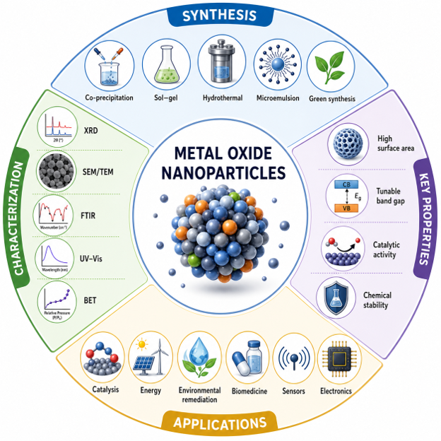
Keywords:
metal oxide nanoparticles
; synthesis methods
; green synthesis
; photocatalysis
; antimicrobial activity
; wastewater treatment
; biomedical applications
; sensors
1. Introduction
Metal oxide nanoparticles are among the most widely studied nanomaterials because of their extraordinary structural, chemical, optical, electrical, magnetic and biological properties. These materials are typically made up of metal cations bound to oxygen anions, and the particle sizes are in the nanometer range, typically 1-100 nm. Scaled down to this size, the properties of metal oxides are very different from those of bulk materials. The decrease in particle size results in a large surface-area-to-volume ratio, an increase in surface reactivity, quantum size effects, enhanced catalytic activity and increased interaction with biological and environmental systems. Due to all these properties, metal oxide nanoparticles are important materials in the present science and technology [1].
Danish, Mir Sayed Shah, et al. stated that metal oxide nanoparticles like zinc oxide, titanium dioxide, iron oxide, copper oxide, magnesium oxide, nickel oxide, cerium oxide, tin oxide, aluminium oxide, and manganese oxide have been of interest in recent years. They are related to the numerous applications where they are used for photocatalysis, environmental remediation, wastewater treatment, antimicrobial systems, drug delivery, biosensing, gas sensing, energy storage systems, solar cells, agriculture and industrial catalysis. The use of nanoparticles as photocatalytic degradation of organic pollutants with materials like titanium dioxide and zinc oxide nanoparticles, as well as magnetic separation, biomedical imaging and targeted drug delivery with iron oxide nanoparticles, are examples of possible applications. Copper oxide and magnesium oxide nanoparticles are recognized for their outstanding antimicrobial activity, while cerium oxide nanoparticles are prized for their antioxidant and catalytic properties [2].
Olawade, David B., et al. stated that the rising interest in metal oxide nanoparticles is also connected to the rising environmental and health issues. The need for advanced, efficient and sustainable materials is great due to rapid industrialization, population growth, water pollution, microbial resistance, energy demand and agricultural stress. Metal oxide nanoparticles are promising solutions as they can be tailored to have specific size, shape, surface chemistry, and functional properties. They have high surface activity, are able to interact strongly with pollutants, microorganisms, gases, biomolecules and light. Hence, they are applied for the degradation of pollutants, the removal of heavy metal, inhibition of bacteria, cancer therapy, sensor development and energy conversion [3].
Synthesis of metal oxide nanoparticles is one of the most crucial aspects. Their size, morphology, crystallinity, surface charge, porosity, band gap, stability and biological response are strongly dependent on the synthesis method. A number of physical, chemical and biological methods have been developed for the preparation of these nanoparticles. High purity nanoparticles can be generated through physical means like laser ablation, vapour deposition and mechanical milling, but can be costly in terms of equipment and energy. Chemical methods such as sol–gel, co-precipitation, hydrothermal, solvothermal, sonochemical, microwave-assisted, combustion, and microemulsion methods are widely used to achieve a better control of particle size and morphology[4]. On the other hand, toxic solvents, harsh reaction conditions and harmful reducing and stabilizing agents are used in some chemical routes [5].
Suppiah, Durga Devi, et al. stated that to address such limitations green synthesis has come across as an eco-friendly and cost effective approach. In green synthesis, natural reducing, stabilizing and capping agents such as plant extracts, bacteria, fungi, algae and biomolecules are used. Use of plant mediated synthesis is particularly appealing due to presence of phytochemicals such as flavonoids, phenols, alkaloids, terpenoids, proteins and sugars, which aid in the formation and stabilization of nanoparticles. This method decreases the amount of harmful chemicals used and allows for the creation of biocompatible nanoparticles for biomedical and environmental purpose. But, there are still some issues for green synthesis including reproducibility, control of particle size, purity and large scale production [6].
The special properties of metal oxide nanoparticles are primarily due to their nanoscale size and surface properties. They have a high surface area which means that they have more active sites for adsorption, catalysis and interactions with target molecules. Their tunable band gap can be employed in photocatalysis, solar cells and optoelectronic devices. Many metal oxide nanoparticles also exhibit very good thermal and chemical stability making them applicable under harsh environment or industrial conditions. The magnetic properties of some metal oxides enable separation and re-use of these after use. Others demonstrate potent antimicrobial activity through production of Reactive Oxygen Species (ROS), damage to the microbial cell membrane, and interaction with proteins and DNA molecules of the microbe [7].
Another important property of metal oxide nanoparticles is the capability to be modified or even combined with other materials. Their performance can be significantly enhanced with surface functionalization, defect engineering, and composite formation as well as doping. Doping, for example, can decrease the band gap and enhance the activity for photocatalysis in the visible-light range. Stability, selectivity and biocompatibility can be improved by surface modification. Hybrid systems can be obtained by combining metal oxide nanoparticles with polymers, carbon-based materials, metal–organic frameworks or other nanomaterials, which can lead to enhanced adsorption, catalytic, mechanical and biological properties. These strategies have paved the way to new approaches in the design of multifunctional materials for advanced applications [8].
Chaudhary, Ratiram G., et al. stated that although metal oxide nanoparticles have many benefits, they also have a number of challenges. They are very small in size and react rapidly, which may cause aggregation, which infers a reduction in surface area, and hence reduces their performance. Another key issue is toxicity, particularly for biomedical, agricultural and environmental applications. Particles may cause oxidative stress, cell damage or accumulation in living organisms. Hence, awareness of their toxicity, fate in the environment and safe use is important before they are applied on a large scale. Besides that, some of the studies are still in a lab-scale, and for use in a practical application, cost-effective synthesis, stability, reusability and performance under real environmental conditions are required [9].
In this review, synthesis, properties and applications of metal oxide nanoparticles are discussed. It presents the major methods of synthesis (physical, chemical and green) and how the conditions of the synthesis influence the characteristics of nanoparticles. The review also emphasizes crucial attributes like surface area, morphology, crystallinity, optical attributes, catalytic activity, antimicrobial effectiveness, and stability. Besides that, it covers the environmental, biomedical, energy, sensors, agriculture and industrial catalysis applications of metal oxide nanoparticles.
The primary goal of this review is to provide a clear and detailed understanding of the synthesis methods, property control and the impact of these properties on the metal oxide nanoparticles in various application fields. The connection between synthesis method, structure of nanoparticles and final application efficiency is highlighted. The review also covers the existing gaps, safety issues, and future research directions. Altogether, the objective of this work is to offer valuable guidelines for researchers involved in the design and application of metal oxide nanoparticles for sustainable environmental, biomedical, energy and technological development.
2. Classification of Metal Oxide Nanoparticles
Metal oxide nanoparticles can be categorized in various modes according to their chemical nature, structure, dimensionality, morphology and functional behavior. This classification is significant due to the fact that the performance of metal oxide nanoparticles is closely associated with their composition and physical structure. Optical, electrical, magnetic, catalytic, antimicrobial and adsorption properties of various metal oxides vary. Likewise, changes in particle shapes, sizes and dimensional structure can significantly impact surface area, reactivity, stability and interaction with pollutants, microbes, biomolecules or light. Thus, the classification of metal oxide nanoparticles is important for the selection of appropriate materials for various applications [10].
Gindose, Teketel Girma, et al. stated that according to the metal content, metal oxide nanoparticles can be classified into single-metal oxide, mixed-metal oxide, doped metal oxide, and composite metal oxide. Single-metal oxide nanoparticles consist of one type of metal element and oxygen such as ZnO, TiO₂, CuO, MgO, Fe₂O₃, NiO, CeO₂, Al₂O₃, and SnO₂. These materials have been extensively studied due to their ease of preparation and to the fact that they possess well-defined properties. Examples of such uses include the use of ZnO and TiO₂ in photocatalysis and antimicrobial applications, and the use of Fe₂O₃ in magnetic and environmental applications. CuO and MgO are known for their antimicrobial and catalytic behavior while SnO₂ is a widely used gas sensor and electronic device [11].
Mixed-metal oxide nanoparticles consist of a combination of two or more metal elements in the oxide. Materials have been devised to synthesize the benefits of various metal oxides into a single system. Mixed oxides generally possess high catalytic activities, high stability, high electrical conductivity and high adsorption ability than the separate oxides. These include ferrites like CoFe₂O₄, ZnFe₂O₄, NiFe₂O₄ and MnFe₂O₄, perovskite-type oxides and spinel oxides. These nanoparticles have a great application in photocatalysis, magnetic separation, sensors, supercapacitors and wastewater treatment. Their composition can be modified to vary their band gap, magnetic, surface charge and redox activity [12].
Mehtab, Amir, et al. stated that another class of interest is that of doped metal oxide nanoparticles. These materials are ones where a very small amount of foreign metal or non-metal element is added to the crystal lattice of the host oxide. The primary application of doping is to enhance the optical, electrical, magnetic and photocatalytic properties. Doping with elements like Fe, Cu, Ag, N, S, or C can for example reduce the band gap, enhance the absorption in the visible range and boost the photocatalytic efficiency of ZnO or TiO₂. Doping can also induce the formation of oxygen vacancies and defect sites, which can improve charge separation and surface reactions. But the concentration and kind of dopants need to be precisely regulated and the use of too many dopants can disrupt the crystal structure and affect performance [13].
The composite metal oxide nanoparticles are obtained by combining metal oxides with other compounds like polymers, carbon nanotubes, graphene oxide, activated carbon, metal–organic frameworks, or other inorganic nanoparticles. The function of composite formation is to enhance stability, to avoid aggregation, to increase surface area, to improve charge transfer and to provide multifunctional characteristics. For instance, the photocatalytic activity of the TiO₂/graphene oxide composite is enhanced as a result of the better electron transport and the Fe₃O₄-based composite can be easily separated from water under an external magnetic field. This type of composites are very interesting for applications, since they are often more efficient and re-usable than pure nanoparticles [14].
Another classification for metal oxide nanoparticles is by dimensionality and morphology. Dimensionality as it relates to the material refers to the number of directions that the material is nanoscale. Small clusters, nanospheres and quantum dots are examples of zero dimensional metal oxide nanoparticles (MONPs), in which all the three dimensions are in the nano scale range. The typical feature of these particles is that they possess a high surface area, and powerful size-dependent properties, which are beneficial for both catalysis and imaging, sensing and antimicrobial applications. Their small size makes them very interactive with surrounding molecules, yet they can easily aggregate if not properly stabilized [15].
Abraham, Jiji, et al. stated that the one-dimensional metal oxide nanostructures are nanorods, nanowires, nanotubes, and nanobelts. These are structures that have two dimensions at the nanoscale, and one dimension extended. One-dimensional materials are important because they provide direct pathways for electron transport and have high aspect ratios. Examples include ZnO nanorods, TiO₂ nanotubes, SnO₂ nanowires, and CuO nanorods. They have been employed in many applications, such as sensors, photocatalysis, solar cells, batteries, and electronic devices. They have elongated shape which increases charge movement and enhances contact with target molecules or light [16].
Nanosheets, nanoplates and thin films are all two-dimensional metal oxide nanostructures. These are nanoscale thickness materials with a larger lateral dimension. They are sheet-like in nature and offer high exposed surface area and numerous active sites that are useful for adsorption, catalysis and energy storage. Metal oxide nanosheets demonstrate enhanced ion transport, greater surface interaction, and electrochemical performance. They are such as TiO₂ nanosheets, MnO₂ nanosheets, NiO nanosheets, Co₃O₄ nanosheets, etc. These materials are especially useful in the fields of supercapacitors, batteries, photocatalysis, and environmental remediation [17].
The assembly of lower-dimensional nanoscale building blocks into hierarchical architectures results in the formation of three-dimensional nanostructures of metal oxides. These are flower-like, porous, hollow, core–shell, mesoporous and sponge-like. Three-dimensional structures are very valuable as they possess high surface area, good porosity, improved mass transfer and structural stability. For instance, flower-like ZnO, hollow TiO₂ spheres, porous Fe₂O₃ and core–shell metal oxide structures have been reported to be used in photocatalysis, gas sensing, drug delivery and pollutant removal. The hierarchical structure offers more active sites and better accessibility of the reactants, hence enhancing the overall performance [18].
Negrescu, Andreea Mariana, et al. stated that morphology is also another important criterion which has been used to classify metal oxide nanoparticles. The size and shape of metal oxide nanoparticles can be spherical, rod-shaped, wire-shaped, tubular, sheet-like, cube-shaped, plate-shaped, flower-like, star-shaped, needle-shaped or porous. The synthesis method, precursor concentration, pH, temperature, solvent, reaction time, and capping agents are significant factors influencing the morphology. Variations in the morphologies offer variations in surface energies, exposed crystal facets, and active sites. For instance, rod-like or wire-like structures can facilitate electron transport and porous or hollow structures can facilitate adsorption and diffusion. Flower-like structures can have a large surface area with several sites for reaction and thus can be used for catalytic and sensing applications [19].
Zinc oxide, among the most investigated metal oxide nanoparticles, is important because of its large band gap, high exciton binding energy, high chemical stability, low cost and excellent antimicrobial property. ZnO nanoparticles have the potential application in photocatalysis, UV protection, antibacterial coatings, cosmetics, sensors, drug delivery, and wastewater treatment. Surface defects and oxygen vacancies are important to their photocatalytic and antimicrobial performance since they are related to the generation of reactive oxygen species [20].
The high photocatalytic activity, chemical stability, non-toxicity and strong oxidizing ability of the nanoparticles of this material under light irradiation are also the reason for their extensive use. TiO₂ comes in three different phases, namely anatase, rutile, and brookite, where anatase phase is often more photocatalytic. The TiO₂ nanoparticles have been widely used in dye degradation, water purification, air treatment, self-cleaning coatings, solar cells and biomedical applications. On the other hand, because of their large band gap, they are not able to absorb visible light and hence doping and composite formation are used to enhance their activity under visible light [21].
Pourmadadi, Mehrab, et al. stated that the magnetic, biocompatible, and separation applications of iron oxide nanoparticles, especially Fe₂O₃ and Fe₃O₄, make them of great significance. These are widely used in the field of wastewater treatment, magnetic drug targeting, magnetic resonance imaging, removal of heavy metals, and catalysis. They have the magnetic property, which makes them easily recoverable from aqueous systems by the application of an external magnetic field, an important advantage for environmental applications. They frequently need to be modified at the surface to enhance their stability and reduce aggregation [22].
The copper oxide nanoparticles have been studied because of their low gap, their catalytic activity, antimicrobial activity and their low price. CuO nanoparticles have a wide range of applications including sensors, photocatalysis, antimicrobial materials, batteries, and environmental remediation. They have antibacterial and antifungal properties due to their ability to produce ROS and release of copper ions. But their toxicity should be thoroughly studied in biomedical and agricultural applications [23].
Chinthala, Mahendra, et al. stated that Magnesium oxide nanoparticles are characterized by their high thermal stability, basic surface and antimicrobial properties. MgO nanoparticles are employed in catalysis, adsorbents, flame retardants, antibacterial agents and cleaning the environment. They possess surface basic sites which are useful for adsorption and catalytic reactions. They are also relatively safe in comparison to some other metal oxide nanoparticles, with dose/exposure conditions being important [24].
Avinash, B., et al. stated that Nickel oxide nanoparticles are p type semiconducting material which have good electrochemical, magnetic and catalytic properties. NiO nanoparticles are applied in the fields of supercapacitors, batteries, sensors, photocatalysis and electrochromic devices. They are highly redox active and thus can be applied to energy storage and catalytic systems. But nickel-based materials can cause toxicity and environmental issues; thus they must be handled safely and used carefully [25].
The nanoparticles of cerium oxide are special as they can change between the oxidation states Ce³⁺ and Ce⁴⁺. This redox activity enables them to bind oxygen, free oxygen, and makes them a potent antioxidant, catalyst, and oxygen buffer. CeO₂ nanoparticles have been applied in catalysis, fuel cells, biomedical applications, control of oxidative stress, sensors and environmental remediation. The defect structure of them and oxygen vacancies are essential and responsible for their activity [26].
The nanoparticles of Al2O3 are in wide use due to their high hardness, thermal stability, chemical resistance and insulating property. Al₂O₃ nanoparticles are used in ceramics, coatings, catalysts, adsorbents, drug delivery systems and composite materials. Due to their high surface area and stability, they can be used as catalyst supports and adsorbents for the treatment of water. They also are used to enhance the mechanical and thermal properties of polymers and other materials[27].
Tin oxide nanoparticles are important semiconductor materials having excellent electrical and gas sensing properties[28]. SnO₂ nanoparticles have many applications such as gas sensors, transparent conducting films, lithium-ion batteries, photocatalysis, and electronic devices[29]. The size, surface oxygen species, defects and morphology of the particles play a significant role in their sensing performance. SnO₂ based sensors have been extensively studied for environmental monitoring and safety due to their sensitivity to various gases, including CO, NO₂, H₂ and ethanol[30].
In general, the classification of metal oxide nanoparticles is a helpful framework which helps in understanding the behavior of nanoparticles and thus their selection for various applications. The basic chemical and electronic properties are determined by composition, while the dimensionality and morphology play a significant role in surface area, reactivity, charge transport, and interaction with the target molecules. The metal oxide nanoparticles, including zinc oxide, titanium dioxide, iron oxide, copper oxide, magnesium oxide, nickel oxide, cerium oxide, aluminum oxide, and tin oxide are still majorly used in environment, biomedical, energy, sensing, agriculture and industry. They can be further improved in performance by doping, surface modification, composite formation and morphology control. The figure is shown below that illustrates the classification of metal oxide according to various aspects:
Figure 1.
Classification of Nanoparticles on the basis of different aspects(Source:Own made).

3. Synthesis Methods of Metal Oxide Nanoparticles
3.1. Physical Methods
Physical methods are commonly used for the synthesis of metal oxide nanoparticles since large quantities of chemical reducing or stabilizing agents are typically not required[31]. These techniques are primarily based on the evaporation and condensation of bulk matter, laser energy, mechanical grinding or impact with high energy forces to reduce matter to a nanosized particle[32]. Physical methods are often claim to be clean and are beneficial in obtaining high purity nanoparticles, but some of them are expensive, have high energy consumption, need controlled conditions and sometimes yield less compared to chemical and green synthesis methods. The common physical synthesis methods for the metal oxide nanoparticles are physical vapor deposition, laser ablation and ball milling[33].
3.1.1. Physical Vapor Deposition
Physical vapor deposition (PVD) is a vapor phase deposition process in which a solid metal or metal oxide source material is vaporized and deposited onto a substrate or collected as nanometer sized particles[34]. This is normally done under vacuum or controlled atmosphere. The source material is evaporated or sputtered or thermally vaporized and the vaporized atoms or molecules then condense to form thin films or nanoparticles. Sputtering is one of the most widely used methods for the fabrication of nanostructured metal oxide materials among various PVD methods[35].
Tämm, K., et al. stated that paramount variables like temperature, pressure, deposition rate, type of substrate and composition of gas can be programmed to obtain a desirable particle size, shape, purity, crystallinity and film thickness for metal oxide nanoparticles[36]. It is also possible to add reactant gases like oxygen gas in the process of the deposition to produce oxide nanoparticles from metallic targets [37]. It is applicable for the preparation of nanoparticles and nanostructured coatings of various metal oxides such as TiO₂, ZnO, CuO, SnO₂, Fe₂O₃[38,39,40,41,42,43,44,45].
The primary benefit of PVD is that materials produced are highly pure and uniform, and have minimal chemical contamination. It is particularly useful in applications where surface quality and controlled composition is a key factor such as Biomedical coatings, protective films, photocatalysts, sensors and optoelectronic devices. But the technique is not without some drawbacks, such as its high equipment expense, requirement of vacuum systems, low production scale, and problems in the production of large quantities of free nanoparticles[46] The key application fields of pH sensors in environmental monitoring, biotechnology, healthcare, food processing, agriculture and industrial process control are shown in Figure 2a. Proper monitoring of pH is crucial in these industries as pH affects biological processes, chemical reactions, product quality, and environmental impact. pH sensors are employed for biomedical applications such as monitoring cellular activities, enzymatic reactions, tissue engineering, and disease diagnosis. In environmental systems, they are vital for water quality assessment, pollution monitoring and wastewater treatment. Likewise, in agriculture, the pH sensor is commonly used for soil quality management and nutrient optimization, and in the food industry, it plays a crucial role in ensuring product safety and quality control. Reliable, miniaturized pH sensing devices are becoming in increasing demand, leading to the development of advanced metal oxide thin films prepared by Physical Vapor Deposition (PVD) techniques. Because of their outstanding chemical stability, high sensitivity, fast response and microelectronic compatibility, PVD derived metal oxide films have become attractive materials for next generation pH sensing technologies in a wide range of technological fields.. Thus, PVD is better suited for thin film and coating applications than bulk production of nanoparticles[47]. The Physical Vapor Deposition (PVD) technique is used to fabricate the thin films of PbO as shown in Figure 2b. Here, the high purity lead powder is thermally evaporated in a high vacuum and then deposited on a glass substrate to form a thin layer of oxide. The PVD method is a technique that can provide accurate thickness, composition and surface morphology control for the films, allowing the creation of high quality metal oxide coatings with high uniformity. The thin films formed yield a polycrystalline microstructure and good optical characteristics such as high visible transparency and semiconducting behavior. These properties make PVD-fabricated films of PbO materials of great interest for use in optoelectronic devices, gas sensors, dielectric layers, and energy-related devices. The figure shows how easily and effectively a functional metal oxide thin film with a precisely adapted physicochemical properties can be fabricated using the PVD method[48] Figure 2c shows schematic illustration of the aerosol chemical vapor deposition (ACVD) technology for the preparation of thin films of nanostructured metal oxides with controlled architecture. The various deposition conditions and growth kinetics can be carefully controlled to yield films of TiO₂ with a variety of morphologies, such as dense, columnar, granular, and branched tree-like structures. The ACVD method can also be used to produce columnar NiO thin films with desired structural features. Understanding the morphology and how it can be controlled is very important to define the functionality of the deposited films, especially in solar energy conversion. The photocatalytic water-splitting efficiency of columnar TiO₂ structures is much higher than that of dense films, due to the enhanced surface area and improved charge transport properties of columnar structures. Also, the use of nanostructured NiO electrodes illustrates the potential of vapor deposition methods to produce advanced metal oxide systems for hydrogen generation, and other renewable energy applications. The figure highlights the strong correlation between deposition conditions, film morphology, and device performance, emphasizing the potential of vapor-phase deposition methods for engineering high-performance metal oxide nanomaterials[49].
3.1.2. Laser Ablation
Svetlichnyi, Valery A., et al. stated that laser ablation is also one of the significant physical processes that use the laser to produce the production of metal oxide nanoparticles. In this technique, a high-energy laser beam is focused on the surface of a solid target material. This high energy laser produces material from the target surface in the form of atoms, ions, clusters and plasma. The species then cool very quickly and nucleate to form nanoparticles[50]. Laser ablation may be done in a gas or in a liquid medium. The use of laser ablation in liquid is especially appealing because it is able to generate colloidal nanoparticles without the need to add any other chemical reducing agents or surfactants[51].
Metal oxide nanoparticles can be synthesized by ablation of the target metal in water, ethanol, or another solvent that is chosen, in which the process of oxidation and formation of nanoparticles occurs at the same time[50,52]. The properties of the resulting nanoparticles are determined by various factors: laser wavelength, laser pulse duration, laser fluence, irradiation time, nature of target material, nature of solvent, surrounding atmosphere. These effects can be used to obtain nanoparticles that have various sizes, shapes, phases, and surface properties[53].
Laser ablation is regarded as a clean and flexible method as the produced nanoparticles are highly pure and less polluted[54]. It can be used for the preparation of several metal oxide nanoparticles like ZnO, TiO₂, CuO, Fe₂O₃, NiO and SnO₂[55,56]. Another good thing is that the method does not typically involve harmful chemicals and so is relatively friendly to the environment. On the other hand, the method has its drawbacks, which include that the equipment used is costly, the yields are low, and it is difficult to carry out the synthesis on a large scale, and the particles may aggregate during synthesis[57]. Despite these drawbacks, laser ablation is still useful for producing high-purity nanoparticles for biomedical, catalytic, sensing and optical applications[58].
3.1.3. Ball Milling
A very simple and common top-down physical method for the synthesis of nanoparticles of metal oxides is ball milling[59]. Chen, Fan, et al. stated that this process involves putting the bulk powder materials into a milling container with hard balls of materials, which can be stainless steel, zirconia or tungsten carbide[60]. The repeated impact of the balls and powder particles during milling creates vigorous mechanical energy, which causes the particles to be broken down from the bulk or microscale to the nanoscale. The process may also result in crystal defects, an increase in the surface area, and increased reactivity of the material[61].
Ball milling can also be employed for the reduction of preformed metal oxide powders to make metal oxide nanoparticles, or for mechanochemical reaction between solid metal oxide precursors[62]. Final particle size, particle morphology, crystallinity and particle purity of the nanoparticles are all strongly affected by varying the milling time, ball-to-powder ratio, milling speed, ball size, atmosphere, and type of milling medium. In the case of metal oxide nanoparticles, it has been reported that mechanochemical processing is a useful method for their preparation in the solid-state via well-separated reactions[63].
The key benefits of ball milling are the simplicity, low cost, scalability and the processing of a large amount of material. It can be carried out without using any complex solvent, or any reaction medium with high temperature, which make it appealing for industrial production[64]. Several metal oxide nanoparticles (ZnO, TiO₂, Fe₂O₃, CuO, NiO, MgO and Al₂O₃) have been prepared using ball milling[65,66,67].
But there are some disadvantages to the method as well. Excessive milling time may lead to contamination from the milling media or container, particle agglomeration and wide particle size distributions. Sometimes further calcination, washing, and size separation processes are needed to enhance crystallinity and clean the contaminants. Despite these drawbacks, ball milling is one of the most convenient physical techniques for nanoparticles of metal oxides, particularly when large quantities and solvent-free synthesis are required.
3.2. Chemical Methods
Chemical methods are one of the most commonly used methods for synthesizing metal oxide nanoparticles as they have better control of particle size, shape, crystallinity, surface chemistry, and composition when compared to many physical methods[68]. Metal salts or organometallic compounds are typically applied in the methods as precursors and reacted by hydrolysis, condensation, precipitation, oxidation, reduction or thermal decomposition to create the metal oxide nanoparticles[69]. Reaction parameters like precursor concentration, pH, temperature, reaction time, type of solvent, stirring rate, calcination temperature, surfactant/stabilizing agent play a significant role in the final properties of the nanoparticles[70].
Chemical synthesis routes are especially advantageous because they can be used to obtain nanoparticles with uniform size distribution and high surface area[71]. They are also used to synthesize doped, functionalized and composite metal oxide nanoparticles. Sol gel synthesis, precipitation and co-precipitation, hydrothermal and solvothermal synthesis, microemulsion synthesis, chemical vapor deposition, combustion synthesis and thermal decomposition are among the common chemical methods[72,73,74,75]. Chemical methods are highly effective and will be useful for many applications, but they are accompanied by toxic solvents, high temperature, expensive precursors or post synthesis treatment. Thus, efficient optimization is vital to reduce these methods to be more economical, repeatable, and accepted in the environment[76,77].
3.2.1. Sol–Gel Method
One of the most frequently used chemical methods for synthesis of metal oxide nanoparticles is the sol–gel technique[78]. A metal alkoxide or metal salt is dissolved in an appropriate solvent and then water, acid or base is added to initiate the hydrolysis. Metal precursor reacts with water to form metal hydroxide species during hydrolysis. The species are then condensed to form metal–oxygen–metal bonds and slowly build up a three-dimensional gel network. Gel obtained is dried and calcined to get rid of organic residues and enhance the crystallinity[79].
It is a common synthesis procedure for metal-oxide NPs like TiO₂, ZnO, SiO₂, Fe₂O₃, SnO₂, ZrO₂, and Al₂O₃[80,81,82,83]. The advantages of the sol–gel method are that high quality in terms of composition, particle size, homogeneity and purity can be achieved. The reaction proceeds at fairly low temperature, so it can be used to synthesize thermally sensitive materials and coatings. It also enables to incorporate easily a dopant or functional group into the metal oxide structure[84,85]. Figure 3a illustrates the fundamental steps involved in the sol–gel synthesis of metal oxide nanoparticles. The process begins with the mixing of metal precursors and solvents, followed by hydrolysis reactions that generate reactive hydroxyl species. Subsequent condensation reactions lead to the formation of a colloidal sol containing dispersed nanometric particles. As the reaction progresses, these particles interconnect to form a three-dimensional porous gel network. The gel then undergoes aging and drying, during which the solvent is removed and the structural integrity of the network is strengthened. Depending on the drying technique employed, different porous materials can be obtained, including aerogels through supercritical drying, xerogels through thermal drying, and cryogels through freeze-drying. Finally, calcination is performed to eliminate residual organic species, enhance crystallinity, and produce the desired metal oxide nanoparticles. The versatility of the sol–gel method allows precise control over particle size, porosity, morphology, and composition, making it one of the most widely used approaches for synthesizing high-performance metal oxide nanomaterials for catalytic, environmental, energy, and biomedical applications[86]. Figure 3b shows in a schematic way the evolution of materials in the sol–gel process and a special attention is given to the effect of the drying conditions on the porous structure. First a homogeneous precursor solution is hydrolysed and condensed into a stable sol where colloidal particles are dispersed. As particles grow and cross-link more, a three dimensional gel network forms. After the gelation, the drying method is important in determining the characteristics of the gel structure after drying. Conventional drying in air produces xerogel, that is, pores collapse partially due to the capillary forces created during the drying process, making porosity and surface area smaller. Unlike supercritical drying, which avoids the presence of a liquid/gas interface, thus reducing capillary stresses, porous framework is maintained and high porous aerogels with a large surface area are obtained. This versatility of the sol–gel method in controlling the porosity and morphology of the material makes it a suitable process for the synthesis of metal oxide nanostructures for many applications, including catalysis, sensing, energy storage, environmental remediation, and biomedical applications[87]. Fig 3c shows the basic steps needed to make metal oxide nanoparticles using the sol–gel process. The first step is to prepare a precursor solution of metal salts or metal alkoxides in a suitable solvent. By hydrolysis and polycondensation reactions, a stable (sol) colloidal suspension is obtained, in which the molecular species slowly combine to form polymeric chain. As more water condenses, chain growth occurs leading to gelation and the creation of a 3-D network of interconnected chains, forming a wet gel. After the evaporation of the solvents, a xerogel with a porous inorganic framework is formed. Last, the residual organic species are removed and the crystallinity is increased through a heat treatment (calcination), to produce the desired metal oxide ceramic material or nanoparticles. The figure emphasizes the ease and versatility of sol-gel method, which allows precise control of the particle size, shape, composition and microstructure, and is one of the most used methods for the preparation of high quality nanomaterials of metal oxides[88]. Figure 3d illustrate a typical sol–gel synthesis procedure for anatase TiO₂ nanoparticles starting from titanium tetraisopropoxide (TTIP). First, TTIP is dissolved in 2-propanol, and a few drops of HCl are added to set the pH of the solution at about 1.89 to control the hydrolysis rate and to enhance the stability of the precursors. The precursor solution is then subjected to reflux at 70 °C, leading to the formation of a homogeneous sol. Distilled water is then added to encourage hydrolysis and condensation and a gel is formed. The resulting gel is allowed to dry at 70 °C overnight to remove the excess solvent and yield a stable xerogel. Lastly, the amorphous gel network is transformed to the crystalline anatase TiO2 nanopowder by calcination at 500 °C. This synthetic route illustrates the various steps involved in sol–gel processing and emphasises the effect of the reaction conditions, pH control, drying and calcination on the phase purity, crystallinity and properties of the resulting metal oxide nanoparticles[89].
The sol–gel method also has some drawbacks, On the other hand. Drying may lead to shrinkage, cracking and agglomeration of particles. There are expensive, moisture sensitive and handling-unfriendly metal alkoxide precursors available. Also, often the calcination step is necessary to obtain crystalline nanoparticles, which can lead to sintering and increase the size of the nanoparticles. Although there are some of these disadvantages, the sol–gel method is one of the significant ones used in the synthesis of metal oxide nanoparticles due to its simplicity, versatility, and its ability to produce highly uniform nanomaterials.
Figure 3.
a):General steps involved in sol-gel synthesis of MO-NPs[86] (Reproduced with Permission). Fig 3b: Schematic illustration of the formation of a sol in a liquid phase, a gel that has infinite viscosity, a xerogel that shrinks, and an aerogel without shrinkage[87] (Reproduced with Permission). Fig 3c: Schematic representation of the sol–gel synthesis process for metal and metal oxide nanomaterials, illustrating the hydrolysis and condensation of molecular precursors, gel formation, drying, and calcination steps. The method enables precise control over composition, morphology, surface area, and physicochemical properties, resulting in highly homogeneous materials for catalytic, environmental, energy, and biomedical applications[88] (Reproduced with Permission). Fig 3d): Representative sol–gel preparation pathway for TiO₂ nanoparticles, demonstrating the transformation of titanium alkoxide precursors into crystalline anatase TiO₂ through hydrolysis, condensation, gelation, and thermal treatment, enabling controlled particle size and phase formation[89] (Reproduced with Permission).
Figure 3.
a):General steps involved in sol-gel synthesis of MO-NPs[86] (Reproduced with Permission). Fig 3b: Schematic illustration of the formation of a sol in a liquid phase, a gel that has infinite viscosity, a xerogel that shrinks, and an aerogel without shrinkage[87] (Reproduced with Permission). Fig 3c: Schematic representation of the sol–gel synthesis process for metal and metal oxide nanomaterials, illustrating the hydrolysis and condensation of molecular precursors, gel formation, drying, and calcination steps. The method enables precise control over composition, morphology, surface area, and physicochemical properties, resulting in highly homogeneous materials for catalytic, environmental, energy, and biomedical applications[88] (Reproduced with Permission). Fig 3d): Representative sol–gel preparation pathway for TiO₂ nanoparticles, demonstrating the transformation of titanium alkoxide precursors into crystalline anatase TiO₂ through hydrolysis, condensation, gelation, and thermal treatment, enabling controlled particle size and phase formation[89] (Reproduced with Permission).
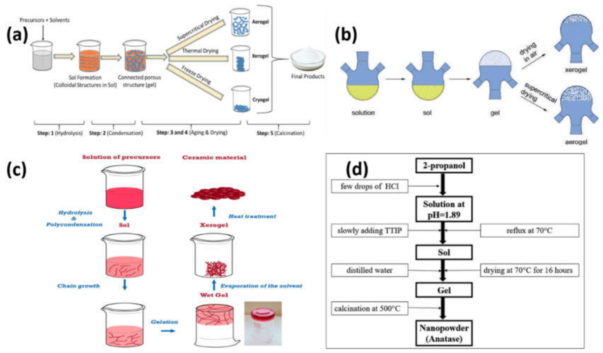

Table 1.
Comparative Table of Sol–Gel Synthesized Metal Oxide Nanoparticles.
| Serial No | Metal Oxide Nanoparticle | Typical Precursor | Key Properties | Major Applications | Reference |
|---|---|---|---|---|---|
| 01 | Ti2O3 | Titanium isopropoxide | High photocatalytic activity, UV absorption | Photocatalysis, solar cells, self-cleaning coatings | [90] |
| 02 | Mesoporous Titanium Dioxide (TiO₂) | Titanium isopropoxide | Pure anatase crystal phase, Mesoporous structure, High dye adsorption ability and good photo-electrochemical performance | Dye-sensitized solar cells (DSSC), Photovoltaic devices, Photoelectrochemical systems, Light harvesting and energy conversion | [91] |
| 03 | Titanium Dioxide Nanoparticles | Titanium (IV) Isopropoxide, water | Anatase and rutile crystal phase, Spherical particle shape, good light absorption in the UV region, Good thermal stability optical absorption | Solar cells, DSSCs Photoelectrochemical devices, Light harvesting and energy conversion applications | [92] |
| 04 | Titanium Dioxide Nanomaterials | Titanium Tetrachloride (TiCl₄), DI water and Ethanol | Anatase TiO₂ crystal phase, High purity and crystallinity, Polygonal morphology, good thermal stability and high homogeneity | Photocatalysis, Solar cells and photovoltaic devices, UV-blocking coatings, Sensors and Energy conversion applications | [93] |
| 05 | Titanium Dioxide NPs | TiCl₄ and Ethanol | Anatase-to-rutile phase transformation, good crystallinity, strong UV photocatalytic activity | Photocatalysis, wastewater treatment, dye degradation, environmental remediation and self-cleaning coatings | [94] |
| 06 | Graphene–Nitrogen-Doped Titanium Dioxide (GR–N/TiO₂) Nanocomposite | Titanium tetraisopropoxide (TTIP), Triethylamine (nitrogen source), Graphene sheets | Reduced band gap, visible-light active, enhanced charge separation, improved photocatalytic efficiency | Wastewater treatment, water purification, dye degradation, photocatalysis and environmental remediation |
[95] |
| 07 | Kaolin/TiO₂ Nanocomposite | TiO₂ precursor, Kaolin clay | High photocatalytic activity, sunlight responsive, stable and reusable, effective pollutant degradation | Wastewater treatment, tannery effluent treatment, water purification, photocatalysis, environmental remediation | [96] |
| 08 | Zn-doped TiO₂ (Mesoporous nanocrystalline) | TiO₂ precursor, Zinc source, surfactant | Anatase phase, mesoporous, reduced band gap, high surface area, enhanced photocatalytic activity | Photocatalysis, wastewater treatment, environmental remediation | [97] |
| 09 | N–TiO₂/SWCNT Nanocomposite | TiO₂ precursor, nitrogen dopant source, SWCNT | Visible-light active, high photocatalytic efficiency, high wettability, enhanced charge separation | Self-cleaning solar panels, photocatalysis, anti-dust coatings and environmental remediation | [98] |
| 10 | ZnO/TiO₂/GO–MWCNT Hybrid Nanocomposite (sand-supported) | Zinc precursor, Titanium precursor, sand, GO, MWCNTs | High surface area, strong conductivity, improved charge separation, nanorod structure, high photocatalytic efficiency | Photocatalysis, wastewater treatment, environmental cleanup, self-cleaning surfaces | [99] |
| 11 | Ag₂O (Silver oxide nanoparticles | Silver precursor (sol–gel route), oxygen source | Strong antibiofilm activity, effective against bacteria like K. pneumoniae and P. aeruginosa | Antibacterial coatings, biomedical devices, water disinfection and anti-biofilm surfaces | [100] |
| 12 | Ag₂O nanoparticle | AgNO₃ + NaOH (co-precipitation/sol–gel method) | Highly crystalline, semi-spherical morphology | Antibacterial coatings, sensors, catalysis and environmental applications | [101] |
| 13 | Silver nanoparticles (Ag NPs) | AgNO₃, CH₃COONa, N₂H₄, water | Crystalline, spherical, high purity, stable and eco-friendly synthesis | Electronics, catalysis, medicine, cosmetics, food industry and sensors | [102] |
| 14 | Ag₂O Nanoparticles) | Silver precursor and sol–gel reagents | Small crystallite size, high surface area, spherical morphology, high purity and good crystallinity | Antibacterial coatings, photocatalysis, catalysis, sensors, wastewater treatment and environmental applications | [103] |
| 15 | CuO–Ag₂O Nanocomposite | CuO precursor, Ag₂O precursor | Good crystallinity, nanoscale particles, strong antibacterial and antibiofilm activity | Antibacterial coatings, biofilm control, medical devices, water treatment and antimicrobial materials | [104] |
| 16 | Ag nanoparticles in silica (SNPs–SiO₂) | Silver precursor, silica sol–gel matrix | Temperature-dependent formation, strong optical absorption and stable nanocomposite | Plasmonic devices, sensors, antimicrobial coatings, optical materials and catalysis | [105] |
| 17 | Ag-doped MgO thin films | MgO precursor, silver dopant, glass substrate | Light-dependent capacitance, improved electrical response under violet light and morphology changes with doping | Capacitors, optoelectronics, sensors, thin-film electronics and energy storage | [106] |
| 18 | Ag–TiO₂ / Ag–ZrO₂ nanocomposites | TiO₂ and ZrO₂ precursors, Ag nanoparticles | Plasmonic absorption, tunable optical properties, thin film and crack-free dense coatings | Photocatalysis, plasmonic devices, optical coatings, sensors and luminescent systems | [107] |
| 19 | Ag₂O/Chitosan nanocomposite | Silver oxide (Ag₂O), chitosan polymer, sol–gel in-situ method | Silver oxide (Ag₂O), chitosan polymer, sol–gel in-situ method | Food packaging, antibacterial coatings, antifungal materials and biomedical applications |
[108] |
| 20 | Ag/SiO₂ (Silver–Silica Nanocomposite) | AgNPs, glucose-containing cyclosiloxane, TEOS | High surface area, stable porous structure, good catalytic activity and well-dispersed Ag nanoparticles | Catalysis, environmental cleanup, sensors, antimicrobial materials and catalytic supports | [109] |
| 21 | Fe₂O₃ (Iron Oxide, Hematite) | Iron precursor | Hematite phase, magnetic, size ~34–36.7 nm, band gap 2.4–2.7 eV, thermally stable | Sensors, catalysis, magnetic storage, biomedical applications, environmental cleanup | [110] |
| 22 | Fe₃O₄ (Magnetite) | Iron precursor, powdered coconut water (PCW) | Ferrimagnetic, high saturation magnetization, SAR heating ability and low cytotoxicity | Magnetic hyperthermia, biomedical imaging, drug delivery, cancer therapy, separation technologies | [111] |
| 23 | Iron oxide systems (Fe₂O₃, Fe₃O₄, FeO mixtures) | Fe(NO₃)₃, ethylene glycol, sol–gel method, controlled atmospheres (N₂, O₂, air) | Phase-controlled iron oxides, crystallite size 18–110 nm, magnetic behavior varies with phase | Magnetic devices, catalysis, biomedical imaging, sensors, environmental cleanup | [112] |
| 24 | Fe₂O₃ (Hematite iron oxide) | Iron precursor, sol–gel autocombustion reagents, ultrasonic method | Hematite phase, ~36–76 nm size, good UV photocatalytic activity, multiple band gaps | Photocatalysis, dye degradation, wastewater treatment, environmental remediation | [113] |
| 25 | Nanoporous Iron Oxide (Fe₂O₃ / FeOx) Aerogel-type Oxidizer Nanoparticles | Iron(III) salt (Fe³⁺ source), epoxide (for sol–gel reaction), ethanol (volatile solvent), aerosol-based sol–gel (aero-sol–gel) process | Nanoporous iron oxide structure,High reactivity due to large surface area - Suitable for energetic and redox reactions with fuels |
- Energetic materials (explosives and propellants) - Oxidizer in nano-thermite systems - Combustion and ignition systems - High-energy materials research - Catalytic oxidation reactions |
[114] |
| 26 | Fe₂O₃–SiO₂ Nanocomposite | TEOS (silica source), Fe(NO₃)₃ (iron source), sol–gel gelation method | Superparamagnetic at low size (3–4 nm), phase transition γ-Fe₂O₃ → α-Fe₂O₃, size- and temperature-dependent magnetism | Biomedical imaging, sensors, catalysis, magnetic storage, environmental applications | [115] |
| 27 | SPION@SiO₂ (Iron oxide–silica core–shell nanoparticles) | Iron oxide nanoparticles (ferrofluid), TEOS (silica precursor), sol–gel method | Superparamagnetic core, tunable silica shell (2–100 nm), fluorescent functionalization, magnetic + optical behavior | Biomedical imaging, drug delivery, biosensing, targeted therapy, multimodal diagnostics | [116] |
| 28 | CeO₂–Fe₂O₃ Nanocomposite | Ce(NO₃)₃·6H₂O, Fe(NO₃)₃·9H₂O, ethanol, propylene oxide (sol–gel method) | High thermal stability, redox active, stable cycling performance, good porosity and surface area | Solar fuels, H₂ production, CO₂ splitting, thermochemical energy conversion | [117] |
| 29 | Fe₂O₃–SiO₂ Nanocomposite Aerogel | FeCl₃·6H₂O, TMOS or TEOS, ethanol, organic epoxide (sol–gel method) | 5–20 nm particles, highly porous, very high surface area (350–450 m²/g), uniform Fe₂O₃/SiO₂ dispersion, stable aerogel structure | Catalysis, adsorption, sensors, environmental remediation, energy applications, advanced nanocomposites | [118] |
| 30 | Fe₃O₄ (Magnetite) in PANI/Fe₃O₄ Hybrid Nanocomposite | Fe₃O₄ nanoparticles, aniline monomer, sol–gel reagents | Magnetic, electrically conductive, good nanoparticle dispersion, combined magnetic and electrical properties | Sensors, drug delivery, biomedical devices, electronics, smart nanocomposites | [119] |
| 31 | Bi₂O₃ (Bismuth Oxide Nanoparticles) | Bismuth salt precursor, sol–gel reagents | α-phase, homogeneous nano powder, high purity, good crystallinity | Photocatalysis, sensors, electronics, fuel cells, environmental and catalytic applications | [120] |
| 32 | Bi₂O₃ NPs | Bi (NO₃) ₃·5H₂O, citric acid, HNO₃ | Mixed phases (α and γ), band gap tuning with temperature. | Photocatalysis, dye degradation, wastewater treatment, environmental cleanup | [121] |
| 33 | BiFeO₃ (Bismuth Ferrite Nanoparticles) | Bi (NO₃) ₃·H₂O, Fe (NO₃) ₃·9H₂O | Multiferroic, ferroelectric + magnetic at room temperature, rhombohedral structure | Cancer treatment, antimicrobial applications, biomedical uses, sensors, multiferroic devices | [122] |
| 34 | BiFeO₃ (Microwave-assisted sol–gel NPs) | Bi nitrate (Bi source), Fe nitrate (Fe source), | Phase-pure perovskite structure, ferromagnetic behavior at high power | Multiferroic devices, sensors, memory devices, capacitors, spintronics, electronic applications | [123] |
| 35 | BiFeO₃ (Bismuth Ferrite NPs) | Bismuth precursor, iron precursor | Multiferroic, ferromagnetic, nanocrystalline, high Néel and Curie temperatures | Sensors, memory devices, spintronics, magnetoelectric devices, advanced electronics | [124] |
| 36 | TiO₂–Bi₂O₃ Nanocomposite | Titanium precursor (TTIP or Ti alkoxide), bismuth salt precursor | High crystallinity, visible-light active, reduced band gap | Photocatalysis, dye degradation, wastewater treatment, solar energy conversion, environmental cleanup | [125] |
| 37 | BiFeO₃–Graphene Nanocomposite | Bi nitrate, Fe nitrate, graphene sheets | High conductivity, enhanced capacitance, good electron transport | Supercapacitors, energy storage, electrochemical devices, high-performance capacitors | [126] |
| 38 | Bi₂O₃@PANI Nanocomposite | Bismuth oxide precursor, aniline monomer | High sensitivity, fast response, high conductivity | Sensors, environmental monitoring, toxic chemical detection, electrochemical devices | [127] |
| 39 | TiO₂–Bi₂O₃ / Bismuth Titanate–GO Thin Films | Ti precursor, Bi precursor, graphene oxide | Mixed titanate phases, enhanced charge transport and photocatalytic activity with GO | Photocatalysis, dye degradation, wastewater treatment, environmental remediation | [128] |
| 40 | 3DOM Bi₂O₃/TiO₂ Nanocomposite | Ti precursor, Bi precursor, PS latex spheres, P123 template, sol–gel + calcination | Ordered macroporous structure, high surface area, good recyclability | Photocatalysis, dye degradation, wastewater treatment, environmental cleanup, solar energy applications | [129] |
| 41 | MONPs: TiO₂, ZnO, SnO₂, WO₃ Nanoparticles | Metal salts/alkoxides | High purity, uniform size, good stability | Photocatalysis, photovoltaics, sensors, biomedical systems, purification, optical devices | [86] |
| 42 | Metal oxides and mixed (e.g., TiO₂, ZnO, Fe₂O₃, and mixed oxide systems using different metal combinations) | Metal salts such as chlorides and nitrates; metal-organic compounds (alkoxides); chelating agents and polyols; heterometallic alkoxides | High purity materials, Uniform structure and good chemical homogeneity | Catalysis and photocatalysis, Electronics and optical devices, Sensors and energy materials, General applications across materials science and nanotechnology | [130] |
| 43 | Tin Oxide–Zinc Oxide (SnO₂–ZnO) Nanocomposite | Tin precursor (Sn salt/alkoxide), zinc precursor (Zn salt/alkoxide) | Flower-like morphology, Increased band gap compared to bulk materials | Photocatalysis, Optical and photoluminescent devices Gas sensors, Environmental sensing applications | [131] |
| 44 | Metal NPs embedded in oxide films such as (SiO₂),(TiO₂), (ZrO₂), (Al₂O₃) and hybrid oxide materials, combined with metals like Ag, Au, and Ag–Au alloys |
Metal salts: Ag, Au, Cu salts (e.g., nitrates or chlorides) Oxide precursors (sol–gel): Tetraethoxysilane (TEOS) / tetramethoxysilane (TMOS) → for silica (SiO₂) Titanium alkoxides → for TiO₂ Zirconium alkoxides → for ZrO₂ Aluminum precursors → for Al₂O₃ |
Very small particle size (nanometer scale), High surface energy and high reactivity, Enhanced optical properties, Unique plasmonic properties (especially for Ag and Au nanoparticles) | Optical devices (sensors, coatings, photonic materials) Catalysis and photocatalysis, Environmental purification and pollutant removal, Biomedical and antimicrobial materials and electronic and sensing devices | [87] |
| 45 | Copper salts (for CuO), Manganese salts (for Cu–Mn system), Cobalt salts (for Cu–Co system), Polyvinyl alcohol (PVA) | CuO NPs, Cu–Mn NCs, Cu–Co NCs, Prepared using sol–gel method with PVA (polyvinyl alcohol) as stabilizer | Nanometer-sized particles, good antioxidant activity), Strong enzyme inhibition activity |
Biomedical applications, Antioxidant and anti-oxidative stress systems, Enzyme inhibition–based drug development, Potential therapeutic agents and Biochemical and pharmaceutical research | [132] |
| 46 |
Multicomponent mesostructured metal oxides (MMMOs) Mixed metal oxide systems (e.g., rare-earth oxides and transition metal oxides) |
Metal alkoxides (main metal sources), Polymers / block copolymers, Solvent system: acetic acid, hydrochloric acid, ethanol and Metal ions stabilized using acetic acid binding |
Nanometer-sized particles, highly porous mesostructured, good condensation and self-assembly behavior | Catalysis and heterogeneous catalysis, Photocatalysis Photovoltaic and solar energy systems, Energy storage devices, Electronic materials and thin films, membranes, and coating technologies |
[133] |
| 47 |
Nickel oxide / Zinc oxide nanocomposites (NiO/ZnO NCs) |
Nickel salts → source of NiO nanoparticles Zinc salts → source of ZnO nanoparticles, Triton X-100 → templating/structuring agent to control morphology | Nanosized mixed oxide particles with mesoporous structure, High surface area and good crystallinity |
Photocatalytic degradation of organic pollutants (wastewater treatment), Self-cleaning and environmental cleanup materials, Anti-corrosion coatings for metals (e.g., aluminum surfaces), Protective thin-film coatings, Solar-light driven catalytic systems | [134] |
| 48 | Various metal oxide nanoparticles and nanostructures |
Metal alkoxides, Solvents such as water, alcohol (ethanol, etc.), Chemical reagents for hydrolysis and alcoholysis reactions | High control over composition and purity, homogeneous and fine nanoscale materials |
Optical and electronic devices Energy-related materials (solar, batteries, etc.) Surface coatings and corrosion protection Biosensors and medical/pharmaceutical applications Separation technologies (e.g., chromatography) Ceramic materials and industrial coatings Building insulation and protective materials |
[135] |
| 49 | Metal oxide/silica (MOx/SiO₂) nanocomposites, Metal/silica (M/SiO₂) nanocomposites | (RO)₃Si–X–M (RO)₃Si = alkoxysilane group M = metal ion or metal alkoxide X = organic linker |
Controllable nanoparticle size, high homogeneity and purity, good thermal and chemical stability | Catalysts and photocatalysts, Optical and photonic materials, electronic devices, sensors |
[136] |
| 50 | Cobalt oxide–cerium oxide nanocomposite (Co₃O₄/CeO₂) | Cobalt precursor, Cerium precursor,Citric acid, maleic acid, succinic acid, and trimesic acid used as stabilizers |
Nanocomposite with controlled particle size and morphology, good crystallinity and stable structure | Photocatalytic degradation of dyes Wastewater and water treatment, Environmental remediation |
[137] |
3.2.2. Precipitation and Co-Precipitation Methods
Precipitation is a simple and inexpensive chemical technique to synthesize nanoparticles of metal oxides. This technique involves adding a precipitating agent like sodium hydroxide, ammonium hydroxide, potassium hydroxide and sodium carbonate to an aqueous solution of a metal salt. The reaction results into metal hydroxide or metal carbonate precipitates, which are rinsed, dried and calcined to metal oxide nanoparticles[138,139,140].
For instance, zinc salts can be reacted with sodium hydroxide to produce zinc hydroxide, and then, ZnO nanoparticles can be formed by heating[141,142]. In a similar manner, the iron salts can be precipitated to form iron oxide nanoparticles. Particle size and morphology can be controlled by varying pH, temperature, concentration of the reactants, stirring speed and aging time[143].
A modified form of precipitation in which two or more metal ions are precipitated from the same solution is known as co-precipitation[144]. Such a method is particularly useful for the synthesis of mixed metal oxides, ferrites, doped oxides and for the synthesis of nanoparticles of the spinel type. One such example is the magnetic nanoparticles of iron oxide (Fe₃O₄) which are usually prepared from the co-precipitation of Fe²+ and Fe³+ salts in the alkaline medium[145,146].
The precipitation and co-precipitation methods have the main advantages of simplicity, low cost, mild reaction conditions and applicability to large-scale production. All these methods are not based on complicated instruments and are applicable in the aqueous media[147]. If the reaction conditions are not well controlled, On the other hand, they can generate nanoparticles with large size distribution and high aggregation. Repeated washing of the final product is sometimes necessary to remove impurities and calcination is necessary to enhance its crystallinity. Despite these drawbacks, precipitation methods are common due to their simplicity, low cost, and versatility in the production of various metal oxide nanoparticles[74,139].
3.2.3. Hydrothermal and Solvothermal Methods
Hydrothermal and solvothermal synthesis are significant chemical processes for metal oxide nanoparticles synthesis with well-controlled morphology and highly crystallinity[148,149,150]. The hydrothermal method involves conducting the reaction at high temperatures and pressures, in water, in a closed pressure vessel known as an autoclave[151]. The solvothermal process is similar to the previous method, except that an organic solvent, e.g. ethanol, methanol, ethylene glycol, or dimethylformamide, is employed in place of water[152]. The sealed environment allows the reaction to proceed above the boiling point of the solvent, and so high pressure conditions are created which favour nucleation and crystal growth[149,153].
The metal salts are usually dissolved in water with a base, a surfactant or a mineralizing agent in a typical hydrothermal synthesis. The solution goes into an autoclave, lined with Teflon, and is heated at a certain temperature for several hours[154]. Metal hydroxide or oxide nuclei are produced during heating and then a controlled crystal growth occurs. The product is collected and washed, dried and sometimes calcined after cooling[155].
For the synthesis of nanoparticles of metal oxides hydrothermally and solvothermal methods have been widely used. These techniques are especially suitable for the synthesis of nanoparticles of various shapes, including nanorods, nanowires, nanospheres, nanoplates, or flowers. The size and shape of the nanoparticles can be easily controlled by changing the solvent, pH, temperature, reaction time, and surfactant[156]. Conventional solvothermal and green hydrothermal methods were used to prepare copper and cobalt nanoparticles as shown in Figure 4a. The solvothermal method involves dissolving the metal nitrate precursors in deionized (DI) water and reacting them with ethylene glycol, which is used as both solvent and reducing agent. In the green hydrothermal approach, Medicago sativa extract is used as a sustainable bioreducing and stabilizing agent in place of the commonly used chemical reagents. The precursor solutions are then agitated and poured into the sealed reactors and are subjected to hydrothermal treatment at 180 °C for 16 h which leads to the nucleation and growth of nanoparticles under high temperature and pressure. The products are then further purified by centrifugation and washing and then dried at 70 °C to generate stable nanopowders. The comparison shows the benefits of green hydrothermal synthesis such as low amount of chemicals used, high synthesis efficiency, good environmental compatibility, particle size and morphology control[157]. Representative hydrothermal synthesis route of Bi₂O₃/Bi₂WO₆ heterostructured photocatalysts is shown in Figure 4b. During this process, the bismuth nitrate was dissolved in acetic acid and was slowly added to a suspension of WO₃ nanoparticles in water, ensuring the homogeneous mixing of the two. A precipitating and pH-regulating agent, urea is then introduced which allows for controlled nucleation and crystal growth. The resulting suspension goes to a Teflon-lined stainless-steel autoclave and undergoes a hydrothermal treatment at 160 °C for 24 h. When these conditions are satisfied, high temperature and autogenous pressure will favor the in-situ growth of a heterojunction between the semiconductor phases Bi2O3/Bi2WO6 with good contact and crystallinity of both phases. The product is recovered after hydrothermal processing by centrifugation, repeatedly washed with deionized water and ethanol and then dried and finally calcined at 500 °C to increase the phase purity and crystallinity. The hydrothermal synthesis was proved to be an effective method for fabricating advanced metal oxide photocatalysts, and the synthesized Bi₂O₃/Bi₂WO₆ composite showed the enhanced visible-light photocatalytic performance, in which the efficient charge separation and the reduction of electron–hole recombination at the heterojunction interface were observed[158]. The reaction pathway of the hydrothermal synthesis process of Cu nanoparticles using Cu formate with the aid of formic acid is shown in figure 4C. Transient formation of Cu₂O as an intermediate phase during the reduction process was observed by in situ synchrotron X-ray diffraction. The two reduction pathways suggested were the direct reduction of Cu₂O by formic acid adsorption/decomposition on the oxide surface and the indirect reduction pathway, in which the reduction is carried out by the production of hydrogen gas from the hydrothermal decomposition of formic acid. The results show that the most important pathway is (i) with the appearance of Cu₂O intermediate and pressure independent activation energy for Cu formation, which are in full agreement with this. The figure depicts the dynamic phase evolution of Cu²⁺ species towards Cu₂O and subsequently to the metallic Cu, which underscores the significance of hydrothermal conditions and reducing agents in nanoparticle nucleation and growth[159].These methods have a significant advantage because the nanoparticles are produced in a highly crystalline form, and very high calcination temperatures are not required. They also provide a good control of the morphology and the phase purity. They do need sealed autoclaves, longer reaction times, and careful handling because of the high pressure On the other hand. Autoclave size and safety may also be the issue for large scale production. On the other hand, hydrothermal/solvothermal processes are still valuable for the synthesis of well-defined nanocrystals of metal oxides for catalysis, sensing, energy storage, photocatalysis and biomedical applications.
Table 2.
Metal Oxide Nanoparticles Synthesized by Hydrothermal and Solvothermal Methods.
| Serial No | Metal Oxide Nanoparticle | Synthesis Method | Key Structural Features | Functional Properties | Major Applications | Reference |
|---|---|---|---|---|---|---|
| 01 | TiO₂ (Titania) nanoparticles | Green hydrothermal method using Morinda citrifolia leaf extract | Rutile tetragonal phase, quasi-spherical particles, good structural stability | Strong antimicrobial activity, good optical, stable nanoparticle formation with plant-based capping agents | Antimicrobial agents, biomedical applications, disinfection, water purification, antimicrobial coatings | [160] |
| 02 | Titanium dioxide (TiO₂), rutile phase | Hydrothermal method | Well-formed nanoscale TiO₂ structure, surface functional groups, uniform morphology, stable colloidal nature | Cytotoxic activity against cancer cells, apoptosis induction, strong larvicidal and pupicidal activity against Aedes aegypti | Cancer therapy research, mosquito control (larvicides/pupicides), biomedical applications, antimicrobial and environmental control agents | [161] |
| 03 | SiO₂-modified TiO₂ (silica–titania nanocomposite) | Hydrothermal method | Pure anatase TiO₂ phase, high surface area, silica prevents rutile formation and controls crystal growth | Enhanced photocatalytic activity, increased band gap with silica content, high stability even at high temp | Photocatalysis, wastewater treatment, environmental cleanup, high-temperature catalytic applications | [162] |
| 04 | TiO₂ nanorods + nanoparticles (TiO₂ NRs/NPs) | One-step hydrothermal method | Pure anatase crystalline phase; mixed morphology (nanorods + nanoparticles); high surface area | High photocatalytic/electrochemical performance; improved charge transport; reduced charge recombination | Dye-sensitized solar cells (DSSCs), solar energy conversion, photocatalysis, energy storage, photoelectrochemical devices | [163] |
| 05 | Reduced graphene oxide/TiO₂ nanocomposite (rGO/TiO₂) | One-step hydrothermal synthesis | TiO₂ NPs uniformly distributed on RGO sheets, narrow particle size distribution | Improved electrochemical performance, higher thermal stability | Heavy metal sensing (Hg²⁺ detection), electrochemical sensors, environmental monitoring | [164] |
| 06 | TiO₂–Ti₃C₂Tₓ nanocomposite |
One-step hydrothermal | TiO₂ nanoparticles (~30 nm) attached to layered Ti₃C₂Tₓ sheets | High surface area, improved electron transport, enhanced structural stability, tunable nanoparticle size | Photocatalysis, energy storage devices, sensors, environmental remediation, electrochemical and optoelectronic applications | [165] |
| 07 | Titanium oxide (TiO₂) | Solvothermal synthesis | Multiple morphologies obtained: nanowires nanorods, nanofibers, nanoparticles, nanobelts, nanocubes, nanosheets | Tunable size and shape, morphology-dependent properties, potential for enhanced catalytic, optical, and electronic performance | Photocatalysis, sensors, solar cells, environmental remediation, coatings, nanodevices, energy storage and conversion | [166] |
| 08 | TiO₂ (Anatase titanium dioxide) nanoparticles | Sol–gel method coupled with solvothermal treatment | High-purity anatase phase, nanosized particles formed at relatively low temperature and short reaction time | Strong photocatalytic activity, efficient degradation of organic pollutants | Wastewater treatment, dye degradation, environmental remediation, self-cleaning and purification systems | [167] |
| 09 | BaTiO₃ (Barium titanate) perovskite nanocrystals | Solvothermal colloidal synthesis | Single-crystalline, non-aggregated nanoparticles, high dispersibility, self-assembly into 2D and 3D superstructures | Ferroelectric behavior, polar ordering, excellent colloidal stability, self-assembly capability | Energy conversion devices, data storage systems, electronic materials, biomedical applications | [168] |
| 10 | Mixed-phase TiO₂ nanocrystals | Low-temperature solvothermal process | Tunable phase composition, formation of interparticle connections via hydrogen bonding | Improved charge separation in mixed phases; photocatalytic activity depends on phase ratio | Photocatalysis, environmental pollutant degradation, solar energy conversion, self-cleaning surfaces, water treatment |
[169] |
| 11 | Silver oxide nanoparticles (AgO NPs) | Hydrothermal method using wild shrimp extract + Ag₂NO₃ salt | Cubic crystal structure; size depends on method | Strong antibacterial activity (especially against S. aureus); tunable band gap, optical emission changes with method | Antibacterial coatings, medical disinfectants, antimicrobial materials, water purification, and biomedical applications | [170] |
| 12 | Ag₂Se nanoparticles / Ag₂Se–rGO nanocomposite | Hydrothermal method | well-dispersed nanocomposite structure with enhanced active sites | Excellent electrocatalytic activity for oxygen evolution reaction (OER), fast reaction kinetics, very low charge transfer resistance | Water splitting, hydrogen/oxygen production, energy conversion, electrocatalysts for renewable energy systems | [171] |
| 13 | Fe₃O₄/RGO) nanocomposite | One-pot hydrothermal method | Well-dispersed Ag nanoparticles and Fe₃O₄ nanoparticles anchored on reduced graphene oxide sheet | High catalytic activity (efficient reduction of 4-nitrophenol); strong antibacterial activity against E. coli; good reusability and stability | Wastewater treatment, pollutant degradation, antibacterial materials, environmental remediation, catalytic applications | [172] |
| 14 | GO–Fe₃O₄–Ag ternary nanocomposite | One-pot hydrothermal method | Well-crystalline Fe₃O₄ nanoparticles anchored on GO sheets, controlled particle size | Improved magnetic properties (high saturation magnetization); good stability; high crystallinity | Photocatalysis, environmental pollutant degradation, solar energy conversion, self-cleaning surfaces, water treatment | [173] |
| 15 | Ag₂O–MgO/rGO nanocomposite | Green hydrothermal synthesis | rGO sheets loaded with Ag₂O–MgO nanoparticles, smaller effective particle size | Highest photocatalytic efficiency, strong ROS generation, highest cytotoxicity vs cancer cells (MCF-7) | Wastewater treatment (dye removal), anticancer applications | [174] |
| 16 | Ag/ZnO micro–nanocomposite | Solvothermal method | ZnO nanorods with Ag nanoparticles on surface | Improved photocatalytic activity, enhanced surface reactivity, Ag improves charge separation and antibacterial behavior | Wastewater treatment, dye degradation, photocatalysis, antibacterial coatings, environmental remediation | [175] |
| 17 | Ag₂O-supported WO₃ nanorods (Ag₂O/WO₃) | Solvothermal method with mixed surfactants | Nanorod structure of WO₃ decorated with Ag₂O, mesoporous structure, high surface area | Strong visible-light absorption; improved charge separation; high photocatalytic activity | CO₂ conversion to methanol (CH₃OH) under visible light; environmental photocatalysis and carbon reduction | [176] |
| 18 | CDs/Ag/TiO₂ (ternary nanocomposite) | One-pot solvothermal (green-assisted) | TiO₂ (Degussa P25) crystal phase unchanged; Ag nanoparticles and carbon dots (CDs) uniformly attached on TiO₂ surface | Strong visible + UV light absorption; reduced electron–hole recombination due to Ag SPR and CDs synergy | Photocatalytic degradation of dyes (e.g., methylene blue), wastewater treatment, environmental cleanup | [177] |
| 19 | AgFe₃O₄ / Graphene nanoplatelet (GNP) nanocomposite | Solvothermal method | Ag and Fe₃O₄ nanoparticles well dispersed on graphene sheets; good crystallinity; ferromagnetic behavior | Magnetic behavior, good electron transport, stable nanocomposite, good chemical interaction with graphene | Magnetic sensors, digital signal processors, environmental remediation, biomedical applications | [178] |
| 20 | Surface-modified iron oxide nanoparticles (α-Fe₂O₃ / Fe₃O₄) | Aqueous-phase heating of FeSO₄ at 473 K (Hydrothermal) | Nanoparticles coated with organic surface layer, shape changes depending on modifier | Improved surface functionality,tunable particle shape; enhanced stability and dispersibility in aqueous systems | Catalysis, environmental remediation, biomedical applications and surface-engineered nanomaterials | [179] |
| 21 | Fe₃O₄ (magnetite) nanoparticles | One-step hydrothermal synthesis | Highly crystalline NPs, narrow size distribution, size-dependent magnetic behavior | Tunable magnetic properties, high saturation magnetization, size-dependent detection sensitivity | Cancer diagnosis, cancer treatment (drug delivery, imaging), biomedical applications, magnetic sensing | [180] |
| 22 | Iron oxide nanoparticles (mainly maghemite, Fe₂O₃ / Fe₃O₄-related phase) | Hydrothermal method using a homemade autoclave reactor | Spherical nanoparticles; strong aggregation due to magnetic nature | Magnetic behavior, optical band gap, good crystallinity; Fe–O bonding confirmed | Magnetic applications, nanotechnology devices, biomedical uses sensors | [181] |
| 23 | SnO₂/Fe₂O₃ (tin oxide/iron oxide) nanocomposites | Hydrothermal method | Sheet-like structures coated with SnO₂ NPs/NR, morphology changes from nanosheets → nanorods with reaction time | Combined optical + magnetic behavior; paramagnetic nature; tunable structure depending on synthesis time | Sensors, environmental applications, magnetic devices, optoelectronic applications | [182] |
| 24 | Graphene/Fe₃O₄ (magnetite) hierarchical nanocomposites | One-step hydrothermal method | Fe₃O₄ nanocrystals and clusters anchored on graphene; GO reduced to graphene during synthesis | Strong magnetic properties, good water dispersibility, strong photoluminescence | MRI imaging, bio separation, bioimaging, optical devices, biomedical applications | [183] |
| 25 | Iron oxide (Fe₃O₄) magnetic nanoparticle clusters | Modified solvothermal method | Cluster size, citrate gives smaller superparamagnetic clusters, PVP leads to larger aggregates | High magnetization, low remanence, controllable aggregation | Magnetic control systems, bio-separation, drug delivery guidance, microwave/nano wave absorbing materials, biomedical uses | [184] |
| 26 | Iron oxide nanoparticles (mixed phases: γ-Fe₂O₃, Fe₃O₄, ε-Fe₂O₃) | Alkaline solvothermal method | Mixed crystal phases, particle size, surfactants control shape and stability | Magnetic behavior, good thermal stability, surface functional groups | Wastewater treatment, magnetic separation, drug delivery, catalysis, sensors | [185] |
| 27 | rGO/Fe₃O₄ nanocomposites | Solvothermal method with different mass ratios | Fe₃O₄ nanoparticles uniformly embedded on rGO sheets; microsphere size | Superparamagnetic behavior, improved optical properties, good photocatalytic activity and stability over repeated use | Wastewater treatment, magnetic separation, photocatalysis, environmental remediation | [186] |
| 28 | rGO–Fe₃O₄ | One-step solvothermal method | Fe₃O₄ NPs uniformly deposited on rGO sheets, good dispersion, no aggregation due to rGO support | Superparamagnetic behavior, moderate saturation magnetization, strong adsorption ability for dyes | Wastewater treatment, dye removal (e.g., methylene blue), magnetic separation, environmental remediation | [187] |
| 29 | Bi₂O₃ (bismuth oxide) nanoparticles | Hydrothermal method | Pure phase Bi₂O₃ confirmed; rod-like morphology | Stable crystalline structure; tunable particle size; good structural purity | Photocatalysis, environmental remediation, sensors, antibacterial applications | [188] |
| 30 | Bi₂O₃ nanoparticles | Hydrothermal synthesis using bismuth nitrate |
Monoclinic Bi₂O₃ phase; rod-like morphology;,size controlled, higher precursor concentration | High atomic number (Z) → strong radiation interaction; low cytotoxicity at tested dose | Cancer radiotherapy enhancement, medical imaging, radiosensitizers in oncology, biomedical applications |
[189] |
| 31 | BVO/BWO (Bismuth vanadate / Bismuth tungstate) composite | Hydrothermal method | Rod-like BiVO₄ deposited on flake-ball Bi₂WO₆ particles; strong interfacial contact | Enhanced photocatalytic activity under UV–visible and visible light; improved charge separation | Photocatalysis, water splitting, environmental cleanup, solar energy conversion | [190] |
| 32 | Graphene–Bi₂O₃ (bismuth oxide/graphene) composite | Solvothermal reduction + thermal treatment | Bi₂O₃ nanoparticles anchored on graphene sheets; uniform dispersion; porous composite structure | Very high specific capacitance, excellent charge storage; good rate capability, enhanced conductivity due to graphene | Supercapacitors, energy storage devices, electrochemical capacitors, high-performance electrodes | [191] |
| 33 | BiPO₄–graphene (BP–RGO) nanocomposite | One-step solvothermal method using ethylene glycol/water | Rod-shaped BiPO₄ grown and uniformly deposited on RGO; increased surface area | Strong photocatalytic activity; high dye adsorption; improved charge separation | Wastewater treatment, photocatalytic dye degradation (e.g., MO), environmental remediation | [192] |
| 34 | RGO–BiPO₄ (RGO–bismuth phosphate) nanocomposite | One-pot solvothermal method | BiPO₄ NPs anchored on RGO sheets, strong interfacial contact, porous structure | High photocatalytic activity, improved charge separation, enhanced dye degradation | Wastewater treatment, photocatalytic dye degradation, environmental remediation | [193] |
| 35 | Ti₀. ₉₀Sn₀. ₁₀O₂, CuO–Ti–Sn–O₂₋δ, Ti–Sn–Fe–O₂₋δ nanocomposites | Hydrothermal method | Doped TiO₂-based mixed oxide systems; incorporation of Sn, Cu, and Fe ions; oxygen vacancies (O₂₋δ | Room-temperature ferromagnetism; strong visible-light photocatalytic activity; improved charge separation | Wastewater treatment, photocatalysis (dye degradation), magnetic–photocatalytic devices, environmental remediation | [194] |
| 36 | NiO, Ni (OH)₂, Co₃O₄, Co₃O₄@Ni–Co–O, Co carbonate hydroxide, Co₃₋ₓFeₓO₄, ZnₓCo₃₋ₓO₄ nanoarrays | Hydrothermal synthesis (structure-designed growth) | 1D nanorods, 2D nanowalls, hierarchical nanoarrays; mixed-metal oxide structures | Excellent electrochemical activity; high charge storage capability; improved conductivity and catalytic activity | Supercapacitors, electrocatalysis, energy storage devices, batteries, environmental catalysis | [195] |
| 37 | CuO/NiO/ZrO₂ (CNZr) mixed metal oxide composite | Facile hydrothermal method | Ternary mixed-oxide structure; strong interaction between CuO, NiO, ZrO₂; high surface area; | Excellent electrocatalytic activity, high H₂ generation rate, low charge-transfer resistance | Water splitting, hydrogen production, renewable energy devices, electrocatalysis for clean fuel generation | [196] |
| 38 | CeO₂, CuO, ZrO₂, γ-Fe₂O₃, TiO₂, Cu₂O, NiO, BaTiO₃, LiNbO₃ | Solvothermal method | Nanosized metal oxides with tunable morphology, high crystallinity and variable particle size | High surface reactivity; good catalytic, magnetic, optical, and electronic properties depending on material | Catalysis, photocatalysis, sensors, energy storage, electronics, biomedical applications, environmental remediation | [148] |
| 39 | Various metal oxide–based nanocomposites (e.g., TiO₂, NiO, Co₃O₄, ZnO with graphene/polymers | Mainly chemical synthesis methods (hydrothermal, solvothermal, sol–gel, etc.) | Nanoscale composite structures with high surface area, porous morphology | High electrochemical performance; improved charge storage; low cost; high stability; fast charge–discharge capability | Supercapacitors, batteries, energy storage devices for electronics (smartphones, computing devices), renewable energy systems | [197] |
3.2.4. Microemulsion Method
The microemulsion method is one of chemical synthesis where nanoparticles are synthesized within nanosized droplets of a microemulsion system[198]. A microemulsion typically contains water, oil, and surfactant and, in some cases, co-surfactant. These components create small droplets that function as nanoreactors to control the growth of nanoparticles and hence their size and shape[199].
In this technique, the metal precursor solutions and precipitating agents are typically formulated in different microemulsion systems. Mixing the two microemulsions results in the collision of the droplets, which causes the exchange of reactants and nucleation and the formation of nanoparticles in the droplet space. The droplet size is primarily influenced by the water/surfactant ratio, the type of surfactant, the oil phase and the reaction conditions, and the size of the nanoparticles depends on the size of the droplets[200,201,202].
On the other hand, this method has some disadvantages. May need large quantities of surfactant and organic solvents, sometimes leading to higher costs and environmental issues[203]. It can also be challenging to remove the surfactant from the nanoparticles surface, which may involve multiple washing or calcination steps[204]. Also, the harvest is frequently small relative to other methods of production . The microemulsion method is still valuable for situations where careful control over size and uniformity of the nanoparticles is needed despite its drawbacks[205].
3.2.5. Combustion Method
The combustion method is a fast and energy efficient chemical route for the preparation of nanoparticles of metal oxides[206]. This approach is to combine a metal salt, typically a nitrate, with an organic fuel, commonly urea, glycine, citric acid or hydrazine[207]. A self sustaining exothermic reaction takes place between the oxidizing agent and fuel when the mixture is heated. A considerable amount of heat and gases are released in this reaction and fine powder of metal oxide is formed[208].
The combustion typically takes a short amount of time and isn't the result of long heating. Gases formed in the reaction avoid massive particle agglomeration and formation of porous structures[209].
The advantages of combustion synthesis are the simplicity, low cost, short reaction time and production of highly porous nanoparticles. It also can be used to prepare doped and mixed metal oxide nanoparticles[210]. The reaction is, On the other hand, very fast, which makes it difficult to control the particle size and morphology. The high temperature that exists locally during combustion can also cause sintering of the particles, or the formation of unwanted phases. Post calcination is sometimes necessary to enhance the crystallinity and to eliminate the carbon residues. Nevertheless, combustion synthesis is still a good alternative method for rapid and cost-effective synthesis of metal oxide nanoparticles[211].
3.2.6. Thermal Decomposition Method
Thermal decomposition is a chemical technique that involves the decomposition of the metal-containing precursors at high temperature to produce metal oxide nanoparticles[212]. The precursor can be a metal nitrate, acetate, carbonate, hydroxide, oxalate or organometallic compound. Heating of the precursor results in the release of gases (water vapor, carbon dioxide, nitrogen oxides, or organic fragments) and the formation of metal oxide nanoparticles[213,214,215]. The size, morphology and crystallinity of the nanoparticles are dependent on the type of precursor, decomposition temperature, heating rate, reaction atmosphere and time of heating. Sometimes, capping agents and/or surfactants are added to regulate particle growth and to avoid particle aggregation[216,217].
The advantage of thermal decomposition is the fact that it can yield crystalline nanoparticles of metal oxides in a relatively simple process. It can also be used for the preparation of nanoparticles of well-controlled phase composition, based on the selection of precursors and conditions for their decomposition. But, in high temperatures, the particles may grow and form agglomerates[218]. The cost and/or toxicity of some organometallic precursors may be high, and harmful gases may be released during the process. Hence, a proper control of the reaction and safe handling are required[219,220].
3.2.7. Chemical Vapor Deposition
Chemical vapor deposition is a vapor phase chemical technique to prepare metal oxide nanoparticles, thin films and nanostructured coatings. Previously, the precursors for making the volatile chemicals are transported in the vapor phase and convert or decompose on a heated surface into a solid metal oxide material. To facilitate oxide formation oxygen or another active gas can be added[49,221].
CVD has been widely applied in the manufacture of high quality metal oxide materials like TiO₂, ZnO, SnO₂, WO₃ and In₂O₃[222,223,224,225,226]. It offers a good control of film thickness, composition, crystallinity and surface morphology. This method is particularly significant for the fabrication of sensor, solar cell, photocatalytic, electronic, and protective coatings of metal oxides[227].
The greatest benefit of CVD is that it can be used to produce uniform and highly adherent high purity coatings[228]. Also it can be used to make nanowires, nanotubes and other 1D metal oxide nanostructures. The process does, On the other hand, need special equipment, high temperatures and volatile precursors. Some precursors can be toxic, unstable or costly. This means that, CVD is more often applied for advanced coating and device applications than for large scale production of powders[229].
3.3. Green and Biological Synthesis
Green and biological synthesis has gained a significant importance as an alternative to physical and chemical synthesis of metal oxide nanoparticles[230]. Green synthesis avoids high temperature, high pressure, toxic solvents, strong reducing agents and expensive equipment used in the traditional methods, but natural biological resources are used as reducing, stabilizing, and capping agents[231]. These biological resources comprise plant extracts, bacteria, fungi, algae, enzymes, proteins, amino acids, polysaccharides, vitamins and other biomolecules occurring in nature[232,233,234].
The principle of green synthesis is the interaction between metal precursor salts in biological systems with bioactive compounds[235]. These compounds are involved in the reduction, hydrolysis, oxidation, precipitation and stabilization of metal ions to generate metal oxide nanoparticles. Functional groups in phytochemicals and biomolecules include hydroxyl groups, carbonyl groups, carboxyl groups, amine groups and sulfhydryl groups[5,236,237]. They can bind to metal ions, induce nucleation, regulate particle growth, and prevent excessive particle aggregation in these groups[238,239]. Figure 5 a,b,c,d explains Green and biological synthesis of metal oxide nanoparticles involves the reduction of metal ions using plant extracts or microbial metabolites that act as reducing, capping, and stabilizing agents. The reduced ions undergo nucleation and growth to form nanoparticles with controlled size and morphology. Plants, bacteria, fungi, and algae are commonly employed as biological resources, while subsequent purification and calcination steps are often used to obtain highly crystalline metal oxide nanoparticles such as CuO, ZnO, MgO, and Fe₃O₄[5].
The improvement in biocompatibility and surface functionality of nanoparticles obtained from green synthesis is particularly interesting for biomedical, antimicrobial, photocatalytic, agricultural, environmental and wastewater-treatment applications. But it is also associated with some drawbacks, such as difficulties in the reproducibility, heterogeneities in the biological composition, and limitations in scaling up as well as controlling the particle size. In spite of these difficulties, green and biological synthesis are one of the most promising methods for the sustainable production of Metal oxide nanoparticles.
3.3.1. Plant Extract-Mediated Synthesis
Most popular green approach for synthesis of Metal oxide Nanoparticles is by using plant extract. This approach involves the use of various plant parts such as leaves, roots, stems, flowers, fruits, seeds, bark, peel and whole plant extracts[240,241,242,243]. Plant extracts contain naturally occurring phytochemicals which can serve as reducing, stabilizing, and capping agents in the formation of nanoparticles. Flavonoids, polyphenols, tannins, alkaloids, terpenoids, saponins, steroids, glycosides, sugars, amino acids and organic acids are common phytochemicals involved in this process[244].
The process of plant mediated synthesis involves the selection of plant, washing, drying, cutting into small pieces or powder. It is then boiled or soaked in distilled water, ethanol, methanol or any suitable solvent to obtain out the active compounds[245]. The formation of nanoparticles is usually accompanied by a visible color change, precipitation or turbidity. The product is then centrifuged/filtrated, washed, dried and subjected to calcination if required to get the crystalline metal oxide nanoparticles[246].
Typically, the synthesis of metal oxide nanoparticles by using plant extracts involves multiple stages. Metal ions initially bind with functional groups in the phytochemicals. Second, these complexes undergo nucleation; small nuclei start to form[247]. Third, their nuclei are allowed to grow into nanoparticles by controlled aggregation and crystal growth. Finally, some phytochemicals are absorbed on the surface of the nanoparticles and serve as capping agents to prevent uncontrolled growth and aggregation of nanoparticles. These natural capping agents can also enhance the stability and biological activity of the nanoparticles[248].
Some reaction parameters play significant role on the properties of plant mediated metal oxide nanoparticles. These are the kind of plant extract, concentration of metal precursor, ratio of plant extract to metal precursor, pH, temperature, reaction time, type of solvent, and calcination temperature. For instance, an alkali pH may lead to the more rapid formation of nanoparticles of metals oxides and high temperatures may increase crystallinity. But high temperatures can result in particle growth and agglomeration. Likewise, increasing the amount of plant extract may increase the number of stabilizing molecules, but the excess amount of plant extract may result in an excess organic coating on the surface of the nanoparticles[249].
Plant mediated synthesis has been widely used for the preparation of nanoparticles of ZnO, TiO₂, CuO, Fe₂O₃, NiO, MgO, CeO₂, SnO₂, and Au Nps[78,230,250,251,252]. Of these, the use of nanoparticles of ZnO is very common due to its ease of synthesis with aqueous plant extract and its antimicrobial, antioxidant, photocatalytic and biomedical potential. It is also found that TiO₂ and CuO nanoparticles prepared using plant extracts have exhibited good activity in photocatalysis, antibacterial treatment, dye degradation and environmental remediation[253].
The major benefit of the plant extract mediated synthesis is that it is simple. It does not need any complex microbial culture conditions, expensive instruments or any highly toxic chemicals. Plant materials are readily available, renewable, biodegradable and in many cases inexpensive. The method is performed under mild conditions, which makes it more environmental friendly, compared to many chemical routes. Another key benefit is the ability to retain biomolecules on the surface of the nanoparticles derived from them; this can increase their colloidal stability and enhance biological interactions[254].
Plant-mediated synthesis, On the other hand, also has its limitations. Plant extracts may vary in chemical composition among plant species, in different stages of growth, in different geographic locations and seasons, in different extraction methods and storage conditions. This variation may impact the size, morphology, yield and activity of nanoparticles. Besides, plant extracts are composed of several compounds and it is difficult to determine which molecules are responsible for the formation of the nanoparticles[255]. Reproducibility is thus a big concern. Some of the organic matter can also be left over in the final product and therefore sometimes calcination may be needed. Increased crystallinity by calcination may result in loss of beneficial surface biomolecules and larger particles[256].
Nevertheless, synthesis using plant extract is still one of the most feasible and desirable green methods for the synthesis of metal oxide nanoparticles. It is particularly preferable in applications where low toxicity, surface biofunctionality and environmental compatibility are significant factors.
Table 3.
Plant Extract-Mediated Synthesis of Different Metal Oxide Nanoparticles.
| Serial No | Metal Oxide Nanoparticle | Representative Plant Extract | Key Properties Obtained | Major Applications | References |
|---|---|---|---|---|---|
| 01 | Titanium dioxide nanoparticles (TiO₂ NPs) | Eclipta prostrata leaf extract | Eco-friendly synthesis, non-toxic, low-cost, spherical nanoparticle clusters | Wastewater treatment, photocatalysis, environmental remediation, coatings, cosmetics, food additives | [257] |
| 02 | TiO₂ NPs | Catharanthus roseus (leaf aqueous extract) | Green synthesis, nanocrystalline structure, rutile and anatase phases, particle size | Antiparasitic control, control of blood-feeding flies (Hippobosca maculata), control of sheep-biting lice (Bovicola ovis) | [258] |
| 03 | Titanium Dioxide Nanoparticles | H. thelbiecea and Ananos seneglensis leaves | Green synthesis, crystalline anatase phase, strong antimicrobial activity | Antibacterial agents, antifungal agents, biomedical applications, infection control | [259] |
| 04 | TiO₂ NPs | Calotropis gigantea (leaf, seed, flower extracts) | Strong photocatalytic activity via radical formation (•OH, O₂•⁻), 96% dye degradation in 60 min | Wastewater treatment, dye degradation, solar cells, environmental remediation | [260] |
| 05 | Au/TiO₂ nanocomposite | Averrhoa bilimbi fruit extract + Pandanus amaryllifolius leaf extract | Green synthesis, Improved charge separation → more reactive radicals → faster dye breakdown | Photocatalytic degradation of dyes (e.g., methylene blue), wastewater treatment | [261] |
| 06 | Zinc–Titanium Dioxide Nanocomposite (Zn–TiO₂ NC) | Lemon (Citrus limon) extract | Green Synthesis, Strong antibacterial, antioxidant, anti-inflammatory activity | Biomedical drugs, antimicrobial coatings, pharmaceutical and therapeutic uses | [262] |
| 07 | TiO₂–ZnO nanocomposite | Green tea extract | Green synthesis, Mixed morphology (spherical + rhomboid), heterogeneous structure, enhanced stability | Medical-grade coatings, especially dental implants, antimicrobial surface coatings | [263] |
| 08 | Silver/Silver Oxide Nanoparticles (Ag/Ag₂O NPs) | Olea europaea (olive leaf) extract | Green synthesis (water-based), spherical shape, particle size, strong antimicrobial and antioxidant activity | Antibacterial agents, antioxidant applications, drug delivery systems, chemotherapeutic carriers | [264] |
| 09 | Silver Oxide Nanoparticles (Ag₂O NPs) | Callistemon lanceolatus leaf extract | Green synthesis, spherical and hexagonal shapes, strong antioxidant activity, dose-dependent cytotoxic activity | Antioxidant applications, antimicrobial/biomedical uses, pharmacological applications | [265] |
| 10 | Silver ferrite nanoparticles (AgFeO₂ NPs) | Amaranthus blitum leaf extract (green synthesis) | Smaller particle size, better dispersion (monodispersity), higher surface area, strong antibacterial activity (better against E. coli), | Antibacterial agents, antioxidant materials, biocatalysis, biotechnology applications, wastewater and biomedical-related treatments | [266] |
| 11 | Silver oxide nanoparticles (AgONPs) | Prunella vulgaris aqueous leaf extract (green synthesis) | spherical/oval shape, confirmed functional groups, strong antibacterial activity against Staphylococcus aureus and Klebsiella pneumoniae | Antibacterial agents, treatment of drug-resistant bacterial infections, biomedical antimicrobial coatings, water disinfection and environmental cleanup | [267] |
| 12 | Silver–Zinc oxide nanocomposite (Ag–ZnO NCs) | Lawsonia inermis (henna) leaf extract | Strong antibacterial activity (against E. coli, P. aeruginosa, S. aureus), antifungal activity (Aspergillus flavus, A. niger), acaricidal activity (against Hyalomma marginatum), hemocompatible (safe with blood) | Antibacterial coatings, antifungal agents, pest control (acaricidal applications), biomedical and pharmaceutical applications, infection control materials | [268] |
| 13 | Silver-based nanocomposites (Ag/AgO, Ag/Ag₂O, Ag/AgCl) | Prunus mahaleb L. (stem, leaf, and fruit pericarp extracts) | Green synthesis using different plant parts, crystalline structure, strong antibiofilm activity (especially Ag/AgCl), significant cytotoxic activity | Antibacterial and antibiofilm coatings, biomedical applications, anticancer and cytotoxic agents, infection control materials, potential drug development for resistant microbes | [269] |
| 14 | Iron oxide nanoparticles (Fe₃O₄ NPs) | Solanum tuberosum (potato) aqueous extract | Eco-friendly green synthesis, nanoscale formation, good antibacterial activity, strong antioxidant properties | Antibacterial agents, antioxidant applications, biomedical engineering, drug delivery systems, environmental and wastewater treatment applications | [270] |
| 15 | Iron oxide nanocomposite (Fe₂O₃/Fe₃O₄/FeO – FePPE) | Peltophorum pterocarpum leaf extract (polyphenol-rich) | Magnetic behavior chain-like NPs, strong catalytic activity, high pollutant removal efficiency (~95% RhB degradation), fast initial adsorption | Wastewater treatment, dye degradation, Fenton-like catalytic oxidation, magnetic separation-based water purification, environmental remediation | [271] |
| 16 | Iron oxide nanoparticles | Celosia argentea leaf extract | Green synthesis, high antibacterial activity (E. coli, S. aureus), strong antioxidant (97%), anti-inflammatory (93%), anti-diabetic (87%), larvicidal and anticancer (MTT inhibition 86%) | Antibacterial agents, antioxidant and anti-inflammatory drugs, anti-diabetic formulations, larvicides (mosquito control), anticancer research, biomedical and pharmaceutical applications | [272] |
| 17 | Iron oxide nanoparticles (Fe₃O₄ NPs) | Mikania mikrantha leaf extract | Green synthesis, stabilized by phytochemicals (flavonoids, terpenoids, phenolic acids, proteins) | Antimicrobial agents, antifungal coatings, biomedical applications, pharmaceutical and medical antimicrobial drug development | [273] |
| 18 | Iron oxide nanoparticles | Echinochloa frumentacea grains extract | Green synthesized, strong antibacterial activity (S. aureus, S. typhi), high antioxidant (95.10%), anti-inflammatory (92.10%), anti-diabetic (91.68%), strong anticancer activity (94.36% against A549), biocompatible (non-toxic to HEK293) | Biomedical applications, antibacterial and anticancer therapy, drug delivery systems, larvicides/insecticides, agricultural growth promotion, pharmaceutical applications | [274] |
| 19 | (RC-Fe₃O₄-Chitosan) nanocomposite | Ricinus communis (Castor plant) extract | Green synthesized magnetic nanocomposite, reusable up to 7 cycles, good stability | Removal of Pb²⁺ from drinking water and wastewater, heavy metal remediation, water purification | [275] |
| 20 | Ferric oxide nanoparticles with attapulgite clay | Radix paeoniae rubra (RPR) extract | Improved dispersion and stability, strong platelet and red blood cell adhesion, antioxidant activity, improved wound-healing potential | Hemostatic agents (bleeding control), wound dressings, wound healing materials, antibacterial biomedical products, tissue repair and regenerative medicine | [276] |
| 21 | Alpha bismuth oxide nanoparticles (α-Bi₂O₃ NPs) | Rubus ellipticus (Himalayan raspberry) fruit and leaf extracts | Green synthesized, High Congo red dye degradation efficiency (84–89%), antibacterial activity against Gram-positive and Gram-negative bacteria | Wastewater treatment, dye degradation, environmental remediation, antibacterial agents, water purification | [277] |
| 22 | Bismuth oxide nanoparticles (Bi₂O₃ NPs) | Melanin | Stable, antibacterial, anti-biofilm, membrane-disrupting activity | Antibacterial agents, anti-biofilm coatings, infection control, medical and healthcare applications | [278] |
| 23 | Bismuth oxide nanoparticles (BiONPs) | Moringa oleifera bark extract | Green synthesized, stable nanostructure, strong antibacterial activity against Gram-positive and Gram-negative bacteria | Antibacterial agents, biomedical applications, therapeutic products, infection control, pharmaceutical and diagnostic applications | [279] |
| 24 | Bismuth oxide (Bi₂O₃, α and β phases) | Millettia pinnata pod extract | Green synthesized, strong photocatalytic activity under visible light, efficient reduction of 4-nitrophenol and 4-nitroaniline, high dye degradation (98.83% for AB-10B) | Wastewater treatment, dye degradation, reduction of toxic nitro compounds, environmental remediation, photocatalysis under visible light | [280] |
| 25 | Bi₂O₃ NPs and Bi₂O₃–RGO nanocomposites | Swietenia macrophylla plant extract | Green synthesis, urface area, strong reduction of toxic compounds (4-nitrophenol → 4-aminophenol), low toxicity in biological model (Drosophila melanogaster) | Wastewater treatment, catalytic reduction of toxic nitro compounds, environmental cleanup, safer nanomaterials for biomedical | [281] |
| 26 | Ag/Bi₂O₃ and Ag/Bi₂O₃–curdlan nanocomposites | Sargassum latifolium (seaweed extract) | Strong antibacterial, antioxidant, biocompatible, enhanced bioactivity with curdlan support | Antibacterial agents, antioxidant materials, biomedical applications, infection control, water and environmental disinfection | [282] |
| 27 | CeO₂/BiOCl nanocomposites (cerium oxide–bismuth oxychloride) | Allium sativum (garlic) and Helianthus annuus (sunflower petal) extracts | High crystallinity, nanoflower structure, strong antibacterial effect, ROS production, piezoelectric-enhanced activity | Antibacterial agents, treatment of multidrug-resistant infections, water disinfection, environmental remediation, biomedical antimicrobial coatings | [283] |
| 28 | CuO/Bi₂O₃ nanocomposites | Bamboo leaf extract | Enhanced photocatalytic activity, high Rhodamine B dye degradation efficiency (95.6%), improved electron–hole separation | Wastewater treatment, dye degradation, photocatalysis, environmental cleanup, industrial effluent treatment | [284] |
| 29 | TiO₂, ZnO, MgO (metal oxide nanoparticles) | Plant-derived biomolecules (general plant extracts; not specific plant named) | Strong antibacterial and antifungal activity, ROS (reactive oxygen species) generation, biocidal properties, eco-friendly synthesis | Antibacterial/antifungal coatings, medical device coatings, water disinfection, environmental sanitation, antimicrobial surfaces in healthcare and industry | [285] |
| 30 | General metal/metal oxide nanoparticles (Ag, Au, Fe, Cu, Pt, Zn, Pd oxides etc.) | Various plant parts (leaves, stems, roots, fruits; phytochemical-rich aqueous extracts) | Eco-friendly synthesis, strong chemical reactivity, biologically active due to phytochemical capping | Biomedical applications, antimicrobial agents, drug delivery, water treatment, catalysis, sensors, environmental remediation | [286] |
|
31 |
ZnO and Fe₃O₄ nanoparticles | Pomegranate peel extract | High photocatalytic activity (ZnO: 95% methylene blue degradation), high stability, phytochemical capping improves charge transfer and performance | Wastewater treatment, photocatalysis, energy storage (supercapacitors), renewable energy applications | [256] |
| 32 |
Al₂O₃ (aluminum oxide) nanoparticles | Plant extracts rich in phytochemicals (polyphenols, flavonoids, quercetin, alkaloids) | High surface area, strong adsorption ability, antimicrobial and antioxidant activity, efficient pollutant removal (93–98%) | Water purification, removal of dyes, fluoride and nitrate, antibacterial and antifungal agents, biomedical applications | [287] |
| 33 | ZnO–carbon nanofiber nanocomposite (ZnO–CNFs) | Thymus daenensis and Stachys pilifera extracts | High photocatalytic degradation of tetracycline (up to ~93%), strong antibacterial activity against E. coli and B. subtilis, antifungal activity against Candida albicans, | Wastewater treatment, antibiotic removal, antimicrobial coatings, antifungal applications, environmental purification | [288] |
| 34 | Copper and copper-based nanoparticles/ nanocomposites | Plant extracts (green synthesis using various plant biomolecules; no specific plant mentioned) | Strong antibacterial activity stable nanostructures, eco-friendly synthesis, high catalytic activity, good biosensing response | Antibacterial agents, catalysis, biosensors, drug delivery, environmental remediation, medical applications | [289] |
| 35 | Cadmium–Cesium (Cd–Cs) mixed oxide nanocomposite | Trachyspermum ammi (ajwain seeds) | Antioxidant activity, strong corrosion inhibition, protective film formation, stable surface chemistry | Corrosion protection of mild steel in acidic environments, industrial anti-corrosion coatings, surface engineering, antioxidant-based protective materials | [290] |
3.3.2. Microbial Synthesis
The use of microorganisms like bacteria, fungi, yeast and actinomycetes as microbial synthesis for the production of metal oxide nanoparticles. Microorganisms can interact with metal ions via their cell walls, enzymes, proteins, metabolites, and extracellular polymeric substances[291]. These biological components can reduce, oxidize, precipitate and stabilize metal ions, resulting in the creation of nanoparticles within or outside the cells[292].
Microbial synthesis can be divided into two types: intracellular synthesis and extracellular synthesis. Intracellular synthesis involves the uptake of the metal ions into the microbial cells and the interaction with the enzyme and biomolecules inside the cell. From this, the nanoparticles are then created in the cytoplasm, cell membrane or periplasmic space. In extracellular synthesis, microorganisms are allowed to liberate enzymes, proteins, organic acids and other metabolites in the culture medium, where the interaction between these metabolites and the metal ions leads to the formation of nanoparticles in the culture medium. Generally, extracellular synthesis is preferred because the nanoparticles produced can be easily collected and purified as compared to the nanoparticles produced intracellularly[293].
Due to their ability to grow rapidly, withstand varying environmental conditions and produce several enzymes and metabolites, bacteria are used commonly for microbial synthesis. The carboxyl, phosphate, hydroxyl and amine functional groups found in bacterial cell walls can bind metal ions and facilitate the formation of nanoparticles. Certain bacteria can also alter the oxidation state of metal ions by enzymatic reactions and this can favour the production of metal oxide nanoparticles[294].
Fungi are also excellent biological agent for the synthesis of nano particles. Fungi are able to generate greater amounts of extracellular enzymes and proteins than bacteria, which means that they can enhance the yield of nanoparticles. Fungal biomass is not so demanding and can withstand higher concentration of metals. Reductase, oxidases, proteins and organic acids are secreted by fungi and they help in the formation and stabilization of the nanoparticles. Fungal mediated synthesis is also preferred for the synthesis of nanoparticles in the outside of the cell, due to their high secretion ability. The fungal mediated synthesis of ZnO, TiO₂, Fe₂O₃, CuO and MgO nanoparticles have been reported[295,296,297,298,299,300].
Other microorganisms also have been investigated for the production of nanoparticles, such as yeasts and actinomycetes. Yeasts are good eukaryotic microbial systems with respect to metal binding capacities and stress conditions. Actinomycetes are worth while for their ability to synthesize a variety of bioactive metabolites and extracellular enzymes. These microorganisms can not only participate in the synthesis of nanoparticles but also boost the antimicrobial and biomedical activity of the nanoparticles[301].
The mechanism of microbial synthesis includes biosorption of metal ions on the microbial surface, enzymatic transformation of metal ions, nucleation of small particles, growth of nanoparticles and stabilization by proteins or extracellular polymers. Both reducing and capping agents are microbial proteins and enzymes. Nanoparticles can also be surrounded by EPS to avoid aggregation. Sometimes, microbial metabolites serve as a template, and the shape of the resulting crystals or phases is controlled by them[302].
Microbial strain, growth medium, pH, temperature, incubation time, metal salt concentration, oxygen availability and biomass concentration are some of the factors affecting microbial nanoparticles synthesis. The age of the microbial culture is also important as the amount of enzymes produced and the metabolic activity of the culture can differ at different stages of growth. These conditions must be optimised to achieve the nanoparticles of the desired size, shape, crystallinity and biological activity[303].
There are several benefits to microbial synthesis. It is environmentally friendly, economical and can synthesize nanoparticles under mild conditions. Enzymes from microorganisms can be used for natural pathway for the formation of nanoparticles and microorganisms can be cultured in large numbers by simple culture media. Protein capping can also give good surface stability to the nanoparticles produced by microbial systems. Biologically capped nanoparticles can be applied in antimicrobial, anticancer, drug delivery, biosensing and environmental applications[304].
But the microbial synthesis also has significant drawbacks. To prevent contamination sterile conditions must be kept. Synthesis process is sometimes slower than chemical methods. A decrease in yields can be caused by metal ion toxicity that some microorganisms are sensitive to. For intracellular synthesis, extra steps for cell disruption and nanoparticle recovery are required. Besides that, particle size and morphology may be difficult to control, as many biological factors may influence the microbial systems. Culture conditions also have to be carefully controlled for scale-up[305,306].
Despite these difficulties, microbial synthesis is a promising method for the production of biologically functionalized metal oxide nanoparticles. Especially it is useful if surface biocompatibility and eco-friendly synthesis are needed.
3.3.3. Biomolecule-Assisted Synthesis
Biomolecule-assisted synthesis involves the use of purified or isolated biomolecules to synthesize metal oxide nanoparticles and stabilize them. As compared to plant-, microbial- and algal-extracted materials which are complex mixtures of many compounds, biomolecule-assisted-synthesis employs selected compounds like protein, enzymes, amino acids, peptides, polysaccharides, sugars, vitamins, DNA, organic acids and natural polymers. This is useful in order to understand the function of certain functional groups in the synthesis process and to handle it accordingly[307].
Biomolecules can be synthesized and serve several roles during the synthesis of nanoparticles. They can adsorb the metal ions, regulate the nucleation, direct the growth of crystal, stabilize the surface of nanoparticles and prohibit the aggregation of them. The important groups responsible for the interaction with the metal ions are mostly the functional groups like amino group, carboxyl group, hydroxyl group, thiol group, phosphate group and carbonyl group. The function of these groups is to coordinate with the metal precursor and acts on the formation of metal–O bonds in the development of the nanoparticles[308].
A protein is a biomolecule which is transversely applied in biomolecule-assisted synthesis, as is an enzyme. Amino acid residues in proteins possess various functional groups which have the ability to interact with the metal ions and to serve as capping agent. One can obtain controlled forming of nanoparticles through either a redox reaction or a hydrolysis reaction that an enzyme can catalyze. Protein-capped nanoparticles typically exhibit enhanced stability in aqueous systems, which may also contribute to their compatibility with biological environments. Enzyme mediated synthesis is especially interesting since it can take place under mild conditions and exhibits high specificity[309].
The use of amino acids and peptides is also effective to control the shape/size of metal oxide nanoparticles. Amino acids can be coordinating to metal ions via amino and carboxyl groups whereas peptides can serve as templates for crystal growth. Excessive aggregation is prevented by regulating nucleation (some amino acids). Controlled morphology can be achieved with peptides that have particular sequences, that selectively bind to certain crystal face[310,311,312].
The Polysaccharides are another important group of biomolecules that are used in green synthesis. Natural polysaccharides, like starch, cellulose, chitosan, alginate, pectin, dextran, and gum-based polymers can serve as stabilizing and directing agents. Many hydroxyl and carboxyl groups are present in these materials and enable them to bind metal ions to form polymer–metal complexes. These complexes during synthesis work to control the growth of the nanoparticles and prevent them from aggregating. The highly useful is that chitosan also comprises amino groups which offer a strong interaction with metal ions and can also enhance antimicrobial activity[313,314,315].
Biomolecules are no exceptions when it comes to vitamins and small organic molecules. For instance, ascorbic acid can be used as a reducing and stabilising agent, or citric acid can be used as a chelator of metal ions and growth inhibitor of particles. Mild reducing agent/stabilizers: glucose and others. The molecules' virtues are that they are relatively safe, inexpensive and biodegradable[316].
Biomolecule assisted synthesis presents major positive in that it provides a degree of control that is greater than that seen with crude biological extracts. Because the use of specific biomolecules, the mechanism of the reaction can be more easily studied. This method also decreases toxic chemicals use and makes it possible to synthesize under mild conditions. Also, biomolecules can impart functional groups onto the surface of the nanoparticle which can further be manipulated[317].
There are also drawbacks associated with biomolecule-assisted synthesis processes, On the other hand. Biomolecules purified can be costly, particularly such as enzymes and synthesized peptides. There are biomolecules that are sensitive to heat, pH and the storage conditions. Yields of synthesis may not be as good as chemical methods or further purification steps may be necessary. In certain circumstances, effective biomolecule coating could cause alteration of active area of the nanoparticles and could influence their catalysis. Hence, the concentration and the type of biomolecule should be well optimized[318,319,320,321].
Biomolecule-assisted synthesis serves as a path toward greening chemistry and controlling the creation of nanomaterials. It is especially useful in cases where reproducibility, surface functionality and compatibility with life is desirable.
4. Characterization Techniques
Characterization techniques play a central role in understanding the structure, composition, morphology, surface chemistry, optical behavior, and functional performance of metal oxide nanoparticles. Since the properties of nanoparticles are strongly influenced by crystal phase, crystallite size, defects, surface functional groups, and particle morphology, proper characterization is necessary to confirm successful synthesis and to relate structural features with practical applications. In review studies, characterization is generally discussed as a bridge between synthesis and application because it explains how different preparation routes, reaction conditions, calcination temperatures, stabilizing agents, and precursors affect the final properties of the nanoparticles.
Among the different characterization approaches, structural characterization is particularly important because it provides information about phase purity, crystallinity, crystal structure, lattice parameters, defects, and molecular bonding. Techniques such as X-ray diffraction, Raman spectroscopy, and Fourier-transform infrared spectroscopy are commonly used to identify the structural and chemical nature of metal oxide nanoparticles. These methods complement each other: XRD mainly confirms the crystalline phase and average crystallite size, Raman spectroscopy provides information about vibrational modes and structural defects, while FTIR identifies surface functional groups and metal–oxygen bonding[322].
4.1. Structural Characterization
Structural characterization by x-ray crystallography defines the arrangement of atoms inside and the nature of the crystallinity of metal oxide nanoparticles. It is used to establish if the oxide phase is the desired one, or is present secondary phases, impurities, unreacted precursor or amorphous component. Metal oxide nanoparticles may be present in a few polymorphic phases so structural analysis is necessary to determine the appropriate crystal form. TiO₂ can be present as anatase, rutile or brookite; iron oxide can be found as hematite, magnetite or maghemite; and the structure of ZnO is normally hexagonal and known as wurtzite. The physical, chemical, optical and catalytic properties of each phase are different[323].
Synthesis parameters (concentration of precursors, pH, type of solvents, reaction temperature, reaction time, drying method, calcination temperature and stabilizing or capping agents) have a strong influence on the structural features of the nanoparticles. Crystallinity, particle size, phase formation and defect density may change with a slight variation in these conditions. Thus, it is critical to carry out structural characterization not only to determine the formation of nanoparticles, but also to investigate the influence of the synthesis conditions on the final material properties[324].
Commonly employed structural characterization methods for metal oxide nanoparticles are X-ray diffraction, Raman spectroscopy, and FTIR. XRD is useful for understanding crystallinity and phase composition, Raman spectroscopy can provide useful information regarding the vibrational fingerprint, structural disorder, and defects in the nanoparticles, and FTIR will provide information regarding the metal–oxygen vibrations and the presence of organic/biological functional groups on the surface of the nanoparticles[325].
4.1.1. X-Ray Diffraction
X-ray diffraction is one of the most commonly used methods to structurally characterize metal oxide nanoparticles. It is mainly used to determine crystal structure, phase purity, crystallinity, crystallite size, and lattice parameters. This method involves diffracting X-rays off the regular arrangement of atoms of a crystalline material at certain angles. The peaks are typical for a specific crystal structure and can be compared to the standard reference pattern to determine the synthesized nanoparticles' phase.XRD pattern typically presents sharp or broad diffraction peaks for metal oxide nanoparticles which depend on the degree of crystallinity and crystallite size. The sharp, distinct peaks are characteristic of regions of high crystallinity while the broad peaks are typical of small crystallite size or structural disorder. Peak broadening is often seen for very small crystallites in nanoscale materials. The average crystallite size can be estimated from the broadening of the diffraction peak, by applying the Scherrer equation. This is a good way to approximate a size, but is widely adopted to compare the effect of different synthesis conditions on the size of nanoparticles[326].XRD can be especially beneficial in determining if the desired oxide phase has been achieved. For example, peaks associated with the hexagonal wurtzite structure are typically seen in ZnO nanoparticles, and peaks associated with anatase, rutile or a mixture of both are typically seen in TiO₂ nanoparticles. Likewise, the diffraction peaks of the iron oxide nanoparticles can be identified as hematite, magnetite, or maghemite. The XRD pattern does not show any extra peaks that would be typical of good phase purity, and the presence of other peaks could be due to impurities, secondary oxide phases, or incomplete conversion of the precursor. XRD may also give information concerning changes in lattice parameters and of lattice strain in addition to the phase identification. These changes can be caused by doping, surface defects, oxygen vacancies or foreign ions entering the crystal lattice. The shift of diffraction peak position can be a sign of lattice expansion or contraction and the intensity change of diffraction peaks can be a sign of preferred orientation or variations in crystallinity. So XRD is not only a confirmation technique but a valuable technique for understanding the structural modification of the metal oxide nanoparticles. There are, On the other hand, some limitations for XRD. Not so effective in determining amorphous materials or in determining very small amounts of impurities[327].
X-ray diffraction (XRD) analysis is a commonly used technique for assessing the crystalline nature and phase composition of the synthesized metal oxide nanoparticles (CeO₂, CuO, NiO, Mn₃O₄, SnO₂, and ZnO) as shown in Figure 6a. In all of the samples, the diffraction pattern shows a clear set of peaks related to the crystallographic planes of the targeted metal oxide phases, and there are no obvious peaks from impurity phases. The positions of the peaks obtained and their relative intensities are matching with the standard diffraction data of the fluorite type CeO₂ with monoclinic CuO, cubic NiO, spinel Mn₃O₄, tetragonal rutile SnO₂ and wurtzite ZnO structure, which shows good phase purity of the synthesized nanomaterials. One important thing in all the diffraction patterns is the broadening of peaks which is characteristic of nanocrystalline materials and indicates the formation of nanoparticles of small crystallite size. This broadening of peaks may be explained by the presence of size induced lattice strain and the synthesis of the crystals resulting in limited crystal growth (e.g. precipitation). The variations in sharpness and intensity of the peaks also indicate the differences in the crystallinity and crystallite size and the manner in which the crystallites grow for each metal oxide system. The XRD results overall show that all the six metal oxides are successfully synthesized in nanoscale crystalline form which is an important factor affecting the photocatalytic performance in dye degradation applications[328].
In catalysis and related areas, research activity of X-ray diffraction (XRD) has been steadily increasing over the last few decades and is still expanding rapidly as summarized in Figure 6b. The data show that publications using the XRD techniques are generally growing very well, and it is well known that XRD is a basic characterization tool in materials science. The literature concerning XRD is vast, and the work related to catalysis is a significant part of the whole, showing the large use of diffraction methods in the study of catalysis.More specialized methods like in situ and operando XRD have a lower but similar and growing publication trend especially in recent years. This development is a response to the growing appreciation of the need to study catalysts in conditions that are more representative of reaction conditions, where the evolution of structure, phase and the nature of the active sites can be correlated with catalytic activity. Interestingly, operando XRD is the most recent and pronounced, suggesting the new and growing forth an emerging powerful tool to investigate both structural and functional characteristics of catalysts in operation.Summarizing the overall trend, it is seen that there is a strong emphasis on understanding catalysts under the reaction conditions, and on using advanced time-resolved characterisation techniques, rather than traditional ex situ characterisation techniques[329].
Figure 6c shows the X-ray diffraction (XRD) patterns for the formation of distinct copper-based crystalline phases whose formation is greatly influenced by NaOH concentration. It is found that with increasing NaOH content, from 0.25 to 1.0 M, a systematic phase evolution occurs from cubic Cu₂O to monoclinic CuO to orthorhombic CuNa₂(OH)₄. The diffraction peaks of each sample have good agreement with the standard JCPDS data, which indicates that the sample is phase-pure and the synthesis process can be successfully controlled.The pattern obtained at low concentration of NaOH indicates the cubic Cu₂O (JCPDS No. 01-077-0199) and intermediate concentration indicates monoclinic CuO (JCPDS No. 01-080-0076) with better crystallinity. It can be seen that the CuNa₂(OH)₄ orthorhombic structure (JCPDS No. 01-079-0696) is formed at the highest level of NaOH synthesis conditions, which reflects the obvious transition of the structure. The crystallite sizes derived from the broadening of the peaks by Debye-Scherrer equation are still in the nanometer range (~21-25 nm). The results overall indicate the important role of NaOH concentration in guiding the phase-selective synthesis of Cu based nanostructures[330].
Sometimes the peaks produced by the extremely small particles and/or poorly crystallized samples may be weak and broad, which can complicate phase identification. Hence, sometimes other methods besides XRD are used to complement this technique to gain a better insight into the structure and morphology, as examples Raman spectroscopy, FTIR, TEM or SEM.
Figure 6.
a): X-ray diffraction (XRD) patterns of synthesized metal oxide nanoparticles (CeO₂, CuO, NiO, Mn₃O₄, SnO₂, and ZnO), confirming their crystalline phase purity, characteristic crystal structures, and nanoscale crystallite dimensions. Structural characterization clearly show the successful synthesis of highly crystalline metal oxide nanomaterials suitable for photocatalytic degradation of organic pollutants[328] (Reproduced with Permission). Fig 6b): annual publication activity for all catalytic studies (violet), catalytic studies performed using XRD (green), in situ XRD (orange), and operando XRD (red). For comparison, all XRD studies (blue), in situ XRD (yellow), and operando XRD (pink) are presented. Numbers indicate publications of a certain topic in total[329] (Reproduced with Permission).Fig 6c): Representative XRD analysis of copper-based metal oxide nanomaterials showing the effect of synthesis parameters on phase formation, crystal structure evolution, and crystallite size, which are critical factors governing their physicochemical and functional properties[330] (Reproduced with Permission).
Figure 6.
a): X-ray diffraction (XRD) patterns of synthesized metal oxide nanoparticles (CeO₂, CuO, NiO, Mn₃O₄, SnO₂, and ZnO), confirming their crystalline phase purity, characteristic crystal structures, and nanoscale crystallite dimensions. Structural characterization clearly show the successful synthesis of highly crystalline metal oxide nanomaterials suitable for photocatalytic degradation of organic pollutants[328] (Reproduced with Permission). Fig 6b): annual publication activity for all catalytic studies (violet), catalytic studies performed using XRD (green), in situ XRD (orange), and operando XRD (red). For comparison, all XRD studies (blue), in situ XRD (yellow), and operando XRD (pink) are presented. Numbers indicate publications of a certain topic in total[329] (Reproduced with Permission).Fig 6c): Representative XRD analysis of copper-based metal oxide nanomaterials showing the effect of synthesis parameters on phase formation, crystal structure evolution, and crystallite size, which are critical factors governing their physicochemical and functional properties[330] (Reproduced with Permission).
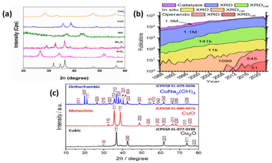
4.1.2. Raman Spectroscopy
The Raman spectroscopy is one of the most important vibrational spectroscopic methods for the characterization of the structural and molecular features of Metal oxide Nanoparticles. It is based on the inelastic scattering of light, where light scatters upon the molecular vibration producing the characteristic Raman bands. The bands give information on crystal structure, vibration modes, defects, phase composition and local structural disorder[331].
Raman spectroscopy is also very useful for metal oxide nanoparticles due to the fact that many oxide phases have unique fingerprints. It can determine the crystalline phases that are not easy to identify by XRD, particularly in case of mixed phases or poorly crystalline areas. The Raman bands are characteristic of various phases of TiO₂, such as anatase and rutile, so that it is possible to make a clear identification of phases. Likewise, Raman spectroscopy can be used to differentiate between the various phases of iron oxide and to detect structural changes in metal oxides such as ZnO, CuO, CeO₂, and others[332].
The advantage of Raman spectroscopy is that it can be sensitive to local structural disorder and defects. The presence of oxygen vacancies, lattice distortions, surface defects and changes induced by dopants in metal oxide nanoparticles significantly influences their optical, catalytic, antimicrobial and sensing properties. Changes in crystallite size, strain, defect concentration, or interaction with surface species can be suggested by Raman peak shifting, peak broadening, or changes in band intensity. Raman analysis is thus particularly useful for investigating the properties of nanometer size materials associated with defects[333].
Doped and composite metal oxide nanoparticles can also be studied using Raman spectroscopy. Raman bands can shift and new bands can appear if the metal ions, the carbon-based material, the polymers or other functional components are added to the matrix of the oxide. These changes may validate the interaction of components and offer information on the modification of the structure. For photocatalytic and sensing materials, the defects and oxygen vacancies in these materials can affect charge separation, surface reactions, and adsorption behavior, so Raman spectroscopy is often used to understand the role of the defects and oxygen vacancies[334].
One of the major advantages of Raman spectroscopy is that minimal sample preparation is typically required and can be used on powders, thin film and supported nanoparticles. It is also in most cases not destructive. There were some restrictions to be taken into account, On the other hand. In some cases, weak Raman signals can be hidden by the fluorescence signal, especially when organic residues or biological extracts are present in samples. If high power is applied to the laser, it can also heat the sample, causing problems if the sample is sensitive[335].
A typical micro-Raman spectroscopy setup to characterize the structure and vibrations of materials at the micro-scale is represented in the schematic shown in Figure 7a. In this setup, a laser is sent through a microscope objective and sent to the sample on a motorized XYZ translation stage to be precisely positioned and mapped. The light scattered by the sample is again collected via the same objective and filtered through an optical filter to remove the Rayleigh scattering.The filtered Raman signal is subsequently sent to a spectrograph where it is dispersed by wavelength and fed to a charge coupled device (CCD) detector to acquire the spectrum. The system also includes a computer for data processing and analysis, and a video monitoring unit for real-time visualization and alignment of the samples. In this setup, high-resolution, non-destructive probing of vibrational modes is obtained, which makes it very useful for research on the structural fingerprints, crystallinity and defect states of nanomaterials[336].
Hence, it is important to carefully select the wavelength, power, and acquisition conditions for the laser, in order to be able to perform accurate analysis.
Figure 7.
Raman spectroscopy as a characterization tool for metal oxide nanoparticles, illustrating the Raman scattering mechanism and a typical micro-Raman experimental setup used to probe crystal structure, phase composition, lattice defects, and chemical bonding at the nanoscale[336] (Reproduced with Permission).
Figure 7.
Raman spectroscopy as a characterization tool for metal oxide nanoparticles, illustrating the Raman scattering mechanism and a typical micro-Raman experimental setup used to probe crystal structure, phase composition, lattice defects, and chemical bonding at the nanoscale[336] (Reproduced with Permission).
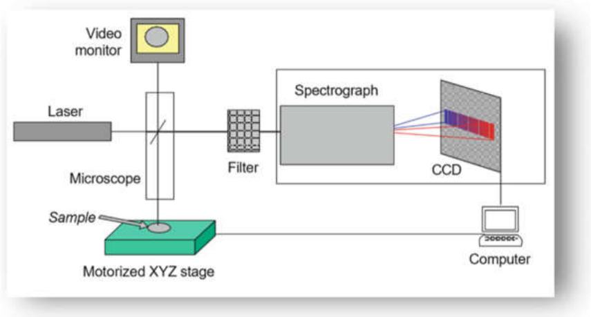
4.1.3. Fourier-Transform Infrared Spectroscopy
Fourier-transform infrared spectroscopy is widely used to identify functional groups, chemical bonds, and surface species present in metal oxide nanoparticles. It is based on the absorption of infrared radiation by molecular vibrations. When a sample absorbs infrared light, specific vibrational bands appear, corresponding to stretching, bending, or other vibrational modes of chemical bonds. In metal oxide nanoparticles[337], FTIR is especially useful for confirming metal–oxygen bond formation and detecting surface functional groups introduced during synthesis.In the FTIR spectra of metal oxide nanoparticles, absorption bands in the lower wavenumber region are commonly assigned to metal–oxygen stretching vibrations.FTIR is particularly important in green and biological synthesis of metal oxide nanoparticles because plant extracts, microorganisms, algae, and biomolecules contain many natural compounds that act as reducing, stabilizing, or capping agents. Functional groups such as hydroxyl, carbonyl, carboxyl, amine, phenolic, and alkyl groups may appear in the FTIR spectra, indicating the involvement of biomolecules in nanoparticle formation and stabilization. These surface functional groups can improve dispersion, prevent agglomeration, and enhance biological compatibility[338].
In chemically synthesized nanoparticles, FTIR can also detect residual solvents, precursor species, surfactants, polymers, or capping agents present on the surface. For example, broad bands around the higher wavenumber region are often associated with hydroxyl groups or adsorbed water, while bands in the mid-infrared region may correspond to organic molecules, nitrate residues, acetate groups, or other surface-bound species. The disappearance, shift, or reduction of specific bands after calcination or washing can indicate removal of organic residues and improvement in oxide purity[339].
FTIR is also helpful for studying surface modification and composite formation. When nanoparticles are functionalized with polymers, biomolecules, ligands, or other materials, changes in FTIR bands can confirm interaction between the oxide surface and the modifying agent. Such interactions may occur through hydrogen bonding, coordination, electrostatic attraction, or covalent bonding. In adsorption, photocatalysis, antimicrobial, and biomedical applications, FTIR is often used before and after treatment to understand surface interactions between nanoparticles and target molecules[340].
The FTIR spectra presented in Figure 8a compare the vibrational fingerprints of iron oxide phases, namely hematite (α-Fe₂O₃), maghemite (γ-Fe₂O₃), and magnetite (Fe₃O₄), highlighting their distinct structural characteristics. All spectra exhibit characteristic Fe–O lattice vibrations in the low wavenumber region, which serve as the primary indicator for phase identification in iron oxide systems. For magnetite, a strong absorption band appears near ~570 cm⁻¹, corresponding to Fe–O stretching vibrations in the inverse spinel structure. In the case of maghemite, multiple bands in the ~530–650 cm⁻¹ region are observed, reflecting cation-deficient spinel distortions and the presence of defect-induced lattice vibrations. Hematite shows prominent bands at higher wavenumbers in the ~450–550 cm⁻¹ region, consistent with the vibrational modes of its corundum-type structure. In addition, weak features around ~3640 cm⁻¹ in all samples are attributed to surface –OH stretching from adsorbed moisture.
Overall, the systematic shift and splitting of Fe–O vibrational bands clearly differentiate the three iron oxide phases, confirming successful phase identification and structural evolution from. These complementary techniques can be used together to provide versatile insight into the composition and bonding of materials in bulk, surface and non-transparent samples. magnetite to maghemite and hematite[341].
The working principles of three FTIR sampling modes are shown in the schematic presented in Figure 8b: transmission (TS), attenuated total reflectance (ATR), and photoacoustic spectroscopy (PAS). In the transmission mode, the IR radiation is transmitted through a thin sample, and gives bulk vibrational information. The evanescent wave ATR probe, On the other hand, comes from a high-refractive-index crystal, and only samples the surface region, making it ideal for minimal preparation surface analysis. PAS detects acoustic signals produced by periodic heating of the sample when it absorbs IR radiation, allowing the analysis of opaque or highly scattering samples[337].
The FTIR spectrum shown in Figure 8c displays key functional groups present in the sample. The broad band at ~3249 cm⁻¹ corresponds to O–H stretching vibrations of surface hydroxyl groups and adsorbed water, while the peak at ~1632 cm⁻¹ is due to H–O–H bending. Bands in the range ~1318–1158 cm⁻¹ are associated with C–O related vibrations, and the peak near ~627 cm⁻¹ is attributed to metal–oxygen (M–O) lattice vibrations, confirming the formation of the inorganic framework[342].
Although FTIR is a simple, rapid, and informative technique, it has some limitations. It does not directly provide detailed crystallographic information and cannot accurately determine particle size or morphology. Overlapping bands may also make interpretation difficult, especially in green-synthesized nanoparticles containing complex biological molecules. Therefore, FTIR is usually used together with XRD, Raman spectroscopy, SEM, TEM, and other techniques to provide a complete characterization profile of metal oxide nanoparticles.
Figure 8.
a): Representative FTIR spectra of major iron oxide nanomaterials, including hematite, maghemite, and magnetite, illustrating the characteristic metal–oxygen vibrational modes used for phase identification and structural characterization of iron oxide nanoparticles[341] (Reproduced with Permission). Fig 8b): Schematic representation of Fourier-transform infrared (FTIR) spectroscopy techniques employed for nanoparticle characterization, including transmission spectroscopy (TS), attenuated total reflectance (ATR), and photoacoustic spectroscopy (PAS). These methods provide valuable information on surface functional groups, chemical bonding, molecular interactions, and structural properties of metal oxide nanoparticles[337] (Reproduced with Permission). Fig 8c): Representative FTIR characterization of plant-mediated iron oxide nanoparticles, highlighting the role of naturally occurring phytochemicals in nanoparticle synthesis, surface capping, and enhanced colloidal stability for biomedical and environmental applications[342] (Reproduced with Permission).
Figure 8.
a): Representative FTIR spectra of major iron oxide nanomaterials, including hematite, maghemite, and magnetite, illustrating the characteristic metal–oxygen vibrational modes used for phase identification and structural characterization of iron oxide nanoparticles[341] (Reproduced with Permission). Fig 8b): Schematic representation of Fourier-transform infrared (FTIR) spectroscopy techniques employed for nanoparticle characterization, including transmission spectroscopy (TS), attenuated total reflectance (ATR), and photoacoustic spectroscopy (PAS). These methods provide valuable information on surface functional groups, chemical bonding, molecular interactions, and structural properties of metal oxide nanoparticles[337] (Reproduced with Permission). Fig 8c): Representative FTIR characterization of plant-mediated iron oxide nanoparticles, highlighting the role of naturally occurring phytochemicals in nanoparticle synthesis, surface capping, and enhanced colloidal stability for biomedical and environmental applications[342] (Reproduced with Permission).
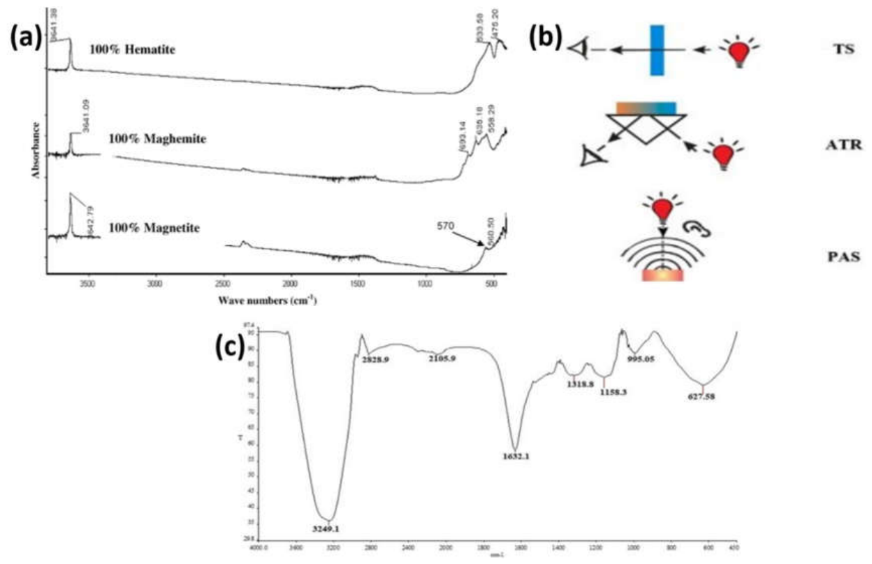
4.2. Morphological Characterization
Morphological characterization is essential for understanding the external shape, particle size, surface texture, aggregation behavior, and structural arrangement of metal oxide nanoparticles. The morphology of nanoparticles strongly influences their surface area, reactivity, adsorption capacity, optical properties, catalytic activity, antimicrobial behavior, and interaction with biological systems. Even nanoparticles with the same chemical composition may show different performance when their size, shape, porosity, or surface roughness changes.
The morphology of metal oxide nanoparticles is mainly controlled by synthesis conditions such as precursor concentration, pH, reaction temperature, reaction time, solvent system, calcination temperature, surfactants, templates, stabilizing agents, and biological reducing agents. Different synthesis methods can produce spherical, rod-like, flower-like, sheet-like, wire-like, cubic, porous, or irregular nanostructures. Therefore, morphological analysis helps explain how synthesis routes affect the final structure and performance of nanoparticles.
Scanning electron microscopy, transmission electron microscopy, and atomic force microscopy are commonly used to study nanoparticle morphology. SEM provides surface images and general particle arrangement, TEM gives detailed internal and nanoscale structural information, while AFM provides three-dimensional surface topography and roughness analysis[343].
4.2.1. Scanning Electron Microscopy
The scanning electron microscopy is among the widely used methodologies to study the surface morphology of metal oxide nanoparticles. Generates high resolution images through scanning the surface of the sample with a focused electron beam. When the electron beam encounters the sample, it produces signals which are used to create detailed images of the surface structure. SEM is particularly helpful for studying the shape, size distribution, agglomeration, porosity, surface texture and arrangement of nanoparticles[344].
SEM has become a common technique for confirming the homogeneity of the morphology of synthesized metal oxide nanoparticles in metal oxide nanoparticle studies. Can expose if the nanoparticles are spherical, rod-shaped, flake-shaped, flower-shaped, porous or clustered. For instance, the ZnO nanoparticles could be nanorods, nanoflowers, nanospherical or hexagonal particles, depending on the synthesis conditions. Other oxides, such as TiO₂, Fe₂O₃, CuO, CeO₂ can exhibit various morphologies depending on the reaction medium, reaction temperature and the process of calcination[345].
SEM can be used to determine the influence of various synthesis techniques on the morphology of the particles. Chemical precipitations can yield compact or agglomerated nanoparticles and hydrothermal and solvothermal processes can yield well-defined structures. The green synthesized nanoparticles can exhibit capping or coating effect as a result of biomolecules from plant and microorganisms, and this can significantly reduce the aggregation of nanoparticles and improve the stability of surfaces. Therefore, SEM images can complement the discussion of the influence of the synthesis conditions on the formation of nanoparticles[345].
The other significant use of SEM is for the research on aggregation behavior. Often nanoparticles have a high surface energy, which causes them to tend to agglomerate. SEM can be used to determine whether particles are isolated, loosely attached or strongly aggregated. Agglomeration impacts surface area, dispersibility, adsorption efficiency, catalytic activity and biological interaction. SEM observations can therefore be used to understand the practical performance of the metal oxide nanoparticles[346].
SEM can also be used in conjunction with the energy-dispersive X-ray spectrometer (EDX) for elemental analysis of nanoparticles. This enables researchers to validate the presence of the metal and the oxygen element and identify impurities and dopants. SEM-EDS mapping can reveal the elemental distribution on the surface of a sample in the case of doped or composite metal oxide nanoparticles.
But there are a few drawbacks to SEM. It primarily serves to give information on surface morphology and does not provide detailed information about the internal crystal structure detail. If the very small nanoparticles are highly aggregated, they may not be accurately measured. In certain samples a conductive material needs to be coated to the sample surface for analysis, particularly when the sample is not conductive. In spite of these drawbacks, SEM is still an important technique and is widely employed for the morphological characterization of metal oxide nanoparticles[347].
4.2.2. Transmission Electron Microscopy
Transmission electron microscopy is a very high power technique used for the characterization of the nanoscale morphology, particle size, shape, internal structure and crystallographic features of metal oxide nanoparticles. In contrast to SEM, which focuses on the surface, TEM involves a beam of electrons passing through an ultra thin sample, yielding highly magnified images. This makes it possible to see the individual nanoparticles in question and it is much more powerful than traditional microscopy[345].
TEM is especially significant for nanoparticles of metal oxides, since it can help to identify the real size and shape at the nanometer level. It is widely applied to identify the shape of nanoparticles such as spherical, rod-like, cubic, hexagonal, plate-like, hollow, porous or irregular. It can also indicates if the particles are well dispersed or aggregated. The crystallite size derived from TEM is sometimes compared to the size derived from the XRD and can be quite different because XRD measures the size of the crystalline domains and TEM measures the size of the whole particles.
High resolution TEM can give detail information of lattice fringes and crystalline planes. The distance of lattice fringes can be measured, and compared with the spacing of lattice planes of metal oxides. This is to support the confirmation of crystallinity and phase formation. Another application of TEM is selected area electron diffraction for the identification of the single crystalline, polycrystalline or amorphous character of the nanoparticles. The presence of rings in a SAED pattern typically suggest that the material is polycrystalline and sharp spots suggest single-crystalline[348].
TEM is also applicable for the investigation of core–shell structures, doped nanoparticles and nanocomposites, and surface coatings. TEM can illustrate the contact between the various components and the distribution of nanoparticles on supports, like graphene oxide, carbon nanotubes, polymers, clays or other oxide matrices in composite materials. In the case of nanoparticles that are synthesized using green methods, TEM can show organic shells or capping agents that are used to synthesize the nanoparticles, which come from plants or biological molecules.
Another important advantage of TEM is its ability to provide reliable particle size distribution. Average particle size and size distribution can be calculated by measuring the size of a large number of particles in the TEM images. Such information is well important as the size of the nanoparticles affects the surface area, band gap, photocatalytic activity, antimicrobial effects, cytotoxicity, and adsorption properties.
Even though there are many benefits to TEM, it does have its drawbacks. Sample preparation is more critical and time consuming than for SEM. Sample should be as thin as possible and well dispersed on an appropriate grid. Natural dispersion state of nanoparticles may be altered by drying effects. Also, TEM provides information on a very small part of the sample and therefore the part examined must be representative. Further, the instrument is costly and needs expertise to operate it. Despite these drawbacks, TEM is one of the most accurate methods for the detailed characterization of metal oxide nanoparticles at the nanoscale[349].
4.2.3. Atomic Force Microscopy
The three-dimensional topography, surface roughness, particle height, grain size, and surface features of metal oxide nanoparticles and thin films can be studied using a surface imaging technique called atomic force microscopy. AFM is not based on an electron beam as is SEM or TEM. It is instead based on a very sharp probe tip which is scanned across the surface of the sample. The interaction between the tip and surface is captured to give a high resolution topographical image.
AFM is particularly suited to the analysis of nanoparticles deposited on solid substrates, thin films, coatings, membranes and supported nanostructures. It has the ability to give details of the surface level and roughening, which is not possible to find in the same manner from SEM or TEM. Surface roughness is also an important parameter for metal oxide nanoparticles in applications such as sensors, photocatalysis, biomedical coatings, solar cells and membranes, as it governs surface contact, adsorption sites, wettability, charge transport, and biological interactions.
For metal oxide nanoparticles, AFM can provide information on the distribution of particles, surface grains, particle clustering, and surface uniformity. It also proves to be useful in comparing films prepared by different methods including sol–gel coating, spin coating, dip coating, sputtering, chemical bath deposition, and hydrothermal growth. A smoother or rougher surface may be an indication of changes that may have occurred in nucleation, growth rate, particle packing, or post-treatment conditions.
AFM quantitatively gives the average roughness, root mean square roughness, particle height and surface profile. These values are used to characterize the surface quality and the textured structure of the nanoparticles or the nanostructured films. For instance, it can be desirable to increase the surface roughness to increase the photocatalytic or sensing activity, since the more surface sites, the greater the activity. On the other hand, for electronic or optical applications, it may be desirable to have more uniform surfaces.
Another benefit of AFM is the ability to function under various environment such as in air, liquid or controlled environment. This is useful in studying biological and surface interactions particularly in biomedical and environmental applications of nanoparticles. AFM can also be applied to study mechanical properties, adhesion forces and interactions at the nanoscopic level.
But there are some drawbacks to AFM. It scans a relatively small surface area, and may not be representative of the sample if it is very heterogeneous. The image quality is determined by the sharpness of the probe tip and the conditions of scanning. The loosely attached or soft nanoparticles can be displaced during the scanning process. AFM is also not as well suited for studying large quantities of powder samples unless they are deposited on a flat substrate. So, AFM is typically used together with SEM and TEM, to obtain a more comprehensive picture of the morphology and surface structure of nanoparticles[350].
4.3. Optical and Surface Characterization
Optical and surface characterization are particularly crucial for assessing the light absorption profile, electronic transitions, defect sites, surface area, surface chemistry, charge state and colloidal stability of metal oxide nanoparticles. These properties have significant impact on their applications as nanoparticles in photocatalysis, sensing, antimicrobial action, adsorption, energy storage, environmental remediation and biomedical action. As many of the optical and surface properties of metal oxide nanoparticles are size dependent, these techniques provide an understanding of the correlation between the synthesis conditions, structural characteristics, and functional behavior.
The optical properties of metal oxide nanoparticles are mainly studied using UV–visible spectroscopy and photoluminescence spectroscopy. UV–visible spectroscopy can be used to obtain the information on light absorption and band gap energy, while photoluminescence spectroscopy can be used to get information on electron–hole recombination, defect states, oxygen vacancies, and surface traps. BET surface area analysis, X-ray photoelectron spectroscopy and zeta potential measurements are typical methods for analysis of surface related properties. BET analysis gives information on surface area, pore volume, pore size while XPS determines surface elements, oxidation states, and chemical bonding and zeta potential analysis on surface charge and dispersion stability.
In combination, these are useful methods for gaining a general idea of the reactivity and use potential of metal oxide nanoparticles as a function of their surface and optical properties. A small band gap can enhance visible light absorption, high surface area can facilitate more adsorption and higher catalytic activity, high absolute value of zeta potential can enhance colloidal stability, and oxygen vacancy can promote more charge separation[351].
4.3.1. UV–Visible Spectroscopy
UV–visible spectroscopy is a very popular method to investigate optical absorption property of metal oxide nanoparticles. It introduces the information about the absorption of ultraviolet and visible light by the material and is important to know its information about electronic transitions, optical band gap, particle size effects, and light-harvesting ability. It is a very significant technique for metal oxide nanoparticles applied in photocatalysis, solar cells, sensors, antimicrobial systems and optoelectronic devices.
For the metal oxide nanoparticles, the UV–visible spectra typically exhibit characteristic absorption bands associated with the transition of electrons from the valence band to the conduction band. The location and intensity of the absorption edge is influenced by the type of metal oxide, particle size, crystallinity, morphology, defect concentrations and synthesis techniques. For instance, the nanoparticles of ZnO and TiO₂ are typically more absorptive in the ultraviolet part of the spectrum because they are wide-band gap materials. But, doping, defect formation, surface modification or composite formation can be used to alter the absorption edge to become more visible light active.
The Tauc plot method is a common method used to estimate the optical band gap of nanoparticles from UV–visible absorption data. Thus, the band gap calculated can be used for deciding the suitability of the material for applications such as UV or visible light driven applications. The bigger the band gap, the higher the ultraviolet absorption ability and the less the band gap, the higher the photocatalytic ability under visible light. The band gap energy may be altered by quantum confinement, lattice defects, oxygen vacancies, doping, or by the interaction with other semiconductor materials.
UV–visible spectroscopy is another tool that can be used for monitoring the generation of the nanoparticles during synthesis. The presence of a certain peak of absorption in some cases can help to attest to the formation of nanoparticles. The height of the absorption peak can be increased as the time of the reaction increases, showing growth or increase in concentration of nanoparticles. UV–visible analysis is also a preliminary tool for the confirmation of nanoparticles formation in green synthesis as the plant extracts, microorganisms and biomolecules are capable of reducing the metal ions and stabilizing the oxide nanoparticles.
In addition, UV–visible spectroscopy can be utilized to track the degradation of organic pollutants like dyes, pharmaceuticals and other contaminants for photocatalytic applications. If the absorbance of the pollutant solution decreases over time, it means that degradation or removal of pollutant solution is taking place as a result of the nanoparticle catalyst. Hence, the application of this technique does not only help to characterize the nanoparticles but also it can be used for the assessment of the photocatalytic activity of the nanoparticles.
Despite the fact that UV – visible spectroscopy is simple, fast, and non – destructive, there are some limitations. It does not give any direct information on morphology, surface chemistry and crystal structure. Sample preparation, concentration, and solvent type, as well as particle aggregation and scattering, can also influence the absorption spectrum. Hence, the UV–visible results are typically complemented with the results of XRD, TEM, PL, BET and XPS to get a more complete understanding of the material[352].
4.3.2. Photoluminescence Spectroscopy
Photoluminescence (PL) spectroscopy is a vital optical method for investigation of electronic structure, defect states, charge carrier recombination and surface related emission of metal oxide nanoparticles. In this method, the material is stimulated with a light of the appropriate energy, and the light emitted is collected. The emission spectrum reveals the behavior of the excited electrons and holes, which is very relevant when considering photocatalytic, sensing, antimicrobial and optoelectronic applications.
The origin of photoluminescence emission in metal oxide nanoparticles can be attributed to near-band-edge transitions, surface defects, oxygen vacancies, interstitial defects and trapped charge carriers. High PL intensity is likely to be a sign of high probability of recombination of the photo-generated electrons and holes and thus lower photocatalytic efficiency. Conversely, lower PL intensity typically indicates more efficient charge separation and slower recombination, which is beneficial for photocatalytic reactions and for photoelectrochemical performance.
The defect related properties of metal oxide nanoparticles are often investigated by the technique of Photoluminescence Spectroscopy. Defects such as oxygen vacancies, zinc interstitial, titanium defects, surface hydroxyl groups as well as other trap states can establish new energy levels inside the band gap. These defect states can give rise to the observed emission bands and have dramatic consequences on the reactivity of the nanoparticles. Controlled defect formation can enhance charge separation and ROS generation, and overabundant defects can serve as recombination centers in photocatalysis.
The PL spectra of ZnO nanoparticles generally consist of a UV peak corresponding to the band-edge emission and a visible peak corresponding to defect emission. Emission behavior in the case of TiO₂ is often attributed to the presence of self-trapped excitons, oxygen vacancies, and surface states. Other metal oxides, e.g. CeO₂, CuO, Fe₂O₃, or SnO₂, also exhibit defect-dependent PL properties, which are pertinent to the understanding of their optical and catalytic properties.
The photoluminescence spectroscopy can also be applied to the analysis of doped and composite metal oxide nanoparticles. The intensity and peak position of photoluminescence can be modified by doping with metal and non-metal elements or by combining with carbon materials, polymers, and other semiconductors. Typically a drop in emission intensity after modification means that charge separation has been better and recombination has been less. It is frequently employed to interpret the improvements of photocatalytic, antibacterial or sensing activity.
But the interpretation of photoluminescence can be difficult since the emission bands can overlap and a number of defect types can overlap in the same region of the spectrum. Other factors that can influence the results include excitation wavelength, sample concentration, particle size, and surface adsorbates. Hence, the data obtained by PL should be discussed in conjunction with the results obtained from the UV–visible spectroscopy, XPS, Raman spectroscopy and the photocatalytic activity data[353].
4.3.3. BET Surface Area Analysis
BET surface area analysis is commonly used for the measurement of the specific surface area, pore volume, pore size distribution and porosity of metal oxide nanoparticles. The method involves the physical adsorption and desorption of gas molecules (usually nitrogen) on the surface of the material at low temperature. BET analysis is an important tool to evaluate the functional potential of many applications of metal oxide nanoparticles which rely heavily on available surface area and porosity.
A high specific surface area is generally preferred since it offers a greater number of active sites for the adsorption, catalytic reaction, photocatalytic degradation, antibacterial interaction, gas sensing and energy storage. Due to their small size and high surface to volume ratio, nanoparticles tend to have larger surface area than bulk materials. Agglomeration, On the other hand, and high calcination temperature and particle growth can lead to a reduction in surface area owing to fewer surface sites being available.
The BET analysis is of significance for the metal oxide nanoparticles as adsorbents and photocatalysts. The larger the surface area, the more pollutant molecules can interact with the adsorbent surface in adsorption applications, and the more suitable the pore structure is. The high surface area in photocatalysis leads to better contact between the pollutant molecules and the catalyst and thereby enhances the chances of surface reactions. Likewise, porous materials can be used in gas sensing for the diffusion of gas molecules into the material and reactions to active sites.
The pore size distribution can be derived from the adsorption–desorption isotherm and is used to classify the material, as microporous, mesoporous, and macroporous. Such mesoporous metal oxide nanoparticles are particularly interesting for environmental and catalytic applications as the pores enable efficient mass transport and provide accessible reaction sites. The adsorption/desorption isotherm shape and adsorption/desorption hysteresis can give adsorption and pore structure, capillary condensation, and particle aggregation information.
BET surface area and porosity are quite sensitive to synthesis conditions. For instance, a lower calcination temperature may maintain a smaller particle size, and higher surface area; too high a temperature, On the other hand, may lead to sintering and collapse of the pores. Porosity and surface area can be enhanced by the use of templates, surfactants, green stabilizing agents, or controlled precipitation. Hence, BET analysis can be used to account for the differences in adsorption, catalytic, or biological activity of nanoparticles synthesized via various methods.
BET analysis is useful to obtain information about surface area, but does not directly reveal particle shape, crystal phase or surface chemical composition. It is also necessary to degas samples before measurement to remove moisture and adsorbed gases. Surface area values may not be accurate if the sample is not properly prepared. On the other hand, BET results are often used in conjunction with SEM, TEM, XRD and XPS for the better understanding of the relationship between morphology, structure, and surface properties[354].
4.3.4. X-Ray Photoelectron Spectroscopy
X-ray photoelectron spectroscopy is a powerful surface sensitive technique which can be used to investigate elemental composition, oxidation state, chemical bonding, surface defect, and electronic environment of the metal oxide nanoparticles. It is derived from the emission of photoelectrons from the surface of the sample after being exposed to X-rays. The amount of energy required to detach these ejected electrons gives clues to what elements are on the surface and how they are chemically bound.
XPS is particularly significant for metal oxide nanoparticles as many reactions will take place at the surface. Surface composition and chemical environment play a significant role in photocatalytic, adsorption, sensing, antimicrobial and biomedical interaction applications. XPS can detect surface contaminants, dopants, hydroxyl groups, oxygen vacancies, or adsorbed species, and can confirm the presence of metal and oxygen, and can identify oxidation states.
The metal-core level spectra can be used to obtain information on the oxidation state of the metal ions. For instance, XPS can be used to differentiate between the Fe²+/Fe3+ states, Ce3+/Ce4+, Cu+/Cu2+ or Ti3+/Ti4+. The significance of this lies in the fact that mixed oxidation states can be beneficial in terms of redox behaviour, oxygen storage capacity, photocatalytic performance, and catalytic activity. XPS may be used to determine if the dopant is part of the lattice or only on the surface of nanoparticles of doped metal oxides.
The XPS spectra of the Oxygen region is also very informative. The various oxygen species can be attributed to lattice oxygen, oxygen vacancies, surface hydroxyl groups, adsorbed water, or chemisorbed oxygen. The relative proportions of these species can be used to explain the reactivity of the surface. Oxygen vacancies are especially crucial in nanoparticles of metal oxides, where they can influence the charge separation, visible-light absorption, adsorptive properties, and the creation of reactive oxygen species.
Another application of XPS is to examine surface modification and the formation of composite. Modification of nanoparticles with polymers, functionalization with biomolecules, association with carbon materials, or modification with other oxides results in a change in binding energy and peak intensity, which can confirm the chemical interaction between the components. These interactions can enhance charge transfer, surface stability, adsorption capacity or biological compatibility.
A major advantage of XPS is its high surface sensitivity – typically the top few nanometres of the material will be analysed. This renders it very attractive for use in the study of nanoparticles, in which the surface chemistry plays a key role in determining performance. But this surface sensitivity can also be a disadvantage as XPS can not necessarily reflect the bulk composition of the material. It also needs high vacuum conditions and attention to the problem of overlapping peaks. Thus, XPS is generally employed along with the other techniques such as XRD, Raman spectroscopy, FTIR, and electron microscopy, to characterise materials in its entirety[355].
4.3.5. Zeta Potential Analysis
The surface charge and colloidal stability of metal oxide nanoparticles in liquid suspension can be assessed using zeta potential analysis. It is a measure of the electric potential that exists at the plane of shear surrounding dispersed particles. This value gives a lot of clues about the stability of particles, the tendency of aggregation, surface functionalization and interaction with ions, biomolecules, pollutant or cells in its surroundings.
When metal oxide nanoparticles are dispersed in water or other solvents, surface charge is formed as a result of the ionization of surface hydroxyl groups, adsorption of ions, and pH changes or the presence of stabilizers. Zeta potential is a measure of electrostatic repulsion between particles; the greater the magnitude of the zeta potential, the greater the electrostatic repulsion between particles. If the value of the zeta potential is high and positive or negative, then this would indicate good colloidal stability as the particles are strongly repulsive and cannot approach each other for aggregation. Values near zero are generally associated with low stability with high tendency of agglomeration.
pH has a significant effect on zeta potential. Most metal oxide nanoparticles have an isoelectric point, at which point the net surface charge is zero. Below this point the surface may be positively charged and above this point, it may be negatively charged. The pH dependent surface charge plays a crucial role in the adsorption, photocatalysis, antimicrobial activity and biomedical applications, as it governs the interaction of nanoparticles with the charged pollutant, charged bacterial membrane, proteins and biological fluids.
Zeta potential can be used to explain the adsorption behavior in environment application. The interaction of positively charged nanoparticles with negatively charged contaminants can be more attractive than the interaction of negatively charged nanoparticles with positively charged contaminants. Surface charge in antimicrobial use, influences the interaction with cell walls and cell membranes of bacteria. For biomedical applications, zeta potential can affect nanoparticle stability in physiological environment, protein binding to nanoparticles and their uptake by cells, and potential toxicity.
Zeta potential analysis can also be used to confirm surface modification. If nanoparticles are coated with polymers, biomolecules, surfactants, plant extract compounds or functional groups, the value of the zeta potential can change. This shift shows the successful surface functionalization and surface chemistry changes. In green synthesis, zeta potential is often used to evaluate the stabilizing effect of phytochemicals or microbial biomolecules on nanoparticle dispersions.
While zeta potential is useful to indicate the colloidal stability, it is not a good direct measure of particle size, morphology, or chemical composition. Solvent, pH, ionic strength, temperature, concentration and measurement conditions affect the measured value. Hence, the interpretation of the zeta potential results should be taken with caution and corroborated with particle size analysis, FTIR, XPS, SEM, TEM, and stability observations[356].
5. Applications of Metal Oxide Nanoparticles
Metal oxide nanoparticles (MONPs) is a topic of great interest in scientific and industrial fields due to their unique and outstanding physicochemical properties such as high surface to volume ratio, surface tunability, catalytic activity, optical absorption, redox properties and antimicrobial activity. These unique features allow MONPs to be used in various applications such as environment, biomedical, agriculture, electronic and energy. Of these, environmental remediation has become one of the most significant applications areas in the framework of the growing worldwide worry about water pollution, air contamination, industrial wastes and ecosystem degradation[357].
Due to their nanoscale size and reactivity, the environmental performance of MONPs is closely related to their properties. The nanoparticles like TiO₂, ZnO, Fe₃O₄, CuO, CeO₂, MnO₂ and MgO have shown some extraordinary adsorption, photocatalytic, oxidation–reduction and antimicrobial properties which are highly beneficial for removal of pollutants and for purification of environment[358,359]. The efficiency of them can be further enhanced by controlling the synthesis parameters, morphology, crystal phase, surface defects, porosity and band-gap engineering. In recent years, considerable research has been dedicated to the development of multifunctional/sustainable MONPs that can simultaneously degrade pollutants, adsorb toxic metals and kill pathogenic microorganism[360].
In addition, some advanced nanocomposites of MONPs have been prepared by combining MONPs with carbon materials, polymers, biopolymer and other semiconductors to improve the stability, recyclability and catalytic activity of MONPs. Green-synthesized metal oxide nanoparticles also have received immense attention for environmental application since they involve the use of less hazardous chemicals and less risk to the environment during the fabrication of the nanoparticles. Major environmental applications of MONPs are discussed below, namely: Photocatalytic degradation of dyes, Photocatalytic degradation of organic pollutants, Wastewater treatment, Heavy metal removal, and Air purification technologies[361].
Figure 9.
a):Graphical Representation of applications of metal oxide NPs[361] (Reproduced with Permission).
Figure 9.
a):Graphical Representation of applications of metal oxide NPs[361] (Reproduced with Permission).
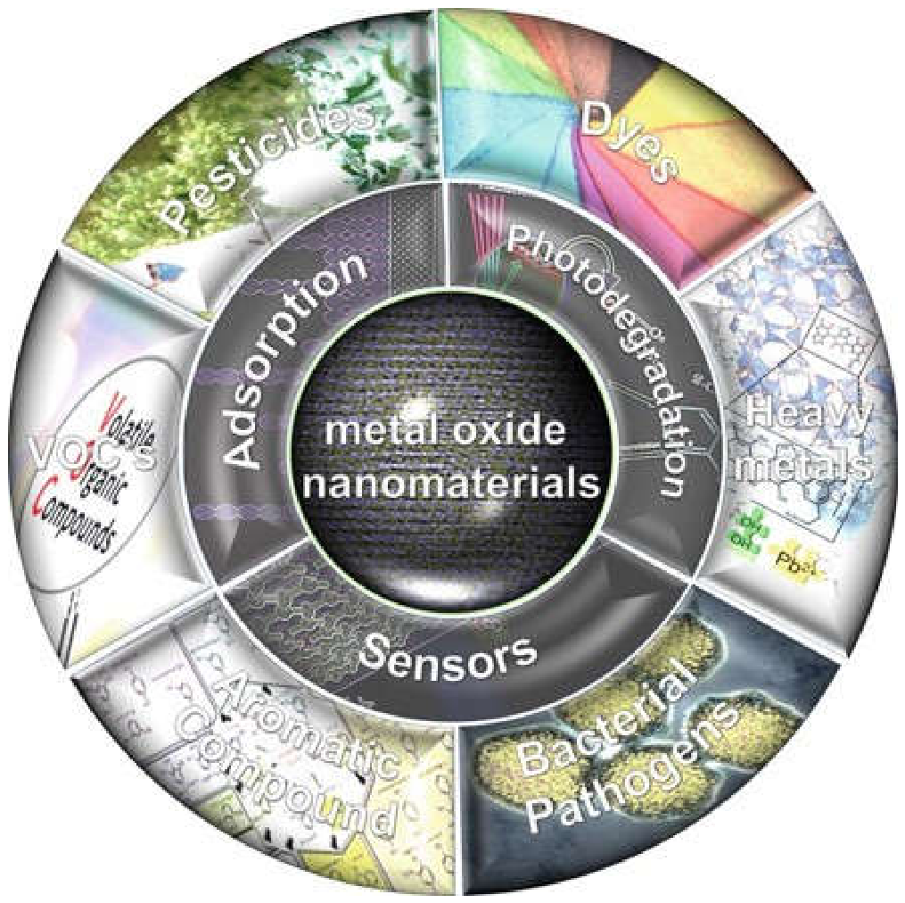
5.1. Environmental Applications
The pollution of the environment due to the rapid industrialization, urbanization and agricultural activities is a key issue in the world. Pollution of water sources, release of toxic gases, deposition of heavy metals, and discharge of organic wastes pose a risk to human health and ecological balance. Traditional treatment technologies have a number of drawbacks, including incomplete removal of pollutants, high operating costs, the formation of secondary pollutants, and low efficiency in treatment of persistent pollutants. Metal oxide nanoparticles with its excellent catalytic, adsorptive, magnetic and antimicrobial properties have emerged as a potential solution for environmental remediation[362].
MONPs have large active surface area and many active sites which can make it easier to interact with pollutants. Semiconductor metal oxides like TiO₂ and ZnO are capable of producing ROS upon illumination that can also degrade toxic organic species into innocuous products. The ease of separation and recovery of magnetic nanoparticles, such as Fe₃O₄, from the treated water enhances the reusability and operational convenience. Besides that, porous and surface-functionalized MONPs have excellent adsorption properties towards heavy metals and harmful ions[222,363].
Recent developments in nanotechnology and material engineering also enhance environmental applications of MONPs. The pollutant removal efficiency and the photocatalytic performance are significantly improved by surface modification, doping, heterojunction formation, and incorporation into membranes or nanocomposites. Besides that, green synthesis by plant extract and microbial systems offer eco-friendly pathways for the preparation of MONPs for sustainable remediation technologies. These benefits have led to growing research on MONPs as wastewater treatment materials, dye and pesticide degradation, air purification devices, antimicrobial coatings and environmental monitoring devices[364].
5.1.1. Photocatalytic Degradation of Dyes and Organic Pollutants
Metal oxide nanoparticles have been studied in environmental applications to the greatest extent, and one of these applications is photocatalytic degradation. Industries like textile, leather, pharmaceuticals, printing, cosmetics and dye manufacturing, generate huge amounts of coloured effluents and toxic organic compounds into aquatic environment. The majority of these pollutants are non-biodegradable, carcinogenic, mutagenic and can't be easily treated with conventional treatment methods. Semiconductor metal oxide nanoparticles have shown excellent ability to degrade such contaminants under the ultraviolet (UV) or visible light irradiation[365,366].
Of the various materials investigated, titanium dioxide (TiO₂) is the most popular photocatalyst because of its high oxidation potential, stability, low toxicity, cheapness and resistance to photocorrosion. Upon absorption of photons, whose energies are equal to or higher than the band gap of TiO₂ nanoparticles, electrons can be excited from the valence band to the conduction band, creating positive holes in the valence band. These electron–hole pairs can interact with water and oxygen molecules to produce reactive oxygen species that oxidize added organic contaminants to carbon dioxide, water and mineralized products. The ZnO nanoparticles also have similar photocatalytic properties and frequently give a higher quantum efficiency due to its excellent electron mobility and large exciton binding energy[367].
Recent studies have been directed towards increasing the efficiency of the photocatalytic effect, by doping and nanocomposite formation. Doping of TiO₂ or ZnO with transition, rare earth or nonmetals can lower the electron–hole recombination and to prolong the light absorption into the visible range. Ag doped TiO₂, Fe doped ZnO, and CuO/TiO₂ heterostructures are examples of such materials, which have demonstrated improved degradation performance for methylene blue, rhodamine B, methyl orange, and phenolic species when exposed to visible light irradiation. Likewise, the combination of MONPs with graphene oxide, activated carbon, carbon nanotubes or conducting polymers enhances charge separation and enhances adsorption capacity[368,369,370].
The morphology, which is also very important for photocatalytic activity, is also crucial. Larger active surface areas and efficient light harvesting are achieved using nanorods, nanotubes, nanoflowers, hollow spheres and mesoporous structures. To obtain highly crystalline photocatalysts with controlled morphologies, hydrothermal/solvothermal synthesis methods are often used. Besides that, the generation of oxygen vacancy defects and surface defects can greatly promote photocatalytic reactions, such as in the form of active sites for adsorbing and transferring pollutants[371].
MONPs can be used for the removal of pesticides, antibiotics, endocrine-disrupting chemicals, pharmaceutical residues and volatile organic compounds apart from dye degradation. Photocatalytic nanoparticles are emerging as an effective technology for sustainable wastewater treatment, as they are capable of degradation with high efficiency, low sludge yield and have a potential to utilize solar-light[372].
5.1.2. Wastewater Treatment
Wastewater sources such as industrial, domestic, agriculture and medical wastes are characterized by a variety of contaminants, which may include dyes, pathogens, pharmaceuticals, oils, pesticides, heavy metals, and suspended solids. Wastewater treatment plays a crucial role in safeguarding aquatic ecosystems and water resources. Metal oxide nanoparticles are multifunctional materials that exhibit various properties such as adsorption, photocatalytic action, antimicrobial activity, enhancement of membranes, and catalytic oxidation, making them highly efficient materials for wastewater treatment[373].
The magnetic properties and high adsorption capacity of iron oxide nanoparticles such as Fe₃O₄ and γ-Fe₂O₃ make them an attractive choice in wastewater treatment. These nanoparticles are capable of effectively removing contaminants like arsenic, chromium, fluoride, phosphate and organic pollutants from water. Due to their magnetic property, they are easily separated after treatment, which can reduce secondary pollution and ease recovery process. The adsorption efficiency and selectivity are also improved by surface functionalization with polymers, silica or organic ligands[374].
The ZnO and TiO₂ nanoparticles are commonly used in photocatalytic wastewater treatment systems to degrade persistent organic pollutants and disinfect pathogenic microorganisms. These nanoparticles generate ROS under light irradiation, which can break down the cell wall of bacteria, the structure of viruses and toxic organic molecules. CuO and MgO nanoparticles also possess high antimicrobial activity against both gram positive and gram negative bacteria that are widely present in the wastewater system[253,358,375].
The application of metal oxide nanoparticles is being introduced in the membrane technologies with an aim to enhance the water filtration capacity. Nanocomposite membranes with the incorporation of TiO₂, Al₂O₃, ZnO or SiO₂ nanoparticles exhibit increased hydrophilicity, increased permeability, decreased fouling, increased thermal stability and increased mechanical strength. These membranes can effectively filter out microorganisms, organic compounds and dissolved contaminants and minimise membrane fouling issues that are typically associated with conventional filtration systems[376,377].
The wastewater treatment based on the adsorption process by MONPs has also been much focused due to its simple and efficient process. Porous nanoparticles have a high surface area, which offers many adsorption sites for the contaminants to bind. Nanoparticles of cerium oxide, manganese oxide and magnesium oxide have displayed outstanding adsorption capabilities to dyes, fluoride ions and toxic metals. Also, the hybrid nanocomposites of metal oxides with activated carbon, biochar, chitosan or derivatives of graphene have shown synergic effects that have been beneficial for adsorption kinetics and pollutant removal[378].
In recent years, research focus is on sustainable and green wastewater treatment technologies based on the biologically synthesized MONPs. Nano particles prepared using plants are less toxic and more biocompatible and possess good catalytic and antimicrobial properties. Although great progress has been made, there are still many issues to be addressed before large-scale implementation including the recovery efficiency, long-term stability, environmental safety and aggregation of the nanoparticles[379].
Table 4.
Application of Metal Oxide Nanoparticles in Wastewater Treatment and Photocatalysis.
| Serial no | Metal Oxide Nanoparticle | Target Contaminants | Treatment Mechanism | Key Advantages | Major Applications | References |
|---|---|---|---|---|---|---|
| 01 | TiO₂ NPs | Organic waste in olive mill wastewater (high COD), phenolic compounds, suspended solids | Absorb and trap pollutants on their surface, reducing pollution (COD) | Fast cleaning, strong removal of pollution, works with small amount of TiO₂, effective process, reduces high organic load in wastewater | Treatment of industrial wastewater, especially olive mill wastewater; removal of organic pollutants and phenolic compounds | [380] |
| 02 | Titanium dioxide (TiO₂) nanoparticles | Heavy metals, organic dyes, pharmaceuticals, and other toxic pollutants in wastewater | TiO₂ nanoparticles break down harmful pollutants using photocatalysis (light-driven reaction) | High efficiency in cleaning water, large surface area, works under light, eco-friendly, can degrade different types of pollutants | Wastewater treatment, removal of toxic pollutants from water, environmental cleaning | [381] |
| 03 | Titanium dioxide (TiO₂) nanoparticles | Organic dyes (e.g., methylene blue, rhodamine B), phenol and derivatives, pharmaceuticals (diclofenac, sulfadiazine, acetaminophen), microplastics, industrial wastewater pollutants | TiO₂ works as a photocatalyst. Under light (UV, solar, or visible), it produces reactive oxygen species (ROS) that break down and degrade harmful pollutants into less toxic substances. | Very high degradation efficiency, works under different light sources, reusable materials, eco-friendly, non-toxic, strong stability | Wastewater treatment, removal of dyes and pharmaceutical waste, water purification systems, photocatalytic membranes | [382] |
| 04 | TiO₂ and its composites with graphene (G) and graphene oxide (GO) | Water pollutants such as organic dyes, toxic chemicals, and other contaminants in wastewater | Under light, TiO₂ generates reactive species that break down pollutants. Graphene/GO improves electron movement, and increases photocatalytic efficiency. | Higher photocatalytic activity than pure TiO₂, works under UV–visible light, improved stability, faster pollutant degradation, cost-effective | Wastewater treatment, removal of dyes and toxic chemicals, water purification systems, | [383] |
| 05 | (GO/TiO₂ nanocomposites) | Dyes, heavy metals, oil, aromatic pollutants, and other toxic substances in wastewater | Photocatalysis: under light, TiO₂ produces reactive species that break down pollutants. GO helps reduce electron–hole recombination and improves pollutant adsorption and degradation. | Complete degradation of pollutants, eco-friendly, cost-effective, improved efficiency with GO, reduced secondary pollution | Wastewater treatment, environmental cleanup, removal of industrial dyes and oils | [384] |
| 06 | TiO₂–zeolite nanocomposite | Industrial dye pollutants and trace dye residues in wastewater | Combines adsorption and photocatalysis. Zeolite first adsorbs dye molecules, then TiO₂ breaks them down under light using photocatalytic reactions. | Easy separation and recovery after treatment, high efficiency at low dye concentration, strong adsorption + degradation ability | Advanced industrial wastewater treatment, especially dye removal and water purification in textile and chemical industries |
[385] |
| 07 | RGO/TiO₂) nanocomposite | Oil-water emulsions, oily wastewater, and asphaltene-stabilized oil droplets | RGO/TiO₂ moves to the oil–water interface, breaks oil droplet stability by adsorbing or replacing asphaltenes, and separates oil from water | Fast separation (within 30 minutes), high efficiency, works in salty, acidic, and neutral conditions, strong interfacial activity, good stability | Treatment of oily wastewater, oil spill cleanup, industrial wastewater purification, and petroleum industry wastewater management | [386] |
| 08 | TiO₂ combined with cellulose nanocrystals | Heavy metals, toxic organic molecules, drugs/pharmaceutical residues, and other water pollutants | Cellulose provides a high surface area and rich –OH groups that help adsorb pollutants, while TiO₂ breaks them down using photocatalysis under light. | Eco-friendly, biodegradable, high adsorption ability, strong interaction with pollutants, cost-effective, suitable for large-scale industrial use | Wastewater treatment, removal of heavy metals and pharmaceutical waste, environmental purification, and industrial water cleaning | [387] |
| 09 | Silver oxide nanoparticles (Ag₂O NPs) | Toluidine Blue (TB) dye and other organic dye pollutants in contaminated water | Under light exposure, they generate electron–hole pairs and reactive species that break down dye molecules into less harmful substances. | Eco-friendly green synthesis, cost-effective, high photocatalytic efficiency (98.5% dye removal), fast degradation rate, and good stability | Wastewater treatment, dye-contaminated water purification, environmental pollution remediation | [388] |
| 10 | Silver oxide nanoparticles (Ag₂O NPs) | Heavy metals (especially lead, Pb), chemical pollutants (high COD), turbidity-causing particles, dissolved contaminants, nitrates, and phosphates in industrial wastewater | Ag₂O NPs adsorb and oxidize pollutants due to their high surface area and redox activity. They trap heavy metals and help break down chemical contaminants, reducing COD and turbidity. | Very high removal efficiency (92% Pb, 88% turbidity, 85% COD),within 24 hours), high surface area, strong redox activity, effective against heavy metals and persistent pollutants | Industrial wastewater treatment, heavy metal removal, reduction of COD and turbidity, water purification, and environmental remWastewater treatment, dye remediation | [389] |
| 11 | Silver oxide nanoparticles | Organic dyes such as methylene blue and methyl orange, bacteria (E. coli, S. aureus, Shigella dysenteriae, Listeria monocytogenes), and reactive oxygen species (ROS) |
Under UV light, breaking down dye molecules into less harmful compounds. They also kill bacteria by damaging cell membranes and generating ROS. | Green and eco-friendly synthesis, utilizes agricultural waste, antioxidant properties, effective dye degradation | Wastewater treatment, dye removal, environmental remediation, antibacterial agents, antioxidant applications | [390] |
| 12 | Silver oxide nanoparticles | Methylene Blue (MB) (industrial dye pollutant in wastewater) |
Ag₂O NPs act as a photocatalyst. Under light, they generate reactive species that break down MB dye into less harmful substances. |
Green and eco-friendly synthesis, good crystallinity, effective dye degradation, simple and low-cost preparation | Wastewater treatment, dye removal from industrial effluents, environmental pollution control, and photocatalytic degradation processes | [391] |
| 13 | silver oxide and TiO₂) NPs incorporated into polyacrylonitrile (PAN) nanofibers (PAN-TA composite) | Methylene Blue (MB) dye and other dye pollutants in wastewater |
The nanofibers adsorb dye molecules onto their surface and the Ti/Ag oxide nanoparticles provide photocatalytic degradation. The nanofibers can then be easily separated from water. | Complete dye removal within 20 minutes, high adsorption capacity (155.4 mg/g), easy recovery from water, reusable for multiple cycles | Wastewater treatment, dye removal from industrial effluents, water purification, antimicrobial filtration systems | [392] |
| 14 | Ag₂O + Graphene oxide (GO) | Industrial dyes (Safranin-O), E. coli bacteria | Strong light absorption → fast electron transfer → high ROS production (•OH, O₂⁻ radicals) | Industrial wastewater treatment, antibacterial surfaces, solar-driven purification systems | Works under visible–NIR light, very fast dye degradation, high antibacterial efficiency, improved stability | [393] |
| 15 | Ag NPs on reduced graphene oxide (rGO) | Dye pollutants (methyl orange), total dissolved solids (TDS) in wastewater | Photocatalysis under visible light + adsorption on rGO surface → breakdown of dye molecules and pollutant trapping | Wastewater treatment, dye removal, environmental purification systems | High efficiency under visible light, strong adsorption, reusable, stable nanocomposite | [394] |
| 16 | Ag decorated reduced graphene oxide Nanocomposites | Toxic dye (Nile Blue) in wastewater | Strong adsorption on graphene surface + photocatalytic degradation enhanced by light → dye breakdown | Wastewater treatment, dye removal from industrial effluents, environmental cleanup | Very high removal efficiency (up to 94%), fast treatment (60 min), reusable, enhanced performance under light | [395] |
| 17 | Iron oxide nanoparticles (Fe₂O₃ / Fe₃O₄ and modified composites) | Dyes, heavy metal ions, industrial toxic pollutants | Adsorption of pollutants on surface + photocatalytic degradation → breakdown/removal from water | Wastewater treatment, dye removal, heavy metal cleanup, industrial effluent treatment | Easy magnetic separation, reusable, effective for multiple pollutants, improved stability in composites | [396] |
| 18 | Iron-based nanomaterials (iron oxides and composites) | Industrial pollutants, wastewater contaminants, general water pollutants | Adsorption of pollutants + filtration through modified membranes + surface interaction trapping contaminants | Water purification, wastewater treatment, industrial effluent cleaning | Low cost, abundant availability, eco-friendly, high efficiency, improves membrane performance | [397] |
| 19 | Iron oxide nanomaterials (Fe₂O₃ / Fe₃O₄) | Industrial wastewater pollutants, dyes, heavy metals, general toxic contaminants | Adsorption (nanosorbent action) + photocatalysis + immobilization of pollutants on surface | Wastewater treatment, pollutant removal, environmental cleanup systems | Easy magnetic separation, reusable, high efficiency, multifunctional (adsorption + photocatalysis), eco-friendly | [398] |
| 20 | Iron oxide nanoadsorbents (various Fe oxide phases) | Wastewater pollutants, dissolved toxins, dyes, mixed contaminants | Adsorption of pollutants onto nanoparticle surface, fixed-bed column adsorption for continuous removal | Drinking water purification, industrial wastewater treatment, | Efficient removal, works under different conditions (pH, temperature), reusable | [399] |
| 21 | Iron oxide nanoparticles and their nanocomposites (Fe₂O₃ / Fe₃O₄ based systems) | Organic dyes, heavy metals, pharmaceuticals, pesticides | Adsorption on high-surface-area particles + surface interaction + pollutant trapping and remova | Wastewater treatment, drinking water purification, removal of industrial pollutants |
Highly selective, works for multiple pollutant types, efficient and reusable | [400] |
| 22 | Fe₃O₄ + Graphene oxide (GO) coated with alginate (Alg–Fe₃O₄@GO composite) | Cationic dyes: Methylene Blue (MB), Malachite Green (MG) | Adsorption (high surface area + porous structure) + photocatalytic degradation under visible light → dye breakdown | Wastewater treatment, dye removal from industrial effluents, | Very high removal efficiency (up to 98.5%), reusable for multiple cycles | [401] |
| 23 | Superparamagnetic iron oxide (Fe₃O₄) on rectorite clay (REC-Fe₃O₄) | Dyes: MB, Neutral Red (NR), Methyl Orange (MO) | Adsorption of dye molecules on surface + magnetic separation after treatment | Wastewater treatment, dye removal from textile and industrial effluents | Easy magnetic separation, improved adsorption for specific dyes, fast recovery from water | [402] |
| 24 | Magnetic iron oxide nanocomposites | Organic pollutants, industrial wastewater contaminants | Adsorption + photocatalytic oxidation + chemical oxidation → breakdown and removal of pollutants | Wastewater treatment, industrial effluent purification, removal of organic compounds | Low cost, easy recovery using magnet, high efficiency, works in multiple treatment methods, | [403] |
| 25 | Bismuth-based nanoparticles (Bi-based NPs and derivatives | Organic pollutants, industrial dyes, heavy metals in wastewater | Visible-light catalytic degradation + adsorption + multi-pathway oxidation → breakdown/removal of pollutants | Wastewater treatment, dye degradation, heavy metal removal, environmental cleanup systems | Works under visible light, high efficiency, multifunctional degradation pathways, strong catalytic performance |
[404] |
| 26 | Bismuth oxide nanoparticles (BiONPs) synthesized via plant extracts | Organic dyes (MB, MG), bacteria (E. coli, S. aureus, K. pneumoniae), mosquito larvae (Aedes spp.) | Photocatalysis (light-driven dye degradation) + antibacterial action (cell membrane damage) + larvicidal toxicity | Wastewater treatment, dye degradation, antibacterial coatings, mosquito control, environmental cleanup | Green and low-cost synthesis, strong dye removal efficiency (up to 90%), broad antimicrobial activity, | [405] |
| 27 | Bismuth oxide nanoparticles (BiO-NPs) | Dye pollutants (Congo Red, Brilliant Green), toxic organic compounds (4-nitrophenol) | Photocatalysis (light-driven degradation), radical formation (•OH, electrons), and catalytic reduction (NaBH₄-assisted conversion) | Eco-friendly synthesis, high dye removal efficiency (~90%), reusable (up to 4 cycles) | Wastewater treatment, dye removal from textile industry effluents, chemical detoxification, environmental cleanup | [406] |
| 28 | Pure and doped bismuth oxide (Bi₂O₃) nanoparticles | Organic dye pollutants in wastewater | Photocatalytic degradation under light irradiation (visible-light activation enhanced by doping → better light absorption and charge separation) | Improved visible-light absorption, enhanced photocatalytic activity after doping, nanorod structure, | Wastewater treatment, dye degradation in textile effluents, solar-driven environmental cleanup, | [407] |
| 29 | Bismuth–TiO₂ nanotube (Bi–TNT) composites | Industrial wastewater pollutants (organic contaminants, mixed industrial dyes/chemicals) | Visible-light photocatalysis; Bi improves charge separation and light absorption in TiO₂ nanotubes, enhancing photodegradation of pollutants | Stronger photocatalytic activity, improved visible-light response, good stability and recyclability, efficient one-step synthesis | Industrial wastewater treatment, photocatalytic degradation of organic pollutants, environmental remediation under visible light | [408] |
| 30 | Zeolite/Bi₂O₃ nanocomposite | Rhodamine B (RhB) dye in industrial wastewater | Mainly adsorption with chemisorption (pseudo-second-order kinetics, dye molecules bind to active surface sites of the nanocomposite | Very high removal efficiency, fast treatment (10 min), reusable (stable up to multiple cycles), eco-friendly and sustainable | Industrial dye wastewater treatment, removal of toxic textile dyes, real wastewater purification, environmental cleanup | [409] |
| 31 | Bi₂O₃@GO nanocomposite | Rhodamine B in industrial wastewater | Mainly adsorption via surface interaction; follows Langmuir and Temkin isotherms (monolayer adsorption); dye molecules attach to active sites on GO and Bi₂O₃ surface | Higher adsorption capacity, high dye removal efficiency (~80.7%), stable and reusable, Efficient pollutant binding | Wastewater treatment, removal of textile dye pollutants, industrial effluent purification, | [410] |
| 32 | Bi/Bi₂O₃@Al₂O₃ nanocomposite | 4-nitrophenol (4-NP) and related toxic organic pollutants in wastewater | Photocatalytic reduction under UV–visible light; p–n heterojunction improves charge separation; electrons reduce pollutants efficiently | Very high removal efficiency (~99%), fast reaction (≈40 min), strong charge separation, self-cleaning ability | Wastewater treatment, catalytic reduction of toxic chemicals, self-cleaning surfaces | [411] |
| 33 | Metal oxide nanoparticles and their nanocomposites | Toxic dyes, heavy metals, antibiotics, oils, and industrial chemicals in wastewater | Mainly adsorption (binding pollutants on high-surface-area nanoparticle surfaces); sometimes combined with catalytic effects depending on material | Easy operation, low cost, high efficiency, simple maintenance, strong pollutant removal ability, | Industrial wastewater treatment, removal of dyes and heavy metals, water purification, pre-treatment for reuse of wastewater | [412] |
| 34 | Iron oxide, (TiO₂), MgO, Aluminium oxide (Al₂O₃), Cerium oxide (CeO₂) nanocomposites | Water pollutants such as dyes, heavy metals, organic compounds, and toxic industrial waste | Combination of adsorption (pollutants stick to surface) and photocatalysis (light-driven breakdown into harmless products) | High surface area, strong adsorption ability, efficient pollutant breakdown, reusable and stable materials | Wastewater treatment, industrial effluent purification, dye removal, heavy metal cleanup and water recycling | [373] |
| 35 | Metal oxide nanocomposites (ZnO, TiO₂, CuO, AgO, graphene oxide-based systems) | Industrial wastewater pollutants such as dyes, organic contaminants, pathogens (bacteria), and toxic chemicals | Adsorption (pollutant trapping), photocatalysis (light-driven degradation), disinfection (killing microbes), and membrane filtration | Improved stability in nanocomposite form, enhanced surface activity, multifunctional (adsorption + disinfection + catalysis) | Wastewater treatment, water purification, antimicrobial disinfection, industrial effluent treatment | [413] |
| 36 | Spinel ferrites (MFe₂O₄) and their nanocomposites (with ZnO, TiO₂, CeO₂, etc.) | Dye pollutants and industrial wastewater contaminants | Photocatalysis under UV/visible light; generation of free radicals (•OH, O₂•⁻) that break down pollutants | Magnetic recovery stable structure, reusable, enhanced photocatalytic activity in composites | Wastewater treatment, dye degradation, industrial effluent purification, water recycling systems | [414] |
| 37 | Metal oxide semiconductors and ternary nanocomposites (various combinations of metal oxides) | Dyes, pesticides, surfactants, oil & grease, antibiotics, heavy metal ions, and industrial chemicals | Heterogeneous photocatalysis: light activation generates electron–hole pairs that form reactive radicals (•OH, O₂•⁻) to degrade pollutants into harmless products | Better visible-light absorption, reduced electron–hole recombination, tunable properties through multi-metal combinations | Industrial wastewater treatment, degradation of toxic organic pollutants, water purification, environmental remediation | [415] |
| 38 | TiO₂, ZnO, CuO (and doped TiO₂ nanocomposites such as Bi–S co-doped TiO₂) | Industrial dye pollutants (Indigo Carmine, Malachite Green, Methylene Blue) | Photocatalysis under UV/visible/solar light: light activation creates electron–hole pairs → generates reactive radicals (•OH, O₂•⁻) that degrade dyes | Very high dye degradation efficiency (up to ~100%), fast reaction rates, effective under visible/UV/solar light (especially doped forms) | Wastewater treatment, textile dye removal, industrial effluent purification, solar-driven environmental cleanup | [416] |
| 39 | Various metal oxide nanoparticles and nanocomposites | Heavy metal ions, organic & inorganic dyes, pesticides, and small toxic molecules in wastewater | Mainly adsorption, sometimes combined with catalytic removal | High surface area, efficient pollutant capture, low material usage, easy separation after treatment | Wastewater treatment, removal of heavy metals, dye removal from textile effluent | [417] |
| 40 | Plant-synthesized metal oxide nanocomposites | Toxic dyes, heavy metal ions, and industrial wastewater pollutants | Combination of adsorption and photocatalysis (light-driven breakdown into harmless products) | Eco-friendly synthesis, high surface area, good stability, reusable, strong pollutant removal, low cost | Wastewater treatment, industrial effluent purification, dye degradation | [418] |
5.1.3. Heavy Metal Removal
The contamination of heavy metal is a serious environmental problem brought about by mining operations, electroplating factories, batteries, fertilizer manufacturing, pesticides and industrial waste effluents. Poisonous heavy metals (lead, cadmium, mercury, chromium, arsenic, nickel) are indestructible and have a tendency to bioaccumulate and are responsible for severe health issues as neurological diseases, kidney damage, cancer and others. Thanks to their great adsorption capacities, high surface reactivity, and tunable surface chemistry, metal oxide nanoparticles have exhibited outstanding potential in heavy metal remediation[362,419,420,421].
Among the most effective adsorbents that can be used to remove heavy metal is the iron oxide nanoparticles. Fe₃O₄ nanoparticles also have a large surface area and magnetic properties to enable the easy separation of the nanoparticles from the aqueous solution. These nanoparticles can successfully adsorb Arsenic, Cr, Pb, and Cd ions in the form of electrostatic attraction, surface complexation and redox reaction. adsorption selectivity and stability can be greatly improved through surface modification by using polymers, amino groups, silicon coating or chelating agents[422,423].
On the other hand, Titanium Dioxide (TiO2) and Zinc Oxide (ZnO) nanoparticles have a high affinity towards toxic metal ions. Surface hydroxyl groups of these nanoparticles can undergo ion exchange and coordination reaction with the metal ions. Due to their high oxidative and adsorptive properties, manganese oxide nanoparticles have a special efficacy to remove lead and arsenic. In the same way, cerium oxide nanoparticles contain vacancies of oxygen atoms, which can enhance the interaction with the species of heavy metals and hence improve the adsorption ability[424,425,426].
Heavy metal adsorption efficiency is influenced by a number of factors like pH, temperature, ionic strength, size of nanoparticles, surface charge and contact time etc. The smaller is the particle, the more the adsorption capacity is larger because of the increased surface area and active sites. But aggregation of nanoparticles can decrease the level of adsorption and repel separation. Hence, it is important to immobilise the MONPs onto supporting materials like activated carbon, clay, biochar, zeolites, cellulose, and polymeric materials to achieve their practical applicability characteristics[427].
In most cases, the adsorption property of nanogels incorporating more than one metal oxide possesses synergistic adsorption property along with higher mechanical stability of the adsorbent. Fe₃O₄/TiO₂ and MnO₂/graphene composites for the removal of arsenic ions and chromium ions, respectively, have exhibited improved removal efficiency[428]. Magnetic nanocomposites are particularly interesting because they can be easily recovered with external magnetic fields, allowing its reusability and the reduction of operation costs[429].
While MONPs are one of the most effective tools for heavy metal remediation, they may be toxic to human health, may persist in the environment and may need to be disposed of after heavy metals have been removed by the adsorption process. Going forward one should work on nanomaterials that are reusable, low in impurities, and are not polluting the environment, and that they could be implemented in large-scale water purification systems[430].
5.1.4. Air Purification
Industrial emissions, emissions from vehicles, combustion processes, volatile organic compounds (VOCs), particulate matter, and toxic gases like NOₓ, SO₂, CO and ozone are all important sources of air pollution, which is a serious environmental and public health concern. Polluted air is a risk factor of respiratory diseases, cardiovascular illnesses, lung cancer and climate change. Due to properties of catalytic, adsorptive, antimicrobial and gas-sensing, metal oxide nanoparticles have been emerged as a very promising material for the air purification technology[431].These nanoparticles can produce Reactive Oxygen Species (ROS) under UV or visible light, which can oxidize the airborne pollutants to less harmful compounds. Photocatalytic coatings based on TiO₂ are widely used for degradation of VOCs, NOx and microbial contaminants on building surfaces, on glass, filters and on air purification systems. Another feature of using TiO₂ nanostructures is for self-cleaning surfaces which can prevent the accumulation of organic deposits and improve the quality of the indoor air[432].
The nanoparticles of ZnO and CuO exhibit excellent antimicrobial properties against airborne bacteria, fungi and viruses. These nanoparticles cause disruption of microbial membranes by a combination of oxidative stress and release of metal ions, and can be applied in air filters, face masks, ventilation systems and hospital sterilization technologies. The antiviral activity of metal oxide nanomaterials to limit airborne transmission of infectious pathogens has also been recently studied.
Metal oxide nanoparticles are of critical importance in the development of gas sensors and pollution monitoring systems. Gas sensors include nanoparticles of SnO₂, WO₃, ZnO, and In₂O₃ which change their electrical conductivity when they come in contact with the target gas. Nanoscale structures offer gas detection limits, high sensitivity, and fast response times for many different gases, including ammonia, hydrogen sulfide, carbon monoxide and nitrogen dioxide. These sensors are now being widely used in smart environmental monitoring systems and industrial safety devices[433].
The membrane filters designed using MONPs in the air filtration sector have improved ability to capture pollutants and have also been shown to have antimicrobial properties. The addition of TiO₂, ZnO, or Ag-doped metal oxides in polymeric filter materials enhances the removal of particulates and suppress growth of microorganisms on the surfaces of the filter materials. Also, cerium oxide nanoparticles are commonly employed in the manufacture of catalytic converters used in the exhaust system of automobiles to decrease the emission of toxic gases in redox reactions[434]. The schematic shown in Figure 10 illustrates nanotechnology-based approaches for air purification, highlighting both fundamental mechanisms and practical applications. The left panel summarizes the core strategies of filtration, adsorption, and catalytic conversion, where pollutants such as VOCs and NOx are removed from air streams through physical capture or chemical transformation into benign products like H₂O and CO₂.The right panel clearly show passive and active air purification systems. In passive systems, sunlight-driven photocatalytic coatings facilitate the degradation of airborne pollutants on surfaces, while active systems integrate filters and carbon materials within engineered devices to continuously treat polluted air and release cleaned air. Overall, the figure emphasizes the integration of nanomaterials in efficient, multifunctional air purification technologies[435].
Even though of their effectiveness, concerns about nanoparticle release into the atmosphere and long-term inhalation toxicity are important challenges. Thus, future investigations should aim at enhancing the immobilization of nanoparticles, the safety of the nanoparticles to the environment, their recyclability and the energy-efficient purification of air for sustainable environmental applications.
Figure 10.
Multifunctional roles of metal oxide nanoparticles in ambient air purification, including particulate matter capture, toxic gas adsorption, photocatalytic decomposition of volatile organic compounds, and catalytic removal of atmospheric pollutants for environmental and public health protection[435] (Reproduced with Permission).
Figure 10.
Multifunctional roles of metal oxide nanoparticles in ambient air purification, including particulate matter capture, toxic gas adsorption, photocatalytic decomposition of volatile organic compounds, and catalytic removal of atmospheric pollutants for environmental and public health protection[435] (Reproduced with Permission).
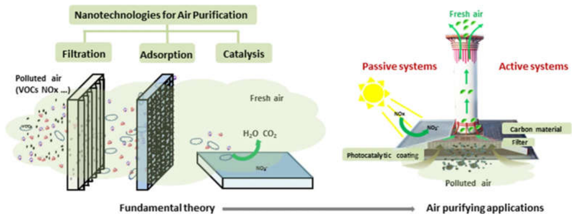
5.2. Biomedical Applications
Metal oxide nanoparticles (MONPs) are promising materials in biomedical science owing to their unique physicochemical and biological properties. They are nanoscale in size, possess a large surface area, can be tuned to various morphologies and optical properties, can be magnetic, are catalytically active and can be easily functionalized on their surface, thus offering a wide range of possibilities for medical and healthcare applications. MONPs not only offer advantages such as superior bioavailability and specific interaction with biological systems but also control drug release, excellent imaging capabilities, and potent antimicrobial activity, which are not available in traditional materials[436].
Among the various metal oxides, some are more extensively explored for biomedical applications such as ZnO, TiO₂, Fe₃O₄, CuO, CeO₂, MgO, MnO₂ and SiO₂-based oxides[437,438]. These nanoparticles may interact with cells, proteins, nucleic acids and microorganisms in several ways including through the generation of reactive oxygen species, release of ions, through electrostatic interaction and through surface adsorption. Their biological activity can be further modified by surface modification, polymers, biomolecules, surfactants, antibodies, peptides and targeting ligands[439].
With the recent progress in nanotechnology, it has become possible to prepare multifunctional MONPs that are able to perform therapeutic, diagnostic, antimicrobial and imaging roles in a single platform. Multi-functional systems are becoming of growing interest in fields such as cancer therapy, targeted drug delivery, biosensing, tissue engineering, wound dressing materials and diagnostic imaging. Another interest in green-synthesized MONPs is their potential biocompatibility and toxicity reduction as compared to chemically synthesized nanoparticles.
The schematic shown in Figure 11a illustrates the biological interaction pathways of metal and metal oxide nanoparticles relevant to biomedical applications. After entry via inhalation, skin penetration, or oral uptake, nanoparticles can distribute through the bloodstream and accumulate in major organs such as the lungs, liver, brain, spleen, and kidneys.At the cellular level, their effects are mainly governed by ion release, ROS generation, and oxidative stress, which may lead to apoptosis and other cellular responses. Additional processes such as membrane interaction and intracellular accumulation also contribute to their biological impact.Overall, the figure highlights both the biomedical relevance and safety considerations of metal oxide nanoparticles, emphasizing their dose- and property-dependent behavior[440].
The schematic shown in Figure 11b highlights the broad biomedical and biological applications of nanoparticles along with their major material classes. At the center, nanoparticles serve as a multifunctional platform enabling diverse applications including drug delivery, antimicrobial activity, gene therapy, biosensing, bioimaging, molecular diagnostics, and tissue engineering.
These applications are enabled by a wide range of nanomaterials such as metal oxide nanoparticles doped metal oxide systems, metal sulfides, and metal–organic frameworks (MOFs). Each class offers tunable physicochemical properties that determine their suitability for specific biomedical functions.Overall, the figure emphasizes the versatility of nanoparticles in biomedical and biological systems, where their functional diversity enables integration into multiple diagnostic and therapeutic platform[441].
On the other hand, several challenges remain, such as ensuring the safety of the cells, their elimination from the body, their long-term stability, immune response, and cytotoxicity. Thus, it is critical to understand MONP-biological interactions to create nanomedicines that are safe and effective. The subsequent sections will describe the important biomedical applications of metal oxide nanoparticles.
Figure 11.
a): Overview of nanotoxicity mechanisms associated with metal oxide nanoparticles, illustrating oxidative stress pathways, ion release, cellular signaling disruption, and organ accumulation that govern nanoparticle biocompatibility and safety[440] (Reproduced with Permission). Fig 11b): Overview of biomedical applications of metal and metal oxide nanoparticles, highlighting their roles in antimicrobial therapy, drug delivery, cancer treatment, bioimaging, tissue engineering, biosensing, and regenerative medicine. Their unique physicochemical properties enable enhanced therapeutic efficacy, targeted delivery, and improved clinical outcomes[441] (Reproduced with Permission).
Figure 11.
a): Overview of nanotoxicity mechanisms associated with metal oxide nanoparticles, illustrating oxidative stress pathways, ion release, cellular signaling disruption, and organ accumulation that govern nanoparticle biocompatibility and safety[440] (Reproduced with Permission). Fig 11b): Overview of biomedical applications of metal and metal oxide nanoparticles, highlighting their roles in antimicrobial therapy, drug delivery, cancer treatment, bioimaging, tissue engineering, biosensing, and regenerative medicine. Their unique physicochemical properties enable enhanced therapeutic efficacy, targeted delivery, and improved clinical outcomes[441] (Reproduced with Permission).
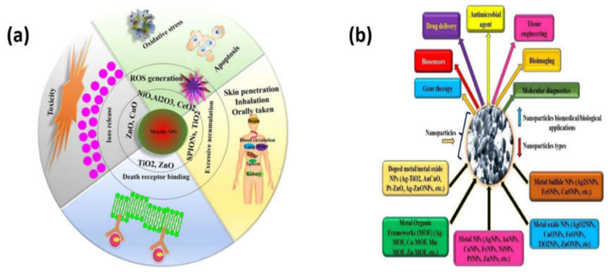
Table 5.
Biomedical applications of Metal Oxide nanoparticles.
| S.No | Metal Oxide Nanoparticle | Key Biological Property | Biomedical Application | Mechanism of Action | Advantages | References |
|---|---|---|---|---|---|---|
| 01 | TiO₂ nanoparticles | Highly biocompatible, chemically stable, have low toxicity, and show strong photocatalytic behavior. | cancer treatment (photodynamic therapy), drug delivery systems, cell imaging and biosensors | When exposed to light, TiO₂ produces reactive oxygen species that can kill cancer cells or microbes | Safe for biological systems, versatile for many biomedical uses, | [442] |
| 02 | Titanium dioxide (TiO₂) nanoparticles |
stable, biocompatible, can be chemically modified (tunable surface) | Drug delivery systems, cancer therapy, antimicrobial coatings for medical devices and implants | TiO₂ NPs can attach to drugs and release them in a controlled way at target sites; under light, they produce reactive oxygen species that kill cancer cells and microbes | Enable targeted and controlled drug delivery, provide minimally invasive cancer treatment, show strong antimicrobial effects | [443] |
| 03 | TiO₂ NPs | More environmentally friendly, have fewer toxic impurities | Drug-related applications, antimicrobial uses, and other medical technologies. | TiO₂ interacts with biological systems in a controlled and stable way, improving medical performance. | Low cost, less toxicity, energy-efficient production, better environmental safety | [444] |
| 04 | Titanium dioxide (TiO₂) nanotubes | Biocompatible and their surface structure strongly influences how cells attach, grow, and behave. | Used mainly in medical implants (especially bone implants) to improve bone integration, support stem cell growth | The nanotube surface controls cell behavior; improve cell adhesion and growth this affects osteoblast and stem cell activity and improves bone formation. | Enhances bone-implant bonding (osseointegration), improves cell attachment and tissue regeneration, provides antibacterial properties | [445] |
| 05 | TiO₂ nanotubes (TNTs) decorated with manganese oxide (MnO)NPs. | Bioactive (can interact with body fluids), support mineral formation (apatite) | Bone-related implants and biomaterials where improved bone bonding and regeneration are needed | They promote apatite (bone-like mineral) formation in body fluid, improving bone integration with implants. | Stronger bone bonding ability, improved bioactivity, better apatite (bone mineral) formation | [446] |
| 06 | TiO₂ NPs reinforced in calcium phosphate bioceramic nanocomposites | Biocompatible, support bone-like mineral formation, and provide high surface area with porous nanostructure | Used mainly in orthopedic and dental applications | Enhances cell attachment, bone cell growth, and formation of new bone (osteointegration and osteoinduction). | Better bone regeneration, improved surface area for cell growth, enhanced mechanical strength, improved integration with natural bone | [447] |
| 07 | TiO₂ NPs incorporated into a PLGA (poly lactic-co-glycolic acid) porous 3D scaffold | Biocompatible, biodegradable, high porosity, and ability to support bone cell growth | Bone tissue engineering (BTE), orthopedic regeneration, antimicrobial implants | The porous scaffold provides space for cell growth, while TiO₂ NPs improve antibacterial activity, bioactivity, and photocatalytic breakdown of harmful substances | High antibacterial effect (~99%), good bone-like mineral formation, excellent cell compatibility, tunable pore for better tissue growth | [448] |
| 08 | Green silver oxide nanoparticles (Ag₂O NPs | Biocompatible, low toxicity, hemocompatible (safe with blood), antioxidant, and strong biological activity against microbes and cancer cells. | Used for anticancer therapy, antimicrobial treatments, anti-parasitic applications (Leishmania), enzyme inhibition studies, and potential use in nano-pharmaceutical drug development. | Ag₂O nanoparticles damage cancer cells and microbes by releasing active silver-based species that disrupt cell membranes, proteins, and DNA | Eco-friendly green synthesis, cost-effective production, strong anticancer and antimicrobial effects, good biocompatibility with blood, low toxicity, and broad biomedical potential for future drug development. | [449] |
| 09 | Silver oxide nanoparticles (Ag₂O NPs) | Bio-interaction properties, show cytotoxic effects against cancer cells | Cancer therapeutics, especially against liver cancer cells (HepG2 cell line) | Ag₂O nanoparticles interact with cancer cells, inducing phototoxicity and cytotoxicity that can damage or kill tumor cells | Strong anticancer potential, effective interaction with cancer cells, unique physicochemical properties | [450] |
| 10 | Silver oxide nanoparticles (AgNPs/Ag₂O NPs) | Biocompatible, low toxicity, antioxidant activity, antimicrobial activity, enzyme inhibition ability |
Antimicrobial treatments, anti-leishmanial therapy, antioxidant applications, pharmaceutical formulations | Inhibit the growth of microbes and parasites, neutralize harmful free radicals through antioxidant activity, and inhibit enzymes such as protein kinase and α-amylase. | Eco-friendly synthesis, low toxicity to human cells and red blood cells, strong antimicrobial and antioxidant effects, anti-parasitic activity, good biocompatibility | [451] |
| 11 | Bimetallic silver–copper oxide nanoparticles (Ag–CuO NPs | Strong antibacterial activity, anticancer activity, good biocompatibility, low cytotoxicity, and ability to inhibit tumor growth and spread. | Antibacterial agents against pathogenic bacteria and MRSA, anticancer therapy, anti-metastatic treatment | Inhibit bacterial growth and suppress the mecA resistance gene in MRSA. Inhibit cancer cell migration, and block angiogenesis (formation of new blood vessels that feed tumors) | Broad-spectrum antibacterial activity, effective against antibiotic-resistant bacteria (MRSA), strong anticancer effects, prevents tumor spread | [452] |
| 12 | Silver oxide nanoparticles (Ag₂O NPs) incorporated into hydroxyapatite (HAP) and GO-based nanocomposites | Excellent biocompatibility, promotes osteoblast (bone cell) growth | Used in bone tissue engineering, orthopedic and dental implants, bone regeneration materials, and antibacterial biomedical coatings. | Ag₂O NPs provide antibacterial effects by inhibiting bacterial growth, while HAP supports bone formation and GO improves surface properties | Improved bone cell growth and viability, strong antibacterial activity against Escherichia coli and Staphylococcus aureus | [453] |
| 13 | Ag NPs incorporated into chitosan/polyethylene oxide (CS/PEO) nanocomposite films. | Strong antibacterial activity and good compatibility with polymer matrices | Used in antibacterial wound dressings, biomedical coatings, tissue engineering materials, and infection-control applications. | Silver NPs release silver ions that damage bacterial cell membranes, proteins, and DNA, leading to the death of both Gram-positive and Gram-negative bacteria | Effective against Escherichia coli and Staphylococcus aureus, enhanced thermal stability, uniform nanoparticle distribution | [454] |
| 14 | AgO NPs embedded in a PVC/PMMA polymer blend to form ternary nanocomposites | Strong antibacterial activity and ability to interact with biological systems | Antibacterial coatings and biomedical materials to prevent infections, also in medical devices and flexible optoelectronic devices | AgO NPs release active silver species that damage bacterial cell membranes and inhibit microbial growth. The polymer matrix (PVC/PMMA) helps distribute NPs evenly | Strong antibacterial effect against both Gram-positive and Gram-negative bacteria (S. aureus, B. subtilis, E. coli), improved optical and electronic properties | [455] |
| 15 | Iron oxide (Fe₂O₃) nanoparticles | High surface area, porous structure, good magnetic behavior, high thermal stability, and good mechanical strength | Drug delivery systems (nanocarriers), tissue engineering scaffolds, wound healing materials | Fe₂O₃ NPs interact with biological systems due to their magnetic and surface properties, allowing controlled drug loading/release, improved cell attachment. | High stability, strong mechanical strength, good drug delivery capability, large surface area for interaction, useful magnetic properties | [22] |
| 16 | Fe2O3 NPs | Magnetic behavior, tunable surface chemistry, good interaction with biological systems | MRI imaging, drug delivery systems, magnetic hyperthermia (heat-based cancer therapy), in vitro diagnostics, and theranostic | Iron oxide NPs respond to external magnetic fields for imaging and targeting. They can also generate heat under magnetic fields for killing cancer cells and can carry drugs to specific sites due to surface modification. | Highly versatile, improved diagnostic and therapeutic efficiency, targeted treatment with less damage to healthy tissues | [456] |
| 17 | Iron oxide nanoparticles and composite iron oxide nanoparticles | High biocompatibility in the body, good colloidal stability, and strong magnetic properties useful for imaging. | (MRI, high-resolution diagnostic imaging systems, and combined imaging–therapy applications. | Iron Oxide NPs respond to magnetic fields, allowing them to be detected in MRI scans. They improve signal strength and imaging resolution in the body. | Safe and stable in biological systems, tunable size/shape for better performance, enhanced detection accuracy | [457] |
| 18 | Iron oxide NPs and hybrid nanolipidic magnetic nanocomposites |
Strong magnetic behavior, good biocompatibility, ability to interact with biological systems | Used in cancer diagnosis, cancer therapy, nanotheranostics, and multimodal cancer treatment systems. | Iron oxide NPs respond to external magnetic fields to generate heat (killing cancer cells in hyperthermia) and improve imaging. | Improves diagnostic accuracy, supports combined therapy and imaging, reduces damage to healthy tissues | [458] |
| 19 | Fe2O3 NPs combined with optical probes to form magneto-optical nanocomposites | Magnetic behavior, good nanoscale imaging ability, and biocompatibility | MRI imaging, early disease detection, targeted drug delivery | Optical probes enhance light-based imaging and detection; together they improve visualization and enable controlled therapy in the body. | Improved disease detection accuracy, ability for targeted treatment, and potential for real in vivo biomedical applications | [459] |
| 20 | Superparamagnetic iron oxide nanoparticles | Strong magnetic properties, good biocompatibility, high stability, tunable size and shape | Drug delivery, MRI, magnetic-activated cell sorting, nanobiosensors, magnetic hyperthermia, tissue engineering | The NPs respond to external magnetic fields for imaging, cell separation, and targeted drug delivery. Surface coatings improve stability and biocompatibility | Excellent magnetic responsiveness, targeted delivery capability, improved MRI contrast, enhanced stability through surface modification | [460] |
| 21 | Iron (II) oxide NPs functionalized with chitosan to form a CS/FeO nanocomposite. | Biocompatible, biodegradable, antibacterial, and anticancer activity | Used as an antibacterial agent against pathogenic bacteria and as a potential anticancer material for lung cancer treatment | The FeO NPs and chitosan work together to inhibit bacterial growth and reduce the proliferation of cancer cells. | Strong antibacterial activity against E. coli, B. subtilis, and S. aureus, anticancer effects against A549 lung cancer cells | [461] |
| 22 | Bismuth oxide (Bi₂O₃) nanoparticles | High radiopacity, tunable surface properties, potential cytotoxic activity against diseased cells | Used as active drugs, diagnostic imaging agents, drug delivery systems, and theragnostic platforms | Medical imaging due to its radiopacity. Surface-modified bismuth materials can deliver drugs, and may induce cytotoxic effects in diseased tissues | Excellent imaging capability, multifunctional diagnosis-and-treatment potential, customizable surface properties | [462] |
| 23 | Bismuth oxide nanoparticles (Bi₂O₃ NPs) | High biocompatibility, low toxicity, strong X-ray attenuation, NIR absorption, good photothermal conversion | Used in cancer therapy, photothermal therapy, multimodal imaging, theranostics drug delivery | BiNPs absorb NIR light and convert it into heat to destroy cancer cells (photothermal therapy), enhance X-ray imaging and radiation therapy | Non-toxic and cost-effective, excellent imaging contrast, effective cancer treatment under NIR light, multifunctional antibacterial activity, promotes bone regeneration | [463] |
| 24 | Bi₂O₃ NPs |
Can interact strongly with cells and induce oxidative stress, leading to toxicity at higher doses and longer exposure times. | Cosmetics, dental materials, pulp capping, and biomedical imaging; also studied for anticancer potential in lab research. | Increase oxidative stress in cells, damage mitochondria, reduce antioxidant defenses (GSH, SOD, catalase), and activate apoptosis pathways | biomedical imaging and dental/cosmetic applications, shows strong interaction with cancer cells in research | [464] |
| 25 | Bi₂O₃ NPs combined with hydroxyapatite (HAP) and graphene oxide to form a ternary nanocomposite (HAP/Bi₂O₃/GO). | High biocompatibility, improved cell viability, and strong antibacterial and bioactive properties | Bone tissue engineering, biomaterials for implants, antibacterial coatings | HAP supports bone-like growth, GO improves dispersion and reduces particle aggregation, while Bi₂O₃ enhances bioactivity and antibacterial effects. | High cell viability (~98.7%), strong antibacterial activity against E. coli and S. aureus, and enhanced performance for bone-related biomedical applications. | [465] |
| 26 | Bi₂O₃ and antimony oxide (Sb₂O₃) composite nanoparticles (Sb₂O₃@Bi₂O₃ | High electrochemical activity, good stability | Monitoring of methotrexate (anticancer drug) levels in blood and urine for safe drug dosage control. | Detects methotrexate through strong electrochemical signals generated when the drug interacts with the Sb₂O₃@Bi₂O₃ surface, allowing accurate measurement of drug concentration. | Very high sensitivity, low detection limit, wide detection range, strong selectivity in complex biological samples, and useful for safe monitoring of anticancer drug levels. |
[466] |
| 27 | Bi₂O₃ NPs incorporated into graphene oxide and polyvinyl alcohol (PVA) hydrogel system (Bi₂O₃/GO/PVA composite). | Biocompatible, strong antibacterial activity, antioxidant (free radical scavenging), and improved optical properties | Antibacterial wound dressings, drug delivery systems, bio-imaging materials, and biomedical hydrogel-based therapies. | Bi₂O₃ NPs provides antibacterial effects, while GO enhances dispersion, reduces oxidative stress (ROS control). Together they damage bacterial cells, and improve healing environment | Good antioxidant activity, improved optical clarity for imaging, better light absorption, enhanced biocompatibility, multifunctional performance (healing + drug delivery + antimicrobial), |
[467] |
| 28 | Bismuth oxide/copper oxide/graphene oxide (Bi₂O₃/CuO/GO) nanocomposites. | Strong antibacterial activity, good biocompatibility, low in vivo toxicity | Potential wound infection control materials, and safe nanomaterials for future biomedical and therapeutic applications | Nanocomposites kill bacteria by interacting with bacterial cell walls and disrupting their function, enhances oxidative stress and prevents bacterial growth | Strong antibacterial effect against both Gram-positive and Gram-negative bacteria, confirmed safety in animal studies (non-toxic at tested dose) | [468] |
| 29 | Various metal oxide nanoparticles and their hybrid nanocomposites | Non-toxic nature, large surface area, suitable band gap, and high biological activity. | Used in anticancer therapy, antibacterial treatments, photocatalysis-based biomedical systems | Producing reactive species (like ROS) under light or biological conditions, which can destroy cancer cells and bacteria | Eco-friendly and less toxic, high efficiency in killing cancer cells and bacteria, strong optical and photocatalytic performance |
[469] |
| 30 | Polymer/metal oxide nanocomposites. | Good biocompatibility, improved electrical conductivity, and stable physical properties suitable for biological environments. | Used in biomedical products, tissue engineering materials, biosensors, conductive medical devices, implants, and diagnostic systems. | The polymer matrix provides flexibility and biocompatibility, while metal/metal oxide nanoparticles improve mechanical strength, electrical conductivity, sensing capability | High strength and durability, enhanced stability, multifunctional performance, suitability for sensors and medical devices, and potential for advanced biomedical applications. | [470] |
| 31 | Transition metal oxide nanoparticles and their nanocomposites | High biocompatibility, strong biomolecule-capturing ability, excellent electrocatalytic activity | Used in electrochemical biosensors for detecting and monitoring biomarkers in body fluids, disease diagnosis | Enhance electron transfer and electrocatalytic reactions at the sensor surface. They capture target biomarkers and generate measurable electrical signals, enabling sensitive detection. | Rapid detection, high sensitivity, excellent selectivity, good durability, strong stability, low detection limits, real-time monitoring capability | [436] |
| 32 | Various metal oxide nanoparticles incorporated into hydrogel networks to form nanocomposite hydrogels | Excellent biocompatibility, high water content, porous structure and ability to mimic the natural tissue environment | Used in tissue engineering, drug delivery systems, wound healing, regenerative medicine, pharmaceutical formulations, and biotechnology applications. | MO NPs reinforce the hydrogel structure and improve physical, chemical, and biological properties. The porous hydrogel network supports cell growth, nutrient transport, and controlled drug release. | Mimics natural tissues, improved mechanical strength, controlled drug delivery, high hydration, and suitability for advanced biomedical and pharmaceutical applications. | [471] |
| 33 | Various metal oxide nanoparticles (e.g., iron oxide, titanium dioxide, zinc oxide, copper oxide, bismuth oxide, etc.) used as advanced nanobiomaterials. | Biocompatibility, antimicrobial, antifungal, antiviral activity, high surface area, tunable physicochemical properties | Used in tissue therapy, immunotherapy, disease diagnosis, dentistry, regenerative medicine, wound healing, biosensors | Metal oxide nanoparticles interact with cells, tissues, and microorganisms through their surface properties. They can deliver therapeutic effects, support tissue regeneration, enhance biosensing signals | High biomedical performance, multifunctional use (therapy and diagnosis), strong antimicrobial activity, improved wound healing, support for tissue regeneration, sensitive biosensing capabilities, | [8] |
5.2.1. Antibacterial and Antifungal Activity
Metal oxide nanoparticles have shown a potent antibacterial and antifungal activity against a wide range of pathogenic microorganisms. Multidrug-resistant bacteria and fungal infections have emerged rapidly and there is an urgent need for the development of alternative antimicrobial drugs. MONPs can be used as antimicrobial materials, due to their multiple mechanisms of action against microorganisms, which make the development of resistance less likely[472].
Zinc oxide (ZnO) nanoparticles are getting a lot of attention among different MONPs because of their remarkably strong antibacterial activity and their ability to kill gram-positive and gram-negative bacteria such as Escherichia coli (E. coli), Staphylococcus aureus (S. aureus), Pseudomonas aeroguinosa (P. aeroguinosa), and Klebsiella pneumonia (K. pneumonia). The nanoparticles of ZnO induce oxidative stress through the production of reactive oxygen species including hydroxyl radicals, superoxide ions and hydrogen peroxide that cause oxidative damage to the microbial membranes, proteins, lipids and DNA. The free Zn²+ ions also help disrupt the bacterial cells and inhibit the metabolic processes[473].
The strong redox activity and ion release capability of copper oxide (CuO) nanoparticles also make them excellent antimicrobials. CuO nanoparticles are able to enter the cell walls of the microbes, leading to leakage of the cell membrane, denaturation of proteins and inactivation of enzymes. In the same way, the magnesium oxide (MgO) nanoparticles have important bactericidal activity related to surface alkalinity and oxidative stress. Titanium dioxide (TiO₂) nanoparticles are particularly effective under UV irradiation due to their photocatalytic activity in the generation of reactive oxygen species, which can lead to the damage of microbial cells[473].
Due to their magnetic properties and biocompatibility, iron oxide nanoparticles are widely used in the development of antimicrobial coatings and biomedical devices. Biofilm formation is a major problem in medical implants and hospital-acquired infections, and can be effectively inhibited by surface-functionalized iron oxide nanoparticles. The antioxidant and antimicrobial properties of cerium oxide nanoparticles have also garnered interest, as they can exhibit both properties based on their oxidation state and environment[375].
MONPs have been shown to have antifungal activity against pathogenic fungi like Candida albicans, Aspergillus niger and Fusarium species. The nanoparticles of ZnO and CuO affect the integrity of the fungal membrane, spore germination, and the induction of oxidative stress in fungal cells. Recently, it has been demonstrated that MONPs composites with polymers, chitosan, graphene oxide and silver nanoparticles are synergistically antimicrobial[474].
The applications of antimicrobial MONPs are limited to biomedical (wound dressing, implant coating, dental materials), food packing, water disinfection, and textile coating. The level of cytotoxicity, On the other hand, and dosage should be carefully analyzed as over production of the reactive oxygen species can also harm healthy mammalian cells[475].
5.2.2. Anticancer Activity
Metal oxide nanoparticles have shown selective cytotoxic effects in tumor cells that has drawn an immense attention in cancer therapy. The limitations of conventional cancer therapies like radiation therapy and chemotherapy are: nonspecific toxicity, multidrug resistance, severe side effects and poor targeting efficiency. MONPs will provide new opportunities to develop targeted and multifunctional anticancer drugs with better therapeutic outcomes[476].
ZnO nanoparticles (NPs) are one of the most widely investigated anticancer nanomaterials. They are selectively toxic to cancer cells, mainly due to their ability to induce excessive production of reactive oxygen species, to disrupt mitocondrial functions, to damage DNA and to induce apoptosis. Under normal conditions, the basal level of ROS is higher in cancer cells than in healthy cells, and the metabolism of cancer cells is different from that of healthy cells, which makes them more sensitive to the effects of oxidative stress. ZnO nanoparticles have demonstrated to have anticancer activity against breast, lung, liver, cervical, prostate and colon cancer cell lines[477].
For cancer therapy, nanoparticles of TiO2 are the subject of extensive research for both photodynamic and photocatalytic applications. TiO₂ nanoparticles generate reactive oxygen species on exposure to light, which help to break down the membrane, proteins and nucleic acids of cancer cells. The surface modification and doping are often used to boost visible light responsiveness and targeting efficiency. In a like manner, copper oxide nanoparticles (CuO-NPs) have been shown to possess potent anticancer activity via induction of oxidative stress and apoptosis in tumor cells and reduction in the proliferation of cells[478].
Among the nanoparticles, iron oxide nanoparticles hold special significance in cancer theranostics due to its therapeutic and diagnostic properties. Magnetic Fe₃O₄ nanoparticles could be targeted to the tumour tissue by external magnetic fields for targeted drug delivery and hyperthermia treatment. Magnetic hyperthermia therapy involves the use of iron oxide nanoparticles to selectively heat tumors cells in the presence of alternating magnetic fields and minimize heating to healthy tissues[479].
The presence of both Ce³⁺ and Ce⁴⁺ oxidation states results in unique redox switching behavior of cerium oxide nanoparticles. They can help to prevent oxidative damage to healthy cells or cause oxidative stress in cancer cells, depending on the environment. Tumor microenvironment responsive drug delivery and imaging applications are also explored using manganese oxide nanoparticles[480].
Recent research involves multifunctional nanoplatforms, which involve incorporating MONPs with anticancer drugs, antibodies, peptides, polymers and targeting ligands. Such hybrid systems result in increased tumor selectivity, cellular uptake and decreased systemic toxicity. Although the laboratory results are hopeful, there are still many problems with nanoparticles and their biodistribution, immune response, long-term toxicity and clinical translation that needs to be investigated[481].
5.2.3. Drug Delivery
One of the most crucial biomedical uses of metal oxide nanoparticles is drug delivery. Traditional drug delivery techniques are often limited by low bioavailability, limited solubility, rapid degradation, low specificity and high side effects. Advanced nanocarrier systems are offered by MONPs, which have the potential to enhance drug stability, controlled release, targeted delivery, and therapeutic efficiency[482].
Due to their high surface area and surface chemistry tunability, the easy loading of therapeutic molecules is possible via adsorption, covalent bonding, electrostatic interaction, or encapsulation. Iron oxide nanoparticles are especially interesting for drug delivery applications due to their magnetic properties and biocompatibility. Fe₃O₄ nanoparticles can be guided into tissues of interest by an external magnetic field which increases the amount of drug in the target tissues and decreases systemic toxicity[483].
Controlled drug release is a very common application of nanoparticles with mesoporous structures and large loading capacities, such as metal oxide nanoparticles coated with mesoporous silica. ZnO nanoparticles are also of great interest due to their pH responsive dissolution behaviour. The acidic environment of cancer cell microenvironments promotes the dissolution of ZnO nanoparticles, which allows for selective release of anticancer drugs inside cancer cells. Stable carriers for photosensitive drugs and therapeutic biomolecules are prepared using titanium dioxide nanoparticles[484].
Surface functionalization is an important process to enhance the biocompatibility and targeting capability of nanoparticles. The PEGylation or coating with chitosan, dextran, proteins, antibodies, or folic acid of MONPs leads to a prolonged circulation time, decreased immune system recognition, and greater selective uptake by diseased tissues. Multifunctional nanocarriers with drug delivery, imaging and therapeutic functions are being studied in greater depth in the field of personalized medicine[485].
The stimuli-responsive MONPs that are able to release drug upon adjustment of pH, temperature, magnetic fields, enzymes or light have received considerable attention. Magnetic nanoparticles can also be used for hyperthermia and simultaneous release of drugs under alternating magnetic fields. In addition, hybrid nanocomposites of MONPs and liposomes, hydrogels or polymeric nanoparticles offer improved stability and sustained drug release profiles[486].
On the other hand, long-term toxicity, biodegradability, aggregation of nanoparticles and regulatory approval are significant issues for clinical application of MONP-based drug delivery systems[487].
5.2.4. Wound Healing
The antimicrobial effect, anti-inflammatory activity, oxygen-producing property, and tissue-regenerating capability of metal oxide nanoparticles have made them a promising material for wound healing applications. The ability to manage wounds is very important to avoid complications with chronic wounds and burn injuries which are highly susceptible to microbial infections and delayed healing[488].
The widespread use of ZnO nanoparticles in wound dressings is attributed to their broad spectrum antimicrobial activity and cell proliferation and collagen synthesis. The Zn²⁺ ions liberated from the ZnO nanoparticles are crucial for enzymatic functions, wound healing, and immune system modulation. Another advantage of ZnO based Nanocomposites Dressing is that it possesses a very good UV-blocking property and also has moisture holding capacity which would provide a favourable environment for wound healing[489].
Photocatalytic antimicrobial activity and biocompatibility of titanium dioxide nanoparticles are their main effect in wound healing. In the presence of light, dressings containing TiO₂ can prevent the growth of bacteria and minimise the risk of infection. Copper oxide nanoparticles have been found to induce angiogenesis and collagen deposition, which enhances the wound healing and tissue regeneration process. In the same way, cerium oxide nanoparticles have antioxidant and anti-inflammatory activity, which can decrease the oxidative stress in the wound area[490].
Due to their magnetic properties and antimicrobial activity, iron oxide nanoparticles are more widely used in the development of smart wound dressing. Magnetic nanocomposite dressings are able to promote cell migration and tissue regeneration with the magnetic stimulation. Besides, MONPs have been used in conjunction with hydrogels, electrospun nanofibers, biopolymers and chitosan matrices to create novel wound healing materials for enhanced moisture control, flexibility and mechanical strength[491].
Due to the biocompatibility and low toxicity of green-synthesized nanoparticles, green-synthesized MONPs have attracted significant attention in wound healing applications. Recent research has shown that plant-mediated ZnO nanoparticles and TiO₂ nanoparticles have the potential to greatly promote wound contraction and to enhance the re-epithelization in experimental models[492].
Although promising results have been obtained, optimisation of the concentration, release rate and the long-term safety of nanoparticles is still crucial to prevent potential inflammatory responses and cytotoxicity in clinical wound management systems[493].
5.2.5. Bioimaging and Diagnostics
Due to their magnetic, optical, fluorescent and catalytic properties, metal oxide nanoparticles have been increasingly recognized as being of great significance in the field of bioimaging and diagnostics. Accurate imaging and early diagnosis of disease are essential to the effectiveness of treatment and patient management, and MONPs are more sensitive, more multifunctional and have a higher contrast than conventional imaging agents[494].
For magnetic resonance imaging (MRI), one of the most common nanoparticles used are those composed of iron oxide. Fe₃O₄ nanoparticles are superparamagnetic, which means they create a contrast effect and enhance the sensitivity and clarity of the image. Their magnetic properties can also be used for targeted imaging and simultaneous therapeutic uses, which is useful for theranostic systems[495].
Considering the strong paramagnetic properties of manganese ions, manganese oxide nanoparticles have attracted attention as alternative MRI contrast agents. These nanoparticles can offer better imaging capabilities and some benefits over traditional gadolinium-based imaging agents. Oxidative stress monitoring and fluorescence imaging are the fields of interest in which cerium oxide nanoparticles are investigated because of their redox-responsive behavior[496].
The optical and fluorescent properties of quantum-sized metal oxide nanoparticles like ZnO and TiO₂ are unique and suitable for cellular imaging and biosensing. The ZnO nanoparticles exhibit excellent luminescence properties and can be modified with biomolecules to selectively detect biomolecules such as proteins, DNA, glucose, pathogens and cancer biomarkers. Owing to the high stability and photocatalytic properties, TiO₂ nanoparticles also have applications in the field of photoelectrochemical biosensors and imaging systems[497].
Targeting efficiency and biocompatibility of nanoparticles for imaging can be greatly improved by surface modification. Functionalization makes it possible to recognize selectively diseased tissues and biomolecules by using antibodies, peptides, aptamers and fluorescent dyes. The simultaneous imaging, drug delivery, and therapy of multifunctional MONPs are being increasingly investigated for personalized medicine and precision diagnostics[495].
As diagnostic application, MONPs are applied into the structure of biosensors to rapidly detect the biological analytes with high sensitivity. Metal oxide-based electrochemical and optical sensors exhibit high sensitivity, low detection limits and fast response time for detecting disease biomarkers, toxins, pathogens and metabolites. These technologies are gaining increasing adoption in POC diagnostics and wearable health care devices.[498]
While MONPs have great promise for imaging and diagnostics, issues of long-term accumulation, biodegradation, immune response and potential toxicity must be carefully addressed before their widespread clinical use[494].
5.3. Energy Applications
Due to the growing need for clean, sustainable and efficient energy technologies worldwide, research activities on advanced nanomaterials used in energy generation, conversion and storage systems has been accelerated. Metal oxide nanoparticles (MONPs) have become as extremely promising materials in this field due to their unique electrical, optical, catalytic, magnetic and electrochemical properties. They are also nanoscale, which means that they have large surface areas, short ion diffusion paths, tunable band gaps, and good charge transport characteristics; all of these are useful for a range of renewable energy applications[499].
Many types of metal oxide-based nanocrystals, such as TiO₂, ZnO, Fe₃O₄, MnO₂, Co₃O₄, NiO, CuO, WO₃, SnO₂ and CeO₂, have been popular for energy applications. Some of their properties like particle size, morphology, crystallinity, surface defects, porosity, and synthesis method can have a great impact on their performance. The electrochemical and catalytic properties of nanostructures, like nanorods, nanotubes, nanosheets, hollow spheres, and mesoporous frameworks, can be superior to bulk materials[500,501,502,503,504,505].
In recent years, the hybrid and multifunctional nanocomposites of MONPs and graphene, carbon nanotubes, conducting polymers and other semiconductors are developed due to the recent advances of nanotechnology. These composite systems offer better conductivity, greater stability, better electron transfer and higher energy conversion efficiency. Also, new green synthesis methods and scalable fabrication techniques are being developed to facilitate energy technologies that are environmentally friendly[499].
MONPs are crucial components of solar energy harvesting, electrochemical energy storage, fuel generation, photocatalytic water splitting, and hydrogen evolution systems. Their use in solar cells, supercapacitors, batteries and hydrogen production are described below.
5.3.1. Solar Cells
The optical transparency, electron transport capacity, chemical stability, and tunable electronic structure make metal oxide nanoparticles extremely promising for use in solar cell technologies. By increasing the absorption of light, separation of charges and electron transport in a PV device, MONPs can greatly enhance the efficiency of solar cells[506].
One of the most significant materials for dye-sensitized solar cells (DSSCs) is titanium dioxide (TiO₂) nanoparticles. Nanocrystalline TiO₂ films have large surface areas for the adsorption of the dye and allow efficient light harvesting and injection of electrons. TiO2 has a mesoporous structure which promotes fast electron transport and reduces charge recombination loss. The electron mobility and chemical stability of TiO₂ nanoparticles has made them a popular choice for electron transport materials in perovskite solar cells and organic photovoltaic devices[507].
In this regard, considerable attention has been focused on the alternative compounds to TiO₂ like zinc oxide (ZnO) because of its similar band gap, higher electron mobility and ease to be synthesized as various nanostructures like nanorods, nanowires and nanotubes. The possibility of direct pathway for electron transport in ZnO nanostructures increases charge collection efficiency and decreases charge recombination. But, in some cases, the stability problem of the perovskite and acidic dyes arises in the ZnO-based solar cell[508].
High-efficiency perovskite solar cells are now being increasingly used with tin oxide (SnO₂) nanoparticles as the electron transport layer. SnO₂ is highly transparent in the optical range, has high electron mobility, a low processing temperature, and a good band alignment. In the same way, NiO nanoparticles are widely utilized as hole transport material in inverted solar cell structure[509].
The recent research has been directed towards increasing the efficiency of solar cells by doping, heterojunction formation, surface engineering of MONPs. Adding plasmonic nanoparticles, rare earth dopants or carbon-based materials in the material increases light absorption and charge separation. TiO₂/graphene, ZnO/carbon nanotubes and metal oxide/perovskite interfaces have been shown to exhibit enhanced photovoltaic efficacy and stability[510].
Morphology control also is an important factor for the efficiency of solar energy conversion. The 1D nanowire and nanotube structures can facilitate directional charge transport, and the porous and hierarchical structures can enhance light scattering and dye loading. The preparation methods for MONPs used in solar cells have been a subject of interest for green synthesis methods to reduce the environment impact and production cost[511].
The summary of the PV performance of the metal oxide based photoelectrodes (V₂O₅, MoO₃ and WO₃) is shown in the schematic in Figure 12a, where the key solar cell parameters before and after the annealing treatment are summarized. These are the parameters: Open circuit voltage (Vₒc), Fill Factor (FF), short circuit current density (Jₛc) and Power Conversion Efficiency (PCE).From the comparison it is evident that annealing has a significant effect on the device performance due to the changes of charge transport and recombination in the metal oxide layers. V₂O₅ and MoO₃ both demonstrate a decrease in efficiency parameters after annealing, whereas WO₃ clearly show greater stability and improved photovoltaic response, suggesting improved charge separation and transport. The differences indicate the criticality of structure optimization and heat treatment of photoelectrodes of metal oxides for the solar cell performance[512].
Although significant progress has been made, some issues, including instability, interfacial recombination, and large-scale production of MONP-based PV systems, are still important hurdles for the commercialization of these photovoltaic systems.
Figure 12.
Changes in photovoltaic performance parameters of transition metal oxide-based devices following thermal annealing, illustrating the role of post-treatment processes in optimizing charge separation, carrier transport, and overall device efficiency[512] (Reproduced with Permission).
Figure 12.
Changes in photovoltaic performance parameters of transition metal oxide-based devices following thermal annealing, illustrating the role of post-treatment processes in optimizing charge separation, carrier transport, and overall device efficiency[512] (Reproduced with Permission).
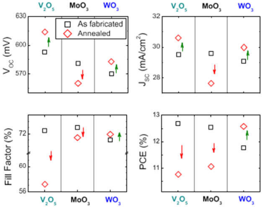
5.3.2. Supercapacitors
Supercapacitors are modified capacitors with high power density, fast charge/discharge property and long life. The metal oxide nanoparticles are the most important material as an electrode for supercapacitors due to their high theoretical capacitance, electrochemical stability, and the redox activity they exhibit[513].
Supercapacitor applications are widely studied for transition metal oxides like MnO₂, RuO₂, NiO, Co₃O₄, Fe₃O₄ and CuO. In particular, manganese oxide (MnO₂) is a material that is very attractive due to its multiple oxidation states, low cost, environmental friendliness and high theoretical capacitance. MnO₂ nanoparticles are able to store energy by fast and reversible surface redox reactions, which can be utilized in pseudocapacitive energy storage[514].
Nanoparticles such as NiO and Co₃O₄ also have excellent electrochemical properties as this material has high electrical conductivity and redox activity. Nanosheets, nanoflowers, and porous frameworks of NiO nanostructured electrodes have a large electroactive surface area and a short ion-diffusion pathway, which leads to better capacitance and rate capability. Likewise, the Co₃O₄ nanoparticles show high energy density and cycling stability in the asymmetric supercapacitor devices[515].
The use of iron oxide nanoparticles is growing in intensity due to their low toxicity, variety of oxidation states and abundance. The low conductivities of the majority of the MONPs are, On the other hand, quite a limiting factor in the design of a high-performance supercapacitors. MONPs are often mixed with conductive carbon materials like graphene, reduced graphene oxide (rGO), activated carbon (AC) and carbon nanotubes (CNTs) to overcome this problem. Such hybrid nanocomposites offer enhanced charge transport efficiency, mechanical stability and electrical conductivity[516].
The morphology engineering has a great effect on the performance of the supercapacitors. Nanorods, nanosheets, hollow spheres, mesoporous structures and hierarchical architectures promote infiltration of the electrolyte and enhance the amount of electroactive sites. The synthesis of nanostructured metal oxide electrodes with controlled morphologies is typically achieved through the use of hydrothermal, solvothermal, electrodeposition and template-assisted approaches[517].
Supercapacitors made from MONP nanocomposites also received significant attention for smart technologies and portable electronic devices, which have to operate without the use of batteries. While great progress has been made, there is still room for increased energy density, durability upon cycling, and scalable manufacturing for practical commercial implementation[518].
5.3.3. Batteries
As a result of their high electrochemical activity, large surface area and improved ion transport properties, metal oxide nanoparticles are particularly important for modern battery technologies. Portable electronics, electric vehicles, renewable energy storage and grid stabilization systems rely on rechargeable batteries. MONPs are extensively explored as electrode materials in lithium ion, sodium ion, zinc ion and other advanced batteries[519].
Transition metal oxides like Fe₃O₄, Co₃O₄, MnO₂, NiO, TiO₂ and SnO₂ can be reversibly reduced and oxidized in multiple electron transfer reactions, which is the reason for their high theoretical capacities. Fe₃O₄ nanoparticles, among these materials, are of great interest due to their low cost, environmental compatibility, high lithium storage capacity. But any significant volume change of the crystal during charge/discharge cycles can lead to structural degradation and capacity fading[140].
Titanium dioxide nanoparticles are used as anode materials because of their excellent structural stability, safety and long cycle life. The nanostructured TiO₂ offers short lithium diffusion pathway and enhanced charge transfer kinetics. While the theoretical capacity of TiO₂ is lower than certain transition metal oxides, it is compensated by its outstanding cycling stability, which makes it a suitable material for high-performance battery systems[520].
The lithium storage capacity of tin oxide (SnO₂) nanoparticles is extremely high, but the volume expansion of the nanoparticles during cycling will affect their long-term stability. To resolve this problem, SnO₂ nanoparticles are usually mixed with graphene, carbon nanotube, polymer matrix etc. to enhance the conductivity and to meet the mechanical stresses. In a similar fashion, Co₃O₄ and MnO₂ nanoparticles exhibit excellent electrochemical properties, but do need to be nanostructured and composed[521].
MONP incorporation has also been beneficial to lithium–sulfur and sodium-ion batteries. The metal oxide nanoparticles could inhibit the dissolution of polysulfide, increase the conductivity of the electrodes and promote the diffusion of ions. Nanostructured metal oxides are being investigated as low cost alternatives in sodium ion batteries as sodium resources are more plentiful than lithium[522].
Recent studies are directed towards the creation of hierarchical nanostructures, porous electrodes and hybrid nanocomposites for enhanced battery performance. In order to improve the conductivity and electrochemical stability, surface coating, doping and defect engineering are commonly employed. Sustainable synthesis methods are also becoming more significant to produce sustainable batteries[523].
On the other hand, challenges like low cycling stability, aggregation of nanoparticles, compatibility of electrolyte, and the high cost of mass production must be overcome before the material can be used for commercial batteries[524].
5.3.4. Hydrogen Production
Hydrogen is a clean and sustainable energy carrier as it leaves no emissions when combusted, and is energy dense. The metal oxide nanoparticles are widely highlighted in the field of hydrogen production technologies because of their remarkable photocatalytic, electrocatalytic and catalytic properties. MONPs have been extensively studied for water splitting, photocatalytic hydrogen evolution and reforming reactions towards sustainable hydrogen production[525].
Titanium dioxide (TiO₂) nanoparticles have been the most extensively studied photocatalysts for water splitting to produce hydrogen. TiO₂ produces electron–hole pairs under ultraviolet (UV) irradiation, which can be used to carry out reduction and oxidation reactions and to generate hydrogen and oxygen from water molecules. On the other hand, the band gap of TiO₂ is wide and absorbs only ultraviolet light, which is only a small portion of the solar energy[526].
Since this is a limiting factor, there has been much research into band gap engineering via doping and heterojunction formation. Doping of TiO₂ with transition metals, nonmetals or rare-earth elements provides the ability to absorb light in the visible region, and enhances charge separation efficiency. The photocatalytic hydrogen evolution performance is also improved by combining TiO₂ with other semiconductors like ZnO, WO₃, CdS and g-C₃N₄[527].
The photocatalytic properties of zinc oxide nanoparticles are similar and they are actively studied for solar-driven hydrogen production. The ZnO nanorods, nanosheets and porous structure offer high active surface area and efficient charge transport pathways. Also, such photoelectrochemical water splitting is studied by tungsten oxide (WO₃), cerium oxide (CeO₂), and iron oxide nanoparticles due to their attractive and advantageous electronic structures and catalytic activities[528].
Metal oxide nanoparticles have become increasingly used as electrocatalysts for water electrolysis systems. Transition metal oxides (NiO, Co₃O₄, MnO₂ and mixed metal oxides) have demonstrated high catalytic activity for oxygen evolution reaction and hydrogen evolution reaction. Nanostructured electrocatalysts offer greater active surface area, lower overpotential, and the faster reaction kinetics[529,530,531].
Incorporating noble metals, conducting polymers, graphene and carbon nanotubes into hybrid nanocomposites with MONPs can result in better photocatalytic and electrocatalytic properties because of better conductivity and efficient charge separation. Oxygen vacancy, surface defects and heterostructure engineering also contribute substantially to increase the efficiency of hydrogen generation[532].
On the other hand, challenges such as low solar to hydrogen conversion efficiency, photocorrosion, catalyst stability and upscaling of hydrogen production technologies are still key research areas when it comes to MONP based hydrogen production technologies. Research on visible-light-responsive photocatalysts, low-cost electrocatalysts, and integrated renewable hydrogen production systems for sustainable energy applications are expected to be the future developments[533].
5.4. Sensor Applications
Metal oxide nanoparticles (MONPs) are highly important materials due to their excellent electrical conductivity, high surface reactivity, catalytic behavior, optical properties and large surface to volume ratio for sensor technologies. The unique features facilitate quick interactions between the target molecules and give rise to very sensitive and selective sensing performances. MONPs have advantages over conventional sensing materials in terms of faster response time, lower detection limit, higher stability and capability of miniaturization[530].
There are a number of metal oxide nanomaterials, such as ZnO, SnO₂, TiO₂, WO₃, Fe₃O₄, CuO, NiO, In₂O₃, and CeO₂, which are intensively studied for sensing applications. They rely heavily on particle size, shape, crystallinity, porosity, oxygen vacancy and surface functionalization for their sensing performance. Nanostructures like nanowires, nanorods, nanosheets, hollow spheres, and porous architectures offer considerable active sites and can be used to improve adsorption of target analytes[534].
With the advent of nanotechnology advances, the highly efficient sensor system based on MONP composites and heterostructures has been developed. Significant improvement of conductivity, selectivity and signal amplification can be achieved by integration with graphene, carbon nanotubes, conducting polymers, noble metals and biomolecules. Besides that, flexible and wearable sensors utilizing MONPs are also emerging for applications in healthcare monitoring, environmental analysis, industrial safety, and smart electronic devices[535].
Sensors based on metal oxide nanoparticles are used for the detection of gases, biomolecules, toxic chemicals, humidity, pathogens and environmental pollutants. Applications of these in gas sensors, biosensors, chemical sensors, and humidity sensors, are discussed below[37].
5.4.1. Gas Sensors
One of the most intensely investigated applications of nanoparticles based on metal oxides is gas sensing. The detection of toxic, flammable, and dangerous gases is crucial for environmental monitoring, industrial safety, medical diagnostics, and home security systems. MONPs are very appropriate for gas detecting since the adsorption of a gas molecule on the surface of MONPs will induce large variations in their electrical resistances and conductances[536].
Tin oxide (SnO₂) nanoparticles are one of the most widely used gas sensing material because of their excellent sensitivity, thermal stability and fast response characteristics. SnO₂-based sensors have the ability to detect gases like carbon monoxide, methane, hydrogen, ethanol, ammonia and nitrogen dioxide. The sensing mechanism is typically based on the adsorption of the oxygen species on the surface of the nanoparticles, and the subsequent charge transfer interactions between the target gas molecules and the oxygen species that affect the electrical conductivity of the nanoparticles[537,538,539].
Another wide application of zinc oxide (ZnO) nanoparticles is in gas sensing due to its high electron mobility, chemical stability and tunable nanostructures. A large active surface area and efficient electron transport pathways from the ZnO nanorods, nanowires, and porous structures lead to high sensing performance. ZnO based sensors have been found to be effective in detecting hydrogen sulfide, ammonia, acetone and VOCs[540].
The tungsten oxide (WO₃) nanoparticles show extraordinary sensitivity towards the nitrogen oxides and ozone gases. Likewise, TiO₂ nanoparticles are commonly utilized in gas sensing systems based on photocatalytic or ultraviolet (UV) radiation. As p-type semiconductors, copper oxide (CuO) and nickel oxide (NiO) are of significance for selective detection of reducing gases and they are capable of forming p–n heterojunction with n-type oxides for increasing sensitivity and selectivity[541].
The recent studies are towards morphology engineering, doping and composite formation of the gas sensors to improve its performance. The use of Ag, Au, or Pt nanoparticles for the decoration of the noble metal increase the catalytic activity and decrease the operating temperature. Hybrid nanocomposites with a combination of MONPs and graphene or carbon nanotubes result in better conductivity, lower power consumption, and faster response–recovery behavior[542,543].
Flexible and wearable gas sensors using MONPs are recently investigated for health care monitoring and smart environment systems. Although there have been great achievements, selectivity, interference from humidity, long-term stability and high operating temperatures are still needs for optimization[544].
5.4.2. Biosensors
Metal oxide nanoparticles, because of their biocompatibility, catalytic activity, ability to transfer electrons and surface modification, have become very interesting technologies for biosensors. A biosensor is an instrument for measuring biological (biochemical, biochemical-electronic, or biochemical-optical) interactions that generates an electrical, optical, or electrochemical signal. Biosensors based on MONP are more sensitive, faster, have lower detection limit, and are more stable than the traditional sensing systems[545].
The high isoelectric point, biocompatibility and excellent electron transport properties of ZnO nanoparticles make them common use in biosensors. They can effectively immobilize enzymes, antibodies, DNA, and proteins without losing activity and retain the biological activity of the immobilized molecules. ZnO nanostructures have been used for glucose sensing, cholesterol detection, DNA analysis, and pathogen monitoring[546].
The magnetic properties and ease of surface functionalization make iron oxide nanoparticles ideal for biosensing applications. The external magnetic field can be used to control the aggregation and separation of the Fe₃O₄ nanoparticles, which facilitates the fast separation and concentration of biomolecules. Biosensors based on magnetic nanoparticles are widely applied for detection of cancer biomarkers, for immunoassays and for identification of pathogens[547].
Titanium dioxide nanoparticles have high chemical stability and photo catalytic characteristics, which can be utilized for the development of photoelectrochemical biosensors. Biosensors based upon TiO₂ have been produced for the detection of glucose, hydrogen peroxide, nucleic acid, and toxins. The reversible redox properties and enzyme-mimicking catalytic activity of cerium oxide nanoparticles are also crucial for the sensitivity of biosensors[548].
Recent development activities are related to multifunctional and miniaturized biosensors based on the integration of MONPs with graphene, conducting polymers, quantum dots and noble metals. These hybrid systems not only enhance signal amplification, but also create more efficient electron transfers and more target-specific systems. Selectivity and detection accuracy is further improved by using antibodies, aptamers, peptides and molecularly imprinted polymers in the modification of the surface[549].
In medical diagnostics, food safety analysis, environmental monitoring, and wearable health care devices, the creation of MONP-based biosensors is rapidly being used to monitor a variety of parameters. The metal oxide nanomaterials based point-of-care diagnostic systems offer rapid and portable disease biomarker and infectious pathogen detection. But, the problems of reproducibility, long term stability and large-scale fabrication are still significant issues for commercial application[544].
5.4.3. Chemical Sensors
Metal oxide nanoparticles are the key components of chemical sensors for the detection of toxic chemicals, industrial pollutants, explosives, pesticides, pharmaceuticals and hazardous ions. Thanks to the outstanding surface reactivity and catalytic properties MONPs can interact with chemical analytes in a sensitive manner resulting in measurable optical, electrical or electrochemical changes[550].
Due to its strong electron transfer characteristics and photocatalytic activity, titanium dioxide and zinc oxide nanoparticles have been widely applied to the electrochemical and optical chemical sensors. These sensors are able to sense various heavy metal ions and organic compounds, pesticides, and phenols with great sensitivity. Due to their high surface activity and conductivity, ZnO nanoparticles are well suited to detect nitrite, hydrazine and pharmaceutical compound[551].
Due to their catalytic redox properties, copper oxide (CuO) nanoparticles have been found to be very useful for electrochemical sensing applications. CuO-based sensors have been extensively studied in glucose, hydrogen peroxide and toxic chemical sensing. Likewise, NiO nanoparticles have strong electrocatalytic activity and are used as sensors for environment pollutants, urea and alcohol[552].
Important nanoparticles in the detection of volatile organic compounds and industrial chemicals are tin oxide and tungsten oxide. Nanostructuring, surface functionalization and heterojunction engineering can be used to significantly enhance their sensing performance. Catalytic activity and selectivity is increased with the incorporation of noble metals like Au, Ag and Pt which helps facilitate electron transfer reactions[552,553,554,555].
Recent progress focuses on chemical sensors developed as flexible, portable and wearable sensors. Incorporation of MONPs with carbon-based materials like graphene and carbon nanotubes (nanocomposites) improve the conductivity and mechanical flexibility. A growing area of interest is the use of optical chemical sensors involving both fluorescence, photoluminescence, and colorimetric changes of MONPs for fast and visual detection systems[556].
MONPs based chemical sensors have wide applications in environmental monitoring, industrial process control, food quality analysis, forensic science and healthcare diagnostics. On the other hand, the ability to improve selectivity, reduce interference effects, and achieve long-term operational stability are still important research challenges[557].
5.4.4. Humidity Sensors
Humidity monitoring is critical to many applications such as environmental control, agriculture, food storage, healthcare, industrial processing and electronic device protection. Metal oxide nanoparticles have been found to be extremely suitable for humidity sensors as they have porous structures, hydrophilic surface, high adsorption capacity and conductivity changes in response to moisture[558].
Metal oxide humidity sensor is based on adsorption and desorption of water molecules onto the surface of nanoparticles which causes change in electric resistance, capacitance or impedance. A large active surface area and easy diffusion of the water molecules within the nanostructured materials gives rise to fast response and recovery times of the sensor[559].
The high sensitivity, low cost and ease of fabrication of ZnO nanoparticles as nanorods, nanowires and porous structures make them widely used for humidity sensing. Water molecules adsorb on the ZnO surfaces, leading to alteration of charge carrier concentration and electrical conductivity, which allows efficient detection of humidity. Owing to its high hydrophilicity and chemical stability, TiO₂ nanoparticles are also widely used[559].
Improving the performance of the sensor by morphology engineering, doping and composite formation is the recent research of interest. Incorporating MONPs into hybrid nanocomposites of graphene oxide, conducting polymers, cellulose, or carbon nanotubes results in improved sensitivity, flexibility and mechanical stability. Humidity sensors are widely studied for the applications in smart textiles, respiratory monitoring, and healthcare where it is possible to put the sensor in wearables[560].
The performance of humidity sensors vary with a number of factors such as particle size, pore structure, operating temperature and the environment. While the MONP-based humidity sensor possesses many merits like high sensitivity and low power consumption, the problems of hysteresis, long-term stability and interference from other gases are still important topics for future investigations[561].
5.5. Agricultural Applications
Agriculture is facing major global challenges due to rapid population growth, climate change, soil degradation, water scarcity, and increasing incidence of plant diseases. Traditional farming with high chemical fertilization and pesticide use can lead to pollution of the environment, loss of nutrients, decrease in soil productivity and adverse impact on human health. In this regard, the application of nanotechnology in agriculture is a novel solution to achieve sustainable farming. Metal oxide nanoparticles (MONPs) are also of great interest among various nanomaterials due to their unique physicochemical properties such as high surface area, controlled release behaviour, catalyzing ability, antimicrobial activity and many more[562].
The exploration of metal oxide nanoparticles in the field of agriculture is increasing, including nanoparticles such as ZnO, TiO₂, Fe₃O₄, CuO, MgO, MnO₂, SiO₂, and CeO₂. The use of these nanoparticles can help in better nutrient delivery to the plant, stimulate plant growth, enhance photosynthesis, inhibit plant pathogens and boost their tolerance to environmental stresses. Their nanosize size helps them to interact with plant tissues efficiently and uptake and translocation is also better than conventional agrochemicals[563].
Thanks to recent development in the field of nanotechnology, nano-enabled fertilizers, nanopesticides, nanosensors and smart delivery systems have been developed for precision agriculture. The importance of green synthesis of MONPs with plant extracts and biological materials has also been highlighted for its ability to decrease the toxic impact on the environment and promote eco-friendly farming practices. Nevertheless, a thorough evaluation of the accumulation, phytotoxicity, interactions with soil and ecological consequences of nanoparticles is needed prior to large-scale applications in agriculture[562].
The schematic diagram presented in Figure 13a represents the environmental and agricultural behaviour of metal oxide nanoparticles (NPs) in soil–plant system. Upon application, nanoparticles undergo sorption, aggregation and ion dissolution reactions with soil constituents that significantly impact the mobility and bioavailability of the nanoparticles.The surface modification may be necessary for uncoated nanoparticles to control their stability and controlled dissolution, and they can agglomerate and secondary aggregate. Nanoparticle's rhizosphere interactions with bacteria and biofilms, and uptake by plant roots and translocation into the aerial parts. Part of these may also enter soil pore water and groundwater and part of them may be trapped or modified within soil matrices[562].
Under various biochemical environments, the interaction mechanisms between Fe₂O₃ nanoparticles and plant root system are illustrated in schematic, with the role of organic acids in plant uptake and surface processes highlighted in this figure (Figure 13b). With the absence of (CK), the Fe₂O₃ nanoparticles interact with roots primarily via Fe exchange and adsorption on the surface. On the other hand, the complexation with OTC and the changes in surface–Fe interactions will be affected by the presence of glycine, which will affect the behavior of accumulation and uptake by the roots of the nanoparticles. citric acid facilitates the reductive dissolution of Fe₂O₃ nanoparticles, enhances the mobility of these nanoparticles, and creates Fe–citrate complexes that influence Fe available at the surface of nanoparticles and the uptake of OTC by the roots. Overall, the combined exposure of Fe₂O₃ nanoparticles, glycine and OTC changes the surface interactions and also reduces overall adsorption effects, indicating competitive and synergistic processes in the rhizosphere[564].
Applications of MONPs in agriculture like nanofertilizers, plant growth promoters, antimicrobial protection of plants and stress tolerance are discussed below.
Figure 13.
a): Influence of protective surface coatings on the stability, transport, fate, and bioavailability of metal and metal oxide nanoparticles in agricultural soils. Surface modification reduces nanoparticle agglomeration, enhances mobility in the rhizosphere, improves nutrient delivery, and promotes effective uptake by plants[562] (Reproduced with Permission). Fig 13b): Schematic illustration of the interactions between iron oxide nanoparticles (Fe₂O₃ NPs), antibiotics, and rice plants in agricultural systems. The figure highlights nanoparticle uptake, accumulation, root-surface adsorption, and the influence of root exudates on nanoparticle bioavailability, transport, and plant growth under environmentally relevant conditions[564] (Reproduced with Permission).
Figure 13.
a): Influence of protective surface coatings on the stability, transport, fate, and bioavailability of metal and metal oxide nanoparticles in agricultural soils. Surface modification reduces nanoparticle agglomeration, enhances mobility in the rhizosphere, improves nutrient delivery, and promotes effective uptake by plants[562] (Reproduced with Permission). Fig 13b): Schematic illustration of the interactions between iron oxide nanoparticles (Fe₂O₃ NPs), antibiotics, and rice plants in agricultural systems. The figure highlights nanoparticle uptake, accumulation, root-surface adsorption, and the influence of root exudates on nanoparticle bioavailability, transport, and plant growth under environmentally relevant conditions[564] (Reproduced with Permission).
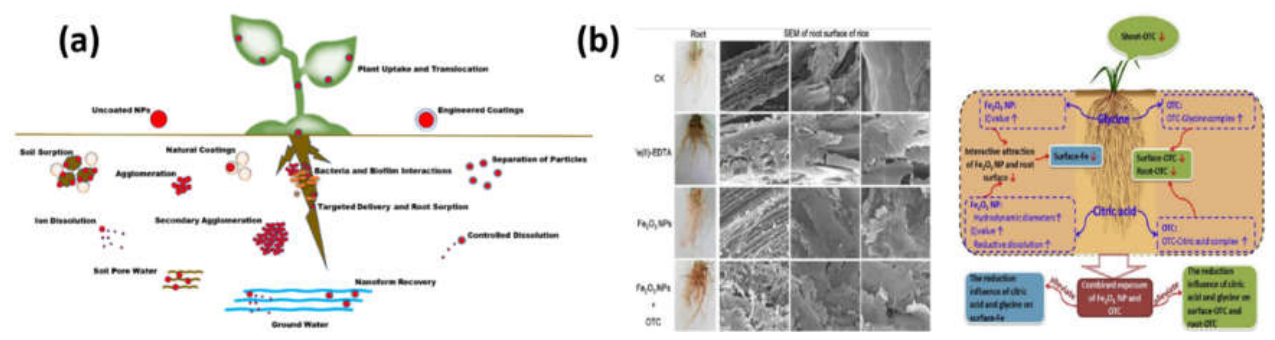
Table 6.
Agricultural Applications of Metal Oxide Nanoparticles.
| S.No | Metal Oxide Nanoparticle | Agricultural Application | Mechanism of Action | Key Benefits | References |
|---|---|---|---|---|---|
| 01 | Titanium Dioxide Nanoparticles (TiO₂ NPs) | Crop growth enhancement, crop protection, improving food production | Improves light absorption and photosynthesis, provides antimicrobial activity against plant pathogens | Better nutrient utilization, protection from diseases, enhanced agricultural productivity | [565] |
| 02 | Titanium Dioxide Nanoparticles | Crop improvement, sustainable agriculture, plant growth promotion | Enhance photosynthesis and light utilization, provide antimicrobial effects against harmful microorganisms | Higher crop yield, better nutrient use efficiency, enhanced resistance to environmental stress | [566] |
| 03 | Titanium Dioxide Nanoparticles (TiO₂ NPs) | Crop growth enhancement, increased yield, stress tolerance, nutrient management, plant protection | Improves photosynthesis, increases root hair formation, and provides antimicrobial protection against plant pathogens | Improved resistance to biotic (diseases) and abiotic (drought, salinity, heat) stresses |
[567] |
| 04 | Titanium Dioxide Nanopowder (TiO₂ NPs) | Pre-sowing seed treatment (e.g., white cabbage), seed germination improvement, seedling growth enhancement | Applied as aqueous suspension to seeds before sowing. Improves seed–water interaction and early metabolic activation, supporting faster germination and early growth processes. | Enhanced early plant vigor Positive effect at low concentration (0.3 g/L) More effective when particles have high surface area and porosity |
[568] |
| 05 | Titanium / Titanium Dioxide NPs (Ti / TiO₂ NPs) | Used in agriculture to improve plant growth and yield, applied in crop production systems as growth enhancers and plant stimulants | Taken up by plants and transported internally (roots → shoots). Can stimulate or sometimes disturb plant metabolic activity depending on dose and conditions. | Enhanced physiological activity Beneficial at optimal concentrations |
[569] |
| 06 | ZnO/TiO₂ Nanocomposite (Mt-supported ZnO/TiO₂) | Used in plant protection systems, antibacterial sprays for crops, leaf surface treatment (e.g., cucumber leaves), crop disease control | Acts as an antibacterial agent by damaging bacterial cells. ZnO and TiO₂ interaction increases active sites, enhances adsorption of bacteria, and improves surface wetting on leaves for better coverage. | Strong antibacterial activity against plant pathogens (e.g., S. aureus) Improved disease control in crops Environment-friendly and low-cost crop protection |
[570] |
| 07 | Titanium Dioxide (TiO₂) in Chitosan Composite (Chitosan–TiO₂) | Used as bioprotective material in agriculture, soil treatment, fertilizer enhancement, controlled nutrient delivery systems | TiO₂ is embedded in a biodegradable chitosan polymer matrix, maintaining effective concentration over time and improving interaction with plants. | Improved nutrient uptake by plants Better fertilizer efficiency (reduced fertilizer use) Controlled and sustained release of TiO₂ |
[571] |
| 08 | Titanium Dioxide (TiO₂) in PEDOT-PSS/PVA Nanocomposite | Used in agricultural sensing systems for soil moisture monitoring and relative humidity (RH) measurement, helping smart irrigation and crop management | TiO₂ is embedded in a conductive polymer matrix (PEDOT-PSS + PVA). Changes in soil moisture alter the electrical resistance of the nanocomposite film due to water adsorption on TiO₂ surface and polymer interaction, improves charge transport pathways and conductivity response. | High sensitivity to humidity (up to 96%) Fast response (10 s) and recovery (50 s) Accurate soil moisture detection Improved conductivity and stability Low-cost and easy fabrication |
[572] |
| 09 | Graphene Oxide–Titanium Dioxide (GO@TiO₂) Nanocomposite | Used in agricultural environmental protection for degradation of pesticide residues (e.g., carbaryl, imidacloprid) in soil and water systems | Acts as a photocatalyst. Graphene oxide improves electron transport and reduces band gap, enhancing light absorption and photocatalytic efficiency. | Efficient degradation of toxic pesticides Improved environmental safety in agriculture Reduced pollution in soil and water |
[573] |
| 10 | TiO₂/Cu₂(OH)₂CO₃ Nanocomposite | Used in crop protection as an antimicrobial agent against plant pathogens (bacteria like E. coli and fungi like Fusarium graminearum) | Under simulated sunlight, the nanocomposite acts as a photocatalyst. Cu₂(OH)₂CO₃ improves light absorption and helps better charge separation in TiO₂, increasing reactive species generation. | Strong antibacterial and antifungal activity Better performance than pure TiO₂ Useful for protecting crops from diseases |
[574] |
| 11 | Green-synthesized Silver Oxide Nanoparticles (Ag₂O NPs) | Seed/plant growth enhancement (e.g., Vigna unguiculata), pest and insect control (larvicidal and insecticidal action), plant protection against microbes | Bioactive compounds from plant extract help form Ag₂O NPs. These nanoparticles release reactive silver species that damage microbial and insect cells, disrupting membranes, enzymes, and metabolic processes. | Improved plant growth, Strong antibacterial activity (S. aureus, S. typhi), Antioxidant and anti-inflammatory effects |
[575] |
| 12 | Silver Oxide Nanoparticles (Ag₂O NPs) | Control of fungal disease in crops (especially Macrophomina phaseolina causing charcoal rot in strawberry), crop protection under lab, greenhouse, and field conditions | Ag₂O nanoparticles inhibit fungal growth directly by disrupting cellular processes. They also trigger plant defense systems by increasing expression of defense-related genes (e.g., PR-1) through salicylic acid signaling pathway, enhancing plant immunity. | Strong antifungal activity against plant pathogens Activation of plant defense genes Improved crop growth and yield Effective under lab, greenhouse, and field conditions |
[576] |
| 13 | Silver Oxide Nanoparticles (Ag₂O NPs) – green synthesized using leaf extract | Used for plant disease control, microbial protection in agriculture, and potential water purification systems for irrigation safety | Ag₂O nanoparticles interact with microbial cells, disrupting cell membranes and metabolic activity. They release silver-based reactive species that inhibit bacterial growth | Strong antimicrobial activity against Gram-positive and Gram-negative bacteria, Eco-friendly green synthesis method | [577] |
| 14 | Silver Oxide Nanoparticles (Ag₂O NPs) – green synthesized using Artocarpus heterophyllus leaf extract | Potential use in agriculture for plant disease control, crop protection, and antimicrobial treatment in agricultural systems | Phytochemicals from plant extract act as reducing and stabilizing agents to form Ag₂O NPs. These nanoparticles can release silver species (Ag⁺) that interact with microbial cells, damaging membranes and inhibiting microbial growth. | Eco-friendly and low-cost synthesis Anticipated strong antimicrobial activity Stable, well-crystallized nanoparticles (fcc structure) |
[578] |
| 15 | Ag/Ag₂O composite nanoparticles (Silver / Silver oxide) | Insect control in agriculture (aphids, ants), crop pest management using water spray formulations | Toxic to insects by disrupting cell membranes and vital enzymes; causes physiological stress and mortality in pests | Effective insecticidal activity against aphids and ants, useful for eco-friendly pest control | [579] |
| 16 | GO–AgNPs nanocomposite (Graphene oxide–Silver nanoparticles) | Crop disease control (fungal leaf spot), plant protection against pathogens, antifungal sprays in agriculture | Damages fungal spores and hyphae by physical contact (sharp GO sheets), releases reactive oxygen species (ROS) that kill microbes, and disrupts cell structure of pathogens | Eco-friendly and low-cost synthesis Anticipated strong antimicrobial activity Tunable properties via reaction conditions |
[580] |
| 17 | Ag₃⁺-enriched AgO/Ag/SnO₂ nanocomposite (HOSBTO) | Control of plant and fish pathogens (fungi, bacteria, oomycetes), especially in crops like strawberry | Highly oxidized silver species and Ag⁺ ions damage microbial cell walls, proteins, and DNA; generates oxidative stress that kills pathogens | Strong, broad-spectrum anti-pathogenic activity; effective at different doses; works against multiple types of pathogens | [581] |
| 18 | FRGO–Ag/AgO/Ag₂O nanocomposite (Reduced graphene oxide + silver/silver oxide system) | Wastewater treatment (dye removal), environmental remediation, antimicrobial protection in agriculture-related systems | Generates reactive oxygen species (ROS) that break down dyes and kill microbes; silver species damage microbial cell membranes and enzymes; graphene supports electron transfer and improves reactions | Strong photocatalytic + antibacterial + antioxidant activity; eco-friendly synthesis using food-based materials; efficient pollutant degradation | [582] |
| 19 | γ-Fe₂O₃–Ag₂O nanocomposite (with AgFeO₂ phase) | Antibacterial and antifungal protection in agriculture, crop disease control, possible use in plant protection sprays and environmental sanitation | Releases silver ions (Ag⁺) and iron oxide reactive species that damage microbial cell walls, proteins, and DNA; induces oxidative stress leading to pathogen death | Strong broad-spectrum antimicrobial activity against bacteria and fungi; effective against multiple pathogens; eco-friendly plant-assisted synthesis | [583] |
| 20 | Ag₂O–COLBN nanocomposite (Silver oxide + Chromolaena odorata biochar) | Water purification for rural/agricultural use, removal of microbial contamination in borehole water | Silver oxide releases Ag⁺ ions that kill bacteria by damaging cell walls, proteins, and DNA; biochar adsorbs pollutants and heavy metals due to high surface area | Strong antibacterial activity, excellent water disinfection, removes turbidity and contaminants, low-cost sustainable material | [584] |
| 21 | FeO nanoparticles (magnetite/maghemite forms) synthesized using Eucalyptus globulus extract | Soil remediation in agriculture, removal of toxic heavy metals (Cr (VI), Cd, Pb) from agricultural soils | Adsorbs and binds heavy metal ions through oxygen-rich active sites on nanoparticle surface; magnetic nature helps interaction and separation from soil systems | Efficient removal of toxic metals, improves soil quality, eco-friendly green synthesis, very small particle size improves activity | [585] |
| 22 | Iron oxide nanoparticles (Maghemite γ-Fe₂O₃ / Magnetite–Maghemite nanoparticles) | Seed priming (pre-sowing treatment), foliar spray for crops, plant growth promotion, sustainable agriculture | Nanoparticles are absorbed by seeds and plants, improving nutrient uptake, stimulating germination, enhancing plant metabolism | Enhances plant growth, increases plant productivity, eco-friendly alternative to excessive agrochemical use | [586] |
| 23 | Fe₂O₃ nanoparticles (Iron oxide nanoparticles) | Compost improvement, organic waste management, seed germination enhancement, seedling growth promotion | Accelerate decomposition of organic matter, stimulate beneficial microbial enzymes (dehydrogenase and urease) | Faster composting, better compost quality, higher seed germination rate, improved seedling growth | [587] |
| 24 | Iron Oxide Nanoparticles (Fe₂O₃, Fe₃O₄) | Soil improvement, plant growth enhancement, seed germination, compost quality improvement, nutrient management | High surface area helps interact with soil and microbes; improves composting processes, enhances enzyme activity, and can be easily separated or directed using magnetic properties | Enhances compost quality, increases organic matter degradation, non-toxic and biocompatible, easy recovery due to magnetic behavior | [588] |
| 25 | Iron oxide nanoparticles (Fe₂O₃ NPs | Used in farming (pot experiments on peanut plants) to improve plant growth, especially in crops like peanut that need iron for healthy development. | Fe₂O₃ NPs release and supply iron to plants, improve nutrient availability in sandy soil, regulate plant hormones, and increase antioxidant enzyme activity, | Improves root length, plant height, biomass, and chlorophyll (SPAD value); increases iron content in plants; works better in sandy soil | [589] |
| 26 | Biochar-supported Iron Oxyhydroxide Nanocomposites (BC@α-FeOOH and BC@β-FeOOH) | Removal of pesticide residues from agricultural wastewater; treatment of water contaminated with pesticides such as Endosulfan (ES) and 4,4-DDD | Biochar provides a high surface area for adsorption of pesticides. Under sunlight, FeOOH NPs generate reactive species (hydroxyl radicals •OH, superoxide radicals O₂•⁻, and holes h⁺) that break down pesticide molecules into less harmful products. | High pesticide degradation efficiency (up to 98%), reusable and stable for multiple cycles, converts agricultural waste (citrus peels) into useful biochar, helps reduce environmental pollution. | [590] |
| 27 | Hydrocalumite–Iron Oxide Magnetic Nanocomposite (HC-DS/Fe) | Removal of pesticide residues (atrazine, chlorpyrifos, thiamethoxam, and acetamiprid) from agricultural wastewater, irrigation water, and contaminated river water | Iron oxide-based magnetic adsorbent captures and holds pesticide molecules on its surface through adsorption. The magnetic property allows easy separation and recovery of the adsorbent from water after treatment. | Efficient pesticide removal, reusable and easy to recover using a magnet, environmentally friendly water treatment method, suitable for agricultural wastewater purification. | [591] |
| 28 | Tungsten Oxide (WO₃), Iron Oxide (MNP), and Copper-Doped Iron Oxide (MNP-Cu) Nanocomposites | Pest control and crop protection against insect pests such as Spodoptera littoralis (cotton leafworm) when used together with insecticides like cyromazine | Nanoparticles enhance the effectiveness of the insecticide. The combination disrupts insect growth and development, prolongs larval and pupal stages, reduces pupation and adult emergence, and stimulates stress and immune responses in insects. | Enhances insecticide performance, may allow lower pesticide use, and offers a promising approach for sustainable crop protection. | [592] |
| 29 | Fe₃O₄@Chitosan–AgNP Magnetic Nanocomposite | Plant disease control, crop protection, and management of fungal pathogens such as Colletotrichum coccodes, Aspergillus niger, and Pyricularia species | Iron oxide provides magnetic recovery, chitosan acts as a biocompatible carrier, and silver nanoparticles damage fungal cells by disrupting cell membranes and cellular functions, leading to inhibition of fungal growth. | Strong antifungal activity, effective control of agricultural pathogens, reusable due to magnetic separation, environmentally friendly use of waste-derived materials | [593] |
| 30 | Bismuth Oxide Nanoparticles (Bi₂O₃ NPs) | Water purification for agriculture, removal of heavy metal contaminants (Co²⁺, Ni²⁺, Cu²⁺) from irrigation water, control of plant-pathogenic bacteria and fungi | Bi₂O₃ nanoparticles adsorb and trap heavy metal ions on their surface. They also exhibit antimicrobial activity by interacting with bacterial and fungal cells, damaging cell structures and inhibiting their growth. | Antibacterial and antifungal activity, eco-friendly green synthesis using plant extract (Beta vulgaris), nanosized particles with high surface area | [594] |
| 31 | Bismuth Oxide Nanoparticles (Bi₂O₃ NPs) | Control of soil-borne bacterial pathogens in crops, reduction of contamination in agricultural environments, improving crop safety | Bi₂O₃ NPs interact with bacterial cells (Gram-positive and Gram-negative), damaging cell membranes and inhibiting bacterial growth. Their high surface area improves contact with microbes, leading to stronger antibacterial action. | Eco-friendly green synthesis (lemon peel extract + microwave method), effective against harmful bacteria like E. coli and Salmonella, stable nanoparticles with good surface charge | [595] |
| 32 | Bismuth oxide (Bi₂O₃) nanoparticles (green synthesized using lemon peel extract) | Control of soil-borne bacterial pathogens in crops (e.g., E. coli, Salmonella spp.); protection of crop health and food safety; environmental sanitation in agricultural soils | Nanoparticles attach to bacterial cells and damage their cell wall/membrane; release of reactive species disrupts metabolism; small size allows strong contact with microbes leading to cell death | Eco-friendly and plant-based synthesis, effective antibacterial activity, useful for environmental and agricultural protection | [595] |
| 33 | NiO–Bi₂O₃ (nickel oxide–bismuth oxide) nanocomposite | Photocatalytic degradation of agricultural soil pollutants (ASP); removal of dye contaminants (methylene blue, methyl orange) from wastewater used in agricultural | Under visible light, the composite absorbs light due to reduced band gap; generates reactive oxygen species (ROS) that break down organic pollutants; NiO–Bi₂O₃ junction improves charge separation, increasing photocatalytic efficiency | High pollutant removal efficiency (up to 95%), works under visible light, reusable multiple times, stable nanocatalyst; effective for cleaning contaminated agricultural water | [596] |
| 34 | γ-Bi₂O₃ (gamma-phase bismuth oxide) combined with graphene oxide (GO) nanocomposite electrode | Detection of herbicide diuron (DU) residues in agricultural water and soil samples to monitor pesticide pollution | Acts as an electrochemical sensor; γ-Bi₂O₃ improves electron transfer while GO increases conductivity and surface area; together they enhance oxidation of diuron, producing a measurable electrical signal | High sensitivity and low detection limit, fast and cost-effective detection, useful for real-time monitoring of pesticide contamination in agriculture | [597] |
| 35 | Bismuth ferrite (BiFeO₃) / N-doped reduced graphene oxide (N-rGO) nanocomposit | Electrochemical detection of monocrotophos (MCP) pesticide residues in agricultural runoff, water, and environmental samples | BiFeO₃ interacts strongly with pesticide molecules (especially organophosphorus groups) and helps electron transfer during oxidation; N-rGO improves conductivity and signal transport; together they enhance electrochemical response for sensitive detection | Very high sensitivity and low detection limit, fast and cost-effective sensing, good selectivity against interfering ions, potential for portable agricultural water testing devices | [598] |
| 36 | Fe₂O₃, ZnO, CuO, Al₂O₃, TiO₂, MnO₂ (metal oxide nanoparticles) | Nanofertilizers, nanopesticides, antimicrobial agents, plant growth enhancers, and nanobiosensors for detecting nutrients, pathogens, and toxins in soil and water | Enter plants through roots/leaves and move inside plant tissues; improve photosynthesis by better light use; increase antioxidant defense enzymes; regulate gene expression; also kill or inhibit pests | Better crop yield and growth, higher resistance to stress, pests, and diseases, reduced need for chemical fertilizers, improved soil and plant health monitoring | [562] |
| 37 | Nanocomposites (including metal oxide–based NCs) | NC-based pesticides for crop protection and pest management in agriculture | Act against pests by multiple pathways such as disrupting cell membranes, causing oxidative stress, blocking metabolism, and improving targeted delivery of active agents | More sustainable alternative to conventional pesticides, improved pest control efficiency, reduced resistance development | [599] |
| 38 | TiO₂, ZnO, Ag nanoparticles in chitosan-based bio-nanocomposites | Active food packaging for agricultural products, protection of fresh produce from spoilage and microbial contamination | Antimicrobial action through direct contact with microbes, release of metal ions, and generation of reactive oxygen species (ROS) that damage bacterial/fungal cells | Biodegradable and eco-friendly packaging, strong antimicrobial protection, reduces food spoilage and waste, safer alternative to plastic preservatives | [600] |
| 39 | Metal and metal-oxide nanoparticles with surface coatings (polymer, biomolecule, inorganic, zwitterionic, humic acid coatings) | Controlled delivery of nanoparticles in soil for crop growth, nutrient delivery, and soil health improvement | Surface coatings prevent nanoparticle clumping (aggregation), control dissolution rate, and improve stability in soil, humic acid can increase bioavailability | Better nanoparticle stability in soil, enhanced nutrient delivery, cost-effective and safer agricultural nanotechnology application | [601] |
| 40 | Metal / metal oxide nanocomposites (general antibacterial nanocomposites) | Plant disease control, antimicrobial coatings for agricultural tools, protection of stored crops and agro-based materials | Penetrate microbial cell walls and membranes, disrupt essential cellular processes, prevent biofilm formation; overcome antibiotic resistance, can work synergistically with antibiotics to enhance killing of pathogens | Strong antibacterial activity against resistant microbes, reduces infection spread in agriculture, helps control biofilms, biodegradable and biocompatible | [602] |
5.5.1. Nanofertilizers
One of the most significant applications of metal oxide nanoparticles in agriculture is nanofertilizers. On the other hand, conventional fertilizers have poor nutrient use efficiency due to the high losses of nutrients from leaching, volatilization, runoff, or fixation in soil. Nanofertilizers offer a way to control and target the delivery of nutrients, enhancing the efficiency of nutrient uptake, and minimizing environmental pollution[603,604,605,606].
The micronutrient sources for plants are the metal oxide nanoparticles like ZnO, Fe₂O₃, CuO, MgO and MnO₂. Zinc oxide nanoparticles are very important because zinc is an essential micronutrient that plays a role in enzyme activation, chlorophyll production and protein metabolism. Nanofertilizers of zinc oxide (ZnO) can boost seed germination rate, root growth, photosynthesis rate and crop production at a relatively low concentration. In the same way, iron oxide nanoparticles boost the synthesis of chlorophyll and also assist in avoiding iron deficiency chlorosis in plants[562,607].
Nanofertilizers have several benefits compared to the conventional ones such as slow and controlled release of nutrients, better absorption by plants, lower nutrient losses and lower frequency of fertilizer application. They have a very large surface area that allows them to interact well with the soil particles and plant roots, increasing the availability of nutrients. Nanoparticles can also be designed to respond to different stimuli, such as the pH of the soil, moisture, or the plant's nutrient needs, and release their cargo onto the surface[608].
Titanium dioxide nanoparticles have been found to positively affect the uptake of nutrients and the efficiency of the photosynthesis process by increasing light absorption and nitrogen metabolism. Magnesium oxide nanoparticles help in the formation of chlorophyll, and manganese oxide nanoparticles aid in the process of photosynthesis and plant's antioxidant defense mechanisms[609].
The current studies are aimed at producing multifunctional nanofertilizer with the combination of MONPs and polymers, biochar, chitosan and biodegradable matrix. These systems help retain nutrients, limit toxicity and promote sustainability. Plant-mediated green-synthesis of nanofertilizers is also trending due to the reduced risk of harmful chemical residues and environmental hazards.
While potentially beneficial, overloading the soil with nanoparticles could also have impacts on the soil's microbial life and on plant growth. Hence, optimization of dosage, methods of use and long-term environmental safety is crucial for sustainable use in agriculture[610].
5.5.2. Plant Growth Promotion
Metal oxide nanoparticles have shown the interesting potential for the promotion of plant growth and crop productivity. Their beneficial effects relate to increased nutrient uptake, stimulation of photosynthesis, increased enzyme activity and regulation of plant metabolic processes. The nanosize of MONPs ensures their superior ability to enter plant tissues as compared to bulk materials and to interact with plant cellular components at the molecular level[611].
ZnO nanoparticles are one of the most popular nanomaterials studied as growth promoters. Zinc is a microelement that plays a key role in auxin production, protein synthesis and carbohydrate metabolism. For some crops such as wheat, maize, rice, soybean and tomato, it has been reported that the application of ZnO nanoparticles leads to enhancement in the germination of seeds, root elongation, shoot growth, chlorophyll content, biomass production[611].
The use of titanium dioxide nanoparticles increases the efficiency of photosynthesis by boosting light absorption and electron transport in the process of photosynthesis. TiO₂ nanoparticles can boost the production of chlorophyll, nitrogen metabolism, and antioxidant enzymes, resulting in better plant growth and productivity. Likewise, iron oxide nanoparticles increase the availability of Fe and aid in the production and formation of chlorophyll, resulting in better photosynthesis and plant strength[562,612].
The silicon dioxide nanoparticles also have come in the limelight since silicon makes the cell wall of plants stronger, increases the water retention in them and enhances their resistance to environmental stress and pathogens. The nanoparticles of magnesium oxide assist in the synthesis of chlorophyll and activation of enzymes and manganese oxide nanoparticles help in the process of photosynthetic oxygen evolution and antioxidant defense system[613].
Low concentrations of MONPs have been found to positively regulate phytohormones, improve uptake efficiency of nutrients and induce beneficial microbial interactions in the rhizosphere, as shown in several studies. Seed priming by nanoparticles has been proven to be a great approach to improve seed germination and seedling development. Foliar application of MONPs is also the subject of much research for nutrient delivery and growth performance[614].
The influence of MONPs on plants, On the other hand, is highly dependent on the type of nanoparticles, concentrations, size, morphology, duration of exposure and type of plants. High levels of nanoparticles could lead to oxidative stress, growth inhibition and phytotoxicity. Careful optimization and risk assessment is therefore, necessary to ensure safe agricultural application[615].
5.5.3. Antimicrobial Protection in Crops
Bacterial, fungal, viral and other diseases of plants take a huge toll on productivity and quality of crops all over the world. These traditional pesticides and fungicides can cause environmental contamination, resistance and adverse effects on non-target species. The importance of metal oxide nanoparticles as antimicrobial agents for crop protection is supported by their strong antimicrobial activity, stability and multifunctionality[616].
The antibacterial and antifungal activity of ZnO and CuO nanoparticles has been extensively studied against several plant pathogens including Xanthomonas, Pseudomonas, Fusarium, Alternaria and Aspergillus species. These nanoparticles produce ROS, disrupt the microbial membranes, interfere with enzyme systems, and damage nucleic acids, which causes the death of the microbial cell. The antimicrobial activity is also due to the release of metal ions[617].
Copper oxide nanoparticles are known to be effective against fungal diseases as copper ions have the ability to inhibit the growth of fungi and spore germination. ZnO nanoparticles have demonstrated to exhibit strong activity against bacterial leaf spot, wilt disease and fungal infection in a number of crops. The photocatalytic antimicrobial properties of TiO₂ nanoparticles when illuminated with light can be used for the control of pathogens and sterilization of surfaces[618].
SiO2 nanoparticles improve the protection of the crops by fortifying the cell walls and stimulating defense responses in plants. The use of iron oxide nanoparticles for targeted delivery of antimicrobial compounds and nanopesticides is being extensively studied. When incorporated in bio-polymer, essential oil, chitosan or traditional pesticide, the nanocombination of MONPs and these materials generally shows synergistically enhanced antimicrobial activity and controlled release behavior[619].
The recent advances are based on the green-synthesized MONPs for sustainable crop protection. Nanoparticles delivered via plants have often been found to have higher biocompatibility and less environmental impact. There is also development of nano enabled anti microbial coatings and smart delivery systems for precision agriculture applications[620].
While the MONPs have great potential as alternatives to conventional agrochemicals, their persistence in soil, toxicity to beneficial microorganisms and accumulation in edible plant tissues are concerns. Thus, detailed environmental and toxicological evaluations are required prior to large-scale applications in agriculture[621].
5.5.4. Stress Tolerance Improvement
Drought, salinity, extreme temperature, heavy metal and oxidative stress are among the environmental factors that significantly influence the growth of plants and agricultural productivity. Metal oxide nanoparticles have demonstrated significant potential for improving plant tolerance to abiotic and biotic stress through physiological, biochemical and molecular regulation[622].
The antioxidant activity of the enzymes and the reduced oxidative damage by ROS, which is mediated by ZnO nanoparticles, contribute to stress tolerance. It is reported that the application of ZnO nanoparticles increases the drought resistance, chlorophyll content and water-use efficiency in different crops. These nanoparticles also control the osmolyte accumulation and membrane stability in stressful situations[623].
Nanoparticles of TiO2 improve the photosynthetic activity and in the environmental stress the antioxidant defense. TiO₂ nanoparticles can induce activity of antioxidant enzymes like catalase, superoxide dismutase and peroxidase, which help plants to scavenge harmful reactive oxygen species. Likewise, the cerium oxide nanoparticles have high redox activity and can function as ROS scavengers, thereby safeguarding plants against ROS[624].
Among the various types of nanoparticles, silicon dioxide nanoparticles play a significant role in salinity and drought stress tolerance. Silicon is a component of the plant tissues that makes plants stronger to hold water, balance ions and helps them withstand environmental stresses. Besides that, iron oxide nanoparticles also aid in stress tolerance through enhanced nutrient uptake and chlorophyll synthesis under stress[625].
Nanoparticle manganese oxide (MnO) and magnesium oxide (MgO) helps to improve the photosynthetic activity, enzyme regulation when subjected to stress. Nanoparticle treated plants have been observed to have better seed germination, root development, biomass accumulation and yield under unfavourable environmental conditions in different studies.
MONPs may affect molecular aspects of stress signalling pathways, antioxidant defense, hormonal control. Seed Priming and foliar application using nanoparticles are emerging methods to enhance crop stress tolerance to climate change[626].
Although the positive results have been achieved, the mechanism of the stress tolerance caused by use of nanoparticles is not completely understood. The long lasting effect of MONP accumulation in soil – plant systems and the possible ecological risk should also be studied in detail to guarantee the safe and sustainable use of MONPs in agricultural applications[627].
5.6. Industrial and Catalytic Applications
Due to their extraordinary physicochemical characteristics such as high surface area, catalytic activity, thermal stability, optical behaviour, electrical conductivity, corrosion resistance and mechanical strength, metal oxide nanoparticles (MONPs) have gained great importance in the industrial and catalytic fields. They have a huge number of active sites and interactions with reactants are better as they are nanoscale and thus are able to perform better than traditional bulk materials. Consequently, MONPs are used more and more in industrial manufacturing, catalytic systems, coatings, textiles, packaging materials, electronic devices and optoelectronic technologies[628].
The metal oxides nanoparticles that are commonly used in industries are TiO₂, ZnO, Fe₃O₄, CeO₂, CuO, Al₂O₃, SiO₂, SnO₂, WO₃, NiO, Co₃O₄. These properties of the nanoparticles can be customized by their synthesis, morphology, doping and surface modification. The large surface areas and better charge transport characteristics of nanostructures like nanorods, nanotubes, porous frameworks, nanosheets, and hollow spheres frequently result in their demonstrating improved catalytic and functional properties[5,629,630,631].
In recent years, with the development of nanotechnology, multifunctional nanocomposites have been developed, including MONPs and polymers, carbon materials, fibers, and other semiconductors. The hybrid materials offer a new durability, conductivity, mechanical strength and multifunctionality to industrial products. In addition, to minimize the impact on the environment and to lower production costs, green synthesis and sustainable fabrication techniques are also investigated[628].
Below, MONPs' major industrial and catalytic applications, such as heterogeneous catalysis, coatings and paints, textiles and packaging, electronics and optoelectronics are discussed.
5.6.1. Heterogeneous Catalysis
One of the largest industrial uses of metal oxide nanoparticles is as heterogeneous catalysts. Catalysts are important for chemical manufacturing, petroleum refining, environmental remediation, energy conversion and pharmaceutical production. Due to the high surface to volume ratio, MONPS have a large number of active sites for catalytic reactions, making it a highly effective heterogeneous catalyst[632].
Titanium dioxide (TiO₂) nanoparticles have been widely applied as photocatalysts to decompose organic pollutants, in self-cleaning surfaces, and in systems for environmental purification. TiO₂ generates reactive oxygen species under UV and visible light irradiation which can oxidize organic compounds to harmless products. ZnO nanoparticles have also demonstrated outstanding photocatalytic activity and are of great interest for environmental and industrial catalytic applications[633].
The nanoparticles of Cerium oxide (CeO₂) are important catalysts because they can store and release oxygen, via reversible Ce³⁺/Ce⁴⁺ redox transitions. CeO₂ is a common component of automobile catalytic converter, which is used for oxidation of carbon monoxide and hydrocarbons and reduction of nitrogen oxides. Likewise, iron oxide nanoparticles are used in Fenton-like catalytic systems, hydrogenation reaction and waste-water treatment processes.
Transition metal oxides like Co₃O₄, NiO, MnO₂ and CuO, are very good catalysts for oxidation, reduction and electrochemical reactions. Copper oxide nanoparticles have been extensively used in carbon monoxide oxidation, methanol synthesis and organic transformations. Manganese oxide nanoparticles are excellent catalysts in oxidation reactions and energy related catalytic processes[634,635].
Current studies have been directed towards enhancing the catalytic activity by morphology engineering, doping, defect engineering and heterojunction formation. Porous or mesoporous nanostructures enhance mass transfer and accessibility of the active sites. The hybrid catalysts of noble metals and graphene, activated carbon and carbon nanotubes with MONPs show optimized catalytic activity, stability and electron transfer efficiency[636].
Among them, magnetic metal oxide nanoparticles like Fe₃O₄ are particularly interesting as they can be easily separated and recycled by applying external magnetic field, thus minimizing catalyst loss and operational expenses. Although tremendous advances have been made, catalyst deactivation, nanoparticle agglomeration, and long-term stability are challenges for industrial catalytic systems[637].
5.6.2. Coatings and Paints
Metal oxide nanoparticles are commonly used in coatings and paints for mechanical durability, corrosion resistance, UV protection, antimicrobial properties, self-cleaning properties, and thermal stability. Problems with environmental degradation, microbial contamination, and ultraviolet radiation are common with conventional coatings. The use of MONPs greatly improves the coating performance and prolongs its service life[638].
Due to its excellent photocatalytic and UV-blocking properties, titanium dioxide nanoparticles are one of the most widely used nanomaterials in coatings and paints. Photocatalytic degradation of organic contaminants on surfaces is achieved by TiO₂ based coatings, giving self cleaning and anti-fogging surfaces. They are widely applied to glass, ceramics, building materials and solar panels[639].
The ZnO nanoparticles also show excellent UV absorbing property and antimicrobial activity, which can be applied in protective coatings for outdoors applications, healthcare facilities and food contact surfaces. Scratch resistance, hardness, transparency and thermal stability of coatings is increased by silica (SiO₂) nanoparticles. Aluminium oxide nanoparticles are used to increase wear resistance and mechanical strength in industrial paints and protective layers[638].
In the field of corrosion, cerium oxide nanoparticles are becoming a promising corrosion inhibitor in metal coatings, as they can generate protective oxide layers which can lessen the degradation of the metal. The use of copper oxide nanoparticles as coating material is dedicated to marine, medical, and industrial applications in which it inhibits the growth and biofouling of microorganisms[640].
By incorporating MONPs into nanostructured coatings, the resulting coatings have enhanced adhesion, hydrophobicity, chemical and environmental stress resistance properties. In self-cleaning, superhydrophobic coating is prepared by silica and titanium dioxide nanoparticles, which give water-repellent properties. Functionalized MONPs are also being used to develop smart coatings that respond to various stimuli such as temperature, light, humidity, and mechanical damage[640].
Recent research has been geared toward environmentally friendly water based nanocoatings and green synthesis methods to lower VOC emissions and environmental toxicity. On the other hand, the dispersion, stability, and working safety of the nanoparticles in manufacturing processes still need to be explored[641].
5.6.3. Textiles and Packaging
Metal oxide nanoparticles have gained the significant importance in the textile and packaging industry due to its antimicrobial activity, UV protection, barrier activity, durability and self cleaning. The integrated MONPs in fibers, fabrics and packaging materials, greatly improve the product performance and functionality[67].
The functional textiles are widely used with ZnO and TiO₂ nanoparticles for their antibacterial, antifungal and UV-blocking properties. The antimicrobial effect of ZnO nanoparticles on the surface of textile products can be achieved by generating reactive oxygen species and the release of the zinc ions, which can inhibit the growth of microorganisms and prevent the formation of odors and infections. TiO₂ nanoparticles impart self-cleaning and stain-resistant properties to the product by photocatalytic degradation of organic contaminant when exposed to light[642].
Metal oxide nanocomposites containing silver are increasingly being used in healthcare textiles, sportswear and protective clothing for their improved antimicrobial properties. The addition of silica nanoparticles increases the strength of the fabric, wrinkle resistance, and water repellency, and aluminum oxide nanoparticles increase the thermal and abrasion resistance[643].
In food packaging, MONPs enhance barrier characteristics to oxygen, moisture, ultraviolet rays and microbial contamination. Incorporation of nanoparticles such as ZnO, TiO₂ or MgO into a packaging material has been shown to help in prolonging the shelf life of food and the preservation of quality product since the nanoparticles act as inhibitors for bacterial and fungal growth. Active and intelligent packaging systems, based on MONPs can also be used for the monitoring of the freshness of the product, detection of product spoilage and response to the environment[644].
Copper oxide nanoparticles have been found to have a potent antimicrobial activity and are being studied for the use in food packaging for antimicrobial protection. The preparation of biodegradable polymer nanocomposites containing MONPs are also being studied extensively as they are biodegradable and have improved mechanical and antimicrobial properties[645].
The recent advances focus on eco-friendly and multi-functional coatings on textiles, which can impart flame resistance, antistatic property, moisture control, and environmental sensing properties. But the issue of how nanoparticles are released during the washing process, as well as the potential for environmental buildup, and human exposure, still needs to be addressed[644].
5.6.4. Electronics and Optoelectronics
The exceptional electrical, magnetic, optical, semiconducting and dielectric properties of metal oxide nanoparticles make them valuable for many modern applications in electronics and optoelectronics. They have nanoscale dimensions which allow their miniaturization, improvement of charge transport, and improvement of their performance in electronic systems[646].
Due to their wide band gap, high electron mobility and strong luminescence properties, ZnO nanoparticles are one of the most extensively studied semiconducting materials for optoelectronic applications. Light-emitting diodes, photodetectors, transistors, transparent conductive films and ultraviolet lasers make use of ZnO nanostructures. They are also used for nanogenerators and flexible electronic devices because of their piezoelectric properties[647].
With their high refractive index and excellent electron transport capability, particles of titanium dioxide are widely used in photovoltaic devices, photocatalytic systems and optical coatings. Tin oxide (SnO₂) nanoparticles are used in transparent conducting materials for displays, touch screens and solar cells. Another important transparent conductive nanoparticles are the nanoparticles of indium oxide and indium tin oxide which are used for optoelectronic devices[647].
Iron oxide nanoparticles have unique magnetic properties and are used for magnetic storage devices, spintronics, sensors, and electromagnetic shielding materials. Due to their good redox and abrasive properties, Cerium oxide nanoparticles have been applied in polishing processes of semiconductor manufacturing and optical devices[648].
Many transition metal oxides, including NiO, Co₃O₄, and WO₃, have been studied for the application in memory devices, electrochromic windows, and smart electronic systems. The tungsten oxide nanoparticles can change color when stimulated with an electric current, which can be helpful in display technology and smart windows. The semiconductor use of CuO nanoparticles is gaining more and more attention due to the low cost of the material and the potential for flexible electronics[649].
The current developments are in the field of flexible, wearable and transparent electronics using MONP nanocomposites. Hybrid MONP-Graphene, conducting polymers and carbon nanotubes show increased conductivity, flexibility and mechanical durability. Optical and electronic properties are also manipulated via quantum confinement effects and defect engineering, for advanced applications[650].
Although significant technological advances have been made, there are still important issues associated with large-scale fabrication, long-term stability, interfacial defects, and environmental safety that should be addressed for future development of electronic and optoelectronic devices based on MONPs[651,652,653].
6. Challenges and Limitations
Although there has been a huge amount of research done on metal oxide nanoparticles (MONPs) and their applications in environmental, biomedical, energy, sensing and industrial applications, the use of these nanoparticles from the laboratory to real applications is still limited. There remain a number of basic and application challenges that prevent commercialization and dependable large-scale deployment.One of the main is the lack of control in the physicochemical characteristics of their synthesis, because changes in the reaction parameters cause large variations in particle size, shape, crystallinity and surface chemistry. This incompleteness is further exacerbated by the lack of standard synthesis and characterization protocols, which hinders comparisons across studies and industry adoption. While Green synthesis approaches are environmentally appealing, they also exhibit extra variability related to non-uniform biological precursors.High surface energy and strong interparticle interactions continue to be a challenge for aggregation and long-term instability. This results in the decrease of active surface area and loss of catalytic, photocatalytic, sensing and biomedical activity during use. While some of these effects can be reduced by surface modification and support matrices, the dispersion is not ensured for long-term periods in realistic environments.Safety related issues with toxicity and environmental risks are not adequately addressed. The production of Reactive Oxygen Species (ROS) and induction of oxidative stress and DNA damage by MONPs, combined with the release of ions from metal oxides, can increase cytotoxicity. Still, long term systematic toxicological tests are still not widespread, especially in realistic exposures, and the potential impacts to the environment and human health are not fully known.Another significant hurdle is scalability; most synthesis methods are energy intensive, the raw materials used are expensive and the processes are batch oriented and hard to reproduce on an industrial scale. Sustainable approaches are green, but often have lower yields, slower reaction rates, and limited reaction control. Also, uniform production in large scale is not solved because of the changes of heat and mass transfer conditions. Economic feasibility and operational durability is still inconclusive for commercialization. The catalytic, sensing and energy-storage systems degrade significantly under various degradation mechanisms including aggregation, oxidation, phase transformation and photocorrosion; all of which can cause high production costs.
7. Future Perspectives
Metal oxide nanoparticles (MONPs) are promising nanomaterials because of their unique physicochemical, catalytic, optical, magnetic and biological properties, which allow them to be used in various fields of environment, energy storage, sensing, agriculture and biomedicine. Their large-scale commercialization is however hindered by the issues of synthesis control, reproducibility, stability, toxicity, and cost-effectiveness. Future studies should then be directed towards the development of safer, scalable, and multifunctional MONP systems for real-life applications.One of the big trends is the emergence of Green and Sustainable synthesis techniques with the use of plants, microorganisms and biomolecules which lower the use of toxic chemicals and energy. Reproducibility and scalability, possibly by AI-assisted optimization, are necessary. Multifunctional nanoparticle designs are another crucial aspect that combines catalysis, sensing, antimicrobial activity, imaging, and drug delivery in a single platform for applications in smart agriculture and theranostic medicine.Surface engineering will continue to be essential to enhance stability, dispersion and biocompatibility, and smart and controlled functionality may be achieved by stimuli-responsive coatings. It is believed that the combination of MONPs with polymers, carbon materials and metal–organic frameworks (MOFs) will improve their conductivity, stability and catalytic activity.Scalable production, standardized protocols, and comprehensive toxicity and life-cycle assessments are necessary for industrial translation. Finally, regulatory requirements and interdisciplinary collaboration will play a key role in closing the loop between research in the lab and commercialization. It is expected that MONPs will contribute in the future to sustainable and highperformance technologies on the whole.
8. Conclusions
Metal oxide nanoparticles (MONPs) are highly versatile nanomaterials with exceptional physicochemical, optical, catalytic, magnetic, and biological properties, enabling wide-ranging applications in environmental remediation, energy storage and conversion, sensing, agriculture, biomedical science, and industrial processes. Extensive research has established a strong foundation in their synthesis, characterization, surface modification, and functional applications.Among various synthesis approaches, green methods have gained significant attention due to their sustainability, low toxicity, and cost-effectiveness. However, nanoparticle performance is strongly dependent on synthesis parameters, which critically influence size, morphology, crystallinity, and surface properties. Advanced characterization techniques remain essential for understanding structure–property relationships and ensuring reproducibility.Despite their promising performance, large-scale commercialization of MONPs is still limited by challenges such as aggregation, toxicity concerns, poor reproducibility, high production costs, stability issues, and scale-up limitations. In addition, the lack of standardized protocols and long-term safety data restricts their industrial translation.Future progress will rely on the development of sustainable and scalable synthesis strategies, advanced surface engineering, and hybrid nanocomposites integrating polymers, carbon materials, and metal–organic frameworks. The incorporation of artificial intelligence and machine learning for material design and optimization, along with robust toxicity evaluation and regulatory frameworks, will be crucial for safe and efficient deployment. MONPs hold strong potential to contribute to next-generation technologies, provided that current scientific and engineering challenges are effectively addressed through interdisciplinary research and innovation.
Author Contributions
Muhammad Kashif: Conceptualization, methodology, literature survey, data curation, writing original draft preparation, visualization, and manuscript revision. Misbah Gul: Literature collection, data analysis, writing—original draft preparation, review and editing. Natasha Shahzad: Investigation, literature review, preparation of figures and tables, writing and editing. Hao Sun: Conceptualization, supervision, validation, critical review, and editing of the manuscript. SK.A. Shezan: Data interpretation, validation, review and editing of the manuscript. Hakan Tozan: Supervision, project administration, funding acquisition, conceptual guidance, critical review, and editing. Oumayma Hamlaoui: Literature survey, data curation, validation, and manuscript review. Naveed Ahmad: Investigation, visualization, proofreading, review and editing.
Data Availability Statement
No datasets were generated or analyzed during the current study.
Acknowledgments
The authors extend their appreciation to the Deanship of Scientific Research at Northern Border University, Arar, KSA, for funding this research work through the project number “NBU-FFR-2026-1902-05". The author also acknowledged the use of Gemini Ai for the preparation of graphical abstract .
Conflicts of Interest
The authors declare no conflicts of interest.
| Abbreviation | Full Form |
| AFM | Atomic Force Microscopy |
| MONPs | Metal oxide Nanoparticles |
| Ag NPs | Silver Nanoparticles |
| AQY | Apparent Quantum Yield |
| AuNPs | Gold Nanoparticles |
| BET | Brunauer–Emmett–Teller (surface area analysis) |
| BOD | Biological Oxygen Demand |
| CB | Conduction Band |
| CBM | Combustion Method |
| CV | Cyclic Voltammetry |
| COD | Chemical Oxygen Demand |
| DNA | Deoxyribonucleic Acid |
| DLS | Dynamic Light Scattering |
| ECSA | Electrochemically Active Surface Area |
| EDX/EDS | Energy Dispersive X-ray Spectroscopy |
| EIS | Electrochemical Impedance Spectroscopy |
| FESEM | Field Emission Scanning Electron Microscopy |
| FTIR | Fourier Transform Infrared Spectroscopy |
| GB | Band Gap Energy |
| GO | Graphene Oxide |
| H2 | Hydrogen Gas |
| H2O2 | Hydrogen Peroxide |
| HER | Hydrogen Evolution Reaction |
| HM | Hydrothermal Method |
| HR-TEM | High-Resolution Transmission Electron Microscopy |
| LOD | Limit of Detection |
| LSV | Linear Sweep Voltammetry |
| LIB | Lithium-Ion Battery |
| MB | Methylene Blue (model pollutant in photocatalysis) |
| MFC | Microbial Fuel Cell |
| MIC | Minimum Inhibitory Concentration |
| MW | Microwave-Assisted Synthesis |
| MRI | Magnetic Resonance Imaging |
| NPs | Nanoparticles |
| OER | Oxygen Evolution Reaction |
| OH• | Hydroxyl Radical |
| PEC | Photoelectrochemical |
| PL | Photoluminescence Spectroscopy |
| PV | Photovoltaic |
| PVD | Physical Vapor Deposition |
| QE | Quantum Efficiency |
| ROS | Reactive Oxygen Species |
| SC | Supercapacitor |
| SEM | Scanning Electron Microscopy |
| SG | Sol–Gel Method |
| SNR | Signal-to-Noise Ratio |
| SS | Sonochemical Synthesis |
| SV | Solvothermal Method |
| TGA | Thermogravimetric Analysis |
| TiO2 | Titanium Dioxide |
| TMO | Transition Metal Oxides |
| TOF | Turnover Frequency |
| TON | Turnover Number |
| TEM | Transmission Electron Microscopy |
| UV–Vis | Ultraviolet–Visible Spectroscopy |
| VB | Valence Band |
| VOCs | Volatile Organic Compounds |
| XPS | X-ray Photoelectron Spectroscopy |
| XRD | X-ray Diffraction |
| ZP | Zeta Potential |
References
- Alhalili, Z.J.M. Metal oxides nanoparticles: general structural description, chemical, physical, and biological synthesis methods, role in pesticides and heavy metal removal through wastewater treatment . 2023, 28(7), 3086. [Google Scholar] [CrossRef] [PubMed]
- Danish, M.S.S.; et al. Photocatalytic applications of metal oxides for sustainable environmental remediation . 2021, 11(1), 80. [Google Scholar] [CrossRef]
- Olawade, D.B.; et al. Metal and metal oxide nanomaterials for heavy metal remediation: novel approaches for selective, regenerative, and scalable water treatment . 2024, 6, 1466721. [Google Scholar] [CrossRef]
- Kashif, M.; et al. Synthesis Strategies and Multifaceted Applications of Molybdenum-Based Nanomaterials: A Comprehensive Review . 2026, 2026(1), 1764400. [Google Scholar] [CrossRef]
- Radulescu, D.-M.; et al. Green synthesis of metal and metal oxide nanoparticles: a review of the principles and biomedical applications . 2023, 24(20), 15397. [Google Scholar] [CrossRef] [PubMed]
- Suppiah, D.D.; et al. Eco-friendly green synthesis approach and evaluation of environmental and biological applications of Iron oxide nanoparticles . 2023, 152, 110700. [Google Scholar] [CrossRef]
- Manuja, A.; et al. Metal/metal oxide nanoparticles: Toxicity concerns associated with their physical state and remediation for biomedical applications . 2021, 8, 1970–1978. [Google Scholar] [CrossRef] [PubMed]
- Nikolova, M.P.; Chavali, M.S.J.B. Metal oxide nanoparticles as biomedical materials . 2020, 5(2), 27. [Google Scholar] [CrossRef] [PubMed]
- Chaudhary, R.G.; et al. Metal/metal oxide nanoparticles: toxicity, applications, and future prospects . 2019, 25(37), 4013–4029. [Google Scholar] [CrossRef] [PubMed]
- Khan, Y.; et al. Classification, synthetic, and characterization approaches to nanoparticles, and their applications in various fields of nanotechnology: a review . 2022, 12(11), 1386. [Google Scholar] [CrossRef]
- Gindose, T.G.; et al. A Review on Metal-Doped Zinc Oxide–Based Single and Multiple Phase Mixed Oxide Nanomaterials for Photocatalytic Applications . 2025, 2025(1), 9925834. [Google Scholar] [CrossRef]
- Abdel-Aziz, A.-A.B.; et al. Synthesis, Characterization, and Versatile Applications of Metal Oxides and Mixed Metal Oxide Nanoparticles . 2025, 172(2), 023503. [Google Scholar] [CrossRef]
- Mehtab, A.; et al. Rare earth doped metal oxide nanoparticles for photocatalysis: a perspective . 2022, 33(14), 142001. [Google Scholar] [CrossRef]
- Qu, H.-j.; et al. A review of graphene-oxide/metal–organic framework composites materials: characteristics, preparation and applications . 2021, 28(6), 1837–1865. [Google Scholar] [CrossRef]
- Khan, S.; Hossain, M.K. Classification and properties of nanoparticles, in Nanoparticle-based polymer composites; Elsevier, 2022; pp. 15–54. [Google Scholar]
- Abraham, J.; et al. One-dimensional (1D) nanomaterials: Nanorods and nanowires; nanoscale processing, in Nanoscale processing; Elsevier, 2021; pp. 71–101. [Google Scholar]
- Timmerman, M.A.; et al. Metal oxide nanosheets as 2D building blocks for the design of novel materials . 2020, 26(42), 9084–9098. [Google Scholar] [CrossRef] [PubMed]
- Sang, Y.; Liu, M.J.C.S. Hierarchical self-assembly into chiral nanostructures . 2022, 13(3), 633–656. [Google Scholar] [CrossRef] [PubMed]
- Negrescu, A.M.; et al. Metal oxide nanoparticles: review of synthesis, characterization and biological effects . 2022, 13(4), 274. [Google Scholar] [CrossRef] [PubMed]
- Mandal, A.K.; et al. Current research on zinc oxide nanoparticles: synthesis, characterization, and biomedical applications . 2022, 12(17), 3066. [Google Scholar] [CrossRef] [PubMed]
- Mohd Raub, A.A.; et al. Photocatalytic activity enhancement of nanostructured metal-oxides photocatalyst: a review . 2024, 35(24), 242004. [Google Scholar] [CrossRef]
- Pourmadadi, M.; et al. Role of iron oxide (Fe2O3) nanocomposites in advanced biomedical applications: a state-of-the-art review . 2022, 12(21), 3873. [Google Scholar] [CrossRef] [PubMed]
- Jayasimha, H.; et al. Green synthesis of CuO nanoparticles: A promising material for photocatalysis and electrochemical sensor . 2024, 100254. [Google Scholar] [CrossRef]
- Chinthala, M.; et al. Synthesis and applications of nano-MgO and composites for medicine, energy, and environmental remediation: a review . 2021, 19(6), 4415–4454. [Google Scholar] [CrossRef]
- Avinash, B.; et al. NiO bio-composite materials: Photocatalytic, electrochemical and supercapacitor applications . 2021, 3, 100049. [Google Scholar] [CrossRef]
- Sims, C.M.; et al. Approaches for the quantitative analysis of oxidation state in cerium oxide nanomaterials . 2019, 30(8), 085703. [Google Scholar] [PubMed]
- C Thomas, S.; Mishra, P. Kumar; C., S.J. Talegaonkar, Ceramic nanoparticles: fabrication methods and applications in drug delivery . 2015, 21(42), 6165–6188. [Google Scholar] [CrossRef]
- Lee, K.; et al. Effect of sintering temperature on the electrical and gas sensing properties of tin oxide powders . 2021, 47(16), 22794–22800. [Google Scholar] [CrossRef]
- Pavitra, V.; et al. Sonochemical synthesis of SnO2–CuO nanocomposite: diverse applications on Li-ion battery, electrochemical sensing and photocatalytic activity . 2020, 31(11), 8737–8749. [Google Scholar] [CrossRef]
- Ren, J.; et al. A review of high-entropy materials with their unique applications . 2025, 8(2), 195. [Google Scholar] [CrossRef] [PubMed]
- Szczyglewska, P.; Feliczak-Guzik, A.; Nowak, I.J.M. Nanotechnology–general aspects: A chemical reduction approach to the synthesis of nanoparticles . 2023, 28(13), 4932. [Google Scholar] [CrossRef] [PubMed]
- Jagdeo, K.R. Physical methods for synthesis of nanoparticles, in Nanochemistry; CRC Press, 2023; pp. 66–76. [Google Scholar]
- Madani, M.; et al. Green synthesis of nanoparticles for varied applications: Green renewable resources and energy-efficient synthetic routes . 2022, 11(1), 731–759. [Google Scholar] [CrossRef]
- Mattox, D.M. Handbook of physical vapor deposition (PVD) processing; William Andrew, 2010. [Google Scholar]
- Colligon, J.; Vishnyakov, V.J.S. I.S.V.A.o.S.S. I, Thin films: sputtering, PVD methods, and Applications . 2020, 9, 1–55. [Google Scholar] [CrossRef]
- Tämm, K.; et al. Parametrization of nanoparticles: Development of full-particle nanodescriptors . 2016, 8(36), 16243–16250. [Google Scholar] [CrossRef] [PubMed]
- Chavali, M.S. M.P.J.S.a.s. Nikolova, Metal oxide nanoparticles and their applications in nanotechnology . 2019, 1(6), 607. [Google Scholar] [CrossRef]
- Amaral, S.P.; Grosche, L.C.; Sousa, J.P.J.M.T.C. Beyond core-shell: enhanced shielding and mutual protection using SiO₂/UVabs and TiO₂ nanoparticles for photostable polymeric films . 2026, 53, 103532. [Google Scholar]
- Kavitha, R.; Manjunatha, C. S.G.J.C.J.o.C. Kumar, ZnO-based S-scheme heterojunction: Design principles, preparation methods and photocatalytic activity . 2026, 83, 54–95. [Google Scholar] [CrossRef]
- Lungu, I.I.; et al. Green-Synthesized Copper and Copper Oxide Nanoparticles: Structural Characterization and Evaluation of Biological Activity . 2026, 15(3), 339. [Google Scholar] [CrossRef] [PubMed]
- Zhang, W.; Tian, H.J.M.T.C. Plasmonic coupling behavior of Ag–SnO₂ nanoparticle dimers . 2026, 51, 114849. [Google Scholar] [CrossRef]
- Saleem, H.; et al. Biogenic Fe₂O₃ nanoparticles enhance carotenoid pathway gene expression and suppress verticillium root rot in marigold (Tagetes erecta) . 2026. [Google Scholar] [CrossRef] [PubMed]
- Parveen, F.; et al. Rational design of SnO2/Fe2O3/GO nanocomposite for efficient photocatalytic degradation of organic dyes . 2026, 116793. [Google Scholar] [CrossRef]
- Abdelfattah, I.; El-Shamy, A.J.S.R. A comparative study for optimizing photocatalytic activity of TiO2-based composites with ZrO2, ZnO, Ta2O5, SnO, Fe2O3, and CuO additives . 2024, 14(1), 27175. [Google Scholar] [CrossRef] [PubMed]
- Gul, M.; Kashif, M.; Shahid, K.J.P.R.J. Eco-friendly approaches in the synthesis of ZnO nanoparticles using plant extract: a review . 2024, 3(2), 1–25. [Google Scholar] [CrossRef]
- Minello, L.V.P.; et al. Surface Engineering of Nanoparticles via Physical Vapor Deposition (PVD): Opportunities and challenges for sustainable agriculture . 2025, 100187. [Google Scholar] [CrossRef]
- Nur-E-Alam, M.; et al. Physical-vapor-deposited metal oxide thin films for pH sensing applications: last decade of research progress . 2023, 23(19), 8194. [Google Scholar] [CrossRef] [PubMed]
- Abdulrahman, A.F.; et al. Synthesis of lead oxide thin films by using physical vapor deposition technique . 2021, 42, 2752–2755. [Google Scholar] [CrossRef]
- An, W.-J.; Thimsen, E. P.J.T.J.o.P.C.L. Biswas, Aerosol-chemical vapor deposition method for synthesis of nanostructured metal oxide thin films with controlled morphology . 2010, 1(1), 249–253. [Google Scholar]
- Svetlichnyi, V.A.; et al. Metal oxide nanoparticle preparation by pulsed laser ablation of metallic targets in liquid, in Applications of Laser Ablation-Thin Film Deposition, Nanomaterial Synthesis and Surface Modification; IntechOpen, 2016. [Google Scholar]
- Zhang, J.; et al. Colloidal metal nanoparticles prepared by laser ablation and their applications . 2017, 18(9), 986–1006. [Google Scholar] [CrossRef] [PubMed]
- Lou, W.; et al. Present and future of metal nanoparticles in tumor ablation therapy . 2023, 15(44), 17698–17726. [Google Scholar] [CrossRef] [PubMed]
- Attallah, A.H.; et al. Effect of liquid and laser parameters on fabrication of nanoparticles via pulsed laser ablation in liquid with their applications: a review . 2023, 18(4), 1307–1323. [Google Scholar] [CrossRef]
- Jiang, Z.; et al. Progress in laser ablation and biological synthesis processes:“Top-Down” and “Bottom-Up” approaches for the green synthesis of Au/Ag nanoparticles . 2022, 23(23), 14658. [Google Scholar] [CrossRef] [PubMed]
- Drmosh, Q.A.Q. Synthesis and Characterization of Nano-Structure Metal Oxides and Peroxides Prepared by Laser Ablation in Liquids; King Fahd University of Petroleum and Minerals (Saudi Arabia), 2010. [Google Scholar]
- Gondal, M.; et al. Growth of metal oxide nanoparticles using pulsed laser ablation technique . In in Oxide-based Materials and Devices II; SPIE, 2011. [Google Scholar]
- Ahmed, N.; et al. Laser ablation and laser-hybrid ablation processes: a review . 2016, 31(9), 1121–1142. [Google Scholar]
- Sportelli, M.C.; et al. The pros and cons of the use of laser ablation synthesis for the production of silver nano-antimicrobials . 2018, 7(3), 67. [Google Scholar] [CrossRef] [PubMed]
- Yadav, T.P.; et al. Mechanical milling: a top down approach for the synthesis of nanomaterials and nanocomposites . 2012, 2(3), 22–48. [Google Scholar] [CrossRef]
- Chen, F.; et al. Synthesis of nanomaterials using top-down methods; 2022; pp. 37–60. [Google Scholar]
- Kukovecz, Á.; et al. Long-time low-impact ball milling of multi-wall carbon nanotubes . 2005, 43(5), 994–1000. [Google Scholar] [CrossRef]
- Šepelák, V.; Bégin-Colin, S.; Le Caer, G.J.D.T. Transformations in oxides induced by high-energy ball-milling . 2012, 41(39), 11927–11948. [Google Scholar] [CrossRef] [PubMed]
- Tsuzuki, T.J.C.C. Mechanochemical synthesis of metal oxide nanoparticles . 2021, 4(1), 143. [Google Scholar] [CrossRef] [PubMed]
- Wei, L.K.; et al. Producing metal powder from machining chips using ball milling process: A review . 2023, 16(13), 4635. [Google Scholar] [CrossRef] [PubMed]
- Amraoui, R.; Younes, A.; Abada, A.J.T.I.J.o.A.M.T. Effect of Al2O3, ZnO, and SiO2 on the magnetic and structural properties of FeTiO3 nanocomposite synthesized by mechanical milling technique . 2025, 139(1), 273–288. [Google Scholar] [CrossRef]
- Kaviarasu, C.; et al. Nanomaterials Through Powder Metallurgy: Production, Processing, and Potential Applications Toward Energy and Environment . 2021, 859–897. [Google Scholar] [CrossRef]
- Jose, S.; Thomas, S.; Kadumudi, F.B. Metal and Metal Oxide Nanoparticles in Textile Applications; Woodhead Publishing, 2026. [Google Scholar]
- Soukupová, G.; et al. Tailored silicon nanostructures in hydrogel-derived conductive binders: Role of size, structure, and surface chemistry in enhancing Li-ion battery performance . 2026, 661, 238620. [Google Scholar] [CrossRef]
- Zheng, J. Iron-Group Metal Compound Nanomaterials: Preparation, Characterization, Structure, and Applications; John Wiley & Sons, 2026. [Google Scholar]
- Wu, T.; et al. Self-driving lab for the photochemical synthesis of plasmonic nanoparticles with targeted structural and optical properties . 2025, 16(1), 1473. [Google Scholar] [CrossRef] [PubMed]
- Mahajan, M.; et al. Green synthesis of ZnO nanoparticles using Justicia adhatoda for photocatalytic degradation of malachite green and reduction of 4-nitrophenol . 2025, 15(4), 2958–2980. [Google Scholar] [CrossRef] [PubMed]
- Mahapatro, J.; Khan, A.; Agrawal, S. Synthesis Techniques, in Luminescence Spectroscopy and Microscopy: Methods and Applications; Springer, 2026; pp. 19–33. [Google Scholar]
- Longchar, C.; et al. Synthesis of Ferrite Nanomaterials: A comparative Analysis of Sol-Gel Auto-Combustion and Co-Precipitation Methods-A Review . 2025, 1(2), 29–48. [Google Scholar] [CrossRef]
- Khan, M.M.J.P.s. and c. methods, Precipitation and coprecipitation synthesis methods; 2025; pp. 11–25.
- Nancy, R.V.; et al. 2 Synthesis Methods; 2025; p. 16. [Google Scholar]
- Parmar, M.; et al. Zeolites in the age of advanced materials: from structural refinement to scalable green synthesis and emerging applications . 2025, 8(6), 4063–4107. [Google Scholar] [CrossRef]
- Ansari, S.M.; et al. Cobalt ferrite nanoparticles: The physics, synthesis, properties, and applications . 2025, 12(2). [Google Scholar] [CrossRef] [PubMed]
- Devesa, S.J.S.-G.-A.V. and W. Technology, Comparative overview of metal oxide nanoparticle synthesis methods: conventional sol-gel versus green approaches . 2025. [CrossRef] [PubMed]
- Khandaker, T.; et al. Recent progress in gel catalysts: boosting efficiency for sustainable energy applications . 2025, 15(5), 1357–1389. [Google Scholar] [CrossRef]
- Suchikova, Y.; et al. Binary Oxide Ceramics (TiO₂, ZnO, Al₂O₃, SiO₂, CeO₂, Fe₂O₃, WO₃) for Solar Cell Applications: A Comparative and Bibliometric Analysis . 2025.
- Srilatha, N.; et al. Developing Hybrid and Functional Materials with Sol-gel Chemistry–A Review . 2025, 14(3), 540–558. [Google Scholar] [CrossRef]
- Zhou, Y.; et al. Functional mesoporous thin films: synthesis, properties, and applications . 2026, 1–29. [Google Scholar] [CrossRef] [PubMed]
- Kumar, P.; et al. Structure, Properties and Application of Metal Oxide, in Metal Oxides; CRC Press, 2025; pp. 3–21. [Google Scholar]
- Nassar, K.I.; Teixeira, S.S.; Graça, M.P.J.G. Sol–gel-synthesized metal oxide nanostructures: advancements and prospects for spintronic applications—a comprehensive review . 2025, 11(8), 657. [Google Scholar] [CrossRef] [PubMed]
- Hjiri, M.; Mustapha, N.; Chouiref, L.J.J.o.M.S.M.i.E. Engineered metal oxide nanostructures via sol–gel chemistry for next-generation spintronic devices . 2026, 37(7), 536. [Google Scholar] [CrossRef]
- Parashar, M.; Shukla, V.K.; Singh, R.J.J.o.M.S.M.i.E. Metal oxides nanoparticles via sol–gel method: a review on synthesis, characterization and applications . 2020, 31(5), 3729–3749. [Google Scholar] [CrossRef]
- Sui, R.; Charpentier, P.J.C.r. Synthesis of metal oxide nanostructures by direct sol–gel chemistry in supercritical fluids . 2012, 112(6), 3057–3082. [Google Scholar] [CrossRef] [PubMed]
- Esposito, S.J.M. “Traditional” sol-gel chemistry as a powerful tool for the preparation of supported metal and metal oxide catalysts . 2019, 12(4), 668. [Google Scholar] [CrossRef] [PubMed]
- Sadek, O.; et al. Synthesis by sol-gel method and characterization of nano-TiO2 powders . 2022, 66, 456–458. [Google Scholar] [CrossRef]
- Lavino, S. Characterization of titanium dioxide coatings for self-cleaning surfaces . 2021. [Google Scholar]
- Khan, M.A.; Akhtar, M.S.; Yang, O.-B.J.S.e. Synthesis, characterization and application of sol–gel derived mesoporous TiO2 nanoparticles for dye-sensitized solar cells . 2010, 84(12), 2195–2201. [Google Scholar] [CrossRef]
- Sharma, A.; Karn, R.; Pandiyan, S.J.J.B.A.E.R. Synthesis of TiO2 nanoparticles by sol-gel method and their characterization . 2014, 1(9), 1–5. [Google Scholar]
- Hayle, S.T. G.G.J.A.J.o.N. Gonfa, and Nanotechnology, Synthesis and characterization of titanium oxide nanomaterials using sol-gel method . 2014, 2(1), 1. [Google Scholar] [CrossRef]
- Sabry, R.S.; et al. Synthesis and photocatalytic activity of TiO2 nanoparticles prepared by sol–gel method . 2016, 78(2), 299–306. [Google Scholar] [CrossRef]
- Azizi. F.J.J.o.M.S.M.i.E., Synthesis and characterization of graphene–N-doped TiO2 nanocomposites by sol–gel method and investigation of photocatalytic activity . 2017, 28(15), 11222–11229. [Google Scholar] [CrossRef]
- Mustapha, S.; et al. Sol–gel synthesis of kaolin/TiO2 nanocomposites for photocatalytic degradation of tannery wastewater, in Industrial Wastewater Treatment: Emerging Technologies for Sustainability; Springer, 2022; pp. 323–343. [Google Scholar]
- Aware, D.V.; Jadhav, S.S.J.A.N. Synthesis, characterization and photocatalytic applications of Zn-doped TiO2 nanoparticles by sol–gel method . 2016, 6(7), 965–972. [Google Scholar]
- Appasamy, J.S.; Kurnia, J.C.; Assadi, M.K.J.S.e. Synthesis and evaluation of nitrogen-doped titanium dioxide/single walled carbon nanotube-based hydrophilic self-cleaning coating layer for solar photovoltaic panel surface . 2020, 196, 80–91. [Google Scholar] [CrossRef]
- Idris, N.J.; et al. Photocatalytic performance improvement by utilizing GO_MWCNTs hybrid solution on sand/ZnO/TiO2-based photocatalysts to degrade methylene blue dye . 2021, 28(6), 6966–6979. [Google Scholar] [PubMed]
- Abbas, A.; et al. Fabrication of Silver Oxide Nanoparticles using sol-gel technique and Their Role in Inhibiting Bacterial Biofilm Development . 2026, 4(1), 163–173. [Google Scholar] [CrossRef]
- Kumar, A.; et al. Synthesis and characterization of silver oxide and silver nanoparticle by sol-gel method . 2010, 29(2), 101. [Google Scholar]
- Shahjahan, M.; et al. Synthesis and characterization of silver nanoparticles by sol-gel technique . 2017, 3(1), 34–39. [Google Scholar] [CrossRef]
- Maesaroh, K.; et al. The effect of different synthesis with chemical and biological methods on properties of silver oxide nanoparticles . 2023, 20(3), 4350–4350. [Google Scholar] [CrossRef]
- Abed, A.A.; Hassouni, M.H.; Banimuslem, H.A. Antibacterial activity of Cuo: Ag2O nanoparticles synthesized using sol-gel method . AIP Conference Proceedings, 2025; AIP Publishing LLC. [Google Scholar]
- Ahlawat, D.S.; et al. Synthesis and characterization of sol–gel prepared silver nanoparticles . 2014, 13(01), 1450004. [Google Scholar] [CrossRef]
- Kariper, I. F.M.J.J.o.E.S. Tezel, Synthesis and characterization of magnesium oxide/silver oxide electrode for supercapacitors by simple Sol-Gel process . 2020, 32, 101958. [Google Scholar] [CrossRef]
- Dolgov, L.; et al. Sol–gel derived metal oxides doped with silver nanoparticles as tunable plasmonic materials . 2010, 207(5), 1166–1169. [Google Scholar] [CrossRef]
- Zagloul, H.; et al. Multifunctional Ag2O/chitosan nanocomposites synthesized via sol-gel with enhanced antimicrobial, and antioxidant properties: A novel food packaging material . 2024, 264, 129990. [Google Scholar] [CrossRef] [PubMed]
- Racles, C.; Nistor, A. M.J.C.E.J.o.C. Cazacu, A silica-silver nanocomposite obtained by sol-gel method in the presence of silver nanoparticles . 2013, 11(10), 1689–1698. [Google Scholar] [CrossRef]
- Kayani, Z.N.; et al. Synthesis of iron oxide nanoparticles by sol–gel technique and their characterization . 2014, 50(8), 1–4. [Google Scholar] [CrossRef]
- Jesus, J.; et al. Green sol–gel synthesis of iron oxide nanoparticles for magnetic hyperthermia applications . 2024, 16(12), 1578. [Google Scholar] [CrossRef] [PubMed]
- Da Costa, G.; et al. Synthesis and characterization of some iron oxides by sol-gel method . 1994, 113(2), 405–412. [Google Scholar] [CrossRef]
- Parhizkar, J.J.N.R. Synthesis, characterization and photocatalytic properties of Iron oxide nanoparticles synthesized by sol-gel autocombustion with ultrasonic irradiation . 2017. [Google Scholar] [CrossRef] [PubMed]
- Prakash, A.; McCormick, A.V. M.R.J.C.o.m. Zachariah, Aero-Sol− Gel synthesis of nanoporous iron-oxide particles: A potential oxidizer for nanoenergetic materials . 2004, 16(8), 1466–1471. [Google Scholar] [CrossRef]
- Ennas, G.; et al. Characterization of iron oxide nanoparticles in an Fe2O3− SiO2 composite prepared by a sol− gel method . 1998, 10(2), 495–502. [Google Scholar] [CrossRef]
- Lu, Y.; et al. Modifying the surface properties of superparamagnetic iron oxide nanoparticles through a sol− gel approach . 2002, 2(3), 183–186. [Google Scholar] [CrossRef]
- Bhosale, R.R.; et al. Sol–gel derived CeO2–Fe2O3 nanoparticles: Synthesis, characterization and solar thermochemical application . 2016, 42(6), 6728–6737. [Google Scholar] [CrossRef]
- Clapsaddle, B.J.; et al. Silicon oxide in an iron (III) oxide matrix: the sol–gel synthesis and characterization of Fe–Si mixed oxide nanocomposites that contain iron oxide as the major phase . 2003, 331(1-3), 190–201. [Google Scholar] [CrossRef]
- Hussain, N.H.I.; et al. Synthesis of PANI/iron (II, III) oxide hybrid nanocomposites using sol-gel method . 2018, 10(1). [Google Scholar] [CrossRef] [PubMed]
- Mallahi, M.; et al. Synthesis and characterization of bismuth oxide nanoparticles via sol-gel method . 2014, 3(4), 162–165. [Google Scholar] [CrossRef]
- Astuti, Y.; et al. Bismuth oxide prepared by sol-gel method: variation of physicochemical characteristics and photocatalytic activity due to difference in calcination temperature . 2021, 21(1), 108–117. [Google Scholar]
- Rameshkumar, C.; Gayathri, R.; Subalakshmi, R.J.M.T.P. Synthesis and characterization of undopped bismuth ferrite oxide nanoparticles for the application of cancer treatment . 2021, 43, 3662–3665. [Google Scholar] [CrossRef]
- Majid, F.; et al. Microwave-assisted sol–gel synthesis of BiFeO 3 nanoparticles . 2015, 74, 310–319. [Google Scholar] [CrossRef]
- Majid, F.; et al. Sol-gel synthesis of BiFeO3 nanoparticles . 2015, 2(10), 5293–5297. [Google Scholar] [CrossRef]
- Solís-Casados, D.; et al. Bismuth–titanium oxide nanopowders prepared by sol–gel method for photocatalytic applications . 2016, 172, 11–19. [Google Scholar] [CrossRef]
- Nayak, S.; et al. Sol–gel synthesized BiFeO3–graphene nanocomposite as efficient electrode for supercapacitor application . 2018, 29(11), 9361–9368. [Google Scholar] [CrossRef]
- Khan, A.; et al. Toward designing efficient rice-shaped polyaniline@ bismuth oxide nanocomposites for sensor application . 2015, 76(3), 519–528. [Google Scholar] [CrossRef]
- Torres-Hernández, A.O.; et al. Microstructural, optical, and photocatalytic properties of TiO2–Bi2O3–GO thin films obtained by sol-gel for applications with sunlight . 2024, 22, 100556. [Google Scholar] [CrossRef]
- Li, L.; et al. Multi-layer three-dimensionally ordered bismuth trioxide/titanium dioxide nanocomposite: synthesis and enhanced photocatalytic activity . 2015, 443, 13–22. [Google Scholar] [CrossRef] [PubMed]
- Sharma, N.; et al. Effective removal of phenol by activated charcoal/BiOCl composite under UV light irradiation . 2022, 1254, 132344. [Google Scholar] [CrossRef]
- Mabuea, B.P.; et al. Sol-gel synthesis and photoluminescent properties of metal oxide-metal oxide coupled nanocomposites . 2024, 675, 415600. [Google Scholar] [CrossRef]
- Tahir, H.; et al. Synthesis of metal oxide nano particles by sol-gel method and investigation of its biomedical applications . 2024, 9(1), 19–27. [Google Scholar] [CrossRef]
- Fan, J.; Boettcher, S.W. G.D.J.C.o.M. Stucky, Nanoparticle assembly of ordered multicomponent mesostructured metal oxides via a versatile sol− gel process . 2006, 18(26), 6391–6396. [Google Scholar] [CrossRef]
- El-Katori, E.E.; et al. Sol–gel synthesis of mesoporous NiO/ZnO heterostructure nanocomposite for photocatalytic and anticorrosive applications in aqueous media . 2022, 636, 128153. [Google Scholar] [CrossRef]
- Bokov, D.; et al. Nanomaterial by sol-gel method: synthesis and application . 2021, 2021(1), 5102014. [Google Scholar] [CrossRef]
- Schubert, U. J.A.E.M., Metal Oxide/Silica and Metal/Silica Nanocomposites from Organofunctional Single-Source Sol-Gel Precursors . 2004, 6(3), 173–176. [Google Scholar] [CrossRef]
- Hassanpour, M.; et al. Sol-gel synthesis and characterization of Co3O4/CeO2 nanocomposites and its application for photocatalytic discoloration of organic dye from aqueous solutions . 2021, 28(6), 7001–7015. [Google Scholar] [PubMed]
- Smith, S.J.; et al. Synthesis of metal oxide nanoparticles via a robust “solvent-deficient” method . 2015, 7(1), 144–156. [Google Scholar] [CrossRef] [PubMed]
- Patil, S.; Jagadale, S. Co-precipitation methods for the synthesis of metal oxide nanostructures, in Solution methods for metal oxide nanostructures; Elsevier, 2023; pp. 39–60. [Google Scholar]
- Fadojutimi, P.; et al. Mesoporous Metal. Oxides As Catal. Des. Prop. Perform. 2026, 1–24. [CrossRef]
- McBride, R.A.; Kelly, J.M. D.E.J.J.o.M.C. McCormack, Growth of well-defined ZnO microparticles by hydroxide ion hydrolysis of zinc salts . 2003, 13(5), 1196–1201. [Google Scholar] [CrossRef]
- Osman, D.A.M.; Mustafa, M.A.J.J.N.N. Synthesis and characterization of zinc oxide nanoparticles using zinc acetate dihydrate and sodium hydroxide . 2015, 1(4), 248–251. [Google Scholar]
- Besenhard, M.O.; et al. Co-precipitation synthesis of stable iron oxide nanoparticles with NaOH: New insights and continuous production via flow chemistry . 2020, 399, 125740. [Google Scholar] [CrossRef]
- Aydin, F.A.; Soylak, M.J.T. A novel multi-element coprecipitation technique for separation and enrichment of metal ions in environmental samples . 2007, 73(1), 134–141. [Google Scholar] [CrossRef] [PubMed]
- Revathi, J.; et al. Synthesis and characterization of CoFe2O4 and Ni-doped CoFe2O4 nanoparticles by chemical Co-precipitation technique for photo-degradation of organic dyestuffs under direct sunlight . 2020, 587, 412136. [Google Scholar] [CrossRef]
- Salih, S.J.; Mahmood, W.M.J.H. Review on magnetic spinel ferrite (MFe2O4) nanoparticles: From synthesis to application . 2023, 9(6). [Google Scholar] [CrossRef] [PubMed]
- Cam, T.S.; et al. Synthesis of inexpensive ternary metal oxides by a co-precipitation method for catalytic oxidation of carbon monoxide . 2023, 18(22), e202300683. [Google Scholar] [CrossRef] [PubMed]
- Khapate, S.R.; Siddiqui, T.A.; Mane, R.S. Solvothermal technique for the synthesis of metal oxide nanostructures, in Solution Methods for Metal Oxide Nanostructures; Elsevier, 2023; pp. 95–108. [Google Scholar]
- Komarneni, S.; et al. Solvothermal/hydrothermal synthesis of metal oxides and metal powders with and without microwaves . 2010, 65(8), 1033–1037. [Google Scholar] [CrossRef]
- Chetouani, A.; et al. Comparison of hydrothermal and solvothermal treatments for the recycling of Nd-Fe-B magnets: effects on microstructure and magnetic properties . In in Journal of Physics: Conference Series; IOP Publishing, 2026. [Google Scholar]
- KUMARI, T. Synthesis of Metal Oxides for Energy and Environmental Application; 2014. [Google Scholar]
- Hosokawa, S.J. J.o.t.C.S.o.J., Synthesis of metal oxides with improved performance using a solvothermal method . 2016, 124(9), 870–874. [Google Scholar] [CrossRef]
- Stankic, S.; et al. Pure and multi metal oxide nanoparticles: synthesis, antibacterial and cytotoxic properties . 2016, 14(1), 73. [Google Scholar] [CrossRef] [PubMed]
- Taheri, M.M.; et al. Surfactant-assisted hydrothermal synthesis of fluoridated hydroxyapatite nanorods . 2015, 41(8), 9867–9872. [Google Scholar] [CrossRef]
- Adschiri, T.; et al. Hydrothermal synthesis of metal oxide nanoparticles at supercritical conditions . 2001, 3(2), 227–235. [Google Scholar] [CrossRef]
- Titirici, M.-M.; Antonietti, M. A.J.C.o.m. Thomas, A generalized synthesis of metal oxide hollow spheres using a hydrothermal approach . 2006, 18(16), 3808–3812. [Google Scholar] [CrossRef]
- Caguana, T.; et al. Metal Nanoparticles Obtained by Green Hydrothermal and Solvothermal Synthesis: Characterization, Biopolymer Incorporation, and Antifungal Evaluation Against Pseudocercospora fijiensis . 2025, 15(5), 379. [Google Scholar] [CrossRef] [PubMed]
- Sayed, M.A.; et al. Hydrothermal synthesis and structural optimization of Bi2O3/Bi2WO6 nanocomposites for synergistic photodegradation of Indigo Carmine dye . 2025, 15(1), 17260. [Google Scholar] [CrossRef] [PubMed]
- Kasai, H.; Maeda, K.; Nishibori, E.J.A.A.N.M. Reaction pathway in the hydrothermal synthesis of metal nanoparticles using formic Acid . 2024, 7(23), 26698–26705. [Google Scholar] [CrossRef]
- Sundrarajan, M.; et al. Obtaining titanium dioxide nanoparticles with spherical shape and antimicrobial properties using M. citrifolia leaves extract by hydrothermal method . 2017, 171, 117–124. [Google Scholar] [CrossRef]
- Murugan, K.; et al. Hydrothermal synthesis of titanium dioxide nanoparticles: mosquitocidal potential and anticancer activity on human breast cancer cells (MCF-7) . 2016, 115(3), 1085–1096. [Google Scholar] [PubMed]
- Li, Z.; et al. Hydrothermal synthesis, characterization, and photocatalytic performance of silica-modified titanium dioxide nanoparticles . 2005, 288(1), 149–154. [Google Scholar] [CrossRef] [PubMed]
- Govindaraj, R.; et al. Preparation of titanium dioxide nanorods/nanoparticles via one-step hydrothermal method and their influence as a photoanode material in nanocrystalline dye-sensitized solar cell . 2018, 449, 166–173. [Google Scholar] [CrossRef]
- Chang, B.Y.S.; et al. Facile hydrothermal preparation of titanium dioxide decorated reduced graphene oxide nanocomposite; 2012; pp. 3379–3387. [Google Scholar]
- Zhang, X.; et al. One-step hydrothermal synthesis of a TiO2-Ti3C2Tx nanocomposite with small sized TiO2 nanoparticles . 2017, 43(14), 11065–11070. [Google Scholar] [CrossRef]
- Xie, R.-C.; J., J.K.J. Shang, Morphological control in solvothermal synthesis of titanium oxide . 2007, 42(16), 6583–6589. [Google Scholar] [CrossRef]
- Valencia, S.; et al. Sol–gel and low-temperature solvothermal synthesis of photoactive nano-titanium dioxide . 2013, 251, 175–181. [Google Scholar] [CrossRef]
- Caruntu, D.; et al. Solvothermal synthesis and controlled self-assembly of monodisperse titanium-based perovskite colloidal nanocrystals . 2015, 7(30), 12955–12969. [Google Scholar] [CrossRef] [PubMed]
- Li, G. K.A.J.C.o.m. Gray, Preparation of mixed-phase titanium dioxide nanocomposites via solvothermal processing . 2007, 19(5), 1143–1146. [Google Scholar] [CrossRef]
- Kadhim, F.J.; et al. Novel comparison of silver oxide nanoparticle preparation from mixing the wild shrimp extract with Ag2NO3 salt via hydrothermal with and without laser for staphylococcus bacteria activity . 2025, 182, 112109. [Google Scholar] [CrossRef]
- Bibi, I.; et al. Reduced graphene oxide-based silver selenide nanocomposite synthesized through hydrothermal method for oxygen evolution reaction . 2024, 993, 174626. [Google Scholar] [CrossRef]
- Joshi, M.K.; et al. One-pot synthesis of Ag-iron oxide/reduced graphene oxide nanocomposite via hydrothermal treatment . 2014, 446, 102–108. [Google Scholar] [CrossRef]
- Huong, P.T.L.; et al. Microstructure and chemo-physical characterizations of functional graphene oxide-iron oxide-silver ternary nanocomposite synthesized by one-pot hydrothermal method . 2018, 18(8), 5591–5599. [Google Scholar] [CrossRef] [PubMed]
- Alaizeri, Z.M.; et al. Green hydrothermal synthesis and characterization of Ag2O-supported MgO/rGO nanocomposites by using Phoenix leaf extract: a promising approach for enhanced photocatalytic and anticancer activities . 2024, 31(31), 44136–44149. [Google Scholar] [CrossRef] [PubMed]
- Muñoz-Fernandez, L.; et al. Solvothermal synthesis of Ag/ZnO micro/nanostructures with different precursors for advanced photocatalytic applications . 2017, 28(1), 83–92. [Google Scholar] [CrossRef]
- Mohamed, R.M. A.J.J.o.E.C.E. Shawky, Ag2O-decorated WO3 nanorods synthesized by soft-template-aided solvothermal route for endorsed visible-light photoreduction of CO2 into methanol . 2023, 11(3), 110167. [Google Scholar] [CrossRef]
- Wang, C.; et al. One-pot solvothermal synthesis of carbon dots/Ag nanoparticles/TiO2 nanocomposites with enhanced photocatalytic performance . 2018, 44(18), 22481–22488. [Google Scholar] [CrossRef]
- Iqbal, J.; et al. Solvothermal synthesis and enhancement of microstructural and magnetic properties of g-agfe3o4 nanocomposites . 2023, 27(3), 171–176. [Google Scholar]
- Takami, S.; et al. Hydrothermal synthesis of surface-modified iron oxide nanoparticles . 2007, 61(26), 4769–4772. [Google Scholar] [CrossRef]
- Ge, S.; et al. Facile hydrothermal synthesis of iron oxide nanoparticles with tunable magnetic properties . 2009, 113(31), 13593–13599. [Google Scholar] [CrossRef] [PubMed]
- Taha, A.B.; Essa, M.S.; Chiad, B.T.J.C.M. Spectroscopic study of iron oxide nanoparticles synthesized via hydrothermal method . 2022, 6(12), 977–984. [Google Scholar]
- Wang, W.-W. J.-L.J.T.J.o.P.C.C. Yao, Hydrothermal synthesis of SnO2/Fe3O4 nanocomposites and their magnetic property . 2009, 113(8), 3070–3075. [Google Scholar] [CrossRef]
- Ren, L.; et al. One-step preparation of hierarchical superparamagnetic iron oxide/graphene composites via hydrothermal method . 2011, 258(3), 1132–1138. [Google Scholar] [CrossRef]
- Li, S.; et al. Solvothermal synthesis and characterization of monodisperse superparamagnetic iron oxide nanoparticles . 2015, 379, 226–231. [Google Scholar] [CrossRef]
- Mishra, D.; et al. Synthesis and characterization of iron oxide nanoparticles by solvothermal method . 2014, 50(5), 628–631. [Google Scholar] [CrossRef]
- Vinodhkumar, G.; et al. Solvothermal synthesis of magnetically separable reduced graphene oxide/Fe3O4 hybrid nanocomposites with enhanced photocatalytic properties . 2020, 580, 411752. [Google Scholar] [CrossRef]
- Liu, Y.-W.; et al. Facile and straightforward synthesis of superparamagnetic reduced graphene oxide–Fe3O4 hybrid composite by a solvothermal reaction . 2013, 24(2), 025604. [Google Scholar] [PubMed]
- Zulkifli, Z.A.; Razak, K.A.; Rahman, W.N.W.A. Effect of hydrothermal reaction time on size of bismuth oxide nanoparticles synthesized via hydrothermal method . In AIP Conference Proceedings; AIP Publishing LLC, 2017. [Google Scholar]
- Zulkifli, Z.; et al. Synthesis and characterisation of bismuth oxide nanoparticles using hydrothermal method: the effect of reactant concentrations and application in radiotherapy . In in Journal of Physics: Conference Series; IOP Publishing, 2018. [Google Scholar]
- Ketwong, P.; et al. Hydrothermal synthesis and photocatalytic activities of stabilized bismuth vanadate/bismuth tungstate composites . 2018, 6(2), 2048–2054. [Google Scholar] [CrossRef]
- Wang, H.-W.; et al. Facile solvothermal synthesis of a graphene nanosheet–bismuth oxide composite and its electrochemical characteristics . 2010, 55(28), 8974–8980. [Google Scholar] [CrossRef]
- Wang, C.; et al. A facile one-step solvothermal synthesis of bismuth phosphate–graphene nanocomposites with enhanced photocatalytic activity . 2014, 435, 156–163. [Google Scholar] [CrossRef] [PubMed]
- Luan, X.; et al. Facile synthesis of bismuth oxide nanoparticles by a hydrolysis solvothermal route and their visible light photocatalytic activity . 2015, 14(3), 703–707. [Google Scholar] [CrossRef]
- Ganeshraja, A.S.; et al. Simple hydrothermal synthesis of metal oxides coupled nanocomposites: structural, optical, magnetic and photocatalytic studies . 2015, 353, 553–563. [Google Scholar] [CrossRef]
- Yang, Q.; et al. Metal oxide and hydroxide nanoarrays: Hydrothermal synthesis and applications as supercapacitors and nanocatalysts . 2013, 23(4), 351–366. [Google Scholar] [CrossRef]
- Kannan, K.; et al. Hydrothermally synthesized mixed metal oxide nanocomposites for electrochemical water splitting and photocatalytic hydrogen production . 2023, 48(93), 36412–36426. [Google Scholar] [CrossRef]
- Yaqoob, A.A.; et al. Synthesis of metal oxide–based nanocomposites for energy storage application; 2022; pp. 611–635. [Google Scholar]
- Lopez Perez, J.; et al. Advances in the preparation of magnetic nanoparticles by the microemulsion method . 1997, 101(41), 8045–8047. [Google Scholar] [CrossRef]
- Morán, D.; et al. Synthesis of controlled-size starch nanoparticles and superparamagnetic starch nanocomposites by microemulsion method . 2023, 299, 120223. [Google Scholar] [CrossRef] [PubMed]
- Islam, M.B.; et al. Synthesis of Cobalt Ferrite Nanoparticles using microemulsion method: Structure, morphology, and magnetic properties . 2022, 13(1), 81–87. [Google Scholar] [CrossRef]
- Daikh, Z.E.; et al. Synthesis of shape-controlled silica nanoparticles via dual soft templates: A comparative study between aqueous and microemulsion synthesis for the impregnation of procaine anaesthesia drug . 2026, 104(3), 1262–1282. [Google Scholar]
- Orhan, Z.; Daş, E.; Bozkurt, G.J.J.o.M.S.M.i.E. Microemulsion synthesis of SnO2 nanoparticles and their integration in Au/n-Si/Al device structure . 2025, 36(2), 158. [Google Scholar] [CrossRef]
- Kumar, S.; et al. Advantages and disadvantages of metal nanoparticles, in Nanoparticles reinforced metal nanocomposites: mechanical performance and durability; Springer, 2023; pp. 209–235. [Google Scholar]
- Inobeme, A.; et al. Strategies to synthesize, advantages, and disadvantages of pharmaceutical nanoparticles, in Nanotechnology for drug delivery and pharmaceuticals; Elsevier, 2023; pp. 371–385. [Google Scholar]
- Amiri-Rigi, A.; Abbasi, S. M.N.J.F.R.I. Emmambux, Background, limitations, and future perspectives in food grade microemulsions and nanoemulsions . 2023, 39(8), 5048–5086. [Google Scholar]
- Serrano-Bayona, R.; et al. Flame synthesis of carbon and metal-oxide nanoparticles: flame types, effects of combustion parameters on properties and measurement methods . 2023, 16(3), 1192. [Google Scholar] [CrossRef] [PubMed]
- Li, F.-t.; et al. Solution combustion synthesis of metal oxide nanomaterials for energy storage and conversion . 2015, 7(42), 17590–17610. [Google Scholar] [CrossRef] [PubMed]
- Yang, S.; et al. A flame metal combustion method for production of nanoparticles . 2010, 197(3), 170–176. [Google Scholar] [CrossRef]
- Yin, Z.; et al. A review on the synthesis of metal oxide nanomaterials by microwave induced solution combustion . 2023, 13(5), 3265–3277. [Google Scholar] [CrossRef] [PubMed]
- Seabra, A.B.; Durán, N.J.M. Nanotoxicology of metal oxide nanoparticles . 2015, 5(2), 934–975. [Google Scholar] [CrossRef]
- Carlos, E.; et al. Solution combustion synthesis: towards a sustainable approach for metal oxides . 2020, 26(42), 9099–9125. [Google Scholar] [CrossRef] [PubMed]
- Salavati-Niasari, M.; Davar, F.J.M.L. Synthesis of copper and copper (I) oxide nanoparticles by thermal decomposition of a new precursor . 2009, 63(3-4), 441–443. [Google Scholar] [CrossRef]
- Hachem, K.; et al. Methods of chemical synthesis in the synthesis of nanomaterial and nanoparticles by the chemical deposition method: a review . 2022, 12(3), 1032–1057. [Google Scholar] [CrossRef]
- Pomogailo, A.D.; Dzhardimalieva, G.I. Thermolysis of metallopolymers and their precursors as a way for synthesis of nanocomposites, in Nanostructured Materials Preparation via Condensation Ways; Springer, 2014; pp. 287–388. [Google Scholar]
- Matus, E.; et al. Preparation of carbon nanotubes with supported metal oxide nanoparticles: effect of metal precursor on thermal decomposition behavior of the materials . 2019, 21(4), 303–316. [Google Scholar] [CrossRef]
- Odularu, A.T. J.B.c. and applications, Metal nanoparticles: thermal decomposition, biomedicinal applications to cancer treatment, and future perspectives . 2018, 2018(1), 9354708. [Google Scholar] [CrossRef] [PubMed]
- Salavati-Niasari, M.; et al. A novel precursor in preparation and characterization of nickel oxide nanoparticles via thermal decomposition approach . 2010, 493(1-2), 163–168. [Google Scholar] [CrossRef]
- Navaladian, S.; et al. Thermal decomposition as route for silver nanoparticles . 2006, 2(1), 44. [Google Scholar] [CrossRef] [PubMed]
- Hufschmid, R.; et al. Synthesis of phase-pure and monodisperse iron oxide nanoparticles by thermal decomposition . 2015, 7(25), 11142–11154. [Google Scholar] [CrossRef] [PubMed]
- Davar, F.; et al. Thermal decomposition route for synthesis of Mn3O4 nanoparticles in presence of a novel precursor . 2010, 29(7), 1747–1753. [Google Scholar] [CrossRef]
- Young, C.; et al. Controlled chemical vapor deposition for synthesis of nanowire arrays of metal–organic frameworks and their thermal conversion to carbon/metal oxide hybrid materials . 2018, 30(10), 3379–3386. [Google Scholar] [CrossRef]
- Zade, R.; Mude, B.; Bodkhe, P.J.A.E.S. SURFACE MODIFICATION OF METAL OXIDES FOR FUNCTIONAL MATERIALS . 2026, 58(1), 945–951. [Google Scholar]
- Rodrigues, J.; Shimpi, N. Nano-Enabled Sensors for Environmental Monitoring, in Advanced Nanotechnology Applications for Environmental Sustainability; Springer, 2026; pp. 135–152. [Google Scholar]
- Jin, J.-a.; et al. Advanced Gas Sensors via Nanoscale Structure Engineering and Fabrication Strategies; 2026. [Google Scholar]
- Sugisaki, S.; et al. Resistive switching characteristics of aluminum-doped gallium oxide synaptic device fabricated using mist chemical vapor deposition . 2026, 65(1), 011002. [Google Scholar] [CrossRef]
- Kuddus, A.; H.J.J.o.V.S. Shirai, T. A, Conformal deposition of amorphous TiOx in high-aspect-ratio trenches by atmospheric-pressure mist chemical vapor deposition . 2026, 44(3). [Google Scholar] [CrossRef]
- Sugisaki, S.; et al. Resistive Switching Properties of Tin Oxide Film Fabricated Using Mist Chemical Vapor Deposition . 2026.
- Soe, T.N.; Nakamura, Y.; Yatabe, Z.J.J.o.t.C.S.o.J. Deposition of ternary hafnium aluminum oxide alloy thin films via single-source mist chemical vapor deposition . 2026, 134(5), 341–346. [Google Scholar] [CrossRef]
- Guo, X.; et al. Rapid Atmospheric Vapor Deposition of H: In2O3 Transparent Conducting Oxide Thin Films . 2026. [Google Scholar] [CrossRef] [PubMed]
- Annu, A.A. S.J.H.o.e. Ahmed, Green synthesis of metal, metal oxide nanoparticles, and their various applications; 2018; pp. 1–45. [Google Scholar]
- Md Ishak, N.; Kamarudin, S.; Timmiati, S.J.M.R.E. Green synthesis of metal and metal oxide nanoparticles via plant extracts: an overview . 2019, 6(11), 112004. [Google Scholar] [CrossRef]
- El Ouardy, K.; et al. Green synthesis of metal and metal oxide nanoparticles: a pathway to sustainable energy and sensing applications . 2026, 15(1), 20250207. [Google Scholar] [CrossRef]
- Amin, M.; et al. Phyto-Mediated Green Synthesis and Physicochemical Characterization of Titanium Dioxide Nanoparticles for Environmental and Pharmacological Applications . 2026, 1(4), 17–56. [Google Scholar]
- Vimala, G.; et al. Biogenic synthesis of iron oxide nanoparticles using Soymida febrifuga bark extract: characterization, antibacterial, and antioxidant evaluation . 2026, 103115. [Google Scholar] [CrossRef]
- Chanyal, P.; et al. Green Synthesis of Iron Oxide Nanoparticles Using Hedychium spicatum Leaf and Rhizome Extract: GC-MS Profiling and Evaluation of Antioxidant, Antidiabetic, and Antibacterial Activities . 2026, 139601. [Google Scholar] [CrossRef]
- Gebre. S.H.J.J.o.C.S., Bio-inspired synthesis of metal and metal oxide nanoparticles: the key role of phytochemicals . 2023, 34(2), 665–704. [Google Scholar]
- Jeevanandam, J.; et al. Green approaches for the synthesis of metal and metal oxide nanoparticles using microbial and plant extracts . 2022, 14(7), 2534–2571. [Google Scholar] [CrossRef] [PubMed]
- Acharya, C.; et al. Synthesis of metallic nanoparticles using biometabolites: mechanisms and applications . 2025, 38(1), 21–54. [Google Scholar] [PubMed]
- Balasubramani, S.R.; et al. A critical review on green synthesis of copper oxide nanoparticles: characterization and its application in wastewater treatment . 2025, 14(1), 797–819. [Google Scholar] [CrossRef]
- Bouafia, A. and S.E.J.M.-r.i.o.c. Laouini, Plant-mediated synthesis of iron oxide nanoparticles and evaluation of the antimicrobial activity: A review . 2021, 18(6), 725–734. [CrossRef]
- Rana, A.; et al. Recent advancements in plant-and microbe-mediated synthesis of metal and metal oxide nanomaterials and their emerging antimicrobial applications . 2023, 6(10), 8106–8134. [Google Scholar] [CrossRef]
- Ojha, I.; et al. Plant-mediated synthesis of silver nanoparticles using Alcea rosea leaf aqueous extract and evaluation of the biological activities . 2026. [Google Scholar] [CrossRef] [PubMed]
- Bapan, B.; et al. Biogenic Synthesis of Nanoparticles by Plant Latex and Green Nanotherapeutics; 2026; pp. 75–97. [Google Scholar]
- Khan, S.; et al. Plant-Mediated Green Synthesis of Nanomaterials for Sustainable Energy Applications . 2026, 2(1), 1–10. [Google Scholar]
- Das, A.; et al. Synthesis of Nanoparticles Using Different Biogenic Wastes . 2026, 97–143. [Google Scholar] [CrossRef]
- El Alouani, M.; et al. A Comprehensive Review on Green Synthesis and Characterization of Plant-Based Nanoparticles for Water Treatment Applications: Adsorption and Photodegradation of Organic and Inorganic Pollutants . 2026, 18(6), 2721. [Google Scholar] [CrossRef]
- Saif, S.; et al. Plant mediated green synthesis of CuO nanoparticles: comparison of toxicity of engineered and plant mediated CuO nanoparticles towards Daphnia magna . 2016, 6(11), 205. [Google Scholar] [CrossRef] [PubMed]
- Kulkarni, N.; Muddapur, U.J.J.o.N. Biosynthesis of metal nanoparticles: a review . 2014, 2014(1), 510246. [Google Scholar] [CrossRef]
- Azad, A.; et al. Factors influencing the green synthesis of metallic nanoparticles using plant extracts: a comprehensive review . 2023, 5(03), e117–e131. [Google Scholar] [CrossRef]
- Shanker, U.; Vipin; Rani, M. Metal oxides–based nanomaterials: green synthesis methodologies and sustainable environmental applications . In Handbook of Green and Sustainable Nanotechnology: Fundamentals, Developments and Applications; Springer, 2023; pp. 1–27. [Google Scholar]
- Ramakrishnan, V.; John, N.S. Chemical synthesis of hybrid nanoparticles based on metal–metal oxide systems, in Morphology Design Paradigms for Supercapacitors; CRC Press, 2019; pp. 163–202. [Google Scholar]
- Gul, M.; et al. Various methods of synthesis and applications of gold-based nanomaterials: a detailed review . 2025, 25(7), 2227–2266. [Google Scholar] [CrossRef]
- Bopape, D.A.J.J.o.W.P.E., Exploring the synthesis and application of ZnO-, TiO₂-, CuO-ZnO-and CuO-TiO₂-carbon sphere (CSs) nanocomposites for the photocatalytic degradation of methylene blue (MB) dye and ciprofloxacin (CIP) antibiotic under ultraviolet (UV) irradiation . 2026, 81, 109392. [CrossRef]
- Nazir, R.; Khalid, S. Plant-Based Synthesis of Metal Nanoparticles for Managing Agroecosystem, in Emerging Nanotechnologies for Agroecosystem Management; Springer, 2026; pp. 45–70. [Google Scholar]
- Yugay, Y.; Shkryl, Y.J.P. Biogenic Approaches to Metal Nanoparticle Synthesis and Their Application in Biotechnology . 2026, 15(2), 183. [Google Scholar] [CrossRef] [PubMed]
- Javed, S.; et al. Green synthesis of plant-extract-mediated metal nanoparticles for renewable energy conversion and storage applications; 2026; pp. 909–925. [Google Scholar]
- Rajakumar, G.; et al. Eclipta prostrata leaf aqueous extract mediated synthesis of titanium dioxide nanoparticles . 2012, 68, 115–117. [Google Scholar] [CrossRef]
- Velayutham, K.; et al. Evaluation of Catharanthus roseus leaf extract-mediated biosynthesis of titanium dioxide nanoparticles against Hippobosca maculata and Bovicola ovis . 2012, 111(6), 2329–2337. [Google Scholar] [PubMed]
- Hassan, H.; et al. Evaluation of Antibacterial Potential of Biosynthesized Plant Leave Extract Mediated Titanium Oxide Nanoparticles using Hypheae Thiebeace and Anannos Seneglensis . 2019, 23(10). [Google Scholar] [CrossRef] [PubMed]
- Pavithra, S.; et al. Photocatalytic and photovoltaic applications of green synthesized titanium oxide (TiO2) nanoparticles by Calotropis gigantea extract . 2023, 960, 170638. [Google Scholar] [CrossRef]
- Yulizar, Y.; Apriandanu, D.O.B.; Wibowo, A.P.J.C.C. Plant extract mediated synthesis of Au/TiO2 nanocomposite and its photocatalytic activity under sodium light irradiation . 2019, 16, 50–56. [Google Scholar] [CrossRef]
- Missier, M.S.; et al. Antibacterial, antioxidant and anti-inflammatory activity zinc-titanium dioxide nanocomposite . 2023, 19(5), 638. [Google Scholar] [PubMed]
- Joseph, S.; et al. Preparation and Characterization of Titanium Oxide, Zinc Oxide based Nanocomposite Material using Green Tea Extract . 2024, 38(1), 14–22. [Google Scholar] [CrossRef]
- Azzi, M.; et al. Plant extract-mediated synthesis of Ag/Ag2O nanoparticles using Olea europaea leaf extract: assessing antioxidant, antibacterial, and toxicological properties . 2024, 14(24), 31309–31322. [Google Scholar]
- Ravichandran, S.; et al. A novel approach for the biosynthesis of silver oxide nanoparticles using aqueous leaf extract of Callistemon lanceolatus (Myrtaceae) and their therapeutic potential . 2016, 11(6), 445–458. [Google Scholar]
- Muthukumar, H.; et al. Plant extract mediated synthesis enhanced the functional properties of silver ferrite nanoparticles over chemical mediated synthesis . 2020, 26, e00469. [Google Scholar] [CrossRef] [PubMed]
- Moges-Tsegaye, M.; et al. Characterization and Antibacterial Activity of Plant-Mediated Silver Oxide Nanoparticles . In in Macromolecular Symposia; Wiley Online Library, 2023. [Google Scholar]
- Shnawa, B.H.; et al. Antimicrobial activity of plant-extract-mediated synthesis of Silver-Zinc Oxide nanocomposites and their acaricidal efficacy on Hyalomma marginatum ticks . 2023, 51, 102765. [Google Scholar] [CrossRef]
- Dashtizadeh, Z.; Kashi, F.J.S.R. Jookar. Comparison of anti-biofilm and cytotoxic activity of Ag/AgO, Ag/Ag2O, and Ag/AgCl nanocomposites synthesized using stem, leaf, and fruit pericarp of Prunus mahaleb L. . 2025, 15(1), 26450. [Google Scholar] [CrossRef] [PubMed]
- Madhu, G.; et al. Solanum tuberosum extract mediated synthesis and characterization of iron oxide nanoparticles for their antibacterial and antioxidant activity . 2019. [Google Scholar] [CrossRef] [PubMed]
- Shah, Y.; et al. Peltophorum pterocarpum leaf extract mediated green synthesis of novel iron oxide particles for application in photocatalytic and catalytic removal of organic pollutants . 2025, 15(22), 28431–28444. [Google Scholar] [CrossRef]
- Velsankar, K.; et al. Celosia argentea leaf extract-mediated green synthesized iron oxide nanoparticles for bio-applications . 2022, 12(4), 625–640. [Google Scholar] [CrossRef]
- Biswas, A.; et al. Mikania mikrantha leaf extract mediated biogenic synthesis of magnetic iron oxide nanoparticles: Characterization and its antimicrobial activity study . 2021, 42, 1366–1373. [Google Scholar] [CrossRef]
- Velsankar, K.; et al. Echinochloa frumentacea grains extract mediated synthesis and characterization of iron oxide nanoparticles: A greener nano drug for potential biomedical applications . 2022, 76, 103799. [Google Scholar] [CrossRef]
- Usman, U.L.; et al. Plant extract mediated synthesis of Fe3O4-chitosan composite for the removal of lead ions from aqueous solution . 2022, 60, 1140–1149. [Google Scholar] [CrossRef]
- Wang, X.; et al. Radix paeoniae rubra extract mediated green synthesis of ferric oxide/mixed-dimensional attapulgite clay nanocomposites for wound hemostasis; 2025; p. 114283. [Google Scholar]
- Chauhan, A.; et al. Phyto-mediated synthesis of pure phase α-Bi2O3 nanostructures using Rubus ellipticus plant extract: photocatalytic activity and antimicrobial efficacy . 2024, 14(20), 25103–25122. [Google Scholar] [CrossRef]
- Mahfouz, A.Y.; et al. Green synthesis of gamma rays-induced melanin-based bismuth oxide nanoparticles for evaluation of the antibacterial and anti-virulence activities against extra-intestinal pathogenic bacteria . 2025, 41(9), 319. [Google Scholar] [CrossRef] [PubMed]
- Singh, A.; et al. A facile green synthesis of bismuth oxide nanoparticles (BiONPs) mediated through bark extract of Moringa Oleifera lam. And characterisation . 2025, 2(1), 201. [Google Scholar] [CrossRef]
- Meena, P.L.; et al. Millettia pinnata plant pod extract-mediated synthesis of Bi2O3 for degradation of water pollutants . 2022, 29(52), 79253–79271. [Google Scholar] [CrossRef] [PubMed]
- Bairy, B.; et al. Evaluation of catalytic and cytotoxic behaviors of Bismuth oxide-reduced graphene oxide nanocomposites synthesized by Swietenia macrophylla plant extract . 2024, 9(3), 245–261. [Google Scholar] [CrossRef]
- Mousa, I.; et al. Harnessing the potential of sargassum latifolium for sustainable synthesis of Ag/Bi2O3-curdlan nanocomposites: Phytochemical characterization and biological applications . 2025, 35(6), 4918–4939. [Google Scholar] [CrossRef]
- Aziz, K.A.; et al. Plant-Assisted Synthesis of CeO2/BiOCl Nanocomposites and Their Antibacterial Activity Against Multidrug-Resistant Bacteria . 2026, 43(1), e00175. [Google Scholar] [CrossRef]
- Negash, A.; et al. Eco-friendly synthesis of CuO/Bi2O3 nanocomposite for efficient photocatalytic degradation of rhodamine B dye . 2024, 14(1), 23393. [Google Scholar] [CrossRef] [PubMed]
- Küünal, S.; Rauwel, P.; Rauwel, E. Plant extract mediated synthesis of nanoparticles, in Emerging applications of nanoparticles and architecture nanostructures; Elsevier, 2018; pp. 411–446. [Google Scholar]
- Jha, S.K.; Jha, A.J.C.G.C. Plant extract mediated synthesis of metal nanoparticles, their characterization and applications: a green approach . 2021, 8(3), 185–202. [Google Scholar] [CrossRef]
- Tran, G.T.; et al. Plant extract-mediated synthesis of aluminum oxide nanoparticles for water treatment and biomedical applications: a review . 2023, 21(4), 2417–2439. [Google Scholar] [CrossRef]
- Arabkhani, P.; et al. Plant extracts-mediated green synthesis of zinc oxide/carbon nanofiber nanocomposites with highly efficient photocatalytic and antimicrobial properties for wastewater treatment . 2023, 54, 104020. [Google Scholar] [CrossRef]
- Wassie, A.T.; et al. Plant extract-assisted green synthesis and characterization of copper nanocomposite, in Metal and metal-oxide based nanomaterials: synthesis, agricultural, biomedical and environmental interventions; Springer, 2024; pp. 43–66. [Google Scholar]
- Naik, S.; et al. Plant-Mediated Synthesis of Electrocatalytically Active Cd–Cs Mixed Oxide Nanocomposites and Their Multifunctional Antioxidant and Anticorrosive Performance . 2025, 8(4), 91. [Google Scholar] [CrossRef]
- Park, T.J.; et al. Advances in microbial biosynthesis of metal nanoparticles . 2016, 100(2), 521–534. [Google Scholar] [PubMed]
- Liu, S.; et al. Mechanisms and applications of microbial synthesis of metal nanoparticles in agri-sectors . 2024, 11(7), 2803–2830. [Google Scholar] [CrossRef]
- Maliszewska, I. Microbial synthesis of metal nanoparticles, in Metal nanoparticles in microbiology; Springer, 2011; pp. 153–175. [Google Scholar]
- Gahlawat, G. A.R.J.R.a. Choudhury, A review on the biosynthesis of metal and metal salt nanoparticles by microbes . 2019, 9(23), 12944–12967. [Google Scholar] [CrossRef] [PubMed]
- Pantidos, N. L.E.J.J.o.N. Horsfall, and Nanotechnology, Biological synthesis of metallic nanoparticles by bacteria, fungi and plants . 2014, 5(5), 1. [Google Scholar]
- Yadav, A.; et al. Fungi as an efficient mycosystem for the synthesis of metal nanoparticles: progress and key aspects of research . 2015, 37(11), 2099–2120. [Google Scholar] [CrossRef] [PubMed]
- Siddiqi, K.S.; Husen, A.J.N.r.l. Fabrication of metal nanoparticles from fungi and metal salts: scope and application . 2016, 11(1), 98. [Google Scholar] [CrossRef] [PubMed]
- Sastry, M.; et al. Biosynthesis of metal nanoparticles using fungi and actinomycete; 2003; pp. 162–170. [Google Scholar]
- Silva, L.P.; et al. Green synthesis of metal nanoparticles by fungi: current trends and challenges 2016, 71–89.
- Bigall, N.C.; et al. Fungal templates for noble-metal nanoparticles and their application in catalysis . 2008, 47(41), 7876–9. [Google Scholar] [CrossRef] [PubMed]
- Maghimaa, M.; et al. Cytotoxicity and targeted drug delivery of green synthesized metallic nanoparticles against oral Cancer: A review . 2025, 173, 113806. [Google Scholar] [CrossRef]
- Ilavenil, K.; Senthilkumar, V.; Kasthuri, A.J.D.C. Green synthesis of metal nanoparticles from three medicinal plants: a review of environmental and health applications . 2025, 2(1), 3. [Google Scholar] [CrossRef]
- Syed, S.M.; et al. A comprehensive review of green synthesis methods and applications of nanoparticles derived from plant extracts and microorganisms . 2026, 1(1), 6. [Google Scholar] [CrossRef]
- Lawal, H.; et al. Green nanotechnology: Naturally sourced nanoparticles as antibiofilm and antivirulence agents against infectious diseases . 2025, 2025(1), 8746754. [Google Scholar] [CrossRef] [PubMed]
- Patel, B.; et al. Green synthesis of Ag–Fe bimetallic nanoparticles using fungal filtrates: unlocking multifunctional medical and environmental applications . 2025, 15(3), 1565–1575. [Google Scholar] [CrossRef] [PubMed]
- Kirubakaran, D.; et al. A comprehensive review on the green synthesis of nanoparticles: advancements in biomedical and environmental applications . 2026, 4(1), 388–413. [Google Scholar]
- Kamble, R.T.; Sujana, M.J.J.o.C.T. Biotechnology, Biomolecule-assisted synthesis of magnetic biochar from betel nut husk and iron ore tailings: characterization and arsenic elimination studies . 2025, 100(8), 1724–1736. [Google Scholar] [CrossRef]
- Chen, X.; et al. Biomolecule-Assisted Synthesis of Blue-Emissive Gold Nanoclusters for Sensitive Analysis of Tetracycline Residues in Food and Feed . 2026. [Google Scholar] [CrossRef] [PubMed]
- Misiak, M.; Jamicka, K.; Bednarkiewicz, A.J.J.o.L. Fluorescence based cadmium detection by protein-assisted synthesis of quantum dots . 2025, 281, 121176. [Google Scholar] [CrossRef]
- Ahmed, S.; et al. Synthesis methods and characterization of iron oxide nanoparticles: A biomedical perspective . 2025, 5(2), 109–128. [Google Scholar] [CrossRef]
- Kala, V.D.; et al. Growth arrest and defect stabilization in nanocrystalline Au and AgI solids synthesized via biomolecule-mediated sol–gel processing . 2026, 128638. [Google Scholar] [CrossRef]
- Gulati, S.; et al. Organometallic-Inspired Green Catalysts: Biogenic Synthesis of Metal-Decorated Multi-Walled Carbon Nanotubes for Efficient Dye Remediation 2025, 123836.
- Malhotra, S.P.K.; Alghuthaymi, M.A.J.A.-w. m.f.p.o.s. nanomaterials, Biomolecule-assisted biogenic synthesis of metallic nanoparticles; 2022; pp. 139–163. [Google Scholar]
- Das, S.K.; Parandhaman, T.; Dey, M.D.J.G.C. Biomolecule-assisted synthesis of biomimetic nanocomposite hydrogel for hemostatic and wound healing applications . 2021, 23(2), 629–669. [Google Scholar] [CrossRef]
- Wang, B.; et al. Biomolecule-assisted synthesis of porous network-like Ni 3 S 2 nanoarchitectures assembled with ultrathin nanosheets as integrated negative electrodes for high-performance lithium storage . 2020, 44(34), 14453–14462. [Google Scholar] [CrossRef]
- Samanta, D.; et al. Biomolecule assisted morphology-controllable synthesis of Zinc Sulphide nanomaterials for efficient photocatalytic activity under solar irradiation . 2020, 844, 155810. [Google Scholar] [CrossRef]
- More, S.G.; et al. Biomolecule-assisted hydrothermal synthesis of copper aluminium sulfide: application for dye degradation . 2024, 25, 1209. [Google Scholar]
- Daniel, T.; et al. Electrochemical Performance of Green Stabilizer-and Biomolecule-Assisted PbWO4 Nanoparticles: Daniel, Balasubramanian, Henry, Sivakumar and Mohanraj . 2020, 49(8), 4680–4690. [Google Scholar] [CrossRef]
- Tan, M.; et al. Low-loading gold in situ doped with sulfur by biomolecule-assisted approach for promoted electrochemical carbon dioxide reduction . 2023, 16(2), 2059–2064. [Google Scholar]
- Alshehri, A.A.; Malik, M.A.J.B. Phytomediated photo-induced green synthesis of silver nanoparticles using Matricaria chamomilla L. and its catalytic activity against rhodamine B . 2020, 10(12), 1604. [Google Scholar] [CrossRef] [PubMed]
- Pudkon, W.; et al. Enhanced visible-light-driven photocatalytic H 2 production and Cr (vi) reduction of a ZnIn 2 S 4/MoS 2 heterojunction synthesized by the biomolecule-assisted microwave heating method . 2020, 10(9), 2838–2854. [Google Scholar] [CrossRef]
- Kumar, A.; Dixit, C.K. Methods for characterization of nanoparticles, in Advances in nanomedicine for the delivery of therapeutic nucleic acids; Elsevier, 2017; pp. 43–58. [Google Scholar]
- Verma, R.; Mantri, B.; Srivastava, A.J.A.M.L. Kumar. Shape control synthesis, characterizations, mechanisms and optical properties of larg scaled metal oxide nanostructures of ZnO and TiO2 . 2015, 6(4), 324–333. [Google Scholar] [CrossRef]
- Lu, P.; et al. Characterization of titanium dioxide and zinc oxide nanoparticles in sunscreen powder by comparing different measurement methods . 2018, 26(3), 1192–1200. [Google Scholar] [CrossRef] [PubMed]
- Hall, J.B.; et al. Characterization of nanoparticles for therapeutics . 2007, 2(6), 789–803. [Google Scholar] [CrossRef] [PubMed]
- Akgul, G.; et al. Structural properties of zinc oxide and titanium dioxide nanoparticles prepared by chemical vapor synthesis; 2013; Volume 554, pp. 177–181. [Google Scholar]
- Akbari, B.; et al. Particle size characterization of nanoparticles–a practical approach . 2011, 8(2), 48–56. [Google Scholar]
- Gnanasekaran, L.; et al. Synthesis and characterization of metal oxides (CeO2, CuO, NiO, Mn3O4, SnO2 and ZnO) nanoparticles as photo catalysts for degradation of textile dyes . 2017, 173, 43–49. [Google Scholar] [CrossRef] [PubMed]
- Bulavchenko, O.A.; Vinokurov, Z.S.J.C. In situ X-ray diffraction as a basic tool to study oxide and metal oxide catalysts . 2023, 13(11), 1421. [Google Scholar] [CrossRef]
- Sudha, V.; Murugadoss, G.; Thangamuthu, R.J.S.r. Structural and morphological tuning of Cu-based metal oxide nanoparticles by a facile chemical method and highly electrochemical sensing of sulphite . 2021, 11(1), 3413. [Google Scholar] [CrossRef] [PubMed]
- Testa-Anta, M.; et al. Raman spectroscopy to unravel the magnetic properties of iron oxide nanocrystals for bio-related applications . 2019, 1(6), 2086–2103. [Google Scholar] [CrossRef] [PubMed]
- Kommula, B.; John, N.S. Noble Metal-Metal Oxide Hybrid Nanoparticles for Surface-Enhanced Raman Spectroscopy-Based Sensors, in Nanosensor Technologies for Environmental Monitoring; Springer, 2020; pp. 309–351. [Google Scholar]
- Gopi Krishna, K.; et al. Optimized n-Butanol Sensing Performance of ZnCeO2 and ZnCuCeO2 Nanocomposites at Room Temperature . 2024, 13(9), 097004. [Google Scholar] [CrossRef]
- Mary Margaret, S.; et al. Investigation on the role of rGO anchoring in hydrothermally synthesized metal oxide (CuO-ZnO/TiO2) nanocomposites composed electrodes toward improved energy storage applications . 2025, 36(36), 2312. [Google Scholar] [CrossRef]
- Wachs, I.E.; Roberts, C.A.J.C.S.R. Monitoring surface metal oxide catalytic active sites with Raman spectroscopy . 2010, 39(12), 5002–5017. [Google Scholar] [CrossRef] [PubMed]
- Soler, M.A.; Qu, F. Raman spectroscopy of iron oxide nanoparticles, in Raman spectroscopy for nanomaterials characterization; Springer, 2012; pp. 379–416. [Google Scholar]
- Pasieczna-Patkowska, S.; Cichy, M.; Flieger, J.J.M. Application of Fourier transform infrared (FTIR) spectroscopy in characterization of green synthesized nanoparticles . 2025, 30(3), 684. [Google Scholar] [CrossRef] [PubMed]
- Khan, S.A.; et al. Fourier transform infrared spectroscopy: fundamentals and application in functional groups and nanomaterials characterization . In Handbook of materials characterization; Springer, 2018; pp. 317–344. [Google Scholar]
- Alalwan, H.A.; et al. Spectroscopic investigation of carbon dioxide interactions with transition metal-oxide nanoparticles . 2023, 46(3), 587–594. [Google Scholar]
- Ismail, A.A.; van de Voort, F.R.; Sedman, J. Fourier transform infrared spectroscopy: Principles and applications . In Techniques and instrumentation in analytical chemistry; Elsevier, 1997; pp. 93–139. [Google Scholar]
- Namduri, H.; Nasrazadani, S.J.C.S. Quantitative analysis of iron oxides using Fourier transform infrared spectrophotometry . 2008, 50(9), 2493–2497. [Google Scholar] [CrossRef]
- Demirezen, D.A.; et al. Green synthesis and characterization of iron oxide nanoparticles using Ficus carica (common fig) dried fruit extract . 2019, 127(2), 241–245. [Google Scholar] [CrossRef]
- Suber, L.; et al. Synthesis, morphology, and magnetic characterization of iron oxide nanowires and nanotubes . 2005, 109(15), 7103–7109. [Google Scholar] [CrossRef] [PubMed]
- Plascencia-Villa, G.; et al. Imaging interactions of metal oxide nanoparticles with macrophage cells by ultra-high resolution scanning electron microscopy techniques . 2012, 4(11), 1358–1366. [Google Scholar] [CrossRef] [PubMed]
- Husaini, M.J. J.o.A.S. and E. Studies, High-Performance Surface-Modified Metal Oxide Nanostructures for Efficient Organic Pollutant Removal and CO₂ Conversion . 2026, 9(2), 165–187. [Google Scholar]
- PL, R.P.; et al. Metal Oxide Functionalized Nanoparticles for the Treatment of Bacterial Infection: Synthesis, Characterization, and Therapeutic Applications . [CrossRef] [PubMed]
- Aisida, S.O.; et al. Synthesis and characterization of iron oxide nanoparticles capped with Moringa Oleifera: the mechanisms of formation effects on the optical, structural, magnetic and morphological properties . 2021, 36, 214–218. [Google Scholar] [CrossRef]
- Saied, E.; et al. Eco-friendly biosynthesis of manganese oxide-silver bimetallic nanoparticles using Cucumis melo peel extract: characterization, antioxidant, antimicrobial, and antiviral activities . 2026, 13(1), 19. [Google Scholar] [CrossRef] [PubMed]
- Pérez Mirabet, L. Synthesis, characterization and functionalization of metal and metal oxide nanoparticles: TEM microscopy study; Universitat Autònoma de Barcelona, 2014. [Google Scholar]
- Rao, A.; et al. Characterization of nanoparticles using atomic force microscopy . in Journal of Physics: Conference Series, 2007. [Google Scholar]
- Rezania, A.; et al. Bioactivation of metal oxide surfaces. 1. Surface characterization and cell response . 1999, 15(20), 6931–6939. [Google Scholar] [CrossRef]
- Lam, S.-M.; et al. Transition metal oxide loaded ZnO nanorods: preparation, characterization and their UV–vis photocatalytic activities . 2014, 132, 378–387. [Google Scholar] [CrossRef]
- Soo, S.-K.; et al. Effect of metal/metal oxide coupling on the photoluminescence properties of ZnO microrods . 2018, 124(11), 783. [Google Scholar] [CrossRef]
- Hwang, N.; T., A.R.J. c.p. Barron, BET surface area analysis of nanoparticles; 2011; pp. 1–11. [Google Scholar]
- Lopinski, G.P.; et al. X-ray photoelectron spectroscopy of metal oxide nanoparticles: chemical composition, oxidation state and functional group content . 2025, 7(6), 1671–1685. [Google Scholar] [CrossRef] [PubMed]
- Wang, N.; et al. Influence of metal oxide nanoparticles concentration on their zeta potential . 2013, 407, 22–28. [Google Scholar] [CrossRef] [PubMed]
- Fard, N.T.; et al. Functionalized metal oxide nanoparticles and their applications in bacterial infections, cancer treatment, and gene therapy: A review 2026, 116149.
- Reddy, S.S.; et al. Nanostructured metal and Metal Oxide Materials for Advanced Water Filtration: Recent Progress and Perspectives . 2026, 13(1). [Google Scholar] [CrossRef] [PubMed]
- Kahveci, O.; et al. Next Materials . [CrossRef] [PubMed]
- Falcaro, P.; et al. Application of metal and metal oxide nanoparticles@ MOFs . 2016, 307, 237–254. [Google Scholar] [CrossRef]
- Ganachari, S.V.; et al. Metal oxide nanomaterials for environmental applications . 2019, 4, 2357–2368. [Google Scholar] [CrossRef]
- Husaini, M.J. J.M.E.S., 17, 406, Metal oxide montmorillonite nanocomposites for heavy metal ion removal: Mechanisms, performance and environmental applications . 2026, p. 423.
- Thang, N.H.; Hien, N.C.J.I. Environmental and energy applications of green-synthesized semiconductor nanomaterials: from photocatalysis to smart sensors . 2026, 1–36. [Google Scholar] [CrossRef]
- Yang, Z.; Shen, J.J.N. A review: metal and metal oxide nanoparticles for environmental applications . 2025, 17(25), 15068–15085. [Google Scholar] [CrossRef] [PubMed]
- Urper, O.; et al. Photocatalytic and antibacterial characteristics of biochar-supported ZnO and TiO2 nanocomposites . 2026, 100481. [Google Scholar] [CrossRef]
- Abati, S.M.; et al. Photocatalysis Using Metal Oxides; 2026; pp. 117–136. [Google Scholar]
- Srivastava, A.; et al. Towards sustainable photocatalytic degradation of organic pollutants through rational design of engineered magnetically retrievable metal oxide@ graphene oxide nanocomposites . 2026, 5(6), 1479–1506. [Google Scholar] [CrossRef]
- Uniyal, K.R.; Singh, S.B.J.I.J.o.R. Technology, Comparative Evaluation of Plant-Mediated Zinc Oxide and Titanium Dioxide Nanoparticles for Photocatalytic Degradation of Industrial Dyes in Wastewater . 2026, 14(1), 790–796. [Google Scholar]
- Zhang, X.; et al. Multifunctional metamaterials based on MXene composite aerogels for integrated load-bearing and multiphysics wave attenuation; 2025; p. 113075. [Google Scholar]
- Yang, W.-Z.; Yang, W.; Liu, Y.; Wang, M.; Wang, X.; Liu, Q.; Lan, L.; Wang, X.; et al. Giant Five-Shell Polyoxometalate Cages and the Single-Cluster-Based Nanowire Superstructures . J. Am. Chem. Soc. 2025, 147(29), 25990–25997. [Google Scholar] [CrossRef]
- Semire, O.F.; et al. Metal and Metal Oxide Nanoparticles for Water Purification, in Nanotechnology and Emerging Contaminants in Drinking Water; CRC Press, 2026; pp. 99–120. [Google Scholar]
- Geldasa, F.T.; et al. Experimental and computational study of metal oxide nanoparticles for the photocatalytic degradation of organic pollutants: a review . 2023, 13(27), 18404–18442. [Google Scholar] [CrossRef] [PubMed]
- Hublikar, L.V.; et al. Functionalized biochar–metal oxide nanocomposites for the catalytic degradation of recalcitrant pollutants in wastewater treatment . 2026, 16(14), 12186–12214. [Google Scholar] [CrossRef] [PubMed]
- Mouele, E.S.M.; et al. Adsorption removal of methyl violet dye by α-Fe2O3 and aminated α-Fe2O3 metal oxide composites synthesized via solvothermal process . 2026, 88, 110194. [Google Scholar] [CrossRef]
- Mwangi, F.; Zia, J.; Riaz, U.J.N.S. Green Synthesis of Metal Oxide Nanoparticles Using Plant Extracts: Applications in Catalysis and Biomedicine . 2026, 7(3), e70117. [Google Scholar] [CrossRef]
- Aleem, S.; et al. Green route synthesis of ZnO/Al2O 3/TiO2/MgO nanocomposite with enhanced performance in sunscreen formulations and photocatalysis . 2026. [Google Scholar] [CrossRef] [PubMed]
- Putri, S.E.; et al. The Effect of Inorganic Acid Activation on the Metal Oxide Content of Natural Montmorillonite from Gowa Regency . 2026, 5(4), 641–648. [Google Scholar] [CrossRef]
- Manchanda, A.; et al. Multifunctional Magnetic Biochar Nanocomposite for Sustainable Water Treatment, Antimicrobial, Antioxidant, and Biocompatibility Investigations . 2026, 11(7), 11790–11807. [Google Scholar] [CrossRef] [PubMed]
- Inamdar, A.K.; et al. A review on environmental applications of metal oxide nanoparticles through waste water treatment . 2023.
- Al Bsoul, A.; et al. Treatment of olive mill effluent by adsorption on titanium oxide nanoparticles . 2019, 688, 1327–1334. [Google Scholar] [CrossRef] [PubMed]
- Hamrayev, H.; et al. Advances in synthesis techniques and environmental applications of TiO2 nanoparticles for wastewater treatment: a review . 2024, 12(1), 1–24. [Google Scholar] [CrossRef]
- Inamdar, A.K.; et al. Applications of titanium dioxide (TiO2) nanoparticles in photocatalysis . 2024, 16(7), 757–771. [Google Scholar] [CrossRef]
- Tayel, A.; Ramadan, A.R.; El Seoud, O.A.J.C. Titanium dioxide/graphene and titanium dioxide/graphene oxide nanocomposites: Synthesis, characterization and photocatalytic applications for water decontamination . 2018, 8(11), 491. [Google Scholar] [CrossRef]
- Kong, E.D.H.; et al. GO/TiO2-related nanocomposites as photocatalysts for pollutant removal in wastewater treatment . 2022, 12(19), 3536. [Google Scholar] [CrossRef] [PubMed]
- Chong, M.N.; et al. Synthesis, characterisation and application of TiO2–zeolite nanocomposites for the advanced treatment of industrial dye wastewater . 2015, 50, 288–296. [Google Scholar] [CrossRef]
- Ye, F.; et al. Preparation of reduced graphene oxide/titanium dioxide composite materials and its application in the treatment of oily wastewater . 2020, 586, 124251. [Google Scholar] [CrossRef]
- Rahman, A.; et al. Hybrid nanocomposites based on cellulose nanocrystals/nanofibrils and titanium oxide: Wastewater treatment, in Cellulose nanocrystal/nanoparticles hybrid nanocomposites; Elsevier, 2021; pp. 141–164. [Google Scholar]
- Gungure, A.S.; et al. Studying the properties of green synthesized silver oxide nanoparticles in the application of organic dye degradation under visible light . 2024, 14(1), 26967. [Google Scholar] [CrossRef] [PubMed]
- Alwan, A.Y.; Jafari, N.; Khaleel, R.I.J.J.o.E.E. Comparative evaluation of silver oxide nanoparticles and spirulina platensis for the remediation of real industrial wastewater . 2025, 26(12), 449–464. [Google Scholar] [CrossRef]
- Sharma, D.; et al. Exploring the Biomedical and Environmental Application of Silver Oxide Nanoparticles Derived From Citrus sinensis Peel: A Valorization Approach . 2026, 14(5), e71818. [Google Scholar] [CrossRef] [PubMed]
- El-Ghmari, B.; et al. A new approach for the green biosynthesis of Silver Oxide nanoparticles Ag2O, characterization and catalytic application . 2021, 16(3), 651–660. [Google Scholar] [CrossRef]
- Xu, Z.; et al. Development of a novel mixed titanium, silver oxide polyacrylonitrile nanofiber as a superior adsorbent and its application for MB removal in wastewater treatment . 2018, 29(3), 560–571. [Google Scholar]
- Dutta, K.; et al. Room temperature synthesis of GO/Ag2O nanocomposite: broad spectral ranged solar photocatalyst and high efficacy antibiotic for waste water treatment . 2022, 10(2), 107175. [Google Scholar] [CrossRef]
- Mangalam, J.; et al. High adsorptivity and visible light assisted photocatalytic activity of silver/reduced graphene oxide (Ag/rGO) nanocomposite for wastewater treatment . 2019, 17, 58–66. [Google Scholar] [CrossRef]
- Natasha, et al. Effective removal of nile blue dye from wastewater using silver-decorated reduced graphene oxide . 2024, 9(17), 19461–19480. [Google Scholar] [CrossRef] [PubMed]
- Mbuyazi, T.B.; Ajibade, P.A.J.D.N. Magnetic iron oxides nanocomposites: Synthetic techniques and environmental applications for wastewater treatment . 2024, 19(1), 158. [Google Scholar] [CrossRef] [PubMed]
- Nizamuddin, S.; et al. Iron oxide nanomaterials for the removal of heavy metals and dyes from wastewater; 2019; pp. 447–472. [Google Scholar]
- Xu, P.; et al. Use of iron oxide nanomaterials in wastewater treatment: a review . 2012, 424, 1–10. [Google Scholar] [CrossRef] [PubMed]
- Nassar, N.N.J.A. Iron oxide nanoadsorbents for removal of various pollutants from wastewater: an overview 2012, 81–118.
- Joshi, P.; Gupta, K. Iron oxide-based nanocomposites for wastewater remediation applications, in Green and Sustainable Synthesis of Iron Oxide-Based Nanomaterials for Energy and Environmental Applications; Elsevier, 2026; pp. 471–492. [Google Scholar]
- Mubarak, M.F.; Mubarak, M. F.; Adaileh, A.; Ahmed, I. A.; Ali, M. A.; Hemdan, M.; et al. Eco-friendly alginate-coated nano iron oxide-graphene oxide nanocomposite for high-performance adsorption and photocatalytic detoxification of harmful dyes in wastewater treatment . J. Clust. Sci. 2025, 36(1)(27 36(1)), 27. [Google Scholar] [CrossRef]
- Wu, D.; et al. Preparation and characterization of magnetic rectorite/iron oxide nanocomposites and its application for the removal of the dyes . 2011, 174(1), 489–494. [Google Scholar] [CrossRef]
- Fadillah, G.; et al. Magnetic iron oxide/clay nanocomposites for adsorption and catalytic oxidation in water treatment applications . 2020, 18(1), 1148–1166. [Google Scholar] [CrossRef]
- Jiang, S.; Zhang, Y.; Gong, J.J.E.S.N. Applications of bismuth-based nanoparticles for the removal of pollutants in wastewater: a review . 2024, 11(4), 1332–1367. [Google Scholar] [CrossRef]
- Moorthy, P.; Kavitha, H.P.J.A.o. Comparative evaluation of bioefficiency and photocatalytic activity of green synthesized bismuth oxide nanoparticles using three different leaf extracts . 2023, 8(16), 14752–14765. [Google Scholar] [CrossRef] [PubMed]
- Guo, Y.; et al. Bio-green synthesis of bismuth oxide nanoparticles using almond gum for enhanced photocatalytic degradation of water pollutants and biocompatibility . 2025, 300, 140222. [Google Scholar] [CrossRef] [PubMed]
- Raza, W.; et al. Synthesis, characterization and photocatalytic performance of visible light induced bismuth oxide nanoparticle . 2015, 648, 641–650. [Google Scholar] [CrossRef]
- Ali, I.; Han, G.-B.; Kim, J.-O.J.E.r. Reusability and photocatalytic activity of bismuth-TiO2 nanocomposites for industrial wastewater treatment . 2019, 170, 222–229. [Google Scholar] [CrossRef] [PubMed]
- Nyakairu, G.W.A.; et al. Synthesis, characterization and application of Zeolite/Bi2O3 nanocomposite in removal of Rhodamine B dye from wastewater . 2024, 1, 100004. [Google Scholar] [CrossRef]
- Das, T.R.; et al. Bismuth oxide decorated graphene oxide nanocomposites synthesized via sonochemical assisted hydrothermal method for adsorption of cationic organic dyes . 2018, 509, 82–93. [Google Scholar] [CrossRef] [PubMed]
- Yang, J.; et al. Synthesis of novel Bi/Bi 2 O 3@ Al 2 O 3 nanocomposites for wastewater treatment, self-cleaning, and corrosion resistance . 2024, 48(37), 16243–16260. [Google Scholar] [CrossRef]
- Ani, I.J.; Egbosiuba, T.C.; Ani, I. J.; Egbosiuba, T. C. Metal oxides and their nanocomposites in wastewater treatment . In Adsorption through advanced nanoscale materials; Elsevier; Elsevier, 2023; p. 185-203 p. 185-203. [Google Scholar]
- Fatima, S. 2024; pp. 128–155.
- Sonia, et al. Spinel ferrites/metal oxide nanocomposites for waste water treatment . 2023, 129(2), 91. [Google Scholar] [CrossRef]
- Tahir, N.; et al. Metal oxide-based ternary nanocomposites for wastewater treatment, in Aquananotechnology; Elsevier, 2021; pp. 135–158. [Google Scholar]
- Gupta, S.; et al. A review of some metal-oxide based nanocomposites for photocatalytic treatment of wastewater . 2023, 14(4), 043003. [Google Scholar] [CrossRef]
- Ghosh, N.; et al. Review on some metal oxide nanoparticles as effective adsorbent in wastewater treatment . 2022, 85(12), 3370–3395. [Google Scholar] [CrossRef] [PubMed]
- Mishra, P.M.; A.P.J.E.S. Devi, P. Research, Current scenario on biogenic synthesis of metal oxide nanocomposites using plant specimens and their application towards treatment of wastewater . 2023, 30(50), 108512–108524. [Google Scholar] [CrossRef] [PubMed]
- Nemeș, N.S.; et al. Heavy Metal Ion Removal: A Global Review of Wastewater Treatment Technologies . 2026, 27(4), 1741. [Google Scholar] [CrossRef] [PubMed]
- Prochaska, C.; Tzitzios, V.; Basina, G.J.S. Clay-supported Fe3O4 magnetic nanocomposites as adsorbents for heavy metal removal from water and wastewater: A mini review on trends and future perspectives . 2026, 18(4), 1745. [Google Scholar] [CrossRef]
- Losetty, V.; et al. Transformative applications of polymer-based metal oxide nanocomposites in medicine, industry, and environmental remediation: a review . 2026, 36(1), 64–96. [Google Scholar]
- Rasulov, A.; et al. Fe3O4 based magnetic nanomaterials for heavy metal removal: comprehensive review (2020–2024 years) . 2026, 9(4), 95. [Google Scholar] [CrossRef]
- Meera, A. Magnetic Nanoparticles for Detection of Heavy Metal Ions and Wastewater Management, in Nanotechtonics: Design, Synthesis, and Applications; Springer, 2026; pp. 361–375. [Google Scholar]
- Rengga, W.D.P.; et al. Optimized batik wastewater filtration using polymeric membranes based on cellulose acetate incorporated with titanium dioxide and zinc oxide nanoparticles . 2025, 30, 1847. [Google Scholar]
- Parveen, N.; et al. Advanced nanostructured oxide materials and composites for efficient heavy metal removal from water . 2026, 69(8), 1428–1451. [Google Scholar]
- Chauke, N.; et al. Transformative advancements in visible-light-activated titanium dioxide for industrial wastewater remediation . 2025, 22(9), 8521–8552. [Google Scholar] [CrossRef]
- Tariq, M.; et al. Recent advances in graphene oxide and metal oxides composites synthesis and their applications in wastewater treatment: Removal of dyes, heavy metals, and pharmaceutical contaminants . 2025, 376, 133944. [Google Scholar] [CrossRef]
- Al-Hamadani, A.J.; et al. Removal of Ni and Cd cations from wastewater using photocatalytic reactions . 2025, 26(4), 161–179. [Google Scholar] [CrossRef]
- Mon, P.P.; et al. Water Purification Methods Using Metal Oxides: Removal of Chemical and Biological Contaminants, in Metal Oxides; CRC Press, 2025; pp. 266–301. [Google Scholar]
- Bhateria, R. R.J.J.o.W.P.E. Singh, A review on nanotechnological application of magnetic iron oxides for heavy metal removal . 2019, 31, 100845. [Google Scholar] [CrossRef]
- Abdelhamid. H.N.J.A.A.P.M., Degradable biopolymer-based nanocomposites in water treatment and air purification: a review . 2025, 7(11), 6576–6596. [Google Scholar] [CrossRef]
- Fan, Z.; et al. Advances in CO catalytic oxidation on typical metal oxide catalysts: performance, mechanism, and optimization . 2025, 13(32), 26129–26165. [Google Scholar] [CrossRef]
- Ayappan, C.; et al. Commercialization aspects for TiO2-based indoor air purification . 2025. [Google Scholar] [CrossRef] [PubMed]
- Cao, J.-j.; Huang, Y.; Zhang, Q.J.C. 2021, 11(11), 1276.
- Cao, J.-j.; Huang, Y.; Zhang, Q.J.C. Ambient air purification by nanotechnologies: from theory to application . 2021, 11(11), 1276. [Google Scholar] [CrossRef]
- Abdullah, W.; et al. Melanin-Driven green synthesis and surface modification of metal and metal-oxide nanoparticles for biomedical applications . 2025, 35(43), 2503017. [Google Scholar] [CrossRef]
- Bhatti, M.T. Porous Metal Oxide Surfaces for Studying Adsorption and Diffusion of Analyte Molecules . 2025.
- Panagiotis, C.J. Advanced Nanocomposite Materials for Enhanced Performance in Oil and Gas Operations–A Comprehensive Review . 2025. [Google Scholar] [CrossRef] [PubMed]
- Andreescu, S.; et al. Biomedical applications of metal oxide nanoparticles, in Fine particles in medicine and pharmacy; Springer, 2011; pp. 57–100. [Google Scholar]
- Malik, A.; et al. Metal nanoparticles: biomedical applications and their molecular mechanisms of toxicity . 2022, 76(10), 6073–6095. [Google Scholar] [CrossRef]
- Narayanan, M.; Sołowski, G.; Özdemir, F.A.J.C. Green synthesis and applications of nanoparticles in biomedical chemistry . 2024, 9(35), e202402472. [Google Scholar] [CrossRef]
- Yin, Z.F.; et al. Recent progress in biomedical applications of titanium dioxide . 2013, 15(14), 4844–4858. [Google Scholar] [CrossRef] [PubMed]
- Irfan, S.; et al. Biomedical Applications of Titanium Dioxide (TiO₂) Nanoparticles . 2024, 3(3), 29–40. [Google Scholar]
- Sagadevan, S.; et al. A comprehensive review on green synthesis of titanium dioxide nanoparticles and their diverse biomedical applications . 2022, 11(1), 44–63. [Google Scholar] [CrossRef]
- Kulkarni, M.; et al. Titanium nanostructures for biomedical applications . 2015, 26(6), 062002. [Google Scholar] [CrossRef] [PubMed]
- Esmaeilnejad, A.; et al. Synthesis of titanium oxide nanotubes and their decoration by MnO nanoparticles for biomedical applications . 2019, 45(15), 19275–19282. [Google Scholar] [CrossRef]
- Delima, S.D.; et al. Synthesis and characterization of nanocomposite powders of calcium phosphate/titanium oxide for biomedical applications; Associacao Brasileira de Ceramica: Sao Paulo, SP (Brazil), 2009. [Google Scholar]
- Pelaseyed, S.S.; et al. PLGA/TiO2 nanocomposite scaffolds for biomedical applications: Fabrication, photocatalytic, and antibacterial properties . 2020, 11(1), 45. [Google Scholar] [CrossRef] [PubMed]
- Gul, F.; et al. Ecofriendly synthesis characterization and biological activities of Eruca sativa mediated silver oxide nanoparticles . 2025, 15(1), 13466. [Google Scholar] [CrossRef] [PubMed]
- Iqbal, S.; et al. Application of silver oxide nanoparticles for the treatment of cancer; 2019; Volume 1189, pp. 203–209. [Google Scholar]
- Minhas, L.A.; et al. Synthesis of silver oxide nanoparticles: A novel approach for antimicrobial properties and biomedical performance, featuring Nodularia haraviana from the Cholistan desert . 2023, 11(10), 2544. [Google Scholar] [CrossRef] [PubMed]
- Chen, Z.; et al. Biosynthesis of bimetallic silver–copper oxide nanoparticles using endophytic Clonostachys rosea ZMS36 and their biomedical applications . 2025, 16, 1581486. [Google Scholar] [CrossRef] [PubMed]
- Algahtani, F.D.; et al. Development of ternary nanocomposites based on hydroxyapatite/silver oxide for biomedical applications: morphology, structure and antibacterial activity . 2022, 20, 60–72. [Google Scholar] [CrossRef]
- Hegazy, D.E.; Mahmoud, G.A.J.A.J.N.S.A. Radiation synthesis and characterization of polyethylene oxide/chitosan-silver nanocomposite for biomedical applications . 2014, 47, 1–14. [Google Scholar] [CrossRef]
- Elsharkawy, W.B.; et al. Tuning the structural, optical properties and antibacterial activity of poly (vinyl chloride)/poly (methyl methacrylate)/silver oxide nanocomposites for potential optoelectronic and medical applications . 2025, 15(1), 41722. [Google Scholar] [CrossRef] [PubMed]
- Magro, M.; et al. New perspectives on biomedical applications of iron oxide nanoparticles . 2018, 25(4), 540–555. [Google Scholar] [CrossRef] [PubMed]
- Pillarisetti, S.; et al. Multimodal composite iron oxide nanoparticles for biomedical applications . 2019, 16(5), 451–465. [Google Scholar] [CrossRef] [PubMed]
- Montiel Schneider, M.G.; et al. Biomedical applications of iron oxide nanoparticles: current insights progress and perspectives . 2022, 14(1), 204. [Google Scholar] [CrossRef] [PubMed]
- Lamichhane, N.; et al. Iron oxide-based magneto-optical nanocomposites for in vivo biomedical applications . 2021, 9(3), 288. [Google Scholar] [CrossRef] [PubMed]
- Elahi, N.; Rizwan, M.J.A.O. Progress and prospects of magnetic iron oxide nanoparticles in biomedical applications: A review . 2021, 45(11), 1272–1299. [Google Scholar] [CrossRef] [PubMed]
- Bharathi, D.; et al. Biogenic preparation, characterization, and biomedical applications of chitosan functionalized iron oxide nanocomposite . 2022, 6(5), 120. [Google Scholar] [CrossRef]
- Bartoli, M.; Jagdale, P.; Tagliaferro, A.J.M. A short review on biomedical applications of nanostructured bismuth oxide and related nanomaterials . 2020, 13(22), 5234. [Google Scholar] [CrossRef] [PubMed]
- Shahbazi, M.-A.; et al. The versatile biomedical applications of bismuth-based nanoparticles and composites: therapeutic, diagnostic, biosensing, and regenerative properties . 2020, 49(4), 1253–1321. [Google Scholar] [CrossRef] [PubMed]
- Alamer, A.; et al. Bismuth oxide nanoparticles induce oxidative stress and apoptosis in human breast cancer cells . 2021, 28(6), 7379–7389. [Google Scholar] [PubMed]
- El-Naggar, M.E.; et al. Biomedical domains of the as-prepared nanocomposite based on hydroxyapatite, bismuth trioxide and graphene oxide . 2022, 19, 3954–3965. [Google Scholar] [CrossRef]
- Yamuna, A.; et al. One-pot synthesis of antimony oxide and bismuth oxide nanocomposites for the selective electrochemical determination of the anticancer drug methotrexate in biomedical samples . 2022, 48(2), 2369–2376. [Google Scholar] [CrossRef]
- Chaturvedi, K.; et al. Effect of graphene oxide on the antibacterial, antioxidant, and optical properties of bismuth oxide and PVA-based hydrogel for the biomedical sector . 2025, 15(1), 189. [Google Scholar] [CrossRef]
- Qayyum, A.; et al. Antibacterial and in vivo toxicological studies of Bi2O3/CuO/GO nanocomposite synthesized via cost effective methods . 2022, 12(1), 14287. [Google Scholar] [CrossRef] [PubMed]
- Kannan, K.; et al. Nanostructured metal oxides and its hybrids for photocatalytic and biomedical applications . 2020, 281, 102178. [Google Scholar] [CrossRef] [PubMed]
- Zare, Y.; Shabani, I.J.M.S. E. C, Polymer/metal nanocomposites for biomedical applications . 2016, 60, 195–203. [Google Scholar] [CrossRef] [PubMed]
- Gaharwar, A.K.; et al. Nanocomposite hydrogels for biomedical applications . 2014, 111(3), 441–453. [Google Scholar] [PubMed]
- Ahmed, I.; et al. Synthesis of CuO/TiO2/ZnO Ternary Nanocomposites using Zea mays Corn Silk Extract: Exploring their Antibacterial Activities . 2026, 16(2), E22106812400277. [Google Scholar] [CrossRef]
- Mimouni, A.; et al. Chitosan-Pyridyl Hybrid Nanocomposites with ZnO and CuO Nanoparticles: A Promising Strategy for Photocatalytic, Antimicrobial and Supercapacitor Applications 2026, 1–22.
- Mayekar, J.; et al. Phytochemical-Mediated Fabrication of CuO Nanostructures Using Aloe barbadensis Miller Extract via Co-precipitation Route . 2026, 15(2), 1728–1738. [Google Scholar]
- Nehra, P.; et al. Antibacterial and antifungal activity of chitosan coated iron oxide nanoparticles . 2018, 75(1), 13–18. [Google Scholar] [PubMed]
- Alsalamah, S.A.; et al. Ecofriendly Production of CuO/ZnO Nanocomposites from Guava Leaf Biomass and their Antimicrobial and Selective Anticancer Activities . 2026, 21(2), 2906. [Google Scholar] [CrossRef]
- Taran, M.; Ali, S.I.; Alavi, M. Micro Nano Bio Aspects . [CrossRef] [PubMed]
- Jain, N.; et al. Biogenic fabrication of metal and metal oxide nanoparticles using plant extracts: a comprehensive overview 2026, 1–19. [CrossRef]
- Ceylan, Y.; et al. Green revolution in nanotechnology: phytogenic CuO nanoparticles for biomedical and pharmaceutical applications . 2026, 56(4), 328–336. [Google Scholar] [CrossRef]
- Seminko, V.; Maksimchuk, P. Cerium Oxide Nanocrystals (CeO2− x): Defects, Luminescence and Redox Activity; Springer Nature, 2026. [Google Scholar]
- Vinardell, M.P.; Mitjans, M.J.N. Antitumor activities of metal oxide nanoparticles . 2015, 5(2), 1004–1021. [Google Scholar] [CrossRef] [PubMed]
- Sheptulina, A.F.; et al. Application of iron oxide nanoparticles in liver drug delivery: insights into biodegradation and toxicity at cellular, tissue, and organ levels . (just-accepted). 2026.
- Rana, B.; et al. Iron oxides for drug delivery, in Inorganic Biomaterials for Drug Delivery; Elsevier, 2026; pp. 189–219. [Google Scholar]
- Sadighian, S. Metal and metal oxide–based nanostructures for drug delivery, in Medical Nanobiotechnology; Elsevier, 2025; pp. 127–145. [Google Scholar]
- Eixenberger, J.E.; et al. On-demand release of encapsulated ZnO nanoparticles and chemotherapeutics for drug delivery applications . 2025, 2(1), 82–93. [Google Scholar] [CrossRef] [PubMed]
- Mohanto, S.; et al. Stimuli-Responsive Inorganic and Metallic Nanoparticles for the Management of Inflammatory Arthritis, in Nanotheranostics; CRC Press; pp. 282–299.
- Vangijzegem, T.; Stanicki, D.; E., S.J. Laurent, Magnetic iron oxide nanoparticles for drug delivery: applications and characteristics . 2019, 16(1), 69–78. [Google Scholar] [PubMed]
- Nabil, Y.; et al. Progress in chitosan/essential oil/ZnO nanobiocomposites fabrication for wound healing applications: A review . 2025, 318, 145123. [Google Scholar] [CrossRef] [PubMed]
- Waheed, R.; et al. Green synthesis of zinc oxide nanoparticles using Bauhinia variegata leaf extract: antibacterial, antioxidant, and wound healing applications . 2025, 15(21), 27831–27843. [Google Scholar] [CrossRef]
- Munusamy, T.; Rajeshkumar, S. Green synthesis of nanoparticles for wound healing, in Recent Advances in Nanomedicines Mediated Wound Healing; Elsevier, 2025; pp. 47–68. [Google Scholar]
- Dawwam, G.E.; El-Sayed, N.S.; Al-Shemy, M.T.J.M.C.F. Bacterial cellulose doped with ZnO as a multifunctional bioactive platform for curcumin and propolis immobilization: synthesis, characterization, and wound healing potential . 2025, 24(1), 196. [Google Scholar] [CrossRef] [PubMed]
- Govea-Alonso, D.O.; et al. Green Synthesis of ZnO Nanoparticles: A Sustainable Approach for Wound Care 2026, 550024.
- Ziv-Polat, O.; et al. Enhancement of incisional wound healing by thrombin conjugated iron oxide nanoparticles . 2010, 31(4), 741–747. [Google Scholar] [CrossRef] [PubMed]
- Das, A.; Ghosh, S.; Pramanik, N. Bioimaging applications of metal and metal oxide nanoparticles, in Multimodal Biomedical Imaging Techniques; Springer, 2025; pp. 181–214. [Google Scholar]
- Singh, R. Metal and Metal Oxide Nanoparticles: Emerging Tools in Cancer Therapy and Bioimaging, in Applications of Nanomaterials for Oncology; IGI Global Scientific Publishing, 2026; pp. 189–230. [Google Scholar]
- Munna, M.S.A.; et al. Multifunctional lanthanide-based iron oxide luminescent–magnetic nanoparticles: synthesis, properties and biomedical applications . 2026, 16(13), 11793–11837. [Google Scholar] [CrossRef] [PubMed]
- Sanattalab, E.; et al. Cutting-Edge Applications of Titanium Dioxide in Biosensors . 2025, 37(9), e70049. [Google Scholar] [CrossRef]
- Jose, D.; et al. Synthesis and Biomedical Applications of Biofunctionalized Metal Nanoparticles and Metal Oxide Nanoparticles, in Metal and Metal Oxide Nanoparticles; CRC Press, 2026; pp. 97–116. [Google Scholar]
- Farooq, U.; et al. Review on metals and metal oxides in sustainable energy production: progress and perspectives . 2023, 37(3), 1577–1632. [Google Scholar] [CrossRef]
- Gonçalves, R.A.; et al. Green synthesis and applications of ZnO and TiO2 nanostructures . 2021, 26(8), 2236. [Google Scholar] [CrossRef] [PubMed]
- Ahmed, W.; et al. Ultrasonic assisted new Al2O3@ TiO2-ZnO/DW ternary composites nanofluids for enhanced energy transportation in a closed horizontal circular flow passage . 2021, 120, 105018. [Google Scholar] [CrossRef]
- Navarro-Gázquez, P.J.; et al. Synthesis and applications of TiO2/ZnO hybrid nanostructures by ZnO deposition on TiO2 nanotubes using electrochemical processes . 2023, 39(7), 1153–1186. [Google Scholar]
- Nie, Y.; et al. Metal organic framework coated MnO2 nanosheets delivering doxorubicin and self-activated DNAzyme for chemo-gene combinatorial treatment of cancer . 2020, 585, 119513. [Google Scholar] [CrossRef] [PubMed]
- Hao, X.; et al. Nanomaterials for bone metastasis . 2024, 373, 640–651. [Google Scholar] [CrossRef] [PubMed]
- Xie, L.; et al. An efficient voltammetric sensing platform for trace determination of Norfloxacin based on nanoplate-like α-zirconium phosphate/carboxylated multiwalled carbon nanotube nanocomposites . 2024, 206, 111451. [Google Scholar] [CrossRef]
- Shanumuganandam, P. S.J.B. Thangavelu . 2026, 39(1), 25–57. [Google Scholar] [PubMed]
- da Cunha, H.; et al. Enhancing solar energy harvesting through TiO2-Fe2O3 composite photoanodes sensitized with natural dyes in photovoltaic cells . 2026, 132(3), 148. [Google Scholar] [CrossRef]
- Rathod, D.; et al. Effect of TiO2 concentration in graphene Oxide/TiO2 nanocomposites as photoanode on the performance of dye-sensitized solar cells . 2026, 132371. [Google Scholar] [CrossRef]
- Kumar, N.S.; et al. Impact of PDMS/SiO2-TiO2 Nanocoating on the Efficiency of Photovoltaic Solar . In in Proceedings of the National Symposium on Recent Advances in Electronic, Optical and Magnetic Materials: RAEOM'25, Coimbatore, India, 6-7 February; Springer Nature, 2026. [Google Scholar]
- Badawi, N. K.M.J.A.o. Batoo, Enhanced Photovoltaic Efficiency in Dye-Sensitized Solar Cells Using rGO/Cu2+: ZnO/Polymer (PMMA/PPy/PANI) Ternary Photoanodes . 2026, 11(10), 16381–16391. [Google Scholar] [CrossRef] [PubMed]
- Sosna-Głębska, A.; et al. Review on metallic oxide nanoparticles and their application in optoelectronic devices . 2019, 30, 5–15. [Google Scholar] [CrossRef]
- Gerling, L.G.; et al. Characterization of transition metal oxide/silicon heterojunctions for solar cell applications . 2015, 5(4), 695–705. [Google Scholar] [CrossRef]
- Raj, R.; Singh, G.P. Advanced Metal Oxides for High-Performance Electrochemical Energy Storage Application, in Metal Oxides; CRC Press, 2025; pp. 77–94. [Google Scholar]
- El-Fawal, E.M.; Amdeha, E.; Morshedy, A.S. Metal Oxide-Based Catalysts for Environmental Applications, in Metal Oxides; CRC Press, 2025; pp. 239–265. [Google Scholar]
- Yelwa, J.; et al. Metal Oxides for Energy Storage Devices, in Metal Oxides; CRC Press, 2025; pp. 53–76. [Google Scholar]
- Afolabi, O.; Olanrewaju, O. Advances in Nanostructured Materials for Energy Storage Applications: A Materials Science and Engineering Perspective . In 2025 IEEE International Conference on Industrial Engineering and Engineering Management (IEEM); IEEE, 2025. [Google Scholar]
- Yousaf, A.B.; et al. The significance of oxygen-deficient metal oxides based on their synthesis and applications perspectives . 2026, 9(6), 134. [Google Scholar] [CrossRef]
- Kandasamy, M.; et al. Recent advances in engineered metal oxide nanostructures for supercapacitor applications: experimental and theoretical aspects . 2021, 9(33), 17643–17700. [Google Scholar] [CrossRef]
- Tathe, P.S. APPLICATIONS OF METAL OXIDE NANOPARTICLES: A COMPREHENSIVE REVIEW . [CrossRef] [PubMed]
- Michael, L. Eco-Friendly Synthesis of Metal Oxide Nanoparticles for High-Performance Lithium-Ion Batteries . 2026, Feb. [Google Scholar]
- Yong, D.; et al. Long-cycle performance of the hollow and sandwich structured of H-TiO2/Fe3O4/C anode material for lithium-ion batteries . 2025, 31(8), 7709–7719. [Google Scholar] [CrossRef]
- Zou, Z.; et al. Ion storage mechanisms in TiO 2 . 2026, 5(2), e9120207. [Google Scholar] [CrossRef]
- Tang, G.; et al. Engineering heterostructured electrocatalysts for enhanced sulfur redox kinetics in metal–sulfur batteries . 2025, 4(3), e9120179. [Google Scholar] [CrossRef]
- Mei, J.; et al. Two-dimensional metal oxide nanomaterials for next-generation rechargeable batteries . 2017, 29(48), 1700176. [Google Scholar] [CrossRef]
- Paúl-Navarrón, M.; et al. Development and characterization of Fe2O3 nanoparticles doped with Al2O3/TiO2 for green hydrogen production . 2025, 51(5), 6579–6586. [Google Scholar] [CrossRef]
- Kumar, N.; et al. Advances in catalysts for hydrogen production: A comprehensive review of materials and mechanisms . 2025, 15(4), 256. [Google Scholar] [CrossRef] [PubMed]
- Chen, Y.; et al. Bimetallic nanoparticles as cocatalysts for photocatalytic hydrogen production . 2026, 36(21), e06279. [Google Scholar]
- Salaheldeen, M.; Abu-Dief, A. M.; El-Dabea, T.J.M. Functionalization of nanomaterials for energy storage and hydrogen production applications . 2025, 18(4), 768. [Google Scholar] [CrossRef] [PubMed]
- Yildirim, O.; Ozkaya, B.J.B.C. Biorefinery, Enhancing fermentation yield for biohydrogen production using eco-friendly nickel and cobalt ferrite nanoparticles . 2025, 15(19), 26613–26622. [Google Scholar]
- Ewere, U.E.; et al. Synthesis and characterization of cobalt/manganese oxide nanomaterials for supercapacitor applications . 2026, 132(6), 557. [Google Scholar] [CrossRef]
- Albalawi, A.N.; et al. Seawater photocatalysis enabled by a multiphase CdO/Co₃O₄/NiO heterojunction and a sulfite-driven redox pathway for hydrogen production; 2026; p. 116816. [Google Scholar]
- Sharma, S. S.J.C.J. SHARMA, Catalytic Enhancement of Hydrogen Storage in MgH₂ Using Metal Oxide Catalysts: Recent Advances and Future Perspectives . 2026, 3(1). [Google Scholar] [PubMed]
- Dubey, P.K.; et al. Synthesis of reduced graphene oxide–TiO2 nanoparticle composite systems and its application in hydrogen production . 2014, 39(29), 16282–16292. [Google Scholar] [CrossRef]
- BOZ, E. Electro-optical/Electro-chromic/Photocatalytic TiO2 Doped with Metaloxides (In2O3, CuO, CeO) for Sensing Smart Windows or Environmental Remediation Applications . 2024.
- Morshedy, A.S.; El-Fawal, E.M.; Amdeha, E. Photocatalytic Applications of Metal Oxide Nanoparticles, in Metal Oxides; CRC Press, 2025; pp. 95–118. [Google Scholar]
- Li, T.; et al. The combination of two-dimensional nanomaterials with metal oxide nanoparticles for gas sensors: a review . 2022, 12(6), 982. [Google Scholar] [CrossRef] [PubMed]
- Rzaij, J.M.; Nawaf, S.O.; Ibrahim, A.K.J.W.J.A.R.R. A review on tin dioxide gas sensor: The role of the metal oxide doping, nanoparticles, and operating temperatures . 2022, 14(01), 051. [Google Scholar] [CrossRef]
- Pandit, N.A.; Ahmad, T.J.M. Tin oxide based hybrid nanostructures for efficient gas sensing . 2022, 27(20), 7038. [Google Scholar] [CrossRef] [PubMed]
- Kharbanda, J.; Priya, R.J.M.T.P. Synthesis and applications of tin oxide nanoparticles: An overview . 2022, 68, 916–921. [Google Scholar] [CrossRef]
- Ahmed, E.H.; Ahmed, M.A.; Sabri, M.M.J.J.o.O. Synthesis of zinc oxide nanoparticles by pulse laser deposition technique for gas sensing applications . 2024, 53(1), 712–719. [Google Scholar]
- Li, X.; et al. Innovations in WO3 gas sensors: Nanostructure engineering, functionalization, and future perspectives . 2024, 10(6). [Google Scholar] [CrossRef] [PubMed]
- Malik, S.B.; et al. Synergistic effect of CeO2 nanoparticles and WO3 nanowires in gas sensing applications . 2024, 400, 134879. [Google Scholar] [CrossRef]
- Zhao, C.; et al. Ultra-efficient trimethylamine gas sensor based on Au nanoparticles sensitized WO3 nanosheets for rapid assessment of seafood freshness . 2022, 392, 133318. [Google Scholar] [CrossRef] [PubMed]
- Moseley, P.T. J.M.S. and Technology, Progress in the development of semiconducting metal oxide gas sensors: a review . 2017, 28(8), 082001. [Google Scholar] [CrossRef]
- Shi, X.; et al. Enzymatic biosensors based on the use of metal oxide nanoparticles . 2014, 181(1), 1–22. [Google Scholar]
- Şerban, I. A.J.F.i.C. Enesca, Metal oxides-based semiconductors for biosensors applications . 2020, 8, 354. [Google Scholar] [CrossRef] [PubMed]
- Wang, Y.; et al. A colorimetric biosensor using Fe3O4 nanoparticles for highly sensitive and selective detection of tetracyclines . 2016, 236, 621–626. [Google Scholar] [CrossRef]
- Subhan, M.A.; et al. Advances in biosensor applications of metal/metal-oxide nanoscale materials . 2025, 13(2), 49. [Google Scholar] [CrossRef]
- Yuan, Q.; et al. Detection of AFB1 via TiO2 nanotubes/Au nanoparticles/enzyme photoelectrochemical biosensor . 2018, 8(3), 90. [Google Scholar] [CrossRef]
- Kaur, N.; et al. 1D titanium dioxide: achievements in chemical sensing . 2020, 13(13), 2974. [Google Scholar] [CrossRef] [PubMed]
- Hahn, Y.-B.; Ahmad, R.; Tripathy, N.J.C.C. Tripathy, Chemical and biological sensors based on metal oxide nanostructures . 2012, 48(84), 10369–10385. [Google Scholar] [CrossRef] [PubMed]
- Farooq, M.; et al. Phytoassisted synthesis of CuO and Ag–CuO nanocomposite, characterization, chemical sensing of ammonia, degradation of methylene blue . 2024, 14(1), 1618. [Google Scholar] [CrossRef] [PubMed]
- Lu, J.Y.; et al. Facile microwave-assisted synthesis of Sb2O3-CuO nanocomposites for catalytic degradation of p-nitrophenol . 2024, 409, 125503. [Google Scholar] [CrossRef]
- Luo, J.; et al. Layered double hydroxides for regenerative nanomedicine and tissue engineering: recent advances and future perspectives . 2025, 23(1), 370. [Google Scholar] [CrossRef] [PubMed]
- Hong, T.; et al. Sustainable methods for the synthesis of chiral nanomaterials . 2025. [Google Scholar] [CrossRef] [PubMed]
- Rahman, M.M.; et al. Hydrothermally prepared Ag2O/CuO nanomaterial for an efficient chemical sensor development for environmental remediation . 2018, 10, 1–9. [Google Scholar] [CrossRef]
- Kolmakov, A.; Moskovits, M.J.A.R.M.R. Chemical sensing and catalysis by one-dimensional metal-oxide nanostructures . 2004, 34(1), 151–180. [Google Scholar] [CrossRef]
- Nunes, D.; et al. Metal oxide nanostructures for sensor applications . 2019, 34(4), 043001. [Google Scholar] [CrossRef]
- Thakur, S.; P.J.S. Patil, A.B. Chemical, Rapid synthesis of cerium oxide nanoparticles with superior humidity-sensing performance . 2014, 194, 260–268. [Google Scholar] [CrossRef]
- Gawli, Y.; et al. Evaluation of n-type ternary metal oxide NiMn2O4 nanomaterial for humidity sensing . 2014, 191, 837–843. [Google Scholar] [CrossRef]
- Taccola, S.; et al. Characterization of free-standing PEDOT: PSS/iron oxide nanoparticle composite thin films and application as conformable humidity sensors . 2013, 5(13), 6324–6332. [Google Scholar] [CrossRef] [PubMed]
- Maity, D.; Gupta, U.; Saha, S.J.N. Biosynthesized metal oxide nanoparticles for sustainable agriculture: next-generation nanotechnology for crop production, protection and management . 2022, 14(38), 13950–13989. [Google Scholar] [CrossRef] [PubMed]
- Jassim, A.Y.; et al. Harnessing nanotechnology for abiotic stress mitigation in agriculture: a sustainable approach to crop resilience . 2025, 20(1), 2586338. [Google Scholar] [CrossRef]
- Xu, M.; et al. Potential effects of metal oxides on agricultural production of rice: A mini review . 2023, 12(4), 778. [Google Scholar] [CrossRef] [PubMed]
- Zainab, L.; et al. Impact of titanium dioxide nanoparticles on agricultural crops performance: A review of efficacy and mechanisms: nanoparticles on agricultural crops . 2024, 12–20. [Google Scholar] [CrossRef]
- Javed, R.; et al. Diverse biotechnological applications of multifunctional titanium dioxide nanoparticles: An up-to-date review . 2022, 16(5), 171–189. [Google Scholar] [CrossRef] [PubMed]
- Loyal, A.; et al. Titanium oxide nanoparticles: plant response, interaction, phytotoxicity, and defence mechanisms, in Nanomaterials and nanocomposites exposures to plants: response, interaction, phytotoxicity and defense mechanisms; Springer, 2023; pp. 263–284. [Google Scholar]
- Khamova, T.V.; et al. The structure and properties of TiO2 nanopowders for use in agricultural technologies . 2021, 11(4), 12285–12300. [Google Scholar] [CrossRef]
- Faraz, A.; et al. Response of titanium nanoparticles to plant growth: Agricultural perspectives, in Sustainable agriculture reviews 41: nanotechnology for plant growth and development; Springer, 2020; pp. 101–110. [Google Scholar]
- Zhan, J.; et al. Mt-supported ZnO/TiO2 nanocomposite for agricultural antibacterial agent involving enhanced antibacterial activity and increased wettability . 2021, 214, 106296. [Google Scholar] [CrossRef]
- Baikin, A.; et al. Method of Preparation, Study of the Structure and Mechanical Properties of the Composite Material “Chitosan–Titanium Dioxide” for Agricultural Use . 2023, 5, 77–82. [Google Scholar] [CrossRef]
- Khasim, S.; et al. PVA treated PEDOT-PSS: TiO2 nanocomposite based high-performance sensors towards detection of relative humidity and soil moisture content for agricultural applications . 2021, 29(2), 612–623. [Google Scholar]
- El-Shafai, N.M.; et al. Fabrication and characterization of graphene oxide–titanium dioxide nanocomposite for degradation of some toxic insecticides . 2019, 69, 315–323. [Google Scholar] [CrossRef]
- Liu, B.; et al. TiO2/Cu2 (OH) 2CO3 nanocomposite as efficient antimicrobials for inactivation of crop pathogens in agriculture . 2020, 107, 110344. [Google Scholar] [CrossRef] [PubMed]
- Velsankar, K.; et al. Green synthesis of silver oxide nanoparticles using Panicum miliaceum grains extract for biological applications . 2022, 33(7), 103645. [Google Scholar] [CrossRef]
- Derbalah, A.; et al. Silver oxide nanostructures as a new trend to control strawberry charcoal rot induced by Macrophomina phaseolina . 2022, 78(11), 4638–4648. [Google Scholar] [CrossRef] [PubMed]
- Redjili, S.; et al. Green synthesis of silver oxide nanoparticles: Eco-friendly approach for sustainable solutions . 2025, 223, 120168. [Google Scholar] [CrossRef]
- Sharma, S.N.; Srivastava, R.J.M.T.P. Silver oxide nanoparticles synthesized by green method from Artocarpus Hetrophyllus for antibacterial and antimicrobial applications . 2020, 28, 332–336. [Google Scholar] [CrossRef]
- Shinde, B.H.; et al. Green synthesis of silver/silver oxide composite nanoparticles using Cuscuta Reflexa plant for the insecticidal applications . 2023, 92, 549–553. [Google Scholar] [CrossRef]
- Chen, J.; et al. Graphene oxide-silver nanocomposite: novel agricultural antifungal agent against Fusarium graminearum for crop disease prevention . 2016, 8(36), 24057–24070. [Google Scholar] [CrossRef] [PubMed]
- Hoque, M.I.U.; et al. Fabrication of highly and poorly oxidized silver oxide/silver/tin (IV) oxide nanocomposites and their comparative anti-pathogenic properties towards hazardous food pathogens . 2021, 408, 124896. [Google Scholar] [CrossRef] [PubMed]
- Lekshmi, G.; et al. Multifunctional oil-produced reduced graphene oxide–silver oxide composites with photocatalytic, antioxidant, and antibacterial activities . 2022, 608, 294–305. [Google Scholar] [CrossRef] [PubMed]
- Rahmah, M.I.; et al. Double hydrothermal synthesis of iron oxide/silver oxide nanocomposites with antibacterial activity . 2021, 30(1), 207–212. [Google Scholar] [CrossRef]
- Sunday, N.F.; et al. Iron (III) oxide and Silver oxide-Chromolaena odorata leaf biochar nanocomposites as effective bio-inorganic material for the treatment of borehole water . 2026, 2(1), 14. [Google Scholar] [CrossRef]
- Andrade-Zavaleta, K.; et al. Green synthesis of superparamagnetic iron oxide nanoparticles with Eucalyptus globulus extract and their application in the removal of heavy metals from agricultural soil . 2022, 27(4), 1367. [Google Scholar] [CrossRef] [PubMed]
- Shilova, O.A.; et al. Aqueous chemical co-precipitation of iron oxide magnetic nanoparticles for use in agricultural technologies . 2021, 10(2), 2215–2239. [Google Scholar]
- Zhang, L.; et al. Impacts of iron oxide nanoparticles on organic matter degradation and microbial enzyme activities during agricultural waste composting . 2019, 95, 289–297. [Google Scholar] [CrossRef] [PubMed]
- Ali, A.; et al. Synthesis, characterization, applications, and challenges of iron oxide nanoparticles; 2016; pp. 49–67. [Google Scholar]
- Rui, M.; et al. Iron oxide nanoparticles as a potential iron fertilizer for peanut (Arachis hypogaea) . 2016, 7, 815. [Google Scholar] [CrossRef] [PubMed]
- Rani, M.; et al. Efficient degradation of endocrine disruptor pesticides by biochar iron oxide-based nanocomposite: Green synthesis, kinetics, and photoactivity . 2024, 14(18), 22657–22672. [Google Scholar]
- Milagres, J.L.; et al. Preparation and evaluation of hydrocalumite-iron oxide magnetic intercalated with dodecyl sulfate for removal of agrichemicals . 2020, 255, 109845. [Google Scholar] [CrossRef] [PubMed]
- Eldesouky, S.E.; et al. Synthesis of tungsten oxide, iron oxide, and copper-doped iron oxide nanocomposites and evaluation of their mixing effects with cyromazine against Spodoptera littoralis (Boisduval) . 2023, 8(47), 44867–44879. [Google Scholar] [CrossRef] [PubMed]
- Tomke, P.D. V.K.J.I.j.o.b.m. Rathod, Facile fabrication of silver on magnetic nanocomposite (Fe3O4@ Chitosan–AgNP nanocomposite) for catalytic reduction of anthropogenic pollutant and agricultural pathogens . 2020, 149, 989–999. [Google Scholar] [CrossRef] [PubMed]
- Flayyih, A.O.; et al. Biosynthesis, characterization, and applications of Bismuth oxide nanoparticles using aqueous extract of beta vulgaris . 2021, 6(8), 620–8. [Google Scholar] [CrossRef]
- Fakhri Merzah, Z.; et al. Biosynthesis of Bi2O3 Nanoparticles via Microwave Irradiation and Evaluation of Physical Properties and Bacterial Activity for Agricultural Soil Improvement . In IOP Conference Series: Earth and Environmental Science; IOP Publishing, 2025. [Google Scholar]
- Masula, K.; et al. Synthesis and characterization of NiO–Bi2O3 nanocomposite material for effective photodegradation of the dyes and agricultural soil pollutants . 2023, 160, 107432. [Google Scholar] [CrossRef]
- Munusamy, N.; et al. Facile fabrication of bismuth oxide anchored graphene oxide for the effective electrochemical sensing of diuron . 2024, 165, 105708. [Google Scholar] [CrossRef]
- Harini, U.; Ponraj, C.J.M.J. Influence of bismuth ferrite/N-doped reduced graphene oxide composites for electrochemical pesticide detection . 2026, 223, 117394. [Google Scholar] [CrossRef]
- Saberi Riseh, R.; et al. Nanocomposites in agriculture as pesticides for plant protection: a review . 2024, 15(2), 023003. [Google Scholar] [CrossRef]
- Babaei-Ghazvini, A.; Acharya, B.; Korber, D.R.J.P. Antimicrobial biodegradable food packaging based on chitosan and metal/metal-oxide bio-nanocomposites: A review . 2021, 13(16), 2790. [Google Scholar] [CrossRef] [PubMed]
- Cartwright, A.; et al. A review of metal and metal-oxide nanoparticle coating technologies to inhibit agglomeration and increase bioactivity for agricultural applications . 2020, 10(7), 1018. [Google Scholar] [CrossRef]
- Mustafa, Y.F.J.B., Modern developments in the application and function of metal/metal oxide nanocomposite–based antibacterial agents . 2023, 13(2), 840–852. [CrossRef] [PubMed]
- Thangavelu, R.M.; et al. Nano-sized metal oxide fertilizers for sustainable agriculture: balancing benefits, risks, and risk management strategies . 2024, 16(43), 19998–20026. [Google Scholar] [CrossRef] [PubMed]
- Al-Azzani, H.; et al. Precision bacterial immunotherapy: an integrated mechanistic taxonomy and translational roadmap against antimicrobial resistance . 2025, 16, 1675682. [Google Scholar] [CrossRef] [PubMed]
- Li, K.; et al. Advances in metal ion–mediated macromolecular drug delivery; 2025; p. 126501. [Google Scholar]
- Chen, X.; et al. Thermophysical and Mechanical performances of (Nd0. 2Sm0. 2Gd0. 2Yb0. 2Y0. 2) 3TaO7 and (Nd0. 2Sm0. 2Gd0. 2Yb0. 2Y0. 2) 3Ta0. 5Nb0. 5O7 High-Entropy Compounds . 2025, 183097. [Google Scholar] [CrossRef]
- Zannat, M.; et al. Potentiality of metal nanoparticles in precision and sustainable agriculture . 2024.
- Malviya, H.; Singh, D.; Singh, J. Various Metal-Oxide Based Nanosensors for Agricultural Practices, in Nanobiosensors for Agricultural and Other Related Sectors; Springer, 2025; pp. 261–285. [Google Scholar]
- Yatoo, S.A.; et al. Economic analysis and environmental aspects of metal oxide materials, in Metal Oxide–Based Carbon Nanocomposites for Environmental Remediation and Safety; CRC Press, 2023; pp. 65–81. [Google Scholar]
- Fatima, F.; et al. Efficacy of nanoparticles as nanofertilizer production: a review . 2021, 28(2), 1292–1303. [Google Scholar] [PubMed]
- Alabdallah, N.M.; et al. Green synthesized metal oxide nanoparticles mediate growth regulation and physiology of crop plants under drought stress . 2021, 10(8), 1730. [Google Scholar] [CrossRef] [PubMed]
- Jamil, A.; et al. Nanoparticles in Vegetable Production: From Soil to Human Health . 2026, 237(12), 727. [Google Scholar] [CrossRef]
- Baram, A.K.; Amooaghaie, R. M.J.J.o.P.G.R. Ghorbanpour, Seed Priming with Silicon Dioxide Nanoparticles Mediates Germination Indices and Pb-induced Oxidative Damage in Quinoa Seedlings Through NO Signaling; 2026; pp. 1–17. [Google Scholar]
- Ahmad, S.; et al. The potential efficiency of plant growth-promoting rhizobacteria and zinc oxide nanoparticles in enhancing pakchoi tolerance to lead stress . 2026, 26(1), 540–563. [Google Scholar]
- Kanwar, M.K.; et al. Impacts of metal and metal oxide nanoparticles on plant growth and productivity, in Nanomaterials and plant potential; Springer, 2019; pp. 379–392. [Google Scholar]
- Pradhan, U.; et al. Synthesis of Sustainable Copper Oxide Nanoparticles with Cordia Macleodii (Griff.) Hook. F. Thomson: Exploring Antimicrobial Activity and Antioxidant Defense . 2026, 198(5), 4034–4058. [Google Scholar] [CrossRef] [PubMed]
- Abegunde, S.M.; Adubiaro, H.O.; Ishaya, F.A.J.H.A. Zinc oxide nanoparticles in smart agricultural fertilization: Current developments and future perspectives 2026, 100637. [CrossRef]
- Thakur, N. A.N.J.N.f.E.E. Yadav, ZnO nanoparticles in biomedical and agricultural applications: a review on double-edged sword of genotoxicity and therapeutic potential . 2026, 11(1), 8. [Google Scholar] [CrossRef]
- El-Zahed, M.M.; et al. SOYBEAN-MEDIATED GREEN SYNTHESIS OF RGO/SIO₂/ZNO NANOCOMPOSITES FOR ANTIMICROBIAL PROTECTION AND SEED NANO-PRIMING ENHANCEMENT . 2026, 15(5), e13967–e13967. [Google Scholar] [CrossRef]
- Selvaraj, K.S.V.; et al. Green synthesis of silicon dioxide nanoparticles using aqueous bulb extracts of Allium sativum: characterization, antioxidant activity and seed germination study . 2026, 16(6), 176. [Google Scholar] [CrossRef]
- Batool, T.; et al. Solar energy driven photo catalytic action and antimicrobial activities of Iron oxide nanoparticles . 2023, 108(3), 655–671. [Google Scholar] [CrossRef]
- Ijaz, M.; et al. Nanoparticles for boosting plant stress tolerance, in Using Stimulants to Improve Plant Health; Elsevier, 2026; pp. 191–215. [Google Scholar]
- Strekalovskaya, E.I.; Perfileva, A.I.; Krutovsky, K.V.J.P. Phytotoxic and Eustress Effects of Metal Oxide Nanoparticles (CuO, MnxOx, and ZnO NPs) on Plants . 2026, 15(9), 1353. [Google Scholar] [CrossRef] [PubMed]
- Ghafar, M.A.; et al. Titanium Dioxide Nanoparticles: Photocatalytic Effects and Defense Priming Against Pathogens and Abiotic Stressors, in Nanoparticles in Abiotic and Biotic Stress Alleviation: Mechanisms and Sustainable Plant Solutions; Springer, 2026; pp. 119–140. [Google Scholar]
- Zaheer, I.E.; et al. Alleviation of tannery wastewater toxicity in Spinacia oleracea through application of metal oxide nanoparticles . 2026. [Google Scholar] [CrossRef] [PubMed]
- Kumari, N. S.J.G.C.i.N.f.S.S. Puri, Characterization,, and A.o. Nanoparticles, Metal Oxide Nanoparticles against Bacterial Biofilms: Emerging Trends and Technologies 2026, 196–213.
- Hayat, F.; et al. Nanoparticles and their potential role in plant adaptation to abiotic stress in horticultural crops: A review. 2023; 321, p. 112285.
- Chaudhari, D.S.; et al. A review on sustainable iron oxide nanoparticles: syntheses and applications in organic catalysis and environmental remediation . 2024, 26(13), 7579–7655. [Google Scholar] [CrossRef]
- Lakhan, M.N.; et al. Integrated MXene and metal oxide electrocatalysts for the oxygen evolution reaction: synthesis, mechanisms, and advances . 2024, 15(38), 15540–15564. [Google Scholar] [CrossRef] [PubMed]
- Mahlaule-Glory, L.M.; Hintsho-Mbita, N.C.J.C. Advances in Plant Mediated Iron Oxide Nanoparticles for Dye Colorant Degradation—A Review . 2026, 5(1), 3. [Google Scholar] [CrossRef]
- Rawat, P.; et al. Role of Artificial Intelligence and Machine Learning in Rare Earth-Doped Metal Oxide Nanoparticles, in Rare Earth-Doped Metal Oxide Nanostructures: For Energy Harvesting and Environmental Sensing; Springer, 2026; pp. 85–103. [Google Scholar]
- Lakhani, P.; A.J.A.E. Srifa, S. Research, Emerging Heterogeneous Catalysis for Valorization of Biomass-Derived Platform Molecules: The Systematic Review Toward Sustainable Biorefinery Applications . 2026, 7(2), e202500402. [Google Scholar] [CrossRef]
- Kushwaha, R.; et al. Rare earth doped metal oxide in heterogeneous catalysis, in Doping of Metal Oxides; Elsevier, 2026; pp. 221–249. [Google Scholar]
- Wagh, A.; Sangle, N.; Patil, N. Cerium Oxide Nanoparticles: Properties, Synthesis, Applications, and Future Perspectives . [CrossRef] [PubMed]
- Cui, Y.; Tang, J.J.I. Catalytic degradation of 4-nitrophenol and methylene blue using ceria-coated gold nanoparticles as a dual-functional catalyst . 2026, 29(4). [Google Scholar] [CrossRef] [PubMed]
- Watanabe, Y.; et al. Initial-step study of metal-support interactions and thermal diffusion of Pt nanoparticles supported on cerium oxide after heat treatment using monodispersed and size-selected Pt10 clusters as the well-defined initial state . 2026, 44(2). [Google Scholar] [CrossRef] [PubMed]
- Schauermann, S.; et al. Nanoparticles for heterogeneous catalysis: new mechanistic insights . 2013, 46(8), 1673–1681. [Google Scholar] [PubMed]
- Watts, M.; et al. Evaluation of Antifungal and Thermal Comfort Properties of Aqueous Paint-Type Coatings Modified with ZnO Nanoparticles Synthesized by Green Chemistry . 2026, 16(4), 418. [Google Scholar] [CrossRef]
- Alcazar, K.; et al. TiO2 Nanoparticles Obtained by Green Synthesis: Characterization and Evaluation of Their Effect on the Self-Cleaning and Antifungal Properties of an Aqueous Paint-Type Coating . 2026, 16(2), 91. [Google Scholar] [CrossRef] [PubMed]
- Elsayed, A.E.; et al. Industrial applications of polymer–metal oxide composites: a comprehensive review . 2026, 55(3), 482–494. [Google Scholar]
- Photochemistry, C.a.P.J. and Photobiology, Photocatalytic coatings for environmental applications¶ . 2005, 81(2), 279–290. [CrossRef] [PubMed]
- Bhandari, V.; et al. Antimicrobial finishing of metals, metal oxides, and metal composites on textiles: a systematic review . 2022, 61(1), 86–101. [Google Scholar] [CrossRef]
- Srinivasan, S.; Jothibas, M.; Nesakumar, N. Fabrication of functional nanoparticles onto textile surfaces with the use of metal (oxide) nanoparticles and biopolymers, in Antiviral and Antimicrobial Coatings Based on Functionalized Nanomaterials; Elsevier, 2023; pp. 421–444. [Google Scholar]
- Rashid, S.; et al. A review of green synthesized magnesium oxide nanoparticles coated textiles . 2025, 55, 15280837251313518. [Google Scholar] [CrossRef]
- Jadoun, S.; et al. Modification of textiles via nanomaterials and their applications; 2020; pp. 135–152. [Google Scholar]
- Alruwaili, A.; et al. Engineered nanoparticle doping, structural, and optical innovations in polyvinyl alcohol composites for advanced optoelectronic applications . 2025, 38(11), 4012–4056. [Google Scholar] [CrossRef]
- Raha, S.; Ahmaruzzaman, M.J.N.a. ZnO nanostructured materials and their potential applications: progress, challenges and perspectives . 2022, 4(8), 1868–1925. [Google Scholar] [CrossRef] [PubMed]
- Abdul Hamza, R.S.; M.A.J.T.o.E. Habeeb, E. Materials, Structural and dielectric parameters of PVA/CMC blend reinforced with SiO2/SnO2 nanoparticles for nanoelectronics applications . 2024, 25(1), 77–88. [Google Scholar]
- Priya, R.; et al. Introduction to SnO2: From Fundamentals to Functional Applications, in Sustainable Futures with SnO2 Nanostructures: Unveiling the Energy, Sensing, and Environmental Applications; Springer, 2026; pp. 1–19. [Google Scholar]
- Chackrabarti, S.; et al. Metal. Oxide Nanocomposite Thin Films Opt. Electr. Charact. 2023, 317–360. [CrossRef]
- Yu, X.; Marks, T.J.; Facchetti, A.J.N.m. Metal oxides for optoelectronic applications . 2016, 15(4), 383–396. [Google Scholar] [CrossRef] [PubMed]
- Li, Z.; et al. Determination of Mn valence states in nanocatalysts during sustainable syngas conversion . 2025, 147(36), 32548–32559. [Google Scholar] [CrossRef] [PubMed]
- Lin, C.; et al. Stimuli-responsive smart materials for biomedical applications. 2026; 167, p. 101126.
Figure 2.
a: This figure shows Overview of major application areas of metal oxide nanoparticle-enabled pH sensing technologies, highlighting their roles in biomedical, environmental, agricultural, food, and industrial sectors[47] (Reproduced with Permission) . Fig 2b: it shows Synthesis of lead oxide thin films by using physical vapor deposition technique [48] (Reproduced with Permission). FIg 2c: Aerosol-Chemical Vapor Deposition Method For Synthesis of Nanostructured Metal Oxide Thin Films With Controlled Morphology[49] (Reproduced with Permission).
Figure 2.
a: This figure shows Overview of major application areas of metal oxide nanoparticle-enabled pH sensing technologies, highlighting their roles in biomedical, environmental, agricultural, food, and industrial sectors[47] (Reproduced with Permission) . Fig 2b: it shows Synthesis of lead oxide thin films by using physical vapor deposition technique [48] (Reproduced with Permission). FIg 2c: Aerosol-Chemical Vapor Deposition Method For Synthesis of Nanostructured Metal Oxide Thin Films With Controlled Morphology[49] (Reproduced with Permission).
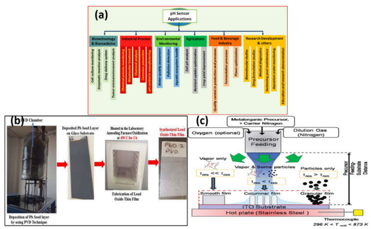
Figure 4.
a): Hydrothermal and solvothermal synthesis strategies for metal oxide nanoparticles and their subsequent incorporation into biopolymer matrices for multifunctional applications, including antifungal activity, environmental protection, and sustainable agriculture[157] (Reproduced with Permission). Fig 4b): . Schematic representation of the hydrothermal-assisted synthesis of Bi₂O₃/Bi₂WO₆ heterojunction nanocomposites, illustrating precursor impregnation, heterojunction formation, and the enhanced separation of photogenerated charge carriers responsible for improved visible-light photocatalytic performance[158] (Reproduced with Permission).Fig 4c): Reaction pathway involved in hydrothermal nanomaterial synthesis, highlighting precursor hydrolysis, intermediate oxide formation, reduction processes, and nanoparticle growth under elevated temperature and pressure conditions[159] (Reproduced with Permission).
Figure 4.
a): Hydrothermal and solvothermal synthesis strategies for metal oxide nanoparticles and their subsequent incorporation into biopolymer matrices for multifunctional applications, including antifungal activity, environmental protection, and sustainable agriculture[157] (Reproduced with Permission). Fig 4b): . Schematic representation of the hydrothermal-assisted synthesis of Bi₂O₃/Bi₂WO₆ heterojunction nanocomposites, illustrating precursor impregnation, heterojunction formation, and the enhanced separation of photogenerated charge carriers responsible for improved visible-light photocatalytic performance[158] (Reproduced with Permission).Fig 4c): Reaction pathway involved in hydrothermal nanomaterial synthesis, highlighting precursor hydrolysis, intermediate oxide formation, reduction processes, and nanoparticle growth under elevated temperature and pressure conditions[159] (Reproduced with Permission).
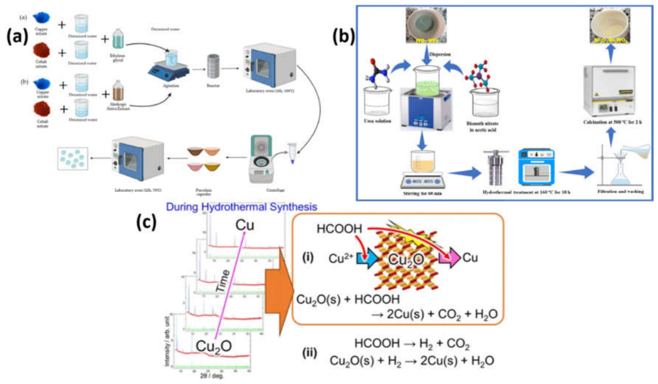
Figure 5.
a): Green synthesis mechanism of metal oxide nanoparticles using plant extracts, highlighting the role of phytochemicals as eco-friendly reducing and capping agents in the formation of stable nanoparticles with tailored physicochemical properties. Fig 5b). Schematic representation of platinum NPs synthesized using plant extracts Fig 5c): Sustainable biosynthesis of CuO nanoparticles using various biological sources and biomolecules. The green synthesis approach provides an environmentally benign route for producing CuO nanomaterials with enhanced physicochemical and biological properties for biomedical and environmental applications. Fig 5d): General mechanism of plant-mediated green synthesis of MgO nanoparticles involving precursor reduction, nucleation, growth, and stabilization by naturally occurring biomolecules[5] (Reproduced with Permission).
Figure 5.
a): Green synthesis mechanism of metal oxide nanoparticles using plant extracts, highlighting the role of phytochemicals as eco-friendly reducing and capping agents in the formation of stable nanoparticles with tailored physicochemical properties. Fig 5b). Schematic representation of platinum NPs synthesized using plant extracts Fig 5c): Sustainable biosynthesis of CuO nanoparticles using various biological sources and biomolecules. The green synthesis approach provides an environmentally benign route for producing CuO nanomaterials with enhanced physicochemical and biological properties for biomedical and environmental applications. Fig 5d): General mechanism of plant-mediated green synthesis of MgO nanoparticles involving precursor reduction, nucleation, growth, and stabilization by naturally occurring biomolecules[5] (Reproduced with Permission).
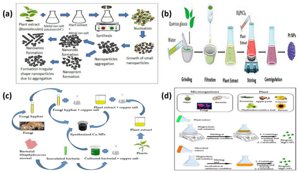
Disclaimer/Publisher’s Note: The statements, opinions and data contained in all publications are solely those of the individual author(s) and contributor(s) and not of MDPI and/or the editor(s). MDPI and/or the editor(s) disclaim responsibility for any injury to people or property resulting from any ideas, methods, instructions or products referred to in the content. |
© 2026 by the authors. Licensee MDPI, Basel, Switzerland. This article is an open access article distributed under the terms and conditions of the Creative Commons Attribution (CC BY) license.
Copyright: This open access article is published under a Creative Commons CC BY 4.0 license, which permit the free download, distribution, and reuse, provided that the author and preprint are cited in any reuse.


