Preprint
Article

This version is not peer-reviewed.

Measurement of Fastening Torque of Rod Securing Spinal Implant Screws

Submitted:

22 June 2026

Posted:

23 June 2026

You are already at the latest version

Abstract

Background/Objectives: The mechanical stability of spinal instrumentation depends on adequate tightening of rod-securing screws. Although some implant systems provide recommended torque values, in many cases tightening remains surgeon-dependent. The objective of this study was to quantify the magnitude and variability of tightening torque applied by spine surgeons and to evaluate the influence of handling technique on torque generation. Methods: A thoracolumbar spine model instrumented with pedicle screws was used in an experimental setup. Twenty-two spine surgeons from six institutions performed a total of 220 screw-tightening procedures using a standard surgical screwdriver. Tightening torque was indirectly determined from measured loosening torque based on previously established calibration ratios. Hand usage, hand position, and surgeon orientation relative to the construct were recorded. Statistical analysis was performed using two-tailed Student’s t-tests with a significance level of 0.05. Results: The mean applied tightening torque was 4.52 Nm, with considerable inter-individual variability. Two-handed tightening resulted in significantly higher torque compared to single-hand techniques (p < 0.05). Screws tightened from the contralateral side relative to the surgeon were subjected to significantly higher torque (4.79 Nm vs. 4.25 Nm, p < 0.05). No clear association was found between institutional practice and applied torque magnitude. Conclusions: Substantial variability exists in the tightening torque applied by spine surgeons, and surgical technique significantly influences torque generation. These findings highlight the importance of standardized torque application and support the use of torque-limiting instruments to improve consistency in spinal implant fixation.

Keywords: 
;  ;  ;  ;  ;  ;  

1. Introduction

Spinal instrumentation systems commonly rely on rod–screw constructs to achieve mechanical stability and maintain spinal alignment. The integrity of these constructs critically depends on the secure fixation of individual components, particularly the tightening of set screws that lock the rods within pedicle screw heads. Insufficient tightening may result in construct loosening and loss of stability, whereas excessive torque may lead to implant damage or compromised mechanical performance.
To ensure the reliability and safety of spinal implants, several standardized testing protocols have been established. Widely used standards [1,2] define methodologies for evaluating the mechanical behavior of spinal implant constructs, including static and fatigue performance as well as the properties of interconnection mechanisms. While these standards provide a robust framework for assessing structural integrity under controlled laboratory conditions, they do not specify clinically relevant tightening torque values for rod-securing screws in many implant systems. Moreover, they do not account for the variability introduced by manual torque application during surgical procedures.
In clinical practice, although some implant manufacturers provide torque-limiting screwdrivers and recommend specific torque values, in many cases the applied torque remains dependent on the surgeon’s subjective judgment. This introduces a potential source of variability that may influence construct stability and long-term outcomes. Despite extensive biomechanical investigations focusing on implant design, fixation strength, and load distribution [3,4,5,6,7,8,9,10,11,12,13,14,15], relatively little attention has been given to the actual torque applied intraoperatively and its variability among surgeons.
Furthermore, the influence of surgical technique—such as hand positioning, number of hands used, and surgeon orientation relative to the implant—on torque generation remains poorly understood. These factors may significantly affect the magnitude of applied torque and contribute to inconsistencies in implant fixation.
The aim of the present study was therefore to quantify the magnitude and variability of tightening torque applied by spine surgeons using a standardized experimental model and to evaluate the effect of handling technique on torque generation.

2. Materials and Methods

2.1. Study Design and Model

A realistic thoracolumbar plastic spine model (Biocalderoni, Budapest, Hungary) was mounted on a portable platform at a height corresponding to standard surgical positioning. Bilateral pedicle tulip-headed screws were inserted in all lumbar vertebrae. Screw heads were modified by incorporating high-tensile female screws (Figure 1).
The measurement apparatus consisted of three components:
  • Standard surgical screwdriver (geometrically identical to instruments used in operating theaters)
  • Calibrated torque-limiting screwdriver (Torqueleader Quickset, Guildford, UK) for calibration
  • Calibrated dial-type torque wrench (WERA 7112BDS, Wuppertal, Germany) for loosening torque measurement

2.2. Calibration Phase (First Measurement Series)

To establish the tightening-loosening torque ratio for each screw, we performed sequential tightening at known torque values using the calibrated torque-limiting screwdriver, followed by loosening torque measurement with the torque wrench. This process was repeated three times. The first series was considered a "break-in" phase and excluded from ratio calculations; ratios were derived from the second and third measurements.
Preliminary analysis revealed that the tightening-loosening ratio varied slightly with torque magnitude. Therefore, two distinct ratios were assigned to each screw: one for tightening torques below 3.5 Nm and another for values exceeding this threshold.

2.3. Surgeon Assessment Phase (Second Measurement Series)

Twenty-two spine surgeons from six Hungarian institutions participated voluntarily. Participants were instructed to tighten screws on the model exactly as they would during surgery. The following parameters were documented for each maneuver:
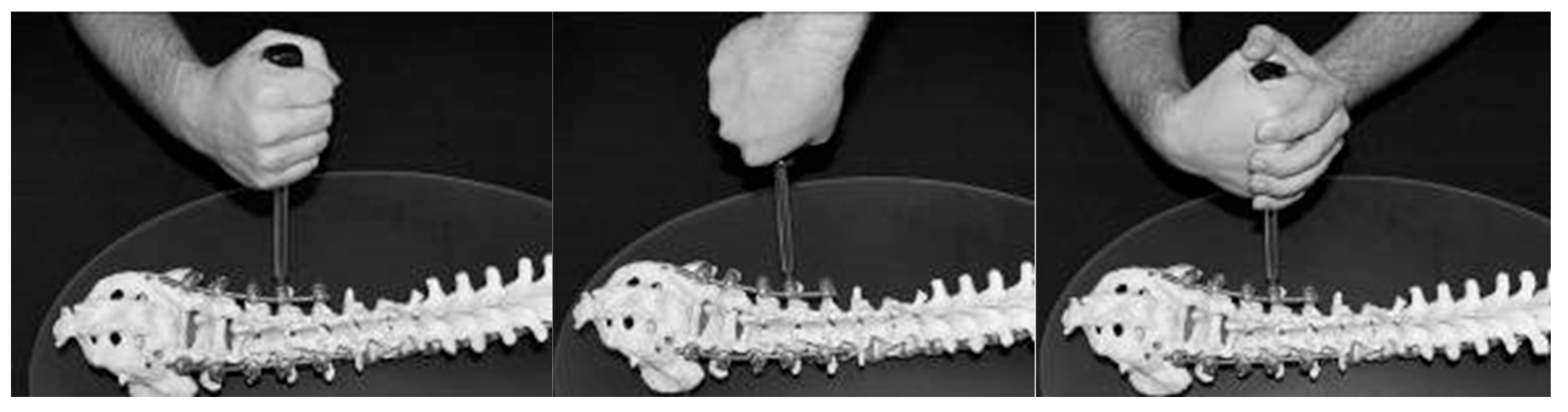
  • Grip technique (thumb-upward, thumb-downward, or two-handed)
  • Hand dominance (right or left)
  • Standing position relative to the model (right or left side)
  • Institutional routine use of torque-measuring instruments
Measurement Protocol:
  • The spine model was secured horizontally at surgical height with pre-loosened screws
  • Surgeons tightened screws in any order using the provided standard screwdriver (Figure 2)
  • Loosening torque was measured using the calibrated torque wrench, recording maximum torque
  • All data were documented in standardized measurement protocols

2.4. Control Phase (Third Measurement Series)

To assess potential screw wear from repeated use, we repeated the calibration measurements (as described in the first series) following the surgeon assessment phase. This allowed detection of any significant changes in screw-screwdriver interface characteristics.

2.5. Statistical Analysis

Comparisons between tightening-loosening ratios from the first and third measurement series, as well as torque values associated with different grip techniques, were analyzed using two-tailed Student's t-tests with a significance level of 5%. Data are presented as means with standard deviations.

3. Results

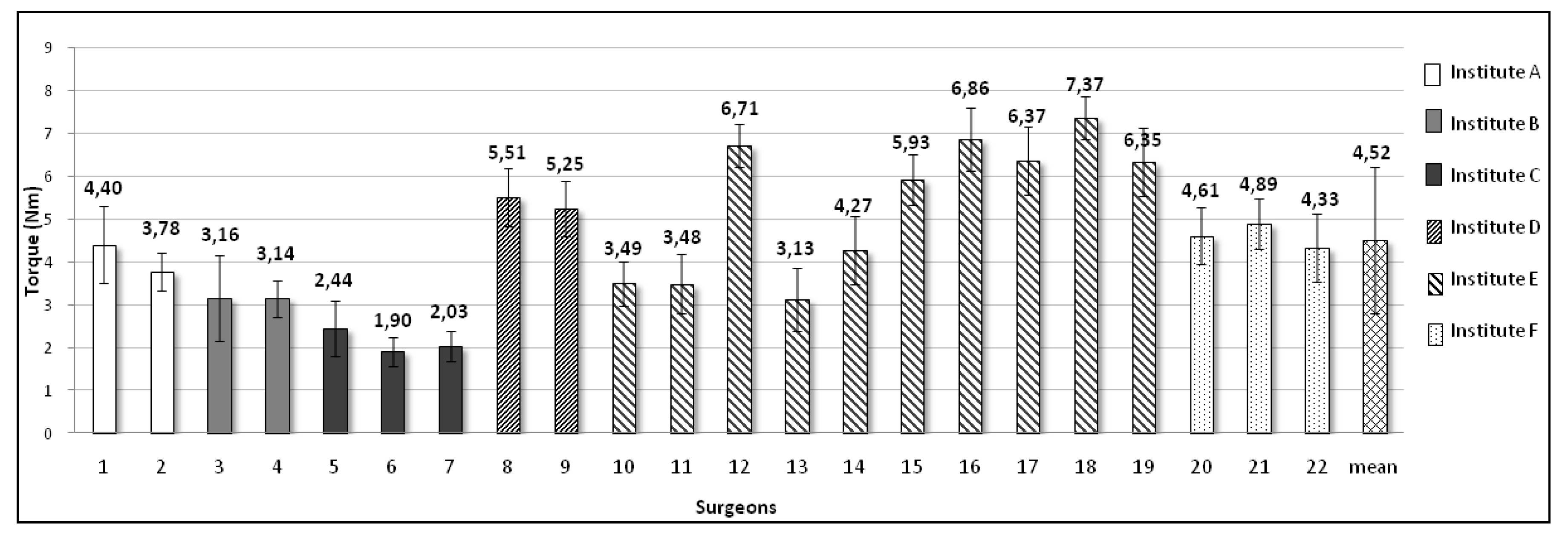
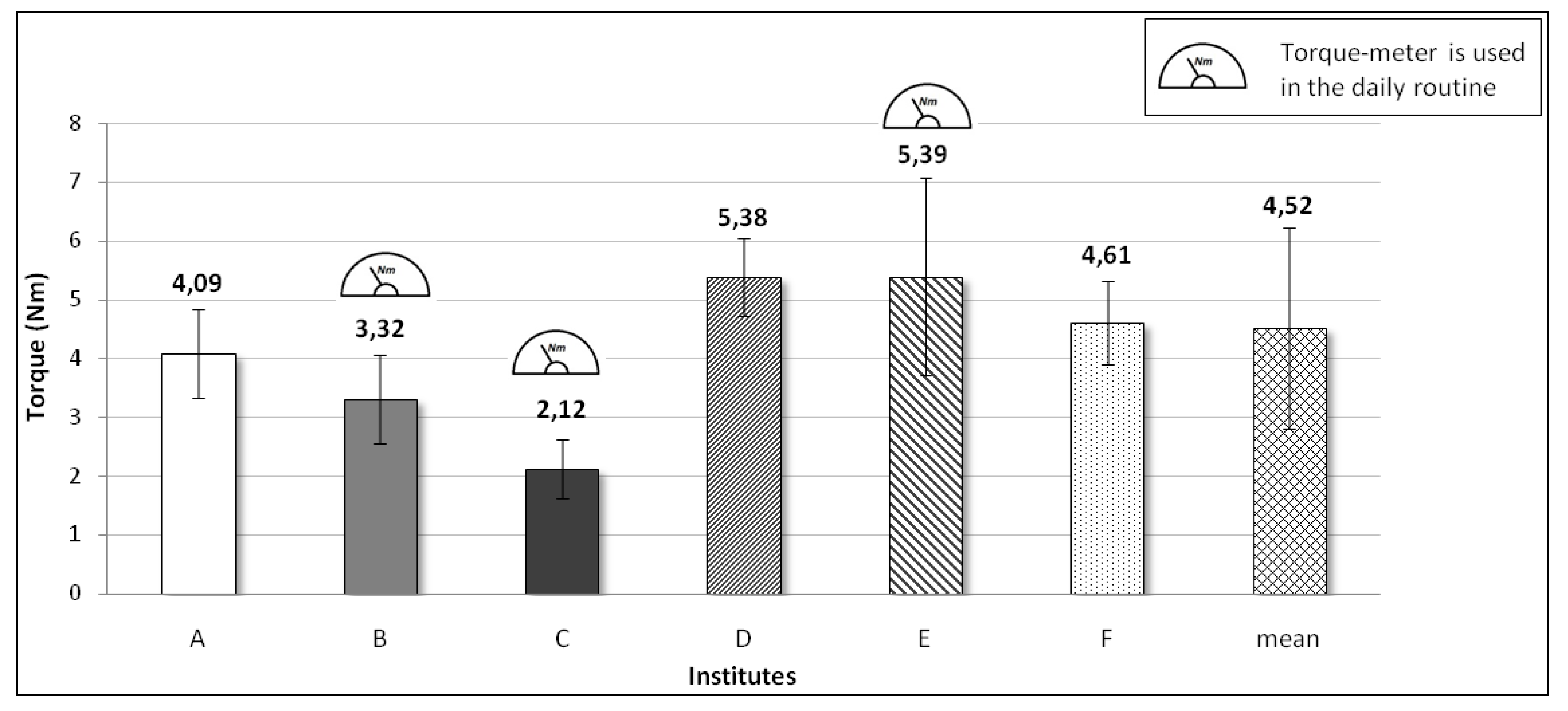
Individual surgeon results and institutional averages are presented in Figure 3 and Figure 4. Substantial inter-surgeon variability was observed, with individual mean torques ranging from 1.8 Nm to 8.9 Nm (overall mean: 4.52 Nm, SD: 1.84 Nm). Institutional averages showed somewhat less variability than individual values.
The control calibration series revealed no significant differences in tightening-loosening ratios between pre- and post-assessment measurements (p=0.412), confirming that screw wear did not significantly influence results.

3.1. Grip Technique Effects

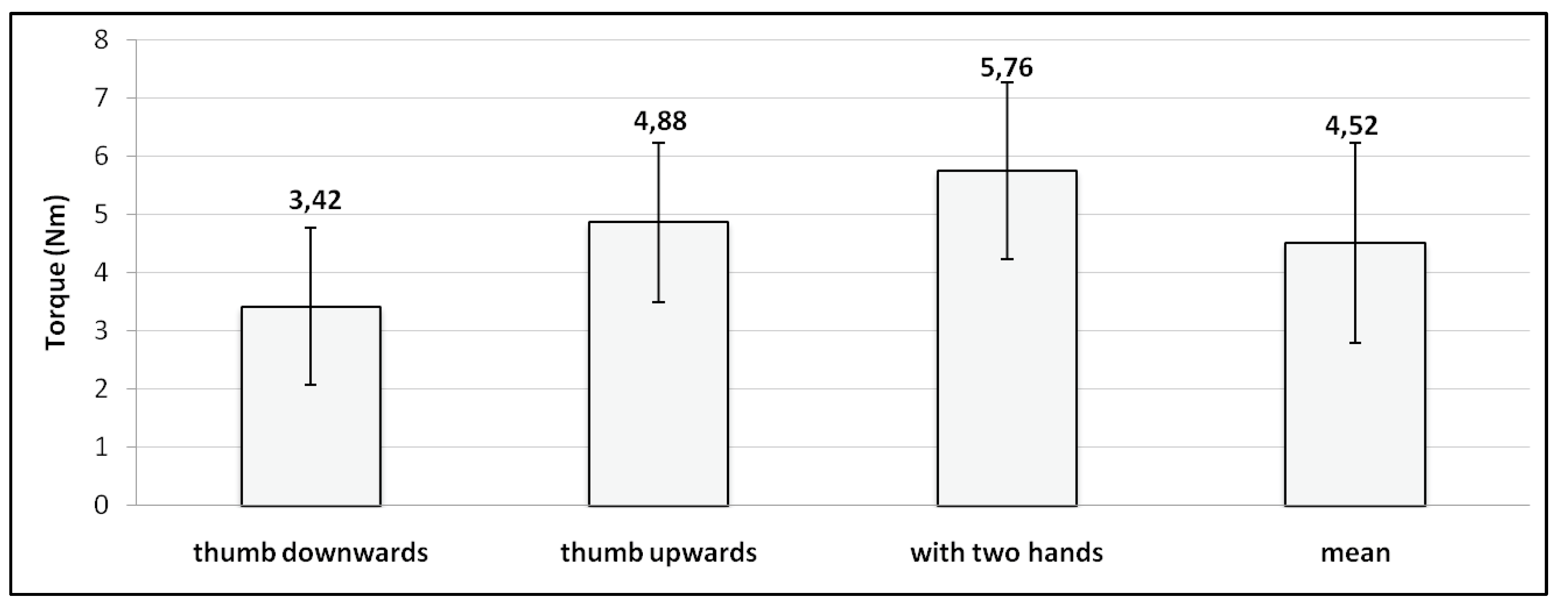
Significant differences in applied torque were observed based on grip technique (Figure 5). Two-handed grip produced significantly higher torque (mean: 6.2 Nm, SD: 1.3 Nm) compared to thumb-downward (mean: 3.9 Nm, SD: 1.1 Nm) and thumb-upward (mean: 4.1 Nm, SD: 1.2 Nm) techniques (p=0.003 for both comparisons). No significant difference was detected between the two one-handed techniques (p=0.382).

3.2. Positioning Effects

Surgeons consistently stood perpendicular to the model on either the right or left side. Analysis of screw position relative to surgeon standing side revealed that screws on the contralateral side were tightened significantly more (4.79 Nm, SD: 1.6 Nm) compared to ipsilateral screws (4.25 Nm, SD: 1.5 Nm, p=0.011).

3.3. Hand Dominance

Only one left-handed surgeon participated, precluding meaningful statistical analysis of handedness effects.

3.4. Torque-Measuring Instrument Use

No significant correlation was found between institutional routine use of torque-measuring instruments and mean applied torque (p=0.287).

4. Discussion

This study demonstrates substantial variability in screw fastening torque among spine surgeons, with individual mean values ranging nearly fivefold (1.8-8.9 Nm). This finding is particularly concerning given that many spinal implant systems lack specific torque guidelines [5]. Such variability could contribute to inconsistent construct stability and potentially compromise clinical outcomes [16,17].
The significant differences associated with grip technique and surgeon positioning highlight the influence of ergonomic factors on applied torque. Two-handed tightening, which is not standard surgical practice, produced significantly higher torque and should be discouraged to prevent excessive loading. The observation that contralateral screws are tightened more forcefully suggests that awkward positioning may compromise tactile feedback, prompting surgeons to apply additional force.
Interestingly, institutional use of torque-measuring instruments did not correlate with torque magnitude. This may reflect inconsistent instrument use, inadequate calibration, or surgeons overriding torque limits based on tactile sensation. It is also possible that surgeons familiar with torque instruments develop better awareness of force application, paradoxically leading to more variable torque values when these instruments are unavailable.
The clinical implications of our findings are significant. Optimal screw tightening torque is critical for construct stability, as inadequate tightening may lead to implant loosening and failure, while excessive torque may risk screw stripping or bone damage [18,19]. The wide variability observed in this study suggests that some patients may receive suboptimal constructs due to inconsistent surgical technique.

4.1. Limitations

Several limitations should be acknowledged. First, the plastic spine model may not fully replicate the tactile feedback of human bone, potentially influencing surgeon behavior. Second, the study was limited to a single screwdriver geometry; results might differ with other instrument designs. Third, the relatively small sample size and single-country setting may limit generalizability. Finally, we did not assess the impact of surgeon experience or fatigue on torque application.

4.2. Recommendations

Based on our findings, we recommend that implant manufacturers provide:
  • Clear specifications for optimal tightening torque for their systems
  • Calibrated torque-limiting screwdrivers with each implant set
  • Surgeon education on the importance of standardized torque application
Hospitals and surgical departments should consider implementing routine torque measurement and documentation as part of quality assurance in spinal surgery.

5. Conclusions

Significant variability exists in the torque applied by spine surgeons during screw fastening, influenced by grip technique, surgeon positioning, and individual practice patterns. These findings support the routine use of torque-limiting instruments in spinal instrumentation to ensure consistent, optimal construct stability and potentially improve clinical outcomes. Future research should investigate the relationship between applied torque and long-term clinical results, as well as develop simplified torque-limiting devices suitable for routine surgical use.

Author Contributions

Conceptualization, methodology, writing—original draft preparation, S.M.; validation, writing—review and editing, T.M. All authors have read and agreed to the published version of the manuscript.

Funding

This research received no external funding.

Data Availability Statement

The original contributions presented in this study are included in the article. Further inquiries can be directed to the corresponding author.

Acknowledgments

The authors thank Eszter Janka for providing professional services.

Conflicts of Interest

The authors declare no conflicts of interest.

References

  1. Serhan, H.; Hammerberg, K.; O'Neil, M.; et al. Intraoperative techniques to reduce the potential of set-screw loosening in long spinal constructs: A static and fatigue biomechanical investigation. J. Spinal Disord. Tech. 2010, 23(6), e31–e36. [Google Scholar] [PubMed]
  2. Cho, W.; Cho, S.K.; Wu, C. The biomechanics of pedicle screw-based instrumentation. J. Bone Jt. Surg. Br. 2010, 92(8), 1061–1065. [Google Scholar] [CrossRef]
  3. Rohlmann, A.; Calisse, J.; Bergmann, G.; et al. Clamping stiffness and its influence on load distribution between paired internal spinal fixation devices. J. Spinal Disord. 1996, 9(3), 234–240. [Google Scholar] [CrossRef] [PubMed]
  4. ASTM F1798-97; Standard Guide for Evaluating the Static and Fatigue Properties of Interconnection Mechanisms and Subassemblies Used in Spinal Arthrodesis Implants. ASTM International, 2008.
  5. ASTM F1717-12; Standard Test Methods for Spinal Implant Constructs in a Vertebrectomy Model. ASTM International, 2012.
  6. Alkaly, R.N. The effects of design and configuration on the biomechanical response of an internal spinal fixator. Proc. Inst. Mech. Eng. H. 1999, 213(2), 137–146. [Google Scholar] [CrossRef]
  7. Alkaly, R.N.; Sharpe, D.; Bader, D.L. A biomechanical analysis of an instrumented spinal fixator under torsional loads. J. Biomech. 2005, 38(4), 865–876. [Google Scholar] [CrossRef]
  8. Sairyo, K.; Scifert, J.; Goel, V.K.; et al. Neurocentral synchondrosis fracture in immature spines associated with pedicle screw type fixation devices. J. Spinal Disord. 1998, 11(2), 142–145. [Google Scholar] [CrossRef] [PubMed]
  9. Defino, H.L.; Rosa, R.C.; Silva, P.; et al. Mechanical performance of cylindrical and dual-core pedicle screws after repeated insertion. Spine 2012, 37(14), 1187–1191. [Google Scholar] [CrossRef] [PubMed]
  10. Defino, H.L.A.; Rosa, R.C.; Silva, P.; et al. The effect of repetitive pilot-hole use on the insertion torque and pullout strength of vertebral system screws. Spine 2009, 34(8), 871–876. [Google Scholar] [CrossRef] [PubMed]
  11. Hee, H.T.; Khan, M.S.; Goh, J.C.; et al. Insertion torque profile during pedicle screw insertion of the thoracic spine with and without violation of the pedicle wall: Comparison between cylindrical and conical designs. Spine 2006, 31(20), E840–E846. [Google Scholar] [PubMed]
  12. Inceoglu, S.; Ferrara, L.; McLain, R.F. Pedicle screw fixation strength: Pullout versus insertional torque. Spine J. 2004, 4(5), 513–518. [Google Scholar] [CrossRef] [PubMed]
  13. Pfeiffer, M.; Hoffmann, S.; Griss, P. Clinical and biomechanical aspects of pedicle screw fixation in the lumbar spine. Spine 1997, 22(1), 100–106. [Google Scholar]
  14. McKinley, T.O.; McLain, R.F.; Yerby, S.A.; et al. The effect of pedicle screw design on screw insertion torque and pullout strength. Spine 1998, 23(8), 901–906. [Google Scholar]
  15. Zindrick, M.R.; Wiltse, L.L.; Widell, E.H.; et al. A biomechanical study of intrapeduncular screw fixation in the lumbosacral spine. Clin. Orthop. Relat. Res. 1986, 203, 99–112. [Google Scholar] [CrossRef]
  16. Sandén, B.; Olerud, C.; Larsson, S. Immediate postoperative stability of a new implant system for anterior cervical fusion. Acta Orthop. Scand. 2000, 71(5), 490–495. [Google Scholar]
  17. Spivak, J.M.; Chen, D.; Kummer, F.J.; et al. The effect of locking fixation screws on the stability of anterior cervical plating. Spine 1999, 24(4), 334–338. [Google Scholar] [CrossRef] [PubMed]
  18. Myers, B.S.; Belmont, P.J., Jr.; Richardson, W.J.; et al. The role of imaging and invasive diagnostic techniques in the evaluation of the spine. Spine 1994, 19((20) Suppl, 2367S–2375S. [Google Scholar]
  19. Ryken, T.C.; Clausen, J.D.; Traynelis, V.C.; et al. Biomechanical analysis of bone mineral density, insertion technique, screw torque, and holding strength of transpedicular screw fixation. J. Neurosurg. 1994, 80(3), 518–525. [Google Scholar]
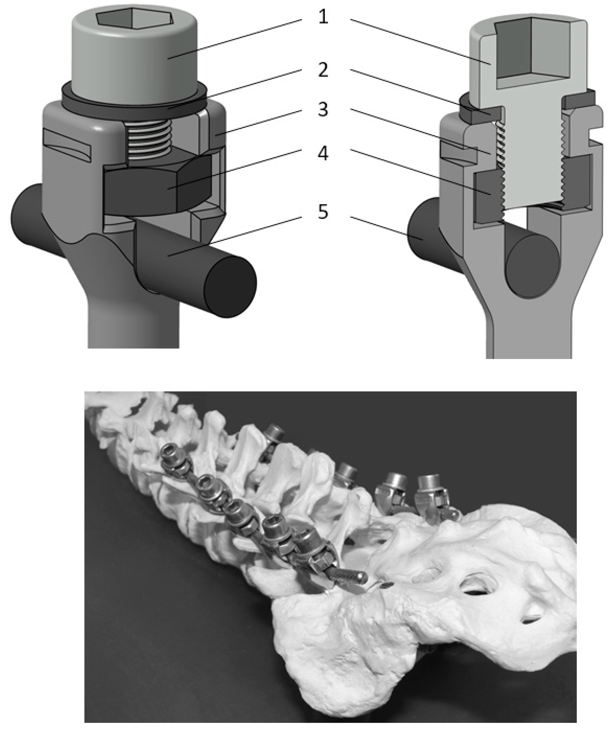
Figure 1. Schematic representation of the modified screw and measurement apparatus. (1) High-tensile screw, (2) washer, (3) modified pedicle screw, (4) high-tensile nut, (5) rod.
Preprints 219747 g001
Figure 2. The three different grip techniques (thumb-upward, thumb-downward, two-handed).
Preprints 219747 g002
Figure 3. Mean fastening torque values by individual surgeon (n=22). Error bars represent standard deviation.
Preprints 219747 g003
Figure 4. Mean fastening torque values by institution (n=6). Error bars represent standard deviation.
Preprints 219747 g004
Figure 5. Mean fastening torque values by grip technique (n=22). Error bars represent standard deviation.
Preprints 219747 g005
Disclaimer/Publisher’s Note: The statements, opinions and data contained in all publications are solely those of the individual author(s) and contributor(s) and not of MDPI and/or the editor(s). MDPI and/or the editor(s) disclaim responsibility for any injury to people or property resulting from any ideas, methods, instructions or products referred to in the content.
Copyright: This open access article is published under a Creative Commons CC BY 4.0 license, which permit the free download, distribution, and reuse, provided that the author and preprint are cited in any reuse.
Prerpints.org logo

Preprints.org is a free preprint server supported by MDPI in Basel, Switzerland.

Subscribe

© 2026 MDPI (Basel, Switzerland) unless otherwise stated

Accessibility

Disclaimer

Terms of Use

Privacy Policy

Privacy Settings