

Submitted:
19 June 2026
Posted:
22 June 2026
You are already at the latest version
Abstract
Stray cats may act as reservoirs of antimicrobial-resistant and potentially pathogenic bacteria, representing a potential public health concern within a One Health framework. This study investigated the occurrence of antimicrobial resistance (AMR), multidrug resistance (MDR), and virulence-associated traits in commensal Escherichia coli isolated from free-living cat colonies in southern Spain. A total of 169 rectal swabs were collected from cats belonging to feline colonies and shelters in Benalmádena (Málaga, Spain). Bacterial isolation and identification were performed using selective culture media, conventional biochemical tests, and MALDI-TOF mass spectrometry. Antimicrobial susceptibility was determined by minimum inhibitory concentration (MIC) testing against 15 antimicrobial agents, and isolates were screened for selected virulence-associated genes associated with major diarrheagenic E. coli pathotypes. A total of 68 E. coli isolates (40.2%) were recovered. The highest resistance frequencies were observed for sulfamethoxazole (25.0%) and ampicillin (20.6%), whereas all isolates showed high susceptibility rates to most of the antimicrobials tested, including azithromycin (100%), as well as gentamicin, amikacin, cefotaxime, ceftazidime, meropenem, colistin, chloramphenicol and tigecycline (98.5%). Six isolates (8.8%) were classified as multidrug-resistant. In addition, six isolates (8.8%) were identified as atypical enteropathogenic E. coli (aEPEC), although none exhibited a multidrug resistance phenotype. These findings demonstrate the presence of both antimicrobial resistance and virulence-associated traits among commensal E. coli circulating in free-living cats. The inclusion of feline colonies or shelters in antimicrobial resistance surveillance programmes may contribute valuable information for integrated One Health monitoring strategies.
Keywords:
stray cats
; Escherichia coli
; antimicrobial resistance
; multidrug resistance
; atypical enteropathogenic E. coli (aEPEC)
; One Health
1. Introduction
The emergence and spread of antimicrobial resistance (AMR) represent a global health problem that requires a One Health approach [1]. In Europe, veterinary antimicrobial consumption was monitored by the ‘European Surveillance of Veterinary Antimicrobial Consumption (ESVAC)’ project from 2009 to 2023 [2]. Currently, in response to European regulations, a modified version of this program has been implemented: the ‘European Sales and Use of Antimicrobials for Veterinary Medicine (ESUAvet)’, as published in European Medicines Agency webpage. This program developed a harmonized approach for collecting and reporting data on the sale and use of antibiotics in animals. However, the monitoring of AMR among animals is not systematic across European countries, as it focuses mainly on food-producing animals, and not on companion animals such as dogs and cats [3,4]. In Spain, the ‘Antimicrobial Resistance National Program’ (PRAN) has been in force since 2014, and for the period 2025–2027, the PRAN intends to include A MR in companion animals within the ‘reduce antibiotics program’ [5].
Pet animals, particularly cats and dogs, are increasingly recognized as potential reservoirs for the transmission of AMR/MDR to humans. This concern arises from two key factors: the frequent and sometimes inappropriate use of broad-spectrum antimicrobial agents in veterinary medicine, and the close, often daily, physical contact between companion animals and their human owners. Such proximity facilitates not only microbial exchange but also the potential for resistant strains to spread across species [5]. Several studies have demonstrated the presence of multidrug-resistant bacteria in clinically healthy pets, indicating that even asymptomatic animals can harbour and disseminate resistant microorganisms. As highlighted by Li et al. [6], antimicrobial resistance has been detected in bacterial isolates obtained from both cats and dogs across the Iberian Peninsula, raising significant public health concerns.
Transmission of AMR bacteria between companion animals and humans, particularly cats, may occur through direct or indirect contact and represents a potential public health concern [7]. Despite all this, it is important to note that the risk of contracting an infection from a veterinarian-supervised domesticated cat is low; however, this risk increases substantially when in contact with stray cats [7]. However, stray cats that are in contact with humans and share the urban environment with them may act as reservoirs and disseminators of antimicrobial-resistant bacteria affecting humans and companion animals.
Free-living cats that live outdoors in public or private urban areas, usually live in colonies, using human activity resources to feed or prey on a broad biodiversity of animals such as birds, rodents, and reptiles [8,9]. Populations of free-living cats could represent an important threat to public health, being a significant factor in the transmission of zoonotic diseases due to their close contact with humans and other pets [10,11]. In fact, previous studies, carried out in different countries, have reported cats as a source of zoonotic infectious and parasitic diseases [8,12,13,14].
Besides antimicrobial resistance, Escherichia coli isolated from companion animals may harbour virulence factors associated with different diarrheagenic pathotypes, including enteropathogenic (EPEC), Shiga toxin-producing (STEC), enterotoxigenic (ETEC), enteroaggregative (EAEC), and enteroinvasive (EIEC) E. coli [15]. Among these, EPEC is responsible for millions of diarrhoeal cases worldwide and represents an important public health concern [16]. EPEC strains are classified as typical (tEPEC) or atypical (aEPEC) according to the presence or absence of the EAF plasmid carrying the bundle-forming pilus gene (bfpA), while both groups possess the eae gene encoding intimin [16,17,18]. Cats have been described as asymptomatic carriers of diarrheagenic E. coli pathotypes, including aEPEC, highlighting their potential role in the maintenance and dissemination of virulence-associated genes.
In European countries, the number of studies focusing on cats is still very limited. This may be due to a lower interest in this species compared to dogs, but also to logistical challenges such as difficulty in handling and sampling these animals or obtaining faecal samples [19,20]. Nonetheless, different publications have reviewed the presence of zoonotic pathogens in feline colonies in Europe. For example, Rosario et al. [21] described for the first time the presence of Salmonella enterica subsp. enterica in feline colonies in Las Palmas de Gran Canaria (Canary Island, Spain), assessing its impact on public health. Additionally, several studies in Italy, using a strong One Health approach, have specifically investigated the presence of Escherichia coli in faecal samples from feline colonies, as well as its AMR phenotype. Thus, Ratti et al. [22] and Gargano et al. [23] confirmed, respectively, the circulation of multidrug-resistant E. coli strains in feral cats, including strains producing ESBL (Extended Spectrum β-Lactamases).
Although several studies in Spain have reported the role of companion animals as reservoirs of AMR [6,24,25], limited information is available regarding the role and circulation of E. coli and AMR-linked strains in free-living cats in our country. In this sense, few studies have focused on the role of stray cats as disseminators of resistance to broad-spectrum antibiotics used in veterinary and human medicine [26].
Considering this, the present study aimed to investigate antimicrobial resistance, multidrug resistance, and virulence-associated traits in commensal E. coli isolated from free-living cats. To achieve this, antimicrobial susceptibility was assessed phenotypically, and the isolates were screened for selected virulence-associated genes related to major diarrheagenic E. coli pathotypes.
2. Material and Methods
2.1. Sample Collection
This collaborative research work was carried out between the Epidemiology, Preventive Medicine, and Health Policy Unit of the Animal Health Department (Veterinary Faculty, University of Córdoba, Spain), the Health and Environment area of the Benalmadena City Council (Málaga province), and the Fenix veterinary clinic located in Benalmadena (Málaga province), a ‘Cat Friendly Silver Level Centre’ Categorized by International Society of Feline Medicine (ISFM).
A total of 169 rectal swabs were collected from cats belonging to free-living colonies or shelters located in Benalmádena (Málaga, Spain) between October 2022 and March 2023. Feline colonies are large groups of free-living cats, mainly in urban areas, originating from abandoned or stray animals and their offspring. In contrast, cat shelters house animals in a semi-domestic environment with appropriate sanitary measures (such as sterilization and deworming), with adoption as their primary goal. The samples were collected at the Fenix clinic at the time of the castration of the animals and from the campaigns carried out on the Malaga province by official institutions for control purposes (Trap-Neuter-Return campaigns) (Figure 1 and Figure 2).
All samples preserved in AMIES transport medium were refrigerated at ≤ 4 ºC and transported to the laboratory of Animal Health Department (University of Cordoba) for microbiological analyses within 24 h of collection.
2.2. Escherichia Coli Isolates Identification
Rectal swabs were pre-enriched in buffered peptone water (BPW; Scharlau, Barcelona, Spain), in 1:10 vol/vol proportion, and incubated at 37 ± 1 ºC for 24 ± 2 h. All the pre-enriched samples were inoculated onto XLD (Xylose Lysine Deoxycholate) agar medium and incubated for 24 hours at 37ºC. Colonies showing morphological characteristics compatible with Escherichia coli were selected and subcultured as necessary to obtain pure cultures.
Isolates were subcultured on brain-heart infusion agar incubated at 37 ºC for 24 h and confirmed as E. coli by biochemical tests including: Gram stain, catalase test, oxidase test and indole production test, before species confirmation by Matrix-Assisted Laser Desorption–Ionization Time-of-Flight Mass Spectrometry (MALDI-TOF) (Bruker MALDI Biotyper).
2.3. Antimicrobial Susceptibility Testing
Antimicrobial susceptibility testing was performed using the Minimum Inhibitory Concentration (MIC) assay with the Salmonella/E. coli EUVSEC Plate (Thermo Scientific, Sensititre Plate Guide, Madrid, Spain), which includes the antimicrobial agents required by Decision (EU) 2020/1729 for monitoring antimicrobial resistance in zoonotic and commensal bacteria. The following antimicrobial agents were tested according to their respective classes and concentration ranges (µg/mL): aminopenicillins (ampicillin, AMP, 1–32), third-generation cephalosporins (cefotaxime, CTX, 0.25–8; ceftazidime, CAZ, 0.5–16), carbapenems (meropenem, MEM, 0.03–4), quinolones (nalidixic acid, NA, 4–64; ciprofloxacin, CIP, 0.015–4), macrolides (azithromycin, AZM, 2–64), aminoglycosides (gentamicin, GEN, 0.25–16; amikacin, AMK, 2–64), polymyxins (colistin, CL, 0.25–4), sulphonamides (sulfamethoxazole, SF, 16–512), diaminopyrimidines (trimethoprim, TMP, 0.25–8), amphenicols (chloramphenicol, CHL, 2–32), tetracyclines (tetracycline, TE, 1–16), and glycylcyclines (tigecycline, TGC, 0.015–2). The results were interpreted using the Sensititre™ SWIN™ Software System based on clinical breakpoints from CLSI M100 and EUCAST [27]. Antimicrobial susceptibility results were interpreted according to EUCAST clinical breakpoints for Enterobacterales whenever available. For nalidixic acid and tetracycline, CLSI breakpoints were applied due to the absence of corresponding EUCAST clinical breakpoints. For azithromycin and chloramphenicol, clinical breakpoints were not available for E. coli in either EUCAST or CLSI; therefore, epidemiological cut-off values (ECOFFs) were used to distinguish wild-type (WT) isolates from isolates with acquired resistance mechanisms (non-WT). For statistical analyses, resistant and non-wild-type (non-WT) isolates were grouped into a single category and compared with susceptible and wild-type (WT) isolates.
Strains showing acquired resistance to at least one antimicrobial agent in three or more antimicrobial classes were classified as multidrug-resistant (MDR) [28].
According to the European Medicines Agency (EMA) classification for the prudent and responsible use of antibiotics in animals [29], the antimicrobials included in this study were categorized into four groups (Table 2): Category D (“Prudence”), considered first-line therapeutic options; Category C (“Caution”), recommended when Category D antimicrobials are not effective; Category B (“Restrict”), corresponding to the Highest Priority Critically Important Antimicrobials (HPCIAs) included in the World Health Organization (WHO) list of Medically Important Antimicrobials (MIAs), including third- and fourth-generation cephalosporins, quinolones, polymyxins, and phosphonic acid derivatives; and Category A (“Avoid”), comprising antimicrobials not authorized for veterinary use and considered last-resort drugs in human medicine [30]. Within the European Union, these antimicrobials are reserved for human use and are not authorized for food-producing animals, although they may be prescribed under exceptional circumstances in companion animals in accordance with current regulations [29].
2.4. Genotypic Characterization
Crude DNA extracts from bacterial isolates were used as DNA templates for PCR assays. Briefly, colonies on agar were picked and suspended in 300 μL of ultrapure water, boiled for 10 min, centrifuged at 13,000 rpm for 30 s and adjusted to a DNA concentration of 10-20 ng/µL using a NanoDrop 2000/2000C spectrophotometer (Thermo Fisher ScientificTM, USA).
Because azithromycin susceptibility is not routinely included in most antimicrobial resistance surveillance studies of commensal E. coli and its interpretation relies on epidemiological cut-off values (ECOFFs), a complementary PCR screening targeting selected macrolide resistance genes was performed to support the phenotypic findings. No additional resistance determinants were investigated, as the primary objective of the study was the phenotypic characterization of antimicrobial susceptibility.
Amplification was carried out using previously described primers targeting a panel of 10 acquired macrolide resistance genes and three chromosomal loci associated with macrolide resistance [31]. Primer sequences, target genes, expected amplicon sizes, and PCR conditions are provided in Table S1.
To determine the presence of diarrheagenic Escherichia coli pathotypes, conventional PCR was performed using six pairs of specific primers (Supplementary Table 2) targeting different virulence-associated genes as previously described [15]. Each PCR assay contained a final reaction volume of 24 µL, consisting of 12 µL of Qiagen PCR Master Mix (Qiagen, Hilden, Germany), 0.5 µL of each primer (forward and reverse), 9 µL of nuclease-free water, and 2 µL of DNA template. Amplification was performed using a cycling protocol with different annealing temperatures depending on each pair of primers: an initial denaturation at 95 °C for 3 min; followed by 35 cycles at 95 °C for 30 s, primer-specific annealing temperature (Supplementary Table S2), and 72 °C for 40 s. A final extension step was performed at 72 °C for 10 min, followed by maintenance at 12 °C.
Positive controls strains carrying the corresponding target genes and a negative control were included in each PCR run.
Amplified products were separated by electrophoresis on 2% (w/v) agarose gels. Samples were mixed with loading buffer containing bromophenol blue prior to loading. DNA bands were visualized under UV illumination following SYBR Safe staining.
Isolates were classified into the corresponding pathotypes according to the virulence gene profiles detected by PCR, following previously established molecular criteria for pathogenic Escherichia coli characterization [15].
2.5. Statistical Analysis
The Multiple Antimicrobial Resistance index (MAR index) value [32] was calculated for each isolate according to Matos et al. [33] as the ratio between the number of antimicrobials to which the isolate was resistant and the total number of antimicrobials tested. The mean MAR index was subsequently calculated for the study population. MAR values <0.2 were considered indicative of isolates originating from environments with low antimicrobial exposure, whereas MAR values ≥0.2 suggested exposure to environments with frequent antimicrobial use [34].
On the other hand, associations between resistance phenotypes were evaluated using Spearman’s rank correlation coefficient (PROC CORR). For this analysis, antimicrobial susceptibility results were categorized as susceptible or resistant, with intermediate isolates considered resistant [35].
Differences were considered statistically significant when p < 0.05.
3. Results
3.1. Escherichia Coli Isolates Identification
From 169 rectal swabs collected from cats housed in free-living colonies and shelters, a total of 68 commensal E. coli strains (40.23%) were identified using conventional biochemical tests and subsequently confirmed by MALDI-TOF mass spectrometry. Only one presumptive E. coli colony was selected per sample, ensuring that each isolate corresponded to a different animal. These strains were stored at −20 °C in brain-heart infusion broth with 15% glycerol until MIC analysis.
3.2. Genotypic Characterization
Among the 68 commensal E. coli strains analysed, 6 (8.8%) carried the eae (intimin) virulence gene. According to their virulence gene profile, these isolates were identified as atypical enteropathogenic E. coli (aEPEC), since they were positive for eae but negative for bfpA. No additional virulence genes included in the study were detected among the analysed strains (Table 1). In addition, none of the isolates carried the macrolide resistance genes investigated.
3.3. Antimicrobial Susceptibility Testing
E. coli isolates showed high susceptibility rates to most of the antimicrobials tested, including azithromycin (100%), as well as gentamicin, amikacin, cefotaxime, ceftazidime, meropenem, colistin, chloramphenicol and tigecycline (98.5%) (Table 2).
The highest frequencies of resistance were observed for sulphonamides (25%) and ampicillin (20.6%), both included in EMA Category D (“Prudence”). Lower resistance rates were detected for nalidixic acid, tetracycline and trimethoprim (7.4% each), and ciprofloxacin (5.9%) (Table 2). Meropenem and tigecycline belong to Category A (“Avoid”) according to the EMA categorization and are considered critically important antimicrobials reserved for human medicine. Resistance to these agents was absent or very low, with resistance rates of 1.5% for tigecycline and meropenem (Table 2).

Table 2.
Antimicrobial resistance profiles of the 68 Escherichia coli isolates.
| Antimicrobial class | Antimicrobial | EMA* | Nº Strains and percentage (%) | |||
| S | % | R | % | |||
| Aminoglycosides | GEN | C | 67 | 98.5 | 1 | 1.5 |
| AMK | C | 67 | 98.5 | 1 | 1.5 | |
| Cephalosporins | CTX | B | 67 | 98.5 | 1 | 1.5 |
| CAZ | B | 67 | 98.5 | 1 | 1.5 | |
| Quinolones | CIP | B | 64 | 94.1 | 4 | 5.9 |
| NA | B | 63 | 92.6 | 5 | 7.4 | |
| Tetracyclines | TE | D | 63 | 92.6 | 5 | 7.4 |
| Sulfonamides | SF | D | 51 | 75.0 | 17 | 25.0 |
| Aminopenicillins | AMP | D | 54 | 79.4 | 14 | 20.6 |
| Carbapenems | MEM | A | 67 | 98.5 | 1 | 1.5 |
| Macrolides | AZM | C | 68 | 100.0 | 0 | 0.0 |
| Polymyxins | CL | B | 67 | 98.5 | 1 | 1.5 |
| Amphenicols | CHL | C | 67 | 98.5 | 1 | 1.5 |
| Glycylcyclines | TGC | A | 67 | 98.5 | 1 | 1.5 |
| Diaminopyrimidines | TMP | D | 63 | 92.6 | 5 | 7.4 |
Abbreviations: GEN, gentamicin; AMK, amikacin; CTX, cefotaxime; CAZ, ceftazidime; CIP, ciprofloxacin; NA, nalidixic acid; TE, tetracycline; SF, sulfamethoxazole; AMP, ampicillin; MEM, meropenem; AZM, azithromycin; CL, colistin; CHL, chloramphenicol; TGC, tigecycline; TMP, trimethoprim. Antimicrobial susceptibility was interpreted according to EUCAST or CLSI clinical breakpoints when available. For antimicrobials lacking clinical breakpoints, epidemiological cut-off values (ECOFFs) were applied. Isolates classified as resistant and non-WT isolates were grouped into a single category.
Overall, nine different resistance phenotypes were identified among the E. coli isolates (Figure 3). The most prevalent phenotypes were resistance to ampicillin alone (11.8%) and sulfamethoxazole alone (10.3%) (Figure 3). Six isolates (8.8%) exhibited resistance to at least one antimicrobial agent in three or more antimicrobial classes and were therefore classified as multidrug-resistant (MDR). Among these, one isolate exhibited resistance to three antimicrobial classes, three isolates showed resistance to five antimicrobial classes and two isolates displayed the most complex resistance profile, involving six antimicrobial classes (Figure 3).
MAR index values ranged from 0.00 to 0.47, with a mean value of 0.046. Ten isolates (14.7%) exhibited MAR values ≥0.2, indicating exposure to environments with frequent antimicrobial use. The highest MAR index (0.47) was observed in two isolates displaying resistance to seven antimicrobials belonging to six antimicrobial classes.
Finally, correlations between antimicrobial resistance phenotypes are shown in Figure 4. Notably, very strong positive correlations were observed between resistance to tetracycline and trimethoprim (Spearman’s ρ = 1.00), ciprofloxacin and trimethoprim (ρ = 0.89), and ciprofloxacin and tetracycline (ρ = 0.89) (Figure 4).
None of the six aEPEC isolates exhibited multidrug resistance. Three isolates (50%) showed resistance to a single antimicrobial agent (two to ampicillin and one to sulfamethoxazole), whereas the remaining three isolates were susceptible to all antimicrobials tested (Table 2). Overall, no apparent association was observed between the presence of virulence genes and multidrug-resistant phenotypes.
4. Discussion
The presence of free-living cats in urban environments has increased considerably in recent decades, leading to the establishment of managed feline colonies in many European countries, including Spain (Boletín Oficial del Estado, España, 2023). These colonies are commonly maintained through Trap–Neuter–Return (TNR) programmes aimed at controlling population growth while improving animal welfare. However, feline colonies also pose several challenges, including potential risks to public health, social conflicts, traffic accidents, animal welfare concerns, and negative impacts on local biodiversity [7]. Consequently, appropriate sanitary and veterinary management of these colonies is essential to safeguard both animal and public health. Measures such as suitable colony location, adequate hygiene conditions, population control and regular health monitoring are fundamental components of responsible colony management.
The present study highlights the importance of monitoring antimicrobial resistance (AMR), multidrug resistance (MDR), and virulence-associated traits in stray cats, as these animals may contribute to the environmental dissemination of resistant and potentially pathogenic bacteria, facilitating their transmission to humans and other animals. Escherichia coli is widely recognized as a sentinel bacterium for antimicrobial resistance surveillance and is included in several official monitoring programmes involving food-producing animals [37]. Furthermore, certain E. coli pathotypes are associated with enteric disease and possess zoonotic potential. As a member of the normal intestinal microbiota of humans and a wide range of animal species, including mammals, birds, and reptiles [38], E. coli represents a valuable indicator for investigating both antimicrobial resistance and the circulation of virulence-associated genes within a One Health framework.
In this study, six isolates (8.8%) were classified as atypical enteropathogenic Escherichia coli (aEPEC). To the best of our knowledge, studies investigating diarrheagenic E. coli pathotypes in feral cats remain scarce, with previous research in these populations focusing primarily on antimicrobial resistance rather than virulence characterization [39]. Although the prevalence detected was relatively low, this finding is epidemiologically relevant because cats have previously been described as asymptomatic carriers and potential reservoirs of diarrheagenic E. coli pathotypes, particularly aEPEC [16,17]. The prevalence observed in the present study is consistent with the generally sporadic detection of diarrheagenic E. coli in feline populations reported elsewhere [40]. Nevertheless, the identification of aEPEC among free-living cats suggests that these animals may contribute to the environmental persistence and dissemination of potentially zoonotic E. coli. Although no clinical signs were observed in the animals included in this study, the detection of aEPEC reinforces the need to include virulence characterization alongside antimicrobial resistance surveillance in feline populations.
None of the aEPEC isolates exhibited multidrug resistance. Three isolates showed resistance to a single antimicrobial agent, whereas the remaining three were susceptible to all antimicrobials tested. Although the limited number of aEPEC isolates precludes definitive conclusions, these findings suggest that the virulence-associated traits detected in the present study were not accompanied by extensive antimicrobial resistance. Previous studies conducted in other animal species have reported contrasting results regarding the relationship between virulence and antimicrobial resistance in Escherichia coli. While positive associations between specific virulence determinants and antimicrobial resistance have been reported among E. coli isolates of ovine origin [41], other authors studying avian E. coli have suggested that antimicrobial resistance and virulence-associated genes are not necessarily co-evolved traits [42]. Likewise, reviews on this topic have highlighted that the relationship between virulence and antimicrobial resistance in E. coli is complex and may vary according to the host, pathotype, phylogenetic background, and ecological context [43].
In addition to the detection of virulence-associated traits, antimicrobial resistance was also identified among the E. coli isolates recovered in this study. Six isolates (8.8%) were classified as multidrug-resistant (MDR). Similar findings have been reported in stray cats from Indonesia [44]. In Europe, relatively few studies have investigated the occurrence of antimicrobial-resistant E. coli in free-living cats. Nevertheless, the results obtained in the present study are broadly consistent with those reported in Italy. In Palermo (Sicily), a phenotypic and genotypic characterization of 75 E. coli isolates recovered from rectal swabs and faecal samples of stray cats revealed that 43% of the isolates were resistant to at least one of the eight antimicrobials tested, while six isolates (19% of resistant strains) were classified as MDR [23]. Furthermore, Ratti et al. [22] identified ESBL/AmpC-producing E. coli in both pet and stray cats in Italy. Despite the growing interest in antimicrobial resistance in companion animals, epidemiological data from free-living cats remain scarce, limiting direct comparisons among studies. The low mean MAR index (0.046) observed in the present study further supports an overall low level of antimicrobial resistance among the E. coli isolates analysed. Nevertheless, 14.7% of the isolates exhibited MAR values ≥ 0.2, indicating that a subset of strains may have originated from environments subjected to higher antimicrobial selective pressure.
Although data from owned cats should be interpreted with caution, they provide valuable information on the epidemiology of antimicrobial resistance in feline populations. In a study conducted in eastern Spain, 72.6% and 34.7% of E. coli isolates from pet cats showed antimicrobial resistance and multidrug resistance, respectively [25]. Similarly, higher MDR frequencies have been reported in pet cats from South Korea (20.9%), [45], Zimbabwe (25%), [46], Panama (29%), [47], Hangzhou, China (30%), [48], Bangladesh (46.34%), [49], Thailand (62.1%), [50], and Poland (66.8%), [51]. Collectively, these findings highlight the widespread occurrence of AMR among feline populations worldwide. Therefore, continuous monitoring of E. coli antimicrobial susceptibility in companion animals is imperative. Furthermore, the integration and application of recommendations for appropriate use of antimicrobials in small animal practice are essential to minimize the emergence of multidrug resistance among E. coli in companion animals.
Regarding the antimicrobial agents evaluated, the highest resistance rates were observed for sulfamethoxazole (25.0%) and ampicillin (20.6%). Although both compounds belong to EMA Category D (“Prudence”), the occurrence of resistant isolates may reflect environmental exposure, horizontal transfer of resistance determinants, or indirect selection driven by the use of other antimicrobial agents. Previous studies have demonstrated the widespread occurrence of antimicrobial residues and antimicrobial-resistant bacteria in aquatic and terrestrial ecosystems, highlighting the potential role of the environment in the maintenance and spread of antimicrobial resistance [52,53,54]. Similar resistance patterns, particularly for ampicillin, have previously been reported in both dogs and cats from different geographical regions [25,45]. Resistance frequencies observed for sulfamethoxazole were higher than those reported in some European antimicrobial resistance surveillance programmes [1]. The detection of resistance to these widely used antimicrobial classes is of particular concern because sulphonamides and aminopenicillins remain important therapeutic options in both human and veterinary medicine. These findings are encouraging, as they suggest a limited circulation of resistance mechanisms affecting critically important antimicrobials within the feline colonies studied. Similar findings have not always been reported in studies involving owned cats, where resistance to some of these compounds has occasionally been detected [25,45,55,56].
An encouraging finding of the present study was the absence of resistance to azithromycin, cefotaxime, ceftazidime and colistin. Furthermore, none of the isolates carried the macrolide resistance genes investigated, further supporting the interpretation that azithromycin susceptibility was not associated with acquired resistance mechanisms. Cefotaxime, ceftazidime, and colistin are classified as highest-priority critically important antimicrobials (HPCIAs) for human medicine, whereas meropenem is considered a last-resort antibiotic. In addition, very low resistance frequencies were observed for gentamicin, amikacin, chloramphenicol, meropenem and tigecycline. These findings indicate a low prevalence of resistance phenotypes affecting critically important antimicrobial classes and suggest a limited circulation of resistance mechanisms within the feline colonies studied. From a One Health perspective, this observation is particularly relevant because it indicates that resistance to critically important antimicrobials remains uncommon in the population investigated, in contrast to findings reported in some studies involving pet cats, where resistance to these compounds has occasionally been detected [25,55,56].
Overall, the results obtained highlight the need to include free-living cats in antimicrobial resistance surveillance programmes. These animals share urban environments with humans, domestic pets, wildlife, and livestock, creating opportunities for the exchange and dissemination of bacteria, antimicrobial resistance determinants, and virulence-associated traits. Consequently, monitoring both antimicrobial resistance and pathogenic Escherichia coli pathotypes circulating among feline colonies should be considered within integrated One Health surveillance strategies.
Several limitations should be considered when interpreting the present findings. First, the sample size was relatively limited and restricted to feline colonies from a specific geographical area, which may limit the extrapolation of the results to other regions. Second, antimicrobial resistance was assessed primarily phenotypically, and a comprehensive molecular characterization of resistance determinants was not performed, although selected macrolide resistance genes were investigated. Likewise, only a limited number of virulence-associated genes were investigated, and other pathogenic Escherichia coli pathotypes may therefore have remained undetected. Future studies involving larger sample sizes, broader geographical coverage, and genomic approaches would contribute to a more comprehensive understanding of the epidemiology of antimicrobial resistance, virulence-associated traits, and pathogenic E. coli circulating among free-living cats.
5. Conclusions
This study provides evidence of antimicrobial resistance and virulence-associated traits in commensal Escherichia coli isolated from free-living cat colonies in Spain. Resistance was most frequently observed to sulfamethoxazole (25.0%) and ampicillin (20.6%), whereas all isolates showed high susceptibility rates to most of the antimicrobials tested, including azithromycin (100%), as well as gentamicin, amikacin, cefotaxime, ceftazidime, meropenem, colistin, chloramphenicol and tigecycline (98.5%). Six isolates (8.8%) were classified as multidrug-resistant. In addition, six isolates (8.8%) were identified as atypical enteropathogenic E. coli (aEPEC), none of which exhibited multidrug resistance. These findings highlight the importance of including free-living cats in antimicrobial resistance surveillance programmes and support the implementation of integrated One Health monitoring strategies.
Supplementary Materials
The following supporting information can be downloaded at the website of this paper posted on Preprints.org.
Author Contributions
Conceptualization, L.G., A.R., B.H, A.G., and R.A.; methodology, L.G., A.R., B.H., A.G., A.M., M.A.M., E.M., and R.A.; formal analysis, L.G., A.R., B.H., A.G., A.M., M.A.M., E.M., and R.A.; investigation, L.G., A.R., B.H., A.G., and R.A.; writing original draft preparation, L.G., A.R., A.G., A.M., and R.A.; writing, review and editing L.G., A.R., A.G., A.M., and R.A.; supervision, L.G., A.R., B.H., A.G., and R.A. All authors have read and agreed to the published version of the manuscript.
Funding
This work was supported by the Group PAIDI AGR-256 (Sanidad Animal: Diagnóstico y control de enfermedades). Antonio Romero Salmoral is contracted by the Córdoba University (7th Research Grants of the Córdoba University, 2023).
Institutional Review Board Statement
The animal study was reviewed and approved by the Experimental Animal Service of the University of Cordoba, concluding that given that routinary veterinary clinical practices are carried out, this study would fall outside the scope of application of the national regulations (Royal Decree 53/2013 of 1 February) and, therefore, this Authorised Authority considers that it does not need to be evaluated.
Acknowledgments
The authors would like to thank Health and Environment Section (Benalmádena Town Hall, Málaga Province) for help with collecting material.
Conflicts of Interest
The authors declare that they have no known competing financial interests or personal relationships that could have appeared to influence the work reported in this paper.
References
- Marco-Fuertes, A.; Marin, C.; Lorenzo-Rebenaque, L.; Vega, S.; Montoro-Dasi, L. Antimicrobial Resistance in Companion Animals: A New Challenge for the One Health Approach in the European Union. Vet. Sci. 2022, 9. [Google Scholar] [CrossRef] [PubMed]
- European Commission Regulation (EU) 2021/578 of 29 January 2021 Supplementing Regulation (EU) 2019/6 of the European Parliament and of the Council as Regards Requirements for the Collection of Data on the Volume of Sales and on the Use of Antimicrobial Medicinal Products in Animals; Luxembourg, 2021.
- Mader, R.; Muñoz Madero, C.; Aasmäe, B.; Bourély, C.; Broens, E.M.; Busani, L.; Callens, B.; Collineau, L.; Crespo-Robledo, P.; Damborg, P.; et al. Review and Analysis of National Monitoring Systems for Antimicrobial Resistance in Animal Bacterial Pathogens in Europe: A Basis for the Development of the European Antimicrobial Resistance Surveillance Network in Veterinary Medicine (EARS-Vet). Front. Microbiol. 2022, 13. [Google Scholar] [CrossRef] [PubMed]
- Ferreira, J.P. Why Antibiotic Use Data in Animals Needs to Be Collected and How This Can Be Facilitated. Front. Vet. Sci. 2017, 4. [Google Scholar] [CrossRef] [PubMed]
- Agencia Española de Medicamentos y Productos Sanitarios (AEMPS) Plan Estratégico 2025–2027 Del Plan Nacional Frente a La Resistencia a Los Antibióticos (PRAN); Madrid, 2025.
- Li, Y.; Fernández, R.; Durán, I.; Molina-López, R.A.; Darwich, L. Antimicrobial Resistance in Bacteria Isolated From Cats and Dogs From the Iberian Peninsula. Front. Microbiol. 2021, 11. [Google Scholar] [CrossRef] [PubMed]
- Fariñas Guerrero, F.; Astorga Márquez, R.J. Zoonosis Transmitidas Por Animales de Compañía. Una Guía de Consulta Para El Profesional Sanitario; Amazing Books: Zaragoza, Spain, 2019; ISBN 978-84-174043-32-4. [Google Scholar]
- Victor Spain, C.; Scarlett, J.M.; Wade, S.E.; McDonough, P. Prevalence of Enteric Zoonotic Agents in Cats Less than 1 Year Old in Central New York State. J. Vet. Intern. Med. 2001, 15, 33–38. [Google Scholar] [CrossRef]
- Philbey, A.W.; Brown, F.M.; Taylor, D.J.; Mather, H.A.; Coia, J.E. Salmonellosis in Cats in the United Kingdom: 1955 to 2007. Vet. Rec. 2009, 164, 120–122. [Google Scholar] [CrossRef] [PubMed]
- Otranto, D.; Dantas-Torres, F.; Mihalca, A.D.; Traub, R.J.; Lappin, M.; Baneth, G. Zoonotic Parasites of Sheltered and Stray Dogs in the Era of the Global Economic and Political Crisis. Trends Parasitol. 2017, 33, 813–825. [Google Scholar] [CrossRef] [PubMed]
- Maggi, R.G.; Halls, V.; Krämer, F.; Lappin, M.; Pennisi, M.G.; Peregrine, A.S.; Roura, X.; Schunack, B.; Scorza, V.; Tasker, S.; et al. Vector-Borne and Other Pathogens of Potential Relevance Disseminated by Relocated Cats. Parasit. Vectors 2022, 15. [Google Scholar] [CrossRef] [PubMed]
- Gerhold, R.W.; Jessup, D.A. Zoonotic Diseases Associated with Free-Roaming Cats. Zoonoses Public Health 2013, 60, 189–195. [Google Scholar] [PubMed]
- Montoya, A.; García, M.; Gálvez, R.; Checa, R.; Marino, V.; Sarquis, J.; Barrera, J.P.; Rupérez, C.; Caballero, L.; Chicharro, C.; et al. Implications of Zoonotic and Vector-Borne Parasites to Free-Roaming Cats in Central Spain. Vet. Parasitol. 2018, 251, 125–130. [Google Scholar] [CrossRef] [PubMed]
- Halim, A.; Bhekharee, A.; Feng, M.; Cheng, X.; Halim, M. Prevalence of Zoonotic Pathogens in Domestic and Feral Cats in Shanghai, with Special Reference to Salmonella. J. Health Care Res. 2021, 2, 1–12. [Google Scholar] [CrossRef]
- Belete, M.A.; Demlie, T.B.; Chekole, W.S.; Tessema, T.S. Molecular Identification of Diarrheagenic Escherichia Coli Pathotypes and Their Antibiotic Resistance Patterns among Diarrheic Children and in Contact Calves in Bahir Dar City, Northwest Ethiopia. PLoS ONE 2022, 17. [Google Scholar] [CrossRef] [PubMed]
- Watson, V.E.; Jacob, M.E.; Flowers, J.R.; Strong, S.J.; Debroy, C.; Gookin, J.L. Association of Atypical Enteropathogenic Escherichia Coli with Diarrhea and Related Mortality in Kittens. J. Clin. Microbiol. 2017, 55, 2719–2735. [Google Scholar] [CrossRef] [PubMed]
- Morato, E.P.; Leomil, L.; Beutin, L.; Krause, G.; Moura, R.A.; Pestana De Castro, A.F. Domestic Cats Constitute a Natural Reservoir of Human Enteropathogenic Escherichia Coli Types. Zoonoses Public Health 2009, 56, 229–237. [Google Scholar] [CrossRef] [PubMed]
- Hernandes, R.T.; Elias, W.P.; Vieira, M.A.M.; Gomes, T.A.T. An Overview of Atypical Enteropathogenic Escherichia Coli. FEMS Microbiol. Lett. 2009, 297, 137–149. [Google Scholar] [CrossRef] [PubMed]
- Salgado-Caxito, M.; Benavides, J.A.; Adell, A.D.; Paes, A.C.; Moreno-Switt, A.I. Global Prevalence and Molecular Characterization of Extended-Spectrum β-Lactamase Producing-Escherichia Coli in Dogs and Cats – A Scoping Review and Meta-Analysis. One Health 2021, 12. [Google Scholar] [PubMed]
- Tateo, A.; Zappaterra, M.; Covella, A.; Padalino, B. Factors Influencing Stress and Fear-Related Behaviour of Cats during Veterinary Examinations. Ital. J. Anim. Sci. 2021, 20, 46–58. [Google Scholar] [CrossRef]
- Rosario, I.; Calcines, M.I.; Rodríguez-Ponce, E.; Déniz, S.; Real, F.; Vega, S.; Marin, C.; Padilla, D.; Martín, J.L.; Acosta-Hernández, B. Salmonella Enterica Subsp. Enterica Serotypes Isolated for the First Time in Feral Cats: The Impact on Public Health. Comp. Immunol. Microbiol. Infect. Dis. 2022, 84. [Google Scholar] [CrossRef] [PubMed]
- Ratti, G.; Stranieri, A.; Penati, M.; Dall’Ara, P.; Luzzago, C.; Lauzi, S. Extended-Spectrum β-Lactamase Producing Escherichia Coli in Stray Cats from Northern Italy. Int. J. Infect. Dis. 2022, 116, S11. [Google Scholar] [CrossRef]
- Gargano, V.; Gambino, D.; Orefice, T.; Cirincione, R.; Castelli, G.; Bruno, F.; Interrante, P.; Pizzo, M.; Spada, E.; Proverbio, D.; et al. Can Stray Cats Be Reservoirs of Antimicrobial Resistance? Vet. Sci. 2022, 9. [Google Scholar] [CrossRef] [PubMed]
- Darwich, L.; Seminati, C.; Burballa, A.; Nieto, A.; Durán, I.; Tarradas, N.; Molina-López, R.A. Antimicrobial Susceptibility of Bacterial Isolates from Urinary Tract Infections in Companion Animals in Spain. Vet. Rec. 2021, 188, no. [Google Scholar] [CrossRef] [PubMed]
- Marco-Fuertes, A.; Jordá, J.; Marin, C.; Lorenzo-Rebenaque, L.; Montoro-Dasi, L.; Vega, S. Multidrug-Resistant Escherichia Coli Strains to Last Resort Human Antibiotics Isolated from Healthy Companion Animals in Valencia Region. Antibiotics 2023, 12. [Google Scholar] [CrossRef] [PubMed]
- Marchetti, L.; Buldain, D.; Gortari Castillo, L.; Buchamer, A.; Chirino-Trejo, M.; Mestorino, N. Pet and Stray Dogs as Reservoirs of Antimicrobial-Resistant Escherichia Coli. Int. J. Microbiol. 2021, 2021. [Google Scholar] [CrossRef] [PubMed]
- Clinical and Laboratory Standards Institute Performance Standards for Antimicrobial Susceptibility Testing, 33rd ed.; Clinical and Laboratory Standards Institute: Wayne, PA, USA, 2023.
- Magiorakos, A.P.; Srinivasan, A.; Carey, R.B.; Carmeli, Y.; Falagas, M.E.; Giske, C.G.; Harbarth, S.; Hindler, J.F.; Kahlmeter, G.; Olsson-Liljequist, B.; et al. Multidrug-Resistant, Extensively Drug-Resistant and Pandrug-Resistant Bacteria: An International Expert Proposal for Interim Standard Definitions for Acquired Resistance. Clin. Microbiol. Infect. 2012, 18, 268–281. [Google Scholar] [CrossRef] [PubMed]
- European Medicines Agency (EMA) Categorisation of Antibiotics Used in Animals Promotes Responsible Use to Protect Public and Animal Health. Available online: https://www.ema.europa.eu/en/news/categorisation-antibiotics-used-animals-promotes-responsible-use-protect-public-animal-health (accessed on 6 February 2022).
- World Health Organization (WHO). WHO’s List of Medically Important Antimicrobials: A Risk Management Tool for Mitigating Antimicrobial Resistance Due to Non-Human Use; Geneva, Switzerland, 2024. [Google Scholar]
- Gomes, C.; Ruiz-Roldán, L.; Mateu, J.; Ochoa, T.J.; Ruiz, J. Azithromycin Resistance Levels and Mechanisms in Escherichia Coli. Sci. Rep. 2019, 9. [Google Scholar] [CrossRef] [PubMed]
- Krumperman, P.H. Multiple Antibiotic Resistance Indexing of Escherichia Coli to Identify High-Risk Sources of Fecal Contamination of Foodst; 1983; Vol. 46. [Google Scholar]
- Matos, A.; Cunha, E.; Baptista, L.; Tavares, L.; Oliveira, M. ESBL-Positive Enterobacteriaceae from Dogs of Santiago and Boa Vista Islands, Cape Verde: A Public Health Concern. Antibiotics 2023, 12. [Google Scholar] [CrossRef] [PubMed]
- da Costa, R.C.; Cunha, F.G.; Abreu, R.; Pereira, G.; Geraldes, C.; Cunha, E.; Chambel, L.; Oliveira, M. Insights into Molecular Profiles, Resistance Patterns, and Virulence Traits of Staphylococci from Companion Dogs in Angola. Animals 2025, 15. [Google Scholar] [CrossRef] [PubMed]
- da Costa, R.C.; Cunha, F.G.; Abreu, R.; Pereira, G.; Geraldes, C.; Cunha, E.; Chambel, L.; Oliveira, M. Insights into Molecular Profiles, Resistance Patterns, and Virulence Traits of Staphylococci from Companion Dogs in Angola. Animals 2025, 15. [Google Scholar] [CrossRef] [PubMed]
- España Ley 7/2023, de 28 de Marzo, de Protección de Los Derechos y El Bienestar de Los Animales; Madrid, Spain, 2023.
- European Food Safety Authority Monitoring AMR in Escherichia Coli. Available online: https://storymaps.arcgis.com/stories/788684f1e7cd48f09238101536577dc4/ (accessed on 1 October 2024).
- Tawfick, M.M.; Elshamy, A.A.; Mohamed, K.T.; El Menofy, N.G. Gut Commensal Escherichia Coli, a High-Risk Reservoir of Transferable Plasmid-Mediated Antimicrobial Resistance Traits. Infect. Drug Resist. 2022, 15, 1077–1091. [Google Scholar] [CrossRef] [PubMed]
- Ratti, G.; Facchin, A.; Stranieri, A.; Giordano, A.; Paltrinieri, S.; Scarpa, P.; Maragno, D.; Gazzonis, A.; Penati, M.; Luzzago, C.; et al. Fecal Carriage of Extended-Spectrum β-Lactamase-/AmpC-Producing Escherichia Coli in Pet and Stray Cats. Antibiotics 2023, 12. [Google Scholar] [CrossRef] [PubMed]
- Puño-Sarmiento, J.; Medeiros, L.; Chiconi, C.; Martins, F.; Pelayo, J.; Rocha, S.; Blanco, J.; Blanco, M.; Zanutto, M.; Kobayashi, R.; et al. Detection of Diarrheagenic Escherichia Coli Strains Isolated from Dogs and Cats in Brazil. Vet. Microbiol. 2013, 166, 676–680. [Google Scholar] [CrossRef] [PubMed]
- Zhao, X.; Lv, Y.; Adam, F.E.A.; Xie, Q.; Wang, B.; Bai, X.; Wang, X.; Shan, H.; Wang, X.; Liu, H.; et al. Comparison of Antimicrobial Resistance, Virulence Genes, Phylogroups, and Biofilm Formation of Escherichia Coli Isolated From Intensive Farming and Free-Range Sheep. Front. Microbiol. 2021, 12. [Google Scholar] [CrossRef] [PubMed]
- Rezatofighi, S.E.; Najafifar, A.; Askari Badouei, M.; Peighambari, S.M.; Soltani, M. An Integrated Perspective on Virulence-Associated Genes (VAGs), Antimicrobial Resistance (AMR), and Phylogenetic Clusters of Pathogenic and Non-Pathogenic Avian Escherichia Coli. Front. Vet. Sci. 2021, 8. [Google Scholar] [CrossRef] [PubMed]
- da Silva, G.J.; Mendonça, N. Association between Antimicrobial Resistance and Virulence in Escherichia Coli. Virulence 2012, 3, 18–28. [Google Scholar] [CrossRef] [PubMed]
- Wibisono, F.J.; Widiasih, D.A.; Mentari, A.O.; Isnaeni, M.; Qurratu’ain, S.H.; Jalal, I.; Islam, A.F.; Fardiansyah, A.; Nguyen-Viet, H. Multidrug Resistance in Stray Cats of The North Surabaya Region, East Java, Indonesia. World’s Vet. J. 2024, 14, 373–379. [Google Scholar] [CrossRef]
- Moon, B.Y.; Ali, M.S.; Kwon, D.H.; Heo, Y.E.; Hwang, Y.J.; Kim, J.I.; Lee, Y.J.; Yoon, S.S.; Moon, D.C.; Lim, S.K. Antimicrobial Resistance in Escherichia Coli Isolated from Healthy Dogs and Cats in South Korea, 2020–2022. Antibiotics 2023, 13. [Google Scholar] [CrossRef] [PubMed]
- Matope, G.; Chaima, K.; Bande, B.; Bare, W.; Kadzviti, F.; Jinjika, F.; Tivapasi, M. Isolation of Multi-Drug-Resistant Strains of Escherichia Coli from Faecal Samples of Dogs and Cats from Harare, Zimbabwe. Vet. Med. Sci. 2024, 10. [Google Scholar] [CrossRef] [PubMed]
- Núñez-Samudio, V.; Pimentel-Peralta, G.; De La Cruz, A.; Landires, I. Multidrug-Resistant Phenotypes of Genetically Diverse Escherichia Coli Isolates from Healthy Domestic Cats. Sci. Rep. 2024, 14. [Google Scholar] [CrossRef] [PubMed]
- Teng, L.; Feng, M.; Liao, S.; Zheng, Z.; Jia, C.; Zhou, X.; Nambiar, R.B.; Ma, Z.; Yue, M. A Cross-Sectional Study of Companion Animal-Derived Multidrug-Resistant Escherichia Coli in Hangzhou, China. Microbiol. Spectr. 2023, 11. [Google Scholar] [CrossRef] [PubMed]
- Rahman, M.H.; Siddique, A.B.; Zihadi, M.A.H.; Soheb Ahmed, S.M.; Sumon, M.S.H.; Ahmed, S. Prevalence and Molecular Characterization of Multi-Drug and Extreme Drug Resistant Escherichia Coli in Companion Animals in Bangladesh. Sci. Rep. 2025, 15. [Google Scholar] [CrossRef] [PubMed]
- Buranasinsup, S.; Wiratsudakul, A.; Chantong, B.; Maklon, K.; Suwanpakdee, S.; Jiemtaweeboon, S.; Sakcamduang, W. Prevalence and Characterization of Antimicrobial-Resistant Escherichia Coli Isolated from Veterinary Staff, Pets, and Pet Owners in Thailand. J. Infect. Public Health 2023, 16, 194–202. [Google Scholar] [CrossRef] [PubMed]
- Rzewuska, M.; Czopowicz, M.; Kizerwetter-͆wida, M.; Chrobak, D.; Błaszczak, B.; Binek, M. Multidrug Resistance in Escherichia Coli Strains Isolated from Infections in Dogs and Cats in Poland (2007-2013). Sci. World J. 2015, 2015. [Google Scholar] [CrossRef] [PubMed]
- Wilkinson, J.L.; Boxall, A.; Kolpin, D.; Pot E, J.; Milakovic, M.; Chaumot, A.; Seidensticker, S.; Melling, M.; Supriatin, A.; Sherif, S. Pharmaceutical Pollution of the World’s Rivers. 2022. [Google Scholar] [CrossRef]
- Huang, X.; Toro, M.; Reyes-Jara, A.; Moreno-Switt, A.I.; Adell, A.D.; Oliveira, C.J.; Bonelli, R.R.; Gutiérrez, S.; Álvarez, F.P.; Rocha, A.D. de L.; et al. Integrative Genome-Centric Metagenomics for Surface Water Surveillance: Elucidating Microbiomes, Antimicrobial Resistance, and Their Associations. Water Res. 2024, 264. [Google Scholar] [CrossRef] [PubMed]
- Loganathan, B.; Phillips, M.; Mowery, H.; Jones-Lepp, T.L. Contamination Profiles and Mass Loadings of Macrolide Antibiotics and Illicit Drugs from a Small Urban Wastewater Treatment Plant. Chemosphere 2009, 75, 70–77. [Google Scholar] [CrossRef] [PubMed]
- Zhou, Y.; Ji, X.; Liang, B.; Jiang, B.; Li, Y.; Yuan, T.; Zhu, L.; Liu, J.; Guo, X.; Sun, Y. Antimicrobial Resistance and Prevalence of Extended Spectrum β-Lactamase-Producing Escherichia Coli from Dogs and Cats in Northeastern China from 2012 to 2021. Antibiotics 2022, 11. [Google Scholar] [CrossRef] [PubMed]
- Nittayasut, N.; Yindee, J.; Boonkham, P.; Yata, T.; Suanpairintr, N.; Chanchaithong, P. Antibiotics Multiple and High-Risk Clones of Extended-Spectrum Cephalosporin-Resistant and Bla NDM-5-Harbouring Uropathogenic Escherichia Coli from Cats and Dogs in Thailand. 2021. [Google Scholar] [CrossRef]
Figure 1.
Rectal swab sample collection (Clínica Veterinaria Fénix, Benalmádena, Málaga).
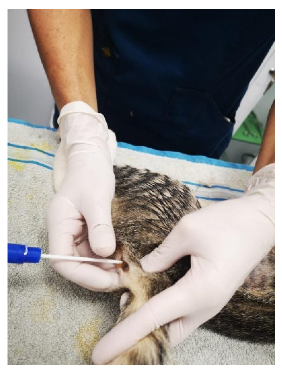
Figure 2.
Shelter for feral cats (Benalmádena, Province of Málaga).
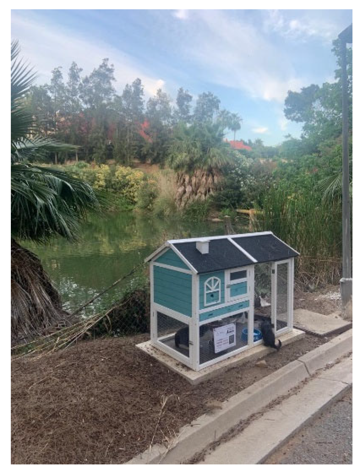
Figure 3.
Distribution of phenotypic antimicrobial resistance profiles (resistotypes) among the 68 Escherichia coli isolates. Bars represent the percentage of isolates exhibiting each resistance profile. Profiles were defined according to the combination of antimicrobial agents to which isolates were classified as resistant or non-wild-type (non-WT). Abbreviations: AMK, amikacin; AMP, ampicillin; CHL, chloramphenicol; CIP, ciprofloxacin; NA, nalidixic acid; SF, sulfamethoxazole; TE, tetracycline; TMP, trimethoprim.
Figure 3.
Distribution of phenotypic antimicrobial resistance profiles (resistotypes) among the 68 Escherichia coli isolates. Bars represent the percentage of isolates exhibiting each resistance profile. Profiles were defined according to the combination of antimicrobial agents to which isolates were classified as resistant or non-wild-type (non-WT). Abbreviations: AMK, amikacin; AMP, ampicillin; CHL, chloramphenicol; CIP, ciprofloxacin; NA, nalidixic acid; SF, sulfamethoxazole; TE, tetracycline; TMP, trimethoprim.
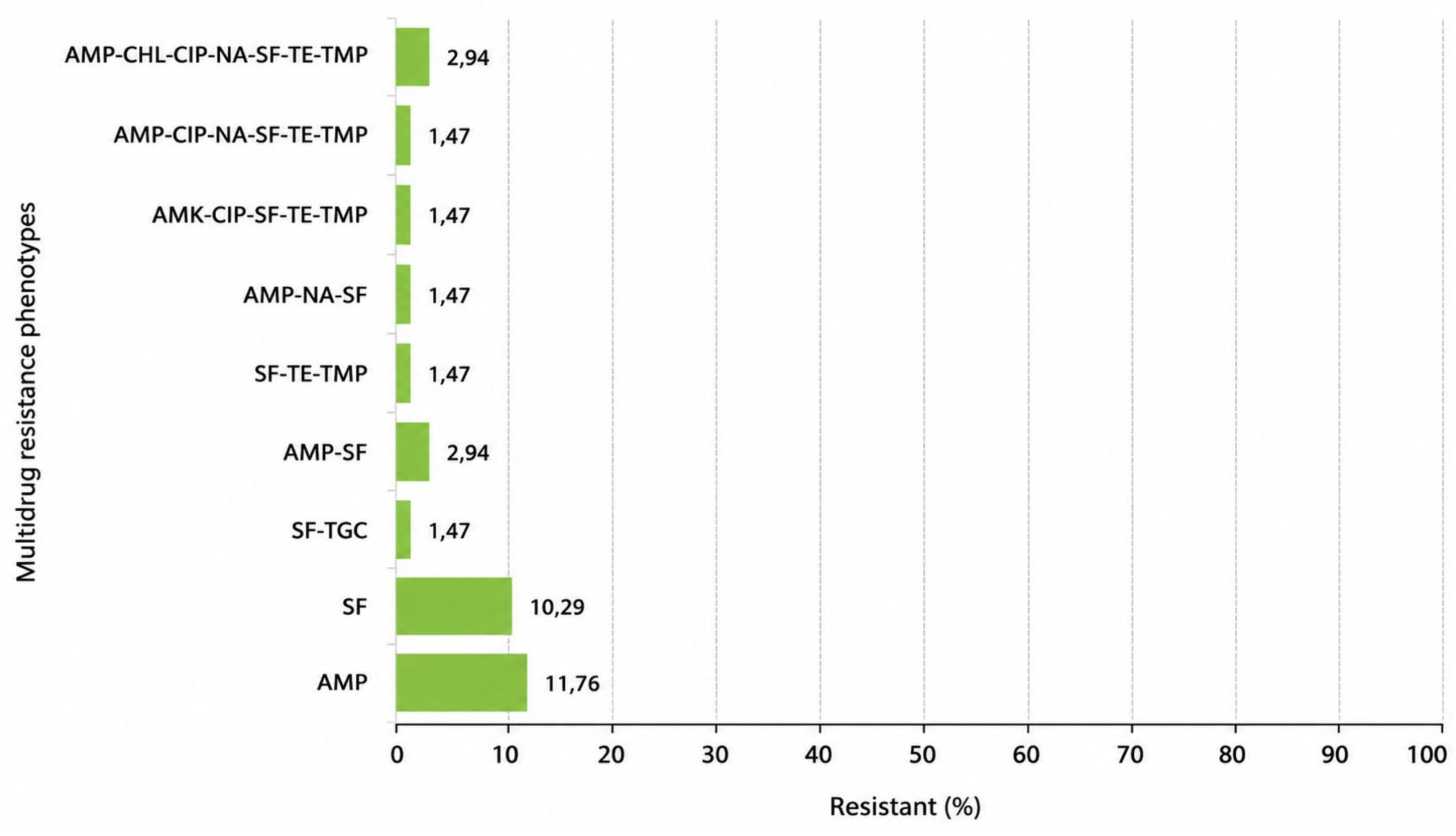
Figure 4.
Correlations between antimicrobial resistance phenotypes in Escherichia coli isolates based on Spearman’s rank correlation coefficients. Correlation strength was classified as very strong (0.80≤ρ≤1.00), strong (0.60≤ρ≤0.79), moderate (0.40≤ρ≤0.59), weak (0.20≤ρ≤0.39), very weak (0.00<ρ≤0.19), and no correlation (ρ≤0). Abbreviations: AMK, amikacin; AMP, ampicillin; CHL, chloramphenicol; CIP, ciprofloxacin; NA, nalidixic acid; SF, sulfamethoxazole; TE, tetracycline; TGC, tigecycline; TMP, trimethoprim. Red numbers indicate correlations that were not statistically significant (p > 0.05).
Figure 4.
Correlations between antimicrobial resistance phenotypes in Escherichia coli isolates based on Spearman’s rank correlation coefficients. Correlation strength was classified as very strong (0.80≤ρ≤1.00), strong (0.60≤ρ≤0.79), moderate (0.40≤ρ≤0.59), weak (0.20≤ρ≤0.39), very weak (0.00<ρ≤0.19), and no correlation (ρ≤0). Abbreviations: AMK, amikacin; AMP, ampicillin; CHL, chloramphenicol; CIP, ciprofloxacin; NA, nalidixic acid; SF, sulfamethoxazole; TE, tetracycline; TGC, tigecycline; TMP, trimethoprim. Red numbers indicate correlations that were not statistically significant (p > 0.05).
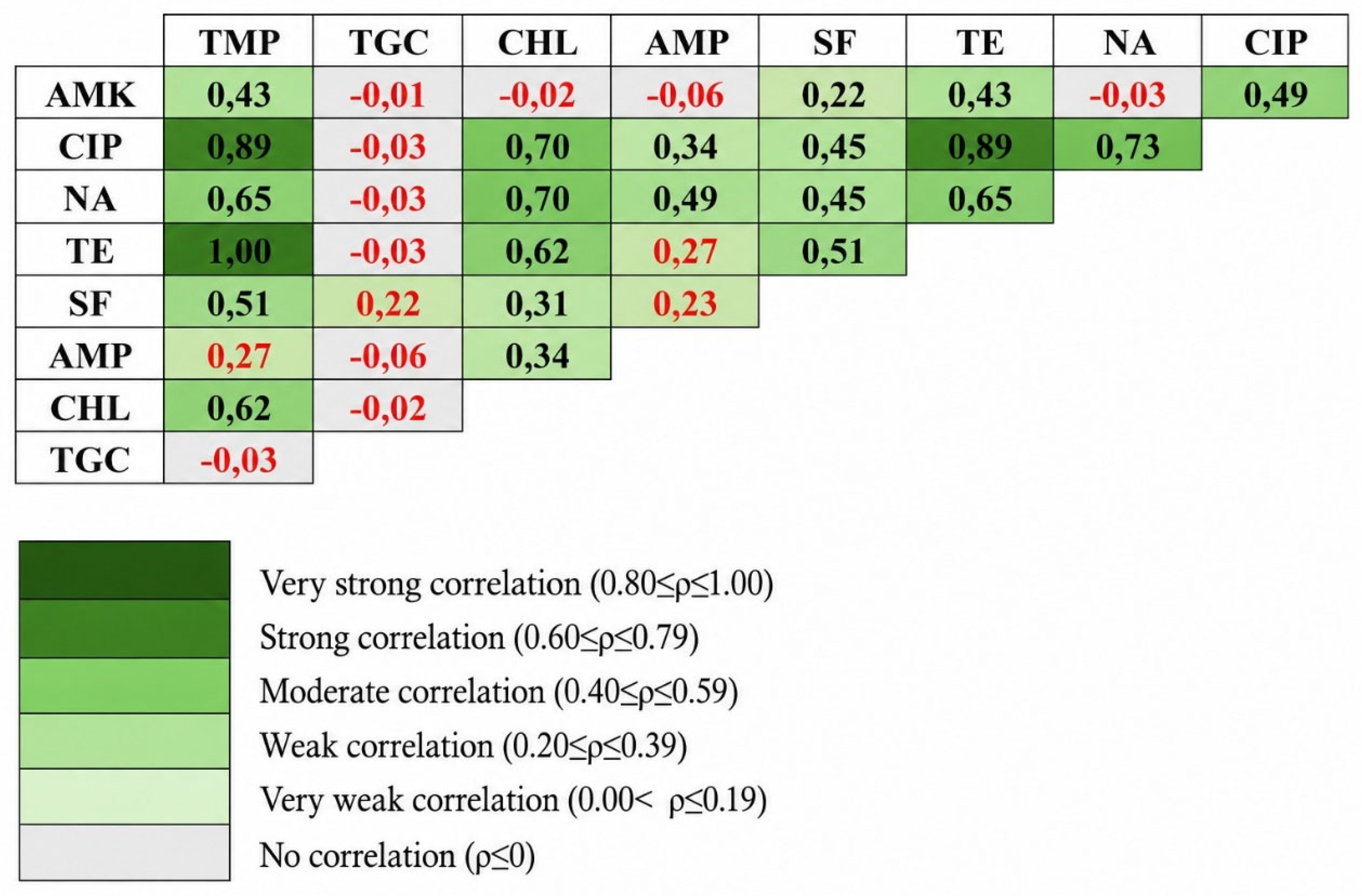
Table 1.
Detection of virulence-associated genes, pathotype classification, and antimicrobial resistance profiles of six atypical enteropathogenic Escherichia coli (aEPEC) isolates.
Table 1.
Detection of virulence-associated genes, pathotype classification, and antimicrobial resistance profiles of six atypical enteropathogenic Escherichia coli (aEPEC) isolates.
| Strain Reference | Gene detection |
Pathotype |
Antimicrobial Resistance |
||||||||||
| eae | stx1 | stx2 | bfpA | st | lt | ||||||||
| R18 | + | - | - | - | - | - | aEPEC | - | |||||
| R20 | + | - | - | - | - | - | aEPEC | AMP | |||||
| R22 | + | - | - | - | - | - | aEPEC | - | |||||
| R31 | + | - | - | - | - | - | aEPEC | AMP | |||||
| R74 | + | - | - | - | - | - | aEPEC | - | |||||
| R132 | + | - | - | - | - | - | aEPEC | SF | |||||
Gene detection legend: eae, intimin; stx1, Shiga toxin 1; stx2, Shiga toxin 2; bfpA, bundle-forming pilus subunit A; st, heat-stable enterotoxin; lt, heat-labile enterotoxin. Isolates positive for eae and negative for bfpA were classified as atypical enteropathogenic Escherichia coli (aEPEC). AMA legend: AMP, ampicillin; SF, sulfamethoxazole.
Disclaimer/Publisher’s Note: The statements, opinions and data contained in all publications are solely those of the individual author(s) and contributor(s) and not of MDPI and/or the editor(s). MDPI and/or the editor(s) disclaim responsibility for any injury to people or property resulting from any ideas, methods, instructions or products referred to in the content. |
© 2026 by the authors. Licensee MDPI, Basel, Switzerland. This article is an open access article distributed under the terms and conditions of the Creative Commons Attribution (CC BY) license (http://creativecommons.org/licenses/by/4.0/).
Copyright: This open access article is published under a Creative Commons CC BY 4.0 license, which permit the free download, distribution, and reuse, provided that the author and preprint are cited in any reuse.


