

Submitted:
19 June 2026
Posted:
22 June 2026
You are already at the latest version
Abstract
This review explores circulating and extracellular vesicle (EV)-associated ncRNAs as functional readouts in cancer liquid biopsy. We discuss their biological origin, carrier state, biofluid context, clinical applications, analytical technologies, artificial intelligence (AI)-assisted integration, standardization barriers, and regulatory requirements. The technology discussion considers sequencing, targeted amplification, and emerging direct or polymerase chain reaction (PCR)-free strategies as complementary translational routes for reliable ncRNA measurement. We propose that ncRNAs should not be viewed as alternatives to ctDNA, but as functional biomarkers that can link tumor genotype, regulatory state, and clinical phenotype within integrated multi-analyte precision oncology.
Keywords:
non-coding RNA
; liquid biopsy
; cancer
; microRNA
; long non-coding RNA
; circular RNA
; extracellular vesicles
; precision oncology
; PCR-free detection
1. Introduction: Liquid Biopsy at a Turning Point
Liquid biopsy has entered routine clinical practice in precision oncology in selected settings [1,2,3,4]. By analyzing tumor- and host-derived material released into body fluids, it enables dynamic and repeatable molecular assessment without exclusive dependence on tissue sampling. Its clinical value is currently most evident for cell-free DNA (cfDNA) and circulating tumor DNA (ctDNA) assays, which are used to identify actionable genomic alterations, support companion diagnostic decisions, monitor molecular evolution, and guide treatment selection in advanced cancer [5,6,7,8,9,10,11,12]. Nevertheless, the maturity of liquid biopsy remains highly dependent on tumor type, clinical indication, analyte class, and analytical platform.
This clinical progress has been driven largely by DNA-based profiling, but a DNA-centered view captures only part of cancer biology. ctDNA is highly informative about the genomic architecture of a tumor, including sequence variants, copy-number changes, structural rearrangements, methylation patterns, and molecular residual disease signatures [13,14,15,16,17,18,19]. However, it does not fully describe the regulatory state of tumor cells, the behavior of the tumor microenvironment, the host inflammatory response, or the functional consequences of therapy-induced selective pressure. Moreover, ctDNA detection can remain challenging in early-stage disease, low-shedding tumors, minimal residual disease (MRD) settings, and anatomical contexts in which tumor-derived DNA is poorly represented in peripheral blood [20,21,22,23,24,25].
Non-coding RNAs (ncRNAs) are among the most informative of these complementary sources. Although they do not encode proteins, they regulate gene expression, chromatin organization, RNA stability, translation, cell signaling, and intercellular communication. In cancer, ncRNAs contribute to proliferation, apoptosis resistance, invasion, angiogenesis, immune evasion, metabolic rewiring, stemness, metastatic colonization, and drug resistance [26,27,28,29,30,31,32,33,34,35,36,37,38,39]. Their detection in plasma, serum, urine, saliva, stool, cerebrospinal fluid, and extracellular vesicles (EVs) therefore offers information that differs from ctDNA: not only evidence of tumor-associated molecular release, but insight into biological programs active within the tumor-host system [40,41,42,43,44,45,46,47,48,49,50,51,52,53,54,55,56].
The central argument of this review is that ncRNAs should be interpreted as functional readouts of cancer liquid biopsy [40,41,42,43,44,45]. They are not merely additional markers for expanded panels, but molecular reporters of tumor activity, host response, immune modulation, tissue damage, metastatic communication, and therapeutic pressure. The following sections therefore examine how ncRNAs can fill the functional gaps left by ctDNA by adding information on carrier state, biofluid context, biological interpretation, clinical intended use, analytical technology, computational integration, and regulatory translation. As liquid biopsy evolves from single-analyte testing toward multi-analyte and multi-omic diagnostic systems, clinically useful assays will increasingly integrate genomic, transcriptomic, proteomic, vesicular, cellular, imaging, and computational information [20,21,22,23,24,25]. This integrated framework is summarized in Figure 1, in which ncRNAs function as the transcriptomic regulatory layer linking tumor genotype, regulatory state, and clinical phenotype.
2. From Liquid Biopsy to the Liquid Transcriptome
Liquid biopsy is often discussed as a single analytical concept, but the molecular entities measured in body fluids differ markedly in origin, stability, abundance, and clinical meaning. ctDNA is released predominantly through cell death [13,14,15,16,17,18,19], whereas circulating RNA can arise from both passive release and active secretion [40,41,42,43,44,45,46,47,48,49,50,51,52]. ncRNAs may circulate as free or protein-bound molecules, in association with lipoproteins, within EVs, in platelets, or as part of apoptotic and necrotic material. This diversity complicates assay design but also expands the biological information available from liquid biopsy.
Circulating ncRNAs can therefore be viewed as part of a liquid transcriptome: a composite molecular signal generated by tumor cells, stromal cells, immune cells, endothelial cells, blood cells, and injured tissues [51,52]. This signal is not equivalent to the transcriptome of the primary tumor. Instead, it is a dynamic mixture shaped by tumor burden, anatomical location, vascularization, inflammation, therapy, sample handling, and the carrier in which each RNA species is found [57,58].
Carrier state is central to interpretation. The same ncRNA may have different diagnostic and biological meaning when measured in total plasma, EVs, platelets, protein complexes, lipoprotein fractions, or tissue-proximal fluids [43,44,45,59,60,61,62,63,64,65,66,67]. For example, a miRNA released during tissue injury may indicate cell damage, whereas the same miRNA enriched in tumor-derived vesicles may reflect active intercellular communication. Similarly, platelet-associated RNA signatures may report tumor-induced platelet education, whereas EV-associated RNA may reflect selective packaging and secretion by tumor or stromal cells [40,41,42,43,44,45,46,47,48,49,50,51,52,59,60,61,62,63,64,65,66,67].
This distinction has practical consequences for clinical translation [6]. Assays measuring total circulating RNA may offer simplicity and sensitivity, but they may integrate signals from multiple tissues and physiological processes. Carrier-enriched assays may improve biological specificity, but they introduce additional pre-analytical and technical variables [56,68,69,70,71,72,73,74,75]. The optimal strategy should therefore be determined by the intended clinical use: early detection may benefit from broad, high-sensitivity signatures, whereas treatment monitoring and resistance prediction may require carrier-defined signals linked more directly to tumor biology [76,77,78,79].
3. Non-Coding RNA Classes in Cancer Liquid Biopsy: From Molecular Categories to Clinical Functions
ncRNAs include a heterogeneous collection of molecules that differ in size, biogenesis, structure, stability, and function [26,27,28,29,30,31,32,33,34,35,36,37,38,39]. In cancer liquid biopsy, the most frequently studied classes include miRNAs, long non-coding RNAs (lncRNAs), circular RNAs (circRNAs), PIWI-interacting RNAs (piRNAs), tRNA-derived fragments (tRFs), small nucleolar RNAs (snoRNAs), small nuclear RNAs (snRNAs), Y RNAs, and other poorly annotated or orphan ncRNAs [53,54,55,56,57,80,81,82,83,84,85,86,87,88,89,90,91,92,93,94,95,96,97,98,99,100,101]. For clinical translation, it is most useful to organize these classes according to the type of biological and clinical information they provide (Table 1).
3.1. MicroRNAs: Stable and Actionable Small-RNA Biomarkers
miRNAs are the most established ncRNA biomarkers in liquid biopsy [40,41,42,43,44,45,68,69,70,71,72]. Their small size, relative stability, disease-associated expression patterns, and compatibility with targeted assays have made them highly attractive for cancer detection, prognosis, and monitoring. In many tumors, miRNA signatures reflect cellular pathways linked to proliferation, apoptosis, epithelial-mesenchymal transition (EMT), angiogenesis, immune regulation, and treatment resistance [26,27,28,29,30,31,32,93]. Their major translational challenge is not biological relevance, but reproducibility across cohorts, specimen types, extraction methods, normalization strategies, and analytical platforms [53,54,55,56,73,74,75].
3.2. Long Non-Coding RNAs: Tissue Specificity and Regulatory Depth
lncRNAs offer a different type of promise. Many lncRNAs show tissue- or cancer-type-enriched expression and can regulate transcription, chromatin state, splicing, RNA stability, and signaling networks [36,37,38,39,80,81,82,83]. Their cancer specificity may support tumor classification and prognostic stratification, but their lower abundance and greater susceptibility to degradation can complicate circulating detection. Extracellular-vesicle enrichment, targeted amplification, or direct capture strategies are particularly relevant for their reliable clinical measurement [102,103,104,105,106,107,108,109,110,111,112].
3.3. Circular RNAs: Structural Stability and Biofluid Robustness
circRNAs are attractive liquid biopsy candidates because their covalently closed structure confers resistance to exonuclease degradation [84,85,86,87,88,89,90]. This structural stability is highly advantageous in harsh biofluid environments such as plasma, serum, urine, saliva, and stool. Functionally, circRNAs can act as miRNA sponges, interact with RNA-binding proteins, modulate transcription, and in some cases encode functional peptides [84,85,86,87,88,89,90,113,114,115,116,117,118,119]. Their presence in EVs and tissue-specific expression patterns make them promising for robust biomarker development, although standardized annotation and quantification remain key operational challenges.
3.4. Emerging Small and Poorly Annotated ncRNAs: High-Dimensional Cancer Fingerprints
Emerging small RNA classes, including tRFs, piRNAs, snoRNAs, Y RNA fragments, and poorly annotated ncRNAs, are expanding the liquid biopsy landscape [57,91,94,95,96,97,98,99,100,101]. These molecules provide high-dimensional cancer fingerprints that are not captured by conventional miRNA panels. Some are particularly useful in artificial intelligence (AI)-driven signatures where diagnostic information emerges from complex global patterns rather than from single canonical biomarkers [74,75,76,77,78,79]. However, this strength creates a translational risk: signatures may be statistically powerful but biologically opaque unless supported by rigorous validation and mechanistic interpretation.
Taken together, these ncRNA classes should not be treated as competing biomarker families, but as complementary molecular layers. miRNAs provide stability and assay practicality; lncRNAs provide regulatory and tissue-specific information; circRNAs offer structural robustness; EV-associated RNAs provide carrier-defined specificity; and emerging small RNAs offer high-dimensional discriminatory power. The most clinically useful liquid biopsy assays will likely combine selected RNA classes according to the intended use rather than relying on a single category.
4. Carrier-Aware ncRNA Biomarkers: Why Molecular Context Matters
The clinical value of a circulating ncRNA depends not only on its sequence and abundance, but also on its biological context. Body fluids contain a complex mixture of RNA carriers, including ribonucleoprotein complexes, lipoproteins, EVs, platelets, apoptotic bodies, cellular debris, and intact rare cells [40,41,42,43,44,45,46,47,48,49,50,51,52,59,60,61,62,63,64,65,66,67]. Each carrier may reflect a different biological process and may require a different analytical workflow. For this reason, future liquid biopsy studies should explicitly define whether they aim to measure total circulating ncRNA, a carrier-enriched fraction, or a tissue-proximal RNA signal [120,121,122,123,124].
Cell-free and protein-bound ncRNAs offer analytical accessibility because they can be measured directly from plasma or serum without complex vesicle enrichment. However, they may also reflect systemic tissue injury, blood cell contamination, inflammation, or sample handling effects [43,44,56,68,69,70,71,72]. Lipoprotein-associated RNAs may provide additional biological information because lipoproteins participate in intercellular transport, metabolism, and inflammatory signaling. Platelet-associated RNAs form another relevant compartment: tumor-educated platelets can acquire altered RNA profiles through interaction with cancer cells and the tumor microenvironment, potentially providing indirect but informative signatures of malignancy [45,66,67].
EV-associated ncRNAs occupy a particularly important position in the field. EVs can protect RNA cargo from degradation and may reflect selective packaging by tumor cells, stromal cells, immune cells, and endothelial cells [46,47,48,49,50,59,60,61,62,63,64,65]. Vesicle-associated RNAs may therefore function both as biomarkers and as mediators of tumor-microenvironment communication. However, the same biological richness creates technical challenges. EV isolation methods differ in yield, purity, scalability, and compatibility with clinical workflows [120,121,122,123,124]. Without rigorous reporting and characterization, apparent EV-ncRNA biomarkers may be confounded by co-isolated protein complexes, lipoproteins, or non-vesicular RNA carriers.
Tissue-proximal biofluids add another dimension. Urine may be especially relevant for urological cancers, saliva for head and neck malignancies, stool for colorectal cancer and gastrointestinal bleeding-related signals, and cerebrospinal fluid for central nervous system tumors [49,50,125,126,127,128,129]. These biofluids can enrich local disease signals that may be diluted in peripheral blood, but they also introduce matrix-specific inhibitors, degradation processes, and pre-analytical variables [58,73]. Figure 2 summarizes this carrier-aware and matrix-aware logic for the rational design of ncRNA liquid biopsy assays.
5. Biological Interpretation: ncRNAs as Functional Disease Sensors
A major conceptual advantage of ncRNAs is their ability to report biological activity. Whereas ctDNA primarily reflects genomic alterations and tumor fraction [13,14,15,16,17,18,19,20,21,22,23,24,25], ncRNAs can provide information about regulatory programs and phenotypic states [26,27,28,29,30,31,32,33,34,35,36,37,38,39,40,41,42,43,44,45]. In this sense, circulating ncRNAs may act as functional disease sensors. They can report tumor presence, tissue-of-origin signals, malignant aggressiveness, host response, immune modulation, metastatic competence, and therapeutic pressure.
For early detection, ncRNA signatures may be useful when tumor DNA shedding is low or intermittent [40,41,42,43,44,45]. For prognosis, specific ncRNA patterns may reflect aggressive biological programs, including EMT, angiogenesis, invasion, stemness, and metastatic niche preparation [26,27,28,29,30,31,32,33,34,35,36,37,38,39,68,69,70,71,72]. For treatment monitoring, changes in circulating ncRNAs may capture therapy-induced stress, immune activation, tumor cell death, or emerging resistance. In immuno-oncology, EV-associated ncRNAs are of particular interest because they may participate in immune escape, antigen presentation modulation, cytokine signaling, and the regulation of immune checkpoint pathways [59,60,61,62,63,130].
The same biological sensitivity that makes ncRNAs attractive also requires careful interpretation. Many ncRNAs are not cancer-exclusive and may be influenced by age, sex, inflammation, infection, medication, tissue injury, metabolic disease, and lifestyle [56,68,69,70,71,72]. Therefore, clinically useful ncRNA signatures should not be interpreted simply as tumor markers, but as disease-context markers. Their value will be highest when the biological question is clearly defined and when comparator populations reflect realistic clinical use rather than idealized health controls [6,73,74,75,76,77,78,79].
A further implication is that ncRNA biomarkers may be most informative when interpreted as dynamic, context-dependent signals rather than as fixed binary indicators of tumor presence. In longitudinal sampling, a change in an ncRNA signature may reflect a shift in tumor biology, treatment-induced tissue injury, immune activation, or remodeling of the tumor microenvironment before these processes become radiologically apparent [40,41,42,43,44,45,46,47,48,49,50,51,52,59,60,61,62,63]. Conversely, stable baseline differences between patients may capture host biology, organ-specific vulnerability, or chronic inflammatory states that influence cancer risk and treatment tolerance. This dual tumor-host origin should be considered a strength rather than a limitation, provided that the intended clinical question is explicit.
For example, an ncRNA pattern used for early detection should be optimized for cancer-associated discrimination, whereas an ncRNA pattern used for treatment monitoring may legitimately incorporate tumor injury, immune response, and organ toxicity signals. The functional interpretation of ncRNAs therefore depends on integrating molecular abundance with carrier state, sampling time point, clinical context, and comparator population [6,68,69,70,71,72,73,74,75,76,77,78,79,120,121,122,123,124,125,126,127,128,129].
6. Clinical Applications in Precision Oncology
The clinical development of ncRNA liquid biopsy should be organized around intended use. A biomarker signature designed for population screening has different performance requirements from a signature designed for recurrence monitoring, therapy response, or resistance prediction [6,74,75]. Early cancer detection requires high sensitivity for low-burden disease and high specificity in clinically relevant populations [20,21,22,23,24,25]. Tumor classification and tissue-of-origin prediction require signatures that distinguish cancer types and account for inflammatory or benign disease controls. Prognostic signatures require association with clinically meaningful outcomes such as progression-free survival, overall survival, metastatic relapse, or treatment failure [58,66,67,125,126,127,128,129].
In therapy selection, ncRNAs may be used to infer pathway activation, immune state, or resistance biology. EV-associated ncRNAs could complement ctDNA by reporting tumor-microenvironment communication or immune escape [59,60,61,62,63,64,65,66,67]. In MRD and recurrence monitoring, ncRNAs may provide complementary information about when ctDNA is undetectable or when active tumor biology precedes radiological progression [13,14,15,16,17,18,19,20,21,22,23,24,25]. Finally, biofluid-specific applications may allow disease-proximal measurements: urine for bladder, prostate, and kidney cancers; saliva for oral and head and neck cancers; stool for colorectal cancer; and plasma or serum for systemic profiling [125,126,127,128,129]. Concrete examples illustrate this potential: profiling of tumor-educated platelet RNA distinguished patients with cancer from healthy individuals with 96% accuracy and identified the tissue of origin across six tumor types with 71% accuracy [66], while a particle-swarm-optimized platelet RNA classifier detected non-small-cell lung cancer with areas under the curve of 0.94 and 0.89 in late- and early-stage validation cohorts, respectively [67]. Such performance is encouraging but still requires confirmation in prospective, intended-use populations with appropriate benign and inflammatory comparators.
Table 2 summarizes the main intended-use scenarios and the corresponding validation focus. These clinical applications should not be collapsed into a single generic biomarker claim. A screening assay must perform in asymptomatic or enriched-risk populations in which disease prevalence is low and false positives may generate unnecessary diagnostic procedures [20,21,22,23,24,25]. A diagnostic classifier must distinguish cancer from benign, inflammatory, or pre-malignant conditions that are likely to appear in the same clinical pathway. A prognostic assay should add information beyond established clinicopathological variables, whereas a monitoring assay should demonstrate that longitudinal molecular changes anticipate or improve clinical decision-making compared with imaging, conventional biomarkers, or ctDNA alone. For therapy response and resistance, the most informative ncRNA signals may be those linked to pathway activity, immune state, or tumor-host communication rather than tumor burden alone [58,59,60,61,62,63,64,65,66,67]. This intended-use discipline is essential for designing appropriate comparator cohorts, statistical endpoints, sampling intervals, and clinical decision thresholds [6,73,74,75,76,77,78,79].
7. Technologies for ncRNA Liquid Biopsy Translation
The transition from biomarker discovery to clinical implementation depends strongly on analytical technology. RNA sequencing and small-RNA sequencing remain powerful discovery tools because they provide broad profiling capacity and can identify canonical and non-canonical RNA species [76,77,78,79]. Targeted sequencing can improve depth and reduce cost, but it still requires careful library preparation, bioinformatic normalization, and batch-effect control. Reverse-transcription quantitative polymerase chain reaction (RT-qPCR) remains widely used because it is sensitive, accessible, and suitable for targeted validation, but it can be affected by reverse-transcription efficiency, primer specificity, amplification bias, and normalization uncertainty [73,75]. Digital polymerase chain reaction (dPCR) can improve absolute quantification and sensitivity for selected targets, although its multiplexing capacity remains more limited than sequencing or bead-based platforms [74]. These established technologies remain essential, but they do not eliminate the need for more direct workflows that reduce sample loss, target conversion bias, and amplification-dependent artifacts.
Direct and polymerase chain reaction (PCR)-free strategies are relevant to ncRNA liquid biopsy because they may reduce dependence on RNA extraction, reverse transcription, and amplification, thereby limiting target loss and analytical bias. Dynamic Chemical Labelling (DCL) represents one example of this broader technology class, using target-guided chemistry on modified peptide nucleic acid probes to convert sequence recognition into optical, chemiluminescent, digital, or bead-based signals. Published studies have demonstrated direct detection of selected miRNAs and compatibility with multiplex bead-based readouts, supporting the feasibility of amplification-free ncRNA measurement [131,132,133,134,135,136,137,138,139]. In cancer liquid biopsy, however, the translational value of DCL, like that of other emerging direct-read technologies, will depend on independent validation in oncological cohorts, clinically relevant biofluids, locked signatures, and comparison with established sequencing, PCR-based, and hybridization-based platforms.
Other emerging direct or low-amplification approaches should be considered alongside chemical-ligation strategies. Clustered regularly interspaced short palindromic repeats (CRISPR)/CRISPR-associated (Cas)-based diagnostics, including Specific High-Sensitivity Enzymatic Reporter UnLOCKing (SHERLOCK) and DNA Endonuclease-Targeted CRISPR Trans Reporter (DETECTR), exploit programmable nucleic-acid recognition and collateral reporter cleavage to generate sensitive sequence-specific readouts [140,141,142]. These systems offer attractive features for liquid biopsy, including programmability, portability, and potential point-of-care formats. At the same time, many implementations still rely on pre-amplification, and their quantitative performance in complex cancer biofluids, carrier-enriched fractions, stool, urine, or low-abundance ncRNA settings requires further analytical and clinical validation.
Surface-enhanced Raman spectroscopy (SERS) and plasmonic biosensors provide another complementary route for sensitive and potentially multiplexed nucleic-acid detection. Recent SERS-based liquid biopsy reviews and miRNA-focused biosensors highlight the capacity of these platforms to analyze circulating nucleic acids, extracellular vesicles, proteins, and miRNA targets with low sample consumption and optical multiplexing [143,144]. Their translational challenges include substrate reproducibility, probe design, matrix interference, spectral standardization, calibration across instruments, and the need for transparent machine-learning pipelines when spectral classifiers are used. Nanopore and direct RNA sequencing approaches may also contribute to future RNA liquid biopsy by enabling longer-read, isoform-level, and potentially epitranscriptomic information; however, input requirements, short-RNA capture, error profiles, and robustness in clinical biofluids remain important limitations [145].
Overall, no single technology is optimal for all stages of ncRNA liquid biopsy development. Discovery may require sequencing or broad profiling, whereas clinical implementation may favor targeted, standardized, cost-effective, and reproducible platforms. The most appropriate assay should be selected according to intended use, target number, required sensitivity, sample matrix, throughput, regulatory pathway, laboratory infrastructure, and the degree to which the platform can be externally validated in real-world clinical cohorts.
8. Artificial Intelligence and Multi-Analyte Integration
High-dimensional ncRNA data are well suited to computational modeling because diagnostic information may arise from patterns distributed across many RNA species rather than from single biomarkers [74,75,76,77,78,79,146]. In current liquid biopsy and precision-oncology studies, model classes commonly include regularized regression, random forests, support vector machines, gradient boosting, and deep-learning architectures, with graph-based or multi-view models becoming increasingly relevant for multi-omic integration. AI and machine learning can integrate canonical ncRNAs, poorly annotated RNAs, ctDNA, proteins, EV markers, imaging, and clinical metadata into composite classifiers [20,21,22,23,24,25,147,148]. This approach may be particularly useful for early detection, tissue-of-origin prediction, and complex response phenotypes such as immunotherapy benefit or resistance [149,150]. As a proof of concept, an AI-normalized cell-free RNA (liquid transcriptome) classifier separated patients with solid tumors from controls with an area under the curve of 0.82, and distinguished myeloid and lymphoid neoplasms with areas under the curve of 0.86 and 0.79, respectively [148], illustrating both the promise and the still-preliminary nature of transcriptome-wide AI classifiers prior to prospective validation.
For clinical translation, the role of AI should be defined before model development rather than after statistical optimization [6,146]. In screening or diagnostic triage, algorithms may be expected to maximize sensitivity while maintaining acceptable specificity in heterogeneous populations; in monitoring or therapy-response settings, they may instead prioritize longitudinal change, calibration, and robustness to repeated measurements [74,75,76,77,78,79,147,148,149,150]. Models based on poorly annotated ncRNAs or multi-analyte inputs should therefore report not only area-under-the-curve metrics, but also calibration, decision thresholds, feature stability, missing-data handling, and performance across clinically relevant subgroups. Interpretability is also important: even when individual ncRNAs are not mechanistically characterized, the final classifier should remain biologically plausible, technically reproducible, and compatible with a predefined clinical action. In this sense, AI should be viewed as a translation tool for integrating complex ncRNA signals, not as a substitute for analytical validation, external replication, or intended-use evidence.
However, computational power does not replace biological and clinical rigor. Models trained on retrospective or highly selected cohorts may not generalize to real-world screening, diagnostic, or monitoring populations [6,146,150]. Overfitting, batch effects, population bias, and inadequate external validation remain major risks [74,75,76,77,78,79]. Future AI-based ncRNA liquid biopsy studies should therefore include locked signatures, independent validation cohorts, transparent reporting, clinically relevant controls, and predefined performance endpoints.
9. Pre-Analytical, Analytical, and Clinical Standardization
Standardization is one of the main barriers to ncRNA liquid biopsy translation. Pre-analytical variables include blood collection tube type, time to processing, centrifugation protocol, hemolysis, platelet contamination, freeze-thaw cycles, storage conditions, and biofluid-specific matrix effects [56,68,69,70,71,72,120,121,122,123,124]. Analytical variables include RNA extraction efficiency, carrier enrichment strategy, reverse transcription efficiency, amplification bias, sequencing depth, adapter ligation bias, normalization method, and inter-platform comparability [73,74,75,76,77,78,79]. Clinical variables include cohort size, case-control definition, benign disease controls, medication, inflammation, comorbidities, and longitudinal sampling design [6].
For EV studies, reporting standards are particularly important because different EV enrichment methods can isolate different mixtures of vesicles, lipoproteins, protein complexes, and ribonucleoprotein particles [120,121,122,123,124]. Transparent reporting of sample handling, EV separation, particle characterization, RNA extraction, and normalization is essential for reproducibility. More broadly, ncRNA liquid biopsy studies should move from exploratory biomarker lists to locked signatures and prospectively defined clinical questions. Table 3 summarizes the key pre-analytical and analytical variables that should be controlled for reproducible ncRNA liquid biopsy development [6,73,74,75,76,77,78,79].
10. Regulatory and Intended Use Roadmap
The clinical translation of ncRNA liquid biopsy requires more than biomarker discovery [6]. A regulatory-grade development pathway should begin with a clearly defined intended use and clinical context. The intended use determines the target population, comparator, acceptable false-positive and false-negative rates, sample type, assay format, and validation endpoint. A screening assay, a diagnostic classifier, a prognostic test, and a therapy-monitoring tool require different evidence packages [74,75].
A practical roadmap includes discovery, technical optimization, analytical validation, signature locking, retrospective clinical validation, prospective intended-use validation, health-economic assessment, regulatory submission, and post-market monitoring. Analytical validation should address sensitivity, specificity, precision, reproducibility, linearity, limit of detection, interference, stability, and matrix effects [73,74,75,120,121,122,123,124]. Clinical validation should demonstrate that the assay answers a clinically relevant question better than, or complementary to, existing standards of care [6,74,75]. Without this roadmap, ncRNA biomarkers risk remaining in the discovery literature without entering clinical practice. This intended-use-driven roadmap is illustrated in Figure 3.
11. Future Perspective: Toward Functional, Multi-Analyte Liquid Biopsy
The future of cancer liquid biopsy will likely not be defined by competition between ctDNA and ncRNAs. Instead, the most informative assays will combine complementary molecular layers. ctDNA can identify genomic alterations and clonal evolution [13,14,15,16,17,18,19,20,21,22,23,24,25]; ncRNAs can reveal regulatory activity, tissue injury, immune modulation, and therapy-induced phenotypic change [26,27,28,29,30,31,32,33,34,35,36,37,38,39,40,41,42,43,44,45,46,47,48,49,50,51,52,59,60,61,62,63,64,65,66,67]; proteins and metabolites can report downstream functional consequences; EV markers can provide carrier and cellular-context information; and imaging or clinical metadata can anchor molecular signals to patient phenotype [20,21,22,23,24,25,151]. Recent literature increasingly emphasizes that this integration will rely on longitudinal, multimodal, and multi-omics datasets, supported by AI-enabled feature selection and clinically validated analytical workflows rather than by simply adding more analytes to existing panels [151,152,153].
In this integrated model, ncRNAs represent the functional transcriptomic layer of precision liquid biopsy [31,32,33,34,35,36,37,38,39,40,41,42,43,44,45]. Their greatest value may emerge when they are not treated as isolated biomarkers, but as context-dependent signals interpreted according to carrier, biofluid, biological process, and clinical question [59,60,120,121,122,123,124,151]. The field should therefore move from descriptive catalogues of differentially expressed RNAs toward clinically locked, mechanistically interpretable, and technologically robust signatures that are benchmarked across platforms, populations, and intended-use settings [73,74,75,76,77,78,79,146,150,151,152,153].
12. Conclusions
ncRNAs may represent the missing functional layer of cancer liquid biopsy. While ctDNA captures the genomic scars of cancer, ncRNAs capture ongoing biological activity. Their detection in blood and other body fluids offers opportunities for early detection, tumor classification, prognosis, treatment monitoring, MRD assessment, and prediction of therapeutic resistance. However, clinical value will depend on overcoming pre-analytical variability, analytical bias, biological confounding, and insufficient prospective validation.
The next generation of ncRNA liquid biopsy should account for carrier state, biological context, and molecular function. It should integrate RNA signals with genomic, proteomic, vesicular, cellular, imaging, and clinical information. Most importantly, it should move from exploratory biomarker discovery to standardized, multiplexed, prospectively validated assays that operate within real diagnostic pathways. ncRNA liquid biopsy should therefore be viewed not as an alternative to ctDNA, but as the functional layer that may complete precision oncology diagnostics.
Author Contributions
Conceptualization, S.P.; literature review and synthesis, S.P.; visualization, S.P.; writing—original draft preparation, S.P.; writing—review and editing, S.P., V.T. and D.G. All authors have read and agreed to the published version of the manuscript.
Funding
This research received no external funding. The article processing charge was waived by the journal as part of the invitation to contribute to the Special Issue.
Institutional Review Board Statement
Not applicable.
Informed Consent Statement
Not applicable.
Data Availability Statement
No new data were created or analyzed in this study. Data sharing is not applicable to this article.
Acknowledgments
During the preparation of this manuscript, the review was conceived, drafted, and critically revised by the authors. FigureLabs was used as an assistive tool for figure preparation; certification for the generated figures is available. The authors reviewed, edited, and approved all content and take full responsibility for the final manuscript.
Conflicts of Interest
S.P. is a shareholder of DESTINA Genomica S.L., a company active in nucleic-acid detection technologies, including Dynamic Chemical Labelling approaches discussed in this review. V.T. and D.G. declare no conflicts of interest. DESTINA Genomica S.L. had no role in the preparation of this review or in the decision to submit the manuscript.
Abbreviations
The following abbreviations are used in this manuscript:
| Abbreviation | Definition |
| AI | artificial intelligence |
| cfDNA | cell-free DNA |
| circRNA | circular RNA |
| ctDNA | circulating tumor DNA |
| DCL | Dynamic Chemical Labelling |
| dPCR | digital polymerase chain reaction |
| EMT | epithelial-mesenchymal transition |
| EV | extracellular vesicle |
| lncRNA | long non-coding RNA |
| miRNA | microRNA |
| MRD | minimal residual disease |
| ncRNA | non-coding RNA |
| PCR | polymerase chain reaction |
| piRNA | PIWI-interacting RNA |
| RT-qPCR | reverse-transcription quantitative polymerase chain reaction |
| snRNA | small nuclear RNA |
| snoRNA | small nucleolar RNA |
| tRF | tRNA-derived fragment |
| Cas | CRISPR-associated |
| CRISPR | clustered regularly interspaced short palindromic repeats |
| DETECTR | DNA Endonuclease-Targeted CRISPR Trans Reporter |
| SERS | surface-enhanced Raman spectroscopy |
| SHERLOCK | Specific High-Sensitivity Enzymatic Reporter UnLOCKing |
References
- Heitzer, E.; Haque, I.S.; Roberts, C.E.S.; Speicher, M.R. Current and future perspectives of liquid biopsies in genomics-driven oncology. Nat. Rev. Genet. 2019, 20, 71–88. [Google Scholar] [PubMed]
- Wan, J.C.M.; Massie, C.; Garcia-Corbacho, J.; Mouliere, F.; Brenton, J.D.; Caldas, C.; Pacey, S.; Baird, R.; Rosenfeld, N. Liquid biopsies come of age: Towards implementation of circulating tumour DNA. Nat. Rev. Cancer 2017, 17, 223–238. [Google Scholar] [CrossRef] [PubMed]
- Siravegna, G.; Marsoni, S.; Siena, S.; Bardelli, A. Integrating liquid biopsies into the management of cancer. Nat. Rev. Clin. Oncol. 2017, 14, 531–548. [Google Scholar] [CrossRef] [PubMed]
- Ignatiadis, M.; Sledge, G.W.; Jeffrey, S.S. Liquid biopsy enters the clinic - implementation issues and future challenges. Nat. Rev. Clin. Oncol. 2021, 18, 297–312. [Google Scholar] [CrossRef] [PubMed]
- Rolfo, C.; Mack, P.C.; Scagliotti, G.V.; Baas, P.; Barlesi, F.; Bivona, T.G.; Herbst, R.S.; Mok, T.S.K.; Peled, N.; Pirker, R.; et al. Liquid biopsy for advanced non-small cell lung cancer: A statement paper from the IASLC. J. Thorac. Oncol. 2018, 13, 1248–1268. [Google Scholar] [PubMed]
- Merker, J.D.; Oxnard, G.R.; Compton, C.; Diehn, M.; Hurley, P.; Lazar, A.J.; Lindeman, N.; Lockwood, C.M.; Rai, A.J.; Schilsky, R.L.; et al. Circulating tumor DNA analysis in patients with cancer: American Society of Clinical Oncology and College of American Pathologists joint review. J. Clin. Oncol. 2018, 36, 1631–1641. [Google Scholar] [CrossRef] [PubMed]
- Crowley, E.; Di Nicolantonio, F.; Loupakis, F.; Bardelli, A. Liquid biopsy: Monitoring cancer-genetics in the blood. Nat. Rev. Clin. Oncol. 2013, 10, 472–484. [Google Scholar] [CrossRef] [PubMed]
- Alix-Panabieres, C.; Pantel, K. Clinical applications of circulating tumor cells and circulating tumor DNA as liquid biopsy. Cancer Discov. 2016, 6, 479–491. [Google Scholar] [CrossRef] [PubMed]
- Bettegowda, C.; Sausen, M.; Leary, R.J.; Kinde, I.; Wang, Y.; Agrawal, N.; Bartlett, B.R.; Wang, H.; Luber, B.; Alani, R.M.; et al. Detection of circulating tumor DNA in early- and late-stage human malignancies. Sci. Transl. Med. 2014, 6, 224ra24. [Google Scholar] [CrossRef] [PubMed]
- Dawson, S.J.; Tsui, D.W.Y.; Murtaza, M.; Biggs, H.; Rueda, O.M.; Chin, S.F.; Dunning, M.J.; Gale, D.; Forshew, T.; Mahler-Araujo, B.; et al. Analysis of circulating tumor DNA to monitor metastatic breast cancer. N. Engl. J. Med. 2013, 368, 1199–1209. [Google Scholar] [CrossRef] [PubMed]
- Diehl, F.; Schmidt, K.; Choti, M.A.; Romans, K.; Goodman, S.; Li, M.; Thornton, K.; Agrawal, N.; Sokoll, L.; Szabo, S.A.; et al. Circulating mutant DNA to assess tumor dynamics. Nat. Med. 2008, 14, 985–990. [Google Scholar] [CrossRef] [PubMed]
- Newman, A.M.; Bratman, S.V.; To, J.; Wynne, J.F.; Eclov, N.C.W.; Modlin, L.A.; Liu, C.L.; Neal, J.W.; Wakelee, H.A.; Merritt, R.E.; et al. An ultrasensitive method for quantitating circulating tumor DNA with broad patient coverage. Nat. Med. 2014, 20, 548–554. [Google Scholar] [CrossRef] [PubMed]
- Abbosh, C.; Birkbak, N.J.; Wilson, G.A.; Jamal-Hanjani, M.; Constantin, T.; Salari, R.; Le Quesne, J.; Moore, D.A.; Veeriah, S.; Rosenthal, R.; et al. Phylogenetic ctDNA analysis depicts early-stage lung cancer evolution. Nature 2017, 545, 446–451. [Google Scholar] [PubMed]
- Chaudhuri, A.A.; Chabon, J.J.; Lovejoy, A.F.; Newman, A.M.; Stehr, H.; Azad, T.D.; Khodadoust, M.S.; Esfahani, M.S.; Liu, C.L.; Zhou, L.; et al. Early detection of molecular residual disease in localized lung cancer by circulating tumor DNA profiling. Cancer Discov. 2017, 7, 1394–1403. [Google Scholar] [CrossRef] [PubMed]
- Tie, J.; Wang, Y.; Tomasetti, C.; Li, L.; Springer, S.; Kinde, I.; Silliman, N.; Tacey, M.; Wong, H.L.; Christie, M.; et al. Circulating tumor DNA analysis detects minimal residual disease and predicts recurrence in patients with stage II colon cancer. Sci. Transl. Med. 2016, 8, 346ra92. [Google Scholar] [CrossRef] [PubMed]
- Reinert, T.; Henriksen, T.V.; Christensen, E.; Sharma, S.; Salari, R.; Sethi, H.; Knudsen, M.; Nordentoft, I.; Wu, H.T.; Tin, A.S.; et al. Analysis of plasma cell-free DNA by ultradeep sequencing in patients with stages I to III colorectal cancer. JAMA Oncol. 2019, 5, 1124–1131. [Google Scholar] [CrossRef] [PubMed]
- Abbosh, C.; Frankell, A.M.; Harrison, T.; Kisistok, J.; Garnett, A.; Johnson, L.; Veeriah, S.; Moreau, M.; Huebner, A.; Swanton, C.; et al. Tracking early lung cancer metastatic dissemination in TRACERx using circulating tumor DNA. Nature 2023, 616, 553–562. [Google Scholar] [CrossRef] [PubMed]
- Parikh, A.R.; Van Seventer, E.E.; Siravegna, G.; Hartwig, A.V.; Jaimovich, A.; He, Y.; Kanter, K.; Fish, M.G.; Fosbenner, K.D.; Miao, B.; et al. Minimal residual disease detection using a plasma-only circulating tumor DNA assay in colorectal cancer patients. Clin. Cancer Res. 2021, 27, 5586–5594. [Google Scholar] [CrossRef] [PubMed]
- Phallen, J.; Sausen, M.; Adleff, V.; Leal, A.; Hruban, C.; White, J.; Anagnostou, V.; Fiksel, J.; Cristiano, S.; Papp, E.; et al. Direct detection of early-stage cancers using circulating tumor DNA. Sci. Transl. Med. 2017, 9, eaan2415. [Google Scholar] [CrossRef] [PubMed]
- Cohen, J.D.; Li, L.; Wang, Y.; Thoburn, C.; Afsari, B.; Danilova, L.; Douville, C.; Javed, A.A.; Wong, F.; Mattox, A.; et al. Detection and localization of surgically resectable cancers with a multi-analyte blood test. Science 2018, 359, 926–930. [Google Scholar] [CrossRef] [PubMed]
- Lennon, A.M.; Buchanan, A.H.; Kinde, I.; Warren, A.; Honushefsky, A.; Cohain, A.T.; Ledbetter, D.H.; Sanfilippo, F.; Sheridan, K.; Rosica, D.; et al. Feasibility of blood testing combined with PET-CT to screen for cancer and guide intervention. Science 2020, 369, eabb9601. [Google Scholar] [CrossRef] [PubMed]
- Klein, E.A.; Richards, D.; Cohn, A.; Tummala, M.; Lapham, R.; Cosgrove, D.; Chung, G.; Clement, J.; Gao, J.; Hunkapiller, N.; et al. Clinical validation of a targeted methylation-based multi-cancer early detection test using an independent validation set. Ann. Oncol. 2021, 32, 1167–1177. [Google Scholar] [CrossRef] [PubMed]
- Cristiano, S.; Leal, A.; Phallen, J.; Fiksel, J.; Adleff, V.; Bruhm, D.C.; Jensen, S.O.; Medina, J.E.; Hruban, C.; White, J.R.; et al. Genome-wide cell-free DNA fragmentation in patients with cancer. Nature 2019, 570, 385–389. [Google Scholar] [PubMed]
- Moss, J.; Magenheim, J.; Neiman, D.; Zemmour, H.; Loyfer, N.; Korach, A.; Samet, Y.; Maoz, M.; Druid, H.; Arner, P.; et al. Comprehensive human cell-type methylation atlas reveals origins of circulating cell-free DNA in health and disease. Nat. Commun. 2018, 9, 5068. [Google Scholar] [PubMed]
- Liu, M.C.; Oxnard, G.R.; Klein, E.A.; Swanton, C.; Seiden, M.V.; CCGA Consortium. Sensitive and specific multi-cancer detection and localization using methylation signatures in cell-free DNA. Ann. Oncol. 2020, 31, 745–759. [Google Scholar] [PubMed]
- Bartel, D.P. MicroRNAs: Genomics, biogenesis, mechanism, and function. Cell 2004, 116, 281–297. [Google Scholar] [PubMed]
- Bartel, D.P. Metazoan microRNAs. Cell 2018, 173, 20–51. [Google Scholar] [CrossRef] [PubMed]
- Calin, G.A.; Croce, C.M. MicroRNA signatures in human cancers. Nat. Rev. Cancer 2006, 6, 857–866. [Google Scholar] [CrossRef] [PubMed]
- Lu, J.; Getz, G.; Miska, E.A.; Alvarez-Saavedra, E.; Lamb, J.; Peck, D.; Sweet-Cordero, A.; Ebert, B.L.; Mak, R.H.; Ferrando, A.A.; et al. MicroRNA expression profiles classify human cancers. Nature 2005, 435, 834–838. [Google Scholar] [CrossRef] [PubMed]
- He, L.; Thomson, J.M.; Hemann, M.T.; Hernando-Monge, E.; Mu, D.; Goodson, S.; Powers, S.; Cordon-Cardo, C.; Lowe, S.W.; Hannon, G.J.; et al. A microRNA polycistron as a potential human oncogene. Nature 2005, 435, 828–833. [Google Scholar] [CrossRef] [PubMed]
- Iorio, M.V.; Croce, C.M. MicroRNA dysregulation in cancer: Diagnostics, monitoring and therapeutics. EMBO Mol. Med. 2012, 4, 143–159. [Google Scholar] [CrossRef] [PubMed]
- Adams, B.D.; Kasinski, A.L.; Slack, F.J. Aberrant regulation and function of microRNAs in cancer. Curr. Biol. 2014, 24, R762–R776. [Google Scholar] [CrossRef] [PubMed]
- Slack, F.J.; Chinnaiyan, A.M. The role of non-coding RNAs in oncology. Cell 2019, 179, 1033–1055. [Google Scholar] [CrossRef] [PubMed]
- Anastasiadou, E.; Jacob, L.S.; Slack, F.J. Non-coding RNA networks in cancer. Nat. Rev. Cancer 2018, 18, 5–18. [Google Scholar] [PubMed]
- Esteller, M. Non-coding RNAs in human disease. Nat. Rev. Genet. 2011, 12, 861–874. [Google Scholar] [CrossRef] [PubMed]
- Huarte, M. The emerging role of lncRNAs in cancer. Nat. Med. 2015, 21, 1253–1261. [Google Scholar] [CrossRef] [PubMed]
- Schmitt, A.M.; Chang, H.Y. Long noncoding RNAs in cancer pathways. Cancer Cell 2016, 29, 452–463. [Google Scholar] [CrossRef] [PubMed]
- Ulitsky, I.; Bartel, D.P. lincRNAs: Genomics, evolution, and mechanisms. Cell 2013, 154, 26–46. [Google Scholar] [CrossRef] [PubMed]
- Rinn, J.L.; Chang, H.Y. Genome regulation by long noncoding RNAs. Annu. Rev. Biochem. 2012, 81, 145–166. [Google Scholar] [CrossRef] [PubMed]
- Mitchell, P.S.; Parkin, R.K.; Kroh, E.M.; Fritz, B.R.; Wyman, S.K.; Pogosova-Agadjanyan, E.L.; Peterson, A.; Noteboom, J.; O'Briant, K.C.; Allen, A.; et al. Circulating microRNAs as stable blood-based markers for cancer detection. Proc. Natl. Acad. Sci. USA 2008, 105, 10513–10518. [Google Scholar]
- Chen, X.; Ba, Y.; Ma, L.; Cai, X.; Yin, Y.; Wang, K.; Guo, J.; Zhang, Y.; Chen, J.; Guo, X.; et al. Characterization of microRNAs in serum: A novel class of biomarkers for diagnosis of cancer and other diseases. Cell Res. 2008, 18, 997–1006. [Google Scholar] [CrossRef] [PubMed]
- Lawrie, C.H.; Gal, S.; Dunlop, H.M.; Pushkaran, B.; Liggins, A.P.; Pulford, K.; Banham, A.H.; Pezzella, F.; Boultwood, J.; Wainscoat, J.S.; et al. Detection of elevated levels of tumour-associated microRNAs in serum of patients with diffuse large B-cell lymphoma. Br. J. Haematol. 2008, 141, 672–675. [Google Scholar] [PubMed]
- Arroyo, J.D.; Chevillet, J.R.; Kroh, E.M.; Ruf, I.K.; Pritchard, C.C.; Gibson, D.F.; Mitchell, P.S.; Bennett, C.F.; Pogosova-Agadjanyan, E.L.; Stirewalt, D.L.; et al. Argonaute2 complexes carry a population of circulating microRNAs independent of vesicles in human plasma. Proc. Natl. Acad. Sci. USA 2011, 108, 5003–5008. [Google Scholar] [CrossRef] [PubMed]
- Turchinovich, A.; Weiz, L.; Langheinz, A.; Burwinkel, B. Characterization of extracellular circulating microRNA. Nucleic Acids Res. 2011, 39, 7223–7233. [Google Scholar] [CrossRef] [PubMed]
- Vickers, K.C.; Palmisano, B.T.; Shoucri, B.M.; Shamburek, R.D.; Remaley, A.T. MicroRNAs are transported in plasma and delivered to recipient cells by high-density lipoproteins. Nat. Cell Biol. 2011, 13, 423–433. [Google Scholar] [PubMed]
- Valadi, H.; Ekstrom, K.; Bossios, A.; Sjostrand, M.; Lee, J.J.; Lotvall, J.O. Exosome-mediated transfer of mRNAs and microRNAs is a novel mechanism of genetic exchange between cells. Nat. Cell Biol. 2007, 9, 654–659. [Google Scholar] [CrossRef] [PubMed]
- Kosaka, N.; Iguchi, H.; Yoshioka, Y.; Takeshita, F.; Matsuki, Y.; Ochiya, T. Secretory mechanisms and intercellular transfer of microRNAs in living cells. J. Biol. Chem. 2010, 285, 17442–17452. [Google Scholar] [CrossRef] [PubMed]
- Skog, J.; Wurdinger, T.; van Rijn, S.; Meijer, D.H.; Gainche, L.; Curry, W.T., Jr.; Carter, B.S.; Krichevsky, A.M.; Breakefield, X.O. Glioblastoma microvesicles transport RNA and proteins that promote tumour growth. Nat. Cell Biol. 2008, 10, 1470–1476. [Google Scholar] [CrossRef] [PubMed]
- Nilsson, J.; Skog, J.; Nordstrand, A.; Baranov, V.; Mincheva-Nilsson, L.; Breakefield, X.O.; Widmark, A. Prostate cancer-derived urine exosomes: A novel approach to biomarkers for prostate cancer. Br. J. Cancer 2009, 100, 1603–1607. [Google Scholar] [CrossRef] [PubMed]
- Noerholm, M.; Balaj, L.; Limperg, T.; Salehi, A.; Zhu, L.D.; Hochberg, F.H.; Breakefield, X.O.; Carter, B.S.; Skog, J. RNA expression patterns in serum microvesicles from patients with glioblastoma multiforme and controls. BMC Cancer 2012, 12, 22. [Google Scholar] [CrossRef] [PubMed]
- Freedman, J.E.; Gerstein, M.; Mick, E.; Rozowsky, J.; Levy, D.; Kitchen, R.; Das, S.; Shah, R.; Danielson, K.; Beaulieu, L.; et al. Diverse human extracellular RNAs are widely detected in human plasma. Nat. Commun. 2016, 7, 11106. [Google Scholar] [CrossRef] [PubMed]
- Srinivasan, S.; Yeri, A.; Cheah, P.S.; Chung, A.; Danielson, K.; De Hoff, P.; Filant, J.; Laurent, C.D.; Laurent, L.D.; Magee, R.; et al. Small RNA sequencing across diverse biofluids identifies optimal methods for exRNA isolation. Cell 2019, 177, 446–462.e16. [Google Scholar] [CrossRef] [PubMed]
- Cortez, M.A.; Bueso-Ramos, C.; Ferdin, J.; Lopez-Berestein, G.; Sood, A.K.; Calin, G.A. MicroRNAs in body fluids - the mix of hormones and biomarkers. Nat. Rev. Clin. Oncol. 2011, 8, 467–477. [Google Scholar] [CrossRef] [PubMed]
- Schwarzenbach, H.; Nishida, N.; Calin, G.A.; Pantel, K. Clinical relevance of circulating cell-free microRNAs in cancer. Nat. Rev. Clin. Oncol. 2014, 11, 145–156. [Google Scholar] [PubMed]
- Sohel, M.H. Extracellular/circulating microRNAs: Release mechanisms, functions and challenges. Achiev. Life Sci. 2016, 10, 175–186. [Google Scholar] [CrossRef]
- Witwer, K.W. Circulating microRNA biomarker studies: Pitfalls and potential solutions. Clin. Chem. 2015, 61, 56–63. [Google Scholar] [CrossRef] [PubMed]
- Yeri, A.; Courtright, A.; Reiman, R.; Carlson, E.; Beecroft, T.; Janss, A.; Siniard, A.; Richholt, R.; Balak, C.; Rozowsky, J.; et al. Total extracellular small RNA profiles from plasma, saliva, and urine of healthy subjects. Sci. Rep. 2017, 7, 44061. [Google Scholar] [CrossRef] [PubMed]
- Weber, J.A.; Baxter, D.H.; Zhang, S.; Huang, D.Y.; Huang, K.H.; Lee, M.J.; Galas, D.J.; Wang, K. The microRNA spectrum in 12 body fluids. Clin. Chem. 2010, 56, 1733–1741. [Google Scholar] [CrossRef] [PubMed]
- van Niel, G.; D'Angelo, G.; Raposo, G. Shedding light on the cell biology of extracellular vesicles. Nat. Rev. Mol. Cell Biol. 2018, 19, 213–228. [Google Scholar] [CrossRef] [PubMed]
- Kalluri, R.; LeBleu, V.S. The biology, function, and biomedical applications of exosomes. Science 2020, 367, eaau6977. [Google Scholar] [CrossRef] [PubMed]
- Tkach, M.; Thery, C. Communication by extracellular vesicles: Where we are and where we need to go. Cell 2016, 164, 1226–1232. [Google Scholar] [CrossRef] [PubMed]
- Becker, A.; Thakur, B.K.; Weiss, J.M.; Kim, H.S.; Peinado, H.; Lyden, D. Extracellular vesicles in cancer: Cell-to-cell mediators of metastasis. Cancer Cell 2016, 30, 836–848. [Google Scholar] [CrossRef] [PubMed]
- Hoshino, A.; Costa-Silva, B.; Shen, T.L.; Rodrigues, G.; Hashimoto, A.; Tesic Mark, M.; Molina, H.; Kohsaka, S.; Di Giannatale, A.; Ceder, S.; et al. Tumour exosome integrins determine organotropic metastasis. Nature 2015, 527, 329–335. [Google Scholar] [CrossRef] [PubMed]
- Melo, S.A.; Luecke, L.B.; Kahlert, C.; Fernandez, A.F.; Gammon, S.T.; Kaye, J.; LeBleu, V.S.; Mittendorf, E.A.; Weitz, J.; Rahbari, N.; et al. Glypican-1 identifies cancer exosomes and detects early pancreatic cancer. Nature 2015, 523, 177–182. [Google Scholar] [CrossRef] [PubMed]
- Kahlert, C.; Melo, S.A.; Protopopov, A.; Tang, J.; Seth, S.; Koch, M.; Zhang, J.; Weitz, J.; Chin, L.; Futreal, A.; et al. Identification of double-stranded genomic DNA spanning all chromosomes with mutated KRAS and p53 in serum exosomes. J. Biol. Chem. 2014, 289, 3869–3875. [Google Scholar] [CrossRef] [PubMed]
- Best, M.G.; Sol, N.; Kooi, I.; Tannous, J.; Westerman, B.A.; Rustenburg, F.; Schellen, P.; Verschueren, H.; Post, E.; Koster, J.; et al. RNA-Seq of tumor-educated platelets enables blood-based pan-cancer, multiclass, and molecular pathway cancer diagnostics. Cancer Cell 2015, 28, 666–676. [Google Scholar] [PubMed]
- Best, M.G.; Sol, N.; In 't Veld, S.G.J.G.; Vancura, A.; Muller, M.; Niemeijer, A.L.N.; Fejes, A.V.; Tjon Kon Fat, L.A.; Huis In 't Veld, A.E.; Leurs, C.; et al. Swarm intelligence-enhanced detection of non-small-cell lung cancer using tumor-educated platelets. Cancer Cell 2017, 32, 238–252.e9. [Google Scholar] [CrossRef] [PubMed]
- Pritchard, C.C.; Kroh, E.; Wood, B.; Arroyo, J.D.; Dougherty, K.J.; Miyaji, M.M.; Tait, J.F.; Tewari, M. Blood cell origin of circulating microRNAs: A cautionary note for cancer biomarker studies. Cancer Prev. Res. 2012, 5, 492–497. [Google Scholar] [CrossRef]
- Kirschner, M.B.; Edelman, J.J.B.; Kao, S.C.H.; Vallely, M.P.; van Zandwijk, N.; Reid, G. The impact of hemolysis on cell-free microRNA biomarkers. Front. Genet. 2013, 4, 94. [Google Scholar] [PubMed]
- Blondal, T.; Jensby Nielsen, S.; Baker, A.; Andreasen, D.; Mouritzen, P.; Wrang Teilum, M.; Dahlsveen, I.K. Assessing sample and miRNA profile quality in serum and plasma or other biofluids. Methods 2013, 59, S1–S6. [Google Scholar] [CrossRef] [PubMed]
- McDonald, J.S.; Milosevic, D.; Reddi, H.V.; Grebe, S.K.; Algeciras-Schimnich, A. Analysis of circulating microRNA: Preanalytical and analytical challenges. Clin. Chem. 2011, 57, 833–840. [Google Scholar] [CrossRef] [PubMed]
- Moldovan, L.; Batte, K.E.; Trgovcich, J.; Wisler, J.; Marsh, C.B.; Piper, M. Methodological challenges in utilizing miRNAs as circulating biomarkers. J. Cell. Mol. Med. 2014, 18, 371–390. [Google Scholar] [CrossRef] [PubMed]
- Bustin, S.A.; Benes, V.; Garson, J.A.; Hellemans, J.; Huggett, J.; Kubista, M.; Mueller, R.; Nolan, T.; Pfaffl, M.W.; Shipley, G.L.; et al. The MIQE guidelines: Minimum information for publication of quantitative real-time PCR experiments. Clin. Chem. 2009, 55, 611–622. [Google Scholar] [CrossRef] [PubMed]
- Huggett, J.F.; Foy, C.A.; Benes, V.; Emslie, K.; Garson, J.A.; Haynes, R.; Hellemans, J.; Kubista, M.; Mueller, R.D.; Nolan, T.; et al. The digital MIQE guidelines: Minimum information for publication of quantitative digital PCR experiments. Clin. Chem. 2013, 59, 892–902. [Google Scholar] [CrossRef] [PubMed]
- Mestdagh, P.; Hartmann, N.; Baeriswyl, L.; Andreasen, D.; Bernard, N.; Chen, C.; Cheo, D.; D'Andrade, P.; DeMayo, M.; Dennis, L.; et al. Evaluation of quantitative miRNA expression platforms in the microRNA quality control (miRQC) study. Nat. Methods 2014, 11, 809–815. [Google Scholar] [PubMed]
- Sorefan, K.; Pais, H.; Hall, A.E.; Kozomara, A.; Griffiths-Jones, S.; Moulton, V.; Dalmay, T. Reducing ligation bias of small RNAs in libraries for next generation sequencing. Silence 2012, 3, 4. [Google Scholar] [CrossRef] [PubMed]
- Robinson, M.D.; McCarthy, D.J.; Smyth, G.K. edgeR: A Bioconductor package for differential expression analysis of digital gene expression data. Bioinformatics 2010, 26, 139–140. [Google Scholar] [PubMed]
- Ritchie, M.E.; Phipson, B.; Wu, D.; Hu, Y.; Law, C.W.; Shi, W.; Smyth, G.K. limma powers differential expression analyses for RNA-sequencing and microarray studies. Nucleic Acids Res. 2015, 43, e47. [Google Scholar] [CrossRef] [PubMed]
- Love, M.I.; Huber, W.; Anders, S. Moderated estimation of fold change and dispersion for RNA-seq data with DESeq2. Genome Biol. 2014, 15, 550. [Google Scholar] [CrossRef] [PubMed]
- Quinn, J.J.; Chang, H.Y. Unique features of long non-coding RNA biogenesis and function. Nat. Rev. Genet. 2016, 17, 47–62. [Google Scholar] [PubMed]
- Statello, L.; Guo, C.J.; Chen, L.L.; Huarte, M. Gene regulation by long non-coding RNAs and its biological functions. Nat. Rev. Mol. Cell Biol. 2021, 22, 96–118. [Google Scholar] [CrossRef] [PubMed]
- Guttman, M.; Rinn, J.L. Modular regulatory principles of large non-coding RNAs. Nature 2012, 482, 339–346. [Google Scholar] [CrossRef] [PubMed]
- Kopp, F.; Mendell, J.T. Functional classification and experimental dissection of long noncoding RNAs. Cell 2018, 172, 393–407. [Google Scholar] [CrossRef] [PubMed]
- Memczak, S.; Jens, M.; Elefsinioti, A.; Torti, F.; Krueger, J.; Rybak, A.; Maier, L.; Mackowiak, S.D.; Gregersen, L.H.; Munschauer, M.; et al. Circular RNAs are a large class of animal RNAs with regulatory potency. Nature 2013, 495, 333–338. [Google Scholar] [CrossRef] [PubMed]
- Jeck, W.R.; Sorrentino, J.A.; Wang, K.; Slevin, M.K.; Burd, C.E.; Liu, J.; Marzluff, W.F.; Sharpless, N.E. Circular RNAs are abundant, conserved, and associated with ALU repeats. RNA 2013, 19, 141–157. [Google Scholar] [PubMed]
- Hansen, T.B.; Jensen, T.I.; Clausen, B.H.; Bramsen, J.B.; Finsen, B.; Damgaard, C.K.; Kjems, J. Natural RNA circles function as efficient microRNA sponges. Nature 2013, 495, 384–388. [Google Scholar] [CrossRef] [PubMed]
- Salzman, J.; Gawad, C.; Wang, P.L.; Lacayo, N.; Brown, P.O. Circular RNAs are the predominant transcript isoform from hundreds of human genes in diverse cell types. PLoS ONE 2012, 7, e30733. [Google Scholar] [CrossRef] [PubMed]
- Chen, L.L. The expanding regulatory mechanisms and cellular functions of circular RNAs. Nat. Rev. Mol. Cell Biol. 2020, 21, 475–490. [Google Scholar] [CrossRef] [PubMed]
- Kristensen, L.S.; Andersen, M.S.; Stagsted, L.V.W.; Ebbesen, K.K.; Hansen, T.B.; Kjems, J. The biogenesis, biology and characterization of circular RNAs. Nat. Rev. Genet. 2019, 20, 675–691. [Google Scholar] [CrossRef] [PubMed]
- Vo, J.N.; Cieslik, M.; Zhang, Y.; Shukla, S.; Xiao, L.; Zhang, Y.; Wu, Y.M.; Dhanasekaran, S.M.; Engelke, C.G.; Cao, X.; et al. The landscape of circular RNA in cancer. Cell 2019, 176, 869–881.e13. [Google Scholar] [CrossRef] [PubMed]
- Cech, T.R.; Steitz, J.A. The noncoding RNA revolution - trashing old rules to forge new ones. Cell 2014, 157, 77–94. [Google Scholar] [PubMed]
- Eddy, S.R. Non-coding RNA genes and the modern RNA world. Nat. Rev. Genet. 2001, 2, 919–929. [Google Scholar] [PubMed]
- Hayes, J.; Peruzzi, P.P.; Lawler, S. MicroRNAs in cancer: Biomarkers, functions and therapy. Trends Mol. Med. 2014, 20, 460–469. [Google Scholar] [CrossRef] [PubMed]
- Kumar, P.; Anaya, J.; Mudunuri, S.B.; Dutta, A. Meta-analysis of tRNA-derived RNA fragments reveals that they are evolutionarily conserved and associate with AGO proteins to recognize specific RNA targets. BMC Biol. 2014, 12, 78. [Google Scholar] [PubMed]
- Goodarzi, H.; Liu, X.; Nguyen, H.C.B.; Zhang, S.; Fish, L.; Tavazoie, S.F. Endogenous tRNA-derived fragments suppress breast cancer progression via YBX1 displacement. Cell 2015, 161, 790–802. [Google Scholar] [CrossRef] [PubMed]
- Dhahbi, J.M.; Spindler, S.R.; Atamna, H.; Boffelli, D.; Mote, P.; Martin, D.I.K. 5' tRNA halves are present as abundant complexes in serum, concentrated in blood cells, and modulated by aging and calorie restriction. BMC Genom. 2013, 14, 298. [Google Scholar] [CrossRef]
- Balatti, V.; Nigita, G.; Veneziano, D.; Drusco, A.; Stein, G.S.; Messier, T.L.; Farina, N.H.; Lian, J.B.; Tomasello, L.; Liu, C.G.; et al. tsRNA signatures in cancer. Proc. Natl. Acad. Sci. USA 2017, 114, 8071–8076. [Google Scholar] [CrossRef] [PubMed]
- Martinez, V.D.; Vucic, E.A.; Thu, K.L.; Pikor, L.A.; Hubaux, R.; Lam, W.L. Unique somatic and malignant expression patterns implicate PIWI-interacting RNAs in cancer-type specific biology. Sci. Rep. 2015, 5, 10423. [Google Scholar] [PubMed]
- Busch, J.; Ralla, B.; Jung, M.; Wotschofsky, Z.; Trujillo-Arribas, E.; Schwabe, P.; Kilic, E.; Fendler, A.; Jung, K. Piwi-interacting RNAs as novel prognostic markers in clear cell renal cell carcinomas. J. Exp. Clin. Cancer Res. 2015, 34, 61. [Google Scholar] [CrossRef] [PubMed]
- Mei, Y.P.; Liao, J.P.; Shen, J.; Yu, L.; Liu, B.L.; Liu, L.; Li, R.Y.; Ji, L.; Dorsey, S.G.; Jiang, Z.R.; et al. Small nucleolar RNA 42 acts as an oncogene in lung tumorigenesis. Oncogene 2012, 31, 2794–2804. [Google Scholar] [PubMed]
- Okugawa, Y.; Toiyama, Y.; Toden, S.; Mitoma, H.; Nagasaka, T.; Tanaka, K.; Inoue, Y.; Mohri, Y.; Kusunoki, M.; Boland, C.R.; et al. Clinical significance of SNORA42 as an oncogene and a prognostic biomarker in colorectal cancer. Gut 2017, 66, 107–117. [Google Scholar] [PubMed]
- Arun, G.; Diermeier, S.; Spector, D.L. Therapeutic targeting of long non-coding RNAs in cancer. Trends Mol. Med. 2018, 24, 257–277. [Google Scholar] [CrossRef] [PubMed]
- Prensner, J.R.; Chinnaiyan, A.M. The emergence of lncRNAs in cancer biology. Cancer Discov. 2011, 1, 391–407. [Google Scholar] [CrossRef] [PubMed]
- Prensner, J.R.; Iyer, M.K.; Balbin, O.A.; Dhanasekaran, S.M.; Cao, Q.; Brenner, J.C.; Laxman, B.; Asangani, I.A.; Grasso, C.S.; Kominsky, H.D.; et al. Transcriptome sequencing across a prostate cancer cohort identifies PCAT-1, an unannotated lincRNA implicated in disease progression. Nat. Biotechnol. 2011, 29, 742–749. [Google Scholar] [CrossRef] [PubMed]
- Bussemakers, M.J.G.; van Bokhoven, A.; Verhaegh, G.W.; Smit, F.P.; Karthaus, H.F.; Schalken, J.A.; Debruyne, F.M.; Ru, N.; Isaacs, W.B. DD3: A new prostate-specific gene, highly overexpressed in prostate cancer. Cancer Res. 1999, 59, 5975–5979. [Google Scholar] [PubMed]
- Hessels, D.; Klein Gunnewiek, J.M.; van Oort, I.; Karthaus, H.F.M.; van Leenders, G.J.L.; van Balken, B.; Kiemeney, L.A.; Witjes, J.A.; Schalken, J.A. DD3(PCA3)-based molecular urine analysis for the diagnosis of prostate cancer. Eur. Urol. 2003, 44, 8–15. [Google Scholar] [PubMed]
- Gupta, R.A.; Shah, N.; Wang, K.C.; Kim, J.; Horlings, H.M.; Wong, D.J.; Tsai, M.C.; Hung, T.; Argani, P.; Rinn, J.L.; et al. Long non-coding RNA HOTAIR reprograms chromatin state to promote cancer metastasis. Nature 2010, 464, 1071–1076. [Google Scholar] [CrossRef] [PubMed]
- Tsai, M.C.; Manor, O.; Wan, Y.; Mosammaparast, N.; Wang, J.K.; Lan, F.; Shi, Y.; Segal, E.; Chang, H.Y. Long noncoding RNA as modular scaffold of histone modification complexes. Science 2010, 329, 689–693. [Google Scholar] [CrossRef] [PubMed]
- Kogo, R.; Shimamura, T.; Mimori, K.; Kawahara, K.; Imoto, S.; Sudo, T.; Tanaka, F.; Shibata, K.; Suzuki, A.; Komune, S.; et al. Long noncoding RNA HOTAIR regulates polycomb-dependent chromatin modification and is associated with poor prognosis in colorectal cancers. Cancer Res. 2011, 71, 6320–6326. [Google Scholar] [CrossRef] [PubMed]
- Gutschner, T.; Hämmerle, M.; Eißmann, M.; Hsu, J.; Kim, Y.; Hung, G.; Revenko, A.; Arun, G.; Stentrup, M.; Gross, M.; et al. The noncoding RNA MALAT1 is a critical regulator of the metastasis phenotype of lung cancer cells. Cancer Res. 2013, 73, 1180–1189. [Google Scholar] [CrossRef] [PubMed]
- Ji, P.; Diederichs, S.; Wang, W.; Boing, S.; Metzger, R.; Schneider, P.M.; Tidow, N.; Brandt, B.; Buerger, H.; Bulk, E.; et al. MALAT-1, a novel noncoding RNA, and thymosin beta4 predict metastasis and survival in early-stage non-small cell lung cancer. Oncogene 2003, 22, 8031–8041. [Google Scholar] [PubMed]
- Bhan, A.; Soleimani, M.; Mandal, S.S. Long noncoding RNA and cancer: A new paradigm. Cancer Res. 2017, 77, 3965–3981. [Google Scholar] [CrossRef] [PubMed]
- Patop, I.L.; Wust, S.; Kadener, S. Past, present, and future of circRNAs. EMBO J. 2019, 38, e100836. [Google Scholar] [CrossRef] [PubMed]
- Kristensen, L.S.; Jakobsen, T.; Hager, H.; Kjems, J. The emerging roles of circRNAs in cancer and oncology. Nat. Rev. Clin. Oncol. 2022, 19, 188–206. [Google Scholar] [PubMed]
- Li, Y.; Zheng, Q.; Bao, C.; Li, S.; Guo, W.; Zhao, J.; Chen, D.; Gu, J.; He, X.; Huang, S. Circular RNA is enriched and stable in exosomes: A promising biomarker for cancer diagnosis. Cell Res. 2015, 25, 981–984. [Google Scholar] [CrossRef] [PubMed]
- Lasda, E.; Parker, R. Circular RNAs: Diversity of form and function. RNA 2014, 20, 1829–1842. [Google Scholar] [CrossRef] [PubMed]
- Zhang, X.O.; Wang, H.B.; Zhang, Y.; Lu, X.; Chen, L.L.; Yang, L. Complementary sequence-mediated exon circularization. Cell 2014, 159, 134–147. [Google Scholar] [CrossRef] [PubMed]
- Chen, L.L.; Yang, L. Regulation of circRNA biogenesis. RNA Biol. 2015, 12, 381–388. [Google Scholar] [CrossRef] [PubMed]
- Yang, Y.; Fan, X.; Mao, M.; Song, X.; Wu, P.; Zhang, Y.; Jin, Y.; Yang, Y.; Chen, L.L.; Wang, Y.; et al. Extensive translation of circular RNAs driven by N6-methyladenosine. Cell Res. 2017, 27, 626–641. [Google Scholar] [CrossRef] [PubMed]
- Thery, C.; Witwer, K.W.; Aikawa, E.; Alcaraz, M.J.; Anderson, J.D.; Andriantsitohaina, R.; Antoniou, A.; Arab, T.; Archer, F.; Atkin-Smith, G.K.; et al. Minimal information for studies of extracellular vesicles 2018 (MISEV2018). J. Extracell. Vesicles 2018, 7, 1535750. [Google Scholar] [PubMed]
- Welsh, J.A.; Goberdhan, D.C.I.; O'Driscoll, L.; Buzas, E.I.; Blenkiron, C.; Bussolati, B.; Cai, H.; Di Vizio, D.; Driedonks, T.A.P.; Erdbrugger, U.; et al. Minimal information for studies of extracellular vesicles (MISEV2023): From basic to advanced approaches. J. Extracell. Vesicles 2024, 13, e12404. [Google Scholar] [CrossRef] [PubMed]
- Lotvall, J.; Hill, A.F.; Hochberg, F.; Buzas, E.I.; Di Vizio, D.; Gardiner, C.; Gho, Y.S.; Kurochkin, I.V.; Mathivanan, S.; Quesenberry, P.; et al. Minimal experimental requirements for definition of extracellular vesicles and their functions. J. Extracell. Vesicles 2014, 3, 26913. [Google Scholar] [CrossRef] [PubMed]
- Witwer, K.W.; Buzas, E.I.; Bemis, L.T.; Bora, A.; Lasser, C.; Lotvall, J.; Nolte-'t Hoen, E.N.; Piper, M.G.; Sivaraman, S.; Skog, J.; et al. Standardization of sample collection, isolation and analysis methods in extracellular vesicle research. J. Extracell. Vesicles 2013, 2, 20360. [Google Scholar] [CrossRef]
- EV-TRACK Consortium; Van Deun, J.; Mestdagh, P.; Agostinis, P.; Akay, O.; Anand, S.; Anckaert, J.; Martinez, Z.A.; Baetens, T.; Beghein, E.; et al. EV-TRACK: Transparent reporting and centralizing knowledge in extracellular vesicle research. Nat. Methods 2017, 14, 228–232. [Google Scholar] [CrossRef] [PubMed]
- Koga, Y.; Yasunaga, M.; Moriya, Y.; Akasu, T.; Fujita, S.; Yamamoto, S.; Matsumura, Y. Exosome can prevent RNase from degrading microRNA in feces. J. Gastrointest. Oncol. 2011, 2, 215–222. [Google Scholar] [CrossRef] [PubMed]
- Link, A.; Balaguer, F.; Shen, Y.; Nagasaka, T.; Lozano, J.J.; Boland, C.R.; Goel, A. Fecal microRNAs as novel biomarkers for colon cancer screening. Cancer Epidemiol. Biomark. Prev. 2010, 19, 1766–1774. [Google Scholar] [CrossRef]
- Ahmed, F.E.; Ahmed, N.C.; Vos, P.W.; Bonnerup, C.; Atkins, J.N.; Casey, M.; Nuovo, G.J.; Naziri, W.; Wiley, J.E.; Mota, H.; et al. Diagnostic microRNA markers for screening sporadic human colon cancer and active ulcerative colitis in stool and tissue. Cancer Genom. Proteom. 2009, 6, 281–295. [Google Scholar]
- Ogata-Kawata, H.; Izumiya, M.; Kurioka, D.; Honma, Y.; Yamada, Y.; Furuta, K.; Gunji, T.; Ohta, H.; Okamoto, H.; Sonoda, H.; et al. Circulating exosomal microRNAs as biomarkers of colon cancer. PLoS ONE 2014, 9, e92921. [Google Scholar] [CrossRef] [PubMed]
- Cheng, L.; Sharples, R.A.; Scicluna, B.J.; Hill, A.F. Exosomes provide a protective and enriched source of miRNA for biomarker profiling compared to intracellular and cell-free blood. J. Extracell. Vesicles 2014, 3, 23743. [Google Scholar]
- Toden, S.; Goel, A. Non-coding RNAs as liquid biopsy biomarkers in cancer. Br. J. Cancer 2022, 126, 351–360. [Google Scholar] [CrossRef] [PubMed]
- Marín-Romero, A.; Robles-Remacho, A.; Tabraue-Chávez, M.; López-Longarela, B.; Sánchez-Martín, R.M.; Guardia-Monteagudo, J.J.; Fara, M.A.; López-Delgado, F.J.; Pernagallo, S.; Díaz-Mochón, J.J. A PCR-free technology to detect and quantify microRNAs directly from human plasma. Analyst 2018, 143, 5676–5682. [Google Scholar] [PubMed]
- Garcia-Fernandez, E.; González-García, M.C.; Pernagallo, S.; Ruedas-Rama, M.J.; Fara, M.A.; López-Delgado, F.J.; Dear, J.W.; Ilyine, H.; Ress, C.; Díaz-Mochón, J.J.; Orte, A. miR-122 direct detection in human serum by time-gated fluorescence imaging. Chem. Commun. 2019, 55, 14958–14961. [Google Scholar] [CrossRef]
- López-Longarela, B.; Morrison, E.E.; Tranter, J.D.; Chahman-Vos, L.; Léonard, J.F.; Gautier, J.C.; Laurent, S.; Lartigau, A.; Boitier, E.; Sautier, L.; et al. Direct detection of miR-122 in hepatotoxicity using dynamic chemical labeling overcomes stability and isomiR challenges. Anal. Chem. 2020, 92, 3388–3395. [Google Scholar] [PubMed]
- Marín-Romero, A.; Tabraue-Chávez, M.; Dear, J.W.; Sánchez-Martín, R.M.; Ilyine, H.; Guardia-Monteagudo, J.J.; Fara, M.A.; López-Delgado, F.J.; Díaz-Mochón, J.J.; Pernagallo, S. Amplification-free profiling of microRNA-122 biomarker in DILI patient serums using the Luminex MAGPIX system. Talanta 2020, 219, 121265. [Google Scholar] [CrossRef] [PubMed]
- Marín-Romero, A.; Tabraue-Chávez, M.; López-Longarela, B.; Fara, M.A.; Sánchez-Martín, R.M.; Dear, J.W.; Ilyine, H.; Díaz-Mochón, J.J.; Pernagallo, S. Simultaneous detection of drug-induced liver injury protein and microRNA biomarkers using dynamic chemical labelling on a Luminex MAGPIX system. Analytica 2021, 2, 130–139. [Google Scholar]
- Marín-Romero, A.; Tabraue-Chávez, M.; Dear, J.W.; Díaz-Mochón, J.J.; Pernagallo, S. Open a new window in the world of circulating microRNAs by merging ChemiRNA Tech with a Luminex platform. Sens. Diagn. 2022, 1, 1243–1251. [Google Scholar]
- Marín-Romero, A.; Regele, V.; Kolanovic, D.; Hofner, M.; Díaz-Mochón, J.J.; Nöhammer, C.; Pernagallo, S. MAGPIX and FLEXMAP 3D Luminex platforms for direct detection of miR-122-5p through dynamic chemical labelling. Analyst 2023, 148, 5658–5666. [Google Scholar] [CrossRef] [PubMed]
- Marín-Romero, A.; Pernagallo, S. A comprehensive review of Dynamic Chemical Labelling on Luminex xMAP technology: A journey towards drug-induced liver injury testing. Anal. Methods 2023, 15, 6139–6149. [Google Scholar] [PubMed]
- Marín-Romero, A.; Di Zeo-Sánchez, D.E.; Tabraue-Chávez, M.; Villanueva-Paz, M.; Pinazo-Bandera, J.M.; Sanabria-Cabrera, J.; García-Cortés, M.; Díaz-Mochón, J.J.; Lucena, M.I.; Andrade, R.J.; et al. Short communication: miRNA122 interrogation via PCR-free method to track liver recovery. PLoS ONE 2025, 20, e0324858. [Google Scholar] [CrossRef] [PubMed]
- Gootenberg, J.S.; Abudayyeh, O.O.; Lee, J.W.; Essletzbichler, P.; Dy, A.J.; Joung, J.; Verdine, V.; Donghia, N.; Daringer, N.M.; Freije, C.A.; et al. Nucleic acid detection with CRISPR-Cas13a/C2c2. Science 2017, 356, 438–442. [Google Scholar] [CrossRef] [PubMed]
- Chen, J.S.; Ma, E.; Harrington, L.B.; Da Costa, M.; Tian, X.; Palefsky, J.M.; Doudna, J.A. CRISPR-Cas12a target binding unleashes indiscriminate single-stranded DNase activity. Science 2018, 360, 436–439. [Google Scholar] [CrossRef] [PubMed]
- Gootenberg, J.S.; Abudayyeh, O.O.; Kellner, M.J.; Joung, J.; Collins, J.J.; Zhang, F. Multiplexed and portable nucleic acid detection platform with Cas13, Cas12a, and Csm6. Science 2018, 360, 439–444. [Google Scholar] [CrossRef] [PubMed]
- Lyu, N.; Hassanzadeh-Barforoushi, A.; Rey Gomez, L.M.; Zhang, W.; Wang, Y. SERS biosensors for liquid biopsy towards cancer diagnosis by detection of various circulating biomarkers: current progress and perspectives. Nano Converg. 2024, 11, 22. [Google Scholar] [CrossRef] [PubMed]
- Martino, S.; Yilmaz, D.; Tammaro, C.; Misso, G.; Esposito, A.; Falco, M.; Cossu, A.M.; Lombardi, A.; Amler, E.; Divin, R.; et al. Flexible 3D nanofiber-based SERS biosensor for detection of miRNA-223-3p in early laryngeal cancer diagnosis. Talanta 2025, 285, 127293. [Google Scholar] [PubMed]
- Katopodi, X.L.; Begik, O.; Novoa, E.M. Toward the use of nanopore RNA sequencing technologies in the clinic: challenges and opportunities. Nucleic Acids Res. 2025, 53, gkaf128. [Google Scholar] [CrossRef] [PubMed]
- Fountzilas, E.; Pearce, T.; Baysal, M.A.; Chakraborty, A.; Tsimberidou, A.M. Convergence of evolving artificial intelligence and machine learning techniques in precision oncology. npj Digit. Med. 2025, 8, 75. [Google Scholar] [CrossRef] [PubMed]
- Eledkawy, A.; Hamza, T.; El-Metwally, S. Precision cancer classification using liquid biopsy and advanced machine learning techniques. Sci. Rep. 2024, 14, 5841. [Google Scholar] [CrossRef] [PubMed]
- Albitar, M.; Charifa, A.; Agersborg, S.; Pecora, A.; Ip, A.; Goy, A. Expanding the clinical utility of liquid biopsy by using liquid transcriptome and artificial intelligence. J. Liq. Biopsy 2024, 6, 100270. [Google Scholar] [CrossRef] [PubMed]
- Foser, S.; Maiese, K.; Digumarthy, S.R.; Puig-Butille, J.A.; Rebhan, C. Looking to the Future of Early Detection in Cancer: Liquid Biopsies, Imaging, and Artificial Intelligence. Clin. Chem. 2024, 70, 27–32. [Google Scholar] [CrossRef] [PubMed]
- Cherukuri, S.P.; Kaur, A.; Goyal, B.; Kukunoor, H.R.; Sahito, A.F.; Sachdeva, P.; et al. Artificial Intelligence-Enhanced Liquid Biopsy and Radiomics in Early-Stage Lung Cancer Detection: A Precision Oncology Paradigm. Cancers 2025, 17, 3165. [Google Scholar] [PubMed]
- Ma, L.; Guo, H.; Zhao, Y.; Liu, Z.; Wang, C.; Bu, J.; Sun, T.; Wei, J. Liquid biopsy in cancer: current status, challenges and future prospects. Signal Transduct. Target. Ther. 2024, 9, 336. [Google Scholar] [CrossRef] [PubMed]
- Borea, R.; Reduzzi, C. The growing field of liquid biopsy and its Snowball effect on reshaping cancer management. J. Liq. Biopsy 2025, 8, 100293. [Google Scholar] [CrossRef] [PubMed]
- Gottardo, A.; Russo, T.D.B.; Perez, A.; Bono, M.; Di Giovanni, E.; Di Marco, E.; et al. Exploring the potential of multiomics liquid biopsy testing in the clinical setting of lung cancer. Cytopathology 2024, 35, 664–670. [Google Scholar] [CrossRef] [PubMed]
Figure 1.
From DNA-centric to integrated multi-analyte liquid biopsy. The left panel summarizes the conventional DNA-centric approach, in which blood/plasma ctDNA/cfDNA analysis generates genomic outputs such as sequence variants, copy-number changes, and methylation patterns, while highlighting key limitations including low tumor fraction, reduced early-stage shedding, and limited regulatory context. The central panel illustrates an expanded layered framework integrating genomic information with an ncRNA-enriched transcriptomic regulatory layer and a functional/phenotypic layer. The right panel shows how genotype, regulatory state, and phenotype/host-response information can be combined to support precision oncology applications, including early detection, tissue-of-origin prediction, prognosis and risk stratification, treatment monitoring, minimal residual disease (MRD)/recurrence assessment, and companion diagnostic integration. Abbreviations: cfDNA, cell-free DNA; ctDNA, circulating tumor DNA; CTCs, circulating tumor cells; EV, extracellular vesicle; ncRNA, non-coding RNA.
Figure 1.
From DNA-centric to integrated multi-analyte liquid biopsy. The left panel summarizes the conventional DNA-centric approach, in which blood/plasma ctDNA/cfDNA analysis generates genomic outputs such as sequence variants, copy-number changes, and methylation patterns, while highlighting key limitations including low tumor fraction, reduced early-stage shedding, and limited regulatory context. The central panel illustrates an expanded layered framework integrating genomic information with an ncRNA-enriched transcriptomic regulatory layer and a functional/phenotypic layer. The right panel shows how genotype, regulatory state, and phenotype/host-response information can be combined to support precision oncology applications, including early detection, tissue-of-origin prediction, prognosis and risk stratification, treatment monitoring, minimal residual disease (MRD)/recurrence assessment, and companion diagnostic integration. Abbreviations: cfDNA, cell-free DNA; ctDNA, circulating tumor DNA; CTCs, circulating tumor cells; EV, extracellular vesicle; ncRNA, non-coding RNA.
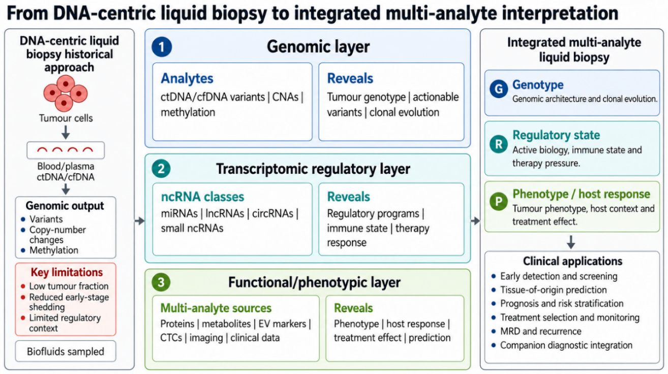
Figure 2.
Carrier-aware sources of circulating and tissue-proximal ncRNA signals. The schematic links cellular and tissue sources to major RNA-containing compartments detectable in blood/plasma, including free or protein-bound RNA, lipoprotein-associated RNA, extracellular vesicles (EVs), tumor-educated platelets, apoptotic bodies, and cellular debris. It also summarizes locally enriched biofluids, including urine, saliva, stool, and cerebrospinal fluid (CSF), and highlights that the same ncRNA may have different meaning depending on carrier type, biological source, sample matrix, and pre-analytical handling. Abbreviations: CSF, cerebrospinal fluid; EV, extracellular vesicle; ncRNA, non-coding RNA.
Figure 2.
Carrier-aware sources of circulating and tissue-proximal ncRNA signals. The schematic links cellular and tissue sources to major RNA-containing compartments detectable in blood/plasma, including free or protein-bound RNA, lipoprotein-associated RNA, extracellular vesicles (EVs), tumor-educated platelets, apoptotic bodies, and cellular debris. It also summarizes locally enriched biofluids, including urine, saliva, stool, and cerebrospinal fluid (CSF), and highlights that the same ncRNA may have different meaning depending on carrier type, biological source, sample matrix, and pre-analytical handling. Abbreviations: CSF, cerebrospinal fluid; EV, extracellular vesicle; ncRNA, non-coding RNA.
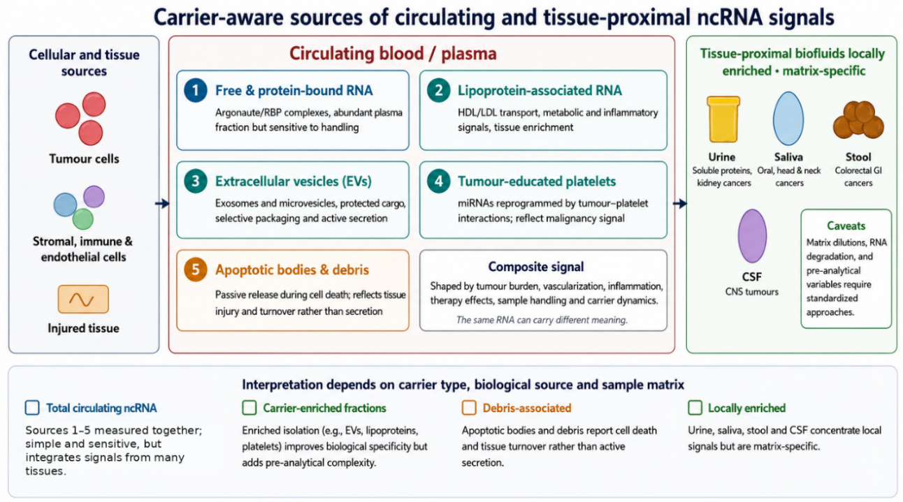
Figure 3.
Intended-use-driven roadmap from ncRNA discovery to clinical liquid biopsy. The upper row defines the principal intended-use contexts—screening, diagnostic classification, prognostic stratification, and therapy monitoring—together with their evidence requirements. The central workflow shows the translational sequence from discovery and technical optimization to analytical validation, signature locking, clinical validation, health-economic assessment, regulatory submission, post-market monitoring, and functional multi-analyte implementation. The lower bars summarize cross-cutting requirements that should be maintained at every stage: biological robustness, technical standardization, and clinical utility. Abbreviations: CDx, companion diagnostic; IVDR, in vitro diagnostic regulation; MRD, minimal residual disease; ncRNA, non-coding RNA.
Figure 3.
Intended-use-driven roadmap from ncRNA discovery to clinical liquid biopsy. The upper row defines the principal intended-use contexts—screening, diagnostic classification, prognostic stratification, and therapy monitoring—together with their evidence requirements. The central workflow shows the translational sequence from discovery and technical optimization to analytical validation, signature locking, clinical validation, health-economic assessment, regulatory submission, post-market monitoring, and functional multi-analyte implementation. The lower bars summarize cross-cutting requirements that should be maintained at every stage: biological robustness, technical standardization, and clinical utility. Abbreviations: CDx, companion diagnostic; IVDR, in vitro diagnostic regulation; MRD, minimal residual disease; ncRNA, non-coding RNA.
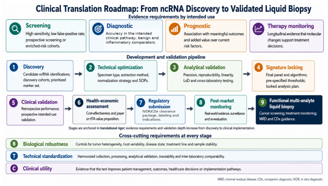

Table 1.
Non-coding RNA (ncRNA) classes and their liquid biopsy relevance. Abbreviations: AI, artificial intelligence; circRNA, circular RNA; lncRNA, long non-coding RNA; miRNA, microRNA; piRNA, PIWI-interacting RNA; snoRNA, small nucleolar RNA; tRF, tRNA-derived fragment.
Table 1.
Non-coding RNA (ncRNA) classes and their liquid biopsy relevance. Abbreviations: AI, artificial intelligence; circRNA, circular RNA; lncRNA, long non-coding RNA; miRNA, microRNA; piRNA, PIWI-interacting RNA; snoRNA, small nucleolar RNA; tRF, tRNA-derived fragment.
| RNA class | Liquid biopsy relevance |
|---|---|
| microRNAs | Stable and abundant disease-associated small RNAs; useful for early detection, tissue injury, prognosis, and therapy monitoring. Key challenges include normalization, hemolysis, extraction bias, and cohort reproducibility. |
| lncRNAs | Often tissue- or cancer-enriched regulatory RNAs; useful for tumor classification, prognosis, and resistance biology. Key challenges include low abundance, degradation, and assay sensitivity. |
| circRNAs | Structurally stable circular RNAs; promising robust biomarkers in plasma, urine, saliva, and stool. Key challenges include annotation, back-splice quantification, and platform comparability. |
| piRNAs/tRFs/snoRNAs/Y RNAs | Emerging high-dimensional small-RNA signatures; useful as cancer fingerprints and AI-assisted classifiers. Key challenges include biological interpretation and standardization. |
| Poorly annotated/orphan ncRNAs | Potentially cancer-informative non-canonical signals; useful for AI-driven early detection and subtype discovery. Key challenges include functional validation and clinical interpretability. |
Table 2.
Intended use scenarios for cancer ncRNA liquid biopsy. Abbreviations: AI, artificial intelligence; circRNA, circular RNA; ctDNA, circulating tumor DNA; EMT, epithelial-mesenchymal transition; lncRNA, long non-coding RNA; MRD, minimal residual disease; ncRNA, non-coding RNA.
Table 2.
Intended use scenarios for cancer ncRNA liquid biopsy. Abbreviations: AI, artificial intelligence; circRNA, circular RNA; ctDNA, circulating tumor DNA; EMT, epithelial-mesenchymal transition; lncRNA, long non-coding RNA; MRD, minimal residual disease; ncRNA, non-coding RNA.
| Intended use | Clinical question and validation focus |
|---|---|
| Early detection | Question: Is cancer likely to be present before symptoms or imaging findings? Signal: broad high sensitivity ncRNA signatures, possibly multi-analyte. Endpoint: sensitivity and specificity in prospective screening or enriched-risk cohorts. |
| Tumor classification | Question: What is the likely tissue of origin or molecular subtype? Signal: tissue-enriched miRNA, lncRNA, circRNA, and AI signatures. Endpoint: correct tumor-source prediction and subtype discrimination. |
| Prognosis | Question: Is the disease biologically aggressive? Signal: ncRNAs linked to EMT, angiogenesis, invasion, inflammation, or metastasis. Endpoint: progression-free survival, overall survival, or relapse risk. |
| Treatment selection | Question: Which therapy is more likely to work? Signal: EV-associated or pathway-linked ncRNA signatures. Endpoint: response rate, durable benefit, immune response, or resistance emergence. |
| Monitoring/MRD | Question: Is residual or recurrent disease biologically active? Signal: longitudinal ncRNA changes combined with ctDNA and imaging. Endpoint: lead time to recurrence, relapse-free survival, and treatment adaptation. |
Table 3.
Pre-analytical and analytical variables affecting ncRNA liquid biopsy. Abbreviations: dPCR, digital polymerase chain reaction; ncRNA, non-coding RNA; RT-qPCR, reverse-transcription quantitative polymerase chain reaction.
Table 3.
Pre-analytical and analytical variables affecting ncRNA liquid biopsy. Abbreviations: dPCR, digital polymerase chain reaction; ncRNA, non-coding RNA; RT-qPCR, reverse-transcription quantitative polymerase chain reaction.
| Variable category | Impact and mitigation |
|---|---|
| Sample collection | Tube type, anticoagulant, and time to processing can affect RNA degradation, blood-cell contamination, and carrier distribution. Mitigation: standardized collection and processing procedures. |
| Hemolysis and platelets | Red blood cell lysis and platelet contamination can falsely elevate blood-cell-derived miRNAs and other RNAs. Mitigation: hemolysis assessment and platelet-depletion protocols. |
| RNA isolation | Extraction kit, carrier enrichment, and input volume can bias recovery across RNA classes. Mitigation: spike-ins, process controls, and platform comparison. |
| Quantification | RT-qPCR, dPCR, sequencing, and hybridization assays have platform-specific sensitivity and bias. Mitigation: analytical validation and cross-platform benchmarking. |
| Data analysis | Normalization, batch effects, and model training can cause false discovery or poor generalization. Mitigation: locked pipelines, external validation, and transparent reporting. |
Disclaimer/Publisher’s Note: The statements, opinions and data contained in all publications are solely those of the individual author(s) and contributor(s) and not of MDPI and/or the editor(s). MDPI and/or the editor(s) disclaim responsibility for any injury to people or property resulting from any ideas, methods, instructions or products referred to in the content. |
© 2026 by the authors. Licensee MDPI, Basel, Switzerland. This article is an open access article distributed under the terms and conditions of the Creative Commons Attribution (CC BY) license (http://creativecommons.org/licenses/by/4.0/).
Copyright: This open access article is published under a Creative Commons CC BY 4.0 license, which permit the free download, distribution, and reuse, provided that the author and preprint are cited in any reuse.