Preprint
Article

This version is not peer-reviewed.

Exogenous Application of an RXLR-Fused CRa Protein Systemically Colonizes Roots and Suppresses Clubroot via Rhizosphere Microbiome Remodeling in Stem Mustard

A peer-reviewed article of this preprint also exists.

Submitted:

17 June 2026

Posted:

22 June 2026

You are already at the latest version

Abstract

Clubroot, caused by the obligate biotrophic protist Plasmodiophora brassicae, is a devastating soil-borne disease threatening global cruciferous crop production. To explore non-transgenic control strategies, this study investigated the efficacy of an RXLR motif-fused recombinant CRa protein against clubroot in tumorous stem mustard (Brassica juncea var. tumida) and its regulatory mechanisms in the rhizosphere. Mechanistically, fluorescence tracking confirmed that CRa protein exploits the xylem stream for systemic translocation, colonizing the stele and vascular tissues within 24 h and significantly reducing the disease index. Ecologically, CRa protein reshaped the rhizosphere microbiome by enhancing α-diversity (Shannon and Simpson indices), increasing network complexity, and specifically enriching beneficial genera such as Pseudomonas. Metabolically, CRa protein induced the reprogramming of root exudates, upregulating defensive compounds including jasmonic acid methyl ester, 4-hydroxyglucobrassicin, and (2R)-2-hydroxy-2-phenethylglucosinolate. Overall, exogenous CRa protein activates a coordinated defense response through three key mechanisms: rapid vascular translocation, recruitment of beneficial microbiota, and metabolic reprogramming. This study provides novel insights for developing non-transgenic, microbiome-based green control strategies.

Keywords: 
;  ;  ;  ;  ;  

1. Introduction

Clubroot, caused by the obligate biotrophic protist Plasmodiophora brassicae, is one of the most destructive soil-borne diseases of cruciferous crops. The pathogen causes severe root hypertrophy and hyperplasia, disrupting water and nutrient uptake and leading to devastating yield losses. The disease is especially severe in major producing regions of tumorous stem mustard (Brassica juncea var. tumida), such as Fuling, Chongqing, where reported yield reductions often exceed 60% [1], threatening the sustainability of the local industry.
Genetic resistance remains the most cost-effective and environmental approach to managing clubroot. The CRa gene, the first cloned clubroot resistance gene in crucifers, encodes a canonical TIR-NB-LRR (Toll/Interleukin-1 receptor-Nucleotide-binding - Leucine-rich repeat) immune receptor and confers high-level, race-specific resistance against certain P. brassicae pathotypes [2]. However, translating CRa into broad-field protection faces persistent challenges. Conventional introgression breeding is time-consuming and hindered by linkage drag and fertility barriers. While transgenic or genome-editing approaches deliver the functional allele directly, they remain entangled in regulatory hurdles, public acceptance issues, and varietal specificity [3]. These constraints motivate the exploration of an alternative strategy: delivering the functional output of a resistance gene as protein rather than DNA.
Advances in NLR (Nucleotide-binding Leucine-rich repeat) protein engineering-such as modifying LRR (Leucine-rich repeat) recognition surfaces or constructing chimeric scaffolds-represent the forefront of next-generation disease resistance breeding [4]. The core challenge lies in the fact that NLRs require intracellular localization to activate immunity. The introduction of the RXLR (Arg-X-Leu-Arg) motif resolves this bottleneck: functioning as a “Trojan horse,” it efficiently delivers the fused CRa protein into the cytoplasm, thereby bypassing genetic modification to directly harness the functional output of a native resistance gene [5]. This exogenous activation of intracellular immunity not only triggers cell-autonomous defense but also reprograms root exudate chemistry, thereby serving as a key signal to orchestrate the rhizosphere microbiome assembly.
The rhizosphere microbiome can suppress pathogens through antagonism or induce systemic resistance [6,7], and their assembly is strongly influenced by root exudate composition [8,9]. Plant resistance genes, particularly those encoding NLR receptors, can modulate root exudate profiles via defense signaling pathways such as salicylic acid and jasmonic acid [10,11], thereby selectively recruiting beneficial microorganisms. Similar microbe-mediated protection has been demonstrated in other systems, such as resistant banana genotypes enriching antifungal taxa to combat Fusarium wilt [11], and in rapeseed clubroot, where rhizosphere microbiome structure correlates with disease severity [12].
In the tumorous stem mustard-clubroot pathosystem, however, it remains unknown whether externally applied CRa protein can similarly influence root exudation and reshape the rhizosphere microbiome to enhance resistance. In this study, we combined high-throughput amplicon sequencing and metabolomic profiling to characterize changes in rhizosphere bacterial communities and root exudate compositions following CRa protein treatment. This integrated analysis aims to clarify the mechanistic link between recombinant CRa protein application, rhizosphere metabolic signaling, microbiome assembly, and clubroot resistance, providing a theoretical basis for novel microbiome-oriented disease management strategies.

2. Materials and Methods

2.1. Experimental Materials and Seedling Preparation

The tumorous stem mustard cultivar “Yong’an Xiaoye” was used as the experimental material. Seeds were surface-sterilized with 75% ethanol for 5 min, rinsed three times with sterile water, and sown evenly on sterile Petri dishes lined with moist filter paper. Each dish was supplemented with 40 mL of half-strength Hoagland nutrient solution and incubated at 23 °C under a 16 h light/8 h dark photoperiod for approximately 5 days to obtain healthy, aseptic seedlings with fully expanded cotyledons for subsequent experiments.

2.2. Recombinant Protein Expression and Purification

The recombinant expression plasmid containing the RXLR motif (a conserved effector domain facilitating host cell entry) [5], the enhanced green fluorescent protein (eGFP) sequence, and the coding sequence of the CRa gene was synthesized by Beijing Genomics Institute (Shenzhen, China) and constructed into the pET-28a (+) vector. The recombinant plasmid was transformed into Escherichia coli BL21 competent cells to generate the expression strain.
A single colony was inoculated into LB liquid medium containing kanamycin (100 mg/L) and cultured at 37 °C with shaking at 200 rpm for 12 h. The culture was then diluted 1:100 into fresh antibiotic-containing LB medium and grown until the OD₆₀₀ reached 0.6–0.8. A 500 μL aliquot of the bacterial culture was mixed with an equal volume of 30% sterile glycerol, flash-frozen in liquid nitrogen, and stored at -80 °C as a glycerol stock. The remaining culture was induced by adding isopropyl β-D-1-thiogalactopyranoside to a final concentration of 1 mmol/L, followed by induction at 16 °C with shaking at 200 rpm overnight (approximately 16 h). After induction, cells were harvested by centrifugation at 4 °C and 5,000 × g for 10 min. The cell pellet was re-suspended in pre-cooled PBS buffer and lysed by sonication on ice. The lysate was centrifuged at 4 °C and 12,000 × g for 20 min, and the supernatant was collected as the crude protein extract containing the recombinant CRa-eGFP fusion protein and stored on ice for immediate use.

2.3. Root Colonization Assay

The tumorous stem mustard seedlings were vertically inserted into 2 mL centrifuge tubes containing 1.0 mL of the crude protein extract, ensuring that the roots were completely submerged. Holes were made in the tube caps to fix the stems. The plants were maintained at 23 °C under a 16 h photoperiod. Six time gradients were set: 1 h, 3 h, 6 h, 9 h, 12 h, and 24 h. At each time point, seedlings were removed using sterile forceps, placed in sterile Petri dishes, and rinsed five times with sterile water (30 s per rinse). Following washing, root tips or root hair zones were sampled to prepare temporary aqueous mounts and immediately observed under a fluorescence microscope for green fluorescent signals to evaluate the adhesion and distribution of the recombinant protein in the root tissue [13].

2.4. Preparation of P. Brassicae Resting Spore Suspension

Resting spores of P. brassicae were isolated from clubbed root tissues of field-infected tumorous stem mustard. Diseased roots were washed thoroughly with tap water, soaked in 75% sodium hypochlorite solution for 10 min, and subsequently rinsed five times with sterile water to remove residual disinfectant. Surface-disinfected gall tissues were minced and homogenized with an appropriate volume of sterile water. The homogenate was filtered through eight layers of sterile cheesecloth, and the filtrate was centrifuged at 4 °C and 5,000 × g for 10 min to collect the spore pellet. The pellet was re-suspended in sterile water, and this process was repeated three times to purify the spores. Finally, the spore suspension was adjusted to a concentration of 1.0 × 10⁸ spores/mL using a hemocytometer and stored at 4 °C until use [14].

2.5. Pot Inoculation Assay

Four treatment groups were established. CRaP group: seedling roots were immersed in the crude protein extract for 24 h, transplanted into sterilized substrate for acclimation (3–5 days), and then inoculated with the P. brassicae spore suspension (1.0 × 10⁸ spores/mL). CRa group: seedling roots were immersed in the crude protein extract for 24 h and transplanted, followed by inoculation with an equal volume of sterile nutrient solution. PT group: seedlings were immersed in sterile PBS buffer instead of the protein extract for 24 h, transplanted, and inoculated with the spore suspension.CK group: seedlings were immersed in sterile PBS buffer for 24 h, transplanted, and inoculated with an equal volume of sterile nutrient solution.
Five weeks post-inoculation, whole seedlings with intact root systems were carefully harvested. Disease incidence was recorded, and disease indices were calculated following the standard grading scale [14]. Concurrently, rhizosphere soil tightly adhering to the root surface was collected for subsequent microbial DNA extraction. Thereafter, the harvested roots were thoroughly rinsed with sterile water to collect root exudates.

2.6. Rhizosphere Soil DNA Extraction, Illumina Sequencing, and Analysis

Total genomic DNA was extracted from 0.5 g of rhizosphere soil using the FastDNA™ SPIN Kit for Soil (MP Biomedicals) following the manufacturer’s protocol. DNA concentration and quality were assessed using a NanoDrop 2000 spectrophotometer and agarose gel electrophoresis. The V3–V4 region of the bacterial 16S rRNA gene was amplified using primers 338F/806R. Purified PCR products were pooled in equal amounts and sequenced on the Illumina MiSeq PE300 platform. Raw data were quality-filtered using fastp, merged using FLASH, and chimera sequences were removed using USEARCH. Unique sequences were identified in QIIME2 based on sample-specific barcodes. Operational taxonomic units (OTUs) were clustered at a 97% similarity level using the UPARSE pipeline [15]. Representative OTU sequences were taxonomically annotated using the RDP Classifier against the SILVA 138 database [16]. Alpha diversity metrics, including Shannon and Simpson indices, were calculated. Venn diagrams were constructed to visualize the overlap of bacterial taxa at the class and genus levels across different treatments. Relative abundance of bacterial groups at class and genus level were calculated. Principal Component Analysis (PCA) based on Bray-Curtis distance was performed. Linear discriminant analysis Effect Size (LEfSe) was employed to identify bacterial genera showing significant differential abundance among groups [17].

2.7. Collection of Root Exudates and Non-Targeted Metabolomics Analysis

Root exudates were collected following the method with slight modifications [18]. Intact, washed root systems were individually immersed in 100 mL of sterile Milli-Q water and allowed to exude under light at 25 ± 0.5 °C for 12 h. The collection fluid was filtered through a 0.22 μm membrane to remove impurities, flash-frozen in liquid nitrogen, and stored at -80 °C for metabolomic analysis.
Non-targeted metabolomics analysis was performed using liquid chromatography-mass spectrometry. Thawed root exudate samples were vortexed, and aliquots were lyophilized. Samples were reconstituted in 70% methanol containing internal standards, vortexed, centrifuged, and the supernatants were analyzed. Quality control samples were inserted every 10 samples during the experimental run. Metabolites were identified using the MetWare database and quantified via multiple reaction monitoring mode on a triple quadrupole mass spectrometer [19].
Venn diagrams were plotted to display shared and unique metabolites among groups. Partial least squares discriminant analysis (PLS-DA) models were constructed, and model validity was evaluated using 200 permutation tests [20]. Differential metabolites were screened based on Variable Importance in Projection (VIP) ≥ 1.5 and fold change ≥ 2 or ≤ 0.5, and visualized using volcano plots.

2.8. Correlation Analysis Between Rhizosphere Bacterial Communities and Root Exudates

To elucidate the interactions between rhizosphere bacterial communities and root exudates, Procrustes analysis was performed on PCA of differential bacterial genera and differential metabolites. Mantel tests based on Spearman correlation were used to assess the overall association between bacterial communities and differential metabolites. Furthermore, Spearman correlation analysis was conducted to explore specific interactions between bacterial genera and metabolites, identifying significant correlations (P < 0.05)[21].

3. Results

3.1. Colonization of Recombinant Protein in Tumorous Stem Mustard Roots

Fluorescence microscopy revealed dynamic temporal progression of protein colonization. One hour post-inoculation (hpi), distinct fluorescent signals were detected in the root cortex (Figure 1a). By 3 hpi, signals expanded laterally within the cortical region (Figure 1b). Initial penetration into ground tissues was observed at 6 hpi (Figure 1c), progressing to widespread distribution throughout inner tissues by 9 hpi (Figure 1d). Characteristic fluorescence appeared in the stele and vascular tissues at 12 hpi (Figure 1e), culminating in complete coverage of root tissues by 24 hpi (Figure 1f). These results demonstrate that the fusion protein systemically colonizes tumorous stem mustard roots within 24 h. Consequently, P. brassicae resting spores were inoculated 24 h after protein treatment to ensure optimal protein distribution within host tissues.

3.2. Control Efficacy Against Clubroot

The CRa protein conferred potent resistance against clubroot, reducing the disease index from 45.8 (PT) to 12.1-a 73.6% reduction in severity (Figure S1a). Visual symptoms were also markedly alleviated compared to the heavily diseased PT (Figure S1b), underscoring the suppressive activity against P. brassicae.

3.3. Alpha Diversity and Taxonomic Composition

At the class level, all treatments shared 55 common bacterial taxa. Notably, the CRa group uniquely harbored 9 exclusive taxa, surpassing other treatments (Figure S2a). At the genus level, 450 taxa were shared across groups, with CRa group again exhibiting the highest number of unique taxa (Figure S2b).
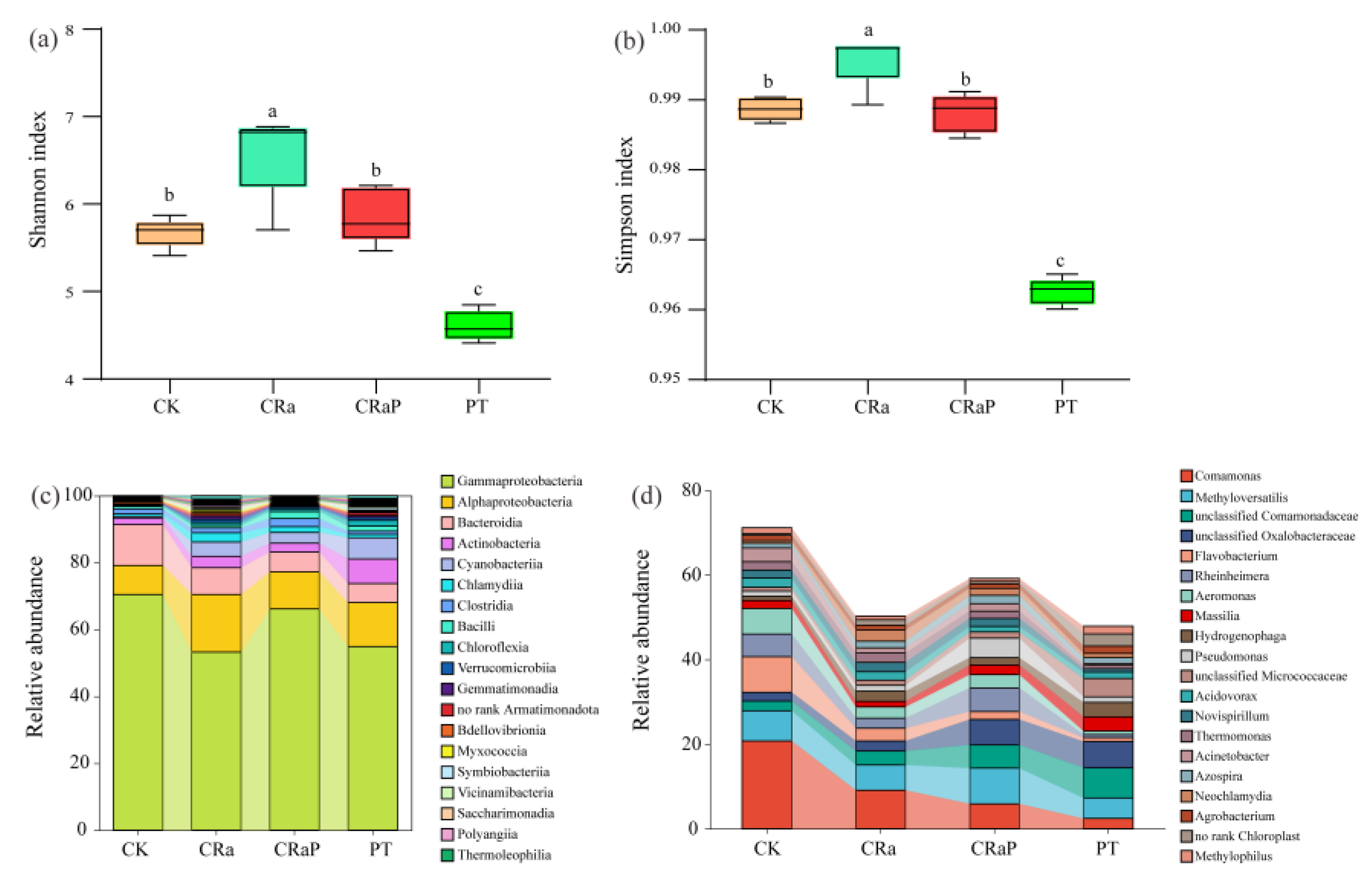
Regarding alpha diversity, the CRa group exhibited the highest Shannon and Simpson indices, followed by CK and CRaP groups, while PT group recorded the lowest values (Figure 2a and Figure 2b). Dominant classes across groups included Gammaproteobacteria, Alphaproteobacteria, Bacteroidia, and Actinobacteria, though their relative abundances varied (Figure 2c). At the genus level, the core taxa comprised Comamonas, Methyloversatilis, and unclassified Comamonadaceae, with varying relative abundances across groups (Figure 2d).

3.4. Beta Diversity and Indicator Species

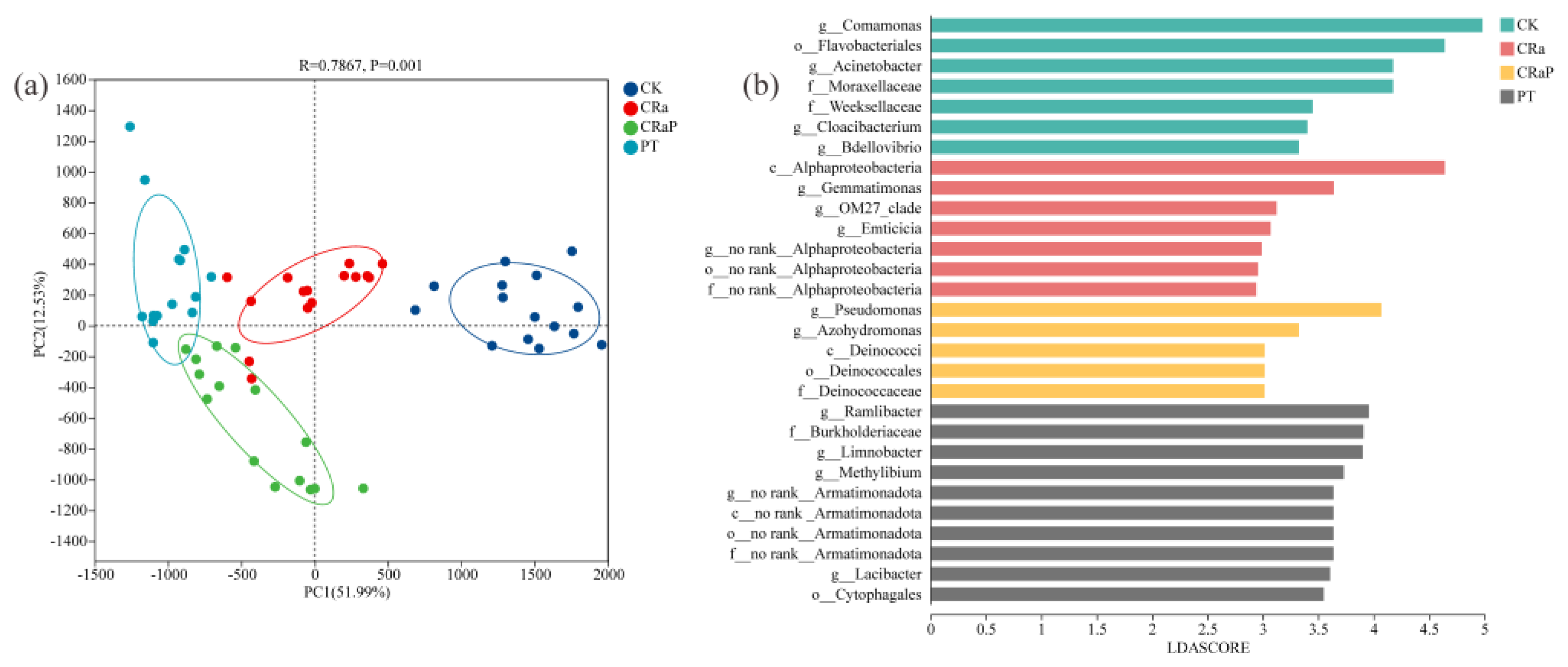
PCA revealed distinct clustering patterns (Figure 3a). CK group localized to the right quadrant, CRa group occupied an intermediate position, CRaP and PT groups to the left. PC1 explained 51.99% of the variance, indicating that treatment type was the primary driver of community structure. CK group formed a distinct cluster separate from other treatments, while CRa group displayed a transitional profile.
Indicator species analysis further delineated these differences (Figure 3b). CK group was characterized by Bdellovibrio, Cloacibacterium, Acinetobacter, and Comamonas. CRa group was marked by Emticicia. CRaP group featured Pseudomonas and Dechloromonas, whereas PT group was dominated by Lacibacter, Methylibium, Limnobacter, and Ramlibacter.

3.5. Co-Occurrence Network Analysis

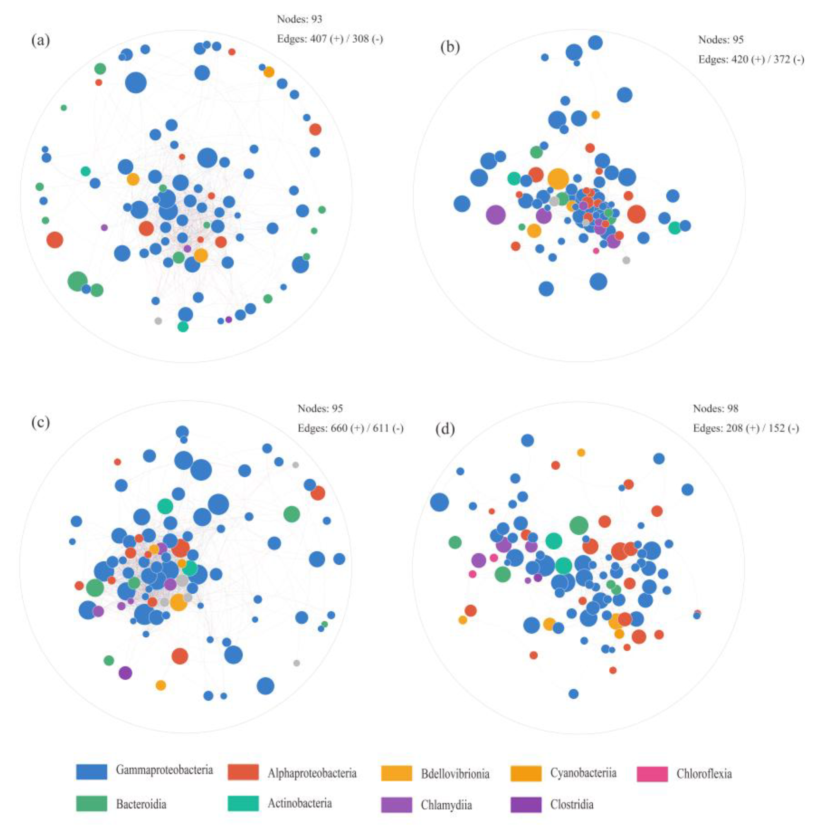
Although the number of nodes within the co-occurrence networks remained relatively consistent across treatments, the edge density varied significantly, indicating distinct patterns of microbial interaction. The CK and CRaP groups exhibited comparable connectivity, with 407 and 420 edges respectively (Figure 4a,b). Notably, the CRa group constructed the most intricate network, comprising 660 edges (Figure 4d), suggesting that the CRa protein promotes the structural complexity of the rhizosphere microbiome. This heightened complexity reflects a highly synergistic and tightly-knit microbial community. Conversely, the PT group exhibited the sparsest network, with only 208 edges (Figure 4d). This drastic reduction in connectivity signifies a state of pathogen-induced dysbiosis, characterized by a breakdown of cooperative relationships and a loss of functional redundancy, ultimately leaving the rhizosphere vulnerable to invasion.

3.6. Root Exudate Metabolomics

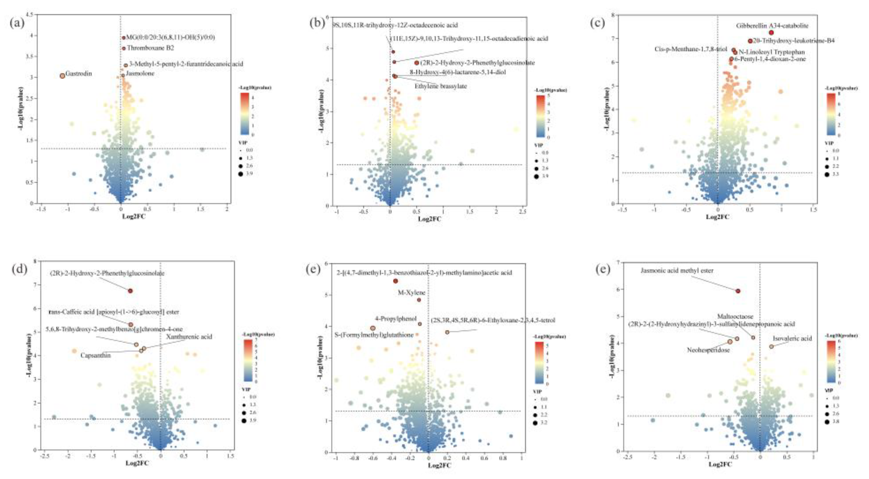
The metabolites were identified across treatments, spanning amino acids, organic acids, fatty acids, nucleotides, and lipids (Figure S3a). Among these, 87.00% (1,392 metabolites) were shared across all groups (Figure S4a). PLS-DA clearly segregated samples into four distinct clusters (Figure S4b). Differential metabolite analysis (VIP > 1.5, P < 0.05) delineated distinct metabolic reprogramming patterns across treatments. Compared to CK, the CRa group selectively elevated levels of the complex glycerolipid MG (0:0/20:3(6,8,11)-OH(5)/0:0) (Figure 5a), whereas the CRaP group triggered a specific enrichment of the defense-related phytoalexin (2R)-2-hydroxy-2-phenethylglucosinolate (Figure 5b). In contrast, plants subjected solely to PT exhibited a marked accumulation of the gibberellin A34 catabolite, indicative of pathogen-induced hormonal disruption (Figure 5c). Direct inter-group comparisons further highlighted the unique biochemical signatures induced by the recombinant protein: CRaP group uniquely accumulated (2R)-2-hydroxy-2-phenethylglucosinolate relative to CRa group (Figure 5d), while the PT group showed up-regulation of (2S,3R,4S,5R,6R)-6-ethyloxane-2,3,4,5-tetrol compared to CRa group (Figure 5e). Finally, CRaP group specifically enhanced jasmonic acid methyl ester levels relative to PT group (Figure 5f).

3.7. Integrated Microbiome-Metabolome Analysis

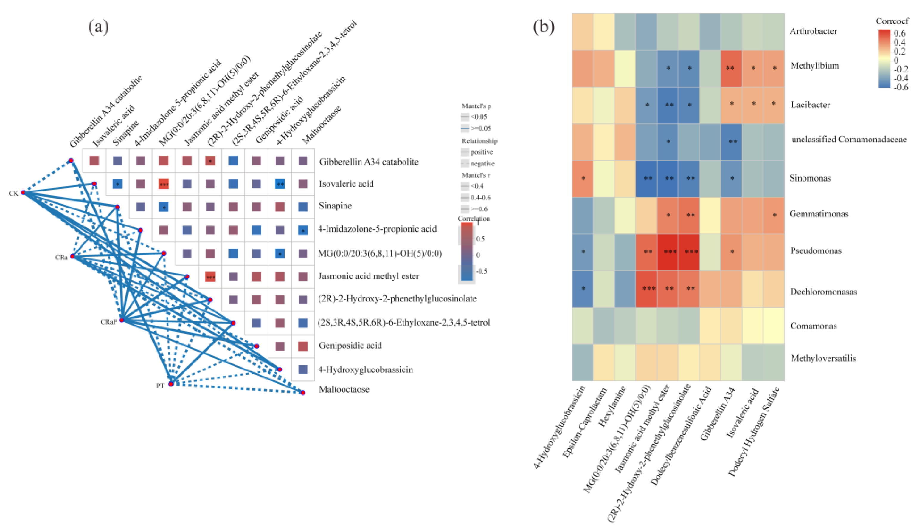
Procrustes analysis revealed significant congruence between rhizosphere microbiome composition and root exudate profiles (M2 = 0.627, P = 0.021; Figure S5), confirming strong microbe-metabolite linkages. Mantel tests further demonstrated that CRaP and CRa group exhibited positive correlations with the majority of differential metabolites, including (2R)-2-hydroxy-2-phenethylglucosinolate, jasmonic acid methyl ester, 4-hydroxyglucobrassicin, MG(0:0/20:3(6,8,11)-OH(5)/0:0), and geniposidic acid (Figure 6a). Conversely, PT showed positive associations with the gibberellin A34 catabolite and (2S, 3R, 4S, 5R, 6R)-6-ethyloxane-2,3,4,5-tetrol. Spearman’s correlation analysis at the genus level revealed distinct interaction networks (Figure 6b). Pseudomonas displayed significant positive correlations with (2R)-2-hydroxy-2-phenethylglucosinolate and jasmonic acid methyl ester, whereas Methylibium was significantly associated with gibberellin A34.

4. Discussion

This study elucidates how a recombinant protein derived from the CRa gene mediates resistance against P. brassicae. Through the integration of colonization kinetics, microbiome profiling, and metabolomics, we reveal that the CRa protein functions as a systemic modulator, capable of simultaneously reshaping the rhizosphere microbial consortium and reconfiguring root exudate profiles to establish disease suppression.

4.1. Engineered Systemic Delivery of CRa-RXLR Fusion Protein Activates Immunity

The efficacy of CRa protein is fundamentally rooted in its molecular design. By fusing the CRa coding sequence with an RXLR motif—a conserved domain facilitating host cell entry [5]—we enabled the protein to bypass traditional endocytosis and exploit the xylem stream for systemic translocation. Fluorescence microscopy confirmed that this strategy facilitated rapid, deep-tissue colonization within 24 h, reaching the stele and vascular tissues. This preemptive positioning at the invasion front proved highly effective, reducing the disease index from 45.8 to 12.1—an efficacy aligning with the established role of NB-LRR proteins in triggering potent defense responses through their NB-ARC domain-mediated signaling [22].

4.2. CRa Protein Reshapes Rhizosphere Microbiome Structure and Enhances Diversity

Beyond direct defense, CRa protein treatment profoundly restructured the rhizosphere microbiome toward a suppressive state. We observed a clear gradient of ecological maturity: the CRa group fostered the most resilient microbiome, achieving the highest alpha diversity indices (Shannon and Simpson) and harboring the greatest number of unique bacterial taxa. This enhanced diversity confers functional redundancy, increasing the probability of containing taxa capable of antagonizing P. brassicae [23].
Co-occurrence network analysis corroborated this transition, revealing that the CRa group possessed the most intricate network (660 edges), indicative of a tightly-knit, cooperative community. In contrast, the pathogen-induced dysbiosis in the PT group led to a simplified network (208 edges), reflecting a degraded niche characterized by the loss of functional redundancy and the enrichment of Methylibium and Lacibacter [24]. Notably, CRaP treatment enriched Pseudomonas as a key indicator genus, a hallmark of disease-suppressive soils capable of producing broad-spectrum antimicrobials [25,26].

4.3. Metabolic Reprogramming Drives Defensive Chemistry in Root Exudates

The structural remodeling of the microbiome was driven by CRa protein-induced metabolic reprogramming. Our integrated analysis revealed a specific upregulation of specialized defense metabolites, most prominently (2R)-2-hydroxy-2-phenethylglucosinolate, jasmonic acid methyl ester, and 4-hydroxyglucobrassicin. These compounds constitute the signature chemical arsenal of the Brassicaceae [27,28]. Their accumulation mirrors the classic defense signature of TIR-NB-LRR activation [29], providing the raw materials for both direct toxicity against plasmodia and the recruitment of beneficial microbiota [30]. Notably, the core differential metabolites identified in this study (geniposidic acid and jasmonic acid methyl ester) are also common defense markers under natural P. brassicae infection [31] and avirulent effector protein treatment [32]. This suggests that the CRa protein essentially amplifies the plant’s natural metabolic reprogramming program to recruit beneficial microbes more efficiently for disease-suppressing.

4.4. Synergistic Metabolite-Microbe Interactions Suppress Disease

The convergence of these findings supports a model wherein CRa protein establishes disease suppression through the synergistic interaction of metabolites and microbes. Spearman’s correlation Pseudomonas was positively coupled to defensive metabolites, exemplifying the “cry-for-help” hypothesis [33,34]. Here, immune activation via jasmonate signaling reshapes root exudate chemistry—specifically elevating glucosinolates and jasmonates to assemble a health-promoting microbiome [35,36]. By priming the plant’s native signaling network, CRa protein effectively transforms root exudates into a recruitment signal for Pseudomonas, thereby constructing a robust, below-ground alliance that is less susceptible to P. brassicae invasion. This teamwork between metabolites and microbes isn’t unique to the CRa protein. The avirulent effector protein 2565 uses the same trick: it recruits Paenibacillus using geniposidic acid to fight the disease [32]. This suggests that using external immune proteins is a broadly applicable new strategy. By adjusting the chemical conversations in the rhizosphere, we can boost the soil’s natural defense system.

5. Conclusions

This study demonstrates that a recombinant CRa protein engineered with an RXLR motif confers robust clubroot resistance by orchestrating a tripartite defense: systemic xylem-stream translocation, metabolic reprogramming that elevates defensive metabolites, and restructuring of the rhizosphere microbiome toward a suppressive state characterized by enriched Pseudomonas and fortified microbial networks. Future studies should elucidate the precise molecular receptors linking CRa protein perception to glucosinolate/JA upregulation. Field validation across diverse soil types is essential to translate this proof-of-concept into a scalable, microbiome-based green control strategy for sustainable agriculture.

Supplementary Materials

The following supporting information can be downloaded at the website of this paper posted on Preprints.org, Figure S1: Assessment of clubroot disease severity in stem mustard under different treatments. (a) Quantitative comparison of disease indices between the CRaP and PT groups. ** indicates a statistically significant difference (P < 0.01).(b) Representative phenotypic symptoms of clubroot infection in stem mustard roots. Figure S2: Venn diagrams illustrating the overlap of bacterial taxa. (a) Overlap of bacterial taxa at the class level across the four treatment groups. (B) Overlap of bacterial taxa at the genus level across the four treatment groups. Figure S3: Overview of root exudate metabolic profiles. (a) Classification of identified metabolites based on chemical taxonomy. (b) Classification of metabolites based on their involvement in metabolic pathways. Figure S4: Shared and unique root exudate metabolites across treatments. (a) Venn diagram showing the number of shared and unique metabolites among the four treatment groups. (b) Partial Least Squares Discriminant Analysis score plot visualizing the separation of metabolic profiles among treatment groups. Figure S5: Procrustes analysis of rhizosphere microbiome composition and root exudate profiles.

Author Contributions

Conceptualization, writing-original draft, data curation, investigation: Q.W.; Methodology, visualization, investigation, formal analysis: J.L.; Software, methodology, investigation and validation: T.C.; Resources, visualization, software, methodology: Z.C.(Zhaoming Cai1); Visualization, methodology, investigation and software: Z.C. (Zhiyi Chen); Writing-original draft, writing-review and editing, conceptualization, supervision: X.T.; Writing-original draft, funding acquisition, project administration, writing- review and editing: D.W. All authors have read and agreed to the published version of the manuscript.

Funding

This work was funded by Chongqing Municipality Natural Science Foundation Innovation and Development Joint Fund (CSTB2023NSCQ-LZX0074), Chongqing Talent Plan “Lump-sum Funding System” Project (CQYC20220302547).

Data Availability Statement

All data supporting this study are included in the manuscript and Supplementary Files. Additional inquiries should be directed to the corresponding authors. The raw 16S rRNA reads have been uploaded to the NCBI Sequence Read Archive (SRA) database with the accession number PRJNA1475733.

Acknowledgments

The authors declare the use of generative artificial intelligence (GenAI) in the preparation of this manuscript. The specific tool employed was DeepSeek-R1 (v1.0), developed by DeepSeek (official website: https://www.deepseek.com). The initial prompt provided to the AI was: “Check for spelling and grammar errors.” The final prompt provided was: “Polish the language and correct grammatical errors.”.

Conflicts of Interest

The authors declare no conflicts of interest.

Abbreviations

The following abbreviations are used in this manuscript:
eGFP Enhanced green fluorescent protein
HPI Hour post-inoculation
LEfSe Linear discr hpi iminant analysis Effect Size
NLR Nucleotide-binding Leucine-rich repeat
OTU Operational taxonomic units
PCA Principal component analysis
PLS-DA Partial least squares discriminant analysis
RXLR Arg-X-Leu-Arg motif
TIR-NB-LRR Toll/Interleukin-1 receptor-Nucleotide-binding -Leucine-rich repeat
VIP Variable importance in projection

References

  1. Cai, Z.; Xiang, M.; Tan, J.; Cheng, C.; Liu, Y.; Shi, J.; Shi, M.L.; Li, J.; Wang, D.D. High-throughput sequencing reveals tuber mustard genes responsive to Plasmodiophora brassicae in the early stage of infection. Physiol. Mol. Plant. Pathol. 2023, 123, 101943. [Google Scholar]
  2. Ueno, H.; Matsumoto, E.; Aruga, D. Molecular characterization of the cra gene conferring clubroot resistance in Brassica rapa. Plant Mol. Biol. 2012, 80, 621–629. [Google Scholar] [CrossRef] [PubMed]
  3. Torti, S.; Schlesier, R.; Thümmler, A.; Bartels, D.; Römer, P.; Koch, B.; Werner, S.; Panwar, V.; Kanyuka, K.; Wirén, N.V.; et al. Transient reprogramming of crop plants for agronomic performance. Nat. Plants 2021, 7, 159–171. [Google Scholar] [CrossRef] [PubMed]
  4. Jones, J.D.G.; Staskawicz, B.J.; Dangl, J.L. The plant immune system: from discovery to deployment. Cell. 2024, 187, 2095–2116. [Google Scholar] [CrossRef] [PubMed]
  5. Dou, D.; Kale, S.D.; Wang, X.; Jiang, R.H.; Bruce, N.A.; Arredondo, F.D.; Tyler, B.M. Rxlr-mediated entry of Phytophthora sojae effector Avr1b into soybean cells does not require pathogen-encoded machinery. Plant Cell. 2008, 20, 1930–1947. [Google Scholar] [PubMed]
  6. Wu, H.M.; Wu, H.M.; Jiao, Y.Y.; Zhang, Z.Z.; Rensing, C.; Lin, W.X. The combination of biochar and PGPBs stimulates the differentiation in rhizosphere soil microbiome and metabolites to suppress soil-borne pathogens under consecutive monoculture regimes. GCB Bioenergy 2021, 14, 84–103. [Google Scholar]
  7. Yu, Y.Y.; Gui, Y.; Li, Z.J.; Jiang, C.H.; Guo, J.H.; Niu, D.D. Induced systemic resistance for improving plant immunity by beneficial microbes. Plants 2022, 11, 386. [Google Scholar] [CrossRef] [PubMed]
  8. Berendsen, R.L.; Pieterse, C.M.J.; Bakker, P.A.H.M. The rhizosphere microbiome and plant health. Trends Plant Sci. 2012, 17, 318–328. [Google Scholar] [CrossRef] [PubMed]
  9. Manamele, D.M.; Lizelle, A.P.; Ian, A.D.; Fidele, T.; Msizi, I.M. Rhizosphere tripartite interactions and PGPR-mediated metabolic reprogramming towards isr and plant priming: a metabolomics review. Biology 2022, 11, 346. [Google Scholar] [CrossRef] [PubMed]
  10. Liu, Q.; Cheng, L.; Nian, H.; Jin, J.; Lian, T.X. Linking plant functional genes to rhizosphere microbes: a review. Plant Biotechnol. J. 2023, 21, 902–917. [Google Scholar] [PubMed]
  11. Liu, S.; Tao, C.; Zhang, L.; Wang, Z.; Xiong, W.; Dandan, S.; Peng, J. Plant pathogen resistance is mediated by recruitment of specific rhizosphere fungi. ISME Journal. 2023, 17, 931–945. [Google Scholar] [CrossRef] [PubMed]
  12. Daval, S.; Gazengel, K.; Belcour, A.; Linglin, J.; Guillerm-Erckelboudt, A.Y.; Sarniguet, A.; Manzanares-Dauleux, M.J.; Lebreton, L.; Mougel, C. Soil microbiota influences clubroot disease by modulating Plasmodiophora brassicae and Brassica napus transcriptomes. Microb. Biotechnol. 2020, 13, 1648–1672. [Google Scholar] [CrossRef] [PubMed]
  13. Hückelhoven, R. Cell wall-associated mechanisms of disease resistance and susceptibility. Annu. Rev. Phytopathol. 2007, 45, 101–127. [Google Scholar] [CrossRef] [PubMed]
  14. Zahr, K.; Sarkes, A.; Yang, Y.; Ahmed, H.; Zhou, Q.; Feindel, D.; Harding, M.W.; Feng, J. Plasmodiophora brassicae in its environment: effects of temperature and light on resting spore survival in soil. Phytopathology. 2021, 111, 1743–1750. [Google Scholar] [CrossRef] [PubMed]
  15. Edgar, R.C. Uparse: Highly accurate otu sequences from microbial amplicon reads. Nat. Methods 2013, 10, 996–998. [Google Scholar] [CrossRef] [PubMed]
  16. Quast, C.; Pruesse, E.; Yilmaz, P.; Gerken, J.; Schweer, T.; Yarza, P.; Glöckner, F.O. The silva ribosomal RNA gene database project: improved data processing and web-based tools. NAR. 2012, 41, D590–D596. [Google Scholar] [CrossRef] [PubMed]
  17. Segata, N.; Izard, J.; Waldron, L.; Gevers, D.; Miropolsky, L.; Garrett, W.S.; Huttenhower, C. Metagenomic biomarker discovery and explanation. Genome Biol. 2011, 12, R60. [Google Scholar] [CrossRef] [PubMed]
  18. Vives-Peris, V.; De Ollas, C.; Gómez-Cadenas, A.; Pérez-Clemente, R.M. Root exudates: from plant to rhizosphere and beyond. Plant Cell Rep. 2020, 39, 3–17. [Google Scholar] [PubMed]
  19. Fraga, C.G.; Clowers, B.H.; Moore, R.J.; Zink, E.M. Signature-discovery approach for sample matching of a nerve-agent precursor using liquid chromatography–mass spectrometry, XCMS, and chemometrics. Anal. Chem. 2010, 82, 4165–4173. [Google Scholar] [PubMed]
  20. Westerhuis, J.A.; Hoefsloot, H.C.; Smit, S.; Vis, D.J.; Smilde, A.K.; Van Velzen, E.J.; Van Duijnhoven, J.P. Assessment of plsda cross model validation. Metabolomics 2008, 4, 81–89. [Google Scholar] [CrossRef]
  21. Dixon, M.M.; Afkairin, A.; Manter, D.K.; Vivanco, J. Rhizosphere microbiome co-occurrence network analysis across a tomato domestication gradient. Microorganisms 2024, 12, 1756. [Google Scholar] [CrossRef] [PubMed]
  22. Stepan, F.; Philip, D.T.; Christopher, H.D.; Gerhard, B.S.; Alba De San Eustaquio, C.; Slootweg, E.J.; Lotte, B.W.; Fleur, G.; Marc, R.K.; Gary, J.S.; et al. The potato nucleotide-binding leucine-rich repeat (NLR) immune receptor Rx1 is a pathogen-dependent DNA-deforming protein. J. Biol. Chem. 2015, 290, 24945–24960. [Google Scholar]
  23. Kang, H.J.; Chai, A.L.; Lin, Z.H.; Shi, Y.X.; Xie, X.W.; Li, L.; Fan, T.F.; Xiang, S.; Xie, J.M.; Li, B.J. Deciphering differences in microbial community diversity between clubroot-diseased and healthy soils. Microorganisms 2024, 12, 251. [Google Scholar] [CrossRef] [PubMed]
  24. Kwak, M.J.; Kong, H.G.; Choi, K.; Kwon, S.-K.; Song, J.Y. Rhizosphere microbiome structure alters in response to soil contamination and host developmental stage in arabidopsis. Sci. Rep. 2018, 8, 6867. [Google Scholar]
  25. Hassani, M.A.; Durán, P.; Hacquard, S. Microbial interactions within the plant holobiont. Microbiome 2018, 6, 58. [Google Scholar] [CrossRef] [PubMed]
  26. Weller, D.M.; Raaijmakers, J.M.; Gardener, B.B.M.; Thomashow, L.S. Microbial populations responsible for specific soil suppressiveness to plant pathogens. Annu. Rev. Phytopathol. 2002, 40, 309–348. [Google Scholar] [PubMed]
  27. Burow, M.; Halkier, B.A. How does a plant orchestrate defense in the face of multiple concurrent attackers? Front. Plant Sci. 2018, 9, 235. [Google Scholar]
  28. Halkier, B.A.; Gershenzon, J. Biology and biochemistry of glucosinolates. Annu Rev. Plant Biol. 2006, 57, 303–333. [Google Scholar] [CrossRef] [PubMed]
  29. Ronaldo, J.D.D.; Daniele, P.; Gabriella, D.A.; Diogo Maciel, M.; Tiago Silva, O.; Marcus, V.M.; Heros José, M.; Marcos Antônio, M. Hypersensitive response: from NLR pathogen recognition to cell death response. Ann. Appl. Biol. 2020, 178, 268–280. [Google Scholar] [CrossRef]
  30. Agerbirk, N.; Olsen, C.E. Glucosinolate structures in evolution. Phytochemistry 2012, 77, 16–45. [Google Scholar] [CrossRef] [PubMed]
  31. Wang, D.; Liao, J.; Cai, Z.; Luo, L.; Shi, J.; Qin, X.; Xu, X.; Li, L.; Tian, X. Root-exudate-mediated modulation of the rhizosphere microbiome in Brassica juncea var. Tumida during Plasmodiophora brassicae infection. Agronomy 2025, 15, 1944. [Google Scholar]
  32. Wang, D.; Liao, J.; Shi, J.; Cai, Z.; Luo, L.; Chen, T.; Zhang, Y.; Tian, X. Candidate avirulent effector protein 2565 reduces clubroot incidence via rhizosphere microbiome restructuring and root exudate modulation. Front. Microbiol. 2025, 16, 1614252. [Google Scholar] [CrossRef] [PubMed]
  33. Rizaludin, M.S.; Stopnisek, N.; Raaijmakers, J.M.; Garbeva, P. The chemistry of stress: understanding the ‘cry for help’ of plant roots. Metabolites 2021, 11, 357. [Google Scholar] [CrossRef] [PubMed]
  34. Rolfe, S.A.; Griffiths, J.; Ton, J. Crying out for help with root exudates: adaptive mechanisms by which stressed plants assemble health-promoting soil microbiomes. Curr. Opin. Microbiol. 2019, 49, 73–82. [Google Scholar] [CrossRef] [PubMed]
  35. Pang, G.; Liu, X.; Liang, Q.; Cai, L.; Chen, W.; Zhang, Y. Root exudate metabolites drive plant–soil feedbacks on growth and defense by shaping the rhizosphere microbiome. Microbiome 2022, 10, 233. [Google Scholar]
  36. Pieterse, C.M.J.; Zamioudis, C.; Berendsen, R.L.; Weller, D.M.; Van Wees, S.C.M.; Bakker, P.A.H.M. Induced systemic resistance by beneficial microbes. Annu. Rev. Phytopathol. 2014, 52, 347–375. [Google Scholar] [CrossRef] [PubMed]
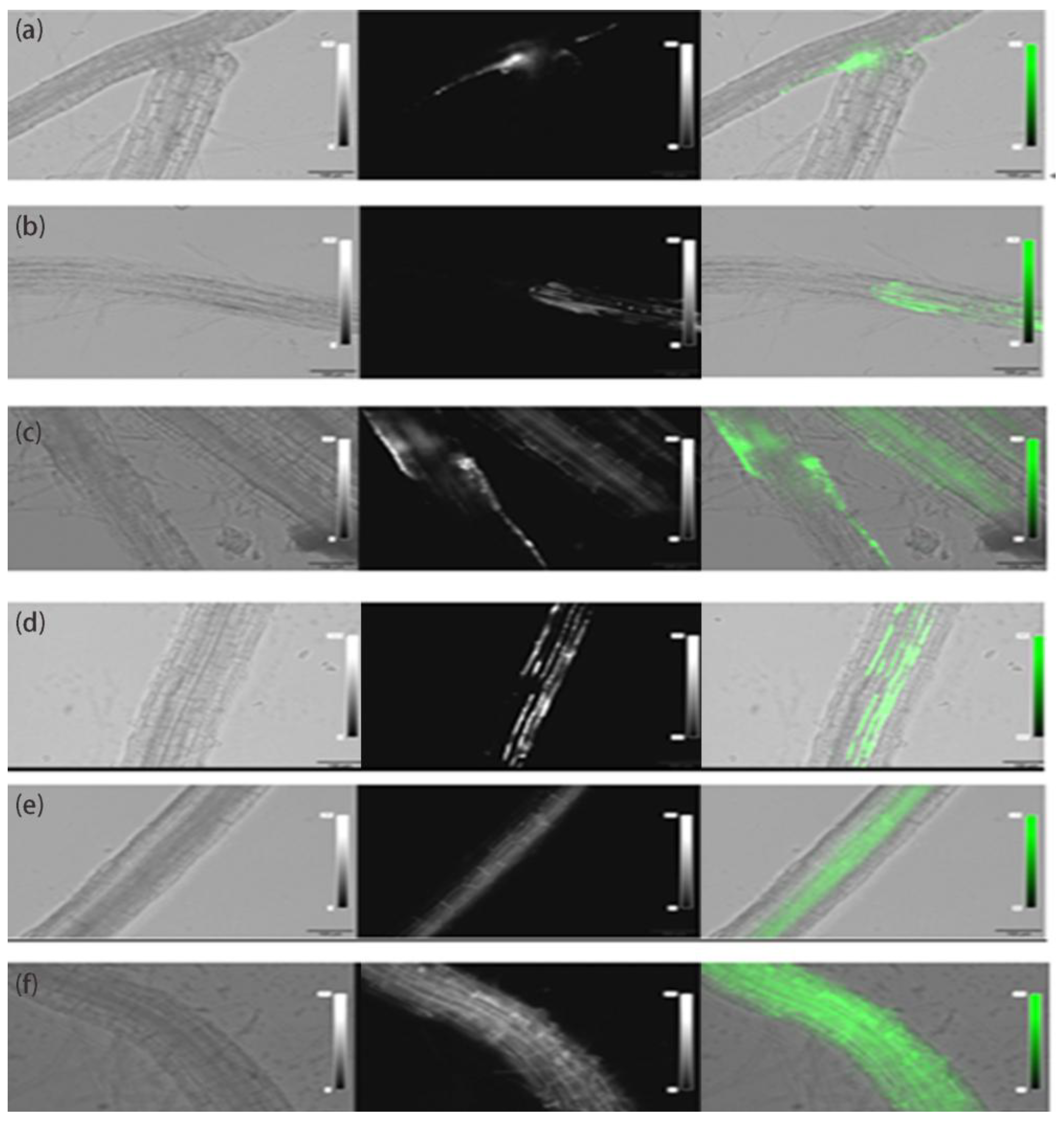
Figure 1. Spatiotemporal dynamics of recombinant CRa protein colonization in tumorous stem mustard roots. (a)1 hpi: Initial detection of fluorescent signals localized in the root cortex. (b)3 hpi: Lateral expansion of fluorescence within the cortical region. (c)6 hpi: Initial penetration of the protein into the ground tissues. (d)9 hpi: Widespread distribution of fluorescence throughout the inner root tissues. (e)12 hpi: Characteristic fluorescence observed in the stele and vascular tissues. (f)24 hpi: Complete coverage of root tissues by the fusion protein, indicating systemic colonization. From left to right, the images represent bright field transmission imaging, fluorescence signal detection, and the merged overlay of both. Scale bars: 100 μm.
Preprints 219048 g001
Figure 2. Alpha diversity and taxonomic composition of the rhizosphere microbiome under different treatments. (a)Shannon index. (b) Simpson index. (c) Relative abundance of dominant bacterial classes. (d) Relative abundance of core bacterial genera at the genus level. Different lowercase letters indicate significant differences between groups (P < 0.05).
Preprints 219048 g002
Figure 3. Beta diversity and indicator species analysis rhizosphere bacterial communities. (a) Principal Component Analysis. (b) Linear Discriminant Analysis Effect Size. Histogram identifying bacterial genera with significant differential abundance among treatments. Only taxa with an LDA score greater than 3.0 are displayed..
Preprints 219048 g003
Figure 4. Co-occurrence network analysis of rhizosphere bacterial communities. (a) CK group. (b) CRaP group. (c) CRa group. (d) PT group. Nodes represent bacterial genera, and edges represent significant correlations (P < 0.05) between genera. Node size is proportional to the degree of connectivity. While the total number of nodes was similar across all groups, edge density varied significantly, reflecting differences in network complexity and stability.
Preprints 219048 g004
Figure 5. Differential metabolite profiling and metabolic reprogramming under stress and protein treatments. (a) CK vs CRa. (b) CK vs CRaP. (c) CK vs PT. (d) CRaP vs CRa. (e) PT vs CRa. (f) CRaP vs PT. Differential metabolites were screened based on VIP ≥ 1 and fold change ≥ 2 or ≤ 0.5.
Preprints 219048 g005
Figure 6. Integrated analysis linking rhizosphere bacterial communities and root exudate metabolic profiles. (a) Mantel test correlations between bacterial community structure and differential metabolite abundance. (b) Spearman correlation hot map of differential genera and key metabolites.
Preprints 219048 g006
Disclaimer/Publisher’s Note: The statements, opinions and data contained in all publications are solely those of the individual author(s) and contributor(s) and not of MDPI and/or the editor(s). MDPI and/or the editor(s) disclaim responsibility for any injury to people or property resulting from any ideas, methods, instructions or products referred to in the content.
Copyright: This open access article is published under a Creative Commons CC BY 4.0 license, which permit the free download, distribution, and reuse, provided that the author and preprint are cited in any reuse.
Prerpints.org logo

Preprints.org is a free preprint server supported by MDPI in Basel, Switzerland.

Subscribe

© 2026 MDPI (Basel, Switzerland) unless otherwise stated

Accessibility

Disclaimer

Terms of Use

Privacy Policy

Privacy Settings