Submitted:
17 June 2026
Posted:
22 June 2026
You are already at the latest version
Abstract
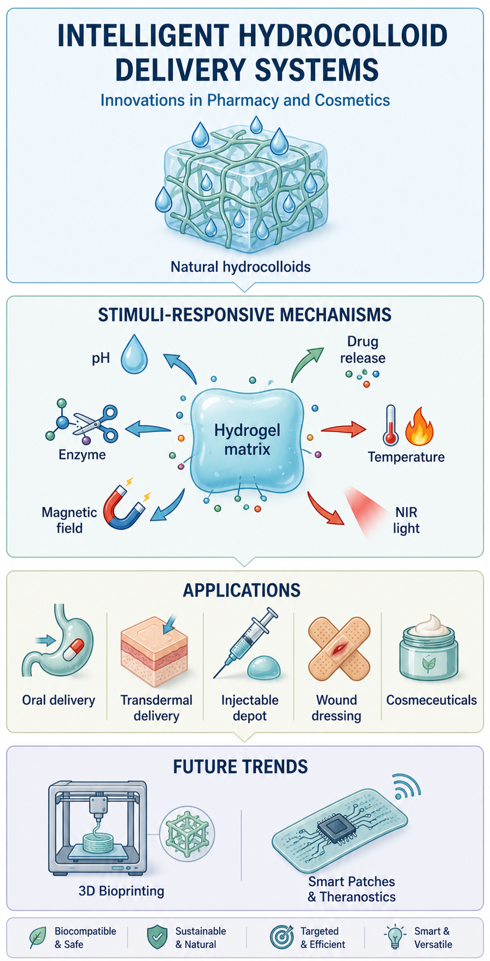
Hydrocolloids, particularly natural polysaccharides and the hydrogels derived from them, have evolved from passive rheology modifiers into functional delivery matrices for phar-maceutical and cosmetic applications. This review focuses specifically on hydrocol-loid-based systems and examines how their material chemistry and network architecture translate into controlled swelling, bioadhesion, mechanical performance, biocompatibility, and stimulus-responsive release. Key classes of hydrocolloids, including alginate, chi-tosan, hyaluronic acid, pectin, carrageenan, dextran, gellan gum, collagen, and cellulose derivatives, are discussed in relation to their physicochemical properties and formulation potential. Rather than reviewing smart polymers in general, we analyse pH-, enzyme-, re-dox-, thermo-, and externally triggered responses in the specific context of hydrocolloid chemistry and hybrid hydrocolloid architectures. Particular attention is given to oral, transdermal, injectable depot, wound-healing, and regenerative applications, as well as to cosmetic and cosmeceutical platforms for active stabilisation, controlled dermal delivery, barrier support, and personalised skincare. By linking material class, trigger mechanism, and application route, the review clarifies the distinct role of hydrocolloids in next-generation delivery systems and identifies the major translational barriers, regulatory constraints, and emerging opportunities, including multi-responsive constructs, 3D printing, and sensor-integrated patches.
Keywords:
stimuli-responsive hydrogels
; natural hydrocolloids
; controlled drug delivery
; oral delivery systems
; transdermal patches
; injectable depots
; wound dressings
; cosmeceuticals
; 3D bioprinting
; theranostic smart patches
1. Introduction
Hydrocolloids, particularly cross-linked hydrogels derived from them, are a unique and highly versatile class of flexible, semi-solid polymers with a high water content, forming three-dimensional (3D) networks of hydrophilic chains [1,2]. In terms of physical, rheological, and mechanical properties, they perfectly mimic the natural extracellular matrix (ECM) of living human tissues [3]. The history of their use in modern biomedical applications dates back to 1960, when O. Wichterle and D. Lim described the first synthetic hydrogels based on poly(2-hydroxyethyl methacrylate) (pHEMA), initially intended for the production of soft contact lenses [4]. Since that breakthrough, these materials have formed the foundation for thousands of therapeutic innovations [5]. Their ability to retain large amounts of physiological fluids without dissolving, resulting from the presence of numerous hydrophilic groups, has made them an indispensable element of modern tissue engineering and delivery systems [6].
Beyond their pharmaceutical role, hydrocolloids are also central to modern cosmetology and cosmeceutical design. Traditionally used as thickeners, stabilisers, and rheology modifiers, they now increasingly serve as functional matrices for the stabilisation of labile actives, prolonged skin residence, controlled dermal delivery, and barrier-support formulations [7]. This transition is particularly evident for polysaccharides such as xanthan, carrageenan, alginate, cellulose derivatives, chitosan, and hyaluronic acid, which com-bine favourable formulation properties with growing relevance in advanced skincare and personalised cosmetic systems [8,9,10,11]. Although the present review is centred primarily on natural hydrocolloids, particularly polysaccharides and hydrocolloid-derived hydrogels, selected semi-synthetic or synthetic polymers are discussed only where they contribute to hybrid architectures, comparative interpretation, or formulation strategies directly relevant to hydrocolloid-based delivery systems. For decades, macromolecular biopolymers—such as gelatine, carrageenan, xanthan gum, or cellulose derivatives—served purely passive and structural functions in pharmacy and cosmetology [12]. They were primarily used as inexpensive thickeners, viscosity modifiers, or stabilisers preventing the syneresis of O/W and W/O emulsions [8]. However, with the rapid development of materials engineering, polymer chemistry, nanostructured carriers, and advanced cross-linking strategies, many hydrocolloid-based systems have evolved from passive excipients into functional and, in some cases, stimuli-responsive delivery platforms [9,13,14].
A schematic timeline (Figure 1) illustrates the historical progression of hydrocolloid-based systems from early hydrophilic cross-linked hydrogels and controlled-release matrices to stimuli-responsive, reinforced, and sensor-integrated personalised platforms. In the context of the present review, this progression should be understood not only as a general increase in material complexity but also as an expansion of hydrocolloid functionality across both pharmaceutical and cosmetic applications, including active stabilisation, controlled dermal delivery, barrier-support systems, and personalised skin-contact formats [10,11].
This review is centred on hydrocolloid-based delivery systems, particularly those built from natural polysaccharides or hydrocolloid-derived hydrogels, rather than on stimuli-responsive polymers as a broad material class. Accordingly, stimulus responsiveness is discussed only insofar as it arises from hydrocolloid chemistry itself or from hybrid architectures in which hydrocolloids remain the primary functional matrix. Section 2 summarises the principal hydrocolloid classes and the material properties that govern their performance. Section 3 links these properties to defined trigger mechanisms and release principles. Section 4 maps specific hydrocolloid classes onto major pharmaceutical applications, including oral, transdermal, injectable, wound-healing, and regenerative systems. Section 5 extends the discussion to cosmetic and cosmeceutical formulations, including the stabilisation of labile actives, controlled dermal delivery, barrier-support skincare, and personalised formats such as 3D-printed masks. The final section critically discusses translational barriers, regulatory constraints, and the convergence of pharmaceutical and cosmetic technologies [15,16,17,18,19].
2. Key Hydrocolloids and Their Functional Properties
Designing highly effective delivery systems for active pharmaceutical ingredients (APIs) requires a detailed understanding of polymer network topology, and its relationship to functional properties [20]. Hydrocolloids are commonly classified according to their origin, chemical composition, ionic character, gelation behaviour and application profile [8,21,22].
2.1. Classification by Origin
2.1.1. Algal and Marine Origin
Representative polymers in this group include alginic acid and its salts isolated from the cell walls of brown algae [23,24]. Alginates, rich in blocks of guluronic and mannuronic acid, exhibit outstanding gelling properties in the presence of divalent cations and are widely used in microencapsulation, oral protection and hydrogel-based delivery systems [23,24,25,26,27]. Fucoidan, a highly sulphated marine polysaccharide, has attracted increasing interest because of its native antioxidant, anti-tumour and antithrombotic properties, as well as its potential role in localised hydrogel-based therapy [9,28,29,30]. Microalgae extracts are also an excellent source of mycosporine-like amino acids (MAAs), which form the basis for modern UV-protective cosmetic strategies [28,29].
2.1.2. Microbial, Animal, and Plant Origin
Bacterial exopolysaccharides (EPS), such as xanthan gum, gellan gum, and dextran, are particularly valuable because their biosynthetic origin enables reproducible chemistry, high hydration capacity, and pronounced rheological functionality [31,32,33,34,35]. Xanthan gum provides pseudoplastic flow and formulation stability, gellan gum forms ion-sensitive gels with ophthalmic and injectable potential, and dextran is readily modified into degradable conjugates and dynamic networks for injectable or anti-tumour systems [31,32,33,34,35,36,37,38,39].
Animal-derived hydrocolloids complement these materials by contributing native bioactivity. Chitosan, obtained by deacetylation of chitin, is the best-known cationic polysaccharide, with intrinsic mucoadhesive, haemostatic, and antimicrobial properties that are especially useful in wound care, mucosal delivery, and injectable matrices [36,40,41,42,43,44,45]. Collagen and hyaluronic acid (HA), in turn, more closely resemble the extracellular matrix of skin and connective tissues, which explains their importance in regenerative medicine, dermal repair, and soft biomaterials for cell-supportive delivery [37,46,47,48,49,50,51].
Plant-derived hydrocolloids extend this functional spectrum by offering a wide range of physicochemical properties linked to their structural diversity. Modified cellulose derivatives (e.g., CMC, HPMC, and CNC-containing systems) are widely used as matrix-forming excipients and controlled-release scaffolds because they combine swelling capacity with robust processing properties [52,53,54,55,56,57]. Pectin, rich in galacturonic acid units, forms pH-sensitive gels via ionic cross-linking, while locust bean gum (LBG), a galactomannan, exhibits resistance to enzymatic degradation in the upper gastrointestinal tract and undergoes selective fermentation in the colon [13,58,59,60]. This makes plant polysaccharides particularly attractive for site-specific gastrointestinal delivery and colon-targeted systems [13,58,59,60,61,62].
Together, microbial, animal, and plant hydrocolloids provide complementary functionalities spanning rheology control, bioadhesion, ionotropic gelation, enzymatic degradability, and site-specific delivery, while also underpinning newer injectable and targeted-delivery strategies based on polysaccharide-rich hydrogel matrices [14,22,63]. Although the present review is focused primarily on natural hydrocolloids, selected synthetic or semi-synthetic polymers are referred to later only where they provide comparative context or form part of hybrid architectures directly relevant to hydrocolloid-based delivery systems.
The classification and key functional properties of these hydrocolloids are summarised in Table 1.
For the hydrocolloids listed in Table 1, gelation, swelling and network mechanics are best treated as formulation-dependent descriptors rather than fixed material constants. Alginate, pectin, carrageenan and gellan gum provide typical examples of ionotropic or ion-strengthened networks; in these systems, the cation species, cation concentration and junction density determine swelling pressure and modulus. Chitosan and hyaluronic acid are more strongly affected by pH-dependent ionisation and cross-linking density, which influence mucoadhesion, water uptake, viscoelasticity, injectability and residence time. Dextran is often modified into dynamic imine, hydrazone, boronate or redox-cleavable networks, so degradation-controlled softening becomes part of the release mechanism. Cellulose derivatives and LBG usually act through hydration, chain entanglement and gel-layer formation, whereas collagen forms ECM-like fibrillar networks whose mechanics depend on concentration, fibrillogenesis and cross-linking [23,24,25,26,27,32,33,34,38,39,49,53,54,55,59,62,80,81,82,83,85,86,87,88,89,90].
2.2. Critical Properties: Thermodynamics of Gelation, Swelling, and Network Mechanics
Cross-linking reactions define not only the chemical structure of hydrocolloid networks but also their thermodynamic stability and mechanical performance, which directly govern drug release behaviour [22,39,63,85]. Physical cross-linking is based on reversible non-covalent interactions (e.g., hydrogen bonding, ionic interactions, hydrophobic associations), whereas chemical cross-linking introduces permanent covalent junctions that increase network integrity and mechanical strength. From a thermodynamic perspective, gelation reflects the formation of a three-dimensional network that minimises the free energy of the system through a balance of polymer–polymer and polymer–solvent interactions.
Swelling is governed by the equilibrium between the osmotic pressure driving solvent uptake and the elastic retractive forces of the polymer network, as described in hydrogel swelling and transport models [62,87,88]. The degree of swelling therefore depends on parameters such as cross-linking density, polymer-solvent affinity, charge density and the presence of ionisable groups. Recent studies on hydrogel-based delivery systems further confirm that cross-linking parameters and matrix composition strongly affect rheological behaviour, release kinetics and biological performance in topical and injectable applications [49,86]. Polyelectrolyte hydrocolloid networks, containing a high density of charged moieties, may exhibit enhanced swelling due to electrostatic repulsion and Donnan osmotic effects, which in turn can accelerate diffusion of encapsulated hydrophilic molecules [62,87,88].
From a mechanical standpoint, the elastic modulus of a hydrogel is directly related to the density and functionality of network junctions. This relationship is particularly relevant in HA-based and hybrid hydrogel systems, where tuning cross-linker architecture may simultaneously influence stiffness, swelling, injectability, and in-use stability [49,86]. Increasing cross-linking density generally enhances stiffness and reduces swelling, while loosely cross-linked networks exhibit higher deformability and permeability. Recent developments, including “click” chemistry (e.g., Diels–Alder reactions) and dynamic covalent bonds such as Schiff bases, enable the design of adaptive and self-healing hydrogels, in which reversible network rearrangements allow recovery after mechanical damage while maintaining structural integrity [91]. A schematic comparison of selected functional properties of hydrocolloid-based hydrogels and hydrophobic polymer matrices relevant to delivery-system design is presented in Figure 2. These material differences are not merely descriptive. They decide whether a hydrocolloid is better suited to a protective oral matrix, a mucoadhesive carrier, an injectable depot, a wound dressing or a dermal/cosmetic hydrogel. In most formulations, higher ionic or covalent cross-link density strengthens the network and slows release, whereas higher charge density, lower junction density or trigger-induced bond cleavage increase swelling, mesh size and permeability [49,62,85,86,87,88,89,90].
Hydrocolloid-based hydrogels score highly for hydration, biocompatibility, biodegradability, responsiveness and protein/peptide loading mainly because their backbones are hydrophilic and often ionisable. Carboxyl, hydroxyl, amino and sulphate groups support water uptake, pH- or ion-dependent swelling and a hydrated microenvironment suitable for labile biomolecules. Hydrophobic polymeric matrices show a different profile. Their lower water content can improve storage stability and mechanical robustness by limiting hydrolysis, microbial risk and premature diffusion, but it may also make hydrophilic drugs, proteins or peptides more difficult to load without additional formulation strategies. Figure 2 therefore compares two useful but different design spaces rather than ranking one material family as universally superior.
2.3. Biocompatibility and “Green” Engineering
Natural biopolymers often offer important advantages in terms of biocompatibility and sustainability, although these properties depend on source, purity, chemical modification, and the intended route of administration [92]. Many hydrocolloids used in pharmaceutical and cosmetic formulations have a favourable safety profile and, in selected cases, GRAS status or comparable regulatory familiarity [93]. Their susceptibility to enzymatic or hydrolytic degradation may reduce long-term persistence in vivo, but degradation behaviour must be assessed individually for each material and formulation [94,95]. From a sustainability perspective, current hydrogel design increasingly considers not only source renewability but also processing requirements, degradation behaviour, and the overall formulation burden associated with translational use [96].
3. Stimuli-Responsive Mechanisms and Design
The development of hydrocolloid delivery systems is closely linked to stimuli-responsive behaviour, but in the context of this review such responsiveness is considered primarily where it arises from hydrocolloid chemistry itself or from hybrid architectures in which hydrocolloids remain the principal functional matrix [12,15,22,97,98,99]. Unlike passive carriers, these systems enable controlled, spatially and temporally modulated release—through changes in ionisation, bond stability, permeability, swelling, or matrix integrity, thereby supporting more selective and application-specific delivery.
Release mechanisms vary with material architecture and conditions [87,100]. In simple systems, drug diffuses from a polymer matrix or degrades with the carrier. Advanced systems respond to stimuli—pH, temperature, enzymes—triggering structural reorganisation and selective release [101]. In situ forming hydrogels are administered as low-viscosity solutions and gel at the application site, creating a long-acting depot with near-zero-order kinetics [17,102,103]. The choice of hydrocolloid and responsiveness mechanism depends on the therapeutic goal, such as tumour-targeted release or chronic wound management.
Hydrocolloids respond to chemical, biological and physical stimuli. Effective design requires balancing material chemistry, network architecture, mass transport, and the trade-off between stability and sensitivity [104]. As summarised in Figure 3, these trigger classes do not act at the same structural level: some primarily alter charge density and swelling, others induce bond cleavage or matrix degradation, while externally applied stimuli more often change permeability, local temperature, or network relaxation. In each case, the final pharmaceutical consequence is a change in mesh size, diffusional resistance, or matrix erosion, which then determines cargo release.
The principal stimulus-responsive mechanisms relevant to hydrocolloid-based delivery systems are summarised in Figure 3.
3.1. Trigger Mechanisms in Stimuli-Responsive Hydrocolloids
Trigger mechanisms are classified by signal nature: endogenous biochemical cues, externally applied physical stimuli, and multi-responsive platforms.
3.1.1. pH-Responsive Systems
pH-responsive systems are well-established internally activated carriers [20,89,105,106,107,108,109]. Physiological and pathological environments exhibit predictable pH differences, including the gastrointestinal gradient, tumour acidity and inflamed or infected wound microenvironments. In hydrocolloid-based systems, the mechanism relies mainly on ionisable groups such as carboxyl and amino functionalities present in alginate, pectin, chitosan, hyaluronic acid derivatives or modified cellulose-based matrices. Changes in pH cause protonation or deprotonation, altering fixed charge density, osmotic pressure and polymer-polymer interactions, which in turn affects swelling, permeability or network destabilisation [20,89,106,107,108,109].
Acidic polymers swell in alkali; basic polymers swell in acid. Amphoteric systems swell at both extremes but collapse near the isoelectric point. pH-responsiveness can be implemented in crosslinked hydrogels (reversible swelling modulating diffusion) or self-assembling systems (disintegration and rapid release). Alternatively, dynamic metal-coordination bonds (e.g., Fe³⁺ with carboxyl groups) create pH-labile networks: stable at neutral pH, degrading in acid for controlled release [110]. From a release perspective, pH-dependent ionisation increases mesh size and water uptake in the swollen state, thereby lowering diffusional resistance and enhancing drug transport; conversely, partial collapse or reduced ionisation may temporarily restrict diffusion and improve gastric or local protection. Recent examples include pH-responsive chitosan hydrogels designed for drug delivery and gastrointestinal dual-polysaccharide systems based on carboxylated chitosan and carboxymethyl cellulose sodium, in which gastric-to-intestinal pH variation drives sol-gel behaviour, swelling and site-selective drug retention [89,111].
3.1.2. Enzyme-Responsive Systems
These systems are modified by enzymes in the biological microenvironment [112,113,114,115]. Pathological sites may overexpress proteases, esterases or matrix metalloproteinases (MMPs), while the colon contains microbial enzymes capable of degrading selected polysaccharides [60,112,113,116]. Enzymatic cleavage of polymer chains or crosslinks increases permeability, degrades the structure and releases the drug. Reactions include hydrolysis of bonds, destabilisation of colloidal complexes and local structural changes. Enzyme specificity can improve selectivity because release occurs preferentially at enzyme-rich sites such as infected wounds, tumour-associated microenvironments or the colon [60,112,113,114,115,116].
3.1.3. Redox- and Metabolite-Responsive Systems
These respond to internal signals such as redox state, reactive oxygen species (ROS) or metabolites [117,118,119,120,121]. Disulphide bonds (-S-S-) are stable in circulation but can be cleaved in reducing environments, while ROS-sensitive moieties (e.g., thioketal bridges) oxidise in inflammatory environments, altering network properties [117,119,120].
Metabolite-responsive systems use dynamic bonds like boronate esters, which reversibly bind diols (e.g., glucose). This creates closed-loop systems: glucose binding reorganises the network, releasing insulin only during hyperglycaemia [121,122,123]. Response can be tuned at two levels: choice of reactive chemical group, and network parameters (crosslinking density, mesh size) that determine whether the chemical change causes gradual modulation or sudden destabilisation [15]. In hydrocolloid-based formulations, these redox- or metabolite-sensitive motifs are frequently introduced into hybrid architectures rather than arising from the native polysaccharide alone, but the hydrocolloid matrix still governs hydration, retention, and the overall release profile once the trigger has been activated. A recent pectin-based example is an amino-acid-modified HG-type pectin hydrogel in which dynamic cross-linking enables dual pH/ROS-responsive release, illustrating how native polysaccharide chemistry can be combined with pathology-associated oxidative cues [90].
3.1.4. Thermo-Responsive Systems
These utilise temperature-induced phase transitions, often based on polymers with a lower critical solution temperature (LCST), such as PNIPAM-containing or poloxamer-based systems [123,124,125]. Below the LCST, chains are hydrated and soluble; above it, hydrophobic interactions dominate, causing chain collapse, viscosity increase or gelation.
Temperature can be endogenous (body heat triggers in situ gelation of an injected sol) [88,89] or externally applied (NIR or magnetic field heating). Injectable systems that flow at room temperature and gel at 37 °C enable minimally invasive depot formation. The sol–gel transition sharply reduces diffusion, enabling prolonged release [18,123]. This concept is illustrated by recent injectable thermosensitive hydrogel depot systems developed for sustained subcutaneous drug delivery, where body-temperature-triggered gelation creates a local depot and slows release [126].
Biocompatibility is critical: crosslinking density affects not only release but also cell infiltration and hypoxia. Degradation must yield non-toxic, renally clearable fragments [124]. Immunogenicity depends on composition and impurities; PEGylation reduces protein adsorption [125]. Design must synchronise gelation, mechanics, degradation, and immune interactions [5]. In the specific context of hydrocolloid-based systems, thermo-responsiveness often appears in hybrid formulations in which a polysaccharide matrix is combined with thermosensitive synthetic segments or copolymers, while the hydrocolloid component provides mucoadhesion, biocompatibility, injectability, or cargo retention [127]. For this reason, temperature-triggered systems should be interpreted here primarily as hydrocolloid-centred hybrid constructs rather than as a separate class of smart polymers.
3.1.5. Other Physically-Triggered Systems
These systems are activated by externally applied energy rather than by a local biochemical change in the microenvironment [16,17,36,128,129,130]. Magnetic hydrogels usually contain iron oxides, ferrites or related particles dispersed in alginate, chitosan, dextran, or hybrid matrices. Under static or alternating magnetic fields, these inclusions can produce magneto-mechanical deformation or magnetothermal heating, which changes mesh size, softens the gel or accelerates diffusion from the loaded depot [17,128]. Such strategies are especially attractive when clinicians need spatial control after administration, because the trigger can be switched on only over the selected anatomical site.
Light- and ultrasound-responsive systems offer a similar principle of remote control. Photoresponsive chromophores or plasmonic nanostructures convert irradiation—most often in the NIR range—into local heat or bond cleavage, resulting in network relaxation, permeability changes, or rapid release [16,129]. Ultrasound, by contrast, acts through acoustic cavitation and pressure waves that transiently disrupt the hydrogel microstructure and enhance mass transport [36]. For hydrocolloid-based systems, the central design challenge is to couple the external energy absorber to a biocompatible matrix strongly enough to ensure responsiveness, but not so strongly that baseline stability or cargo integrity is lost [130]. In practical terms, these externally triggered systems are rarely “all-hydrocolloid” constructs; rather, they are hydrocolloid-based hybrid platforms in which the matrix ensures hydration, conformability, and biological compatibility, while the embedded responsive component converts external energy into a structural or diffusional change.
3.1.6. Multi-Responsive Systems
These combine two or more stimuli for complex control [131]. Integration of e.g., pH and temperature, or magnetic field and light, enables “AND” logic gating—drug release only when multiple pathological conditions coincide, minimising off-target activation [35,132].
Advantages include synergistic responses, sequential activation (first stimulus primes the network, second triggers release), and resistance to false signals. Examples include hydrogels responding to pH + NIR [133] or magnetic field + temperature [134]. Such systems offer higher functionality and adaptability for medical applications [16,135]. For hydrocolloid-based delivery systems, the main value of multi-responsiveness lies in the possibility of combining a biocompatible, highly hydrated matrix with selective trigger combinations that better match the complexity of real disease microenvironments. However, these systems are also the most structurally complex and, in many cases, depend on hybrid rather than purely natural hydrocolloid architectures [63]. The pH/ROS-responsive pectin hydrogel example also illustrates this multi-responsive logic, because release is controlled by both environmental acidity and oxidative stress rather than by a single trigger [90].
3.2. Core Principles for Controlled and Targeted Delivery
Beyond responsiveness, effective design requires mastering principles that govern transport, stability, and targeting. Design involves compromises [136].
3.2.1. Polymer Network Architecture
Polymer network architecture controls not only whether a hydrocolloid behaves as a depot, adhesive film, injectable sol–gel precursor, or swelling matrix, but also how it responds once exposed to a trigger. Key parameters include mesh size, crosslinking density, segment mobility, charge distribution, and structural heterogeneity, all of which determine equilibrium swelling, mechanical resistance, diffusional permeability, and the extent of stimulus amplification [56,87,88]. A loosely crosslinked network may favour rapid hydration and faster release, whereas a dense or multi-network structure can slow transport and better preserve local retention.
Architecture becomes even more important in hybrid hydrocolloid systems. Nanoparticles, micelles, dynamic coordination domains, or interpenetrating networks can create secondary diffusion barriers, additional drug-binding sites, or mechanically reinforced regions. As a result, the same trigger—such as a pH shift or a temperature increase—may generate very different release profiles depending on whether the network responds by gradual deswelling, pore opening, bond cleavage, or bulk erosion. For this reason, the architecture of the carrier should be treated as a design variable equal in importance to polymer chemistry itself. Recent post-2024 literature has further emphasised that in all-polysaccharide and hybrid PEG–polysaccharide systems, crosslinking strategy and network hierarchy are not secondary formulation details but central determinants of responsiveness, self-healing, and release behaviour [63,127].
3.2.2. Transport Mechanisms and Release Kinetics
Release typically combines Fickian diffusion, matrix relaxation (Case II), erosion, and degradation. Kinetics are described by empirical models (zero-order, Higuchi, Korsmeyer-Peppas), but mechanistic models integrating diffusion with network deformation are increasingly used [62,64]. Flory–Rehner theory helps predict erosion [137]. The specific trigger determines which transport contribution becomes dominant. For example, pH-induced ionisation increases water uptake and enlarges effective mesh size, thereby facilitating diffusion; enzyme-triggered cleavage shifts the mechanism toward matrix erosion or permeability increase; and external physical stimuli often act by transiently lowering diffusional resistance through local softening, pore opening, or microstructural disruption. For this reason, Table 2 should be interpreted not as a simple list of trigger classes, but as a summary of the structural routes by which responsiveness is translated into measurable release.
3.2.3. Targeting Strategies and Functional Architectures
Targeting strategies in hydrocolloid-based systems operate at several hierarchical levels. Passive targeting exploits naturally occurring gradients such as the enhanced permeability and retention (EPR) effect, local acidosis, enzyme overexpression, oxidative stress, or mucus interactions, thereby enriching the carrier at the desired site without requiring a specific ligand [65]. Active targeting adds a second recognition layer through surface functionalisation with peptides, antibodies, sugars, folic acid, or related ligands that promote receptor-mediated adhesion, uptake, or tissue retention [138,139,140]. Beyond these two classical modes, functional architectures such as core–shell particles, multilayer films, and compartmentalised hydrogels make it possible to spatially separate protection, targeting, and release functions. This is particularly important for polysaccharide-based systems, where one domain may provide mucoadhesion or enzymatic susceptibility while another controls cargo diffusion or ligand presentation. In practice, hydrocolloids contribute to targeting not only as passive matrix materials but also through intrinsic properties such as bioadhesion, mucosal affinity, enzymatic susceptibility, and local tissue conformability, which may enhance retention even before ligand-mediated active targeting is introduced.
3.2.4. Modelling and Design Trade-Offs
Design is multi-criteria: increasing crosslinking density prolongs release but may exclude large drugs; low density accelerates transport but risks burst release [62,141]. Covalent networks offer stability; dynamic bonds enable stimulus-triggered modulation but require precise modelling. Mechanistic models (Flory–Rehner + transport equations) allow prediction of release profiles at the design stage, reducing empirical optimisation.
Conscious trade-off management between structure, hydration, transport, and degradation is the foundation of rational design. Table 2 provides an overview of the main stimuli-responsive release mechanisms from intelligent hydrogels.
A schematic summary linking material class, trigger logic, and representative application pathways is provided in Figure 4.
4. Pharmaceutical Applications
Hydrocolloids, due to their biocompatibility, high water content, and ability to undergo controlled swelling, form the foundation of modern drug delivery systems [2,3]. Initially used as passive excipients, they have evolved towards technologically advanced carriers capable of precisely modulating the release kinetics of therapeutic substances in response to specific physiological conditions [5,15]. The use of intelligent polymer networks allows for effectively overcoming biological barriers, minimising side effects, and improving the overall efficacy of therapies across various routes of administration. In the context of the present review, the most relevant examples are hydrocolloid-rich systems in which alginate, chitosan, hyaluronic acid, pectin, carrageenan, dextran, gellan gum, collagen, or cellulose derivatives determine protection, swelling, retention, or release behaviour. As schematically summarised in Figure 4, these material classes map differently onto oral matrices, transdermal formats, injectable depots, wound dressings, and regenerative constructs, rather than contributing equally across all routes. The following subsections therefore move from broad material classes to more specific formulation examples: alginate/pectin oral beads for IBD-related delivery, chitosan/cellulose gastrointestinal in situ systems, HA- or chitosan-based microneedle patches, injectable thermosensitive depots, fucoidan-based local chemotherapy hydrogels and smart wound patches that combine trigger-responsive therapy with monitoring [30,111,126,142,144,145].
4.1. Oral Delivery Using Smart Hydrocolloid Systems
The oral route of administration is the most preferred by patients, yet it poses an enormous challenge for molecules sensitive to the acidic environment of the stomach or those poorly soluble in water. Hydrocolloid systems offer effective solutions to these problems through the physicochemical modification of carriers [5,146].
4.1.1. Improving Bioavailability of Poorly Water-Soluble Drugs
Many modern active pharmaceutical ingredients (APIs) exhibit a highly hydrophobic nature, which drastically limits their absorption. The use of hydrogels with incorporated nanostructures (e.g., micelles, cellulose nanocrystals) allows for an increase in the specific surface area of the drug and an improvement in its solubility. Hydrocolloids, such as modified cellulose or hyaluronic acid, act as stabilisers, preventing the aggregation of hydrophobic molecules in the gastrointestinal tract [56,147].
4.1.2. Protection of Peptides and Proteins
Biologic drugs, including insulin and other peptide hormones, undergo rapid denaturation under the influence of low stomach pH and proteolytic enzymes (e.g., pepsin). The use of pH-responsive hydrogels, e.g., based on sodium alginate or DNA-motifs, allows for the physical shrinkage of the matrix in an acidic environment, which effectively hides and protects the sensitive protein [137,148]. Only after passing into the intestines, where the pH rises, does the network undergo deprotonation and rapid swelling, releasing the intact drug [20,42,149].
4.1.3. Controlled and Prolonged Drug Release
Hydrophilic matrices, particularly those based on cellulose derivatives (e.g., HPMC, CMC), are commonly used to create prolonged-release tablets. Upon contact with gastrointestinal fluids, the polymer swells, forming a gel barrier layer that slows down drug diffusion and the erosion of the tablet itself, enabling release kinetics approaching zero-order [57,64].
4.1.4. Site-Specific and Colon-Targeted Delivery
In the treatment of inflammatory bowel disease (IBD), delivering the drug directly to the colon is crucial. Plant and microbial polysaccharides (e.g., guar gum, dextran, locust bean gum—LBG) are utilised for this purpose, as they are completely resistant to digestion in the upper sections of the gastrointestinal tract [13,150]. Their degradation only occurs in the large intestine under the influence of specific bacterial flora producing appropriate enzymes (e.g., azoreductases, glycosidases), which releases the drug directly at the site of inflammation [60,116]. Recent overviews of oral colon-specific systems further confirm that polysaccharides such as chitosan, pectin, alginate, dextran, and guar-based carriers remain among the most attractive platforms because they combine upper-GIT protection with microflora-triggered or pH-assisted release in the colon [42].
4.1.5. Mucoadhesive Systems and Absorption Enhancement
Polymers with mucoadhesive properties, particularly cationic chitosan, can interact electrostatically with the negatively charged mucus layer (mucin) lining the intestines. This causes a significant prolongation of the residence time of the drug formulation at the absorption site [43]. Additionally, chitosan exhibits the ability to transiently open tight junctions between epithelial cells, significantly increasing the paracellular transport of macromolecules [151].
4.2. Transdermal Drug Delivery Systems
Administering drugs through the skin avoids the first-pass effect through the liver and degradation in the gastrointestinal tract, ensuring a constant therapeutic concentration in the plasma. Hydrogels constitute a flexible, biocompatible transfer platform here [152].
4.2.1. The Stratum Corneum Barrier and Its Modulation
4.2.2. Controlled and Sustained Transdermal Release
4.2.3. Microneedle-Integrated Hydrogel Systems
Microneedles have revolutionised the transdermal delivery of macromolecules (vaccines, insulin) by painlessly piercing the stratum corneum without stimulating pain receptors [155]. Dissolvable microneedles are often made from biopolymers such as hyaluronic acid, which rapidly degrade in the skin’s interstitial fluid after application, releasing a precisely loaded dose of the drug directly into the capillaries [66,156]. Recent post-2024 work continues to support hyaluronic-acid-based dissolving or hydrogel-forming microneedles as one of the most practical hydrocolloid-centred strategies for minimally invasive transdermal delivery, particularly where local dermal deposition and controlled dissolution are both required [86].
4.2.4. Stimuli-Responsive Transdermal Hydrogels
Innovative wearable hydrogel patches can release drugs in response to external or internal stimuli. For example, systems based on thermoresponsive poloxamers can increase the release of substances as body temperature rises with inflammation [157,158]. Furthermore, matrices integrated with miniature iontophoresis systems actively drive the drug deep into the tissues using a weak electrical current [159].
4.2.5. Localised and Systemic Drug Delivery
The same transdermal hydrogel platform can be tuned either for local therapy or for systemic administration, but the design priorities differ substantially. For local dermatological use, the objective is usually to maximise retention within the epidermis or superficial dermis while minimising deeper permeation; this is desirable for anti-inflammatory, antimicrobial, anti-psoriatic, or antineoplastic agents intended to act at the site of skin pathology. By contrast, systemic delivery requires steady flux across the full skin barrier and a sufficiently large drug reservoir to maintain therapeutic plasma levels over time. Hydrocolloids enable both strategies because their water content, adhesiveness, and crosslinking density can be adjusted together with the use of microneedles, permeation enhancers, or electrical assistance. Hydrocolloid choice strongly affects this balance: HA- and chitosan-rich microneedle or patch systems often favour local retention and skin compatibility, whereas more reservoir-like cellulose-based matrices are more often developed for sustained systemic flux. In practice, the distinction between local and systemic delivery is not binary but exists on a continuum defined by drug potency, target depth, and acceptable systemic exposure [86,160].
4.3. Injectable Depot Formulations
Injectable in situ forming hydrogels combine the advantages of minimally invasive administration via a syringe needle (as a liquid sol) with the capability of immediate gelation at the target site (at 37 °C), forming a stable drug depot [17,18,102].
4.3.1. Long-Acting Delivery of Small-Molecule Drugs
Depot systems are revolutionising the therapy of chronic and oncological diseases [108]. The injection of a gelling implant releases the drug slowly, in line with the progressive erosion of the matrix. This reduces the frequency of chemotherapy injections (such as paclitaxel or doxorubicin) from daily to a single dose every few weeks, thereby radically reducing systemic toxicity [17]. Recent thermosensitive injectable depots for sustained subcutaneous release illustrate the same principle: after administration, gelation converts a low-viscosity formulation into a local drug reservoir [126].
4.3.2. Injectable Depots for Peptides and Protein Therapeutics
Proteins and growth factors are highly susceptible to rapid in vivo degradation. Injectable hydrocolloid networks shield these macromolecules, maintaining their bioactivity [161]. These systems allow for the controlled release of, e.g., vascular endothelial growth factor (VEGF) directly into the ischaemic heart muscle or damaged tissues, promoting neovascularisation [162,163].
4.3.3. Localised Therapy and Site-Specific Drug Retention
Hydrogels injected directly into postoperative cavities (e.g., after brain glioblastoma resection) prevent tumour recurrence. The use of responsive DNA or chitosan hydrogels allows the carrier to immediately halt postoperative tissue haemorrhage, and then slowly destroy surviving cancer cells by releasing cytotoxins exclusively in the acidic residual microenvironment [71,164]. Fucoidan hydrogel systems proposed for hepatocellular carcinoma provide another example of local chemotherapy, with the polysaccharide contributing both to the matrix structure and to the localised therapeutic concept [30].
4.3.4. Injectable Hydrogels in Regenerative Therapies
In regenerative medicine, injectable hydrogels are valuable not merely as passive cell carriers but as instructive microenvironments. Their shear-thinning or in situ gelling behaviour protects cells during injection, while the hydrated network subsequently supports nutrient transport, cell spreading, and local retention inside irregular defects. For MSC delivery in cartilage, bone, or soft tissue repair, this means that the hydrogel can simultaneously improve post-administration cell survival, shield the cells from washout, and provide biochemical or mechanical cues closer to the native ECM than a simple suspension. In this setting, injectable alginate, hyaluronic acid, chitosan, dextran, or gellan-based depots are particularly attractive because they can be formulated as shear-thinning, ionically crosslinked, or in situ gelling systems that improve retention inside irregular defects while simultaneously serving as carriers for cells, cytokines, or mineral phases [160]. When loaded additionally with growth factors or mineral phases, the same depot can coordinate sequential cell delivery and tissue remodelling, making injectable hydrocolloid systems particularly attractive for minimally invasive regenerative therapies [163,165,166].
4.4. Hydrocolloids for Wound Care and Tissue Repair
Polymer dressings have completely replaced traditional, passive gauzes in treating difficult and chronic wounds, such as diabetic foot ulcers or burns, offering active management of the healing process [167,168].
4.4.1. Moisture Management and Microenvironment Regulation
A key tenet of wound care is maintaining optimal moisture [91]. Hydrogels exhibit a unique, dual capability: they can absorb excess toxic wound exudate (preventing the maceration of healthy edges) while simultaneously donating moisture to dry and necrotic wounds, which accelerates autolytic debridement and keratinocyte migration [85].
4.4.2. Antimicrobial and Anti-Inflammatory Hydrocolloid Systems
Chronic wounds are heavily burdened not only by resistant biofilms but also by a protease-rich inflammatory microenvironment that prevents orderly tissue repair. Advanced hydrocolloid matrices therefore aim to do more than simply kill planktonic bacte-ria. Cationic chitosan can disrupt bacterial membranes, sequester negatively charged cell-wall components, and interfere with biofilm organisation, while silver, zinc oxide, iodine, or antibiotic-loaded particles broaden the antimicrobial spectrum and provide prolonged local action [44,45,130]. Recent reviews of natural polysaccharide wound dressings likewise identify alginate, chitosan, dextran, hyaluronic acid, and composite polysaccharide systems as especially relevant because they combine haemostatic or antimicrobial effects with exudate control and maintenance of a hydrated wound environment [169]. At the same time, the hydrogel matrix can dilute inflammatory mediators, absorb excess exudate, and reduce repeated mechanical trauma during dressing changes. The most effective systems combine these functions so that antimicrobial action, moisture management, and inflammation control occur together rather than as separate treatment steps. This concept is also relevant to natural antimicrobial compositions, including essential-oil-based systems, where carrier composition and physicochemical dispersion parameters can influence biological activity [169,170].
4.4.3. Bioactive and Regenerative Hydrogel Dressings
Modern hydrogel dressings also serve as regenerative microenvironments that ac-tively instruct the healing cascade. By releasing growth factors, peptides, nucleic acids, or extracellular-matrix-derived cues, they can recruit fibroblasts, support keratinocyte migration, and stimulate angiogenesis within the wound bed [50]. Polysaccharide-based regenerative dressings are increasingly designed as composite matrices that release growth factors, peptides, nucleic acids, or cell-derived signals while simultaneously supporting angiogenesis, fibroblast activity, and macrophage polarization [169]. Equally important is immunomodulation: a well-designed hydrocolloid matrix can shift macrophage behaviour away from persistent M1-dominant inflammation toward an M2-like reparative phenotype, thereby promoting matrix deposition, vascularisation, and re-epithelialisation [169,171].
This regenerative concept is especially relevant for diabetic and other chronic wounds, in which healing fails not because of a single deficit but because inflammation, infection, poor vascularisation, and defective matrix remodelling coexist. Hydrocolloid dressings that combine structural support with bioactive release are therefore closer to temporary therapeutic tissues than to conventional passive coverings.
4.4.4. Smart and Responsive Wound Dressings
The highest level of innovation is represented by patches that respond to local pathologies [19]. These dressings detect the overexpression of specific enzymes (e.g., matrix metalloproteinases—MMPs), the alkaline pH of an infected wound, or high levels of reactive oxygen species (ROS). In response to this stimulus, the gel structure loosens, releasing an optimal dose of an antibacterial agent proportional to the severity of the infection [115]. Recent theranostic microneedle platforms take this idea further by combining NIR-triggered antibacterial action with real-time wound pH monitoring, so that diagnosis, external activation and treatment are integrated in one patch format [144].
4.5. Translational Progress and Clinical Perspectives
The transition of smart hydrocolloid systems from the laboratory phase to widespread clinical use (bench-to-bedside) involves specific technological and regulatory challenges [100]. Recent overviews also emphasise that formulation complexity, nanocomponent integration, and hybrid architectures may improve performance, but they simultaneously intensify regulatory, manufacturing, and scalability burdens [163].
Interconnected barriers limiting clinical and commercial transition of hydrocolloid-based systems are summarised in Figure 5.
The barriers in Figure 5 overlap in practice. For naturally derived hydrocolloids, batch variability is not a minor raw-material issue; differences in molecular weight, degree of deacetylation, alginate M/G ratio, sulphation pattern of marine polysaccharides, residual protein or endotoxin content and extraction history can change viscosity, gelation time, swelling ratio, degradation rate and elastic modulus. Such shifts may alter release kinetics or adhesive performance, and during scale-up they often require adjustment of polymer concentration, cross-linker level, sterilisation conditions or drying process. The same variability then affects GMP reproducibility and shelf-life specifications. For responsive or combination systems, the regulatory burden is greater because the product has to be evaluated not only as a material but also as a delivery platform whose performance depends on trigger sensitivity, biological safety, active-substance stability and clinical outcome.
4.5.1. Barriers in Clinical Translation
The fundamental problem remains production scalability (up-scaling). Due to the natural origin of hydrocolloids (algae, animal chitin, plant extracts), these polymers exhibit significant batch-to-batch variability. This instability in molecular parameters forces continuous recalibration in industrial reactors to guarantee reproducible drug release kinetics within GMP standards.
4.5.2. Regulatory Considerations and Safety Assessment
Complex, responsive hydrogel matrices fall into the legislative grey area of “combination products”. Because they serve a dual function (a physical medical device scaffold combined with the release of an active pharmaceutical ingredient), they are subject to dual and highly stringent safety assessment criteria by the FDA and EMA [101]. This results in enormous costs and deployment delays [102,172,173].
4.5.3. Commercially Available Hydrocolloid-Based Products
Currently, the clinical market is dominated by hydrocolloids serving barrier and structural functions [6]. These primarily include classic hydrocolloid burn dressings, hyaluronic acid used as a viscoelastic dermal filler in aesthetic medicine, and ophthalmic gels preventing dry eye [174]. These examples underline that commercially successful hydrocolloid products still rely mainly on barrier, viscoelastic, or structural functions rather than fully closed-loop responsiveness. “Smart” systems are still slowly paving their way towards commercialisation.
4.5.4. Emerging Trends and Future Clinical Directions
The future of pharmacy will be dominated by a personalised therapeutic approach. 3D bioprinting already allows for the creation of multi-compartmental hydrogels perfectly tailored to the anatomy of a specific patient (e.g., irregular tissue defects) [95,103]. Moreover, the integration of bioelectronics is leading to the development of fully bionic smart patches, where built-in microprocessors continuously monitor wound parameters (pH, glucose levels) and automatically dose the drug in a closed-loop theranostics feedback system [99,163].
5. Cosmetic and Cosmeceutical Applications
The conceptual convergence of hydrocolloid-based innovations in pharmaceutical and cosmetic applications is already foreshadowed in Figure 1. Here, however, the overlap is discussed in functional rather than purely chronological terms, with emphasis on how the same material platform may stabilise actives, regulate dermal residence, and support controlled topical delivery.
The current transition towards “smart hydrocolloid systems” should therefore be understood as an extension of classical formulation science rather than its replacement. The same matrix that acts as a thickener or film-former can also protect labile molecules, prolong residence on the skin, modulate dermal penetration, or respond to environmental cues such as pH, temperature, or hydration state. Consequently, hydrocolloids in cosmeceutical design are not merely passive excipients but functional carriers that shape both product performance and the local bioavailability of actives [175].
In contrast to pharmaceutical formulations, cosmetic and cosmeceutical systems are usually intended for topical action with limited or no systemic absorption. Their design therefore prioritises long-term skin compatibility, aesthetics, and formulation stability over systemic pharmacokinetics. Representative hydrocolloid classes, bioactive ingredients, dosage formats, and cosmetic functions discussed in this section are summarised in Table 3.
In this section, hydrocolloids are discussed through formulation functions rather than as passive excipients. Retinoids are used as examples of photochemical stabilisation and dermal retention; vitamin C and polyphenols as examples of oxidation-sensitive actives; curcumin and gamma-oryzanol as examples of nanocarrier-assisted hydrogel deposition; HA- and alginate/carrageenan-based masks as examples of hydration and barrier support; and 3D-printed hydrogel masks as examples of personalisation through spatially controlled composition and geometry.
5.1. Stabilisation of Labile Cosmetic Actives
The stability of cosmetic active ingredients is frequently limited by photodegradation, oxidation, hydrolysis, and poor chemical compatibility with the formulation environment. Hydrocolloid-based systems address these limitations by creating protective microenvironments that reduce exposure to reactive species, limit oxygen and light diffusion, and modulate local pH and water activity. As a result, stabilisation is inherently coupled with controlled release and improved functional availability of the active compound.
Related stabilisation challenges also concern volatile natural compositions such as essential-oil-based systems, for which the carrier medium and dispersion properties may affect both physicochemical behaviour and antimicrobial performance [170].
Retinoids provide one of the clearest examples of cosmetic actives that benefit from hydrocolloid-assisted stabilisation. Retinyl palmitate, widely used in anti-ageing formulations, undergoes rapid photochemical degradation in conventional emulsions. Encapsulation in nanoemulsion-hydrogel systems improves stability during storage and under UV exposure, while chitosan-based nanocapsules can additionally enhance dermal retention, illustrating how stabilisation and delivery can be coupled within a single carrier design [10,178,185,186].
Recent work also indicates that hydrogel architectures can stabilise more labile retinoids such as retinol. Crosslinked polymer networks, including systems incorporating hyaluronic acid or inorganic fillers, appear to limit oxygen access and molecular mobility within the matrix, thereby improving physicochemical stability. Comparable strategies have also been reported for lutein/retinol co-loaded pH-responsive hydrogel beads, where encapsulation protects the actives while preserving the possibility of controlled release [10,84,186,187].
Hydrocolloid systems are likewise relevant for oxidation-prone antioxidants. Ascorbic acid, one of the least stable cosmetic actives, shows improved preservation when immobilised in PEG-, PVA-, or hydrogel-based matrices that restrict oxygen diffusion and reduce local molecular mobility [188,189]. Chitosan-derived carriers have also been investigated as protective systems for vitamin C and related antioxidants, again linking storage stability with more controlled exposure at the skin interface [53].
Similar strategies have been reported for polyphenols and flavonoids such as rutin and curcumin. Chitosan-based films and composite polysaccharide hydrogels can protect these molecules against degradation while simultaneously moderating their release profile [179,190]. In more advanced formulations, hydrogel matrices are combined with nanocarriers, for example lipid nanoparticles or micellar systems, which is particularly advantageous for poorly soluble actives such as curcumin or gamma-oryzanol [10,183,184,186].
An important recent direction is the development of hybrid, multi-component platforms in which the hydrocolloid matrix acts not simply as a passive barrier but as an active stabilising environment. Composite hydrogels, hydrogel microspheres, and nanoemulsion-loaded gels can maintain actives in a dispersed state, reduce aggregation or crystallisation, and preserve controlled-release behaviour during storage and application [184,191].
5.2. Controlled and Enhanced Dermal Delivery
Hydrocolloid systems enable controlled and enhanced dermal delivery by modulating the release kinetics, retention, and penetration of active ingredients within the skin. Unlike conventional formulations, hydrogel matrices act as reservoirs that maintain prolonged contact with the skin surface, regulate diffusion through their network structure, and modify the microenvironment at the skin interface, including hydration and local concentration gradients.
pH-responsive and nanostructured hydrogels provide a representative example of this approach. When active compounds are first encapsulated in nanoparticles and then immobilised within a hydrogel matrix, release becomes environment-dependent and skin retention is often higher than in non-encapsulated controls. This behaviour has been demonstrated for lutein/retinol-loaded hydrogel beads and for chitosan-based systems, where bioadhesion and electrostatic interactions enhance retention within the stratum corneum and facilitate transport into deeper epidermal layers [178,187].
Hybrid hydrogel-nanocarrier systems further improve dermal delivery by coupling controlled release with enhanced penetration. Liposomal hydrogels containing adapalene, for example, increase skin deposition relative to conventional gels, with the hydrogel acting as a reservoir and the liposomes assisting transport across the lipid barrier [192]. Comparable design logic can also be applied to hydrophilic or unstable actives such as vitamin C, for which the hydrogel phase prolongs residence and attenuates burst release.
Curcumin-loaded hydrocolloid systems illustrate the close link between stabilisation and dermal delivery. Hyaluronic-acid-based micelles or nanogels improve the aqueous stability of this hydrophobic compound and enable controlled release, while in vitro, ex vivo, and in vivo studies indicate enhanced deposition in epidermal layers and improved anti-inflammatory performance compared with free curcumin [183,193]. Comparable hybrid strategies have also been explored for gamma-oryzanol and other lipophilic actives integrated into nanogel systems with photoprotective functions [184].
An important direction in this field is the development of multifunctional matrices that integrate delivery with additional protective roles, such as photoprotection, barrier support, or irritation control. In these systems, the hydrocolloid network does not merely carry the active ingredient; it also determines residence time, local distribution, and the balance between superficial retention and deeper permeation [176,184].
Hydrocolloids may additionally contribute to skin conditioning through their own physicochemical or biological properties. Hydration, film formation, bioadhesion, and extracellular-matrix-mimetic behaviour can all enhance the perceived and functional performance of the formulation independently of the encapsulated active, further blurring the distinction between carrier and bioactive component [9,176].
5.3. Bioresponsive Skincare: Moisture Regulation and Barrier Support
Hydrocolloids are widely used in skincare because their water-binding capacity and hydrophilic network structure allow them to modify the microenvironment of the stratum corneum. By absorbing and retaining water, hydrogel matrices can increase surface hydration and reduce transepidermal water loss (TEWL), while their semi-occlusive character helps preserve comfort without fully blocking gas exchange [176,194,195].
Hyaluronic-acid-based hydrogels are among the most intensively studied moisturising materials. Because HA binds water in amounts far exceeding its own mass, topical HA gels and masks can produce measurable increases in skin hydration and, in many studies, concomitant reductions in TEWL [176]. Related barrier-support functions have also been described for marine polysaccharides such as alginate and fucoidan, whose film-forming and extracellular-matrix-associated properties contribute to water retention at the skin surface [9,49].
The hydration effect of hydrogels is closely connected to the barrier they create on the skin. In vivo TEWL measurements show that well-designed hydrogel films can reduce water loss while maintaining a degree of breathability, which is important for comfort and for prolonged cosmetic use [194,195].
Polysaccharide matrices can also work synergistically with bioactive ingredients. For example, carboxymethylated chitosan combined with plant extracts has been reported to improve epidermal moisture while simultaneously supporting antioxidant and antimicrobial functions, illustrating how moisturisation and active delivery can be integrated within a single formulation [180]. Clinical and translational studies likewise suggest that hydrocolloid-containing moisturisers can improve barrier-associated parameters when combined with suitable functional additives [49,196].
A broader implication of these findings is that hydrocolloid films and masks should not be viewed only as passive humectant layers. Their internal architecture governs how water is stored, transferred, and released at the skin surface, and therefore determines whether the formulation acts as a short-lived wetting agent or as a sustained barrier-support system [176,195].
Contemporary bioresponsive skincare thus draws conceptually from medical hydrogel technologies, particularly from wound-dressing research, where adaptive swelling and controlled hydration are used to stabilise the local microenvironment. Transferred to cosmetology, the same principles support sustained moisturisation, reduced application frequency, and more functionally designed barrier-support products [18,176,195].
5.4. The Blurring Line Between Cosmetics and Pharmaceuticals
The previous subchapters show that modern hydrocolloid systems used in cosmetics increasingly rely on design principles well established in pharmaceutical sciences, including controlled release, protection of labile compounds, and responsiveness to environmental stimuli. As a result, the boundary between cosmetics and pharmaceuticals is becoming progressively blurred—not in a regulatory sense, but in terms of functionality and formulation strategy. Hydrocolloids are no longer used only as rheology modifiers or stabilisers but are increasingly engineered as active systems that modulate the local bioavailability and performance of cosmetic ingredients.
A key manifestation of this convergence is the transfer of drug-delivery strategies into cosmetic formulations. Encapsulation in nanocarriers, incorporation into responsive hydrogel matrices, and control over release kinetics are increasingly used to improve the efficacy, tolerability, and predictability of retinoids, antioxidants, vitamins, and other cosmetic actives. In this sense, hydrogel-based platforms provide a common technological framework for stabilisation, delivery, and controlled interaction with biological barriers in both pharmaceutical and cosmetic contexts [176,197].
This overlap becomes especially clear in aesthetic medicine, where injectable hyaluronic-acid-based hydrogels combine mechanical support, hydration, and bioactive signalling functions. Such products sit at the interface between dermal therapeutics, regenerative medicine, and advanced cosmetic intervention, and they are therefore better understood as translational hydrogel systems than as conventional passive cosmetics [51,177].
Skin-booster formulations illustrate this intermediate space particularly well. These lightly crosslinked or low-viscosity hyaluronic-acid systems are designed not for volumetric augmentation but for improving hydration, elasticity, and overall skin quality after intradermal administration. Their development shows how cosmetic objectives are increasingly pursued using formulation principles borrowed from injectable therapeutics, including rheological tuning, controlled tissue distribution, and prolonged local residence [51,177].
Methodological convergence is equally important. In vitro release testing, ex vivo skin permeation models, and advanced imaging of skin distribution—once mostly associated with pharmaceutical development—are now routinely used in cosmeceutical research. This shift enables a more quantitative relationship between formulation design and biological outcome, moving cosmetic science closer to an evidence-based framework [176].
Overall, hydrocolloid-based systems exemplify a broader transformation in cosmetic science, where materials originally developed for drug delivery are adapted to create multifunctional formulations with both protective and bioactive roles. This evolution underpins the growing category of cosmeceuticals, in which hydrogel matrices act not only as carriers but also as active interfaces that dynamically interact with the skin [176,197].
5.5. Personalisation and Advanced Manufacturing: The Case of 3D-Printed Masks
The evolution of hydrocolloid systems increasingly incorporates advanced manufacturing technologies, enabling a shift from standardised formulations toward highly personalised cosmetic products. Among these, extrusion-based 3D printing techniques, adapted from pharmaceutical and biomedical applications, allow the precise spatial deposition of hydrogel materials with controlled composition and architecture. This approach enables not only the fabrication of complex geometries but also the localisation of active ingredients within defined regions of the final product.
In cosmetic applications, 3D printing has already been explored for personalised hydrogel-based facial systems. Extrusion-printed patches or masks based on iota-carrageenan and sodium hyaluronate can be tailored to specific skin types or functional goals by varying humectants, botanical extracts, or other additives, demonstrating the feasibility of personalised hydrocolloid-based cosmetics [11,181].
From a materials perspective, hydrocolloids are especially suitable for such applications because their rheology can be tuned to achieve shear thinning, shape retention, and post-deposition gelation. More broadly, work on hydrogel bioinks shows that balancing viscosity, crosslinking density, and mechanical stability is essential if printability is to be combined with acceptable cosmetic performance [11,182].
An additional advantage of 3D printing is the possibility of structurally programmed delivery. By controlling internal architecture—such as pore size, layer thickness, and spatial distribution of components—it becomes possible to modulate diffusion pathways and release kinetics. This concept, originally developed in pharmaceutical manufacturing, enables the transition from homogeneous formulations to spatially resolved systems with locally optimised functionality.
Although still emerging, these technologies illustrate a broader movement towards digitalisation and personalisation in cosmetology. Hydrocolloid-based printable systems provide a platform that links formulation science with digital design, enabling next-generation cosmeceuticals in which both composition and geometry contribute to final product performance [181,182].
6. Emerging Trends and Future Challenges (Expanded)
6.1. Bioelectronic Integration (Smart Patches)
Hydrocolloid-based delivery systems are increasingly converging with nanoelectronics, giving rise to flexible hydrogel patches that combine sensing, communication, and on-demand actuation while retaining the soft, hydrated, and conformable properties of the underlying matrix [198,199].
In wound care and drug delivery, such devices can monitor local temperature, pH, glucose, uric acid, or inflammatory markers and then modulate release or trigger external intervention through a closed-loop workflow. Because hydrogel substrates are soft, hydrated, and adhesive, they provide a more compatible skin interface than rigid electronics and can be worn for prolonged periods. Future medical applications include integrated wound platforms, biohybrid interfaces, and 4D-bioprinted systems capable of real-time therapeutic adaptation [200].
Although the cosmetic use of fully closed-loop patches is less mature, the same technological logic is already relevant to personalised skincare. Printable hydrogel facial systems, hydrocolloid sheet masks, and other conformal epidermal platforms can be tailored to different skin regions and functional goals, and they therefore represent a plausible cosmetic extension of smart wearable hydrogel technology [181,182,201].
6.2. Regulatory Barriers and Scalability
The fundamental challenge for global implementation is standardising quality during scale-up and transfer to GMP-compatible manufacturing. Due to the natural origin of hydrocolloid materials--e.g., algae, animal chitin or plant extracts--there is substantial molecular variability between batches, depending on geographical factors, harvest conditions, extraction route and processing history [100,163,202]. Table 4 compares the key regulatory requirements for hydrogels under FDA and EMA frameworks.
In practice, scalability is further complicated by purification demands, sterilisation constraints, and the need to maintain reproducible rheology, swelling behaviour, and release kinetics across industrial batches, especially in multi-component or responsive systems [163].
Solutions include early regulator engagement, standardized biopolymer sourcing, impurity and molecular-weight specifications, validated sterilisation routes and process analytical technology (PAT) for real-time quality control [100,163,202]. These measures are particularly important for hydrocolloid-rich systems whose composition may vary with biological source, extraction route or post-processing history.
6.3. New Directions in Development
AI-assisted materials design is becoming increasingly relevant for hydrogels with programmable degradation, tunable cross-linking patterns and multi-stimuli responsiveness [203,204,205,206]. In tumour therapy, recent work on smart responsive hydrogels combined with AI-based design supports the use of data-assisted optimisation to select trigger combinations, anticipate release behaviour and shorten formulation screening [206]. Another important direction is the development of hydrogel platforms for skin therapeutics and personalised regenerative medicine, particularly systems that combine hydrocolloid matrices with LCST- or pH-responsive behaviour to better match local physiological conditions [197,205].
Sustainability-driven substitution of petrochemical excipients with renewable hydrocolloids is also gaining momentum in both pharmaceutical and cosmetic formulation science, although such replacement must still be balanced against processing, purification and performance requirements [96].
6.4. Final Summary
With advances in polymer chemistry and formulation design, hydrocolloids have evolved from largely passive excipients into multifunctional delivery matrices with increasingly sophisticated therapeutic and cosmetic roles.
Responsive matrices capable of reacting to tumour-associated, inflammatory, or wound-related microenvironments illustrate how material chemistry can be translated into more selective and individualised intervention strategies.
At the same time, regulatory complexity, scale-up reproducibility, long-term stability, and manufacturing cost remain decisive barriers to broader clinical and commercial translation. Addressing these constraints will determine how far next-generation hydrocolloid-based systems can move from promising laboratory platforms to robust pharmaceutical and cosmetic products.
Author Contributions
Conceptualization, K.K; investigation, K.K., O.M. and K.M.; writing—original draft preparation, K.K., O.M. and K.M.; writing—review and editing, K.K., O.M. and K.M.; visualization, K.K..; project administration, K.K. All authors have read and agreed to the published version of the manuscript.
Funding
This research was funded by a subsidy from the Ministry of Science and Higher Education for the University of Agriculture in Krakow for 2025.
Institutional Review Board Statement
Not applicable.
Informed Consent Statement
Not applicable.
Data Availability Statement
Data sharing is not applicable.
Conflicts of Interest
The authors declare no conflicts of interest.
Abbreviations
The following abbreviations are used in this manuscript:
| 3D | Three-dimensional |
| AHA | Alpha-hydroxy acid |
| API | Active pharmaceutical ingredient |
| CMC | Carboxymethylcellulose |
| CNC | Cellulose nanocrystals |
| ECM | Extracellular matrix |
| EMA | European Medicines Agency |
| EPS | Exopolysaccharides |
| EPR | Enhanced permeability and retention |
| FDA | Food and Drug Administration |
| FDM | Fused deposition modeling |
| FTIR | Fourier-transform infrared spectroscopy |
| GMP | Good Manufacturing Practice |
| GRAS | Generally Recognized As Safe |
| HA | Hyaluronic acid |
| HPMC | Hydroxypropyl methylcellulose |
| IBD | Inflammatory bowel disease |
| LCST | Lower critical solution temperature |
| LBG | Locust bean gum |
| MAAs | Mycosporine-like amino acids |
| MBBT | Methylene bis-benzotriazolyl tetramethylbutylphenol |
| MMPs | Matrix metalloproteinases |
| MRSA | Methicillin-resistant Staphylococcus aureus |
| MSCs | Mesenchymal stem cells |
| NIR | Near-infrared |
| NLCs | Nanostructured lipid carriers |
| OW | Oil-in-water |
| OTC | Over-the-counter |
| pHEMA | Poly(2-hydroxyethyl methacrylate) |
| PAT | Process analytical technology |
| PEG | Polyethylene glycol |
| PMS | Post-market surveillance |
| PNIPAM | Poly(N-isopropylacrylamide) |
| PLGA | Polylactic-co-glycolic acid |
| PVA | Polyvinyl alcohol |
| PVP | Polyvinylpyrrolidone |
| ROS | Reactive oxygen species |
| RGD | Arg-Gly-Asp |
| SEM | Scanning electron microscopy |
| SVX | Spider silk protein |
| TEWL | Transepidermal water loss |
| TiO₂ | Titanium dioxide |
| UV-Vis | Ultraviolet-visible spectroscopy |
| VEGF | Vascular endothelial growth factor |
| WO | Water-in-oil |
References
- Wichterle, O.; Lím, D. Hydrophilic Gels for Biological Use. Nature 1960, 185, 117–118. [Google Scholar] [CrossRef]
- Hoffman, A.S. Hydrogels for Biomedical Applications. Adv. Drug Deliv. Rev. 2012, 64, 18–23. [Google Scholar] [CrossRef]
- Peppas, N.A.; Hilt, J.Z.; Khademhosseini, A.; Langer, R. Hydrogels in Biology and Medicine: From Molecular Principles to Bionanotechnology. Adv. Mater. 2006, 18, 1345–1360. [Google Scholar] [CrossRef]
- Caló, E.; Khutoryanskiy, V. V. Biomedical Applications of Hydrogels: A Review of Patents and Commercial Products. Eur. Polym. J. 2015, 65, 252–267. [Google Scholar] [CrossRef]
- Li, J.; Mooney, D.J. Designing Hydrogels for Controlled Drug Delivery. Nat. Rev. Mater. 2016, 1, 16071. [Google Scholar] [CrossRef] [PubMed]
- Cascone, S.; Lamberti, G. Hydrogel-Based Commercial Products for Biomedical Applications: A Review. Int. J. Pharm. 2020, 573, 118803. [Google Scholar] [CrossRef] [PubMed]
- Jarzębski, M.; Smułek, W.; Umutoniwase, Y.; Niyobuhungiro, S.; Shirodkar, S.; Huomachi, P.O.; Perła-Kaján, J.; Szwajca, A.; Pal, K. Characterization of Hemp Seed Oil Emulsion Stabilized by Soap Nuts (Sapindus Mukorossi) Extract. Food Hydrocoll. 2024, 156, 110352. [Google Scholar] [CrossRef]
- Alam, M.; Kaur, S.; Dar, B.N.; Nanda, V. Classification, Techno-Functional Properties, and Applications of Diverse Hydrocolloids in Fruits-Based Products: A Concise Review. J. Food Sci. 2025, 90, 1–26. [Google Scholar] [CrossRef] [PubMed]
- Yuan, M.; Wang, J.; Geng, L.; Wu, N.; Yang, Y.; Zhang, Q. A Review: Structure, Bioactivity and Potential Application of Algal Polysaccharides in Skin Aging Care and Therapy. Int. J. Biol. Macromol. 2024, 272, 132846. [Google Scholar] [CrossRef] [PubMed]
- Cheong, K.L.; Chen, Q.; Aweya, J.J.; Ji, X.L.; Zhong, S.; Tan, K. Trends in Polysaccharide-Based Hydrogels for Skin Anti-Aging and Skin Antioxidant. Int. J. Biol. Macromol. 2025, 319, 145366. [Google Scholar] [CrossRef] [PubMed]
- Huang, S.M.; Sun, X.L.; Li, C.C.; Hwang, J.J. Development of an SA/XLG Composite Hydrogel Film for Customized Facial Mask Applications. Polym. 2025, 17, 2410. [Google Scholar] [CrossRef]
- Khizar, S.; Zine, N.; Errachid, A.; Elaissari, A. Introduction to Stimuli-Responsive Materials and Their Biomedical Applications. ACS Symp. Ser. 2023, 1436, 1–30. [Google Scholar] [CrossRef]
- Dhiman, P.; Rani, P.; Zahoor, I.; Singh, P. From Nature to Nurture: A Review on the Escalating Role of Locust Bean Gum in Food, Pharma and Cosmeceuticals. Int. J. Biol. Macromol. 2026, 336, 149376. [Google Scholar] [CrossRef] [PubMed]
- Liao, X.; Lu, J.; Yao, J.; Li, J.; Chen, J.; Liu, J.; Chen, Y. Polysaccharide-Based Injectable Hydrogels for Targeted Drug Delivery: Advances and Challenges. Int. J. Pharm. 2026, 690. [Google Scholar] [CrossRef] [PubMed]
- Zhang, Y.; Wu, B.M. Current Advances in Stimuli-Responsive Hydrogels as Smart Drug Delivery Carriers. Gels 2023, 9. [Google Scholar] [CrossRef] [PubMed]
- Protsak, I.S.; Morozov, Y.M. Fundamentals and Advances in Stimuli-Responsive Hydrogels and Their Applications: A Review. Gels 2025, Vol. 11(Page 30 2025, 11), 30. [Google Scholar] [CrossRef]
- Liu, J.; Du, C.; Huang, W.; Lei, Y. Injectable Smart Stimuli-Responsive Hydrogels: Pioneering Advancements in Biomedical Applications. Biomater. Sci. 2023, 12, 8–56. [Google Scholar] [CrossRef] [PubMed]
- Dimatteo, R.; Darling, N.J.; Segura, T. In Situ Forming Injectable Hydrogels for Drug Delivery and Wound Repair. Adv. Drug Deliv. Rev. 2018, 127, 167–184. [Google Scholar] [CrossRef] [PubMed]
- Das, I.J.; Bal, T. PH Factors in Chronic Wound and PH-Responsive Polysaccharide-Based Hydrogel Dressings. Int. J. Biol. Macromol. 2024, 279, 135118. [Google Scholar] [CrossRef] [PubMed]
- Vegad, U.; Patel, M.; Khunt, D.; Zupančič, O.; Chauhan, S.; Paudel, A. PH Stimuli-Responsive Hydrogels from Non-Cellulosic Biopolymers for Drug Delivery. Front. Bioeng. Biotechnol. 2023, 11, 1–12. [Google Scholar] [CrossRef] [PubMed]
- Bhattacharjee, N.; Parra-Cabrera, C.; Kim, Y.T.; Kuo, A.P.; Folch, A. Desktop-Stereolithography 3D-Printing of a Poly(Dimethylsiloxane)-Based Material with Sylgard-184 Properties. Adv. Mater. 2018, 30, 1800001. [Google Scholar] [CrossRef] [PubMed]
- Zhang, R.; Liu, X.; Zhang, W.; Cui, B.; Du, Y.; Huang, Y.; Li, W.; Liu, Q.; Ren, C.; Tang, Z. A Review of Polysaccharide-Based Hydrogels: From Structural Modification to Biomedical Applications. Int. J. Biol. Macromol. 2025, 310, 143519. [Google Scholar] [CrossRef] [PubMed]
- Hariyadi, D.M.; Islam, N. Current Status of Alginate in Drug Delivery. Adv. Pharmacol. Pharm. Sci. 2020, 2020, 8886095. [Google Scholar] [CrossRef] [PubMed]
- Shaikh, M.A.J.; Gupta, G.; Afzal, O.; Gupta, M.M.; Goyal, A.; Altamimi, A.S.A.; Alzarea, S.I.; Almalki, W.H.; Kazmi, I.; Negi, P.; et al. Sodium Alginate-Based Drug Delivery for Diabetes Management: A Review. Int. J. Biol. Macromol. 2023, 236. [Google Scholar] [CrossRef] [PubMed]
- Kuchta-Gładysz, M.; Wojciechowska-Puchałka, J.; Grzesiakowska-Dul, A.; Kmiecik, M.; Khachatryan, K.; Khachatryan, G. The In Vitro Toxicity Profile of Nanoencapsulated Curcumin in a Chitosan–Alginate Gel Carrier Using Rabbit Lymphocytes: Preliminary Research. Int. J. Mol. Sci. 2025, 26, 8577. [Google Scholar] [CrossRef]
- Khachatryan, G.; Khachatryan, K.; Szczepankowska, J.; Krzan, M.; Krystyjan, M. Design of Carbon Nanocomposites Based on Sodium Alginate/Chitosan Reinforced with Graphene Oxide and Carbon Nanotubes. Polym. 2023, Vol. 15 15, 925. [Google Scholar] [CrossRef]
- Nowak, N.; Grzebieniarz, W.; Khachatryan, G.; Khachatryan, K.; Konieczna-Molenda, A.; Krzan, M.; Grzyb, J. Synthesis of Silver and Gold Nanoparticles in Sodium Alginate Matrix Enriched with Graphene Oxide and Investigation of Properties of the Obtained Thin Films. Appl. Sci. 2021, 11, 3857. [Google Scholar] [CrossRef]
- Hernández Muñoz, A.C.; Rodríguez Martínez, I.A.; Serafini, M.R.; Aragón, D.M. Innovative Applications of Marine-Derived Algae in Cosmetics: A Patent Review (2010−2023). Algal Res. 2024, 84. [Google Scholar] [CrossRef]
- Wang, H.M.D.; Chen, C.C.; Huynh, P.; Chang, J.S. Exploring the Potential of Using Algae in Cosmetics. Bioresour. Technol. 2015, 184, 355–362. [Google Scholar] [CrossRef] [PubMed]
- Xu, H.; Liu, Z.; Wu, T.; Zheng, F.; Huang, Y.; Su, T.; Li, J.; Dai, Y. Therapeutic Potential of a JHCF4-Based Fucoidan Hydrogel for Localized Chemotherapy of Hepatocellular Carcinoma. Carbohydr. Polym. Technol. Appl. 2025, 11, 100999. [Google Scholar] [CrossRef]
- Zhang, C. feng; Yang, Z. lin; Luo, J. bo; Zhu, Q. hong; Zhao, H. nan Effects of Cinnamene Enhancers on Transdermal Delivery of Ligustrazine Hydrochloride. Eur. J. Pharm. Biopharm. 2007, 67, 413–419. [Google Scholar] [CrossRef] [PubMed]
- Petrovici, A.R.; Pinteala, M.; Simionescu, N. Dextran Formulations as Effective Delivery Systems of Therapeutic Agents. Molecules 2023, 28, 1086. [Google Scholar] [CrossRef] [PubMed]
- Destruel, P.L.; Zeng, N.; Seguin, J.; Douat, S.; Rosa, F.; Brignole-Baudouin, F.; Dufaÿ, S.; Dufaÿ-Wojcicki, A.; Maury, M.; Mignet, N.; et al. Novel in Situ Gelling Ophthalmic Drug Delivery System Based on Gellan Gum and Hydroxyethylcellulose: Innovative Rheological Characterization, in Vitro and in Vivo Evidence of a Sustained Precorneal Retention Time. Int. J. Pharm. 2020, 574. [Google Scholar] [CrossRef] [PubMed]
- Biswal, S.; Parmanik, A.; Das, D.; Sahoo, R.N.; Nayak, A.K. Gellan Gum-Based in-Situ Gel Formulations for Ocular Drug Delivery: A Practical Approach. Int. J. Biol. Macromol. 2025, 290, 138979. [Google Scholar] [CrossRef] [PubMed]
- Jadav, M.; Pooja, D.; Adams, D.J.; Kulhari, H. Advances in Xanthan Gum-Based Systems for the Delivery of Therapeutic Agents. Pharmaceutics 2023, 15, 1–27. [Google Scholar] [CrossRef] [PubMed]
- Sun, Y.; Chen, L.G.; Fan, X.M.; Pang, J.L. Ultrasound Responsive Smart Implantable Hydrogels for Targeted Delivery of Drugs: Reviewing Current Practices. Int. J. Nanomed. 2022, 17, 5001–5026. [Google Scholar] [CrossRef] [PubMed]
- Xing, Y.; Zeng, B.; Yang, W. Light Responsive Hydrogels for Controlled Drug Delivery. Front. Bioeng. Biotechnol. 2022, 10, 1–9. [Google Scholar] [CrossRef] [PubMed]
- Huang, S.; Huang, G. Preparation and Drug Delivery of Dextran-Drug Complex. Drug Deliv. 2019, 26, 252. [Google Scholar] [CrossRef] [PubMed]
- Yasmin, R.; Afzaal, M.; Khan, K.N.; Rana, A.S.; Nazar, M.F.; Abid, M.; Sumrra, S.H. Dextran-Based Stimuli-Responsive Hydrogels for Smart Dressings in Wound Healing; Springer US, 2026; Vol. 37, ISBN 1085602506999. [Google Scholar]
- Liu, H.; Wang, C.; Li, C.; Qin, Y.; Wang, Z.; Yang, F.; Li, Z.; Wang, J. A Functional Chitosan-Based Hydrogel as a Wound Dressing and Drug Delivery System in the Treatment of Wound Healing. 2018. [Google Scholar] [CrossRef] [PubMed]
- Krystyjan, M.; Khachatryan, G.; Grabacka, M.; Krzan, M.; Witczak, M.; Grzyb, J.; Woszczak, Ł.; Woszczak, L. Physicochemical, Bacteriostatic, and Biological Properties of Starch/Chitosan Polymer Composites Modified by Graphene Oxide, Designed as New Bionanomaterials. Polymers . 2021, 13, 2327. [Google Scholar] [CrossRef] [PubMed]
- Öngoren, B.; Kara, A.; Serrano, D.R.; Lalatsa, A. Novel Enabling Strategies for Oral Peptide Delivery. Int. J. Pharm. 2025, 681, 125888. [Google Scholar] [CrossRef] [PubMed]
- Bhattarai, N.; Gunn, J.; Zhang, M. Chitosan-Based Hydrogels for Controlled, Localized Drug Delivery. Adv. Drug Deliv. Rev. 2010, 62, 83–99. [Google Scholar] [CrossRef] [PubMed]
- Sun, M.; Zhang, Y.; Zhou, M. Smart-Responsive Chitosan Dressings: From Microenvironmental Sensing to Multifunctional Precision Wound Healing. Food Hydrocoll. Heal. 2025, 8, 100243. [Google Scholar] [CrossRef]
- de Maria Aguiar Carvalho, C.; da Silva, B.B.; Brianezi, S.F.S.; Sanfelice, R.C.; Balogh, D.T.; Assis, L.; Tim, C.R.; Pavinatto, A. Chitosan-Based Structures for Skin Repair: A Literature Review. Int. J. Biol. Macromol. 2025, 306. [Google Scholar] [CrossRef] [PubMed]
- Zhu, J.; Tang, X.; Jia, Y.; Ho, C.T.; Huang, Q. Applications and Delivery Mechanisms of Hyaluronic Acid Used for Topical/Transdermal Delivery—A Review. Int. J. Pharm. 2020, 578. [Google Scholar] [CrossRef] [PubMed]
- Dulińska-Litewka, J.; Dykas, K.; Felkle, D.; Karnas, K.; Khachatryan, G.; Karewicz, A. Hyaluronic Acid-Silver Nanocomposites and Their Biomedical Applications: A Review. Mater. 2022, Vol. 15 15, 234. [Google Scholar]
- PopovPereiradaCunha, M.D.; Aldana, A.A.; Abraham, G.A. Bioinspired and Biomimetic Hydrogels for Regenerative Medicine; 2025; pp. 75–118. [Google Scholar] [CrossRef]
- Murugesan, M.; Mathiyalagan, R.; Ramadhania, Z.M.; Nahar, J.; Luu, C.H.; Phan, V.H.G.; Yang, D.C.; Zhou, Q.; Chan Kang, S.; Thambi, T. Tailoring Hyaluronic Acid Hydrogels: Impact of Cross-Linker Length and Density on Skin Rejuvenation as Injectable Dermal Fillers and Their Potential Effects on the MAPK Signaling Pathway Suppression. Bioact. Mater. 2025, 49, 154–171. [Google Scholar] [CrossRef] [PubMed]
- Jin, H.; Huang, K.; Zhou, R.; Chen, P.; Zhang, M.; Jin, Y. Recent Progress of Collagen-Based Hydrogels for Wound Healing: A Review. Collagen Leather 2026, 8, 1–31. [Google Scholar] [CrossRef]
- Song, L.; Qiu, H.; Chen, Z.; Wang, J.; Xu, Y.; Liu, Z.; Liu, S.; Wang, Z.; Zhu, X.; Zhang, K.; et al. Injectable Hyaluronate/Collagen Hydrogel with Enhanced Safety and Efficacy for Facial Rejuvenation. Collagen Leather 2024 61 2024, 6, 21. [Google Scholar] [CrossRef]
- Dhand, A.P.; Kirkpatrick, B.E.; Garay-Sarmiento, M.; Nelson, B.R.; Miksch, C.E.; Meurer-Zeman, B.; Zlotnick, H.M.; Mandal, A.; Lee, J.S.; Cione, J.; et al. Digital Light Processing of Photoresponsive and Programmable Hydrogels. Sci. Adv. 2025, 11, 1–16. [Google Scholar] [CrossRef] [PubMed]
- Baek, J.; Ramasamy, M.; Willis, N.C.; Kim, D.S.; Anderson, W.A.; Tam, K.C. Encapsulation and Controlled Release of Vitamin C in Modified Cellulose Nanocrystal/Chitosan Nanocapsules. Curr. Res. Food Sci. 2021, 4, 215–223. [Google Scholar] [CrossRef] [PubMed]
- Majumdar, S. Cellulose-Based Hydrogels for Drug Delivery Applications. Cellul. Based Hydrogels Prod. Prop. Appl. 2025, 259–279. [Google Scholar] [CrossRef]
- Nicu, R.; Ciolacu, D.E. Cellulose-Based Hydrogels for Controlled Drug Delivery; 2025; pp. 91–129. [Google Scholar] [CrossRef]
- Sampath Udeni Gunathilake, T.M.; Ching, Y.C.; Chuah, C.H.; Rahman, N.A.; Liou, N.S. Recent Advances in Celluloses and Their Hybrids for Stimuli-Responsive Drug Delivery. Int. J. Biol. Macromol. 2020, 158, 670–688. [Google Scholar] [CrossRef] [PubMed]
- Maderuelo, C.; Zarzuelo, A.; Lanao, J.M. Critical Factors in the Release of Drugs from Sustained Release Hydrophilic Matrices. J. Control. Release 2011, 154, 2–19. [Google Scholar] [CrossRef] [PubMed]
- Dionísio, M.; Grenha, A. Locust Bean Gum: Exploring Its Potential for Biopharmaceutical Applications. J. Pharm. Bioallied Sci. 2012, 4, 175. [Google Scholar] [CrossRef] [PubMed]
- Das, S. Pectin Based Multi-Particulate Carriers for Colon-Specific Delivery of Therapeutic Agents. Int. J. Pharm. 2021, 605. [Google Scholar] [CrossRef] [PubMed]
- De Anda-Flores, Y.; Carvajal-Millan, E.; Campa-Mada, A.; Lizardi-Mendoza, J.; Rascon-Chu, A.; Tanori-Cordova, J.; Luisa Martínez-López, A.; Ribeiro, B.; B Pinto, R.J.; Stana Kleinschek, K.; et al. Polysaccharide-Based Nanoparticles for Colon-Targeted Drug Delivery Systems. Polysaccharides 2021, 2, 647. [Google Scholar] [CrossRef]
- Solanki, R.; Bhatia, D. Stimulus-Responsive Hydrogels for Targeted Cancer Therapy. Gels 2024, Vol. 10 10, 440. [Google Scholar] [CrossRef]
- Caccavo, D. An Overview on the Mathematical Modeling of Hydrogels’ Behavior for Drug Delivery Systems. Int. J. Pharm. 2019, 560, 175–190. [Google Scholar] [CrossRef] [PubMed]
- Friday, C.; Edgar, K. Recent Developments in All-Polysaccharide Hydrogels: Synthesis, Properties, and Biomedical Applications. Carbohydr. Polym. 2026, 376. [Google Scholar] [CrossRef] [PubMed]
- Siepmann, J.; Siepmann, F. Mathematical Modeling of Drug Delivery. Int. J. Pharm. 2008, 364, 328–343. [Google Scholar] [CrossRef] [PubMed]
- Dilliard, S.A.; Siegwart, D.J. Passive, Active and Endogenous Organ-Targeted Lipid and Polymer Nanoparticles for Delivery of Genetic Drugs. Nat. Rev. Mater. 2023 84 2023, 8, 282–300. [Google Scholar] [CrossRef] [PubMed]
- He, J.; Zhang, Y.; Yu, X.; Xu, C. Wearable Patches for Transdermal Drug Delivery. Acta Pharm. Sin. B 2023, 13, 2298–2309. [Google Scholar] [CrossRef] [PubMed]
- Grzesiakowska, A.; Dzióbek, M.; Kuchta-Gładysz, M.; Wojciechowska-Puchałka, J.; Khachatryan, K.; Khachatryan, G.; Krystyjan, M. The In Vitro Toxicity Profile of ZnS and CdS Quantum Dots in Polysaccharide Carriers (Starch/Chitosan). Int. J. Mol. Sci. 2024, Vol. 25 25, 361. [Google Scholar] [CrossRef]
- Woszczak, L.; Khachatryan, K.; Krystyjan, M.; Witczak, T.; Witczak, M.; Gałkowska, D.; Makarewicz, M.; Khachatryan, G. Physicochemical and Functional Properties and Storage Stability of Chitosan–Starch Films Containing Micellar Nano/Microstructures with Turmeric and Hibiscus Extracts. Int. J. Mol. Sci. 2023, Vol. 24 24, 12218. [Google Scholar] [CrossRef]
- Nowak, N.; Grzebieniarz, W.; Khachatryan, G.; Konieczna-Molenda, A.; Krzan, M.; Khachatryan, K. Preparation of Nano/Microcapsules of Ozonated Olive Oil in Chitosan Matrix and Analysis of Physicochemical and Microbiological Properties of the Obtained Films. Innov. Food Sci. Emerg. Technol. 2022, 82, 103181. [Google Scholar] [CrossRef]
- Krystyjan, M.; Khachatryan, G.; Khachatryan, K.; Konieczna-Molenda, A.; Grzesiakowska, A.; Kuchta-Gładysz, M.; Kawecka, A.; Grzebieniarz, W.; Nowak, N. The Functional and Application Possibilities of Starch/Chitosan Polymer Composites Modified by Graphene Oxide. Int. J. Mol. Sci. 2022, Vol. 23 23, 5956. [Google Scholar] [CrossRef]
- Alizadeh, P.; Chermehini, N.Y.; Baharampour, A.; Ghanavati, P.; Babadi, A.J. DNA Hydrogels for Glioblastoma: Programmable, Tumor-Responsive Nanocarriers for Precision Therapy and Theranostics. Cancer Nanotechnol. 2026, 17, 1–27. [Google Scholar] [CrossRef] [PubMed]
- Alipour, S.; Aghamaali, M.R.; Mahdavi, A. Development of a PH-Responsive Pegylated Chitosan Nanocarrier for Targeted Delivery of 17-AAG and Synergistic Therapy in HER2+ Breast Cancer. Sci. Rep. 2026, 16, 1–13. [Google Scholar] [CrossRef]
- Khachatryan, G.; Khachatryan, K.; Grzyb, J.; Fiedorowicz, M. Formation and Properties of Hyaluronan/Nano Ag and Hyaluronan-Lecithin/Nano Ag Films. Carbohydr. Polym. 2016, 151, 452–457. [Google Scholar] [CrossRef] [PubMed]
- Lenart-Boroń, A.; Stankiewicz, K.; Dworak, K.; Bulanda, K.; Czernecka, N.; Ratajewicz, A.; Khachatryan, K.; Khachatryan, G. Hyaluron-Based Bionanocomposites of Silver Nanoparticles with Graphene Oxide as Effective Growth Inhibitors of Wound-Derived Bacteria. Int. J. Mol. Sci. 2024, 25, 6854. [Google Scholar] [CrossRef] [PubMed]
- Hovhannisyan, A.; Janik, M.; Woszczak, L.; Khachatryan, G.; Krystyjan, M.; Lenart-Boroń, A.; Stankiewicz, K.; Czernecka, N.; Duraczyńska, D.; Oszczęda, Z.; et al. The Preparation of Silver and Gold Nanoparticles in Hyaluronic Acid and the Influence of Low-Pressure Plasma Treatment on Their Physicochemical and Microbiological Properties. Int. J. Mol. Sci. 2023, 24, 17285. [Google Scholar] [CrossRef]
- Khachatryan, G.; Khachatryan, K.; Krystyjan, M.; Krzemińska-Fiedorowicz, L.; Lenart-Boroń, A.; Białecka, A.; Krupka, M.; Krzan, M.; Blaszyńska, K.; Hanula, M.; et al. Synthesis and Investigation of Physicochemical and Biological Properties of Films Containing Encapsulated Propolis in Hyaluronic Matrix. Polym. 2023, Vol. 15 15, 1271. [Google Scholar] [CrossRef]
- Khachatryan, K.; Khachatryan, L.; Krzan, M.; Krystyjan, M.; Krzemińska-Fiedorowicz, L.; Lenart-Boroń, A.; Koronowicz, A.; Drozdowska, M.; Khachatryan, G. Formation and Investigation of Physicochemical, Biological and Bacteriostatic Properties of Nanocomposite Foils Containing Silver Nanoparticles and Graphene Oxide in Hyaluronic Acid Matrix. Materials . 2021, 14, 3377. [Google Scholar] [CrossRef] [PubMed]
- Khachatryan, G.; Khachatryan, K.; Krystyjan, M.; Krzan, M.; Khachatryan, L. Functional Properties of Composites Containing Silver Nanoparticles Embedded in Hyaluronan and Hyaluronan-Lecithin Matrix. Int. J. Biol. Macromol. 2020, 149, 417–423. [Google Scholar] [CrossRef] [PubMed]
- Białopiotrowicz, T.; Jańczuk, B.; Fiedorowicz, M.; Khachatryan, G.; Tomasik, P.; Bakos, D. Hyaluronan–Lecithin Foils and Their Properties. Mater. Chem. Phys. 2006, 95, 99–104. [Google Scholar] [CrossRef]
- Lee, S.C.; Kwon, I.K.; Park, K. Hydrogels for Delivery of Bioactive Agents: A Historical Perspective. Adv. Drug Deliv. Rev. 2012, 65, 17. [Google Scholar] [CrossRef] [PubMed]
- Li, D. qiang; Li, J.; Dong, H. lin; Li, X.; Zhang, J. qi; Ramaswamy, S.; Xu, F. Pectin in Biomedical and Drug Delivery Applications: A Review. Int. J. Biol. Macromol. 2021, 185, 49–65. [Google Scholar] [CrossRef] [PubMed]
- Khan, A.K.; Saba, A.U.; Nawazish, S.; Akhtar, F.; Rashid, R.; Mir, S.; Nasir, B.; Iqbal, F.; Afzal, S.; Pervaiz, F.; et al. Carrageenan Based Bionanocomposites as Drug Delivery Tool with Special Emphasis on the Influence of Ferromagnetic Nanoparticles. Oxid. Med. Cell. Longev. 2017, 2017, 8158315. [Google Scholar] [CrossRef] [PubMed]
- Kesavan, S.; Rajesh, D.; Shanmugam, J.; Aruna, S.; Gopal, M.; Vijayakumar, S. Biocompatible Polysaccharide Fabricated Graphene Oxide Nanoparticles: A Versatile Nanodrug Carrier to Deliver κ- Carrageenan against Cancer Cells. Int. J. Biol. Macromol. 2023, 244. [Google Scholar] [CrossRef] [PubMed]
- Lixandru Matei, I.L.; Sava, B.A.; Sarosi, C.; Dușescu-Vasile, C.M.; Ionescu, A.I.; Baioun, A.; Băjan, M.; Brănoiu, G.; Popovici, D.R.; Stănică, A.I.; et al. Influence of Retinol Dermal Delivery Formulation on Its Stability Characteristics. Gels 2025, Vol. 11(Page 935 2025, 11), 935. [Google Scholar] [CrossRef]
- Kolipaka, T.; Pandey, G.; Abraham, N.; Srinivasarao, D.A.; Raghuvanshi, R.S.; Rajinikanth, P.S.; Tickoo, V.; Srivastava, S. Stimuli-Responsive Polysaccharide-Based Smart Hydrogels for Diabetic Wound Healing: Design Aspects, Preparation Methods and Regulatory Perspectives. Carbohydr. Polym. 2024, 324, 121537. [Google Scholar] [CrossRef] [PubMed]
- Braido, B.; Rukavina, Z.; Grimstad, Ø.; Franzè, S.; Cilurzo, F.; Vanić, Ž.; Škalko-Basnet, N.; Hemmingsen, L.M. Liposomes-in-Hydrogel for Topical Drug Delivery: Mechanical, Kinetic, and Biological Insights. J. Drug Deliv. Sci. Technol. 2025, 113, 107380. [Google Scholar] [CrossRef]
- Peppas, N.A.; Bures, P.; Leobandung, W.; Ichikawa, H. Hydrogels in Pharmaceutical Formulations. Eur. J. Pharm. Biopharm. 2000, 50, 27–46. [Google Scholar] [CrossRef] [PubMed]
- Ahmed, E.M. Hydrogel: Preparation, Characterization, and Applications: A Review. J. Adv. Res. 2015, 6, 105–121. [Google Scholar] [CrossRef] [PubMed]
- Rodríguez-Rodríguez, R.; Carreón-Álvarez, C.; Cruz-Medina, C.A.; Knauth, P.; López, Z.; Fletes-Vargas, G.; Sahagún, M.R. A Review of PH-Responsive Chitosan-Based Hydrogels for Drug Delivery Applications. Eur. Polym. J. 2025, 237, 114173. [Google Scholar] [CrossRef]
- Kang, L.; Ma, S.; Su, Z.; Li, H.; Wen, J.; Guo, W.; Chang, X.; Huang, F.; Liang, Z.; Li, X.; et al. PH/ROS Dual-Responsive Release of Oleanolic Acid from Amino Acid–Modified HG-Type Pectin Hydrogel via Dynamic Crosslinking. Food Hydrocoll. 2026, 176, 112586. [Google Scholar] [CrossRef]
- Saghazadeh, S.; Rinoldi, C.; Schot, M.; Kashaf, S.S.; Sharifi, F.; Jalilian, E.; Nuutila, K.; Giatsidis, G.; Mostafalu, P.; Derakhshandeh, H.; et al. Drug Delivery Systems and Materials for Wound Healing Applications. Adv. Drug Deliv. Rev. 2018, 127, 138–166. [Google Scholar] [CrossRef] [PubMed]
- Zainol, I.; Hilles, A.R.; Wan Mahamod, W.R.; Che Razali, H.I.; Mohd Zain, H.H. Topical Fish Scale Collagen Cream: Formulation, Skin Permeation, and Biocompatibility for Cosmetic and Therapeutic Use. Food Hydrocoll. Heal. 2025, 8, 100253. [Google Scholar] [CrossRef]
- Kouassi, M.C.; Grisel, M.; Gore, E. Multifunctional Active Ingredient-Based Delivery Systems for Skincare Formulations: A Review; 2022; Vol. 217, ISBN 0927776522003. [Google Scholar]
- Press, K.; Hadar, N.; Sklan, E.; Gubelbank, M.; Znaid, A.A.; Shen, S. SVX-Enhanced Gradual Release System: A Novel Biotechnological Approach for Alpha-Hydroxy Acids and Hyaluronic Acid in Skincare. J. Cosmet. Dermatol. Sci. Appl. 2024, 14, 357–371. [Google Scholar] [CrossRef]
- Chen, J.; Liufu, C.; Zhang, W.; Luo, C.; Fu, K.; Lin, J.; Liang, J.; Yang, W.; Song, F.; Yang, F. Preparation and Efficacy Verification of Three-Dimensional Printed Partitioned Multi-Effect Precision-Care Gel Facial Mask. Int. J. Cosmet. Sci. 2024, 46, 209–227. [Google Scholar] [CrossRef] [PubMed]
- Ballarano, A.; Trotta, F.; Caldera, F.; Hoti, G.; López-Nicolás, J.M.; Matencio, A. Eco-Hydrogels: The New Frontier of Sustainability: A Review. Sustain. Chem. Pharm. 2025, 48, 102223. [Google Scholar] [CrossRef]
- Neumann, M.; di Marco, G.; Iudin, D.; Viola, M.; van Nostrum, C.F.; van Ravensteijn, B.G.P.; Vermonden, T. Stimuli-Responsive Hydrogels: The Dynamic Smart Biomaterials of Tomorrow. Macromolecules 2023, 56, 8377–8392. [Google Scholar] [CrossRef] [PubMed]
- Zagórska-Dziok, M.; Sobczak, M. Hydrogel-Based Active Substance Release Systems for Cosmetology and Dermatology Application: A Review. Pharmaceutics 2020, 12. [Google Scholar] [CrossRef] [PubMed]
- Derakhshandeh, H.; Kashaf, S.S.; Aghabaglou, F.; Ghanavati, I.O.; Tamayol, A. Smart Bandages: The Future of Wound Care. Trends Biotechnol. 2018, 36, 1259–1274. [Google Scholar] [CrossRef] [PubMed]
- Catoira, M.C.; González-Payo, J.; Fusaro, L.; Ramella, M.; Boccafoschi, F. Natural Hydrogels R&D Process: Technical and Regulatory Aspects for Industrial Implementation. J. Mater. Sci. Mater. Med. 2020 318 2020, 31, 64. [Google Scholar] [CrossRef] [PubMed]
- AbuRahma, A.F.; AbuRahma, Z.T.; Scott, G.; Adams, E.; Mata, A.; Beasley, M.; Dean, L.S.; Davis, E. The Combination of Medical Devices and Medicinal Products Revisited from the New European Legal Framework. Int. J. Pharm. 2021, 607, 120992. [Google Scholar] [CrossRef] [PubMed]
- Sabarees, G.; Sam Jebaraj, Y.; Ezhilarasan, E.; Dravid Ragul, Y. Next-Generation Injectable Hydrogels: Advanced Crosslinking Strategies, Multi-Stimuli Responsiveness, and Translational Advances for Precision Regenerative Medicine. Nano TransMed 2026, 5, 100109. [Google Scholar] [CrossRef]
- Kapoor, D.U.; Pareek, A.; Uniyal, P.; Prajapati, B.G.; Thanawuth, K.; Sriamornsak, P. Innovative Applications of 3D Printing in Personalized Medicine and Complex Drug Delivery Systems. iScience 2025, 28, 113505. [Google Scholar] [CrossRef] [PubMed]
- Rosario, A.J.; Ma, B. Stimuli-Responsive Polymer Networks: Application, Design, and Computational Exploration. ACS Appl. Polym. Mater. 2024, 6, 14204–14228. [Google Scholar] [CrossRef]
- Cao, H.; Duan, L.; Zhang, Y.; Cao, J.; Zhang, K. Current Hydrogel Advances in Physicochemical and Biological Response-Driven Biomedical Application Diversity. Signal Transduct. Target. Ther. 2021 61 2021, 6, 426. [Google Scholar] [CrossRef] [PubMed]
- Kocak, G.; Tuncer, C.; Bütün, V. PH-Responsive Polymers. Polym. Chem. 2016, 8, 144–176. [Google Scholar] [CrossRef]
- Shinn, J.; Kwon, N.; Lee, S.A.; Lee, Y. Smart PH-Responsive Nanomedicines for Disease Therapy. J. Pharm. Investig. 2022, 52, 427–441. [Google Scholar] [CrossRef] [PubMed]
- Liu, X.; Zhou, Q.; Yang, Y.; Wu, X.; Chen, J.; Wang, R.; Chen, E. Hydrogels in Cancer Treatment: Mapping the Future of Precision Drug Delivery. Front. Immunol. 2025, 16, 1–20. [Google Scholar] [CrossRef] [PubMed]
- Wang, D.; Yao, H.; Ye, J.; Wang, Z.; Cong, H.; Yu, B. PH-Responsive Polymers: Classification, Response Mode, Properties and Biomedical Applications. Chem. Eng. J. 2025, 523, 168589. [Google Scholar] [CrossRef]
- Tang, Q.; Zhao, D.; Zhou, Q.; Yang, H.; Peng, K.; Zhang, X. Polyhistidine-Based Metal Coordination Hydrogels with Physiologically Relevant PH Responsiveness and Enhanced Stability through a Novel Synthesis. Macromol. Rapid Commun. 2018, 39, 1800109. [Google Scholar] [CrossRef] [PubMed]
- Zhang, Y.; Liu, K.; Zheng, Y.; He, J.; Huang, Y.; Gao, J.; Chen, X.; Wang, T.; Zhou, H.; Wen, J.; et al. A Dual-Polysaccharide (Carboxylated Chitosan-Carboxymethyl Cellulose Sodium) Gastrointestinal Dynamic Response in Situ Hydrogel for Optimized Oral Targeted Inflammatory Bowel Disease Therapy. Carbohydr. Polym. 2025, 363, 123704. [Google Scholar] [CrossRef] [PubMed]
- Sobczak, M. Enzyme-Responsive Hydrogels as Potential Drug Delivery Systems—State of Knowledge and Future Prospects. Int. J. Mol. Sci. 2022, 23. [Google Scholar] [CrossRef] [PubMed]
- Pi, Y.; Ganabady, K.; Celiz, A.D. Enzyme-Responsive Biomaterials for Biomedical Applications. Commun. Mater. 2025, 6, 1–14. [Google Scholar] [CrossRef]
- Naghib, S.M.; Amiri, S.; Mozafari, M.R. Stimuli-Responsive Chitosan-Based Nanocarriers for Drug Delivery in Wound Dressing Applications: A Review. Carbohydr. Polym. Technol. Appl. 2024, 7, 100497. [Google Scholar] [CrossRef]
- Serpico, L.; Dello Iacono, S.; Cammarano, A.; De Stefano, L. Recent Advances in Stimuli-Responsive Hydrogel-Based Wound Dressing. Gels 2023 2023, Vol. 9 9, 451. [Google Scholar] [CrossRef]
- Vala, A.; Mehta, D.; Patel, B.; Priyadarshi, G.; Gamit, S.; Dudhagara, D.; Patel, A.; Vyas, S. Harnessing the Power of Plant Gums: A Detailed Review of Their Bioactive Compounds and Therapeutic Benefits. Int. J. Biol. Macromol. 2026, 337, 149495. [Google Scholar] [CrossRef] [PubMed]
- Jobdeedamrong, A.; Crespy, D. Redox-Responsive Polyprodrugs: Recent Innovations in Reduction- and Oxidation-Responsive Drug Delivery Systems. Chem. Mater. 2025, 37, 2073–2086. [Google Scholar] [CrossRef]
- Pal, S.; Rakshit, T.; Saha, S.; Jinagal, D. Glucose-Responsive Materials for Smart Insulin Delivery: From Protein-Based to Protein-Free Design. ACS Mater. Au 2025, 5, 239–252. [Google Scholar] [CrossRef] [PubMed]
- Hu, K.; Liang, L.; Song, J. Development of a ROS-Responsive, Glutathione-Functionalized Injectable Hydrogel System for Controlled Drug Release. J. Biomater. Appl. 2025. [Google Scholar] [CrossRef] [PubMed]
- Yi, D.; Wang, T.; Xu, S.; Liu, Y.; Wang, Z.; Li, N.; Wu, Y.; He, Y. Injectable ROS/PH-Responsive Hydrogel for Synergistic Chemo-Photodynamic Tumor Therapy and Enhanced Wound Healing. Adv. Healthc. Mater. 2026, 15, e02349. [Google Scholar] [CrossRef] [PubMed]
- Liu, H.; Ai, R.; Liu, B.; He, L. Dual ROS/Glucose-Responsive Quercetin-Loaded Supramolecular Hydrogel for Diabetic Wound Healing. Biomacromolecules 2025, 26, 1541–1554. [Google Scholar] [CrossRef] [PubMed]
- Knipe, J.M.; Peppas, N.A. Multi-Responsive Hydrogels for Drug Delivery and Tissue Engineering Applications. Regen. Biomater. 2014, 1, 57–65. [Google Scholar] [CrossRef] [PubMed]
- Khan, B.; Arbab, A.; Khan, S.; Fatima, H.; Bibi, I.; Chowdhry, N.P.; Ansari, A.Q.; Ursani, A.A.; Kumar, S.; Hussain, J.; et al. Recent Progress in Thermosensitive Hydrogels and Their Applications in Drug Delivery Area. MedComm—Biomater. Appl. 2023, 2. [Google Scholar] [CrossRef]
- Sun, M.; Sun, H.; Jiang, S.; Tian, Y. Thermosensitive Hydrogels. BioResources 2024, 19, 4015–4039. [Google Scholar] [CrossRef]
- Shi, J.; Yu, L.; Ding, J. PEG-Based Thermosensitive and Biodegradable Hydrogels. Acta Biomater. 2021, 128, 42–59. [Google Scholar] [CrossRef] [PubMed]
- Vu, Q.L.; Chiu, I.H.; Do, L.T.; Nguyen, T.H.P.Q.; Nguyen, T.H.P.Q.; Van Thanh, D.; Suhail, M.; Wu, P.C. Development of an Injectable Thermosensitive Hydrogel Depot System for Controlled and Sustained Subcutaneous Drug Delivery. Int. J. Pharm. X 2026, 11, 100529. [Google Scholar] [CrossRef] [PubMed]
- Damiri, F.; Simińska-Stanny, J.; Rasoulian, F.; Pinal, C.; Kaczmarek-Szczepanska, B.; Patel, C.D.; Patel, H.M.; Papadimitirou, S.A.; Jaradat, E.; Paiva-Santos, A.C.; et al. Emerging Trends in Polysaccharide-Based Smart PEGylated Hydrogels for Biomedical Applications. Carbohydr. Polym. 2026, 375. [Google Scholar] [CrossRef] [PubMed]
- Li, Z.; Li, Y.; Chen, C.; Cheng, Y. Magnetic-Responsive Hydrogels: From Strategic Design to Biomedical Applications. J. Control. Release 2021, 335, 541–556. [Google Scholar] [CrossRef] [PubMed]
- Wang, Y.; Liu, J.; Yang, M.; Wang, Y.; Jiang, L.; Wang, Y.; Hu, L. A Recent Review on Stimuli-Responsive Hydrogel Photonic Materials. Macromol. Rapid Commun. 2025, 46, 2500002. [Google Scholar] [CrossRef] [PubMed]
- Wathoni, N.; Suhandi, C.; Purnama, M.F.G.; Mutmainnah, A.; Nurbaniyah, N.S.; Syafra, D.W.; Elamin, K.M. Alginate and Chitosan-Based Hydrogel Enhance Antibacterial Agent Activity on Topical Application. Infect. Drug Resist. 2024, 17, 791–805. [Google Scholar] [CrossRef] [PubMed]
- Ke, Y.; Lan, K.; Wong, J.Y.; Lu, H.; Gao, S.; Ryu, K.; Chen, F.; Loh, W.W.; Dong, Z.; Lim, J.Y.C.; et al. Sustainable DNA-Polysaccharide Hydrogels as Recyclable Bioplastics. Nat. Commun. 2025, 16. [Google Scholar] [CrossRef] [PubMed]
- Asgari, S.; Pourjavadi, A.; Licht, T.R.; Boisen, A.; Ajalloueian, F. Polymeric Carriers for Enhanced Delivery of Probiotics. Adv. Drug Deliv. Rev. 2020, 161–162, 1–21. [Google Scholar] [CrossRef] [PubMed]
- Lv, H.; Li, J.; Hu, Z.; Wang, Y.; Chen, Y.; Wang, Y. Multi-Stimuli Responsive Photonic Hydrogel Based on a Novel Photonic Crystal Template Containing Gold Nanorods. Soft Matter 2023, 20, 167–177. [Google Scholar] [CrossRef] [PubMed]
- Schütz, P.; Lemich, S.B.; Weißpflog, M.; Körner, P.; Abetz, V.; Hankiewicz, B. Multi-Responsive Hydrogels Based on Magneto-Plasmonic Nanoparticles in a Thermo-Responsive Polymer Matrix. Nano Trends 2025, 10, 100113. [Google Scholar] [CrossRef]
- Garshasbi, H.R.; Soleymani, S.; Naghib, S.M.; Moepubi, M.R. Multi-Stimuli-Responsive Hydrogels for Therapeutic Systems: An Overview on Emerging Materials, Devices, and Drugs. Curr. Pharm. Des. 2024, 30, 2027–2046. [Google Scholar] [CrossRef] [PubMed]
- Massaro, M.; Ghersi, G.; de Melo Barbosa, R.; Campora, S.; Rigogliuso, S.; Sànchez-Espejo, R.; Viseras-Iborra, C.; Riela, S. Nanoformulations Based on Collagenases Loaded into Halloysite/Veegum® Clay Minerals for Potential Pharmaceutical Applications. Colloids Surf. B Biointerfaces 2023, 230. [Google Scholar] [CrossRef] [PubMed]
- Ghosal, K.; Ray, S.D. Alginate/Hydrophobic HPMC (60M) Particulate Systems: New Matrix for Site-Specific and Controlled Drug Delivery. Braz. J. Pharm. Sci. 2011, 47, 833–844. [Google Scholar] [CrossRef]
- Prajapati, A.; Rangra, S.; Patil, R.; Desai, N.; Jyothi, V.G.S.S.; Salave, S.; Amate, P.; Benival, D.; Kommineni, N. Receptor-Targeted Nanomedicine for Cancer Therapy. Recept. 2024, 3, 361. [Google Scholar] [CrossRef]
- Kaushik, N.; Borkar, S.B.; Nandanwar, S.K.; Panda, P.K.; Choi, E.H.; Kaushik, N.K. Nanocarrier Cancer Therapeutics with Functional Stimuli-Responsive Mechanisms. J. Nanobiotechnology 2022 201 2022, 20, 152. [Google Scholar] [CrossRef] [PubMed]
- Davydova, G.A.; Chaikov, L.L.; Melnik, N.N.; Gainutdinov, R. V.; Selezneva, I.I.; Perevedentseva, E. V.; Mahamadiev, M.T.; Proskurin, V.A.; Yakovsky, D.S.; Mohan, A.G.; et al. Polysaccharide Composite Alginate–Pectin Hydrogels as a Basis for Developing Wound Healing Materials. Polymers . 2024, 16. [Google Scholar] [CrossRef] [PubMed]
- Choi, H.; Choi, W.-S.; Jeong, J.-O.A.; Dupin, D.; Choi, H.; Choi, W.-S.; Jeong, J.-O. A Review of Advanced Hydrogel Applications for Tissue Engineering and Drug Delivery Systems as Biomaterials. Gels 2024, Vol. 10 10, 693. [Google Scholar] [CrossRef]
- Liu, X.; Ding, B.; Zou, Y.; Liu, H.; Liu, K.; Qu, W.; Zhang, R.; Yuan, Y.; Tian, W.; Dai, W.; et al. Engineered Alginate/Pectin Hydrogel Beads Co-Delivery of Phycocyanin and Berberine for Therapy of Inflammatory Bowel Disease via Alleviating Inflammation and Modulating Microbiota. Chem. Eng. Sci. 2026, 329, 123834. [Google Scholar] [CrossRef]
- Lu, Y.; Deng, L.; Yin, C.; Yang, J.; Shan, S.; Su, H. A PH-Responsive Drug Delivery System Based on Phenylboronic Acid-Conjugated Dextran Hydrogel for Doxorubicin Release via Dynamic Boronate Ester Bonds. Carbohydr. Polym. 2026, 375, 124774. [Google Scholar] [CrossRef] [PubMed]
- Zhou, L.; Zhao, X.; Liang, N.; Sun, X.; Wang, H.; Gai, T.; Lu, X.; Huang, B.; Zhou, F. A Theranostic Microneedle Patch for NIR-II-Triggered Synergistic Antibacterial Therapy and Real-Time PH Monitoring in Infected Diabetic Wounds. Int. J. Pharm. 2026, 695, 126785. [Google Scholar] [CrossRef] [PubMed]
- Ouyang, Q.; Qu, H.; Quan, W.; Tang, Y.; Zhu, X.; Deng, L.; Xie, X.; Wu, K. An Adhesive Chitosan Microneedle Patch Incorporating a Sargassum-Derived Polysaccharide for Skin Repair. Carbohydr. Polym. 2026, 381, 125135. [Google Scholar] [CrossRef] [PubMed]
- Liu, L.; Yao, W.D.; Rao, Y.F.; Lu, X.Y.; Gao, J.Q. PH-Responsive Carriers for Oral Drug Delivery: Challenges and Opportunities of Current Platforms. Drug Deliv. 2017, 24, 569–581. [Google Scholar] [CrossRef] [PubMed]
- Janaswamy, S.; Youngren, S.R. Hydrocolloid-Based Nutraceutical Delivery Systems. Food Funct. 2012, 3, 503–507. [Google Scholar] [CrossRef] [PubMed]
- Hu, Y.; Gao, S.; Lu, H.; Ying, J.Y. Acid-Resistant and Physiological PH-Responsive DNA Hydrogel Composed of A-Motif and i-Motif toward Oral Insulin Delivery. J. Am. Chem. Soc. 2022, 144, 5461–5470. [Google Scholar] [CrossRef] [PubMed]
- Sabourian, P.; Tavakolian, M.; Yazdani, H.; Frounchi, M.; van de Ven, T.G.M.; Maysinger, D.; Kakkar, A. Stimuli-Responsive Chitosan as an Advantageous Platform for Efficient Delivery of Bioactive Agents. J. Control. Release 2020, 317, 216–231. [Google Scholar] [CrossRef] [PubMed]
- Ahmed, A.M.; Shinoda, S.; Shimamoto, T. Pharmaceutical Applications of Various Natural Gums, Mucilages and Their Modified Forms. Carbohydr. Polym. 2013, 92, 1685–1699. [Google Scholar] [CrossRef] [PubMed]
- Baranwal, J.; Barse, B.; Fais, A.; Delogu, G.L.; Kumar, A. Biopolymer: A Sustainable Material for Food and Medical Applications. Polym. 2022, Vol. 14 14, 983. [Google Scholar] [CrossRef]
- Prausnitz, M.R.; Langer, R. Transdermal Drug Delivery. Nat. Biotechnol. 2008 2611 2008, 26, 1261–1268. [Google Scholar] [CrossRef] [PubMed]
- Al Hanbali, O.A.; Khan, H.M.S.; Sarfraz, M.; Arafat, M.; Ijaz, S.; Hameed, A. Transdermal Patches: Design and Current Approaches to Painless Drug Delivery. Acta Pharm. 2019, 69, 197–215. [Google Scholar] [CrossRef] [PubMed]
- Lee, J.H.; Yeo, Y. Controlled Drug Release from Pharmaceutical Nanocarriers. Chem. Eng. Sci. 2015, 125, 75–84. [Google Scholar] [CrossRef] [PubMed]
- Economidou, S.N.; Uddin, M.J.; Marques, M.J.; Douroumis, D.; Sow, W.T.; Li, H.; Reid, A.; Windmill, J.F.C.; Podoleanu, A. A Novel 3D Printed Hollow Microneedle Microelectromechanical System for Controlled, Personalized Transdermal Drug Delivery. Addit. Manuf. 2021, 38, 101815. [Google Scholar] [CrossRef]
- Qu, M.; Kim, H.J.; Zhou, X.; Wang, C.; Jiang, X.; Zhu, J.; Xue, Y.; Tebon, P.; Sarabi, S.A.; Ahadian, S.; et al. Biodegradable Microneedle Patch for Transdermal Gene Delivery. Nanoscale 2020, 12, 16724–16729. [Google Scholar] [CrossRef] [PubMed]
- Chatterjee, S.; Hui, P.C. leung; Kan, C. wai; Wang, W. Dual-Responsive (PH/Temperature) Pluronic F-127 Hydrogel Drug Delivery System for Textile-Based Transdermal Therapy. Sci. Rep. 2019 91 2019, 9, 11658. [Google Scholar] [CrossRef] [PubMed]
- Li, G.; Wang, Y.; Wang, S.; Liu, Z.; Liu, Z.; Jiang, J. A Thermo- and Moisture-Responsive Zwitterionic Shape Memory Polymer for Novel Self-Healable Wound Dressing Applications. Macromol. Mater. Eng. 2019, 304, 1800603. [Google Scholar] [CrossRef]
- Zoabi, A.; Touitou, E.; Margulis, K. Recent Advances in Nanomaterials for Dermal and Transdermal Applications. Colloids Interfaces 2021, Vol. 5 5, 18. [Google Scholar] [CrossRef]
- Yang, S.; Li, Z.; Li, S.; Zhang, J.; Huang, J.; Ren, J.; Wu, X. Advances in Drug Delivery Systems Based on Liposome-Composite Hydrogel Microspheres. J. Mater. Chem. B 2025, 13, 11885–11908. [Google Scholar] [CrossRef] [PubMed]
- Petrak, K. Recent Trends in Protein and Peptide-Based Biomaterials for Advanced Drug Delivery. Adv. Drug Deliv. Rev. 2020, 156, 133–187. [Google Scholar] [CrossRef] [PubMed]
- Pajooh, A.M.D.; Tavakoli, M.; Al-Musawi, M.H.; Karimi, A.; Salehi, E.; Nasiri-Harchegani, S.; Sharifianjazi, F.; Tavamaishvili, K.; Mehrjoo, M.; Najafinezhad, A.; et al. Biomimetic VEGF-Loaded Bilayer Scaffold Fabricated by 3D Printing and Electrospinning Techniques for Skin Regeneration. Mater. Des. 2024, 238. [Google Scholar] [CrossRef]
- Carballo-Pedrares, N.; Giménez, V.M.M.; Alonso, M.J. Clinical Translation of Injectable Hydrogels: From Bioactive Polymers to Long-Acting Drug Delivery Systems. Drug Deliv. Transl. Res. 2026, 1–24. [Google Scholar] [CrossRef]
- Naghinezhad, J.; Zarifi, N.; Soleymani, S.; Kandovan, M.N.; Chermehini, N.Y.; Hamblin, M.R.; Rezaeeyan, H. Smart DNA Hydrogels for Post-Surgical Hemostasis and Tumor Recurrence Prevention: Bridging Bioengineering and Clinical Translation. Cancer Nanotechnol. 2026, 17. [Google Scholar] [CrossRef]
- Bertsch, P.; Diba, M.; Mooney, D.J.; Leeuwenburgh, S.C.G. Self-Healing Injectable Hydrogels for Tissue Regeneration. Chem. Rev. 2023, 123, 834–873. [Google Scholar] [CrossRef] [PubMed]
- Li, Z.; Zhao, T.; Ding, J.; Gu, H.; Wang, Q.; Wang, Y.; Zhang, D.; Gao, C. A Reactive Oxygen Species-Responsive Hydrogel Encapsulated with Bone Marrow Derived Stem Cells Promotes Repair and Regeneration of Spinal Cord Injury. Bioact. Mater. 2023, 19, 550–568. [Google Scholar] [CrossRef]
- Koehler, J.; Brandl, F.P.; Goepferich, A.M. Hydrogel Wound Dressings for Bioactive Treatment of Acute and Chronic Wounds. Eur. Polym. J. 2018, 100, 1–11. [Google Scholar] [CrossRef]
- Hosseini, S.A.; Noruzi, S.; Kesharwani, P.; Sahebkar, A. Hydrogel-Based Dressing for Wound Healing: A Systematic Review of Clinical Trials. Int. J. Biol. Macromol. 2025, 308, 142322. [Google Scholar] [CrossRef] [PubMed]
- Wang, P.; Pu, Y.; He, B. Natural Polysaccharide-Based Hydrogels for Hemostasis and Wound Healing: A Review. Precis. Med. Eng. 2025, 2. [Google Scholar] [CrossRef]
- Smułek, W.; Jarzębski, M.; Ochowiak, M.; Matuszak, M.; Kaczorek, J.; Stangierski, J.; Pawlicz, J.; Drobnik, P.; Nowakowski, P.T.; Dyrda-Muskus, J.; et al. Microscopic Droplet Size Analysis (MDSA) of “Five Thieves’ Oil” (Olejek Pięciu Złodziei) Essential Oil after the Nebulization Process. Mol. 2023, Vol. 28 28, 4368. [Google Scholar] [CrossRef]
- Ding, K.; Liao, M.; Wang, Y.; Lu, J.R. Advances in Composite Stimuli-Responsive Hydrogels for Wound Healing: Mechanisms and Applications. Gels 2025, 11. [Google Scholar] [CrossRef] [PubMed]
- FDA Executive Summary Classification of Wound Dressings Combined with Drugs Prepared for the Meeting of the General and Plastic Surgery Devices Advisory Panel.
- Reis, M.E.; Bettencourt, A.; Ribeiro, H.M. The Regulatory Challenges of Innovative Customized Combination Products. Front. Med. 2022, 9, 821094. [Google Scholar] [CrossRef] [PubMed]
- Lee, D.; Cho, S.; Park, H.S.; Kwon, I. Ocular Drug Delivery through PHEMA-Hydrogel Contact Lenses Co-Loaded with Lipophilic Vitamins. Sci. Rep. 2016, 6, 1–8. [Google Scholar] [CrossRef] [PubMed]
- Sa’adah, A.N.; Milyawan, G.N.A.; Nadya, T.; Silviana, S. A Mini-Review of Bio-Scrubber Derived from Bacterial Cellulose Impregnated by Flavonoid of Moringa Leaves. IOP Conf. Ser. Earth Environ. Sci. 2022, 963. [Google Scholar] [CrossRef]
- Rashid, F.; Carter, P.; Childs, S. Overview of Hydrogels and the Use of Hyaluronic Acid-Based Hydrogels in Pharmaceutical Transdermal Delivery Systems and Topical Cosmetic Skin Applications. Cosmet. 2025, Vol. 12 12, 265. [Google Scholar] [CrossRef]
- Saeed, H.A.M.; Yang, H. The Versatile World of Cosmetic Facial Masks in Skincare: Fabrication, Properties, and Future Directions: A Review. J. Drug Deliv. Sci. Technol. 2025, 108. [Google Scholar] [CrossRef]
- Oh, H.; Lee, J.S.; Kim, S.; Lee, J.H.; Shin, Y.C.; Choi, W. Il Super-Antioxidant Vitamin A Derivatives with Improved Stability and Efficacy Using Skin-Permeable Chitosan Nanocapsules. Antioxidants 2023, 12, 1913. [Google Scholar] [CrossRef] [PubMed]
- Sionkowska, A.; Lewandowska, K.; Kurzawa, M. Chitosan-Based Films Containing Rutin for Potential Cosmetic Applications. Polym. 2023, Vol. 15 15, 3224. [Google Scholar] [CrossRef]
- Chaiwong, N.; Phimolsiripol, Y.; Leelapornpisid, P.; Ruksiriwanich, W.; Jantanasakulwong, K.; Rachtanapun, P.; Seesuriyachan, P.; Sommano, S.R.; Leksawasdi, N.; Simirgiotis, M.J.; et al. Synergistics of Carboxymethyl Chitosan and Mangosteen Extract as Enhancing Moisturizing, Antioxidant, Antibacterial, and Deodorizing Properties in Emulsion Cream. Polymers . 2022, 14, 178. [Google Scholar] [CrossRef] [PubMed]
- Manousi, E.; Chatzitaki, A.T.; Vakirlis, E.; Karavasili, C.; Fatouros, D.G. Development and in Vivo Evaluation of 3D Printed Hydrogel Patches for Personalized Cosmetic Use Based on Skin Type. J. Drug Deliv. Sci. Technol. 2024, 92, 105306. [Google Scholar] [CrossRef]
- Geevarghese, R.; Żur-Pińska, J.; Parisi, D.; Włodarczyk-Biegun, M.K. A Comprehensive Protocol for Hydrogel-Based Bioink Design: Balancing Printability, Stability, and Biocompatibility. J. Mater. Chem. B 2025, 13, 13750–13768. [Google Scholar] [CrossRef] [PubMed]
- Niu, J.; Yuan, M.; Zhang, Z.; Wang, L.; Fan, Y.; Liu, X.; Liu, X.; Ya, H.; Zhang, Y.; Xu, Y. Hyaluronic Acid Micelles for Promoting the Skin Permeation and Deposition of Curcumin. Int. J. Nanomed. 2022, 17, 4009–4022. [Google Scholar] [CrossRef] [PubMed]
- Badalkhani, O.; Pires, P.C.; Mohammadi, M.; Babaie, S.; Paiva-Santos, A.C.; Hamishehkar, H. Nanogel Containing Gamma-Oryzanol-Loaded Nanostructured Lipid Carriers and TiO2/MBBT: A Synergistic Nanotechnological Approach of Potent Natural Antioxidants and Nanosized UV Filters for Skin Protection. Pharm. 2023, Vol. 16 16, 670. [Google Scholar] [CrossRef]
- Algahtani, M.S.; Ahmad, M.Z.; Ahmad, J. Nanoemulgel for Improved Topical Delivery of Retinyl Palmitate: Formulation Design and Stability Evaluation. Nanomater. 2020, Vol. 10 10, 848. [Google Scholar] [CrossRef]
- Gao, Q.; Xu, C.; Tan, M.; Wang, Y.; Liu, Y.D.; Wang, Y.; Fan, Y.; Tao, X.; Lu, W.; Huang, Y.; et al. Hydrogel-Based Therapies for Photoaging: Current Advances and Future Perspectives. J. Cosmet. Dermatol. 2025, 24, e70295. [Google Scholar] [CrossRef] [PubMed]
- Yin, M.; Qiu, Z.; Xu, M.; Zhang, F.; Wang, X.; Zhou, J.; Liang, X.; Yan, W.; Yu, Y.; Li, M. PH-Responsive Release Hydrophilic Colloids: Preparation and Characterization of Lutein/Retinol Coloaded Nanoparticles and Their Hydrogel Beads. Biomacromolecules 2025, 27, 640–661. [Google Scholar] [CrossRef] [PubMed]
- Nakai, S.; Nakai, A.; Michida, T. Microencapsulation of Ascorbic Acid for Cosmetic by Utilizing Self-Assembly of Phase Separated Polymer. Chem. Pharm. Bull. 2016, 64, 1514–1518. [Google Scholar] [CrossRef] [PubMed]
- Szatkowski, P.; Flis, Z.; Ptak, A.; Molik, E. Hydrogel Dressing Biomaterial Enriched with Vitamin C: Synthesis and Characterization. Int. J. Mol. Sci. 2024, Vol. 25 25, 10565. [Google Scholar] [CrossRef]
- Kulka, K.; Sionkowska, A. Chitosan Based Materials in Cosmetic Applications: A Review. Mol. 2023, Vol. 28 28, 1817. [Google Scholar] [CrossRef]
- Lewicka, K.; Smola-Dmochowska, A.; Dobrzyński, P.; Śmigiel-Gac, N.; Jelonek, K.; Musiał-Kulik, M.; Rychter, P. Microspheres Based on Blends of Chitosan Derivatives with Carrageenan as Vitamin Carriers in Cosmeceuticals. Polymers . 2024, 16, 1815. [Google Scholar] [CrossRef] [PubMed]
- Arooj, A.; Rehman, A.U.; Iqbal, M.; Naz, I.; Alhodaib, A.; Ahmed, N. Development of Adapalene Loaded Liposome Based Gel for Acne. Gels 2023 2023, Vol. 9 9, 135. [Google Scholar] [CrossRef]
- Niu, J.; Yuan, M.; Gao, P.; Wang, L.; Qi, Y.; Chen, J.; Bai, K.; Fan, Y.; Liu, X. Microemulsion-Based Keratin–Chitosan Gel for Improvement of Skin Permeation/Retention and Activity of Curcumin. Gels 2023, Vol. 9 9, 587. [Google Scholar] [CrossRef]
- Milani, M.; Sparavigna, A. The 24-Hour Skin Hydration and Barrier Function Effects of a Hyaluronic 1%, Glycerin 5%, and Centella Asiatica Stem Cells Extract Moisturizing Fluid: An Intra-Subject, Randomized, Assessor-Blinded Study. Clin. Cosmet. Investig. Dermatol. 2017, 10, 311–315. [Google Scholar] [CrossRef] [PubMed]
- Cao, H.; Wang, M.; Ding, J.; Lin, Y. Hydrogels: A Promising Therapeutic Platform for Inflammatory Skin Diseases Treatment. J. Mater. Chem. B 2024, 12, 8007–8032. [Google Scholar] [CrossRef] [PubMed]
- Wang, D.Q.; Li, X.; Zhang, R.Y.; Yuan, C.; Yan, B.; Humbert, P.; Quan, Z.X. Effects of Investigational Moisturizers on the Skin Barrier and Microbiome Following Exposure to Environmental Aggressors: A Randomized Clinical Trial and Ex Vivo Analysis. J. Clin. Med. 2023, 12, 6078. [Google Scholar] [CrossRef] [PubMed]
- Du, N.; Fan, Y.; Huang, H.; Guan, Y.; Nan, K. Stimuli-Responsive Hydrogel Actuators for Skin Therapeutics and Beyond. Soft Sci. 2024;435. 2024, 4, N/A–N/A. [Google Scholar] [CrossRef]
- Sojdeh, S.; Panjipour, A.; Castillo, M.; Arabpour, Z.; Djalilian, A.R. Emerging Smart and Adaptive Hydrogels for Next-Generation Tissue Engineering. Bioengineering 2025, 13, 50. [Google Scholar] [CrossRef] [PubMed]
- Bu, O. Slime-like Hydrogel-Based e-Skin Patch for Sensing Human Health. Nat. Rev. Electr. Eng. 2025 26 2025, 2, 373–373. [Google Scholar] [CrossRef]
- Zhang, M.; Zhao, X. Alginate Hydrogel Dressings for Advanced Wound Management. Int. J. Biol. Macromol. 2020, 162, 1414–1428. [Google Scholar] [CrossRef] [PubMed]
- Lee, M.Y.; Lee, E.S.; Ko, N.Y.; Kim, H.J.; Kim, D.-H.; Cha, G.D.; Koo, J.H. Emerging Roles of Hydrogels, Organogels, and Their Hybrids in Soft Bioelectronics and Bioplatforms. npj Biosensing 2025 21 2025, 2, 35. [Google Scholar] [CrossRef]
- Baronsky-Probst, J.; Möltgen, C. V.; Kessler, W.; Kessler, R.W. Continuous Twin Screw Granulation: Impact of Microcrystalline Cellulose Batch-to-Batch Variability during Granulation and Drying—A QbD Approach. Int. J. Pharm. X 2021, 3, 100077. [Google Scholar] [CrossRef]
- Kwan, J.C.; Pillai, S.; Munguia-Lopez, J.G.; Kinsella, J.M.; Tran, S.D. Smart Hydrogels for Tissue Engineering and Regenerative Medicine: How Far Have We Come. Regen. Med. 2025, 20, 585–608. [Google Scholar] [CrossRef] [PubMed]
- Zhang, S.; Wang, H.; Liu, F.; Su, Y.; Han, K.; Liu, Y.; Guan, F.; Liu, H.; Ma, S. Artificial Intelligence-Enabled Hydrogels: Innovations and Applications. J. Mater. Chem. B 2025, 13, 14967–14981. [Google Scholar] [CrossRef] [PubMed]
- Liao, H.; Hu, S.; Yang, H.; Wang, L.; Tanaka, S.; Takigawa, I.; Li, W.; Fan, H.; Gong, J.P. Data-Driven de Novo Design of Super-Adhesive Hydrogels. Nat. 2025 6448075 2025, 644, 89–95. [Google Scholar] [CrossRef] [PubMed]
- Wang, Q.; Li, J.; Feng, N.; Zhang, Y. Smart Responsive Hydrogels Combined with AI Design for Tumor Therapy. Acta Pharm. Sin. B 2026. [Google Scholar] [CrossRef]
Figure 1.
Timeline of the evolution of hydrocolloid-based systems in pharmaceutical and cosmetic applications, illustrating the progression from early hydrophilic cross-linked hydrogels and controlled-release matrices to stimuli-responsive, reinforced, personalised and sensor-integrated systems. Representative pharmaceutical and cosmetic/cosmeceutical applications associated with each developmental stage are indicated schematically.

Figure 2.
Functional comparison of hydrocolloid-based hydrogels and hydrophobic polymer matrices relevant to delivery-system design. The colour coding separates the two matrix classes and is used only as a visual guide; the qualitative assessment is expressed by the number of plus signs. Scoring: +++ high or favourable, ++ moderate, + limited or strongly formulation-dependent, and +/++ intermediate or formulation-dependent. The comparison is intended as a design-oriented summary rather than an absolute ranking, because each property depends on polymer source, purity, molecular weight, cross-linking chemistry, additives, processing, and formulation format.

Figure 3.
Schematic representation of the principal stimulus-responsive pathways relevant to hydrocolloid-based delivery systems. Depending on trigger type, the hydrocolloid matrix undergoes swelling/deswelling, matrix degradation, bond cleavage, sol-gel transition, or permeability change, which ultimately modulates controlled release.

Figure 4.
Decision-tree framework for hydrocolloid-based delivery-system design. The scheme links route of administration or application site with the main biological barrier, payload type, trigger or design driver, intended delivery objective, formulation format, and representative hydrocolloid chemistry. The framework emphasises that material selection should follow the delivery task rather than polymer origin alone.

Figure 5.
Interconnected barriers limiting the clinical and commercial translation of hydrocolloid-based systems. The arrows indicate dominant relationships rather than exclusive one-to-one dependencies. Variability in biological source and batch composition affects structural, functional, and performance reproducibility, which in turn determines the predictability of in vivo outcomes. Translational control therefore requires standardised sourcing, impurity profiling, in-process rheological and swelling controls, validated sterilisation and drying, early regulatory alignment, and claim-matched clinical evidence.


Table 1.
Classification and key properties of selected hydrocolloids in delivery systems.
| Type of Hydrocolloid | Source of origin | Ionic charge | Key functional properties and applications | Ref. |
|---|---|---|---|---|
| Sodium alginate | Brown algae (marine) | Anionic | API microencapsulation, gelling with Ca2+ ions, strong protection in the stomach. | [23,24,25,26,27,64] |
| LBG gum | Plant seeds | Neutral | Delayed drug release in the colon, cosmetic thickener. | [58,65] |
| Chitosan | Crustacean shells | Cationic | Injectable hydrogels, strong mucoadhesive systems, wound healing. | [26,40,41,66,67,68,69,70] |
| Dextran | Bacterial fermentation | Neutral | Anti-tumour therapies, erosion based on dynamic imine bonds, bio-inks. | [32,38,71] |
| Hyaluronic acid | Animal tissues | Anionic | Dermal fillers in aesthetic medicine, intraosseous delivery, strong occlusive moisturisation. | [46,47,72,73,74,75,76,77,78,79] |
| Pectin | Citrus fruits (plant) | Anionic | pH-sensitive gelation, gastric protection, colonic drug release. | [59,80,81] |
| Carrageenan | Red algae (marine) | Anionic | K+/Ca2+-induced gelling, controlled release matrices. | [82,83] |
| Gellan gum | Bacterial fermentation | Anionic | Thermoreversible gels, ophthalmic and parenteral delivery. | [33,34] |
| Collagen | Animal / marine | Amphoteric | Wound dressings, ECM-mimetic scaffolds, regenerative hydrogels. | [48,84] |
| Cellulose derivatives | Plant | Variable | Oral matrices, films, transdermal hydrogels, sustained release. | [53,54,55] |
Table 2.
Principal stimulus-responsive routes by which hydrocolloid-based matrices translate triggers into release behaviour.
Table 2.
Principal stimulus-responsive routes by which hydrocolloid-based matrices translate triggers into release behaviour.
| Trigger class [refs] |
Representative hydrocolloid / hybrid motif | Primary structural response | Main release consequence | Typical application context |
|---|---|---|---|---|
| pH-responsive [20,50,89,102,105,106,107,108,109,110,111,142] |
Alginate, pectin, chitosan, hyaluronic-acid derivatives | Protonation/deprotonation; swelling/deswelling; ionic dissociation | Altered mesh size, diffusion rate, or local protection | Oral delivery, tumour-associated acidity, inflamed tissue |
| Enzyme-responsive [60,112,113,114,115,116] |
Collagen-, dextran-, gelatin-, or linker-modified polysaccharide systems | Backbone or crosslink cleavage; matrix erosion | Permeability increase or degradation-driven release | Infected wounds, colon-targeted systems, tumour microenvironments |
| Redox / metabolite-responsive [90,117,118,119,120,121,122,123,143] |
Disulphide-, boronate-, or thioketal-containing hybrid hydrocolloids | Bond cleavage or reversible complex disruption | On-demand release after biochemical activation | Tumour, inflammatory, or glucose-responsive depots |
| Thermo-responsive [18,123,124,125,126,127] |
Polysaccharide/poloxamer or PEG–polysaccharide systems | Sol–gel transition; chain collapse; viscosity increase | Depot formation and prolonged release | Injectable depots and local retention systems |
| External physical stimuli [16,17,36,128,129,130,144] |
Magnetic, photoresponsive, or ultrasound-coupled hydrocolloid hybrids | Local heating, permeability change, or microstructural disruption | Remotely triggered or accelerated release | Wearable, implantable, and topical systems |
| Multi-responsive [16,35,63,90,131,132,133,134,135,144] |
Dual- or triple-trigger hybrid hydrocolloid architectures | Sequential or AND-gated network reconfiguration | Improved selectivity and reduced off-target release | Complex disease microenvironments |
Table 3.
Representative hydrocolloid materials, actives, formats, and functions discussed in the cosmetic and cosmeceutical section.
Table 3.
Representative hydrocolloid materials, actives, formats, and functions discussed in the cosmetic and cosmeceutical section.
| Material class | Representative active(s) | Typical format | Main cosmetic / cosmeceutical function | Representative refs |
|---|---|---|---|---|
| Hyaluronic acid | Retinol, antioxidants, peptides | Hydrogel masks, serum gels, skin boosters | Moisturisation, dermal delivery, skin rejuvenation | [49,176,177] |
| Chitosan | Retinoids, polyphenols, plant extracts | Films, nanocapsule-loaded gels | Active stabilisation, barrier support, antimicrobial support | [10,178,179,180] |
| Alginate / carrageenan | Humectants, botanical extracts, soothing actives | Hydrogel masks, printable masks | Hydration, soothing, personalised masks | [11,181] |
| Cellulose derivatives | Vitamins, botanical extracts | Serum gels, patch matrices | Rheology control, prolonged skin residence | [7,8,182] |
| Dextran / hybrid nanogels | Curcumin, gamma-oryzanol, lipophilic antioxidants | Nanogel–hydrogel composites | Stabilisation plus controlled dermal delivery | [86,183,184] |
| Collagen / ECM-mimetic hydrogels | Peptides, growth-supportive actives | Gels, masks, dermal matrices | Skin conditioning and regenerative-cosmetic crossover | [51,177] |
Table 4.
Comparative regulatory and translational considerations relevant to hydrogel-based products under FDA and EMA-oriented frameworks.
Table 4.
Comparative regulatory and translational considerations relevant to hydrogel-based products under FDA and EMA-oriented frameworks.
| Regulatory aspect | FDA perspective | EMA perspective | Practical implication |
|---|---|---|---|
| Product classification | Case-by-case classification as device, drug, biologic, or combination product depending on primary mode of action | Comparable borderline assessment linked to intended purpose and principal mode of action | Early classification strategy is essential for the development plan |
| Source variability and CMC | Strong emphasis on raw-material consistency, impurity profiling, and process control | Strong emphasis on material specification, GMP-compatible characterisation, and reproducibility | Batch-to-batch control is critical for naturally derived hydrocolloids |
| Sterility and microbiology | Route-specific sterilisation validation and microbiological control expectations | Comparable route-specific microbiological and sterilisation requirements | Processing must preserve both sterility and hydrogel function |
| Biocompatibility and degradation | Attention to local tolerance, extractables/leachables, and degradation products | Comparable attention to degradation profile, biocompatibility, and long-term safety | Crosslinkers, additives, and degradation pathways must be justified |
| Clinical evidence and claims | Evidence pathway linked to intended claim strength and product class | Performance and safety claims assessed against intended use and regulatory category | Translational planning should align the data package with the final claim set |
Disclaimer/Publisher’s Note: The statements, opinions and data contained in all publications are solely those of the individual author(s) and contributor(s) and not of MDPI and/or the editor(s). MDPI and/or the editor(s) disclaim responsibility for any injury to people or property resulting from any ideas, methods, instructions or products referred to in the content. |
© 2026 by the authors. Licensee MDPI, Basel, Switzerland. This article is an open access article distributed under the terms and conditions of the Creative Commons Attribution (CC BY) license (http://creativecommons.org/licenses/by/4.0/).
Copyright: This open access article is published under a Creative Commons CC BY 4.0 license, which permit the free download, distribution, and reuse, provided that the author and preprint are cited in any reuse.


