Preprint
Article

This version is not peer-reviewed.

Phytochemical, Antioxidant and Antimicrobial Activity of Hymenocardia acida and Phyllantus amarus Extracts Against Selected Clinical Isolates

Submitted:

16 June 2026

Posted:

17 June 2026

You are already at the latest version

Abstract

The increasing prevalence of antimicrobial resistance and oxidative stress-related disorders necessitates the search for alternative, plant-based therapeutic agents. This study evaluated the phytochemical, antioxidant and antimicrobial activity of Hymenocardia acida and Phyllantus amarus extracts against clinical isolates. The phytochemical composition was evaluated using qualitative and quantitative assay, antioxidant activity using DPPH and FRAP assay, antimicrobial efficacy of Hymenocardia acida and Phyllanthus amarus leaf extracts, using methanol, chloroform, and n-hexane as extraction solvents and MIC was evaluated using microbroth diffusion method. Qualitative screening revealed the presence of key secondary metabolites such as alkaloids, flavonoids, phenols, tannins, saponins, terpenoids, and cardiac glycosides in varying abundance. Quantitative analysis showed that P. amarus methanol extract had the highest total phenol content (125.51 ± 0.80 mg GAE/g), while H. acida recorded higher flavonoid content (41.50 ± 0.09 mg QE/g). Antioxidant assays demonstrated that the methanol extracts of both plants exhibited the strongest DPPH radical scavenging activity, with P. amarus showing 78.15% inhibition and H. acida 57.64% at 500 µg/mL. The FRAP results further confirmed the antioxidant capacity, particularly in the methanol extracts. Antimicrobial evaluation against Staphylococcus aureus, Escherichia coli, Klebsiella pneumoniae, Pseudomonas aeruginosa, and Candida spp. revealed that methanol extracts were most effective, with inhibition zones up to 12.4 mm for P. amarus and 10.8 mm for H. acida. The Minimum Inhibitory Concentration (MIC) tests highlighted the chloroform fraction of H. acida as the most potent, with MICs as low as 3.12 µg/mL against S. aureus and Klebsiella spp. These findings underscore the therapeutic potential of both Hymenocardia acida and Phyllanthus amarus, supporting their ethnomedicinal uses and providing a scientific basis for their development into natural antioxidant and antimicrobial agents. Further studies on compound isolation and in vivo validation are recommended.

Keywords: 
;  ;  ;  ;  

1. Introduction

The worrisome rise of antibiotic resistance in clinical practice has resulted in the appearance of new and re-emerging infectious illnesses, making them more expensive and harder to treat (Yusuf et al., 2018). As a result, more study into new alternatives for these microorganisms is required. Researchers have been searching for drugs to ease pain and treat various illnesses since the beginning of time (Stumbo et al., 2017). From the dawn of humanity and the rise of major civilizations, the therapeutic properties of various medicinal plants have been identified, cataloged, and passed down through subsequent generations (Abdallah et al., 2023).
The benefits of one society were passed down, which developed existing qualities while discovering new ones until the present day. Plants are made up of different parts which performs different functions and contain different constituents in their various forms. Plant parts such as roots, stems, barks, leaves, flowers, fruits, seeds and exudates form an important major constituent of drugs used in traditional herbal medicinal systems. Medicinal plants have always been the critical natural factories of phytochemicals such as flavonoids, tannins, phenols, steroids, alkaloids and terpenoids which are responsible for their biological activities (Mengesha Yessuf A. 2015). The plant is regarded as a valuable source of chemical components for the creation of medications to address a range of diseases (Praptiwi et al., 2020).
Plants are made up of different parts which performs different functions and contain different constituents in their various forms. Various plant components, including roots, stems, bark, leaves, flowers, fruits, seeds, and secretions, are a significant component of many herbal remedies. The therapeutic efficacy of the medications utilized in these systems is highly dependent on the utilization of authentic raw ingredients (Ugbogu et al., 2021). Plants are regarded as key sources of antibiotics demonstrated by the antimicrobial and antioxidant activities of plant extracts and plant products.
With antibiotic resistance becoming a growing problem where bacteria evolve and become immune to the drugs that used to kill them. there’s a pressing need to find new ways to fight infections. This challenge has pushed researchers to look beyond traditional antibiotics and explore other options, including natural remedies. Natural remedies, such as those derived from plants, herbs, and other natural sources. This project, titled “Phytochemical, Antioxidant and Antimicrobial Activities of Hymenocardia acida and Phyllantus amarus Extracts Against clinical isolates,” delves into the investigation of the antimicrobial, Antioxidant properties of Hymenocardia acida (commonly known as the “Heartfruit” or “Olorupa”) and Phyllantusa amarus (commonly known as “Gale of the wind” or “Ewe eyin Olobe”) extracts against microorganisms.
Conventional antibiotics are becoming less effective, making it imperative to explore alternative sources of antimicrobial agents. Hymenocardia acida and Phyllantus amarus are two naturally occurring plants known for their diverse pharmacological properties, including potential antimicrobial activities (Abdallah et al., 2023). Hymenocardia acida has been used in traditional medicine to treat various illnesses in Nigeria and some African countries. It is distributed widely within the savanna region of Nigeria. It is known as "Heartfruit" in English, "janyaro" among Hausas, "yawasatoje" among Fulanis, "ikalaga" among Igbos, "Orunpa” among Yorubas and “Enache” among Idomas (Haruna et al., 2017; Sabo et al., 2017), “ii-kwarto” in Tiv, "emela" in Etulo, "Uchuo" in Igede (Agishi, 2004), "enanche" in Idoma (Abu and Uchendu, 2011). Decoction of its stem bark or powders of its roots is used in the treatment of fever, diarrhoea, jaundice, dysentery, muscle pains, and sexual incapacity (Sabo et al., 2017).
Also, Phyllantus amarusa commonly known in traditional Indian medicine as a key herbal remedy, plays a significant role in the Ayurvedic system for treating various health issues such as genitourinary system, liver, kidney and spleen. It is bitter, stomachic, diuretic, febrifuge and antiseptic. The plant is used in gonorrhea, menorrhagia and other genital affections. Also used for gastropathy such as diarrhoea, dysentery, intermittent fevers, ophthalmopathy, scabies, ulcers and wounds. The plant is regarded as a valuable source of chemical components for the creation of medications to treat a variety of illnesses (Praptiwi et al., 2020).
Antimicrobial and antioxidant activities of medicinal plant extracts and plant products demonstrate that many of these plants are key sources of antibiotics (Abdallah et al., 2023). Herbal medicine is becoming more popular in developing nations, with treatments mostly including the use of plant extracts and other plant products containing bioactive chemicals (Srivastava and Singh, 2016). Phytochemicals, including alkaloids, phenolics, flavonoids, tannins, quinones, saponins, and terpenoids, are naturally occurring compounds found in plants (Abdallah et al., 2023). They play significant roles in the plants' defense mechanisms and also exhibit bioactivity, meaning they can affect biological systems in humans and animals (Abdallah et al., 2023). Alkaloids protect plants from diseases and herbivores while also acting as CNS stimulants (Mousavi et al., 2022). Tannins have long been used to treat diarrhoea and hemorrhage (Abdallah et al., 2023). Phenolic and flavonoids have long been recognized to offer several health advantages, as well as the ability to prevent and treat a variety of diseases. They are also well-known antioxidants (Tungmunnithum et al., 2018).
Parts of Phyllantus amarus and Hymenocardia acida are often used medicinal herbs. Phyllanthus amarus is a small, annual plant that grows to a height of 30–60 cm. Its thin branches spread out, and each branch has two rows of small, elliptic-oblong leaves of 5-10mm long that are arranged alternately. Its radial flowers are star-shaped and of about 2mm in size. It grows well in soil of high moisture with light shade, and reaches maturity in 2–3 months. Phyllanthus amarus has been used in the traditional medicine of various cultures, including Amazonian tribes for the treatment of gallstones and kidney stones; in Ayurvedic and Sinhala medicine for bronchitis, anemia, diabetes; and in Malay traditional medicine for diarrhea, kidney ailments and gonorrhea.
The Hymenocardia acida is an actinomorphic and deciduous species, the trunk is often short up to 30 cm in diameter, while the bark is commonly smooth, pale brown to grey in color, flakes off to reveal a powdery reddish to orange inner bark. The leaves are alternate, simple in arrangement with stipules that are up to 3 mm long; leaf-blade is elliptic to oblong in outline up to 9.5 cm long and 5 cm wide, and a coriaceous surface with golden to orange scales beneath (Sofidiya, 2008). Male flowers are reddish to yellow in color, and appears in axillary clusters of spikes up to 9 cm long. Female flower are green and in a terminal raceme. In traditional medical practice, some communities use leaf and root extracts as part of a regimen to treat malaria, inflammatory related ailments and pain, and various extracts of the plant is used to aid the healing process from skin ailments.
A lot of the medicines we use today come from plants. For centuries, people have relied on plants for their healing power. These plants contain special chemicals, called bioactive compounds, that can help fight off harmful bacteria, viruses, and other microbes. This approach is promising because it taps into a vast resource of plant diversity that has evolved to protect itself from microbes in nature. So, exploring these natural products could lead to new and effective treatments for infections (Dar et al., 2017). This means that bacteria are evolving and becoming harder to kill with traditional medicines. Because of this growing problem, investigating alternative therapies like Phyllantus amarus and Hymenocardia acida may provide successful treatment.
Phyllantus amarus and Hymenocardia acida are plants known in traditional medicine for their potential antimicrobial properties (Arafat et al., 2017). This study seeks to address the problem by evaluating the antioxidants as well as antimicrobial properties of Phyllantus amarus and Hymenocardia acida against pathogens obtained from clinical samples. Generally, antimicrobial properties of plants have been investigated by a number of studies worldwide and many of them are being used as therapeutic alternatives because of their success in the treatment of infectious diseases (Ukaegbu-Obi et al., 2018).The pharmacological properties of plant seeds need to be explored further with the aim of producing drug with nutraceutical properties, hence this investigation on the use of plant extracts of Phyllantus amarus and Hymenocardia acida as a new potential drug pool.
This study will evaluate the antimicrobial properties of Phyllantus amarus and Hymenocardia acida for treatment. These plants, known for their traditional use and antimicrobial properties, contain bioactive compounds like flavonoids, alkaloids, saponins, and phenolic compounds that exhibit antimicrobial activity. By investigating their potency against pathogens. This study is trying to bring together what we have learned from traditional medicine with modern scientific methods.
The goal is to find out if these two plants, Phyllantus amarus and Hymenocardia acida, can work as natural treatments for infections. The potency and efficacy of the extracts in comparison and synergy tends to shed more light on their phytochemical, antimicrobial, and antioxidant properties. This in vitro study is set to fill a gap in knowledge. By combining old wisdom with new research, the study hopes to find effective alternatives to antibiotics, especially since antibiotic resistance is becoming a bigger problem. Essentially, it is about seeing if these plants, which have been used for a long time in traditional medicine, can offer new solutions for treating infection in a way that is backed by scientific evidence
This study aims to synergistically evaluate the Phytochemical, Antioxidant as well as antimicrobial activities of Phyllantus amarus and Hymenocardia acida extracts against organism causing pathogens by evaluating their susceptibility to clinical isolate. The study seeks to determine the effectiveness of the extracts as therapeutic agents.
The research objectives of this study are aimed at the following:
  • To obtain the extract of Phyllantus amarus and Hymenocardia acida extracts using maceration extraction method.
  • To determine the antioxidant level of Phyllantus amarus and Hymenocardia acida extracts against isolates obtained from clinical samples
  • To compare the antimicrobial potency of Phyllantus amarus and Hymenocardia acida against organism isolated from clinical samples.
  • To assess the phytochemical constituents of the extracts of Phyllantus amarus and Hymenocardia acida in comparison using standard protocols.

2. Materials and Methodology

All the extraction solvents used were safe for commercial food usage, thus they were all used without further purification or treatment. The Microbiology Laboratory, Chemistry Laboratory, and Biochemistry Laboratory, Adeleke University, provided all the needed apparatus and equipment used.

Collection and Preparation of Samples

The Hymenocardia acida (commonly known as “Heart fruit”) and Phyllantus amarusa (Gale of the Wind) leaf was collected in the bush from their natural habitats at Ede, Osun State, Nigeria. The identification and authentication of the Phyllantus amarus and Hymenocardia acida was done in the Department of Biological Sciences, Faculty of Science, Adeleke University Ede, Osun State. A voucher specimen was deposited at the Departmental Herbarium. The fresh leaf of Hymenocardia acida and Pyllantus amarus were then washed and transferred into a clean tray and air-dried for a week to a constant weight. The dry leaf of both plants was then pummeled into coarse powder using a sterile laboratory blender separately and the powders were then packed into a sterile glass jar with a lid and kept in a cool dark place and kept away from moisture until they were needed for extraction (Abubakar and Haque, 2020).

Microorganisms Used

Human pathogens with clinical relevance were employed as test organisms which were cultured, isolated and identified at The Uniosun Teaching Hospital, Osogbo, Osun State. They include; Klebsiella spp., Candida spp., Escherichia coli, Pseudomonas spp., and Staphylococcus aureus. Onto nutritional agar and Sabouraud dextrose agar, respectively, were sub cultured all isolates of bacteria and fungus. According to the kind of bacteria, subsequent sub-culturing was performed on sterile plates of Sabouraud dextrose agar, blood agar, Mac-Conkey agar, and chocolate agar for the fungus and for each of the bacteria on nutritive agar, blood agar, and chocolate agar. The Gram staining technique and further biochemical tests, comprising of the methyl red test, motility test, and indole test were used to confirm the bacteria's identity. Using the needle mouth method, fungi's identities were ascertained.

Preparation of Media

All agars used for this study which includes; Mueller-Hinton agar, Peptone water, Mueller-Hinton broth, Nutrient agar and Sabouraud dextrose agar were prepared by calculating the required quantity needed at various points following the manufacturer’s instruction.

Preparation of Hymenocardia Acida and Phyllantus Amarus Extracts

Extraction was done using the maceration method. A roughly ground plant is placed in the presence of a solvent for a predetermined amount of time in a stoppered container with regular motion until soluble material is neutralized. The extraction of the leaves was carried out using methanol as the extracting solvent. 700g of powered leaf of Hymenocardia acida and 93.5g of Phyllantus amarus were mixed separately with the solvent for seventy-two hours (72 hours) at ambient temperature (30ºC) and solubilized with the aid of an orbital shaker. After that, particles were removed by filtering each solution individually through sterile cotton wool and Whatman No. 41 filter paper. The residue that was left in the separatory funnel was twice extracted using the same method, then filtered. A rotary evaporator (Yamato, Rotary Evaporator, model-RE 801) was utilized to evaporate the extraction solvents while applying decreased pressure to the crude extracts. For further analysis, the portions of the crude extracts were dried at 50ºC and stored. Each extract was diluted in its original solvent for the subsequent evaluations of phytochemical profile and antioxidant activity (Abubakar & Haque, 2020)..

Fractionation of the Crude Extracts

Fractionation is a process of separation of plant extracts into various fractions. It further segregates the fractions into portions comprising a number of compounds. The process continues until pure compound is isolated. When several solvents are required for the fractionation, they should be added according to the order of increasing polarity. Fractionation techniques are basically classified into physical or chemical method.
The dried crude methanol extracts of both Hymenocardia acida and Phyllantus amarus were partitioned using solvents of three different polarities (n- hexane, Chloroform, and methanol). 20g of the crude extracts was dissolved in 600ml of distilled water in a measuring cylinder for each extract, which was then agitated and left to stand for 15 minutes. Methanol was added after the mixture was put to a separating funnel, and the mixture was then let to stand for 20 to 30 minutes so that partitioning could occur. The portion was drawn into a sterile conical flask when the separation funnel's tap was opened. Chloroform and n-Hexane underwent the same process. To create concentrate, each fraction was put into a rotary evaporator and evaporated. Each fraction was weighed, documented, transported into sterile beakers after evaporation, and securely covered for usage (Abubakar and Haque, 2020).

Determination of Phytochemical Constituents in Crude Extracts

Phytochemical constituents were determined quantitatively and qualitatively using standard procedures and with slight modifications. Each crude extracts were diluted in its original solvent used for extraction for the subsequent evaluations of phytochemical profile.

Test for Alka Loids

After being dissolved in 5 ml of diluted Hydrogen chloride in a steam bath, 0.1 g of each sample was filtered. After adding a few drops of Mayer's reagent to 1 ml of the filtrate, a cream-colored or pale-yellow precipitate appeared, indicating the presence of alkaloids (Sofowora, 1993).

Test for Tannins

About 20 ml of water were boiled in a test tube containing 0.1 g of the dried powdered sample before it was filtered. By adding a few drops of 0.1 percent ferric chloride and checking for a brownish green or blueback coloring, the presence of tannins was identified (Sofowora, 1993).

Test for Saponins

About 2 g of powdered material was boiled in 20 ml of distilled water, and the liquid was then filtered. To make a stable, long-lasting foam, 10 ml of the filtrate was mixed with 5 ml of distilled water and vigorously agitated, revealing the presence of saponins (Sofowora, 1993).

Test for Steroids

About 2 ml of the extract solution was treated with Sulfuric acid in acetic anhydride. A blue-green tint was seen in the presence of steroids (Sofowora, 1993).

Test for Flavonoids

Before being combined with concentrated sulfuric acid, a fraction of each plant extract's filtrate was mixed with 5 ml of diluted ammonia solution. A yellow hue was seen, which was then cleared. A portion of each filtrate was combined with a few drops of a 1 percent aluminum solution. The presence of flavonoids was indicated by a golden hue (Sofowora, 1993).

Test for Terpenoids

Exactly 1 mL of the Liberman-Buchard reagent was added to 2 mL of extract solution. The solution's hue changed to a dark blue or blackish green, suggesting the presence of terpenoids (Ghoshal et al., 2022).

Test for Polyphenols

After being heated for 5 minutes and filtered, a 2 ml sample was diluted in 10 ml of aquadest. 5 percent Ferric Chloride (by volume) was added in 4-5 drops to the filtrate. Dark blue or blackish-green coloration of the solution was an indication of the presence of phenol (Tiwari et al., 2011).

Determination of Total Phenolic Content

The concentration of phenolic in plant extracts was determined using spectrophotometric method. Folin-Ciocalteu assay method was used for the determination of the total phenol content. The reaction mixture consists of 1 ml of extract and 9 ml of distilled water was taken in a volumetric flask (25 ml). One milliliter of Folin-Ciocalteu phenol reagent was treated to the mixture and shaken well. After 5 minutes, 10 ml of 7 % Sodium carbonate (Na2CO3) solution was treated to the mixture. The volume was made up to 25 ml. A set of standard solutions of Gallic acid (20, 40, 40, 60, 80 and 100μg/ml) were prepared in the same manner as described earlier. Incubated for 90 min at room temperature and the absorbance for test and standard solutions were determined against the reagent blank at 550 nm with an Ultraviolet (UV Visible spectrophotometer). Total phenol content was expressed as mg of GAE/gm of extract.

Determination of Total Flavonoid Content

Total flavonoid content was measured by the aluminum chloride colorimetric assay. The reaction mixture consists of 1 ml of extract and 4 ml of distilled water was taken in a 10 ml volumetric flask. To the flask, 0.30 ml of 5 % sodium nitrite was treated and after 5 minutes, 0.3 ml of 10 % aluminum chloride was mixed. After 5 minutes, 2 ml of 1M Sodium hydroxide was treated and diluted to 10 ml with distilled water. A set of reference standard solutions of Quercetin (20, 40, 60, 80 and 100μg/ml) were prepared in the same manner as described earlier. The absorbance for test and standard solutions were determined against the reagent blank at 510 nm with an UV/Visible spectrophotometer. The total flavonoid content was expressed as mg of QE/g of extract.

Determination of Antioxidant Using Two Parameters

DPPH Scavenging Activity

The molecule 1, 1-diphenyl-2-picrylhydrazyl (2.2-diphenyl-1-picrylhydrazyl; DPPH) is characterized as a stable free radical by virtue of the delocalization of the spare electron over the molecule as a whole, so that the molecule does not dimerize, as would be the case with most other free radicals. The delocalization of electron also gives rise to the deep violet color, characterized by an absorption band in methanol solution centered at about 517 nm. When a solution of DPPH is mixed with that of a substrate (AH) that can donate a hydrogen atom, then this gives rise to the reduced form with the loss of this violet color. In order to evaluate the antioxidant potential through free radical scavenging by the test samples, the change in optical density of DPPH radicals is monitored. According to (Manzocco et al., 1998) with slight modifications the sample extract (1 mL) is diluted with 1mL of DPPH solution (0.3mM) is added. After 30 min, the absorbance is measured at 517 nm. The percentage of the DPPH radical scavenging is calculated using the equation as given below.
% inhibition of DPPH radical Abr -Aar/Abr × 100
Where Abr is the absorbance control, and Aar is the absorbance of the sample reaction has taken place. Procedure as described by (Adegbolagun et al., 2018)

Ferric Ion Reducing Antioxidant Power Assay (FRAP)

Ferric ions reducing power was measured according to the method of Oyaizu with a slightest modification. Sample extracts were mixed with 2.5ml of 20 mM phosphate buffer and 2.5 ml 1%, w/v potassium ferricyanide, and then the mixture was incubated at 50 °C for 30min. Afterwards, 2.5 ml of 10%, w/v trichloroacetic acid (TCA) and 0.5ml 0.1%, w/v ferric chloride were added to the mixture, which was kept aside for 10 min. Finally, the absorbance was measured at 700 nm. Ascorbic acid was used as positive reference standard. All assays were run in triplicates.

Preparation of 5% Mcfarland’s Standard

To make this 1% Barium Chloride solution, 1gram of anhydrous barium chloride was mixed in 100 mL of distilled water. In addition, a 1% Sulfuric acid solution was made by combining 1 mL of concentrated sulfuric acid with 99 mL of distilled water. 0.5 McFarland Solution was then prepared by mixing 0.05 ml (or 50 µl) BaCl2 in 9.95 ml of 1% H2SO4 solution in a test tube. The reaction gave rise to a turbid solution, which was then kept, on the workbench for use.

Preparation of Bacterial and Fungal Suspensions

Using aseptic technique, 2 ml of normal saline was added to test tubes. The test tubes were arranged on a test tube rack and labeled with the names of the various isolates of bacteria and fungus. Each bacterial and fungal isolate was put into a tube labeled with the appropriate designation using a sterile wire loop. After each injection, the test tubes were spun until the isolate solution become turbid. The turbidities of the isolate suspensions were then matched to the turbidities of the standard solutions, such that any with turbidity identical to the standard solution was assumed to have the appropriate number of microbial suspensions per millilitre.

Determination of Antimicrobial Activity

Serial Dilution of Extracts

A 10-fold serial dilution was carried out using sterile distilled water to prepare decreasing concentrations of each extract. Five sterile test tubes were labeled 1 to 5. Into each tube, 9 mL of sterile distilled water was added. Then:
  • 1 mL of each stock extract solution (previously reconstituted in a minimal volume of methanol) was added to Tube 1 and mixed thoroughly to achieve a 10⁻¹ dilution.
  • 1 mL from Tube 1 was transferred to Tube 2 and mixed to make a 10⁻² dilution.
  • This process was repeated through Tube 5, yielding serial dilutions from 10⁻¹ to 10⁻⁵.
  • The procedure was repeated separately for each of the three extracts fractions for both plants (methanol, n-hexane, and chloroform).

Table 2.1. Serial Dilution Setup for Extracts. 
Tube Number Volume of Extract Solution Added (mL) Volume of Distilled Water (mL) Final Dilution Factor
Tube 1 1.0 9.0 10⁻¹
Tube 2 1.0 (from Tube 1) 9.0 10⁻²
Tube 3 1.0 (from Tube 2) 9.0 10⁻³
Tube 4 1.0 (from Tube 3) 9.0 10⁻⁴
Tube 5 1.0 (from Tube 4) 9.0 10⁻⁵
The crude seed extracts were tested against organisms to assess their inhibition zone in comparison and synergy. The antimicrobial activities were determined using the Kirby-Bauer disc diffusion method of disc diffusion technique. Mueller-Hinton agar and Saubroud dextrose agar were placed in the appropriate quantity of sterile petri dishes, inoculated with test organisms, allowed to set, and labeled. Sterile filter paper discs were impregnated with 100 µL of each dilution and placed on the inoculated plates. A positive control was prepared using methanol alone (the solvent used in reconstitution) to distinguish its antimicrobial effect from that of the extracts. A standard antibiotic disc served as a reference. Plates were incubated at 37°C for 24 hours for (bacteria) and 37°C for 48 hours for (fungi). Following incubation, Zones of inhibition were measured in millimeters (mm) using a transparent ruler.

Minimum Inhibitory Concentration

The microbroth dilution technique was used to determine the Minimum Inhibitory Concentration (MIC) of crude and fractionated extracts. The extract will be serially diluted in Mueller-Hinton broth medium and distributed onto the wells of a sterile microtitre plate. After adding the crude and fractionated extracts, each row—including the control—was serially diluted. These were then challenged with tiny inoculums of the test organisms' overnight broth culture. For 18-24 hours, the culture was incubated at 370°C. The minimum inhibitory concentration (MIC) for each extract and fraction was determined.

Statistical Analyis

The data will be analyzed using GraphPad Prism version 9.0.0. The mean and standard error of the means (SEM) for the analyses performed in triplicate were computed. The New Duncan Multiple Range Test (NDMRT) was used to separate the means at p>0.05, and the analysis of variance (ANOVA) was used to evaluate whether there were any significant differences between the means.

3. Results

Extraction of Leafs

The leaf extraction of Hymenocardia acida and Phyllantus amarus was done using the maceration technique, which is a cold extraction method, with methanol as the extraction solvent. This study yielded a concentrated brownish-yellow extract of 700g of Hymenocardia acida and a concentrated greenish black extract of 93.5g of Phyllantus amarus as shown in Table 3.1

Qualitative Phytochemical Screening of Crude Extracts

The qualitative phytochemical screening of Phyllanthus amarus and Hymenocardia acida revealed the presence of ten major classes of bioactive compounds in both plants, though with variations in abundance. Both extracts contained moderate amounts of saponins and flavonoids were abundantly present (+++) in both, highlighting their antioxidant and anti-inflammatory potential. Phyllanthus amarus showed a higher concentration of alkaloids and phenols compared to Hymenocardia acida, suggesting its broader pharmacological applications, particularly for antibacterial and antimalarial uses. While tannins were more prominent in H. acida, which may support its astringent and antimicrobial properties, P. amarus excelled in phenolic content, affirming its superior antioxidant profile shown in Table 3。2.
Additional compounds such as coumarins, steroids, terpenoids, diterpenes, and cardiac glycosides were moderately present in P. amarus, but only slightly or moderately present in H. acida, indicating a richer and more diverse phytochemical composition in P. amarus. The higher abundance of terpenoids and cardiac glycosides in P. amarus suggests potential benefits for cardiovascular health and anti-inflammatory treatment. Overall, Phyllanthus amarus demonstrated a more potent and diverse phytochemical profile, supporting its widespread use in traditional medicine and indicating promising potential for further pharmacological development and drug discovery.

Antioxidant Determination of Crude Extracts Using

2,2-diphenyl-1-Picrylhydrazyl (DPPH) Radical Scavenging Assay

The antioxidant activity assessed by the DPPH assay demonstrated a concentration-dependent inhibition across all samples. Methanol extract of Phyllanthus amarus showed the highest inhibition at 500 µg/mL (78.15 ± 0.08%), followed by methanol extract of Hymenocardia acida (57.64 ± 0.14%) shown in Figure 3.1. These results correlated strongly with their high phenolic and flavonoid contents. Hexane and chloroform extracts exhibited lower activities, affirming that antioxidant compounds are more soluble in polar solvents.

Ferric Reducing Antioxidant Power (FRAP) of Phyllanthus amarus and Hymenocardia acida

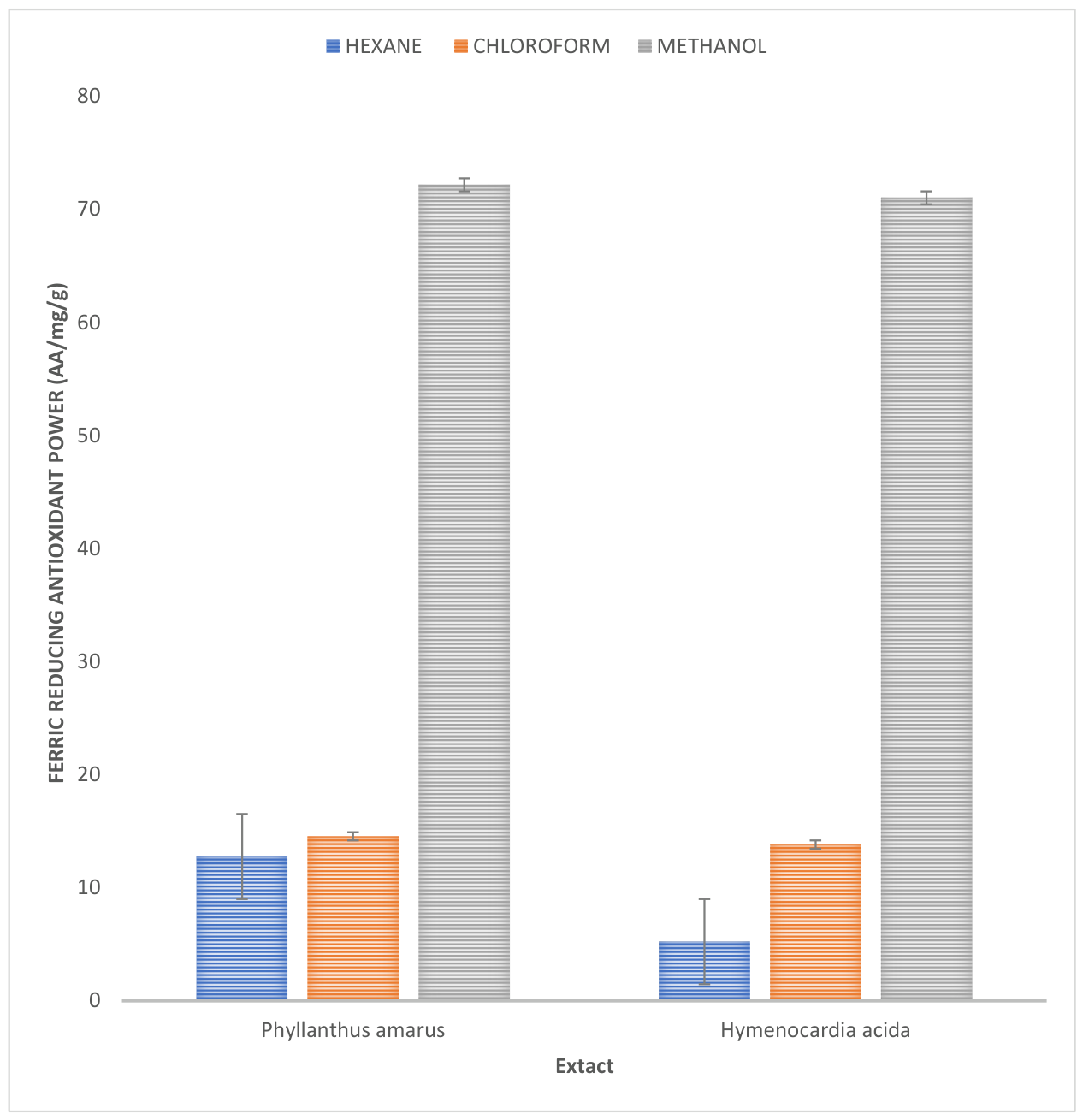
Figure 3.2 illustrates the ferric reducing antioxidant power (FRAP) of Phyllanthus amarus and Hymenocardia acida extracts fractionated with three solvents of varying polarity—hexane, chloroform, and methanol. FRAP measures the ability of antioxidants present in the extracts to reduce ferric (Fe³⁺) to ferrous (Fe²⁺) ions and is expressed in ascorbic acid equivalents (AA/mg/g). Higher FRAP values indicate stronger antioxidant capacity.
FRAP assay results were in concordance with the DPPH assay. Methanol extracts exhibited the highest reducing power with Phyllanthus amarus recording 72.18 ± 0.04 AA/mg and Hymenocardia acida at 71.03 ± 5.81 AA/mg. Lower FRAP values were obtained for hexane and chloroform extracts, reinforcing the superior antioxidant efficiency of polar-extracted fractions.

Total Phenol and Flavonoid Contents of Extracts of Phyllanthus Amarus and Hymenocardia Acida

Table 3.3 presents the total phenol and flavonoid contents of extracts of Phyllanthus amarus and Hymenocardia acida. These bioactive compounds are critical indicators of antioxidant potential due to their capacity to scavenge free radicals, inhibit oxidative enzymes, and chelate metal ions. The total flavonoid content was higher in Hymenocardia acida (28.50 ± 0.11 mg QE/g) compared to Phyllanthus amarus (21.75 ± 0.23 mg QE/g). This suggests that H. acida may have a stronger ability to interfere with oxidative processes, particularly through mechanisms related to flavonoid-mediated enzyme inhibition and reactive oxygen species (ROS) neutralization.
Conversely, Phyllanthus amarus exhibited a significantly higher total phenolic content (66.39 ± 0.59 mg GAE/g) than Hymenocardia acida (49.01 ± 0.47 mg GAE/g). Phenolic compounds are widely recognized for their strong antioxidant properties, which include hydrogen atom donation, radical scavenging, and transition metal ion chelation. The elevated phenol content in P. amarus supports its superior performance in antioxidant assays such as DPPH and FRAP, as also reported in other sections of this study.

Antimicrobial Activity of Phyllanthus amarus Extracts Against Selected Test Organisms

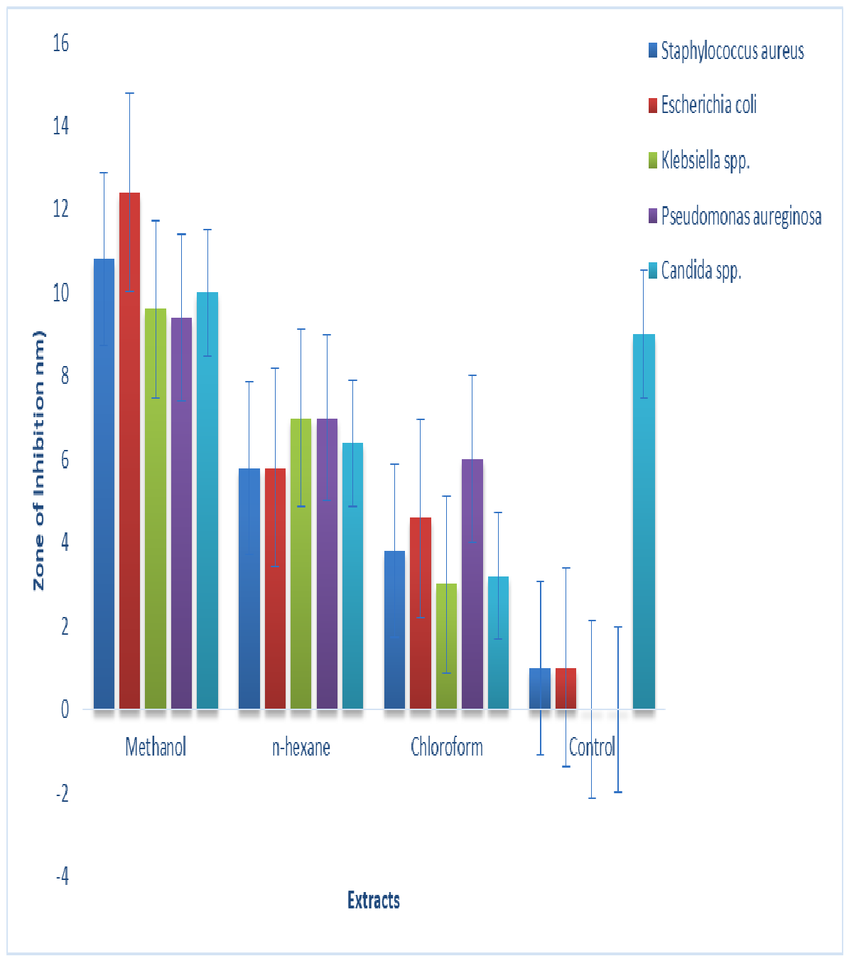
Figure 3.3 presents the zones of inhibition (in mm) observed for methanol, n-hexane, and chloroform extracts of Phyllanthus amarus against five clinically relevant pathogens: Staphylococcus aureus, Escherichia coli, Klebsiella spp., Pseudomonas aeruginosa, and Candida spp., with distilled water/methanol solvent serving as the negative control.
The methanol extract exhibited the highest antimicrobial potency, with inhibition zones ranging from 9.4 mm to 12.4 mm, showing the strongest activity against Escherichia coli (12.4 mm) and Staphylococcus aureus (10.8 mm), followed closely by Candida spp. (10.0 mm). This strong performance highlights methanol's effectiveness in extracting polar bioactive compounds such as phenols, flavonoids, and alkaloids, which are known for their antimicrobial action.
Moderate activity was observed in the n-hexane extract, particularly against Klebsiella spp. and P. aeruginosa (7 mm each), likely due to the presence of non-polar compounds like terpenoids. In contrast, the chloroform extract exhibited the least antimicrobial activity, with inhibition zones between 3.0 mm and 6.0 mm, suggesting a lower yield of active constituents in this fraction. Interestingly, Candida spp. also responded to the negative control (9.0 mm), possibly due to environmental sensitivity or solvent remnants, indicating the need for careful interpretation. Overall, the results confirm that the antimicrobial effects are predominantly due to the plant extracts, with methanol being the most effective solvent for extracting therapeutically relevant phytochemicals.

Antimicrobial Activity of Hymenocardia Acida Extracts Against Selected Test Organisms

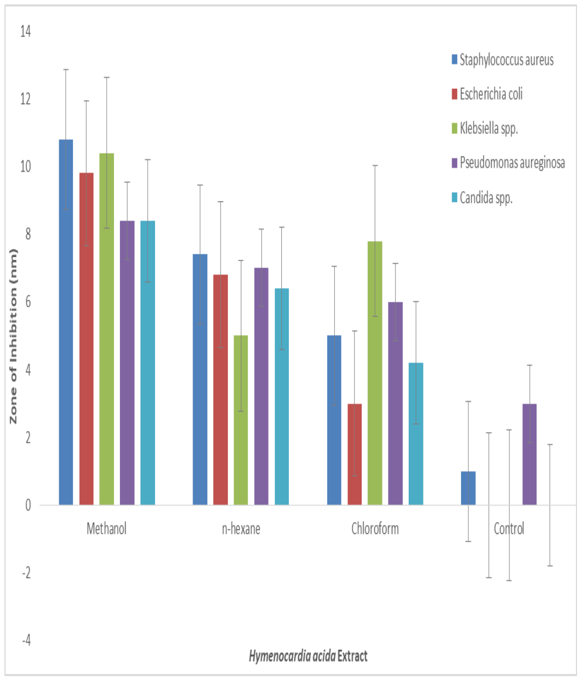
Figure 3.4 illustrates the antimicrobial activity of Hymenocardia acida extracts (methanol, n-hexane, and chloroform) against five clinically important pathogens: Staphylococcus aureus, Escherichia coli, Klebsiella spp., Pseudomonas aeruginosa, and Candida spp., with distilled water/methanol serving as the negative control.
Among the three, the methanol extract demonstrated the highest activity, with inhibition zones ranging from 8.4 mm to 10.8 mm. The strongest inhibition was recorded against Staphylococcus aureus (10.8 mm), followed by Klebsiella spp. (10.4 mm) and Escherichia coli (9.8 mm), while both Pseudomonas aeruginosa and Candida spp. showed moderate susceptibility (8.4 mm each). These results suggest that the methanol extract contains polar phytochemicals such as phenols, flavonoids, and alkaloids that effectively inhibit both bacterial and fungal pathogens, highlighting its broad-spectrum antimicrobial potential.
The n-hexane extract showed moderate antimicrobial activity, particularly against S. aureus (7.4 mm) and P. aeruginosa (7.0 mm), indicating that non-polar bioactive compounds like terpenoids and steroids contributed to the antimicrobial effects, albeit at a lower potency than those in the methanol extract. The chloroform extract demonstrated selective antimicrobial activity, with its highest effect observed against Klebsiella spp. (7.8 mm), suggesting that moderately polar constituents in the extract may be more effective against this specific organism. Minimal or no inhibition in the negative controls (0–3 mm) confirmed that the observed antimicrobial effects were due to the plant extracts rather than the solvents used, underscoring the therapeutic relevance of Hymenocardia acida in traditional medicine.

Minimum Inhibitory Concentration (MIC) of Fractionated Extracts of Phyllanthus Amarus and Hymenocardia Acida Against Test Organisms

The Minimum Inhibitory Concentration (MIC) analysis of Phyllanthus amarus extracts in Table 3.4 revealed varying levels of antimicrobial potency across the different solvent fractions. The methanol extract exhibited its highest efficacy against Klebsiella spp. (10.00 µg/mL) and Pseudomonas aeruginosa (25.00 µg/mL), indicating a moderate to strong antibacterial effect. However, its higher MIC values against Staphylococcus aureus and Candida spp. (100.00 µg/mL each) suggest lower activity against Gram-positive bacteria and fungi. The n-hexane fraction demonstrated uniform moderate activity (25.00–50.00 µg/mL) against bacteria, but was less effective against Candida spp. (100.00 µg/mL), suggesting limited antifungal properties. Interestingly, the chloroform fraction showed broader antimicrobial activity, with MIC values between 25.00 and 50.00 µg/mL for most organisms and a notably low MIC of 25.00 µg/mL against Candida spp., implying that the moderately polar compounds extracted may have better efficacy, especially against fungal pathogens.
Table 3.4a. Minimum Inhibitory Concentration (MIC) of Fractionated extracts of Phyllanthus amarus against test organisms.
Fractionated Extracts Test Organisms Minimum Inhibitory Concentration
(MIC)
Phyllanthus amarus:
Methanol Staphylococcus aureus 100.00
Escherichia coli 50.00
Pseudomonas aureginosa 25.00
Klebsiella spp 10.00
Candida spp. 100.00
n-Hexane Staphylococcus aureus 50.00
Escherichia coli 50.00
Pseudomonas aureginosa 25.00
Klebsiella spp 50.00
Candida spp. 100.00
Chloroform Staphylococcus aureus 25.00
Escherichia coli 50.00
Pseudomonas aureginosa 25.00
Klebsiella spp 50.00
Candida spp. 25.00
Control Staphylococcus aureus 100.00
Escherichia coli 50.00
Pseudomonas aureginosa 100.00
Klebsiella spp 100.00
Candida spp. 100.00
Table 3.4b. Minimum Inhibitory Concentration (MIC) of Fractionated extracts of Hymenocardia acida against test organisms.
Fractionated Extracts Test Organisms Minimum Inhibitory Concentration
(MIC)
Hymenocardia acida
Methanol Staphylococcus aureus 50.00
Escherichia coli 100.00
Pseudomonas aureginosa 50.00
Klebsiella spp 12.5
Candida spp. 100.00
n-Hexane Staphylococcus aureus 12.5
Escherichia coli 25.00
Pseudomonas aureginosa 50.00
Klebsiella spp 6.50
Candida spp. 25.00
Chloroform Staphylococcus aureus 3.12
Escherichia coli 12.50
Pseudomonas aureginosa 6.50
Klebsiella spp 3.12
Candida spp. 50.00
Control Candida spp 25.00
In contrast, the chloroform fraction of Hymenocardia acida demonstrated the most potent antimicrobial activity overall, with remarkably low MICs of 3.12 µg/mL against Staphylococcus aureus and Klebsiella spp., and 6.50 µg/mL against Pseudomonas aeruginosa. These values indicate the presence of highly active semi-polar compounds in H. acida with broad-spectrum efficacy. The n-hexane fraction also performed well, particularly against Klebsiella spp. (6.50 µg/mL) and S. aureus (12.5 µg/mL), likely due to the presence of non-polar bioactives such as terpenoids and steroids. The methanol extract showed moderate activity, with MICs ranging from 12.5 µg/mL to 100.00 µg/mL, suggesting that while polar constituents were bioactive, they were less potent compared to those in the chloroform fraction. Collectively, these results highlight the importance of solvent choice in isolating antimicrobial agents and underscore the superior efficacy of H. acida’s chloroform-soluble components against multiple pathogens.

4. Discussion

Due to rising microbial resistance and an increase in illnesses of microbial origin, the development of novel antibiotic sources has become crucial. Plants generate a diverse spectrum of bioactive compounds, allowing them to be viewed as natural sources of new chemicals with potential use in medicine and biotechnology (Farias et al., 2014). This study explored the phytochemical composition, antioxidant capacity, and antimicrobial activity of Hymenocardia acida and Phyllanthus amarus extracts, fractionated using methanol, chloroform, and n-hexane. The evaluation was performed against five clinical pathogens—Staphylococcus aureus, Escherichia coli, Klebsiella pneumoniae, Pseudomonas aeruginosa, and Candida spp.—to assess the therapeutic potential of these ethnemedicinal plants. The findings are interpreted in detail below and discussed in the context of relevant contemporary studies.
The qualitative phytochemical analysis confirmed the presence of saponins, flavonoids, alkaloids, phenols, tannins, terpenoids, steroids, coumarins, diterpenes, and cardiac glycosides in both plants, though in varying abundance depending on the solvent used. Phyllanthus amarus showed a particularly high presence of alkaloids, flavonoids, and phenols, while Hymenocardia acida was rich in flavonoids and tannins.
Quantitatively, methanol extracts yielded higher concentrations of phytochemicals. P. amarus recorded the highest total phenolic content (125.51 ± 0.80 mg GAE/g), whereas H. acida had a higher flavonoid content (41.50 ± 0.09 mg QE/g). This aligns with foundational extraction principles (Abubakar & Haque, 2020), which demonstrate that methanol, being a highly polar solvent, is highly effective in recovering polar phenolic and flavonoid fractions. These secondary metabolites are known to play critical roles in plant defense and human health due to their antimicrobial, anti-inflammatory, and antioxidant properties (Sofowora, 1993; Panche et al., 2016).
The observed phytochemical profile corroborates the work of Ogbunugafor et al. (2011), who reported a rich abundance of similar radical-scavenging bioactive compounds in Hymenocardia acida extracts. Similarly, Oluba et al. (2020) confirmed the presence of potent phytochemicals in Phyllanthus amarus, supporting its traditional use in treating infections and oxidative stress.
The antioxidant potential of both plants was evaluated using DPPH radical scavenging and FRAP assays. The methanol extract of P. amarus exhibited the highest DPPH inhibition (78.15%) and a strong FRAP profile, indicating an advanced free radical scavenging ability. This was followed closely by H. acida methanol extract (57.64% DPPH inhibition), indicating comparable, high-level antioxidant potential.
These results are consistent with the principles established by Cai et al. (2004), who showed that phenolic and flavonoid content directly correlates with antioxidant performance. The superior antioxidant activity observed in P. amarus is attributable to its higher phenolic concentration, as these molecules act efficiently as hydrogen donors and metal chelators (Oboh et al., 2007). The low antioxidant values observed in chloroform and hexane extracts confirm that the most active antioxidant compounds in these plants are polar, reaffirming the extraction efficiency of methanol. This observation is further supported by Prasad et al. (2011), who emphasized the impact of high solvent polarity on maximizing antioxidant yield during extraction procedures.
The antimicrobial assays revealed concentration-dependent inhibition of microbial growth by both plant extracts. Methanol extracts of P. amarus exhibited the highest inhibition zones against Escherichia coli (12.4 mm), Staphylococcus aureus (10.8 mm), and Candida spp. (10 mm). Likewise, H. acida methanol extract showed strong activity against S. aureus (10.8 mm), Klebsiella spp. (10.4 mm), and E. coli (9.8 mm).
These results corroborate the findings of Silva and Fernandes Júnior (2010), who demonstrated the broad-spectrum antimicrobial activity of flavonoid-rich plant extracts. The observed antimicrobial activity is supported by the phytochemical constituents identified, particularly phenols, flavonoids, and alkaloids, which are known to cause microbial membrane disruption, inhibition of nucleic acid synthesis, and interference with energy metabolism (Ogunyemi et al., 2022).
Chloroform and n-hexane fractions demonstrated moderate activity; notably, the chloroform fraction of H. acida recorded significant selective inhibition against Klebsiella spp. (7.8 mm). This supports findings by Akinyemi et al. (2005), who reported that semi-polar organic solvents like chloroform isolate secondary metabolites with selective, highly focused antimicrobial paths against specific clinical strains.
Interestingly, notable activity was also observed against Candida spp., an opportunistic fungal pathogen. The antifungal activity shown by both plants validates their traditional use in treating fungal infections and supports the observations of regional investigators (Eze et al., 2021), who reported the strong antifungal properties of Nigerian medicinal plants against clinical yeast isolates.
The MIC results revealed the highest potency in chloroform extracts of H. acida, with MIC values as low as 3.12 µg/mL against Staphylococcus aureus and Klebsiella spp., and 6.50 µg/mL against Pseudomonas aeruginosa. For P. amarus, the chloroform extract also exhibited low MIC values (25-50 µg/mL), while its methanol extract recorded its lowest MIC against Klebsiella spp. (10.00 µg/mL). These results are consistent with findings by Okokon et al. (2019), who observed enhanced bioactivity in concentrated semi-polar chloroform plant fractions compared to highly crude polar volumes. The low MIC values discovered in this study fall within the highly effective therapeutic range for plant-derived antimicrobials, confirming the presence of potent molecules suitable for further development (Ogunyemi et al., 2022).

Conclusion

The comparative analysis reveals that Phyllanthus amarus possesses superior antioxidant potential, likely due to its high phenolic content, whereas Hymenocardia acida exhibits stronger antimicrobial efficacy, especially in its chloroform fraction. These complementary effects indicate their potential synergistic application in managing oxidative stress-linked infections. The demonstration of activity against both Gram-positive and Gram-negative bacteria, as well as Candida spp., highlights the broad-spectrum antimicrobial capabilities of these plants. This is particularly important given the global challenge of antimicrobial resistance. The observed MIC values against resistant organisms suggest that these plants may serve as viable leads in the development of new pharmaceutical agents.

Author Contributions

“Conceptualization, James A. Ndako and Amos E. Ohiobo; methodology, Emmanuel O. Yeye.; software, Shammah I. Ayodele .; validation, Bolape A. Oyekanmi., Emmabel O. Onuchukwu. and Omotayo F. Oniyelu.; formal analysis, Suliat D. Tijani ; investigation, Emmanuel O. Yeye.; resources, Christy J.Ndako.; data curation, Omotayo F. Oniyelu.; writing—original draft preparation, Amos E. Ohiobo.; writing—review and editing, Suliat D. Tijani.; visualization, Christy J.Ndako.; supervision,;James A. Ndako.; project administration, Bolape A. Oyekanmi.; All authors have read and agreed to the published version of the manuscript.”

Funding

“This research received no external funding”

Institutional Review Board Statement

“Ethical review and approval were “Not applicable.” for studies not involving humans or animals.

Data Availability Statement

Suggested Data Availability on request.

Acknowledgments

The authors have reviewed and edited the output and take full responsibility for the content of this publication.”

Conflicts of Interest

“The authors declare no conflicts of interest.”

References

  1. Abdallah, E. M.; Alhatlani, B. Y.; de Paula Menezes, R.; Martins, C. H. G. Back to nature: Medicinal plants as promising sources for antibacterial drugs in the post-antibiotic era. Plants 2023, 12(17), 3077. [Google Scholar] [CrossRef] [PubMed]
  2. Abu, A. H.; Uchendu, C. N. Antispermatogenic effects of aqueous ethanolic extract of Hymenocardia acida stem bark in male Wistar rats. J. Med. Plants Res. 2011, 5(13), 2795–2801. [Google Scholar] [CrossRef]
  3. Abubakar, A. R.; Haque, M. Preparation of medicinal plants: Basic extraction and fractionation procedures for experimental purposes. J. Pharm. Bioallied Sci. 2020, 12(1), 1–10. [Google Scholar] [CrossRef] [PubMed]
  4. Agishi, E. C. A Tiv-English dictionary of plant names; Valley Printing and Publishing Company, 2004. [Google Scholar]
  5. Akinyemi, K. O.; Mendie, U. E.; Smith, S. T.; Oyefolu, A. O.; Coker, A. O. Screening of some Nigerian medicinal plants for antibacterial activity against Salmonella typhi and Salmonella paratyphi. Res. Complement. Med. 2005, 12(1), 26–32. [Google Scholar] [CrossRef]
  6. Cai, Y.; Luo, Q.; Sun, M.; Corke, H. Antioxidant activity and phenolic compounds of 112 traditional Chinese medicinal plants associated with anticancer. Life Sci. 2004, 74(17), 2157–2184. [Google Scholar] [CrossRef] [PubMed]
  7. Dar, R. A.; Shahnawaz, M.; Qazi, P. H. General overview of medicinal plants: A review. J. Phytopharm. 2017, 6(6), 349–351. [Google Scholar] [CrossRef]
  8. Eze, F. I.; Nwodo, J. N.; Omeje, E. O. Evaluation of antimicrobial and antifungal activities of secondary metabolites from selected Nigerian medicinal plants. J. Basic Clin. Pharm. 2021, 12(3), 44–51. [Google Scholar]
  9. Farias, D. F.; Souza, T. M.; Viana, M. P.; Soares, B. M.; Cunha, A. P.; Vasconcelos, I. M.; Ricardo, N. M. P. S.; Ferreira, P. M. P.; Melo, V. M. M.; Carvalho, A. F. U. Antibacterial, antioxidant, and anticholinesterase activities of plant seed extracts from Brazilian semiarid region. An. Da Acad. Bras. De Ciências 2014, 86(3), 1195–1210. [Google Scholar] [CrossRef]
  10. Haruna, A.; Pateh, U. U.; Sule, M.; Musa, A. M.; Sani, Y. M.; Mohammed, M.; Lawal, M. D.; Garba, M. A. Evaluation of anticonvulsant activity of methanol leaf extract of Hymenocardia acida, Tul (Euphorbiaceae) in laboratory animals. J. Pharm. Allied Sci. 2017, 14(5), 2540–2547. [Google Scholar]
  11. Oboh, G.; Puntel, R. L.; Rocha, J. B. Hot pepper (Capsicum annuum, Tepin and Capsicum chinense, Habanero) prevents Fe2+-induced lipid peroxidation in brain—in vitro. Food Chem. 2007, 102(1), 178–185. [Google Scholar] [CrossRef]
  12. Ogbunugafor, H. A.; Eneh, F. U.; Ozumba, A. N.; Igwo-Ezikpe, M. N.; Okpuzor, J.; Pagano, I.; Kane, S. T. Physico-chemical and antioxidant properties of Hymenocardia acida King. Int. J. Pharmacol. 2011, 7(2), 238–245. [Google Scholar] [CrossRef]
  13. Ogunyemi, S. O.; Abdallah, Y.; Zhang, M.; Fouad, H.; Hong, X.; Ibrahim, E.; Masum, M. M. I.; Hossain, A.; Mo, J.; Li, B. Green synthesis of zinc oxide nanoparticles using plant extracts and their antibacterial mechanisms against clinical pathogens. Front. Microbiol. 2022, 13, Article 856329. [Google Scholar] [CrossRef]
  14. Okokon, J. E.; Bawo, M. B.; Mbagwu, H. Evaluation of antimalarial, antioxidant and antimicrobial activities of fractions of Mammea africana. Bangladesh J. Pharmacol. 2019, 14(2), 77–86. [Google Scholar]
  15. Oluba, O. M.; Adebisi, K. E.; Eidangbe, G. O.; Ojieh, A. E. Phytochemical composition and antioxidant status of Phyllanthus amarus aqueous extract in alloxan-induced diabetic rats. J. Diet. Suppl. 2020, 17(4), 436–447. [Google Scholar] [CrossRef]
  16. Panche, A. N.; Diwan, A. D.; Chandra, S. R. Flavonoids: An overview. J. Nutr. Sci. 5 2016, e47. [Google Scholar] [CrossRef] [PubMed]
  17. Prasad, K. N.; Yang, B.; Shi, J.; Yu, C.; Zhao, M.; Xue, S.; Jiang, Y. Effect of extraction solvent and technique on the total phenolic content and antioxidant activity of litchi (Litchi chinensis Sonn.) fruit pericarp. Food Chem. 2011, 126(3), 1331–1337. [Google Scholar] [CrossRef]
  18. Silva, N. C. C.; Fernandes Júnior, A. Biological properties of medicinal plants: A review of their antimicrobial activity. J. Venom. Anim. Toxins Incl. Trop. Dis. 2010, 16(3), 402–413. [Google Scholar] [CrossRef]
  19. Sofidiya, M. O. Phytochemical and pharmacological evaluation of Hymenocardia acida Tul. (Hymenocardiaceae) leaves. Doctoral dissertation, University of Lagos, 2008. [Google Scholar]
  20. Sofowora, A. Medicinal plants and traditional medicine in Africa, 2nd ed.; Spectrum Books Ltd, 1993. [Google Scholar]
  21. Srivastava, R.; Singh, R. Herbal medicine: A review of its history, significance, and development in developing countries. Int. J. Green Pharm. 2016, 10(2), 67–75. [Google Scholar] [CrossRef]
  22. Stumbo, P.; Feld, R.; Evans, W.; Weiner, R.; Moran, E.; Hoffman, F. A. The historical pursuit of natural analgesics and antimicrobials. J. Ethnopharmacol. 2017, 204, 89–101. [Google Scholar]
  23. Tungmunnithum, D.; Thongboonyou, A.; Pholboon, A.; Yangsabai, A. Flavonoids and other phenolic compounds from medicinal plants for pharmaceutical and medical applications: An overview. Medicines 2018, 5(3), 93. [Google Scholar] [CrossRef] [PubMed]
  24. Ugbogu, O. C.; Emmanuel, O.; Agi, G. O.; Ibe, C.; Ekweogu, C. N.; Ude, V. C.; Ugbogu, E. A. A review on the traditional uses, phytochemistry, and pharmacological activities of clove basil (Ocimum gratissimum L.). Heliyon 2021, 7(11), e08404. [Google Scholar] [CrossRef] [PubMed]
  25. Ukaegbu-Obi, K. M.; Mereni, C. I.; Oji, O. O. Antimicrobial activity of selected medicinal plants against clinical pathogens. Int. J. Microbiol. Biotechnol. 2018, 3(2), 34–41. [Google Scholar]
  26. Yessuf, A. M. Phytochemical screening and evaluation of biological activities of selected medicinal plants. J. Pharmacogn. Phytochem. 2015, 4(3), 112–118. [Google Scholar]
  27. Yusuf, I.; Arzai, A. H.; Umar, A. M.; Haruna, M.; Yusuf, N. Characterization and antimicrobial susceptibility profile of bacteraemia causing pathogens isolated from febrile children in Kano, Nigeria. Bayero J. Pure Appl. Sci. 2018, 11(1), 143–151. [Google Scholar] [CrossRef]
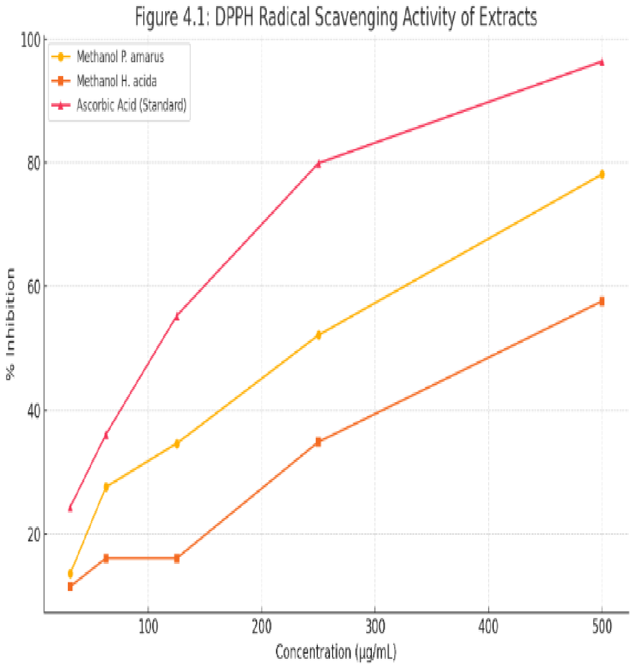
Figure 3.1. illustrating the DPPH radical scavenging activity of methanol extracts of Phyllanthus amarus and Hymenocardia acida compared to ascorbic acid. As evident, P. amarus shows superior antioxidant capacity.
Preprints 218783 g001
Figure 3.2. Ferric Reducing Antioxidant Power (FRAP) of Phyllanthus amarus and Hymenocardia acida.
Preprints 218783 g002
Figure 3.3. Antimicrobial Activity of Phyllanthus amarus Extracts against selected test organisms.
Preprints 218783 g003
Figure 3.4. Antimicrobial Activity of Hymenocardia acida Extracts against selected test organisms.
Preprints 218783 g004
Table 3.1. Gram of Extracts.
Leaf Extracts Gram (g)
Hymenocardia acida 700
Phyllantus amarus 93.5
Table 3.2. Qualitative Phytochemical Screening of Phyllanthus amarus and Phyllanthus amarus Plant Extracts.
Chemical Compounds (Phytochemicals) Phyllanthus amarus Extract Hymenocardia acida Extract
Saponin ++ ++
Alkaloid +++ ++
Flavonoid +++ +++
Tannin + ++
Phenol +++ ++
Coumarin ++ ++
Terpenoid ++ +
Steroid ++ ++
Diterpenes ++ +
Cardiac Glycosides ++ +
Key: +++ = Abundantly present, ++ = Moderately present, + = Slightly present, – = Absent.
Table 3.3. Total Phenol and Flavonoid Contents of plant extracts of Phyllanthus amarus and Hymenocardia acida.
Extracts Total Flavonoid Content
(QE/mg/g)
Mean ± SD
Total Phenol Content
(GAE/mg/g)
Mean ± SD
Phyllanthus amarus 21.75± 0.23 66.39 ± 0.59
Hymenocardia acida 28.50 ± 0.11 49.01 ± 0.47
Values are means of three analyses of the extract ± standard deviation (n=3).
Disclaimer/Publisher’s Note: The statements, opinions and data contained in all publications are solely those of the individual author(s) and contributor(s) and not of MDPI and/or the editor(s). MDPI and/or the editor(s) disclaim responsibility for any injury to people or property resulting from any ideas, methods, instructions or products referred to in the content.
Copyright: This open access article is published under a Creative Commons CC BY 4.0 license, which permit the free download, distribution, and reuse, provided that the author and preprint are cited in any reuse.
Prerpints.org logo

Preprints.org is a free preprint server supported by MDPI in Basel, Switzerland.

Subscribe

© 2026 MDPI (Basel, Switzerland) unless otherwise stated

Accessibility

Disclaimer

Terms of Use

Privacy Policy

Privacy Settings