

Submitted:
06 August 2026
Posted:
10 August 2026
You are already at the latest version
Abstract
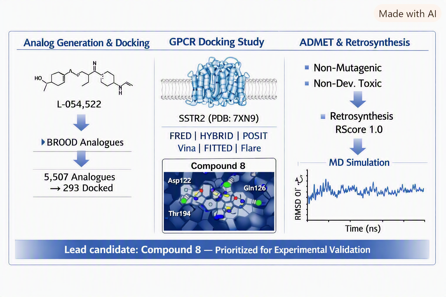
Somatostatin receptor subtype 2 (SSTR2) is overexpressed in high-risk neuroblastoma stage 4 (NBS4) in patients with chromosome 17q gain, making it a potential therapeutic target. This computational study employed a multi-platform drug design workflow to identify novel SSTR2 ligands based on the selective agonist L-054,522. Using BROOD (OpenEye Scientific), 5,507 structural analogues were generated, from which 293 candidates were docked to SSTR2 (PDB: 7XN9) using FRED, HYBRID, and POSIT. Eight compounds achieved GREAT pose confidence (75-100% probability within 2.0 A of the true binding mode). Confirmatory docking with AutoDock Vina, FITTED, and Flare corroborated these results. ADMET profiling using T.E.S.T., DEEP-PK, and SwissADME indicated favourable safety profiles for all eight compounds, with no predicted mutagenicity or developmental toxicity. Retrosynthetic analysis using ChemAIRS and Spaya confirmed synthetic feasibility, with compound 8 achieving an RScore of 1.0. Compound 8 demonstrated consistent docking performance across all platforms and exhibited interaction patterns, including contacts with Asp122, Gln126, and Thr194, that align with residues previously identified as critical for SSTR2 ligand binding. Molecular dynamics simulation further supported the stability of the predicted complex, showing preservation of global structural integrity with local flexibility concentrated mainly in terminal and loop regions. Exploratory docking to CRHR1 and GALR2, two additional chromosome 17q GPCRs expressed in neuroblastoma, suggested that compound 8 may occupy these binding sites, though functional consequences remain unknown. These findings identify compound 8 as a computationally prioritised lead candidate warranting experimental evaluation through binding assays and functional studies to determine its pharmacological activity at SSTR2.
Keywords:
neuroblastoma stage 4
; chromosome 17
; somatostatin receptors
; GPCR
; computational drug design
; molecular docking
; ADMET
; structure–activity relationship
1. Introduction
Neuroblastoma (NB) is the third most common solid tumour in infants and children [1], accounting for approximately 15% of all pediatrics cancer deaths globally. [2,3,4] The disease originates from embryonic sympathoadrenal cells of the neural crest and is closely linked to genetic and molecular characteristics rather than behavioural or environmental factors. [5,6]. Clinical outcomes vary dramatically: some tumours undergo spontaneous regression, while others progress aggressively despite intensive therapy. [7] For patients diagnosed with stage 4 disease (NBS4), survival rates remain between 40–50%, underscoring the urgent need for novel therapeutic strategies.[8]
A hallmark genetic abnormality in high-risk NBS4 is segmental gain of chromosome 17q, detected in over 80% of tumours [9] . The region 17q23.1 to 17qter is most frequently amplified, with gains at 17q25 strongly associated with an unfavourable prognosis. [10] Notably, somatostatin receptor subtype 2 (SSTR2), located at 17q25.1, is overexpressed in high-risk NBS4. [9,10,11,12,13,14] Previous studies have demonstrated that somatostatin receptors, particularly SSTR2, are expressed in many neuroblastoma tumours and cell lines, and that activation of these receptors can inhibit neuroblastoma cell proliferation and tumour growth.[15,16,17]
Somatostatin receptors belong to the G protein-coupled receptor (GPCR) superfamily and mediate diverse physiological effects [18], including inhibition of growth hormone, regulation of neurotransmission, and suppression of cell proliferation. [16] SSTR2 is the most extensively studied mediator of somatostatin's antiproliferative action and the primary pharmacological target of clinically prescribed somatostatin analogues. [19,20] Its activation inhibits adenylyl cyclase, blocks voltage-dependent calcium channels, and upregulates cell-cycle inhibitors such as CDKN1B. [21]
The nonpeptide compound L-054,522 was selected as the structural template for this work. L-054,522 binds SSTR2 with a dissociation constant of 0.01 nM and exhibits greater than 3000-fold selectivity over other somatostatin receptor subtypes[22,23]. Using the co-crystal structure of L-054,522 with SSTR2 (PDB: 7XN9) [24] as a reference, this study employed a multi-platform computational workflow to design and evaluate potential SSTR2 ligands.
Computational approaches offer significant value at early stages of drug discovery by enabling rapid screening of chemical space, predicting binding modes, and prioritising candidates for synthesis and testing.[25] However, docking studies alone cannot determine whether a compound acts as an agonist, antagonist, partial agonist, or inactive binder—such functional characterisation requires experimental validation. [26] The present study therefore aims to identify computationally promising SSTR2 ligand candidates and provide a foundation for subsequent experimental investigation.
Two additional GPCRs on chromosome 17 are also expressed in NB cell lines: corticotropin-releasing hormone receptor 1 (CRHR1) at 17q21.31 and galanin receptor type 2 (GALR2) at 17q25.3. [27,28,29,30,31] As an exploratory analysis, we assessed whether top-ranked SSTR2 ligand candidates might also bind these receptors, though the therapeutic implications of such multi-target binding remain speculative pending experimental studies.
2. Materials and Methods
2.1. Lead Compound and Receptor Selec63
L-054,522 (C₃₅H₄₇N₇O₅; MW 645.80) was identified as a structural template based on its high affinity and selectivity for SSTR2. [22,23] The crystal structure of SSTR2 in complex with L-054,522 (PDB: 7XN9) [24] was retrieved from the Protein Data Bank and used as the primary docking target. The receptor was prepared for docking using the MakeReceptor application (OpenEye), which defines the binding site based on the crystallographic ligand position and generates a shape-based receptor grid.[32]
A second SSTR2 structure (PDB: 7WIG) [33] served as a confirmatory target to assess binding site consistency. For exploratory multi-target analysis, CRHR1 (PDB: 8GTI) [34] and GALR2 (PDB: 7WQ4, 7XBD, 7XJK, 7XJL) [35,36,37] structures were also obtained.
During validation, L-054,522 failed to re-dock successfully using FRED and HYBRID [38,39] ("Undocked") and received a POOR confidence score in POSIT. [32] This likely reflects conformational constraints of the ligand and the sensitivity of pose prediction to starting geometry. The crystallographic pose was used directly as the reference for evaluating analogue binding modes, and the MakeReceptor-defined binding site faithfully represents the experimental ligand position.
2.2. Analogue Generation Using BROOD
The BROOD application (OpenEye Scientific) was used to generate structural analogues of L-054,522 by replacing molecular fragments with bioisosteres of similar shape and electrostatics but different chemical properties. [40,41] BROOD identifies fragments from known chemical space (medicinal chemistry databases) and substitutes them into the lead structure while preserving overall molecular shape and pharmacophore features. Ten rounds of fragment searching were performed:
- Shape & Electrostatics mode: 4 rounds (1,557 compounds)
- Shape & Colour mode: 6 rounds (3,950 compounds)
Druglike filters were applied: molecular weight ≤500, LogP ≤5.0, topological polar surface area ≤150 Ų, Lipinski donor count ≤5, Lipinski acceptor count ≤10, rotatable bonds ≤10, heavy atom count ≤30, and ≤1 Lipinski violation. From each round, the top 20–30 compounds were selected based on Tanimoto combo scores (combined shape and electrostatic/colour similarity), yielding 293 candidates for docking.
2.3. Molecular Docking
Primary docking (OpenEye suite): All 293 compounds were docked to 7XN9 using FRED (fast exhaustive docking) and HYBRID (ligand-guided docking). [38,39] FRED performs systematic, exhaustive sampling of ligand poses within the binding site, while HYBRID uses the co-crystallised ligand to guide pose generation.
Final pose selection and ranking were performed using POSIT [32], which automatically selects the optimal docking algorithm based on 2D (graph) and 3D (structural) similarity between the docked ligand and the bound reference ligand. POSIT assigns pose confidence categories based on the probability that the predicted pose lies within 2.0 Å of the true binding mode:
- GREAT: 75–100% probability
- GOOD: 50–75%
- MEDIOCRE: 33–50%
- POOR: <33%
POSIT probabilities were validated against X-ray crystallography data in the original method development [32], providing a rigorous basis for pose confidence assessment.
Confirmatory docking: Compounds achieving GREAT confidence were re-docked using AutoDock Vina Extended [42,43] and FITTED (Molecular Forecaster) [44] within the Samson Suite, followed by Flare Suite. [45,46,47] Docking was also performed against 7WIG to confirm binding site consistency.
Interpretation of cross-platform results: Different docking programs use different scoring functions with different scales and physical meanings. FRED scores reflect shape complementarity and chemical matching; AutoDock Vina uses an empirical free energy function; FITTED incorporates induced-fit effects. Raw scores are not directly comparable across platforms. Instead, cross-platform consistency—a compound ranking well across multiple independent methods—provides confidence in the predicted binding mode.[48] Compound 8 achieved strong scores across all four platforms, supporting its prioritisation.
Exploratory multi-target docking: The eight top-ranked compounds were docked to CRHR1 (8GTI) and GALR2 (7WQ4) using AutoDock Vina Extended. Binding site similarity across receptors was assessed using Protein Aligner[49]. These results are presented as hypothesis-generating rather than confirmatory.
2.4. ADMET Evaluation
Toxicity profiles were assessed using Toxicity Estimation Software Tools (T.E.S.T.) [50], including:
- Bioconcentration factor
- Mutagenicity (Ames test prediction)
- Oral rat LD₅₀
- 48-hour IGC₅₀ in Tetrahymena pyriformis
- Developmental toxicity
Pharmacokinetic properties were predicted using DEEP-PK [51]:
- Human oral bioavailability (50% threshold)
- Blood–brain barrier penetration
- CYP1A2 substrate status
- Predicted half-life
- Carcinogenicity
Additional ADME parameters (TPSA, LogP, water solubility, GI absorption, skin permeability) were calculated using SwissADME [52].
2.5. Retrosynthetic Analysis
Synthetic feasibility was evaluated using two AI-driven platforms: ChemAIRS [53] and Spaya [54]. Spaya assigns an RScore (0–1), where values above 0.5 indicate well-precedented synthetic routes; lower scores suggest novel or higher-risk transformations. ChemAIRS was run in standard mode where possible; compounds requiring "High Risk" settings were flagged.
2.6. Structure Visualisation and Analysis
Molecular structures and binding interactions were visualised using Discovery Studio Visualizer [55] and VIDA (OpenEye) [56] . Chemical structures were drawn using PICTO (OpenEye) [57] and ChemDraw. [58] Predicted ¹H and ¹³C NMR spectra were generated using MestReNova [59] to support future synthetic characterisation.
3. Results
3.1. Analogue Generation
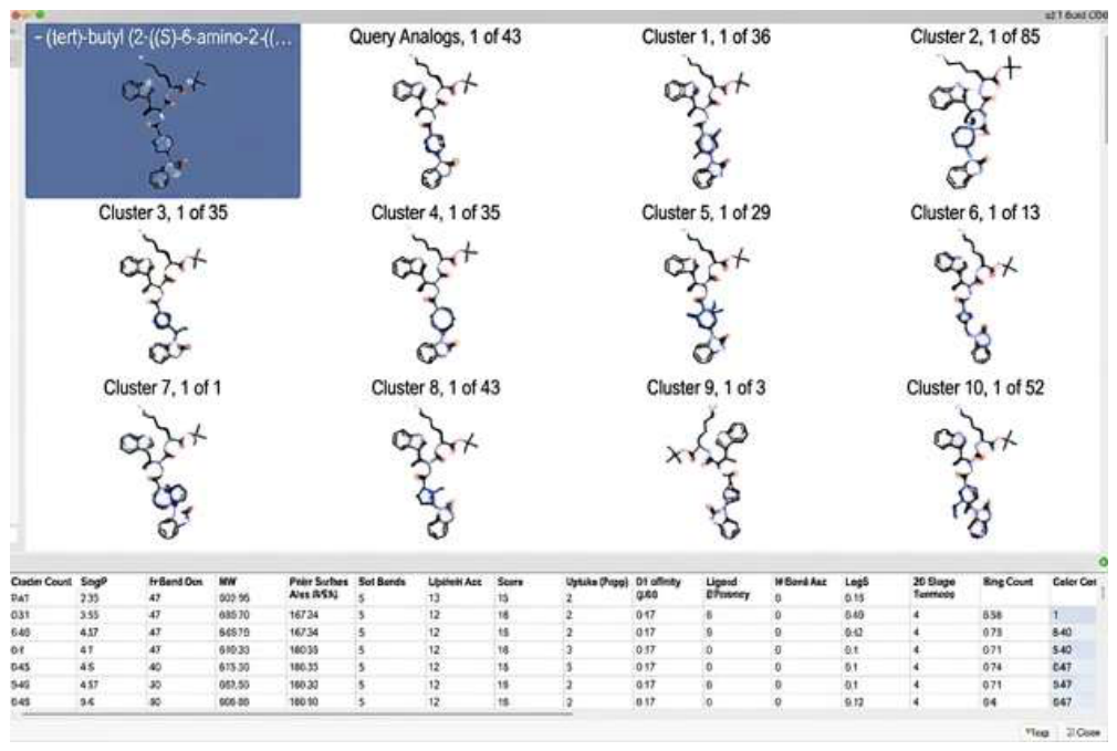
Ten rounds of BROOD searching generated 5,507 candidate structures (Table 1). Following application of druglike filters and Tanimoto-based ranking, 293 compounds were advanced to docking.
Figure 1.
Representative BROOD output from round 6, showing L-054,522 with top-ranked analogues and associated Tanimoto scores.
Figure 1.
Representative BROOD output from round 6, showing L-054,522 with top-ranked analogues and associated Tanimoto scores.
3.2. Primary Docking and Compound Selection
All 293 compounds were docked to SSTR2 (7XN9). POSIT analysis identified eight compounds (8, 12, 13, 14, 17, 22, 27, and 30) with GREAT pose confidence, indicating a 75–100% probability of the predicted pose lying within 2.0 Å of the true binding mode (Table 2).
L-054,522 itself did not dock successfully ("Undocked" in FRED/HYBRID; "POOR" in POSIT), reflecting the conformational constraints discussed in Section 2.1. The crystallographic pose served as the reference for all evaluations.
Figure 2.
Docking of 293 compounds to 7XN9 in FRED, showing binding site occupancy.
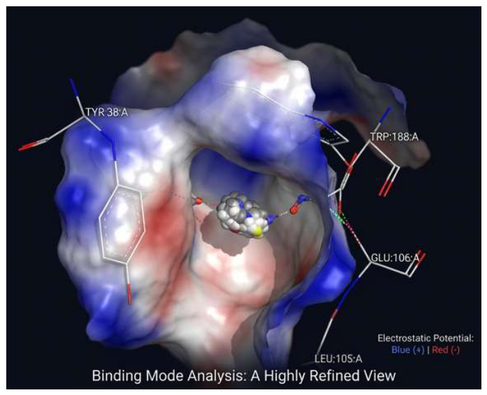
Figure 3.
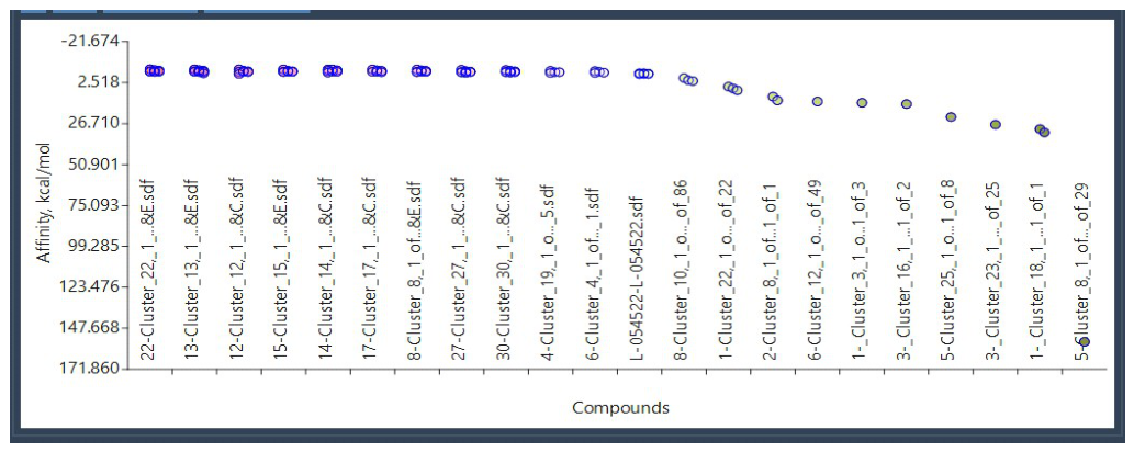
POSIT results in VIDA, highlighting the eight GREAT-confidence compounds.
More negative scores indicate stronger predicted binding. Scores are not comparable across different docking programs.
3.3. Confirmatory Docking
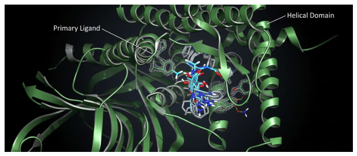
The eight GREAT-confidence compounds were re-docked using Samson Suite (AutoDock Vina Extended, FITTED) and Flare Suite. Compound 8 consistently achieved the strongest or near-strongest scores across all platforms, supporting its prioritisation (Table 3).
Cross-platform consistency (compound 8 ranking well across all methods) is the key finding, not absolute score values.
Figure 4.
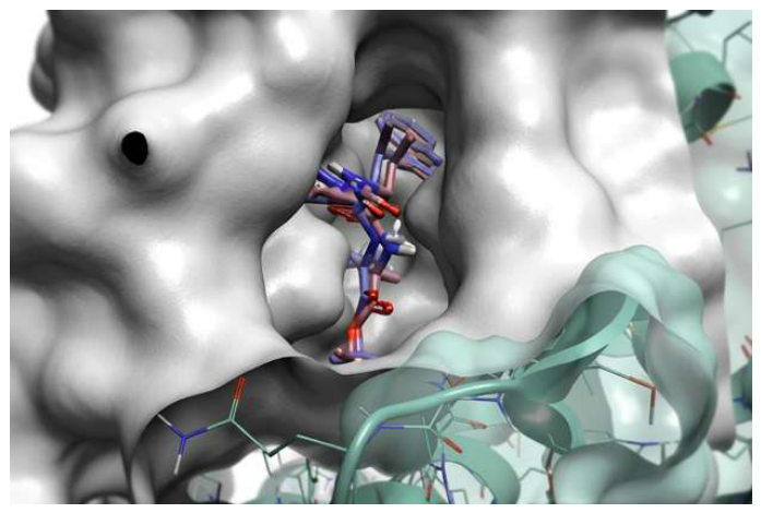
Flare docking results showing compound 8 binding pose.
3.4. Binding Interactions
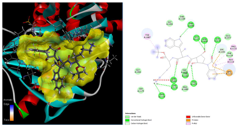
Analysis of compound 8's predicted binding pose revealed the following interactions:
Hydrogen bonds:
- Asp122 (salt bridge)
- Gln126
- Thr194
- Phe208 (backbone)
- Ala283
Hydrophobic contacts:
- Val103
- Phe272
- Phe294
- Leu290
- Val298
Aromatic interactions:
- π-stacking with Phe208, Phe272, Phe275, Phe294
These interactions closely mirror those observed in the L-054,522 crystal structure [24] and align with residues identified in published molecular dynamics studies as critical for SSTR2 ligand binding[60]. Gervasoni et al. demonstrated through multi-microsecond MD simulations that ligands stabilising the active conformation of SSTR2 engage Asp122, Gln126, and the aromatic residues lining the binding pocket [60]. The interaction pattern of compound 8 is consistent with these binding determinants.
While these interactions are consistent with ligand binding, docking cannot determine functional activity. Whether compound 8 acts as an agonist, antagonist, partial agonist, or inactive binder requires experimental determination through binding assays and functional studies.
Figure 5.
Compound 8 interaction map with 7XN9, showing hydrogen bonds, hydrophobic contacts, and aromatic interactions.
Figure 5.
Compound 8 interaction map with 7XN9, showing hydrogen bonds, hydrophobic contacts, and aromatic interactions.
The chemical structures, molecular weights, and names of all eight compounds are presented in Table 4. All retain key structural features: an aromatic moiety capable of π-stacking, a basic centre capable of interacting with Asp122/Gln126, and overall shape similarity to L-054,522.
3.5. ADMET Properties
3.5.1. Toxicity
All eight compounds showed favourable predicted toxicity profiles with no mutagenicity and no developmental toxicity (Table 5). In contrast, L-054,522 was flagged as a developmental toxicant, highlighting a potential safety advantage for the newly identified compounds.
3.5.2. Pharmacokinetics
DEEP-PK predictions indicated that all compounds, including L-054,522, exhibit low oral bioavailability, do not penetrate the blood–brain barrier, are not CYP1A2 substrates, and have safe carcinogenicity profiles (Table 6). Compounds 8, 13, 14, 22, and 27 showed predicted half-lives ≥3 hours, while compounds 12, 17, 30, and L-054,522 showed shorter half-lives.
3.5.3. Physicochemical Properties
SwissADME analysis confirmed that all compounds are poorly water-soluble with low predicted GI absorption (Table 7). These properties are typical of peptide-like GPCR ligands but represent developability challenges that must be addressed.
3.6. Synthetic Accessibility
Retrosynthetic analysis confirmed that all eight compounds have identifiable synthetic routes (Table 8). Critically, compound 8 achieved an RScore of 1.0 in Spaya, indicating a fully precedented pathway with established reaction types. Other compounds required "High Risk" settings in ChemAIRS or achieved lower RScores (0.2–0.4), indicating less established or more challenging synthetic routes.
Given its superior synthetic accessibility, compound 8 is prioritised as the lead candidate. Detailed retrosynthetic schemes for all compounds are provided in Supplementary Materials (Schemes S1–S14).
3.7. Exploratory Multi-Target Analysis
As an exploratory analysis, the eight compounds were docked to CRHR1 (8GTI) and GALR2 (7WQ4)—two additional GPCRs on chromosome 17q that are expressed in neuroblastoma cell lines [27,28,29,30,31]. Protein Aligner [49]comparison indicated structural similarity between the SSTR2 binding pocket and those of CRHR1/GALR2 (Figure 6).
All eight compounds demonstrated predicted binding to both receptors (Table 9). Compound 8 achieved scores comparable to or better than L-054,522 at both targets.
These results demonstrate only that compound 8 can computationally occupy the CRHR1 and GALR2 binding sites. They do not establish whether binding occurs in biological systems, the functional consequence of binding (agonism, antagonism, or no effect), or whether simultaneous modulation of SSTR2, CRHR1, and GALR2 would be therapeutically beneficial in NBS4. The multi-target hypothesis remains speculative and is presented here solely to stimulate future investigation.
3.8. Predicted NMR Spectra
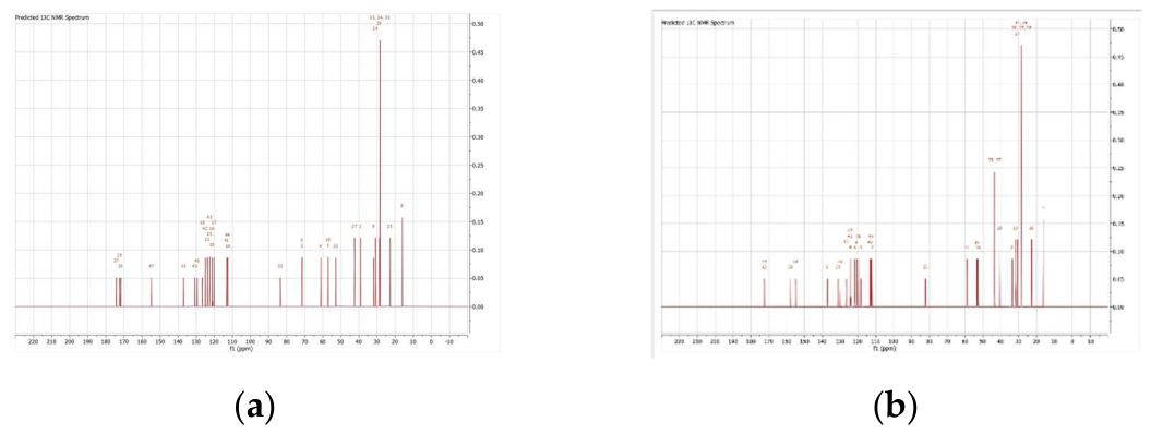
To support future synthetic characterisation, predicted ¹H and ¹³C NMR spectra were generated for compound 8 and L-054,522 using MestReNova (Figure 7).
Figure 7.
Predicted ¹H NMR spectra. (a) Compound 8; (b) L-054,522.
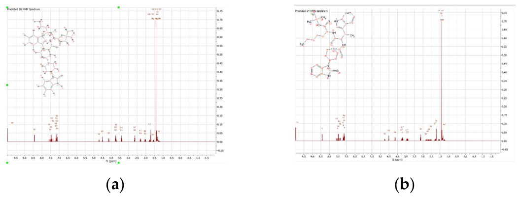
Figure 8.
Predicted ¹³C NMR spectra. (a) Compound 8; (b) L-054,522.
3.9. Cross-Docking with Previously Identified Inhibitors
In previous work, the authors identified twelve potential multi-target inhibitors for NBS4 targeting HDAC, BRD, Hedgehog, TRK, c-Src kinase, and retinoic acid pathways.[61,62] As an exploratory analysis, these twelve compounds were cross-docked with SSTR2 (7XN9) to assess potential overlap.
Several previously identified inhibitors showed predicted binding to SSTR2, suggesting that combination strategies—pairing an SSTR2 ligand such as compound 8 with pathway inhibitors—might merit investigation. However, this remains hypothesis-generating; synergy cannot be inferred from docking alone.
Figure 9.
Cross-docking results in AutoDock Vina Extended showing previously identified inhibitors docked to SSTR2 (7XN9).
Figure 9.
Cross-docking results in AutoDock Vina Extended showing previously identified inhibitors docked to SSTR2 (7XN9).
4. Discussion
This computational study employed a multi-platform workflow to identify potential SSTR2 ligands for high-risk NBS4, a paediatric cancer characterised by frequent 17q gain and SSTR2 overexpression. From 5,507 BROOD-generated analogues of L-054,522, eight compounds achieved GREAT pose confidence in POSIT, and confirmatory docking across three additional platforms corroborated these results. Compound 8 emerged as the most promising candidate based on four converging lines of evidence:
- Consistent docking performance: Compound 8 achieved strong scores across FRED, HYBRID, POSIT, AutoDock Vina, FITTED, and Flare—a level of cross-platform agreement that supports the predicted binding mode.
- Favourable ADMET profile: Unlike L-054,522, compound 8 showed no predicted developmental toxicity. All safety parameters were within acceptable ranges.
- Synthetic accessibility: Compound 8 achieved an RScore of 1.0, indicating a straightforward synthetic route. Other candidates required high-risk transformations, making compound 8 the most practical choice for experimental follow-up.
4.1. Mechanistic Context from Molecular Dynamics and Published SSTR2 Studies
Molecular dynamics simulation performed in the present study provided direct support for the structural stability of the predicted complex over the trajectory. The RMSD profile indicated conformational adjustment from the initial docked pose, while the relatively stable radius of gyration and solvent-accessible surface area suggested preservation of the overall compactness and solvent-exposure pattern of the receptor-ligand system. Residue-level fluctuation analysis showed that the largest motions were concentrated mainly in terminal and flexible regions, whereas the hydrogen-bonding profile remained broadly stable throughout the simulation. Collectively, these findings support a stable but dynamic bound state rather than structural destabilisation of the complex.
These observations can be interpreted in the context of published SSTR2 molecular dynamics studies. [60] Gervasoni et al. showed that the apo receptor is more flexible than ligand-bound states and identified conserved binding-pocket residues, including Asp122 and Gln126, as important determinants of ligand stabilisation in SSTR2. They further reported ligand-dependent behaviour of extracellular loop 2 (ECL2), which closes over the binding site in the presence of the agonist octreotide but remains more open in antagonist-bound and apo states. In the present study, the predicted interaction pattern of compound 8 with Asp122, Gln126, and neighbouring aromatic pocket residues is consistent with these established determinants of SSTR2 ligand recognition, while the MD trajectory supports the view that the complex remains structurally coherent once formed. However, because ECL2 closure and receptor activation were not directly quantified here, the present results should be interpreted as evidence of binding stability rather than proof of agonist-specific conformational activation.
4.2. Limitations of Docking-Based Predictions
Several important limitations must be acknowledged:
- Functional activity cannot be inferred from docking or global MD stability alone. Throughout this manuscript, compound 8 is described as a "ligand" rather than an "agonist" because docking scores and pose geometries cannot distinguish agonists from antagonists, partial agonists, or inactive binders. Functional characterisation requires experimental binding assays (e.g., radioligand displacement) and functional assays (e.g., cAMP inhibition, calcium flux, or β-arrestin recruitment).
- L-054,522 re-docking failure. The lead compound failed to re-dock successfully, which may reflect conformational constraints or sensitivity to starting geometry. This does not invalidate the approach—the crystallographic pose defined the binding site accurately—but it highlights inherent limitations of rigid-body docking.
- The present MD analysis supports structural stability but not a complete activation mechanism. The trajectory supports maintenance of a stable complex and preservation of global structural integrity. However, specific activation-linked features such as ECL2 closure, microswitch transitions, or sustained active-state signalling conformations were not directly quantified.
- ADMET predictions require experimental validation. Computational ADMET tools provide useful filters but have known accuracy limitations. The predicted low GI absorption and poor water solubility of all compounds represent genuine developability concerns.
- Multi-target claims are speculative. The exploratory docking to CRHR1 and GALR2 demonstrates only that compound 8 can occupy these binding sites computationally. No evidence supports therapeutic benefit from simultaneous modulation of these receptors in NBS4.
4.3. Developability Considerations
The predicted low oral bioavailability and poor water solubility of compound 8 (and all analogues) represent significant hurdles for drug development. For a paediatric neuroblastoma population, practical considerations include:
- Intravenous administration: May be clinically acceptable in patients with high-risk neuroblastoma, as treatment commonly involves intensive multimodal regimens incorporating repeated cycles of intravenous chemotherapy, surgery, radiotherapy, stem-cell transplantation, and immunotherapy.[63]
- Formulation strategies: Nanoparticle encapsulation, cyclodextrin complexation, or lipid-based formulations could improve solubility and bioavailability.
- Prodrug approaches: Chemical modification to improve absorption followed by metabolic conversion to the active compound.
These strategies should be explored in parallel with experimental validation of binding and functional activity.
4.4. Comparison with Previous Work
Previous studies established the therapeutic relevance of somatostatin receptors in neuroblastoma. Work in the 1990s identified SSTR2 expression across NB cell lines and tumours and demonstrated antiproliferative effects mediated through somatostatin receptors. [16,17,64] The present study extends this foundation by applying contemporary computational methods to design novel ligand candidates with improved predicted safety profiles compared to L-054,522.
The cross-docking analysis with previously identified multi-target inhibitors [61,62] suggests potential for combination strategies. High-risk NBS4 patients might benefit from treatment combining an SSTR2 ligand with pathway inhibitors targeting HDAC, BRD, Hedgehog, or TRK. However, this hypothesis requires rigorous experimental testing.
4.5. Future Directions
The following experimental studies are required to validate the computational findings:
- Synthesis of compound 8: The RScore of 1.0 indicates straightforward chemistry; synthesis should be prioritised.
- Binding assays: Radioligand displacement or fluorescence polarisation assays to confirm SSTR2 binding and determine affinity (Ki or Kd).
- Functional assays: cAMP inhibition assays to determine whether compound 8 acts as an agonist (inhibits adenylyl cyclase) or antagonist. Additional assays (β-arrestin recruitment, calcium flux) could further characterise the pharmacological profile.
- Selectivity profiling: Assessment of binding to SSTR1, SSTR3, SSTR4, and SSTR5 to determine subtype selectivity.
- Cell-based studies: Evaluation of antiproliferative activity in NB cell lines (e.g., SH-SY5Y, IMR-32, SK-N-BE(2)) expressing SSTR2.
- Extended molecular dynamics analyses: Longer-timescale and replicate MD simulations, together with targeted analysis of ECL2 dynamics, pocket rearrangements, and receptor activation markers, would provide deeper mechanistic insight into the behaviour of the compound 8-SSTR2 complex.
- Free-energy and enhanced-sampling studies: Binding free-energy calculations or enhanced-sampling approaches could help refine prioritisation and distinguish stable binding from activation-relevant conformational effects.
- In vivo studies: If in vitro results are promising, xenograft models of NBS4 could evaluate therapeutic efficacy and safety.
5. Conclusions
This study identified compound 8 as a promising SSTR2 ligand candidate for high-risk neuroblastoma stage 4 through a multi-platform workflow integrating analogue generation, primary docking, confirmatory docking, ADMET profiling, retrosynthetic analysis, and molecular dynamics simulation. Compound 8 demonstrated consistent predicted binding across multiple docking platforms, favourable predicted safety characteristics, and superior synthetic accessibility. The observed interaction pattern involving Asp122, Gln126, and neighbouring aromatic residues aligns with established structural determinants of SSTR2 ligand recognition.
Molecular dynamics simulation further strengthened this prioritisation by showing that the predicted complex remained structurally stable over the simulation period, while retaining the expected local flexibility of a dynamic receptor-ligand system. When interpreted alongside published SSTR2 molecular dynamics work by Gervasoni et al., these findings support the view that compound 8 forms a mechanistically plausible and dynamically stable binding mode within the receptor. However, the present results demonstrate binding stability rather than definitive agonist-specific receptor activation, and experimental studies remain essential to determine pharmacological activity.
Overall, compound 8 emerges as a computationally prioritised lead for further development. The next essential steps are synthesis, receptor-binding studies, functional assays, and more detailed mechanistic evaluation. If these studies confirm SSTR2 engagement and favourable biological activity, compound 8 could represent a useful lead structure for targeted therapeutic development in high-risk NBS4.
Supplementary Materials
The supporting information can be downloaded at the website of this paper posted on Preprints.org and Zenodo (10.5281/zenodo.21361945), Scheme S1–S9: Detailed retrosynthetic routes for compounds 8, 12, 13, 14, 17, 22, 27, 30, and L-054,522; Table S1: SSTR1-5 receptor comparison; Figure S1: BROOD parameter definitions; Additional docking poses and interaction maps.
Author Contributions
Conceptualisation, A.G.; methodology, A.G.; software, A.G.; validation, A.G. and U.C.; formal analysis, A.G.; investigation, A.G.; resources, A.G.; data curation, A.G.; writing—original draft preparation, A.G.; writing—review and editing, A.G. and U.C.; visualisation, A.G.; supervision, U.C.; project administration, A.G. All authors have read and agreed to the published version of the manuscript.
Funding
This research received no external funding.
Institutional Review Board Statement
Not applicable.
Informed Consent Statement
Not applicable.
Data Availability Statement
All data supporting this study are available at Zenodo: 10.5281/zenodo.21361945.
Acknowledgements
The authors thank Corinne Kay for her advice and support over the years and Colin Gaudion for testing some of the programs used in this work.
Dedication: This work is dedicated to the memory of Isabella Gerges (1998–2005), who was diagnosed with neuroblastoma stage 4 in January 2003. Isabella relapsed in March 2005 and passed away in July 2005, one week after her seventh birthday. Her memory continues to inspire this research.
Conflicts of Interest
The authors declare no conflicts of interest. The personal connection to this disease (see Dedication) motivated this research but did not influence the design, execution, or interpretation of the study.
Abbreviations
The following abbreviations are used in this manuscript:
| SSTR2 | Somatostatin receptor subtype 2 |
| NBS4 | Neuroblastoma stage 4 |
| GPCR | G protein-coupled receptor |
| CRHR1 | Corticotropin-releasing hormone receptor 1 |
| GALR2 | Galanin receptor type 2 |
| ADMET | Absorption, distribution, metabolism, excretion, and toxicity |
| SAR | Structure–activity relationship |
| MW | Molecular weight |
| TPSA | Topological polar surface area |
| BBB | Blood–brain barrier |
| MD | Molecular dynamics |
| ECL2 | Extracellular loop 2 |
| PDB | Protein Data Bank |
| FRED | Fast Rigid Exhaustive Docking |
| POSIT | Pose prediction tool (OpenEye) |
| T.E.S.T. | Toxicity Estimation Software Tools |
Appendix A. Retrosynthetic Schemes
Detailed retrosynthetic schemes for compounds 8, 12, 13, 14, 17, 22, 27, 30, and L-054,522 are provided in the Supplementary Materials (Schemes S1–S14).
References
- Cullinane, C.J. Molecular biology and pathology of paediatric cancer; 2003; pp. 332–332. [Google Scholar]
- Paraboschi, I.; Privitera, L.; Kramer-Marek, G.; Anderson, J.; Giuliani, S. Novel Treatments and Technologies Applied to the Cure of Neuroblastoma. Children 2021, Vol. 8 8, 482–482. [Google Scholar] [CrossRef] [PubMed]
- Cullinane, C.J.; Burchill, S.A.; Squire, J.A.; O'Leary, J.J.; Lewis, I.J. Molecular Biology and Pathology of Paediatric Cancer. 2003, 332. [Google Scholar]
- Wilson, L.M.; Draper, G.J. Neuroblastoma, its natural history and prognosis: a study of 487 cases. Br. Med. J. 1974, 3, 301–307. [Google Scholar] [CrossRef] [PubMed]
- Shimada, H.; Ikegaki, N. Neuroblastoma Pathology and Classification for Precision Prognosis and Therapy Stratification; Academic Press, 2019; pp. 1–22. [Google Scholar]
- Kingdom, N.S.C.U. Screen. Neuroblastoma. [CrossRef]
- External review against programme appraisal criteria for the UK National Screening Committee (UK NSC); United Kingdom, February 2015 2015.
- Takahashi, Y.; Sipp, D.; Enomoto, H. Tissue interactions in neural crest cell development and disease. Science 2013, 341, 860–863. [Google Scholar] [CrossRef] [PubMed]
- Paraboschi, I.; Privitera, L.; Kramer-Marek, G.; Anderson, J.; Giuliani, S. Novel Treatments and Technologies Applied to the Cure of Neuroblastoma. Children 2021, 8, 482. [Google Scholar] [CrossRef] [PubMed]
- Simi, L.; Pinzani, P.; Raggi, C.C.; Pazzagli, M.; Orlando, C. Influence of 17q gain and promoter polymorphisms on mRNA expression of somatostatin receptor type 2 in neuroblastoma. Clin. Chim. Acta 2007, 384, 149–154. [Google Scholar] [CrossRef] [PubMed]
- Meddeb, M.; Danglot, G.; Chudoba, I.; Vénuat, A.-M.; Bénard, J.; Avet-Loiseau, H.; Vasseur, B.; Le Paslier, D.; Terrier-Lacombe, M.-J.; Hartmann, O.; et al. Additional copies of a 25 Mb chromosomal region originating from 17q23.1-17qter are present in 90% of high-grade neuroblastomas. Genes Chromosom. Cancer 1996, 17, 156–165. [Google Scholar] [CrossRef]
- Łastowska, M.; Chung, Y.-J.; Cheng Ching, N.; Haber, M.; Norris, M.D.; Kees, U.R.; Pearson, A.D.J.; Jackson, M.S. Regions syntenic to human 17q are gained in mouse and rat neuroblastoma. Genes Chromosom. Cancer 2004, 40, 158–163. [Google Scholar] [CrossRef] [PubMed]
- Vandesompele, J.; Michels, E.; De Preter, K.; Menten, B.; Schramm, A.; Eggert, A.; Ambros, P.F.; Combaret, V.; Francotte, N.; Antonacci, F.; et al. Identification of 2 putative critical segments of 17q gain in neuroblastoma through integrative genomics. Int. J. Cancer 2008, 122, 1177–1182. [Google Scholar] [CrossRef] [PubMed]
- Mlakar, V.; Dupanloup, I.; Gonzales, F.; Papangelopoulou, D.; Ansari, M.; Gumy-Pause, F. 17q Gain in Neuroblastoma: A Review of Clinical and Biological Implications. Cancers 2024, 16. [Google Scholar] [CrossRef] [PubMed]
- Milosevic, J.; Fransson, S.; Svensson, J.; Otte, J.; Olsen, T.K.; Sveinbjornsson, B.; Hertwig, F.; Bartenhagen, C.; Abel, F.; Reinsbach, S.E.; et al. Gain of chromosome 17 is an early genetic abnormality in neuroblastoma with PPM1D emerging as a strong candidate oncogene driving tumor progression. Cancer Lett. 2025, 625, 217769. [Google Scholar] [CrossRef] [PubMed]
- Alexander, N.; Marrano, P.; Thorner, P.; Naranjo, A.; Van Ryn, C.; Martinez, D.; Batra, V.; Zhang, L.; Irwin, M.S.; Baruchel, S. Prevalence and Clinical Correlations of Somatostatin Receptor-2 (SSTR2) Expression in Neuroblastoma. J. Pediatr. Hematol. 2019, 41, 222–227. [Google Scholar] [CrossRef] [PubMed]
- O'Dorisio, M.S.; Chen, F.; O'Dorisio, T.M.; Wray, D.; Qualman, S.J. Characterization of somatostatin receptors on human neuroblastoma tumors. Cell growth & differentiation: the molecular biology journal of the American Association for Cancer Research 1994, 5, 1–8. [Google Scholar]
- Albers, A.R.; O’Dorisio, M.S.; Balster, D.A.; Caprara, M.; Gosh, P.; Chen, F.; Hoeger, C.; Rivier, J.; Wenger, G.D.; O’Dorisio, T.M.; et al. Somatostatin receptor gene expression in neuroblastoma. Regul. Pept. 2000, 88, 61–73. [Google Scholar] [CrossRef] [PubMed]
- Møller, L.N.; Stidsen, C.E.; Hartmann, B.; Holst, J.J. Somatostatin receptors. Biochim. Et. Biophys. Acta (BBA) -Biomembr. 2003, 1616, 1–84. [Google Scholar] [CrossRef] [PubMed]
- Priyadarshini, S.; Allison, D.B.; Chauhan, A. Comprehensive Assessment of Somatostatin Receptors in Various Neoplasms: A Systematic Review. Pharmaceutics 2022, 14, 1394. [Google Scholar] [CrossRef] [PubMed]
- Zou, Y.; Tan, H.; Zhao, Y.; Zhou, Y.; Cao, L. Expression and selective activation of somatostatin receptor subtypes induces cell cycle arrest in cancer cells. Oncol. Lett. 2019, 17, 1723–1731. [Google Scholar] [CrossRef] [PubMed]
- Theodoropoulou, M.; Stalla, G.K. Somatostatin receptors: From signaling to clinical practice. Front. Neuroendocrinol. 2013, 34, 228–252. [Google Scholar] [CrossRef] [PubMed]
- Yang, L.; Berk, S.C.; Rohrer, S.P.; Mosley, R.T.; Guo, L.; Underwood, D.J.; Arison, B.H.; Birzin, E.T.; Hayes, E.C.; Mitra, S.W.; et al. Synthesis and biological evaluation of potent peptidomimetics selective for somatostatin receptor subtype 2. Proc. Natl. Acad. Sci. U.S.A. 1998, 95, 10836. [Google Scholar] [CrossRef] [PubMed]
- Mentlein, R.; Eichler, O.; Forstreuter, F.; Held-Feindt, J. Somatostatin inhibits the production of vascular endothelial growth factor in human glioma cells. Int. J. Cancer 2001, 92, 545–550. [Google Scholar] [CrossRef] [PubMed]
- Zhao, W.; Han, S.; Qiu, N.; Feng, W.; Lu, M.; Zhang, W.; Wang, M.; Zhou, Q.; Chen, S.; Xu, W.; et al. Structural insights into ligand recognition and selectivity of somatostatin receptors. Cell Res. 2022, 32, 761–772. [Google Scholar] [CrossRef] [PubMed]
- Bender, B.J.; Gahbauer, S.; Luttens, A.; Lyu, J.; Webb, C.M.; Stein, R.M.; Fink, E.A.; Balius, T.E.; Carlsson, J.; Irwin, J.J.; et al. A practical guide to large-scale docking. Nat. Protoc. 2021, 16, 4799–4832. [Google Scholar] [CrossRef] [PubMed]
- Kim, S.K.; Gao, Z.G.; Jeong, L.S.; Jacobson, K.A. Docking studies of agonists and antagonists suggest an activation pathway of the A3 adenosine receptor. J. Mol. Graph Model 2006, 25, 562–577. [Google Scholar] [CrossRef] [PubMed]
- Berger, A.; Lang, R.; Moritz, K.; Santic, R.; Hermann, A.; Sperl, W.; Kofler, B. Galanin Receptor Subtype GalR2 Mediates Apoptosis in SH-SY5Y Neuroblastoma Cells. Endocrinology 2004, 145, 500–507. [Google Scholar] [CrossRef] [PubMed]
- Rauch, I.; Kofler, B. The Galanin System in Cancer; Galanin, Hökfelt, T., Eds.; Springer Basel: Basel, 2010; pp. 223–241. [Google Scholar]
- Pozzoli, G.; De Simone, M.L.; Cantalupo, E.; Cenciarelli, C.; Lisi, L.; Boninsegna, A.; Dello Russo, C.; Sgambato, A.; Navarra, P. The activation of type 1 corticotropin releasing factor receptor (CRF-R1) inhibits proliferation and promotes differentiation of neuroblastoma cells in vitro via p27Kip1 protein up-regulation and c-Myc mRNA down-regulation. Mol. Cell. Endocrinol. 2015, 412, 205–215. [Google Scholar] [CrossRef] [PubMed]
- Chen, Z.; Xu, Y.-Y.; Ge, J.-F.; Chen, F.-H. CRHR1 Mediates the Up-Regulation of Synapsin I Induced by Nesfatin-1 Through ERK 1/2 Signaling in SH-SY5Y Cells. Cell. Mol. Neurobiol. 2018, 38, 627–633. [Google Scholar] [CrossRef] [PubMed]
- Schoeffter, P.; Feuerbach, D.; Bobimac, I.; Gazi, L.; Longato, R. Functional, endogenously expressed corticotropin-releasing factor receptor type 1 (CRF1) and CRF1 receptor mRNA expression in human neuroblastoma SH-SY5Y cells. Fundam. Clin. Pharmacol. 1999, 13, 484–489. [Google Scholar] [CrossRef] [PubMed]
- Kelley, B.P.; Brown, S.P.; Warren, G.L.; Muchmore, S.W. POSIT: Flexible Shape-Guided Docking For Pose Prediction. J. Chem. Inf. Model. 2015, 55, 1771–1780. [Google Scholar] [CrossRef] [PubMed]
- Chen, L.-N.; Wang, W.-W.; Dong, Y.-J.; Shen, D.-D.; Guo, J.; Yu, X.; Qin, J.; Ji, S.-Y.; Zhang, H.; Shen, Q.; et al. Structures of the endogenous peptide- and selective non-peptide agonist-bound SSTR2 signaling complexes. Cell Res. 2022, 32, 785–788. [Google Scholar] [CrossRef] [PubMed]
- Kim, H.; Lim, T.; Ha, G.E.; Lee, J.-Y.; Kim, J.-W.; Chang, N.; Kim, S.H.; Kim, K.H.; Lee, J.; Cho, Y.; et al. Structure-based drug discovery of a corticotropin-releasing hormone receptor 1 antagonist using an X-ray free-electron laser. Exp. Mol. Med. 2023, 55, 2039–2050. [Google Scholar] [CrossRef] [PubMed]
- Duan, J.; Shen, D.-D.; Zhao, T.; Guo, S.; He, X.; Yin, W.; Xu, P.; Ji, Y.; Chen, L.-N.; Liu, J.; et al. Molecular basis for allosteric agonism and G protein subtype selectivity of galanin receptors. Nat. Commun. 2022, 13, 1364. [Google Scholar] [CrossRef] [PubMed]
- Heo, Y.; Ishimoto, N.; Jeon, Y.E.; Yun, J.H.; Ohki, M.; Anraku, Y.; Sasaki, M.; Kita, S.; Fukuhara, H.; Ikuta, T.; et al. Structure of the human galanin receptor 2 bound to galanin and Gq reveals the basis of ligand specificity and how binding affects the G-protein interface. PLoS Biol. 2022, 20, e3001714. [Google Scholar] [CrossRef] [PubMed]
- Jiang, W.; Zheng, S. Structural insights into galanin receptor signaling. Proc. Natl. Acad. Sci. U. S. A. 2022, 119, e2121465119. [Google Scholar] [CrossRef] [PubMed]
- McGann, M. FRED pose prediction and virtual screening accuracy. J. Chem. Inf. Model. 2011, 51, 578–596. [Google Scholar] [CrossRef] [PubMed]
- McGann, M. FRED and HYBRID docking performance on standardized datasets. J. Comput.-Aided Mol. Des. 2012, 26, 897–906. [Google Scholar] [CrossRef] [PubMed]
- Wang, L.-h.; Evers, A.; Monecke, P.; Naumann, T. Ligand based lead generation - considering chemical accessibility in rescaffolding approaches via BROOD. J. Cheminformatics 2012, 4. [Google Scholar] [CrossRef]
- OpenEye, C.M.S. BROOD; OpenEye, 2023. [Google Scholar]
- Eberhardt, J.; Santos-Martins, D.; Tillack, A.F.; Forli, S. AutoDock Vina 1.2.0: New Docking Methods, Expanded Force Field, and Python Bindings. J. Chem. Inf. Model. 2021, 61. [Google Scholar] [CrossRef] [PubMed]
- Trott, O.; Olson, A.J. AutoDock Vina: Improving the speed and accuracy of docking with a new scoring function, efficient optimization, and multithreading. J. Comput. Chem. 2010, 31. [Google Scholar] [CrossRef] [PubMed]
- Moitessier, N.; Pottel, J.; Therrien, E.; Englebienne, P.; Liu, Z.; Tomberg, A.; Corbeil, C.R. Medicinal Chemistry Projects Requiring Imaginative Structure-Based Drug Design Methods. Acc. Chem. Res. 2016, 49. [Google Scholar] [CrossRef] [PubMed]
- Cheeseright, T.; Mackey, M.; Rose, S.; Vinter, A. Molecular Field Extrema as Descriptors of Biological Activity: Definition and Validation. J. Chem. Inf. Model. 2006, 46, 665–676. [Google Scholar] [CrossRef] [PubMed]
- Bauer, M.R.; Mackey, M.D. Electrostatic Complementarity as a Fast and Effective Tool to Optimize Binding and Selectivity of Protein–Ligand Complexes. J. Med. Chem. 2019, 62, 3036–3050. [Google Scholar] [CrossRef] [PubMed]
- Kuhn, M.; Firth-Clark, S.; Tosco, P.; Mey, A.S.J.S.; Mackey, M.; Michel, J. Assessment of Binding Affinity via Alchemical Free-Energy Calculations. J. Chem. Inf. Model. 2020, 60, 3120–3130. [Google Scholar] [CrossRef] [PubMed]
- Scardino, V.; Bollini, M.; Cavasotto, C.N. Combination of pose and rank consensus in docking-based virtual screening: the best of both worlds. RSC Adv. 2021, 11, 35383–35391. [Google Scholar] [CrossRef] [PubMed]
- Software, S. Protein Aligner.
- Martin, T.; Harten, P.; Venkatapathy, R.; Young, D. T.E.S.T. EPA-ORD-CCTE Clowder: Public Domain Dedication. 2021. [Google Scholar] [PubMed]
- Myung, Y.; de Sá, A.G.C.; Ascher, D.B. Deep-PK: deep learning for small molecule pharmacokinetic and toxicity prediction. Nucleic Acids Res. 2024, 52, W469–W475. [Google Scholar] [CrossRef] [PubMed]
- Daina, A.; Michielin, O.; Zoete, V. SwissADME: a free web tool to evaluate pharmacokinetics, drug-likeness and medicinal chemistry friendliness of small molecules. Sci. Rep. 2017, 7, 42717. [Google Scholar] [CrossRef] [PubMed]
- ChemAIRS. Available online: https://www.chemical.ai/ (accessed on 10/09/2025).
- Parrot, M.; Tajmouati, H.; da Silva, V.B.R.; Atwood, B.R.; Fourcade, R.; Gaston-Mathé, Y.; Do Huu, N.; Perron, Q.; Parrot, M.; Tajmouati, H.; et al. Integrating synthetic accessibility with AI-based generative drug design. J. Cheminformatics 2023, 15, 1–15. [Google Scholar] [CrossRef] [PubMed]
- Discovery Studio Visualizer, v24.1.0.23298. Dassault Systemes Biovia Corp, 2023.
- VIDA, 5.0.7.0; OpenEye; Cadence Molecular Sciences: Santa Fe, NM, 2024.
- Picto— Applications
- ChemDraw, 25. Revvity Singal Software. 2025. [Google Scholar]
- Williamson, D.; Ponte, S.; Iglesias, I.; Tonge, N.; Cobas, C.; Kemsley, E.K. Chemical shift prediction in 13C NMR spectroscopy using ensembles of message passing neural networks (MPNNs). J. Magn. Reson. 2024, 368, 107795. [Google Scholar] [CrossRef] [PubMed]
- Gervasoni, S.; Guccione, C.; Fanti, V.; Bosin, A.; Cappellini, G.; Golosio, B.; Ruggerone, P.; Malloci, G. Molecular:s of SSTR2 dynamics and interaction with ligands. Sci. Rep. 2023, 13, 4768. [Google Scholar] [CrossRef] [PubMed]
- Gerges, A.; Canning, U.; Gerges, A.; Canning, U. High-Risk Neuroblastoma Stage 4 (NBS4): Developing a Medicinal Chemistry Multi-Target Drug Approach. Molecules 2025, Vol. 30, 2211. [Google Scholar] [CrossRef] [PubMed]
- Gerges, A.; Canning, U. High-risk neuroblastoma stage 4 (NBS4): multi-target inhibitors for c-Src kinases (Csk) and retinoic acid (RA) signalling pathways. Explor. Drug Sci. 2025, 3. [Google Scholar] [CrossRef]
- Excellence, N.-N.I.f.H.a.C. Dinutuximab Beta Treat. Neuroblastoma 2018. [CrossRef]
- Maggi, M.; Baldi, E.; Finetti, G.; Franceschelli, F.; Brocchi, A.; Lanzillotti, R.; Serio, M.; Camboni, M.G.; Thiele, C.J. Identification, Characterization, and Biological Activity of Somatostatin Receptors in Human Neuroblastoma Cell Lines1. Cancer Res. 1994, 54, 124–133. [Google Scholar] [PubMed]
Figure 6.
Protein Aligner comparison of 7XN9, 8GTI, and 7WQ4 binding sites.
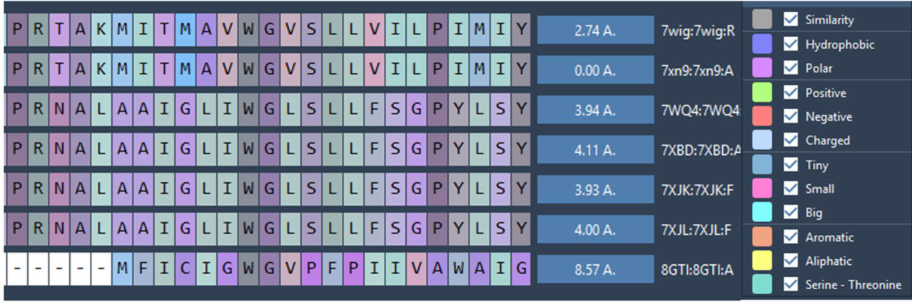

Table 1.
BROOD screening summary.
| Search Mode | Rounds | Compounds Generated |
| Shape & Electrostatics | 4 | 1,557 |
| Shape & Colour | 6 | 3,950 |
| Total | 10 | 5,507 |
Table 2.
Primary docking scores (OpenEye suite) and POSIT confidence.
| Compound | FRED (7XN9) | FRED (7WIG) | HYBRID (7XN9) | HYBRID (7WIG) | POSIT Confidence |
| 8 | −47.67 | −49.69 | −26.50 | −20.23 | GREAT |
| 12 | −49.59 | −52.53 | −24.36 | −20.04 | GREAT |
| 13 | −49.48 | −51.38 | −24.40 | −20.28 | GREAT |
| 14 | −48.92 | −52.22 | −25.00 | −21.32 | GREAT |
| 17 | −49.51 | −53.92 | −20.36 | −19.07 | GREAT |
| 22 | −49.87 | −52.72 | −23.62 | −20.70 | GREAT |
| 27 | −46.47 | −48.40 | −18.12 | −17.01 | GREAT |
| 30 | −45.95 | −49.10 | −26.00 | −18.13 | GREAT |
| L-054,522 | Undocked | Undocked | Undocked | Undocked | POOR |
Table 3.
Confirmatory docking scores (Samson Suite).
| Compound | FITTED (7XN9) | FITTED (7WIG) | AutoDock Vina (7XN9) | AutoDock Vina (7WIG) |
| 8 | −20.91 | −13.78 | −21.24 | −14.96 |
| 12 | −19.31 | −16.04 | −15.61 | −17.18 |
| 13 | −15.78 | −16.06 | −15.40 | −17.01 |
| 14 | −19.59 | −15.83 | −13.94 | −19.11 |
| 17 | −13.85 | −12.33 | −18.02 | −15.59 |
| 22 | −17.05 | −13.90 | −15.38 | −18.79 |
| 27 | −15.77 | −13.13 | −18.14 | −15.05 |
| 30 | −19.74 | −15.41 | −17.93 | −15.59 |
| L-054,522 | −16.60 | −14.54 | −11.39 | −10.29 |
Table 4.
Structures and names of top-ranked compounds.
| Compound | Chemical Structure and Molecular Weight (MW) | Name |
| 8 | [(5S)-5-[[(2R,3S)-2-[[2,5-dihydroxy-3-(2-oxo-3H-benzimidazol-1-yl)cyclopentanecarbonyl]amino]-3-(1H-indol-3-yl)butanoyl]amino]-6-[(2-methylpropan-2-yl)oxy]-6-oxohexyl]azanium. | |
| 12 | [(5S)-5-[[(2R,3S)-2-[[(6S)-6-ethyl-5-(2-oxo-3H-benzimidazol-1-yl)-3,6-dihydro-2H-pyridine-1-carbonyl]amino]-3-(1H-indol-3-yl)butanoyl]amino]-6-[(2-methylpropan-2-yl)oxy]-6-oxohexyl]azanium. | |
| 13 | 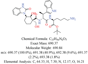 |
[(5S)-5-[[(2R,3S)-2-[[(1R,2S,3R)-2-hydroxy-1-(hydroxymethyl)-3-(2-oxo-3H-benzimidazol-1-yl)cyclohexanecarbonyl]amino]-3-(1H-indol-3-yl)butanoyl]amino]-6-[(2-methylpropan-2-yl)oxy]-6-oxohexyl]azanium. |
| 14 | 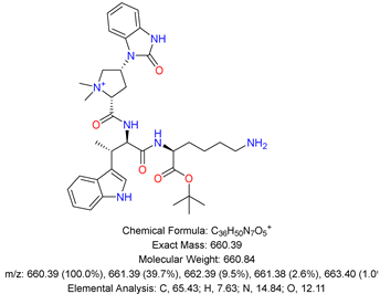 |
[(5S)-5-[[(2R,3S)-2-[[(2R,4R)-1,1-dimethyl-4-(2-oxo-3H-benzimidazol-1-yl)pyrrolidin-1-ium-2-carbonyl]amino]-3-(1H-indol-3-yl)butanoyl]amino]-6-[(2-methylpropan-2-yl)oxy]-6-oxohexyl]azanium. |
| 17 | 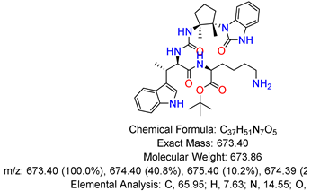 |
[(5S)-5-[[(2R,3S)-2-[[(1S,2S)-1,2-dimethyl-2-(2-oxo-3H-benzimidazol-1-yl)cyclopentyl]carbamoylamino]-3-(1H-indol-3-yl)butanoyl]amino]-6-[(2-methylpropan-2-yl)oxy]-6-oxohexyl]azanium. |
| 22 | 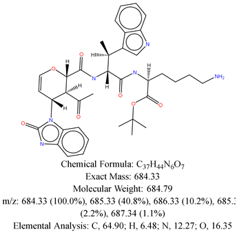 |
[(5S)-5-[[(2R,3S)-2-[[(2S,3S,4R)-3-acetyl-4-(2-oxo-3H-benzimidazol-1-yl)-3,4-dihydro-2H-pyran-2-carbonyl]amino]-3-(1H-indol-3-yl)butanoyl]amino]-6-[(2-methylpropan-2-yl)oxy]-6-oxohexyl]azanium. |
| 27 | 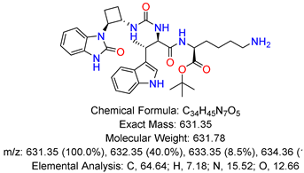 |
[(5S)-5-[[(2R,3S)-3-(1H-indol-3-yl)-2-[[(1S,2S)-2-(2-oxo-3H-benzimidazol-1-yl)cyclobutyl]carbamoylamino]butanoyl]amino]-6-[(2-methylpropan-2-yl)oxy]-6-oxohexyl]azanium. |
| 30 | 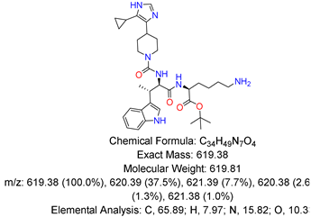 |
[(5S)-5-[[(2R,3S)-2-[[4-(5-cyclopropyl-1H-imidazol-4-yl)piperidine-1-carbonyl]amino]-3-(1H-indol-3-yl)butanoyl]amino]-6-[(2-methylpropan-2-yl)oxy]-6-oxohexyl]azanium. |
Table 5.
T.E.S.T. toxicity predictions.
| Parameter | Compounds 8, 12, 13, 14, 17, 22, 27, 30 | L-054,522 |
| Bioconcentration | Safe | 0.89 |
| Mutagenicity (Ames) | Negative | Negative |
| Oral Rat LD₅₀ | Safe | 1389.4 mg/kg |
| T. pyriformis IGC₅₀ | Safe | Safe |
| Developmental Toxicity | Non-toxicant | Toxicant |
Table 6.
DEEP-PK pharmacokinetic predictions.
| Parameter | 8, 13, 14, 22, 27 | 12, 17, 30, L-054,522 |
| Oral Bioavailability (50%) | Non-bioavailable | Non-bioavailable |
| BBB Penetration | Non-penetrable | Non-penetrable |
| CYP1A2 Substrate | Non-substrate | Non-substrate |
| Half-life | ≥3 hours | <3 hours |
| Carcinogenicity | Safe | Safe |
Table 7.
SwissADME physicochemical predictions.
| Compound | TPSA (Ų) | iLOGP | Water Solubility | GI Absorption | Log Kp (Skin) |
| 8 | 206.18 | 3.51 | Poorly soluble | Low | −8.25 |
| 12 | 167.34 | 4.31 | Poorly soluble | Low | −7.47 |
| 13 | 206.18 | 2.84 | Poorly soluble | Low | −8.48 |
| 14 | 165.72 | −9.96 | Poorly soluble | Low | −8.04 |
| 17 | 177.75 | 3.09 | Poorly soluble | Low | −7.51 |
| 22 | 192.02 | 3.52 | Poorly soluble | Low | −8.49 |
| 27 | 177.75 | 3.90 | Poorly soluble | Low | −7.77 |
| 30 | 159.85 | 4.03 | Poorly soluble | Low | −7.67 |
| L-054,522 | 167.34 | 4.07 | Poorly soluble | Low | −7.76 |
TPSA = topological polar surface area; iLOGP = in-house physics-based LogP; Log Kp = skin permeability coefficient.
Table 8.
Retrosynthetic accessibility summary.
| Compound | ChemAIRS | Spaya RScore | Interpretation |
| 8 | Direct route | 1.0 | Fully precedented; straightforward synthesis |
| 12 | High Risk required | Not scored | Challenging synthesis |
| 13 | High Risk required | Not scored | Challenging synthesis |
| 14 | High Risk required | Not scored | Challenging synthesis |
| 17 | High Risk required | 0.4 | Higher-risk transformations |
| 22 | High Risk required | 0.3 | Higher-risk transformations |
| 27 | High Risk required | 0.4 | Higher-risk transformations |
| 30 | High Risk required | 0.2 | Novel/challenging route |
RScore >0.5 indicates well-precedented routes; lower scores suggest novel transformations with less literature precedent.
Table 9.
Exploratory multi-target docking scores (AutoDock Vina).
| Compound | CRHR1 (8GTI) | GALR2 (7WQ4) |
| 8 | −6.46 | −7.66 |
| 12 | −6.94 | −7.41 |
| 13 | −7.64 | −7.41 |
| 14 | −6.88 | −7.12 |
| 17 | −7.07 | −7.69 |
| 22 | −7.63 | −7.43 |
| 27 | −7.71 | −6.85 |
| 30 | −8.92 | −7.94 |
| L-054,522 | −7.32 | −8.16 |
Disclaimer/Publisher’s Note: The statements, opinions and data contained in all publications are solely those of the individual author(s) and contributor(s) and not of MDPI and/or the editor(s). MDPI and/or the editor(s) disclaim responsibility for any injury to people or property resulting from any ideas, methods, instructions or products referred to in the content. |
© 2026 by the authors. Licensee MDPI, Basel, Switzerland. This article is an open access article distributed under the terms and conditions of the Creative Commons Attribution (CC BY) license (http://creativecommons.org/licenses/by/4.0/).
Copyright: This open access article is published under a Creative Commons CC BY 4.0 license, which permit the free download, distribution, and reuse, provided that the author and preprint are cited in any reuse.