

Submitted:
14 June 2026
Posted:
16 June 2026
You are already at the latest version
Abstract
Urinary schistosomiasis, caused by Schistosoma haematobium, is endemic in many parts of sub-Saharan Africa. The standard microscopic diagnosis is time-consuming and requires expertise to differentiate eggs from artifacts. A study evaluated the performance of a novel staining reagent called Parasimax multicolor® for the rapid detection of Schistosoma haematobium eggs in urine samples from two endemic countries. Samples positive for Schistosoma haematobium were identified by comparing staining techniques, including Parasimax multicolor staining, wet mount preparation and auramide staining. The results showed that Parasimax staining was faster (30 seconds) and more effective for observing Schistosoma haematobium eggs and miracidia, as well as cellular elements such as red blood cells, even when observed with lower magnification lens. These results show that staining with Parasimax reagent is a potential emergency method for detecting eggs and miracidia of Schistosoma haematobium.
Keywords:
schistosomiasis
; diagnosis
; microscopy
; staining
; Parasimax multicolor®
; Schistosoma haematobium
; Mozambique
; Cape Verde
1. Introduction
Schistosomiasis, also known as snail fever, is a neglected tropical disease (NTD) causing significant morbidity in tropical and subtropical regions. Urinary schistosomiasis, caused by Schistosoma haematobium, is endemic in many parts of sub-Saharan Africa, where it poses a persistent public health challenge [1,2]. Schistosomiasis is second only to malaria in terms of the public health burden from parasitic diseases.
Schistosomiasis is a priority neglected tropical disease (NTD) targeted by the World Health Organization (WHO) for control and elimination. The cornerstone of the WHO recommended strategy for reducing morbidity in endemic areas is preventive chemotherapy through the large-scale administration of praziquantel. Control programs are often targeted at high-prevalence groups, particularly school-aged children, who represent the prime reservoir for the parasite and are the most vulnerable to infection and the development of severe morbidity. The school-based delivery of treatment (School-Based Drug Administration programs) has proven to be a cost-effective and logistically viable strategy for achieving high coverage [3,4,5,6].
Although these successful control programs reduce the prevalence and intensity of infection in populations, a new diagnostic challenge emerges. Low-intensity and chronic infections, especially in adults, become increasingly difficult to detect by conventional parasitological methods, such as the microscopic visualization of eggs by traditional staining techniques. The under-detection of these residual infections can lead to a false perception of controlled transmission, allowing infected individuals, even with low parasite burden, to continue serving as reservoirs for the maintenance of the parasite’s life cycle and potentially develop severe long-term pathologies, such as urinary tract fibrosis and bladder carcinoma [7,8].
Therefore, the success of praziquantel-based control programs creates a paradoxical need for more sensitive diagnostic techniques. The accurate identification of individuals with low-level infections is crucial for guiding more targeted control actions, evaluating the true impact of treatment programs, and ultimately advancing towards the goals of eliminating the disease as a public health problem [7,8,9].
The gold standard for diagnosis of schistosomiasis is the microscopic detection of parasite eggs present in urine or stool. However, this method has significant limitations, particularly in the context of control programs. Its sensitivity can be low, especially in low-intensity infections and in chronically infected adults who pass few eggs [10,11]. This often leads to underdiagnosis, as current methods struggle to differentiate eggs from debris and artifacts, requiring skilled technicians and being inherently time-consuming. The need for rapid and accurate diagnosis is paramount, not only for individual patient management but also for monitoring and evaluating the success of mass drug administration (MDA) programs, where infection prevalence and intensity are expected to decline [10,11].
As control efforts intensify and prevalence decreases, the limitations of microscopy become more pronounced. There is a growing need for highly sensitive diagnostic tools to detect low-level and cryptic infections that persist in communities and could serve as reservoirs for continued transmission. Molecular techniques, such as polymerase chain reaction (PCR) targeting specific DNA fragments like the Dra1 repeat sequence, have emerged as powerful alternatives, demonstrating superior sensitivity to microscopy for detecting S. haematobium in urine, even in the absence of visible eggs. Despite their high sensitivity, these molecular methods are often prohibitively expensive, technically demanding, and require sophisticated laboratory infrastructure, making them impractical for routine use in most endemic field settings and primary healthcare laboratories [12,13].
This diagnostic gap highlights the critical need for affordable, rapid, simple, and field-deployable diagnostic improvements that bridge the performance gap between insensitive microscopy and complex molecular assays. This study aims to evaluate the efficacy of a novel multi-color staining reagent, Parasimax multicolor, designed to address these challenges. We hypothesize that Parasimax will significantly reduce the detection time and improve the visual differentiation of S. haematobium eggs from artifacts compared to established methods (direct wet mount, Lugol’s iodine, and Auramine-O fluorescence staining). By enhancing the speed and accuracy of microscopy, the most accessible diagnostic tool Parasimax has shown the potential to be a practical and impactful solution for improving diagnostic efficiency in urinary schistosomiasis control programs in resource-limited settings
2. Materials and Methods
2.1. Study Sites and Sample Collection
Pre-diagnosed positive urine samples for S. haematobium were obtained from a health center in the Marara District of Tete Province, Mozambique (n=1), and from a health center in the Santiago Norte region of Cabo Verde (n=1). Samples were collected and processed according to standard protocols.
2.3. Staining Procedures
The PARASIMAX® reagent, a multicolor stain for auxiliary diagnosis of parasites by light microscopy, was used. The product is registered with the Brazilian Health Regulatory Agency (ANVISA) under number 81649660014, as stated in the manufacturer’s instructions. For each urine sample, four staining techniques were performed in parallel. For the Parasimax technique, 20 µL of pellet was mixed with 10 µL of the Parasimax multicolor reagent on a slide. For the Lugol’s Iodine method, 20 µL of pellet also mixed with 20 µL of Lugol’s iodine solution. The Direct Wet Mount was prepared by applying 20 µL of pellet directly to a slide without any additive. Finally, for the Auramine-O Staining technique, a fixed smear was first stained with a 0.1% Auramine-O solution, then decolorized with an acid-alcohol solution, and subsequently counterstained with methylene blue.
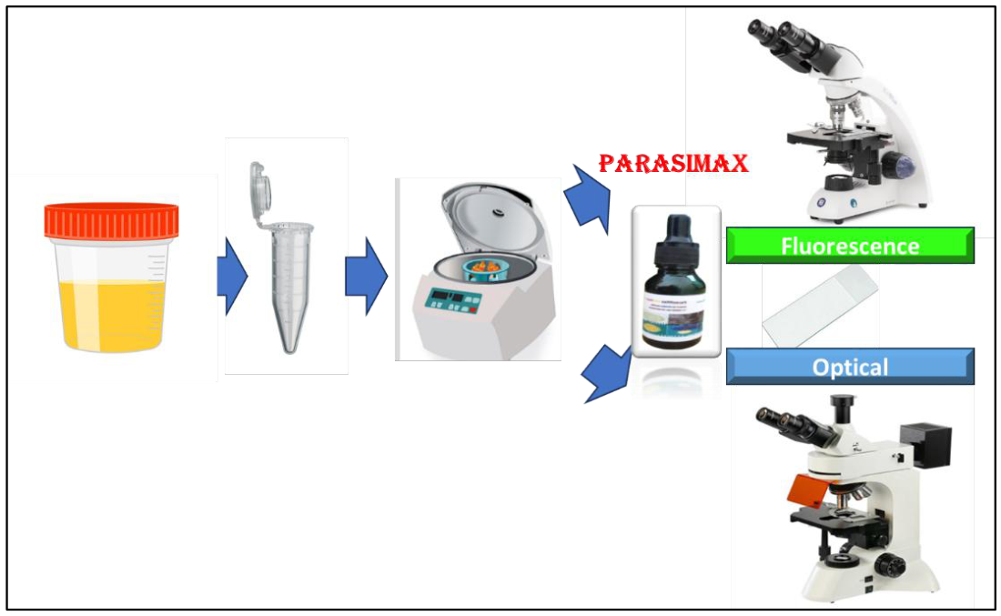
Figure 1.
Procedures for sample observation.
2.4. Microscopy and Analysis
Slides were examined immediately under light microscopy (for Parasimax, Lugol, Direct) and fluorescence microscopy (for Auramine-O). The Mean Detection Time (MDT)—the time taken to visually identify the first egg was recorded for each method at 10x, 20x, and 40x magnifications. The clarity of staining and ease of differentiation from artifacts were qualitatively assessed.
3. Results
Parasimax reagent provided a distinct and consistent color contrast in samples, staining S. haematobium eggs an intense yellow against a blue-green background of other elements and debris (Figure 2a–c). This contrast allowed for rapid egg identification even at lower magnifications (10x).
The Parasimax reagent not only differentiated between the background and the eggs but also allowed the observation of red blood cells and epithelial cells in the samples. This can guide the occurrence of hematuria, which can further guide investigation in suspected negative cases. On the other hand, auramide does not allow the observation of red blood cells due to their destruction after the application of acidic reagents. However, Auramide showed better visualization compared to the direct method due to the concealment of artifacts, as shown in the image (Figure 2d–f).
The Parasimax reagent and fresh observation allowed for the observation of live miracidia moving freely, and also inside the egg. This advantage is not possible with samples treated with auramide. Another major advantage of Parasimax compared to others is the ability to observe eggs and miracidia very quickly with lower magnification. This allows for faster diagnostic speed.
The MDT for Parasimax was consistently and significantly lower than all other methods across all magnifications in both samples (See Table 1). Auramine-O staining, while effective, had the longest preparation and detection time. Lugol’s iodine and the direct method offered no chromatic differentiation. , making egg identification among artifacts more time-consuming and prone to error. This lack of reliable, visible characteristics made species identification subjective, time-consuming, and error-prone (Figure 2g–i). The results were highly consistent between the samples from both Mozambique and Cape Verde.
Massive hematuria is one of the limitations of the standard method due to the hiding of eggs, reducing their visibility. In the figure, we can see the comparison between the two methods. The differential staining allows distinguishing the eggs from red blood cells, which do not stain yellow. In denser areas, this observation would be even more difficult, often being confused with other causes. A feasible way to reduce the interference of these cells is the application of acetic acid. However, this procedure kills the miracidia, preventing their movement, which would otherwise facilitate identification. When observed with a 4x objective lens, it becomes even more difficult, requiring observation with 10x and 40x objectives
Figure 3.
Eggs of Schistosoma haematobium with miracidium in a case of massive hematuria. a) Classical method; b) Parasimax multicolor.
Figure 3.
Eggs of Schistosoma haematobium with miracidium in a case of massive hematuria. a) Classical method; b) Parasimax multicolor.
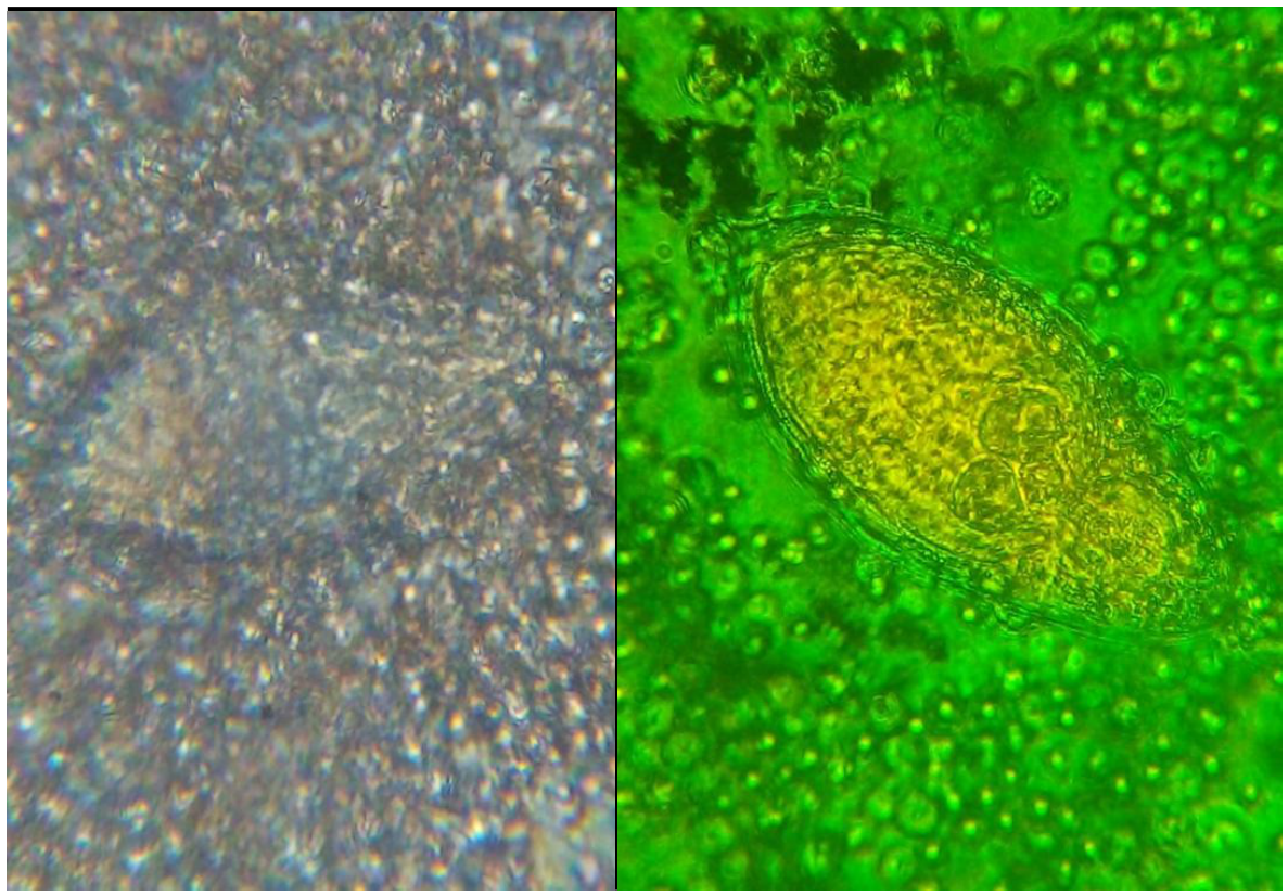
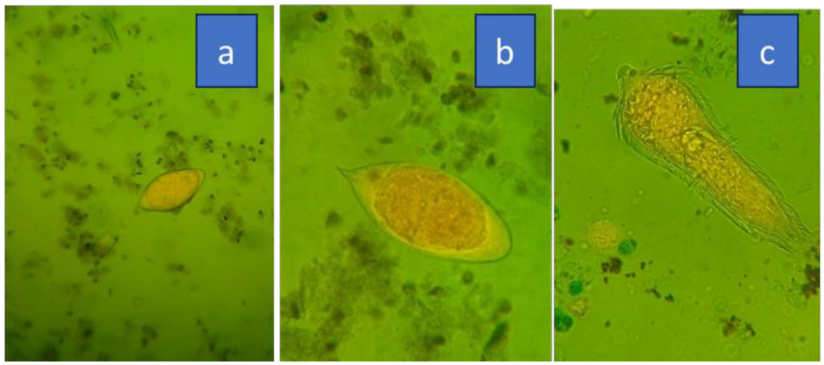
Figure 4.
Schistosoma haematobium eggs (a, b) and miracidium (c) observed with a green filter.
4. Discussion
Some studies have applied different microscopy methods for the observation of Schistosoma haematobium eggs, including the use of 0.4% trypan blue and 1% neutral red, as well as fluorescent stains such as Hoechst 33258. These methods allow the differentiation between viable and non-viable eggs. The first two methods can be performed using light microscopy, while the latter requires fluorescence microscopy. The importance of these methods lies in their ability to assess drug efficacy [14]. In these methods, intact living cells exclude the dye. This approach was applied to observe Schistosoma mansoni eggs. However, the black background does not allow for the observation of other cellular elements that are important for assessing aggressiveness [15,16].
Another study applied special detergents containing calcofluor in their composition, allowing the staining of Schistosoma haematobium eggs for observation under fluorescence microscopy. Lugol’s solution can also be used in combination with a filter membrane. However, the disadvantage is the inactivation of the miracidium [17,18]. The application of Auramine-O provides good contrast; however, its need for expensive equipment and lengthy protocols limits its application in resource constrained settings, where the burden of schistosomiasis is highest. Addressing these limitations could significantly enhance the efficiency of field laboratories and screening programs [19,20,21]. However, the main disadvantage is the inactivation of the miracidium. Hematuria is an important sign of infection by this parasite, which guides the suspicion of infection. Therefore, its detection becomes essential in endemic areas. In most cases, reagent strips are used for primary screening of suspected cases before applying microscopy. Thus, new microscopy methods must have the ability to allow the observation of erythrocytes to also assess the degree of infection-related tissue damage and to evaluate treatment efficacy. Erythrocytes are sensitive cells and undergo lysis if the staining methods are too aggressive, making their observation impossible. Reagent strips are also used in endemic areas where resources for diagnosis [22,23,24].
On the other hand, massive hematuria makes it difficult to observe eggs and miracidia in urine samples, potentially leading to false-negative results when inadequately examined. Therefore, a method that allows clear differentiation of parasites from backgrounds containing cells that interfere with observation is very important. Rapid observation is also an important factor for providing a more appropriate and timely response in emergency situations. Thus, methods that enable faster observation are crucial. Methods that destroy or inactivate miracidia hinder their visualization for diagnosis, as they require an environment with optimal conditions such as osmotic pressure and pH. This issue does not occur when applying the auramine staining method [25].
5. Conclusion
The Parasimax method demonstrated the shortest MDT at all magnifications across both sites (e.g., 30s at 10x), significantly faster than Lugol’s (1 min), direct (1:30 min), and Auramine-O (2 min) methods. Its consistent differential staining (yellow eggs against a blue-green background) facilitated the distinction between eggs and artifacts, improving diagnostic clarity.
The Parasimax multicolor reagent proved to be a rapid, reliable, and user-friendly method for detecting S. haematobium eggs in urine samples. Implementing Parasimax in routine laboratory practice in endemic areas could significantly improve screening efficiency, leading to more timely treatment and bolstering schistosomiasis control efforts. Further validation studies with larger sample sizes are strongly recommended.
Competing interests
The authors declare that they have no competing interests.
Abbreviations
| NTD | Neglected Tropical Disease (NTD) |
| MDT | The Mean Detection Time (MDT |
| LD | Linear dichroism |
| WHO | World Health Organization (WHO) |
| PCR | Polymerase Chain Reaction |
References
- Ally, O.; Kanoi, B. N.; Ochola, L.; Nyanjom, S. G.; Shiluli, C.; Misinzo, G.; Gitaka, J. Schistosomiasis diagnosis: Challenges and opportunities for elimination. PLoS Neglected Trop. Dis. 2024, 18(7), e0012282. [Google Scholar] [CrossRef] [PubMed]
- Cnops, L.; Soentjens, P.; Clerinx, J.; Van Esbroeck, M. A Schistosoma haematobium-specific real-time PCR for diagnosis of urogenital schistosomiasis in serum samples of international travelers and migrants. PLoS Neglected Trop. Dis. 2013, 7(8), e2413. [Google Scholar]
- Gray, D. J.; Ross, A. G.; Li, Y. S.; McManus, D. P. Diagnosis and management ofschistosomiasis. BMJ 2011, 342. [Google Scholar] [CrossRef] [PubMed]
- Gurarie, D.; Yoon, N.; Li, E.; Ndeffo-Mbah, M.; Durham, D.; Phillips, A. E.; King, C. H. Modelling control of Schistosoma haematobium infection: predictions of the long-term impact of mass drug administration in Africa. Parasites Vectors 2015, 8(1), 529. [Google Scholar] [CrossRef] [PubMed]
- Hong, S. T. Review of recent prevalence of urogenital schistosomiasis in Sub-Saharan Africa and diagnostic challenges in the field setting. Life 2023, 13(8), 1670. [Google Scholar] [CrossRef] [PubMed]
- Hotez, P. J. Vaccines in a time of global boiling and megacities. Trends Parasitol. 2025, 41, 167–169. [Google Scholar] [CrossRef] [PubMed]
- King, C. H.; Kittur, N.; Binder, S.; Campbell, C. H., Jr.; N’Goran, E. K.; Meite, A.; Colley, D. G. Impact of different mass drug administration strategies for gaining and sustaining control of Schistosoma mansoni and Schistosoma haematobium infection in Africa. Am. J. Trop. Med. Hyg. 2020, 103((1) Suppl, 14. [Google Scholar] [CrossRef] [PubMed]
- Midzi, N.; Sangweme, D.; Zinyowera, S.; Mapingure, M. P.; Brouwer, K. C.; Kumar, N.; Mduluza, T. Efficacy and side effects of praziquantel treatment against Schistosoma haematobium infection among primary school children in Zimbabwe. Trans. R. Soc. Trop. Med. Hyg. 2008, 102(8), 759–766. [Google Scholar] [CrossRef] [PubMed]
- Nagi, M. A. Evaluation of a programme for control of schistosoma haematobium infection in Yemen. East Mediterr. Health J. 2005, 11(5-6), 977–87. [Google Scholar] [CrossRef] [PubMed]
- Oluwasogo, O. A.; Fagbemi, O. B. Prevalence and risk factors of Schistosoma haematobium infections among primary school children in Igbokuta Village, Ikorodu North Local Government, Lagos State. J. Nurs. Health Sci. 2013, 2(6), 62–68. [Google Scholar] [CrossRef]
- Serra, J. T.; Silva, C.; Sidat, M.; Belo, S.; Ferreira, P.; Ferracini, N.; Conceiçao, C. Morbidity associated with schistosomiasis in adult population of Chókwè district, Mozambique. PLoS Neglected Trop. Dis. 2024, 18(12), e0012738. [Google Scholar] [CrossRef] [PubMed]
- ten Hove, R. J.; Verweij, J. J.; Vereecken, K.; Polman, K.; Dieye, L.; van Lieshout, L. Multiplex real-time PCR for the detection and quantification of Schistosoma mansoni and S. haematobium infection in stool samples collected in northern Senegal. Trans. R. Soc. Trop. Med. Hyg. 2008, 102(2), 179–185. [Google Scholar] [CrossRef] [PubMed]
- Utzinger, J.; Raso, G.; Brooker, S.; De Savigny, D.; Tanner, M.; Ørnbjerg, N.; N’goran, E. Schistosomiasis and neglected tropical diseases: towards integrated and sustainable control and a word of caution. Parasitology 2009, 136(13), 1859–1874. [Google Scholar] [CrossRef] [PubMed]
- Forson, P. O.; Tetteh-Quarcoo, P. B.; Ahenkorah, J.; Aryee, R.; Okine, E. N.; Afutu, E.; Ayeh-Kumi, P. F. Ability of vital and fluorescent staining in the differentiation of Schistosoma haematobium live and dead eggs. Med. Sci. 2019, 7(4), 64. [Google Scholar] [CrossRef] [PubMed]
- Model, M. A.; Anyan, W. K. A promising microscopic technique for detecting Schistosoma mansoni eggs. Diagn. Microbiol. Infect. Dis. 2026, 114(1), 117093. [Google Scholar] [CrossRef] [PubMed]
- Braun, L.; Hazell, L.; Webb, A. J.; Allan, F.; Emery, A. M.; Templeton, M. R. Determining the viability of Schistosoma mansoni cercariae using fluorescence assays: An application for water treatment. PLoS Neglected Trop. Dis. 2020, 14(3), e0008176. [Google Scholar] [CrossRef] [PubMed]
- Ngasala, B.; Juma, H.; Mwaiswelo, R. O. The usefulness of indirect diagnostic tests for Schistosoma haematobium infection after repeated rounds of mass treatment with praziquantel in Mpwapwa and Chakechake districts in Tanzania. Int. J. Infect. Dis. 2020, 90, 132–137. [Google Scholar] [CrossRef] [PubMed]
- Knopp, S.; Ame, S. M.; Hattendorf, J.; Ali, S. M.; Khamis, I. S.; Bakar, F.; Rollinson, D. Urogenital schistosomiasis elimination in Zanzibar: accuracy of urine filtration and haematuria reagent strips for diagnosing light intensity Schistosoma haematobium infections. Parasites Vectors 2018, 11(1), 552. [Google Scholar] [CrossRef] [PubMed]
- Hanscheid, T.; Vieira, S.; Ferreira, P.; Carvalho, T.; Belo, S.; Grobusch, M. P. The fluorescent acid-fast stain, auramine-O, stains schistosome eggs and may be an aid for detection. An old technique with a useful future? Travel Med. Infect. Dis. 2020, 35, 101569. [Google Scholar] [CrossRef] [PubMed]
- Teixeira, C. G.; Candal, M. S.; Pereira, H. S.; da Silva, G. D. C. F.; Oliveira, L. D. O. V.; de Almeira Pereira, D. S. C.; Hanscheid, T. (2024). Auramine stainning and adherence of Schistosoma mansoni eggs onto microscope slides for detection step of Helmintex method. Simpósio Internacional sobre Esquistossomose.
- Panic, G.; Flores, D.; Ingram-Sieber, K.; Keiser, J. Fluorescence/luminescence-based markers for the assessment of Schistosoma mansoni schistosomula drug assays. Parasites Vectors 2015, 8(1), 624. [Google Scholar] [CrossRef] [PubMed]
- Deribew, K.; Yewhalaw, D.; Erko, B.; Mekonnen, Z. Urogenital schistosomiasis prevalence and diagnostic performance of urine filtration and urinalysis reagent strip in schoolchildren, Ethiopia. PLoS ONE 2022, 17(7), e0271569. [Google Scholar] [CrossRef] [PubMed]
- Degarege, A.; Erko, B.; Brett-Major, D. M.; Levecke, B.; Animut, A.; Negash, Y.; Bilder, C. R. Performance of urine reagent test strips in detecting schistosoma haematobium infection in individual and pooled urine samples. Microorganisms 2025, 13(3), 510. [Google Scholar] [CrossRef] [PubMed]
- Ndum, N. C.; Ali, S. M.; Ali, M. N.; Bergelin, J.; Hattendorf, J.; Hoekstra, P. T.; Knopp, S. Evaluation of six different tests for Schistosoma haematobium diagnosis in a near-elimination setting: a prospective observational diagnostic accuracy study. medRxiv 2026, 2026-02. [Google Scholar]
- Ouji, Y.; Hamasaki, M.; Misu, M.; Yoshikawa, M.; Hamano, S. Simple preservation of schistosome eggs with high infectivity up to 12 weeks. Parasitol. Int. 2025, 106, 103020. [Google Scholar] [CrossRef] [PubMed]
Figure 2.
Shistossoma haematobium eggs observed with different technique.
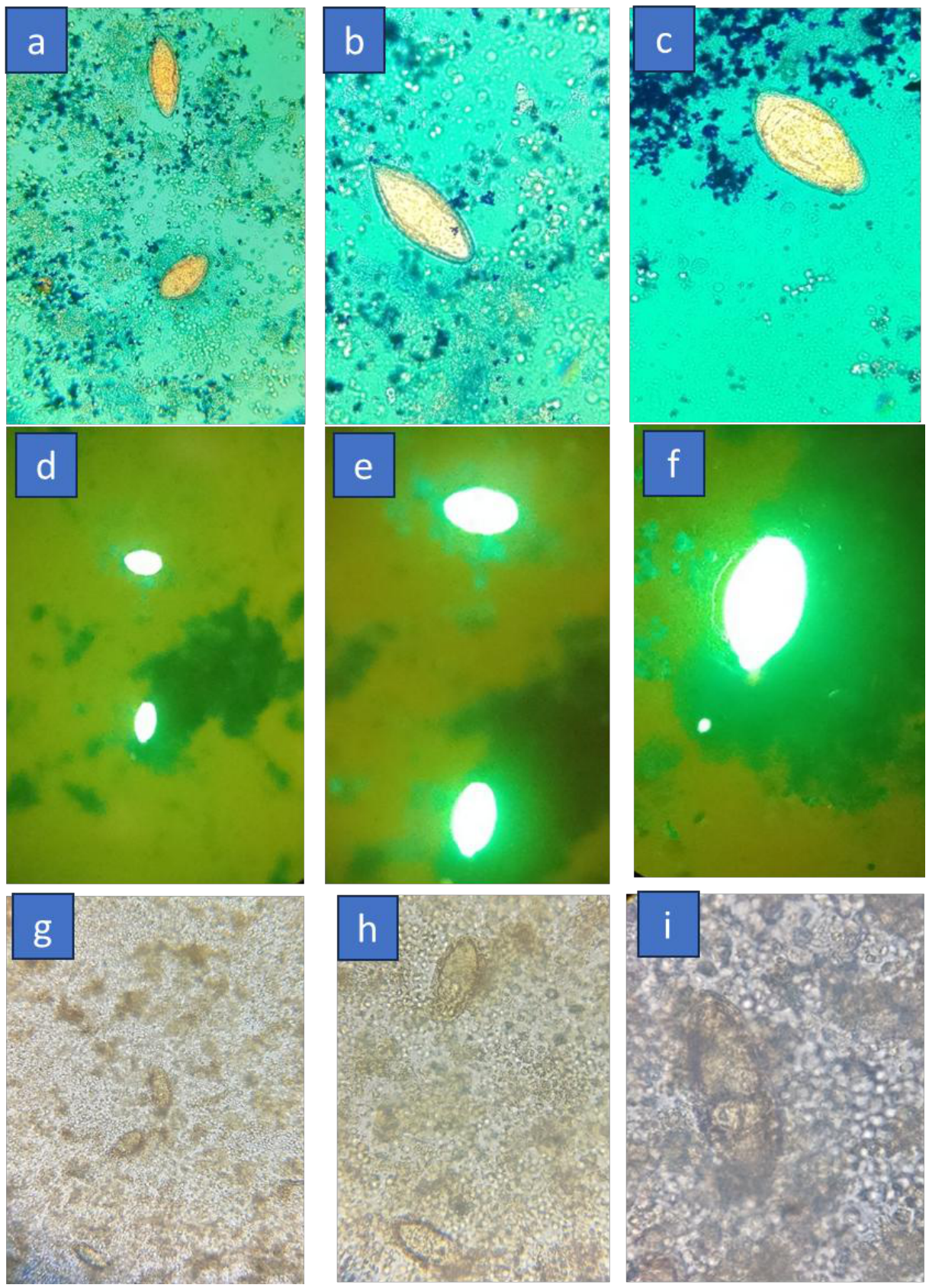

Table 1.
Mean detection time in duplicates for S. haematobium eggs.
| Methods | 100x | 200x | 400x* |
| Direct | 90 | 120 | 180 |
| PARASIMAX | 30 | 40 | 60 |
| Lugol’s Iodine | 60 | 90 | 120 |
| Auramine-O | 120 | 150 | 180 |
*: Microscope magnification.
Disclaimer/Publisher’s Note: The statements, opinions and data contained in all publications are solely those of the individual author(s) and contributor(s) and not of MDPI and/or the editor(s). MDPI and/or the editor(s) disclaim responsibility for any injury to people or property resulting from any ideas, methods, instructions or products referred to in the content. |
© 2026 by the authors. Licensee MDPI, Basel, Switzerland. This article is an open access article distributed under the terms and conditions of the Creative Commons Attribution (CC BY) license (http://creativecommons.org/licenses/by/4.0/).
Copyright: This open access article is published under a Creative Commons CC BY 4.0 license, which permit the free download, distribution, and reuse, provided that the author and preprint are cited in any reuse.


