
Preprint
Hypothesis

This version is not peer-reviewed.

Calpains as Potential Regulators of Microtubule-Associated Cytokinesis: A Cross-Eukaryotic Unifying Hypothesis

Submitted:

12 June 2026

Posted:

12 June 2026

You are already at the latest version

Abstract
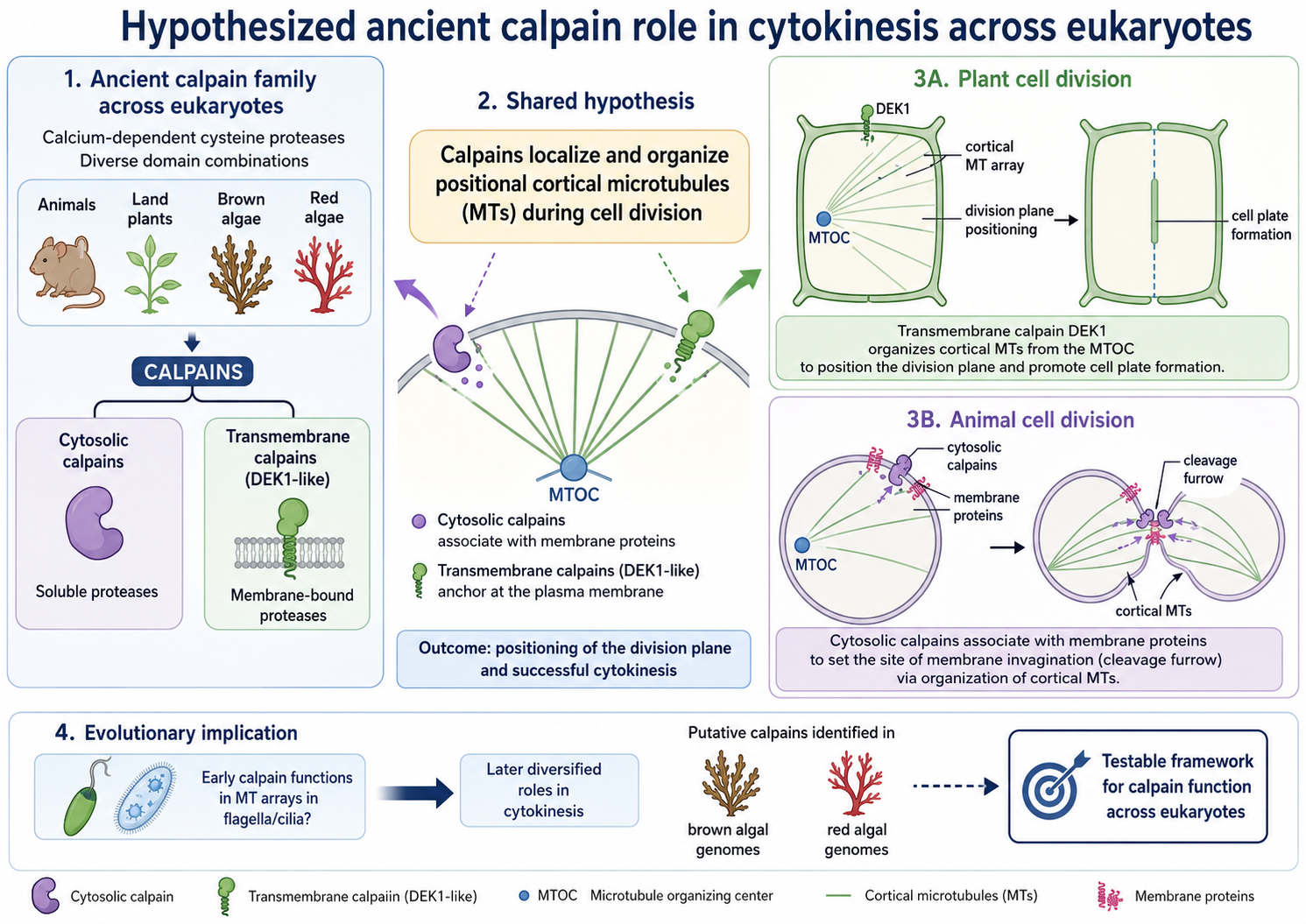
Calpains constitute an ancient, extensive family of calcium-dependent cysteine proteases found in some bacteria and most eukaryotes. They are involved in a wide variety of developmental and cellular processes and are implicated in major human diseases, but whether they share an ancestral or broadly conserved cellular role remains unclear. Beyond their core CysPc catalytic domain, calpains contain diverse domain combinations and can be either cytosolic or membrane bound. Here, we develop the hypothesis that both cytosolic and transmembrane calpains may contribute to cytokinesis through positional anchoring and organization of microtubules (MTs). We propose that during plant cell division, the singular transmembrane calpain DEK1 play a role in localizing and organizing the array of cortical MTs from the microtubule organizing center (MTOC) and may thereby position the cell division plane, potentially affecting preprophase band placement and subsequent cell plate formation. Similarly, during cell division in animals, their cytosolic calpains may be involved in setting the point of membrane invagination via their association with membrane-bound proteins. We discuss this novel model for calpain activity in the context of data from the animal and plant literature, as well as of our discovery of putative calpain sequences in both brown and red algal genomes. These findings are consistent with the view that calpains were present early in eukaryotic evolution and diversified alongside distinct modes of cell division. Finally, we consider the possibility that early calpain functions may have been linked to the formation and function of MT arrays in flagella and cilia, from which later roles in cytokinesis might have evolved. This model is intended as a testable framework for future studies of calpain function across eukaryotes.
Keywords: 
;  ;  ;  ;  ;  ;  ;  

Introduction

The growth and development of all multicellular organisms depend on cell divisions that are controlled by the cell cycle. Progress through the cell cycle is directed by cyclin-dependent-kinases (CDKs) that initiate DNA synthesis and drive the cell through the cycle. The mitotic cell cycle involves chromosome condensation and nuclear envelope breakdown, mitogen-activated protein kinase (MAPK)-mediated attachment of the chromosomes to mitotic spindle fibers, followed by sister chromatid separation and physical division of the cytoplasm and organelles by cytokinesis. A key facet of mitosis is the organization of microtubules (MT) into the mitotic spindle by the animal centrosome or plant microtubule organizing center (MTOC), cytoskeletal components that orient the chromosomes along a set division plane establishing the subsequent division and new cell wall position (Table 1).
Several cell division mechanisms occur in eukaryotes. In animals, the cell division plane is determined by the position of the mitotic spindle anchored at the opposite ends of the cell by the centrosomes, consisting of two centrioles that organize the MT as the main MTOC. Cytokinesis then occurs through the formation of an actomyosin contractile ring that constricts the cell membrane to form a cleavage furrow that divides the cell in two across the short axis (Figure 1A). This ancient evolutionary process developed before the split of the eukaryotic supergroups (Pollard and O'Shaughnessy 2020; Yubuki and Leander 2013).
In most land plant cells, at the onset of mitosis the location of the cell division plane is set by the preprophase band (PPB). The PPB is a transient cortical ring of MT, actin filaments, endoplasmic reticulum, and associated proteins that is assembled and orientated perpendicular to acentrosomal MTOC activity and encircles the nucleus (Schaefer et al., 2017; Domozych and Bagdan 2022). The PPB leaves a positional cue at the future cell division site that guides the activity of phragmoplasts, assemblies of MT and Golgi vesicles that are organized by MTOC activity and fuse to form a cell plate. The cell plate then expands outward into a new cell wall that separates the daughter cells (Figure 1B). This group of organisms lack centrosomes and cell division depends on guidance by distributed MTOCs. Interestingly, the somatic cells of the basal liverwort plant Marchantia polymorpha initially form centrosome-like structures called polar organizers that appear before the PPB and serve as the MTOC (Buschmann et al., 2016). The cells subsequently divide like those of higher plants under direct guidance of the PPB by forming a cell plate (Brown and Lemon 2011). This dual mode of cytokinesis utilizing both centrosome-like structures and PPB is suggested to exemplify a stepwise evolutionary transition from green algae to land plants (Buschmann and Zachgo 2016) and is important to enable the asymmetric cell divisions that are especially important to meristem function, branching and setting 3D tissue orientation (Graham et al., 2000). Like land plants, most fungi also lack centrosomes, and their cell divisions are controlled by a MTOC named the spindle pole body (SPB) that plays a role in MT nucleation and additionally contributes to nuclear envelope breakdown (Jaspersen 2021).
Brown macroalgae, which are part of the SAR supergroup consisting of Stramenopiles, Alveolates and Rhizarians, have a combined set of structures and their cell division occurs via a combination of the two processes. That is, like animals, brown algae contain centrosomes that act as the MTOC (Pickett-Heaps,1974). Yet rather than forming a contractile ring and furrow they divide by centrifugal growth and Golgi vesicle-mediated fusion of wall material from inside to outside to form a cell plate (Nagasato and Motomura, 2002; Motomura et al., 2010), similar to land plants (Aoki et al., 2023) (Figure 1C). The latter behavior is likely explained by the fact that, like plants, brown algae harbor physiologically important cell walls allowing giant kelp species to grow impressive forest canopies, which depends on new cell wall deposition to complete cell division and grow large structures (Choi et al., 2024). Yet brown algae lack the cortical MTs, PPB and phragmoplasts present in land plants (Katsaros et al., 2006; Bellinger et al., 2023).
A fourth eukaryotic supergroup is the Excavata group of unicellular, mostly flagellated protists. Although it is debated whether this group is truly monophyletic (Burki et al., 2020), Excavates are generally characterized by their asymmetrical, “excavated” feeding grove and many undergo a specialized cell division by longitudinal fission where the parental cell splits lengthwise in two. This process is well described in the human parasite Trypanosoma brucei, in which basal bodies that anchor the flagella of the cell serve as centriole-like structures (Vaughan and Gull 2016). The basal body is a MT-composed organelle that like a centriole has a core ninefold-symmetrical MT and cartwheel structure and is physically connected to a kinetoplast consisting of mitochondrial DNA (Figure 1D). At the onset of mitosis, the basal body replicates and the new basal body assembles a new flagellum, the tip of which defines the position where cytokinesis initiates (Kohl et al., 2003). Cytokinesis then proceeds via ingression of a cleavage furrow along the longitudinal axis of the cell from anterior to posterior (Vaughan and Gull 2008; Wheeler et al., 2013; Zhou et al., 2016), without the formation of a contractile ring at the cleavage furrow site (Garcia-Salcedo et al., 2004). In all these cases, the positioning of the MTOC is critical for cytokinesis to proceed in the proper plane and for the correct partitioning of nuclei and organelles into the two daughter cells.
Calcium affects nearly every aspect of life by linking external stimuli to intracellular events via movement through ion channels or by release from cellular stores (Clapham 2007; Carafoli & Krebs 2016; Bootman & Bultynck 2020). Changes in calcium concentrations are universal regulators from bacteria to animals, evolutionary identified across early eukaryote linages before the split leading to the different phylogenetic groups of plants, brown algae, fungi and animals (Cai et al. 2014). Calcium plays a crucial role throughout the cell cycle in nuclear envelope breakdown, chromosome segregation, and cytokinesis (Nugues et al 2022). In fission yeast and animals, calcium spikes at the onset of cytokinesis initiate cleavage furrow ingression and subsequent plate formation (Atilla-Gokcumen et al 2010; Hepler 2015; Poddar et al 2021). At the molecular level, calcium is linked to the cell cycle by motility and motor proteins such as dynein and kinesin, RhoGTPases, initiation of the MAPK cascade and the cytoskeleton (Tan et al. 2006; Cai, 2014; Saternos et al 2020). Despite its pivotal role, a possible universal link remains to be detected between calcium and the many connected biological functions in most living organisms.
F cells occurs via mechanosensitive channels, force-sensing integral membrane proteins found in all kingdoms of life that are activated by mechanical stimuli exerted on cell membranes (Ridone et al., 2019). Among these, Piezo plays an important role in regulating the cell cycle from centrosome integrity through divisions until cell death (David et al. 2022). In mammals, Piezo1 has been shown to anchor cytoskeletal components and functions upstream of calpains (Li et al., 2014; Friedrich et al., 2019; Zhang et al., 2021, Chuntharpursat-Bon et al 2023), Ca2+-activated cysteine proteases that are found in some bacteria and most all eukaryotes (Sorimachi et al. 2011). Mammalian calpains are grouped into classical and non- classical calpains based on their domain organization (Croall and Ersfeld 2007). The classical calpains contain a core CysPC (PC1 and PC2) domain as well as C2, calpain-type ß-sandwich (CBSW), and penta-EF hand (PEF) domains (Veselenviova et al 2022), whereas the non-classical calpains contain additional zinc finger (Zn), microtubule interacting and transport (MIT), and WW domain combinations (Ono and Sorimachi 2012; Safranek et al., 2023). All vertebrate and insect calpains are cytosolic proteins that function in the cytoplasm, not anchored to cell membranes, although they are associated with membranes and in some cases this association is linked to their activation (Araujo et al., 2018; Safranek et al., 2023). Calpains are central to many fundamental cellular processes, including cytoskeletal remodeling, cell signaling and apoptosis, centrosome functions, centrosome duplication and positioning, MT polarization, connecting chromosomes to spindles, chromosome positioning, and cell cycle progression (Honda et al. 2004; Storr et al. 2011; Valls et al. 2025). In mammals 14-15 genes encode calpain proteins, some of which are specific to certain tissues, whereas others are ubiquitous (Opsahl-Sorteberg et al., 2024).
In contrast, plants including primitive mosses and liverworts contain a single unique calpain called DEFECTIVE KERNEL1 (DEK1). Unlike animal calpains, at its amino terminus the DEK1 protein contains an extensive membrane anchor consisting of 21-24 predicted transmembrane domains interrupted by an unstructured loop or channel domain (Lid et al 2002; Kumar et al 2010; Perroud et al 2020). This is followed by an intracellular linker containing a LamininG-like domain (LGL), and the calpain protease comprised of a CysPC and a CBSW domain (Lid et al 2002; Johansen et al 2016). DEK1 is highly expressed in actively dividing cells, including stem cells (Lid et al., 2005; Johnson et al., 2005; Liang et al. 2015), and the maize and Arabidopsis proteins are localized at the plasma membrane, ER and in endosome-like compartments (Tian et al. 2007; Johnson et al. 2008). During 3D growth in the moss Physcomitrella patens, PpDEK1 protein exhibits a more polarized localization restricted to the plasma membrane between recently divided cells (Perroud et al., 2020). Multiple studies have shown that DEK1 is essential for plant embryogenesis and post-embryonic development, epidermal identity, cell-to-cell adhesion, 3D cell orientation, stem cell maintenance and gene regulation, microtubule orientation, and cell wall positioning (Lid et al. 2002; Lid et al. 2005; Johnson et al. 2005; Tian et al. 2007; Johnson et al. 2008; Liang et al. 2015; Demko et al. 2024; Chen et al. 2024). These diverse biological roles have led previous investigators to conclude that DEK1 has a variety of activities within the cell (Demko et al., 2024). Here, we consider whether these activities as well as those of animal cytosolic calpains could be linked by a single core function involving calcium-responsive regulation of membrane-cytoskeleton interactions during cell division.
A unifying hypothesis for calpain involvement in cytokinesis
Here we present our unifying hypothesis that calcium-regulated calpains may contribute to mitosis and cytokinesis by modulating microtubule organization, cell division plane positioning, and cytokinetic progression. This model-generating hypothesis is based on correlations among calpain domain architecture, localization, calcium signaling, cytoskeletal organization, and cytokinesis phenotype datasets from the literature (Table 1). It is intended as a framework for community testing rather than as a demonstrated general mechanism and should not be interpreted as evidence that calpains directly anchor MTOCs or centrosomes in all organisms. Rather, we propose calpain function in MTOC/centrosome positioning as a possible unifying mechanism that can be experimentally tested against alternative explanations, such as multiple indirect roles for calpains in cytoskeletal remodeling, membrane trafficking, mechanotransduction, cell wall deposition, and proteolytic regulation of mitotic factors.
In animals, cell division is mediated by the main centrosomal MTOC positioning that directs MT spindle formation, and then sets the position of the cytokinetic contractile ring forming the furrow that divides the cell. We propose that cytosolic calpains set the centrosome position, thereby directing and facilitating the progress of furrow ingression and enabling the completion of cell division at the proper position (Figure 2A). This activity may occur in response to calcium pulses that coincide with the initiation of furrow ingression (Atilla-Gokcumen et al., 2010; Poddar et al., 2021) and may involve CysPC-mediated degradation of proteins such as RhoA and fodrin, which are confirmed targets of calpain-mediated cleavage (Kulkarni et al., 2002; Sato et al., 2004). RhoA is a small GTPase that, when activated, stimulates actin nucleation and myosin activation to form the contractile ring and is sufficient for furrow initiation (Drechsel et al., 1997; Wagner and Glotzer, 2016). Fodrin is a non-erythroid form of spectrin that functions in metazoans to nucleate MT from centrosomes, elongate spindles, and position chromosomes at the metaphase plane, and is required for mitosis progression along with kinesins and dyneins (Shashikala et al 2013; Nellikka et al 2019; Sridhara and Shimamoto 2024). Thus, the available data are consistent with, although not evidence of, the hypothesis that cytosolic calpains participate in MTOC/centrosome-linked cell division plane control.
Alternatively, such activity could occur via multiple distinct calpain activities, such as proteolytic regulation of cytoskeletal or membrane-associated substrates, interactions with microtubule-associated proteins, or indirect signaling pathways activated by cytokinetic calcium pulses (Atilla-Gokcumen et al., 2010; Poddar et al., 2021). In this scenario calpains would not necessarily serve as physical anchors for centrosomes. Instead, they might help regulate the local protein complexes that connect calcium signaling, cytoskeletal remodeling, and membrane deformation during cytokinesis.
In land plants, cytokinesis is initiated by two MTOC anchored at the opposite ends of the cell and their interconnected MT, which set the perpendicularly oriented cortical MTs into the PPB. The PPB positions the final cytokinetic division by the phragmoplast that directs cell plate formation and deposits cell wall material between the two newly forming daughter cells. The membrane-anchored calpain DEK1 is not required for PPB formation but is necessary for the arrangement of MTs into organized patterns and to correctly position the PPB (Liang et al., 2015). Therefore, we hypothesize that DEK1 may act to anchor the MTOCs and organize the subsequent positioning of the CMTs and PPB (Figure 2B), setting the new cell wall position and also facilitating the deposition of new cell wall material at the correct position by the phragmoplasts (Yan et al 2023). We propose this could take place through direct DEK1 interactions with MT-associated proteins, mechanosensitive calcium signaling, or regulation of proteolytic substrates. Yet we recognize that at present there is no direct evidence that DEK1 physically binds or anchors plant MTOC activity, and therefore DEK1-mediated MTOC or MT anchoring is only one possible core mechanism that could explain the observed developmental and cytoskeletal phenotypes. An alternative explanation is that it could occur indirectly through independent effects of DEK1 on membrane trafficking, cellulose synthase behavior, or cell wall mechanics.
Brown algal somatic cell division combines features that occur separately in animals and land plants. Brown algal mitosis depends on centrosomes that act as MTOCs, like animal cells, followed by plate formation to set the new cell wall, like land plant cells (Table 1). This combination makes brown algae a good model system in which to assess whether membrane-associated calpains might be involved in coordinating centrosome-associated MTs with cell plate formation. Until recently it was not feasible to probe the calpain diaspora among the diverse families of brown algae, but the release of several dozen high-quality brown algal genomes (Diesel et al., 2023; Denoeud et al., 2024; De Weese et al., 2025) has enabled this type of investigation. We used the conserved CysPC domain amino acid sequence from Arabidopsis thaliana to identify calpain sequences across brown algal genomes. We identified a single amino acid sequence in each brown algal genome that aligns well with conserved CysPC domain amino acid sequences from oomycetes, land plants, moss and fungi (Figure 3). This finding suggests that DEK1-like calpain sequences are broadly represented among brown algal genomes. Yet, because genome complexity, gene annotation, and search sensitivity vary among species, our results do not prove that all brown algae contain only a single calpain gene. Future HMM-based searches and reciprocal sequence comparisons using improved genome annotations will be needed to rule out the presence of more divergent or fragmented calpain-like sequences.
The CysPC domain consists of the PC1 (Figure 3A) and PC2 (Figure 3B) domains and contains three amino acids - cysteine, histidine, and asparagine - that make up the catalytic triad in canonical animal and land plant calpains (Berti and Storer, 1995; Hosfield et al., 1999). Among the brown algal CysPC sequences, the asparagine residue in the PC2 domain is conserved in the majority of the brown algae species examined, but has been substituted with a tyrosine residue in F. serratus, P. canaliculata, and A. nodosum, three members of the Fucaceae family (Figure 3B). The histidine residue at position 1848 in the PC2 domain is not conserved in any brown algal or oomycete species in our dataset, having been replaced with an alanine, serine or threonine residue (Figure 3B). Each brown algal and oomycete sequence instead features one or two adjacent histidine residues at positions 1781-82 in the PC2 domain (Figure 3B). The cysteine residue at position 1670 in the CysPC domain also is not present in any brown algae calpain sequence identified, having been substituted with a serine residue in many of them and by a threonine, alanine or glycine reside in the others (Figure 3A). This cysteine residue is likewise replaced by a serine, alanine or glycine residue in the PC1 domain of oomycete calpain sequences (Figure 3A). However, nearly all of the brown algal calpain sequences contain a cysteine residue at position 1632, and the oomycete calpain sequences contain a cysteine residue at position 1630 (Figure 3A). Taken together, these data indicate the presence of conserved calpain-like CysPC domains in brown algae. It remains to be determined whether these proteins retain protease activity via their non-canonical catalytic triad arrangements or function partly or fully through non-proteolytic mechanisms.
Most of the identified brown algal calpain-like sequences contain at least one predicted domain in addition to the CysPC core. However, in five brown algae genomes only a CysPC domain is predicted (Figure 4A), likely due to incomplete sequence information or annotation. The calpain sequences of two additional species, D. mesarthrocarpus and H. paniculata, are annotated as having a CysPC domain and a short C2 domain near the carboxyl terminus and may also be incomplete. We predicted the calpain sequences of Saccharina latissima, S. japonica, S. promiscuus and two other species based on several adjacent contigs (Figure 4A). The calpain sequences in these five and the majority of the other brown algal species are predicted to contain a series of TM domains animo terminal to the CysPC domain, and 18 species across several families are predicted to contain an additional C2 domain close to the amino terminus of the predicted protein (Figure 4A). Unexpectedly, the calpain sequence from Desmarestia dudesnayi uniquely harbors nested among the TM motifs two EF hand domains, which are present in some metazoan calpains but are absent from land plants (Opsahl-Sorteberg et al., 2024). Phylogenetic analysis of the brown algal calpain full-length amino acid sequences confirms the presence of a single calpain sequence in each brown algae genome (Figure 4B), as well as strong sequence conservation within the various brown algal families represented. The domain combinations identified suggest that many brown algal calpain-like proteins are membrane-associated and potentially calcium-responsive. Although domain architecture alone cannot establish cellular function, based on their structural similarity to DEK1 we propose that these transmembrane calpains may likewise contribute to centrosome-associated MT organization and/or cell plate positioning in brown algae – a hypothesis that remains to be tested experimentally.
Beyond the calpain-like sequences identified in brown algal genomes, our searches of red algal genome and transcriptome resources using the conserved CysPC domain amino acid sequence from A. thaliana returned a hit from Rhodosorus marinus and Gracilaria vermiculophylla. The CysPC domain of both red algal calpain sequences contains the Cys-His-Asn catalytic triad in the same positions as in the green algal and land plant CysPC domains (Figure 3). Overall, the predicted Rhodosorus marinus calpain protein features a GRF zinc finger Zf_GRF-CycPC domain configuration also present in green algae, phytoplankton, and all Plasmodium species within the Aveolates (Russo et al., 2009; Zhao et al., 2012). The predicted Gracilaria vermiculophylla calpain protein features a CysPC-CBSW domain architecture present in nearly all eukaryotic supergroups and considered to be one of the ancient calpain types (Zhao et al., 2012). These findings indicate that calpains are present across the red and green algal and land plant lineages of the Archaeplastida supergroup. Broader genome sampling and experimental validation will be required to determine how widely they are distributed and whether they have cytokinetic, cytoskeletal, or other cellular functions.
Our hypothesis that calpains play a primary role in cell mitosis by positioning the MTOC and the cleavage furrow or PPB, and thereby orienting the cell division plane is compatible with known functions for these proteins in the animal cell division process. Cytosolic calpains already have been shown to function in MT-associated processes during mammalian mitosis. In human HeLa cells, CAPN2 protein levels are elevated during mitosis, and this activation is necessary for proper MT attachment to kinetochores and correct alignment of the chromosomes at the spindle pole prior to their segregation into the two daughter cells (Honda et al., 2004). In human breast cancer cell lines, CAPN2 plays a key role during the progression of mitosis and cytokinesis by reducing the levels of the LIM Kinase-1 (LIMK1) protein that phosphorylates the actin-severing protein cofilin-1 (CFL1) at critical steps of mitosis (Rodriguez-Fernandez et al., 2021). A similar mechanistic role for calpains in indirectly modulating the activity of MT binding proteins during mitosis can likewise be envisioned. CAPN2 is also known to degrade fodrin (Siman et al., 1984; Sato et al., 2004), which in brain cells co-localizes with MTs and plays a role in MT spindle organization and mitotic progression by associating with g-tubulin to promote the transport of the tubulin to centrosomes for MT nucleation (Shashikala et al., 2013; Nellikka et al 2019). Fodrin concentrations peak prior to the initiation of mitosis, while the proteins dissociate from the centrosomes after prophase (Shashikala et al., 2013). It has been hypothesized that calpains mediate the cleavage of fodrin from g-tubulin to ensure the proper progression of mitosis (Shashikala et al., 2013). Thus, fodrin may be a key target of cytosolic calpain activity during mitotic microtubule nucleation in metazoans, where it is predominantly found (Sreeja et al 2022).
In addition, some non-canonical mammalian calpains contain a microtubule interacting and trafficking (MIT) domain that is absent from calpains found thus far in bacteria and tetrahymena (Zhao et al., 2012; Opsahl-Sorteberg et al. 2024). The MIT domain forms an asymmetric three- helix bundle structure that resembles the first three helices in a tetratricopeptide repeat (TPR) motif (Scott et al., 2005; Takasu et al., 2005). The MIT domain can bind to ESCRT-III proteins that constrict membranes and mediate fission, including during cytokinesis where they mediate abscission of the dense central array of MTs called the midbody to create the two daughter cells (Wenzel et al. 2022). During cytokinesis in humans, the ESCRT-III subunit IST1 recruits CAPN7 proteins to midbodies, where its proteolytic activity is required for abscission checkpoint maintenance and at the final stage of cell division to complete the abscission process (Wenzel et al. 2022; Paine et al. 2023).
CAPN6 is another non-canonical mammalian calpain, one that lacks an MIT domain as well as the active-site cysteine residues necessary for protease activity (Dear et al., 1997). It functions as a microtubule-stabilizing protein that associates with microtubules via its CBSW domain and colocalizes to microtubule structures, including the central spindle and midbody, during cytokinesis (Tonami et al., 2007). During mitosis, CAPN6 protein is associated with the mitotic spindle and appears to be required for the progression and completion of cytokinesis, similar to fodrin, as its over-expression retards the ingression of the cleavage furrow in human cells (Tonami et al., 2007; Nellikka et al 2019). Taken together, these findings are consistent with our hypothesis that calpains may have a core function in MTOC/centrosome positioning during cell division, although the data do not uniquely support MTOC/centrosome anchoring as the causal mechanism.
In contrast to the body of understanding of cytosolic calpain activity during the animal cell cycle, the cellular mechanism of DEK1 calpain function remains poorly understood. DEK1 is localized to the plasma membrane as well as internal membranes (Tian et al., 2007; Johnson et al., 2008), and regulates cell wall composition and structure (Demko et al., 2014; Galletti et al., 2015; Amanda et al., 2016; Amanda et al., 2017), including the synthesis of cellulose and pectin and the mechanical properties of the primary cell walls (Novakovic et al, 2023). Further, DEK1 is suggested to facilitates mechanosensitive calcium transport through the association of its TM domains with a yet unidentified rapid mechanically activated (RMA) channel in the plasma membrane (Tran et al., 2017). This RMA channel displays electrophysiological properties similar to those of mouse Piezo channels (Coste et al., 2010) and may be encoded by a possible unidentified plant PIEZO gene or a different protein with a similar function. Piezo has 24 TML domains, comparable to DEK1's 23, and the two proteins have seemingly shared functions (Guerringue et al., 2018). Piezo1 and 2 localize to the centrosome and play roles in cell cycle progression (David et al., 2022) and additionally are localized to the cleavage furrow controlling final cell division (Carrillo-Garcia et al., 2021).
One plausible mechanism for DEK1 activity in cytokinesis is that perception in the plasma membrane of mechanical stresses from the internal tension of the dividing cell leads to transiently elevated calcium transport either through the DEK1 loop/channel structure, which itself shows properties of mechanosensory channels (Guerringue et al., 2018; Yan et al 2023), or via a separate RMA channel such as Piezo1. This might then cause an increase in cytosolic calcium concentration that triggers DEK1 auto-activation (Johnson et al., 2008) and release of the activity to affect the cytoskeleton positioning the MTOC and orient the new cell wall. Considering that the protease-deficient human CAPN6 calpain co-localizes to microtubule bundles via its CBSW domain and promotes their formation and stabilization (Tonami et al, 2007), we suggest that DEK1 could act through its CBSW domain to directly bind and stabilize the MTOC to set the new cell division plane.
Although alternative mechanisms for DEK1 function currently cannot be excluded, our hypothesis is consistent with multiple lines of evidence regarding DEK1 activity within cells. First, DEK1 signaling appears to be cell autonomous even though the protein is associated with the plasma membrane, ER, and endosome-like structures (Becraft, 2000; Tian et al., 2007; Johnson et al., 2008). Second, DEK1 affects cell wall orientation, where PPB and cell walls are incorrectly positioned in severely affected Arabidopsis dek1 early embryos (Liang et al., 2015). Third, cortical MTs in dek1 protoderm-like cells are oriented more randomly than in wild-type cells (Liang et al., 2015). An altered cortical MT arrangement was also documented in the epidermal cells of partial loss-of-function dek1-4 plants (Galletti et al., 2015). These data indicate that DEK1 is important for the proper arrangement of the cortical microtubule systems, which in turn may affect the orientation of the phragmoplasts during mitosis and subsequent cell wall deposition. Several studies in Arabidopsis and moss consistently indicate a role for DEK1 in position-dependent cell wall orientation (Demko et al., 2014; Perroud et al., 2014; Liang et al., 2015). In addition, the expression of multiple genes related to the MTOC/centrosome is altered in Arabidopsis dek1 mutants or over-expression lines, such as Human CENP-E and animal KID, MAP65, mitotic kinesin, NEK5, CDKs, RanGAP, E2F and ROPs (Liang et al., 2015). Many of these genes play prominent roles in regulating phragmoplast MT dynamics during cytokinesis (Smertenko et al., 2018). Similarly, moss DEK1 target genes include those involved in reorientation of the phragmoplast and the cell division plane (Demko et al., 2024).
Direct targets of DEK1 protease activity have yet to be identified, but several candidate pathways could connect DEK1 to cell division and cell wall placement. Potential substrates or downstream factors include NAC WITH TRANSMEMBRANE MOTIF 1 (NTM1), a membrane-bound NAC domain transcription factor that is activated by proteolytic cleavage to mediate signaling by the plant hormone cytokinin during cell division in Arabidopsis (Kim et al., 2006). DEK1 also has been proposed to affect cellulose synthase trafficking and mobility at the plasma membrane, either directly by regulating CELLULOSE SYNTHASE (CESA) complexes at the post-translational level or indirectly through interactions with CESA regulatory proteins or cytoskeletal components that guide CESA movement (Safranek et al., 2023). Candidate factors include the endo-1,4-beta-glucanase KORRIGAN (KOR) that is involved in CESA regulation (Mansori et al., 2014; Vain et al., 2014), CSI1/POM2 that facilitates binding between CESA complexes and cortical MTs (Gu et al., 2010; Bringmann et al., 2012), and PATROL1 (PTL1) that interacts with CSI1/POM2 and exocyst complex proteins to deliver CESA complexes to the plasma membrane (Zhu et al., 2018). These candidate pathways illustrate how DEK1 could influence cell division plane orientation through direct or indirect effects on MTs, membrane trafficking, and cell wall formation.
Together the current evidence indicates that DEK1 is important for proper cortical MT organization and cell wall positioning and supports a connection between DEK1 and cytokinesis-related regulatory networks. It does not demonstrate that DEK1 directly binds or positions the plant MTOC, nor does it identify the underlying mechanism. It is consistent with, although not proof of, a direct role in MTOC anchoring and/or MT stabilization. Alternative explanations, including effects on cell wall mechanics, cellulose synthase movement, membrane trafficking, and/or transcriptional regulation, remain plausible. Further investigation to identify DEK1 substrates, interaction partners, and cell cycle-dependent localization patterns will be essential for resolving these alternatives.

Discussion

Calpains are calcium-dependent cysteine proteases crucial to eukaryotic cellular processes including aspects of cell cycle activity, cytoskeletal remodeling, membrane dynamics, and developmental regulation. A vast array of calpain architectural combinations exist in eukaryotes, likely originating from the domain shuffling of four combined variants early during eukaryotic evolution (Zhao et al, 2012). These domains were present in eubacteria and archaea before being combined with the core CysPC domain that defines a calpain based on its catalytic activity. Extant unicellular protists and streptophytes contain long membrane-anchored calpains, and the ciliate Tetrahymena genome encodes up to an impressive 26 calpain genes (Croall and Ersfeld 2007), suggesting that calpains have long been associated with cellular organization in diverse eukaryotic contexts.
Here we put forward the hypothesis that calpains may have a common role in linking calcium signaling to microtubule-associated cytokinetic processes. This hypothesis predicts that the core cellular activity of calpains is to regulate cytokinesis by coordinating MTOC and/or centrosome-associated microtubules with the spatial positioning of cytokinesis and the new cell wall. This hypothesis is speculative but more parsimonious than the broader interpretation that calpains perform multiple independent cellular functions in cytoskeletal remodeling, cell signaling, cell cycle progression, centrosome-related functions, MT polarization and apoptosis. Our proposition therefore should be read as a model-generating hypothesis, not as a theory of a universal calpain mechanism. A key goal is to define experiments that can distinguish a single calpain-mediated direct MTOC/centrosome positioning function from alternative functions in multiple processes.
Mitosis in brown algae occurs through a combination of the processes observed in plants and animals: organization of centrosomes into MTOCs, followed by cell plate and wall formation. This makes brown algae particularly relevant for assessing whether transmembrane calpains could participate in coordinating centrosome-associated MTs with cytokinetic membrane and wall deposition. Previous work revealed the presence of a single DEK1-like TM calpain sequence in the reference genomes of 17 brown algae species spanning multiple families (Denoeud et al., 2024). Our analysis expands this dataset across additional brown algal species and identifies conserved calpain-like CysPC and other domains in the predicted proteins. We demonstrate that the genomes of the more than three dozen brown algal species investigated each contain a CysPC domain sequence consisting of both the PC1 and PC2 regions (Figure 3), although the domains in some species are incomplete likely due to lower genome sequence quality. Our analysis likely underestimates the true number of brown algal calpains, because we manually removed sequences that lacked a full length CysPC domain even if they displayed significant amino acid alignment. Further complete sequencing and annotation of brown algal genomes will provide a more comprehensive picture of the nature of the calpain family in this important group of marine Stramenopiles.
Interestingly, the positioning of the Cys-His-Asn conserved active site residues comprising the catalytic triad is not conserved in the brown algal calpain sequences. Instead, all but three of the brown algal calpain sequences contain an asparagine residue at the conserved position at the end of PC2, but have a cysteine residue at a more amino-terminal position in PC1 (Figure 3A) and one or two histidine residues at a more amino-terminal position in PC2 (Figure 3B). A parsimonious interpretation of our data is that these calpains may lack protease activity, similarly to the mammalian CAPN6 protein (Matena et al 1998; Tonami et al., 2013). However, some animal calpains with substitutions in these residues do not display loss-of-function phenotypes (Spadoni et al., 2003), suggesting either that protease activity is not completely abrogated or that these calpains may have functions beyond proteolysis, such as in signal transduction and/or gene regulation. On the other hand, we note that the positions of the cysteine and histidine residues in the CysPC domains of brown algae calpains are nearly identical to those in oomycetes (Figure 3). The conservation of spacing of these key amino acids within these Stramenopile lineages that shared a last common ancestor over 400 million years ago may indicate that the brown algal and oomycete calpains contain a functional but non-canonical catalytic triad. Further biochemical experiments will be required to determine if this non-canonical triad retains protease activity in vivo.
Analysis of the domain architecture of brown algal calpain sequences revealed that each contains a CysPC domain consisting of PC1 and PC2 along with one or more C2 domains and several stretches of multiple TM domains, as many as 26 in total. The putative calpain sequences identified in a few brown algal species currently lack TM or C2 motifs. However, because only a single CysPC core calpain domain sequence is present in any brown algal genome (Figure 3 and Figure 4), the most parsimonious interpretation of these data is that the sequences are incomplete and that the full domain configuration is likely to be C2-TML-CysPC-C2. Although C2 domains are present in classical animal calpains and TM domains are present in land plant calpains, the brown algal calpain C2-TML-CysPC-C2 domain configuration is so far unique among eukaryotes (Zhao et al., 2012; Safranek et al., 2023). This architecture represents a fifth type of eukaryotic membrane-anchored calpain, most similar to the Type 3 C2-TML-Linker-CysPc-WW version found in various oomycetes (Safranek et al., 2023), which like brown algae are members of the Stramenopiles subclade. The predicted D. dudesnayi C2-TML-EF-TML-EF-TML-CysPC domain structure is also unique, among not only the brown algae but eukaryotes in general, although an EF-CysPC configuration is present in Tetrahymena thermophila (Zhao et al., 2012), an Alveolate member of the SAR supergroup. Further investigation will be required to discern whether these D. dudesnayi calpain putative EF hand motifs are functional and what their evolutionary origin might be.
Our characterization of brown algal calpain-like sequences emphasizes the modularity of these cysteine protease-related proteins across eukaryotes. Beyond the CysPC protease core domain, the C2 domain primarily acts as a calcium-binding motif that is involved in signal transduction and membrane association (Stahelin and Cho 2001; Bondada et al., 2021; Safranek et al., 2023). The combination of C2 and transmembrane domains hints that brown algal calpain proteins may have the capacity to associate with membranes and detect and transmit calcium signals across them, although this inference is based on domain architecture rather than functional data. Their overall sequence similarity to A. thaliana DEK1 raises the possibility that the brown algal calpains may bind and stabilize MTs in their centrosomes during cytokinesis, an unproven but experimentally testable hypothesis for the future. Currently, we cannot rule out that the brown algal calpains have lineage-specific functions unrelated to MT positioning.
The centriole is an ancient MT-based cylindrical organelle that not only nucleates the formation of the centrosome, the main MT organizing center in animal cells, but also can be modified to form basal bodies that template the formation of cilia and flagella. The centrosome templates the formation of the primary cilium from one of its centrioles, which is then assembled at the plasma membrane (Joukov & De Nicolo 2019). There is a direct structural relationship between MT and membrane furrowing during ciliogenesis, where cilia initiate as a basal membrane invagination/ pocket, via interaction with actin-regulating proteins, Rho GTPases and formins (Birkenfeld et al., 2008; Paridaen et al., 2013; Onishi et al., 2020; Wilsch-Bräuninger & Huttner 2021). In fact, the presence of centrioles correlates across the tree of life with the presence of cilia but not of centrosomes, suggesting that an ancestral role of centrioles may have been to direct cilia formation and activity (Carvalho-Santos et al., 2011; Breslow and Holland, 2019).
Calcium signaling is involved in a variety of cilia-dependent biological processes, including mechanosensation, chemosensation, cell cycle control, cell polarity, and cell migration (Saternos et al., 2020), and evidence exists for a role for calcium-dependent calpains in mammalian cilia and flagella. Calpains are colocalized with cilia components in multiple organisms including in the green algae Chlamydomonas reinhardtii flagellar proteome, in Trypanosomes associated with cilia functions as calpains are essential for flagellar attachment/structure, in sea urchins for mediating sperm activation, and in human sperm (Ortiz-Garcia et al., 2023). In mouse fibroblast cells, CAPN6 acts as an inducer primary of ciliogenesis that is proposed to increase the levels of alpha-tubulin as a post-translational modification to promote MT stability and function (Kim et al., 2019). In spermatozoa, which use flagella for motility, calpain activity is essential for physio-logical processes such as capacitation, acrosomal reaction, mobility, and fusion that are required for successful fertilization (Rojas and Moretti-Rojas, 2000; Aoyama et al., 2003; Ozaki et al., 2001; Ashizawa et al., 2006). Specifically, CAPN1 regulates the remodeling of the spectrin cytoskeleton (Bastian et al., 2010) as well as lipid raft rearrangement and activation of the Src kinase family (Maldonado-Garcia et al., 2017). Membrane contractile networks are especially fundamental to cytokinesis and cell motility, depending on the cell structures actin, tubulin and MT and their crosslinking to proteins and membranes. Common to these functions are MT organized in centrosomes and it is possible that these functions may all be controlled by calpains.
One plausible scenario is that early during eukaryotic evolution, calpains contributed to calcium-regulated membrane–microtubule functions associated with cilia, flagella, or basal bodies, and that related activities were later co-opted into cytokinesis and cell division plane control in different lineages. However, this is not the only possible interpretation. Given the diversity of calpain domain architectures and activities across eukaryotes, calpains instead may have undergone multiple lineage-specific co-options into overlapping cytoskeletal, membrane, signaling, and proteolytic roles. Distinguishing between a deeply conserved ancestral role and repeated lineage-specific recruitment will require broader phylogenetic sampling, functional studies in non-model organisms, sub-cellular localization of calpains in the cells of diverse eukaryotes, and comparison of calpain interaction partners across taxa.
Concluding Testable Predictions and Future Experiments
Our hypothesis both raises new questions and opens novel avenues for the investigation of calpain function at the cellular and biochemical levels. One key question is how the cytosolic calpains might set the position of the cell division plane when they themselves are not membrane anchored. The answer may lie in their association with membrane-bound proteins that have a specific sub-cellular localization, but this remains to be determined. The sub-cellular localization of TML calpains during mitosis in different cell types also warrants additional careful investigation. In addition, the proteolytic substrates of transmembrane calpains such as DEK1 are as yet unknown, and whether such TML calpains can directly bind MTs needs to be tested. The relative contribution of calpain proteolytic activity and MT anchoring activity towards facilitating MTOC orientation during cell division also remains to be assessed.
Our hypothesis also generates a number of experimentally testable predictions. First, if calpains directly participate in positioning centrosomes, MTOCs, or cytokinetic MT arrays, then specific calpains should show cell cycle-dependent localization to centrosomes, basal bodies, PPBs, phragmoplasts, midbodies, or related MT-associated structures. Live cell imaging of fluorescently tagged calpains in animal, algal and land plant cells would provide a direct test of this prediction.
Second, if DEK1 contributes directly to MT organization during plant cytokinesis, then the protein should physically interact with MT-associated proteins, cortical MTs, or proteins involved in PPB and phragmoplast positioning. Proximity labeling, co-immunoprecipitation, and in vitro binding assays could test this possibility. If such interactions are not detected, then an indirect mechanism related to transcriptional regulation and/or proteolysis of cell division, cytokinesis and cell wall-related genes and proteins would be favored.
Third, a broader comparative genomics analysis using more complete sequencing, improved gene prediction, HMM-based searches, and reciprocal homology tests will be needed to define the full calpain repertoire in brown and red algae and to assess whether these proteins are conserved as single-copy genes or represent only the currently detectable members of a more diverse family. Further, if brown algal transmembrane calpain-like proteins participate in cytokinesis, they should be expressed during cell division and localize near centrosomes, division planes, or sites of cell plate formation.
Fourth, perturbing brown and red algal calpain gene function should affect centrosome–cell plate coordination or cell wall placement. Loss-of-function or domain-specific mutations should disrupt cell division plane fidelity, centrosome or MTOC positioning, MT organization, or other aspects of mitosis and/or cytokinesis as they do in animals and land plants. Rescue experiments using catalytically inactive proteins, CBSW domain mutants, C2 domain mutants, or transmembrane-domain mutants would help distinguish proteolytic activity from possible structural, scaffolding, or membrane-associated functions. Conversely, the absence of cell division-stage localization or cytokinetic phenotypes would argue against our proposed cellular role for calpains in brown algae.
Finally, the catalytic status of the non-canonical brown algal CysPC domains should be tested biochemically. Purified brown algal CysPC domains could be assayed for calcium-dependent protease activity and compared with those of canonical plant and animal calpains. These experiments would determine whether the conserved but position-shifted cysteine and histidine residues form a functional non-canonical catalytic triad or whether brown algal calpain-like proteins act primarily through non-proteolytic mechanisms.
Together these experiments should allow the proposed hypothesis to be validated, modified, or rejected based on data from specific eukaryotic lineages. We hope that this article provides a useful conceptual framework for designing experiments that clarify how calpains contribute to calcium signaling, MT organization, cytoskeletal regulation and cytokinesis across diverse eukaryotes.

Methods

Brown Algae Genome Mining for Calpain Sequences
To identify putative DEK1-like calpains in brown algae, the Phaeoexplorer brown algae protein database (Denoeud et al., 2024) was queried using BLASTp with a DEK1-like calpain sequence from Ectocarpus sp. 7. The domain compositions of the top 100 hits from this search were predicted individually using InterProScan (Jones et al., 2014), and the output filtered to exclude sequences lacking a predicted CysPC domain. To visualize and compare the domain compositions of the putative brown algal calpains, this filtered output file was uploaded to RStudio. Additionally, the 10 available brown algal proteomes and 26 red algal proteomes in the PhycoCosm database (Grigoriev et al., 2021) were individually queried using BLASTp with the same DEK1-like sequence from Ectocarpus sp. 7. The domain compositions of the top hits from these queries were predicted using InterProScan. The output was filtered as described, and the remaining sequences uploaded to RStudio.
To generate the brown algal calpain domain composition schematic in RStudio, InterProScan domain prediction outputs were first manually reformatted in Excel for compatibility with the drawProteins package (Brennan, 2018). Extraneous annotations and structural descriptions were filtered out, and entry names were optimized for clarity. The ggplot2 package was then used to construct the initial plot, followed by domain architecture visualization using drawProteins. The BiocStyle and knitr packages in RStudio were employed to visually optimize and format the figure.
Multiple Sequence Alignment and Phylogenetic Analysis
To probe the composition of the brown algal CysPC domain, a sequence alignment was generated using canonical CysPC domains from plants and cyanobacteria alongside selected DEK1-like sequences from heterokonts, including brown algae, as identified in Denoeud et al., 2024. Cyanobacterial sequences were obtained from Veselenyiova et al., 2022, and the Physcomitrella patens sequence (Johansen et al., 2016) was downloaded from the reference genome in NCBI GenBank (Rensing et al., 2008). Arabidopsis thaliana and Zea mays sequences were downloaded from Uniprot (Theologis et al., 2000; Lid et al., 2002; Yi et al., 2011). Sequences were aligned by Multiple Alignment using Fast Fourier Transform (MAFFT), followed by automatic trimming using TrimAI (Capella-Gutiérrez et al., 2009; Katoh and Standley, 2013). Following the generation of a trimmed alignment file, the sequences were visualized using JalView (Waterhouse et al., 2009).
To generate the phylogenetic tree, the trimmed alignment file was filtered to include only the heterokont sequences from the Phaeoexplorer database. This alignment file was then loaded into PhyloSuite v2.0dev2 and a treefile generated using the IQ-TREE plugin with Schizocladia ischiensis included as the outgroup (Zhao et al., 2025; Zhang et al., 2019; Xiang et al., 2023). Bootstrap values were calculated with 1000 replicates. The resulting treefile was uploaded to the ITOL webserver (Letunic and Bork, 2024) where the final phylogenetic tree was visualized and formatted. Entry names were optimized for clarity.

Author Contributions

H-G O-S and JCF conceived the article subject and wrote the manuscript. MAB generated the new data, generated figures and commented on the drafts. All authors accepted the final version of the manuscript.

Funding

H.-G.O.-S. was funded by the Norwegian University of Life Sciences faculty of Biosciences and M.A.B. was funded by the University of California, Berkeley Department of Plant and Microbial Biology. J.C.F. was funded by the United States Department of Agriculture (CRIS 2030-21210-001-00D) and the United States National Science Foundation (NSF-OISE 2435380). We had additional support from the International Bioeconomy Macroalgae Center (IBMC) and a Peder Sather Grant.

Acknowledgments

We are grateful to all the authors of published work that have led us to this synthesis and hypothesis. Special thanks to Huw Jones for many walks talking through results leading to the equatorial plane and cell wall positioning by DEK1. Thanks to Mark Cock, Sheila McCormick and Odd-Arne Olsen for bringing us together and leading the way with brown algae and calpains, Sebastian Svensson for his curiosity and long discussions helping forming the hypothesis, and Martin Paliocha for improving our understanding of parallel evolution. Special thanks to Johannes Opsahl Ferstad for generating the video by ChatGPT and opening the AI world to us.

Conflicts of Interest

The authors declare that the research was conducted in the absence of any commercial or financial relationships that could be construed as a potential conflict of interest.

References

  1. Sorimachi, H.; Hata, S.; Ono, Y. Calpain -- an enzyme family under multidisciplinary characterization . Proceedings of the Japan Academy, Series B 2011, 87(6), 287–327. [Google Scholar] [CrossRef] [PubMed]
  2. Šafranek, M.; et al. Membrane-anchored calpains – hidden regulators of growth and development beyond plants? . Frontiers in Plant Science 2023, 14. [Google Scholar] [CrossRef] [PubMed]
  3. Vešelényiová, D.; et al. Calpains in cyanobacteria and the origin of calpains . Scientific Reports 2022, 12(1). [Google Scholar] [CrossRef] [PubMed]
  4. Rawlings, N.D. Bacterial calpains and the evolution of the calpain (C2) family of peptidases . Biology Direct 2015, 10(1). [Google Scholar] [CrossRef] [PubMed]
  5. Bricheux, G.; Coffe, G.; Brugerolle, G. Identification of a new protein in the centrosome-like “atractophore” of Trichomonas vaginalis . Molecular and Biochemical Parasitology 2007, 153(2), 133–140. [Google Scholar] [CrossRef] [PubMed]
  6. Zhao, S.; et al. Massive expansion of the calpain gene family in unicellular eukaryotes . BMC Evolutionary Biology 2012, 12(1). [Google Scholar] [CrossRef] [PubMed]
  7. Sanchez, A.D.; Feldman, J.L. Microtubule-organizing centers: from the centrosome to non-centrosomal sites . Current Opinion in Cell Biology 2017, 44, 93–101. [Google Scholar] [PubMed]
  8. Li, J.; et al. Disruption of Plasmodium falciparum kinetochore proteins destabilises the nexus between the centrosome equivalent and the mitotic apparatus . Nature Communications 2024, 15(1). [Google Scholar] [CrossRef] [PubMed]
  9. Russo, I.; et al. A calpain unique to alveolates is essential in Plasmodium falciparum and its knockdown reveals an involvement in pre-S-phase development . Proceedings of the National Academy of Sciences 2009, 106(5), 1554–1559. [Google Scholar]
  10. Soh, B.Y.; et al. Identification of active Plasmodium falciparum calpain to establish screening system for Pf-calpain-based drug development . Malaria Journal 2013, 12(1). [Google Scholar] [CrossRef] [PubMed]
  11. Liang, Z.; et al. Mesostigma viride Genome and Transcriptome Provide Insights into the Origin and Evolution of Streptophyta . Advanced Science 2019, 7(1). [Google Scholar] [CrossRef] [PubMed]
  12. Liang, Z.; et al. The catalytic domain CysPc of the DEK1 calpain is functionally conserved in land plants . The Plant Journal 2013, 75(5), 742–754. [Google Scholar] [CrossRef] [PubMed]
  13. Buschmann, H. Microtubule dynamics of the centrosome-like polar organizers from the basal land plant Marchantia polymorpha. References. New Phytologist 2016, 209(3), 999–1013. [Google Scholar] [PubMed]
  14. Buschmann, H.; Zachgo, S. The Evolution of Cell Division: From Streptophyte Algae to Land Plants . Trends in Plant Science 2016, 21(10), 872–883. [Google Scholar] [CrossRef] [PubMed]
  15. Aoki, Hinako; Katsaros, Christos; Motomura, Taizo; Nagasato, Chikako. Simultaneous Visualization of the Actin Plate and New Cell Partition Membrane during Cytokinesis in the Brown Alga Sphacelaria Rigidula (Sphacelariales, Phaeophyceae). Phycological Research 2023, 71(no. 2), 107–15. [Google Scholar] [CrossRef]
  16. Aoyama, Tomomi, Yasuhiko Ozaki, Kaoru Suzumori, Yasuhiko Ozaki, Mitoshi Kunimatsu, Makoto Sasaki, Koji Aoki, and Toyohiro Tada. Involvement of μ-Calpain in Human Sperm Capacitation for Fertilization. American Journal of Reproductive Immunology, 2003; 45, no. 1, pp. 12–20. [CrossRef]
  17. Ashizawa, K.; Wishart, G. J.; Katayama, S.; Takano, D.; Maeda, M.; Arakawa, E.; Tsuzuki, Y. Effects of Calpain and Rho-Kinase Inhibitors on the Acrosome Reaction and Motility of Fowl Spermatozoa in Vitro. Reproduction 2006, 131(no. 1), 71–79. [Google Scholar] [CrossRef] [PubMed]
  18. Atilla-Gokcumen, G. Ekin, Adam B. Castoreno, Sofia Sasse, and Ulrike S. Eggert. Making the Cut: The Chemical Biology of Cytokinesis. ACS Chemical Biology 2010, 5(no. 1), 79–90. [CrossRef] [PubMed]
  19. Bastián, Yadira; Roa-Espitia, Ana L.; Mújica, Adela; Hernández-González, Enrique O. Calpain Modulates Capacitation and Acrosome Reaction through Cleavage of the Spectrin Cytoskeleton. Reproduction 2010, 140(no. 5), 673–84. [Google Scholar] [CrossRef] [PubMed]
  20. Bellinger, Marschal A., Aimee N. Uyehara, Lindy Allsman, Pablo Martinez, Michael C. McCarthy, and Carolyn G. Rasmussen. Cortical Microtubules Contribute to Division Plane Positioning during Telophase in Maize. The Plant Cell 2023, 35(no. 5), 1496–512. [CrossRef] [PubMed]
  21. Birkenfeld, Jörg; Nalbant, Perihan; Yoon, Soon-Hee; Bokoch, Gary M. Cellular Functions of Gef-H1, a Microtubule-Regulated Rho-Gef: Is Altered Gef-H1 Activity a Crucial Determinant of Disease Pathogenesis? Trends in Cell Biology 2008, 18(no. 5), 210–19. [Google Scholar] [CrossRef] [PubMed]
  22. Bondada, Vimala; Gal, Jozsef; Mashburn, Charles; Rodgers, David W.; Larochelle, Katherine E.; Croall, Dorothy E.; Geddes, James W. The C2 Domain of Calpain 5 Contributes to Enzyme Activation and Membrane Localization. Biochimica et Biophysica Acta (BBA) - Molecular Cell Research 2021, 1868(no. 7). [Google Scholar] [CrossRef] [PubMed]
  23. Bootman, Martin D.; Bultynck, Geert. Fundamentals of Cellular Calcium Signaling: A Primer. Cold Spring Harbor Perspectives in Biology 2020, 12(no. 1). [Google Scholar] [CrossRef] [PubMed]
  24. Brennan, Paul. Drawproteins: A Bioconductor/R Package for Reproducible and Programmatic Generation of Protein Schematics. F1000Research 2018, 7. [Google Scholar] [CrossRef] [PubMed]
  25. Breslow, David K.; Holland, Andrew J. Mechanism and Regulation of Centriole and Cilium Biogenesis. Annual Review of Biochemistry 2019, 88(no. 1), 691–724. [Google Scholar] [CrossRef] [PubMed]
  26. Bricheux, Geneviève; Coffe, Gérard; Brugerolle, Guy. Identification of a New Protein in the Centrosome-Like “Atractophore” of Trichomonas Vaginalis. Molecular and Biochemical Parasitology 2007, 153(no. 2), 133–40. [Google Scholar] [CrossRef] [PubMed]
  27. Brown, Roy C.; Lemmon, Betty E. Dividing without Centrioles: Innovative Plant Microtubule Organizing Centres Organize Mitotic Spindles in Bryophytes, the Earliest Extant Lineages of Land Plants. AoB PLANTS 2011, 2011. [Google Scholar] [CrossRef] [PubMed]
  28. Büdel, Burkhard; Friedl, Thomas. Algae from Primary Endosymbioses. In Chap. Chapter 4 In Biology of Algae, Lichens and Bryophytes; 2024; pp. 101–217. [Google Scholar]
  29. Burki, Fabien; Roger, Andrew J.; Brown, Matthew W.; Simpson, Alastair G. B. The New Tree of Eukaryotes. Trends in Ecology & Evolution 2020, 35(no. 1), 43–55. [Google Scholar] [CrossRef] [PubMed]
  30. Buschmann, Henrik; Holtmannspötter, Michael; Borchers, Agnes; O'Donoghue, Martin-Timothy; Zachgo, Sabine. Microtubule Dynamics of the Centrosome-Like Polar Organizers from the Basal Land Plant Marchantia Polymorpha. New Phytologist 2016, 209(no. 3), 999–1013. [Google Scholar] [CrossRef] [PubMed]
  31. Buschmann, Henrik; Zachgo, Sabine. The Evolution of Cell Division: From Streptophyte Algae to Land Plants. Trends in Plant Science 2016, 21(no. 10), 872–83. [Google Scholar] [CrossRef] [PubMed]
  32. Cai, Xinjiang; Wang, Xiangbing; Clapham, David E. Early Evolution of the Eukaryotic Ca2+ Signaling Machinery: Conservation of the Catsper Channel Complex. Molecular Biology and Evolution 2014, 31(no. 10), 2735–40. [Google Scholar] [CrossRef] [PubMed]
  33. Capella-Gutiérrez, Salvador; Silla-Martínez, José M.; Gabaldón, Toni. Trimal: A Tool for Automated Alignment Trimming in Large-Scale Phylogenetic Analyses. Bioinformatics 2009, 25(no. 15), 1972–73. [Google Scholar] [CrossRef] [PubMed]
  34. Carafoli, Ernesto; Krebs, Joachim. Why Calcium? How Calcium Became the Best Communicator. Journal of Biological Chemistry 2016, 291(no. 40), 20849–57. [Google Scholar] [CrossRef] [PubMed]
  35. Carrillo-Garcia, Julia; Herrera-Fernández, Víctor; Serra, Selma A.; Rubio-Moscardo, Fanny; Vogel-Gonzalez, Marina; Doñate-Macian, Pablo; Hevia, Covadonga F.; Pujades, Cristina; Valverde, Miguel A. The Mechanosensitive Piezo1 Channel Controls Endosome Trafficking for an Efficient Cytokinetic Abscission. Science Advances 2021, 7(no. 44). [Google Scholar] [CrossRef] [PubMed]
  36. Carvalho-Santos, Zita; Azimzadeh, Juliette; Pereira-Leal, José B.; Bettencourt-Dias, Mónica. Tracing the Origins of Centrioles, Cilia, and Flagella. Journal of Cell Biology 2011, 194(no. 2), 165–75. [Google Scholar] [CrossRef] [PubMed]
  37. Chen, Zexi; Wang, Wenbo; Zhou, Shizhao; Ding, Lulu; Xu, Zhanwu; Sun, Xuwu; Huo, Heqiang; Liu, Li. Single-Cell Rna Sequencing Reveals Dynamics of Gene Expression for 2d Elongation and 3d Growth in Physcomitrium Patens. Cell Reports 2024, 43(no. 8). [Google Scholar] [CrossRef] [PubMed]
  38. Choi, Seok-Wan; Graf, Louis; Choi, Ji Won; Jo, Jihoon; Boo, Ga Hun; Kawai, Hiroshi; Choi, Chang Geun; et al. Ordovician Origin and Subsequent Diversification of the Brown Algae. Current Biology 2024, 34(no. 4), 740–54.e4. [Google Scholar] [CrossRef] [PubMed]
  39. Chuntharpursat-Bon, Eulashini; Povstyan, Oleksandr V.; Ludlow, Melanie J.; Carrier, David J.; Debant, Marjolaine; Shi, Jian; Gaunt, Hannah J.; et al. Piezo1 and Pecam1 Interact at Cell-Cell Junctions and Partner in Endothelial Force Sensing. Communications Biology 2023, 6(no. 1). [Google Scholar] [CrossRef] [PubMed]
  40. Clapham, David E. Calcium Signaling. Cell 2007, 131(no. 6), 1047–58. [Google Scholar] [CrossRef] [PubMed]
  41. Coste, Bertrand; Mathur, Jayanti; Schmidt, Manuela; Earley, Taryn J.; Ranade, Sanjeev; Petrus, Matt J.; Dubin, Adrienne E.; Patapoutian, Ardem. Piezo1 and Piezo2 Are Essential Components of Distinct Mechanically Activated Cation Channels. Science 2010, 330(no. 6000), 55–60. [Google Scholar] [CrossRef] [PubMed]
  42. Croall, Dorothy E.; Ersfeld, Klaus. The Calpains: Modular Designs and Functional Diversity. Genome Biology 2007, 8(no. 6). [Google Scholar] [CrossRef] [PubMed]
  43. David, Liron; Martinez, Laurel; Xi, Qiongchao; Kooshesh, Kameron A.; Zhang, Ying; Shah, Jagesh V.; Maas, Richard L.; Wu, Hao. Piezo Mechanosensory Channels Regulate Centrosome Integrity and Mitotic Entry. Proceedings of the National Academy of Sciences 2022, 120(no. 1). [Google Scholar] [CrossRef] [PubMed]
  44. Dear, Neil; Matena, Kerstin; Vingron, Martin; Boehm, Thomas. A New Subfamily of Vertebrate Calpains Lacking a Calmodulin-Like Domain: Implications for Calpain Regulation and Evolution. Genomics 1997, 45(no. 1), 175–84. [Google Scholar] [CrossRef] [PubMed]
  45. Demko, Viktor; Belova, Tatiana; Messerer, Maxim; Hvidsten, Torgeir R.; Perroud, Pierre-François; Ako, Ako Eugene; Johansen, Wenche; et al. Regulation of Developmental Gatekeeping and Cell Fate Transition by the Calpain Protease Dek1 in Physcomitrium Patens. Communications Biology 2024, 7(no. 1). [Google Scholar] [CrossRef] [PubMed]
  46. Denoeud, France; Godfroy, Olivier; Cruaud, Corinne; Heesch, Svenja; Nehr, Zofia; Tadrent, Nachida; Couloux, Arnaud; et al. Evolutionary Genomics of the Emergence of Brown Algae as Key Components of Coastal Ecosystems. Cell 2024, 187(no. 24), 6943–65.e39. [Google Scholar] [CrossRef] [PubMed]
  47. DeWeese, Kelly; Molano, Gary; Calhoun, Sara; Lipzen, Anna; Jenkins, Jerry; Williams, Melissa; Plott, Christopher; et al. Scaffolded and Annotated Nuclear and Organelle Genomes of the North American Brown Alga Saccharina Latissima. Frontiers in Genetics 2025, 16. [Google Scholar] [CrossRef] [PubMed]
  48. Diesel, Jose; Molano, Gary; Montecinos, Gabriel J.; DeWeese, Kelly; Calhoun, Sara; Kuo, Alan; Lipzen, Anna; et al. A Scaffolded and Annotated Reference Genome of Giant Kelp (Macrocystis Pyrifera). BMC Genomics 2023, 24(no. 1). [Google Scholar] [CrossRef] [PubMed]
  49. Domozych, David S.; Bagdan, Kaylee. The Cell Biology of Charophytes: Exploring the Past and Models for the Future. Plant Physiology 2022, 190(no. 3), 1588–608. [Google Scholar] [CrossRef] [PubMed]
  50. Drechsel, David N.; Hyman, Anthony A.; Hall, Alan; Glotzer, Michael. A Requirement for Rho and Cdc42 during Cytokinesis in Xenopus Embryos. Current Biology 1997, 7(no. 1), 12–23. [Google Scholar] [CrossRef] [PubMed]
  51. Friedrich, Emily E.; Hong, Zhigang; Xiong, Shiqin; Zhong, Ming; Di, Anke; Rehman, Jalees; Komarova, Yulia A.; Malik, Asrar B. Endothelial Cell Piezo1 Mediates Pressure-Induced Lung Vascular Hyperpermeability Via Disruption of Adherens Junctions. Proceedings of the National Academy of Sciences 2019, 116(no. 26), 12980–85. [Google Scholar] [CrossRef] [PubMed]
  52. García-Salcedo, José A.; Pérez-Morga, David; Gijón, Purificación; Dilbeck, Vincent; Pays, Etienne; Nolan, Derek P. A Differential Role for Actin during the Life Cycle of Trypanosoma Brucei. The EMBO Journal 2004, 23(no. 4), 780–89. [Google Scholar] [CrossRef] [PubMed]
  53. Graham, Linda E.; Cook, Martha E.; Busse, James S. The Origin of Plants: Body Plan Changes Contributing to a Major Evolutionary Radiation. Proceedings of the National Academy of Sciences 2000, 97(no. 9), 4535–40. [Google Scholar] [CrossRef] [PubMed]
  54. Grigoriev, Igor V.; Hayes, Richard D.; Calhoun, Sara; Kamel, Bishoy; Wang, Alice; Ahrendt, Steven; Dusheyko, Sergey; et al. Phycocosm, a Comparative Algal Genomics Resource. Nucleic Acids Research 2021, 49(no. D1), D1004–D11. [Google Scholar] [CrossRef] [PubMed]
  55. Guerringue, Yannick; Thomine, Sébastien; Frachisse, Jean-Marie. Sensing and Transducing Forces in Plants with Msl10 and Dek1 Mechanosensors. FEBS Letters 2018, 592(no. 12), 1968–79. [Google Scholar] [CrossRef] [PubMed]
  56. Hepler, Peter K. The Cytoskeleton and Its Regulation by Calcium and Protons. Plant Physiology 2015, 170(no. 1), 3–22. [Google Scholar] [CrossRef] [PubMed]
  57. Honda, Shinobu; Marumoto, Tomotoshi; Hirota, Toru; Nitta, Masayuki; Arima, Yoshimi; Ogawa, Michio; Saya, Hideyuki. Activation of M-Calpain Is Required for Chromosome Alignment on the Metaphase Plate during Mitosis. Journal of Biological Chemistry 2004, 279(no. 11), 10615–23. [Google Scholar] [CrossRef] [PubMed]
  58. Jaspersen, Sue L. Anatomy of the Fungal Microtubule Organizing Center, the Spindle Pole Body. Current Opinion in Structural Biology 2021, 66, 22–31. [Google Scholar] [CrossRef] [PubMed]
  59. Johansen, Wenche; Ako, Ako Eugene; Demko, Viktor; Perroud, Pierre-François; Rensing, Stefan A.; Mekhlif, Ahmed Khaleel; Olsen, Odd-Arne. The Dek1 Calpain Linker Functions in Three-Dimensional Body Patterning in Physcomitrella Patens. In Plant Physiology; 2016. [Google Scholar] [CrossRef] [PubMed]
  60. Johnson, Kim L.; Degnan, Kathryn A.; Walker, J. Ross; Ingram, Gwyneth C. Atdek1 Is Essential for Specification of Embryonic Epidermal Cell Fate. The Plant Journal 2005, 44(no. 1), 114–27. [Google Scholar] [CrossRef] [PubMed]
  61. Johnson; Leonie, Kim; Faulkner, Christine; Jeffree, Chris Edward; Ingram, Gwyneth Christina. The Phytocalpain Defective Kernel 1 Is a Novelarabidopsisgrowth Regulator Whose Activity Is Regulated by Proteolytic Processing. The Plant Cell 2008, 20(no. 10), 2619–30. [Google Scholar] [CrossRef] [PubMed]
  62. Katoh, K.; Standley, D. M. Mafft Multiple Sequence Alignment Software Version 7: Improvements in Performance and Usability. Molecular Biology and Evolution 2013, 30(no. 4), 772–80. [Google Scholar] [CrossRef] [PubMed]
  63. Katsaros, Christos; Karyophyllis, Demosthenes; Galatis, Basil. Cytoskeleton and Morphogenesis in Brown Algae. Annals of Botany 2006, 97(no. 5), 679–93. [Google Scholar] [CrossRef] [PubMed]
  64. Kim; Hye, Bo; Kim, Do Yeon; Oh, Sumin; Ko, Je Yeong; Rah, Gyuyeong; Yoo, Kyung Hyun; Park, Jong Hoon. Deficiency of Calpain-6 Inhibits Primary Ciliogenesis. BMB Reports 2019, 52(no. 10), 619–24. [Google Scholar] [CrossRef] [PubMed]
  65. Kim, Youn-Sung; Kim, Sang-Gyu; Park, Jung-Eun; Park, Hye-Young; Lim, Mi-Hye; Chua, Nam-Hai; Park, Chung-Mo. A Membrane-Bound Nac Transcription Factor Regulates Cell Division Inarabidopsis. The Plant Cell 2006, 18(no. 11), 3132–44. [Google Scholar] [CrossRef] [PubMed]
  66. Kohl, L. Novel Roles for the Flagellum in Cell Morphogenesis and Cytokinesis of Trypanosomes. The EMBO Journal 2003, 22(no. 20), 5336–46. [Google Scholar] [CrossRef] [PubMed]
  67. Kumar, Surya Bhushnan; Kundu, Suman. Alternative Conformational Model of a Seed Protein Dek1 for Better Understanding of Structure-Function Relationship. Journal of Protein and Proteomics 2010, 1(no. 2), 77–90. [Google Scholar]
  68. Letunic, Ivica; Bork, Peer. Interactive Tree of Life (Itol) V6: Recent Updates to the Phylogenetic Tree Display and Annotation Tool. Nucleic Acids Research 2024, 52(no. W1), W78–W82. [Google Scholar] [CrossRef] [PubMed]
  69. Li, Jiahong; Shami, Gerald J.; Liffner, Benjamin; Cho, Ellie; Braet, Filip; Duraisingh, Manoj T.; Absalon, Sabrina; Dixon, Matthew W. A.; Tilley, Leann. Disruption of Plasmodium Falciparum Kinetochore Proteins Destabilises the Nexus between the Centrosome Equivalent and the Mitotic Apparatus. Nature Communications 2024, 15(no. 1). [Google Scholar] [CrossRef] [PubMed]
  70. Li, Jing; Hou, Bing; Tumova, Sarka; Muraki, Katsuhiko; Bruns, Alexander; Ludlow, Melanie J.; Sedo, Alicia; et al. Piezo1 Integration of Vascular Architecture with Physiological Force. Nature 2014, 515(no. 7526), 279–82. [Google Scholar] [CrossRef] [PubMed]
  71. Liang, Zhe; Brown, Roy C.; Fletcher, Jennifer C.; Opsahl-Sorteberg, Hilde-Gunn. Calpain-Mediated Positional Information Directs Cell Wall Orientation to Sustain Plant Stem Cell Activity, Growth and Development. Plant and Cell Physiology 2015, 56(no. 9), 1855–66. [Google Scholar] [CrossRef] [PubMed]
  72. Liang, Zhe; Demko, Viktor; Wilson, Robert C.; Johnson, Kenneth A.; Ahmad, Rafi; Perroud, Pierre-François; Quatrano, Ralph; et al. The Catalytic Domain Cyspc of the Dek1 Calpain Is Functionally Conserved in Land Plants. The Plant Journal 2013, 75(no. 5), 742–54. [Google Scholar] [CrossRef] [PubMed]
  73. Liang, Zhe; Geng, Yuke; Ji, Changmian; Du, Hai; Wong, Chui Eng; Zhang, Qian; Zhang, Ye; et al. Mesostigma Viride Genome and Transcriptome Provide Insights into the Origin and Evolution of Streptophyta. Advanced Science 2019, 7(no. 1). [Google Scholar] [CrossRef] [PubMed]
  74. Lid; Erik, Stein; Gruis, Darren; Jung, Rudolf; Lorentzen, Jennifer A.; Ananiev, Evgueni; Chamberlin, Mark; Niu, Xiaomu; et al. The Defective Kernel 1 (Dek1) Gene Required for Aleurone Cell Development in the Endosperm of Maize Grains Encodes a Membrane Protein of the Calpain Gene Superfamily. Proceedings of the National Academy of Sciences 2002, 99(no. 8), 5460–65. [Google Scholar] [CrossRef] [PubMed]
  75. Lid, Stein Erik; Olsen, Lene; Nestestog, Ragnhild; Aukerman, Milo; Brown, Roy C.; Lemmon, Betty; Mucha, Mark; Opsahl-Sorteberg, Hilde-Gunn; Olsen, Odd-Arne. Mutation in the Arabidopisis Thaliana Dek1 Calpain Gene Perturbs Endosperm and Embryo Development While over-Expression Affects Organ Development Globally. Planta 2005, 221(no. 3), 339–51. [Google Scholar] [CrossRef] [PubMed]
  76. Maldonado-García, Deneb; Salgado-Lucio, Monica L.; Roa-Espitia, Ana L.; Reyes-Miguel, Tania; Hernández-González, Enrique O. Calpain Inhibition Prevents Flotillin Re-Ordering and Src Family Activation during Capacitation. Cell and Tissue Research 2017, 369(no. 2), 395–412. [Google Scholar] [CrossRef] [PubMed]
  77. Matena, Kerstin; Boehm, Thomas; Dear, T. Neil. Genomic Organization of Mousecapn5andcapn6genes Confirms That They Are a Distinct Calpain Subfamily. Genomics 1998, 48(no. 1), 117–20. [Google Scholar] [CrossRef] [PubMed]
  78. Motomura, Taizo; Nagasato, Chikako; Kimura, Kei. Cytoplasmic Inheritance of Organelles in Brown Algae. Journal of Plant Research 2010, 123(no. 2), 185–92. [Google Scholar] [CrossRef] [PubMed]
  79. Nagasato, Chikako; Motomura, Taizo. Influence of the Centrosome in Cytokinesis of Brown Algae: Polyspermic Zygotes of Scytosiphon Lomentaria(Scytosiphonales,Phaeophyceae). Journal of Cell Science 2002, 115(no. 12), 2541–48. [Google Scholar] [CrossRef] [PubMed]
  80. Nellikka; Kumar, Rohith; Sreeja, Jamuna S.; Dharmapal, Dhrishya; John, Rince; Monteiro, Augusta; Macedo, Joana Catarina; Conde, Carlos; et al. α-Fodrin Is Required for the Organization of Functional Microtubules during Mitosis. Cell Cycle 2019, 18(no. 20), 2713–26. [Google Scholar] [CrossRef] [PubMed]
  81. Novaković, Lazar; Yakubov, Gleb E.; Ma, Yingxuan; Bacic, Antony; Blank, Kerstin G.; Sampathkumar, Arun; Johnson, Kim L. Defective Kernel1 Regulates Cellulose Synthesis and Affects Primary Cell Wall Mechanics. Frontiers in Plant Science 2023, 14. [Google Scholar] [CrossRef] [PubMed]
  82. Nugues, Charlotte; Helassa, Nordine; Haynes, Lee P. Mitosis, Focus on Calcium. Frontiers in Physiology 2022, 13. [Google Scholar] [CrossRef] [PubMed]
  83. Onishi, Masayuki; Umen, James G.; Cross, Frederick R.; Pringle, John R. Cleavage-Furrow Formation without F-Actin Inchlamydomonas. Proceedings of the National Academy of Sciences 2020, 117(no. 31), 18511–20. [Google Scholar] [CrossRef] [PubMed]
  84. Ono, Yasuko; Sorimachi, Hiroyuki. Calpains — an Elaborate Proteolytic System. Biochimica et Biophysica Acta (BBA) - Proteins and Proteomics 2012, 1824(no. 1), 224–36. [Google Scholar] [CrossRef] [PubMed]
  85. Opsahl-Sorteberg, Hilde; Evju, Espen; Liang, Zhe; Fletcher, Jennifer. Plant and Animal Calpain Functions, Association with Microtubules and Possible Medical Applications. Medical Research Archives 2024, 12(no. 9). [Google Scholar] [CrossRef]
  86. Ortiz-García, César I.; Salgado-Lucio, Monica L.; Roa-Espitia, Ana L.; Muñoz-Sánchez, Aidé A.; Cordero-Martínez, Joaquín; Hernández-González, Enrique O. Calpain Regulates Reactive Oxygen Species Production during Capacitation through the Activation of Nox2 and Nox4. International Journal of Molecular Sciences 2023, 24(no. 4). [Google Scholar] [CrossRef] [PubMed]
  87. Ozaki, Yasuhiko; Blomgren, Klas; Sugiura Ogasawara, Mayumi; Aoki, Koji; Furuno, Tadahide; Nakanishi, Mamoru; Sasaki, Makoto; Suzumori, Kaoru. Role of Calpain in Human Sperm Activated by Progesterone for Fertilization. bchm 2001, 382(no. 5), 831–38. [Google Scholar] [CrossRef]
  88. Paine, Elliott L.; Skalicky, Jack J.; Whitby, Frank G.; Mackay, Douglas R.; Ullman, Katharine S.; Hill, Christopher P.; Sundquist, Wesley I. The Calpain-7 Protease Functions Together with the Escrt-Iii Protein Ist1 within the Midbody to Regulate the Timing and Completion of Abscission. eLife 2023, 12. [Google Scholar] [CrossRef] [PubMed]
  89. Paridaen, Judith T M. L.; Wilsch-Bräuninger, Michaela; Huttner, Wieland B. Asymmetric Inheritance of Centrosome-Associated Primary Cilium Membrane Directs Ciliogenesis after Cell Division. Cell 2013, 155(no. 2), 333–44. [Google Scholar] [CrossRef] [PubMed]
  90. Perroud, Pierre-François; Meyberg, Rabea; Demko, Viktor; Quatrano, Ralph S.; Olsen, Odd-Arne; Rensing, Stefan A. Dek1 Displays a Strong Subcellular Polarity during Physcomitrella Patens 3d Growth. New Phytologist 2020, 226(no. 4), 1029–41. [Google Scholar] [CrossRef] [PubMed]
  91. Pickett-Heaps, Jeremy. The Evolution of Mitosis and the Eukaryotic Condition. Biosystems 1974, 6(no. 1), 37–48. [Google Scholar] [CrossRef] [PubMed]
  92. Poddar, Abhishek; Sidibe, Oumou; Ray, Aniruddha; Chen, Qian; Martin, Sophie. Calcium Spikes Accompany Cleavage Furrow Ingression and Cell Separation during Fission Yeast Cytokinesis. Molecular Biology of the Cell 2021, 32(no. 1), 15–27. [Google Scholar] [CrossRef] [PubMed]
  93. Pollard, Thomas D.; O'Shaughnessy, Ben. Molecular Mechanism of Cytokinesis. Annual Review of Biochemistry 2019, 88(no. 1), 661–89. [Google Scholar] [CrossRef] [PubMed]
  94. Rao, Lu; Gennerich, Arne. Structure and Function of Dynein’s Non-Catalytic Subunits. Cells 2024, 13(no. 4). [Google Scholar] [CrossRef] [PubMed]
  95. Rawlings, Neil D. Bacterial Calpains and the Evolution of the Calpain (C2) Family of Peptidases. Biology Direct 2015, 10(no. 1). [Google Scholar] [CrossRef] [PubMed]
  96. Rensing, Stefan A.; Lang, Daniel; Zimmer, Andreas D.; Terry, Astrid; Salamov, Asaf; Shapiro, Harris; Nishiyama, Tomoaki; et al. The Physcomitrellagenome Reveals Evolutionary Insights into the Conquest of Land by Plants. Science 2008, 319(no. 5859), 64–69. [Google Scholar] [CrossRef] [PubMed]
  97. Rodríguez-Fernández, L.; Company, S.; Zaragozá, R.; Viña, J. R.; García-Trevijano, E. R. Cleavage and Activation of Lim Kinase 1 as a Novel Mechanism for Calpain 2-Mediated Regulation of Nuclear Dynamics. Scientific Reports 2021, 11(no. 1). [Google Scholar] [CrossRef] [PubMed]
  98. Rojas, Francisco J.; Moretti-Rojas, Ines. Involvement of the Calcium-Specific Protease, Calpain, in the Fertilizing Capacity of Human Spermatozoa. International Journal of Andrology 2001, 23(no. 3), 163–68. [Google Scholar] [CrossRef]
  99. Russo, Ilaria; Oksman, Anna; Vaupel, Barbara; Goldberg, Daniel E. A Calpain Unique to Alveolates Is Essential in Plasmodium Falciparum and Its Knockdown Reveals an Involvement in Pre-S-Phase Development. Proceedings of the National Academy of Sciences 2009, 106(no. 5), 1554–59. [Google Scholar] [CrossRef]
  100. Šafranek, Martin; Shumbusho, Alain; Johansen, Wenche; Šarkanová, Júlia; Voško, Stanislav; Bokor, Boris; Jásik, Ján; Demko, Viktor. Membrane-Anchored Calpains – Hidden Regulators of Growth and Development Beyond Plants? Frontiers in Plant Science 2023, 14. [Google Scholar] [CrossRef] [PubMed]
  101. Sanchez, Ariana D.; Feldman, Jessica L. Microtubule-Organizing Centers: From the Centrosome to Non-Centrosomal Sites. Current Opinion in Cell Biology 2017, 44, 93–101. [Google Scholar] [CrossRef] [PubMed]
  102. Saternos, Hannah; Ley, Sidney; AbouAlaiwi, Wissam. Primary Cilia and Calcium Signaling Interactions. International Journal of Molecular Sciences 2020, 21(no. 19). [Google Scholar] [CrossRef] [PubMed]
  103. Sato, K. Degradation of Fodrin by M-Calpain in Fibroblasts Adhering to Fibrillar Collagen I Gel. Journal of Biochemistry 2004, 136(no. 6), 777–85. [Google Scholar] [CrossRef] [PubMed]
  104. Schaefer, Estelle; Belcram, Katia; Uyttewaal, Magalie; Duroc, Yann; Goussot, Magali; Legland, David; Laruelle, Elise; et al. The Preprophase Band of Microtubules Controls the Robustness of Division Orientation in Plants. Science 2017, 356(no. 6334), 186–89. [Google Scholar] [CrossRef] [PubMed]
  105. Shashikala, Sasidharan; Kumar, Rohith; Thomas, Nisha E.; Sivadasan, Dhanesh; James, Jackson; Sengupta, Suparna. Fodrin in Centrosomes: Implication of a Role of Fodrin in the Transport of Gamma-Tubulin Complex in Brain. PLoS ONE 2013, 8(no. 10). [Google Scholar] [CrossRef] [PubMed]
  106. Siman, R.; Baudry, M.; Lynch, G. Brain Fodrin: Substrate for Calpain I, an Endogenous Calcium-Activated Protease. Proceedings of the National Academy of Sciences 1984, 81(no. 11), 3572–76. [Google Scholar] [CrossRef] [PubMed]
  107. Smertenko, Andrei; Hewitt, Seanna L.; Jacques, Caitlin N.; Kacprzyk, Rafal; Liu, Yan; Marcec, Matthew J.; Moyo, Lindani; et al. Phragmoplast Microtubule Dynamics – a Game of Zones. Journal of Cell Science 2018, 131(no. 2). [Google Scholar] [CrossRef] [PubMed]
  108. Soh; Yul, Byoung; Song, Hyun-Ok; Lee, Yoonji; Lee, Junghyun; Kaewintajuk, Kusuma; Lee, Binna; Choi, Yun-Young; et al. Identification of Active Plasmodium Falciparum Calpain to Establish Screening System for Pf-Calpain-Based Drug Development. Malaria Journal 2013, 12(no. 1). [Google Scholar] [CrossRef] [PubMed]
  109. Sorimachi, Hiroyuki; Hata, Shoji; Ono, Yasuko. Calpain Chronicle — an Enzyme Family under Multidisciplinary Characterization. Proceedings of the Japan Academy, Series B 2011, 87(no. 6), 287–327. [Google Scholar] [CrossRef] [PubMed]
  110. Spadoni, Cesare; Farkas, Attila; Sinka, Rita; Tompa, Peter; Friedrich, Peter. Molecular Cloning and Rna Expression of a Novel Drosophila Calpain, Calpain C. Biochemical and Biophysical Research Communications 2003, 303(no. 1), 343–49. [Google Scholar] [CrossRef] [PubMed]
  111. Sreeja, Jamuna S.; Jyothy, Athira; Nellikka, Rohith Kumar; Ghorai, Sayan; Riya, Paul Ann; James, Jackson; Sengupta, Suparna. The Centrosomal Recruitment of γ-Tubulin and Its Microtubule Nucleation Activity Is α-Fodrin Guided. Cell Cycle 2022, 22(no. 3), 361–78. [Google Scholar] [CrossRef] [PubMed]
  112. Sridhara, Amruta; Shimamoto, Yuta. Microtubule Choreography: Spindle Self-Organization during Cell Division. Biophysical Reviews 2024, 16(no. 5), 613–24. [Google Scholar] [CrossRef] [PubMed]
  113. Stahelin, Robert V.; Cho, Wonhwa. Roles of Calcium Ions in the Membrane Binding of C2 Domains. Biochemical Journal 2001, 359(no. 3). [Google Scholar] [CrossRef]
  114. Storr, Sarah J.; Carragher, Neil O.; Frame, Margaret C.; Parr, Tim; Martin, Stewart G. The Calpain System and Cancer. Nature Reviews Cancer 2011, 11(no. 5), 364–74. [Google Scholar] [CrossRef] [PubMed]
  115. Tan, Yinfei; Wu, Chao; De Veyra, Teresa; Greer, Peter A. Ubiquitous Calpains Promote Both Apoptosis and Survival Signals in Response to Different Cell Death Stimuli. Journal of Biological Chemistry 2006, 281(no. 26), 17689–98. [Google Scholar] [CrossRef] [PubMed]
  116. Theologis, Athanasios; Ecker, Joseph R.; Palm, Curtis J.; Federspiel, Nancy A.; Kaul, Samir; White, Owen; Alonso, Jose; et al. Sequence and Analysis of Chromosome 1 of the Plant Arabidopsis Thaliana. Nature 2000, 408(no. 6814), 816–20. [Google Scholar] [CrossRef] [PubMed]
  117. Tian, Qing; Olsen, Lene; Sun, Beimeng; Lid, Stein Erik; Brown, Roy C.; Lemmon, Betty E.; Fosnes, Kjetil; et al. Subcellular Localization and Functional Domain Studies of Defective Kernel1 in Maize Andarabidopsissuggest a Model for Aleurone Cell Fate Specification Involving Crinkly4 and Supernumerary Aleurone Layer1. The Plant Cell 2007, 19(no. 10), 3127–45. [Google Scholar] [CrossRef] [PubMed]
  118. Tonami, Kazuo; Hata, Shoji; Ojima, Koichi; Ono, Yasuko; Kurihara, Yukiko; Amano, Tomokazu; Sato, Takahiro; et al. Calpain-6 Deficiency Promotes Skeletal Muscle Development and Regeneration. PLoS Genetics 2013, 9(no. 8). [Google Scholar] [CrossRef] [PubMed]
  119. Tonami, Kazuo; Kurihara, Yukiko; Aburatani, Hiroyuki; Uchijima, Yasunobu; Asano, Tomoichiro; Kurihara, Hiroki. Calpain 6 Is Involved in Microtubule Stabilization and Cytoskeletal Organization. Molecular and Cellular Biology 2007, 27(no. 7), 2548–61. [Google Scholar] [CrossRef] [PubMed]
  120. Tran, Daniel; Galletti, Roberta; Neumann, Enrique D.; Dubois, Annick; Sharif-Naeini, Reza; Geitmann, Anja; Frachisse, Jean-Marie; Hamant, Olivier; Ingram, Gwyneth C. A Mechanosensitive Ca2+ Channel Activity Is Dependent on the Developmental Regulator Dek1. Nature Communications 2017, 8(no. 1). [Google Scholar] [CrossRef] [PubMed]
  121. Valls, Andrea; Ruiz-Roldán, Cristina; Immanuel, Jenita; Alonso-Martín, Sonia; Gallardo, Eduard; Fernández-Torrón, Roberto; Bonilla, Mario; et al. "The Role of Integrin β1d Mislocalization in the Pathophysiology of Calpain 3-Related Limb–Girdle Muscular Dystrophy. Cells 2025, 14(no. 6). [Google Scholar] [CrossRef] [PubMed]
  122. Vaughan, Sue; Gull, Keith. The Structural Mechanics of Cell Division in Trypanosoma Brucei. Biochemical Society Transactions 2008, 36(no. 3), 421–24. [Google Scholar] [CrossRef] [PubMed]
  123. Vešelényiová, Dominika; Hutárová, Lenka; Lukáčová, Alexandra; Schneiderová, Mária; Vesteg, Matej; Krajčovič, Juraj. Calpains in Cyanobacteria and the Origin of Calpains. Scientific Reports 2022, 12(no. 1). [Google Scholar] [CrossRef] [PubMed]
  124. Wagner, Elizabeth; Glotzer, Michael. Local Rhoa Activation Induces Cytokinetic Furrows Independent of Spindle Position and Cell Cycle Stage. Journal of Cell Biology 2016, 213(no. 6), 641–49. [Google Scholar] [CrossRef] [PubMed]
  125. Waterhouse, Andrew M.; Procter, James B.; Martin, David M. A.; Clamp, Michèle; Barton, Geoffrey J. Jalview Version 2—a Multiple Sequence Alignment Editor and Analysis Workbench. Bioinformatics 2009, 25(no. 9), 1189–91. [Google Scholar] [CrossRef] [PubMed]
  126. Wenzel, Dawn M.; Mackay, Douglas R.; Skalicky, Jack J.; Paine, Elliott L.; Miller, Matthew S.; Ullman, Katharine S.; Sundquist, Wesley I. Comprehensive Analysis of the Human Escrt-Iii-Mit Domain Interactome Reveals New Cofactors for Cytokinetic Abscission. eLife 2022, 11. [Google Scholar] [CrossRef] [PubMed]
  127. Wheeler, Richard J.; Scheumann, Nicole; Wickstead, Bill; Gull, Keith; Vaughan, Sue. Cytokinesis in Trypanosoma Brucei Differs between Bloodstream and Tsetse Trypomastigote Forms: Implications for Microtubule-Based Morphogenesis and Mutant Analysis. Molecular Microbiology 2013, 90(no. 6), 1339–55. [Google Scholar] [CrossRef] [PubMed]
  128. Wilsch-Bräuninger, Michaela; Huttner, Wieland B. Primary Cilia and Centrosomes in Neocortex Development. Frontiers in Neuroscience 2021, 15. [Google Scholar] [CrossRef] [PubMed]
  129. Xiang, Chuan-Yu; Gao, Fangluan; Jakovlić, Ivan; Lei, Hong-Peng; Hu, Ye; Zhang, Hong; Zou, Hong; Wang, Gui-Tang; Zhang, Dong. Using Phylosuite for Molecular Phylogeny and Tree-Based Analyses. iMeta 2023, 2(no. 1). [Google Scholar] [CrossRef] [PubMed]
  130. Yan, Yu; Sun, Zhenping; Yan, Pengcheng; Wang, Ting; Zhang, Yi. Mechanical Regulation of Cortical Microtubules in Plant Cells. New Phytologist 2023, 239(no. 5), 1609–21. [Google Scholar] [CrossRef] [PubMed]
  131. Yi, Gibum; Lauter, Adrienne M.; Scott, M. Paul; Becraft, Philip W. Thethick Aleurone1mutant Defines a Negative Regulation of Maize Aleurone Cell Fate That Functions Downstream Ofdefective Kernel1. Plant Physiology 2011, 156(no. 4), 1826–36. [Google Scholar] [CrossRef] [PubMed]
  132. Yubuki, Naoji; Leander, Brian S. Evolution of Microtubule Organizing Centers across the Tree of Eukaryotes. The Plant Journal 2013, 75(no. 2), 230–44. [Google Scholar] [CrossRef] [PubMed]
  133. Zhang, Dong; Gao, Fangluan; Jakovlić, Ivan; Zou, Hong; Zhang, Jin; Li, Wen X.; Wang, Gui T. Phylosuite: An Integrated and Scalable Desktop Platform for Streamlined Molecular Sequence Data Management and Evolutionary Phylogenetics Studies. Molecular Ecology Resources 2019, 20(no. 1), 348–55. [Google Scholar] [CrossRef] [PubMed]
  134. Zhang, Yuhao; Su, Sheng-an; Li, Wudi; Ma, Yuankun; Shen, Jian; Wang, Yaping; Shen, Yimin; et al. Piezo1-Mediated Mechanotransduction Promotes Cardiac Hypertrophy by Impairing Calcium Homeostasis to Activate Calpain/Calcineurin Signaling. Hypertension 2021, 78(no. 3), 647–60. [Google Scholar] [CrossRef] [PubMed]
  135. Zhao, Dong; Ye, Tong; Gao, Fangluan; Jakovlić, Ivan; La, Qiong; Tong, Yindong; Liu, Xiang; et al. "Phylosuite V2: The Development of an All-in-One, Efficient and Visualization-Oriented Suite for Molecular Dating Analysis and Other Advanced Features. iMeta 2025, 4(no. 6). [Google Scholar] [CrossRef] [PubMed]
  136. Zhao, Sen; Liang, Zhe; Demko, Viktor; Wilson, Robert; Johansen, Wenche; Olsen, Odd-Arne; Shalchian-Tabrizi, Kamran. Massive Expansion of the Calpain Gene Family in Unicellular Eukaryotes. BMC Evolutionary Biology 2012, 12(no. 1). [Google Scholar] [CrossRef] [PubMed]
  137. Zhou, Qing; Hu, Huiqing; Li, Ziyin. An Ef-Hand-Containing Protein in Trypanosoma Brucei Regulates Cytokinesis Initiation by Maintaining the Stability of the Cytokinesis Initiation Factor Cif1. Journal of Biological Chemistry 2016, 291(no. 28), 14395–409. [Google Scholar] [CrossRef] [PubMed]
Figure 1. Schematics illustrating the process of cytokinesis in the somatic cells of various organisms. (A) Cytokinesis in animal cells. (B) Cytokinesis in land plant cells. (C) Cytokinesis in brown algal cells. (D) Cytokinesis in trypanosome cells. Figure generated using BioRender.
Preprints 218250 g001
Figure 2. Schematics illustrating the proposed, hypothetical activities of calpains during mitosis and cytokinesis in different types of organisms. (A) Possible cytosolic calpain functions during cytokinesis in animal cells. (B-C) Possible transmembrane calpain functions during cytokinesis in (B) land plant cells and (C) brown algal cells. (D) Possible cytosolic calpain function during cytokinesis in trypanosome cells. These diagrams represent a model-generating hypothesis and should not be interpreted as evidence that calpains directly anchor centrosomes or MTOCs in all systems. Figure generated using BioRender.
Preprints 218250 g002
Figure 3. Multiple sequence alignment of the core CysPC domain of calpain sequences from brown and red algae as well as selected land plants and mosses. Alignment of (A) The PC1 subdomain and (B) the PC2 subdomain. Conserved amino acids are highlighted in colored boxes. Red arrows indicate the position of the amino acids in the catalytic triad (Cys-His-Asn). The red arrows indicate the positions of the three amino acids (C, H and N) that comprise the catalytic triad. The asterisk (*) indicates the position of the highly conserved cysteine residue in the brown algal sequences. Open circles indicate the positions of the highly conserved histidine residues in the brown algal and oomycete sequences. Oomycete species are highlighted in blue, and red algal, land plant and moss species are highlighted in yellow. The consensus amino acid sequence is shown along the bottom.
Preprints 218250 g003
Figure 4. Calpain sequences in brown algae. (A) Domain composition of brown algae calpain amino acid sequences. The calpain sequences in five of the brown algae are discontinuous and are shown with the corresponding sequence gaps. (B) Phylogenetic tree of the brown algae calpain sequences. The tree was constructed using the IQtree program embedded in PhyloSuite. Bootstrap values on each node indicate the proportion of recovered nodes out of 1,000 bootstrap replicates. Bright blue indicates C. minutus, purple indicates red algal species, pale green indicates land plant and moss species, yellow indicates oomycete sequences, and dark blue indicates S. ischiensis. The other colors indicate species within the same brown algal family.
Preprints 218250 g004
Table 1. Comparative overview of centrosome or microtubule-organizing center (MTOC) structure types, cytokinesis types, presence of cilia and/or flagella, and calpain domain combinations across major lineages and organisms.
Organism Type of chromosome organizer Cytokinesis type Flagella/
cilia
movement
Additional calpain domains beyond
CysPc 1, 2
Cyanobacteria3 NA Fission NA ND
Dinoflagellate Symbiodinium minutum CL Acrobase furrow + 44 calpain variants4
ND
Trichomonas CL attractophore5 Ventral furrow + TML1 6
Apusomad
Thecamonas
BB Binary fission furrow + TML1&22, CBSW, MIT
Lower fungus
Yeast
SPB Furrow + PalB
Fungus
Aspergillus nidulans
SPB Septation cross walls - CBSW, MIT, PalB
Fungus
Magnaporthe5
SPB Furrow - C27, EF
Parasitic Alveolata SAR
Plasmodium falciparum
Centriolar plaque8 Furrow + CBSW 9, 10
Rhizaria SAR Oomycete
Phytophthora infestans
+ Cleavage furrowing + TML3 & 41, CBSW, MIT, C2, Zf-GRF
Ciliate Alveolata SAR Tetrahymena BB feeding + MTOC Contractile ring Cilia TML1, TMS6,
EF
Stramenopila SAR
Brown algae (BA)
+ Centrifugal growth cell wall inside to out + TML, C2
BA
Desmarestia dudresnayi
+ ND + TML, C2, EF
Red algae
Rhodosorus marinus
Ring shaped
MTOC
Furrow - Zf-GRF
Green algae (GA)
Ostreococcus
SPB Phycoplast furrowing - Zf-GRF
GA
Chlamydomonas
BB/ MTOC Phycoplast furrowing + CBSW11, 12
Unicellular biflagellate charophyte GA
Mesostigma viride
BB Centripetal cleavage + TML, CBSW
Basal land plants
Liverworts,
Marchantia polymorpha
PO13 PPB + 2 TML variants14,
CBSW
Higher land plants MTOC PPB+ ND TML22, CBSW
Animals Opistochonta + Furrow Flagellum C2, CBSW, MIT, PEF
Human CAPN7 most ancestral calpain NA NA NA MIT, PalBH
Drosophila melanogaster + Furrow Flagellum PEF, SOH
Caenorhabditis elegans + Furrow Cilia C2, MIT, PalBH, SOH
Homo sapiens + Furrow Flagellum MIT, CBSW, C2, PEF, PalBH
These entries summarize different types of evidence and should not be interpreted as having equal evidentiary status. Organismal chromosome organizer types, cytokinesis types, and cilia/flagella presence (+) or absence (-) are based on published data where available. Calpain domain combinations are based on sequence prediction, comparative genomics, or previous reports. The table is intended to show patterns that motivated the hypothesis, not to assign demonstrated cytokinetic functions to all listed calpain proteins. Abbrevations: BA: Brown algae; BB: Basal Body; CAPN: Calpain; C2: C2 domain; CBSW: calpain-type ß-sandwich; CL: Centrosome-like; EF: EF-hand; GA: Green algae; MIT: Microtubule Interacting and Trafficking; MTOC: Microtubule Organizing Centre; MTOCL: MTOC-like; NA: Not applicable; ND: Not detected; PalB: calpain-like cysteine protease; PalBH: PalB homologue; PEF: Penta EF-hand; PO: Polar organizer; PPB: Pre Prophase Band; SAR: Stramenopiles, Aveolates, Rhizarians; SOH: SOL homologue; SPB: Spindle Pole Body; TML: Transmembrane Long; TMS: Transmembrane Short; Zf-GRF: Glycine-Rich Zinc Finger.
Disclaimer/Publisher’s Note: The statements, opinions and data contained in all publications are solely those of the individual author(s) and contributor(s) and not of MDPI and/or the editor(s). MDPI and/or the editor(s) disclaim responsibility for any injury to people or property resulting from any ideas, methods, instructions or products referred to in the content.
Copyright: This open access article is published under a Creative Commons CC BY 4.0 license, which permit the free download, distribution, and reuse, provided that the author and preprint are cited in any reuse.
Prerpints.org logo

Preprints.org is a free preprint server supported by MDPI in Basel, Switzerland.

Subscribe

© 2026 MDPI (Basel, Switzerland) unless otherwise stated

Accessibility

Disclaimer

Terms of Use

Privacy Policy

Privacy Settings