

Submitted:
01 June 2026
Posted:
02 June 2026
You are already at the latest version
Abstract
Trichostrongylid nematodes are globally distributed gastrointestinal parasites of major veterinary and, in some cases, medical importance. Among them, Haemonchus contortus is especially important as a highly pathogenic haematophagous species. Reliable diagnosis of trichostrongylids often requires an integrative approach because specimen damage, cryptic diversity, dimorphic species complexes, and possible hybridization may compromise identification based on morphology alone. In the Philippines, and particularly in Mindanao, previous work has been restricted largely to coproscopy, with no published studies on adult trichostrongylids using DNA-based methods or scanning electron microscopy (SEM). Here, we present an integrative characterization of H. contortus and Trichostrongylus axei recovered from a goat in Mindanao, Philippines, in 2023. Identification was based on light microscopy, SEM, morphometry with descriptive statistics, and sequence data from the CoxI and ITS regions. Phylogenetic analyses and a haplotype network for H. contortus indicated that the Philippine material belongs to a widely distributed lineage complex showing weak geographic structuring. In broadly distributed nematodes of small ruminants, the geographic origin of individual genotypes may therefore be difficult to infer from locality alone, as global host movements can facilitate admixture among parasite lineages. This study provides the first molecular record of trichostrongylid nematodes from the Philippines and the first SEM-based documentation of H. contortus in the country. These findings confirm the presence of economically important gastrointestinal nematodes in Philippine small ruminants and underscore the value of integrative diagnosis for accurate species delimitation. Broader sampling is needed to clarify parasite diversity, distribution, and genetic variation across the country.
Keywords:
Iligan
; small ruminants
; trichostrongylids
1. Introduction
Nematodes of the family Trichostrongylidae are among the most important gastrointestinal parasites of domestic ruminants worldwide [1]. They are responsible for substantial economic losses through reduced weight gain, decreased milk production, poor body condition, and, in severe cases, mortality [2]. The veterinary and epidemiological importance of this group is further emphasized by reports of certain trichostrongylid species infecting humans [3,4], as well as domestic and wild herbivorous mammals.
Among these nematodes, Haemonchus contortus is particularly important because of its high pathogenicity and blood-feeding behavior [5], whereas Trichostrongylus axei is a common and widespread parasite that may also contribute to production losses in infected animals [6]. Both species are not restricted to small ruminants and have been recorded from a broad range of domestic and wild ruminants, including Arctic reindeer [7,8,9]. It suggests extensive opportunities for dispersal and complex distributional histories shaped by host movements [10,11].
Reliable identification of trichostrongylids is fundamental for parasite surveillance, epidemiology, and studies of geographic distribution. Although morphological characters remain essential in nematode taxonomy, species identification may be difficult when specimens are damaged, incomplete, morphologically similar, or hybrids [12]. In addition, some trichostrongylid taxa belong to dimorphic species complexes expressed as major and minor morphs, which have historically been described as separate taxa [13,14]. Therefore, molecular markers are routinely used to complement morphology. The nuclear ribosomal internal transcribed spacer region (ITS) and the mitochondrial cytochrome c oxidase subunit I gene (CoxI) are among the most widely used loci for species identification and comparative phylogenetic analysis in parasitic nematodes [15,16,17]. For species-level discrimination, ITS region is particularly useful, whereas CoxI often provides greater sensitivity for detecting genetic variation among isolates.
The Philippines, as a tropical archipelago with extensive smallholder livestock production systems, provides favorable conditions for the persistence and transmission of gastrointestinal nematodes in ruminants [18]. Nevertheless, the available information on the gastrointestinal nematode fauna of small ruminants in the country remains limited and fragmented, particularly in Mindanao. Most published studies from the Philippines are based on coprological surveys [19,20,21,22], whereas records supported by the examination of adult worms and confirmed by both morphology and DNA sequence data were not found.
Integrative data on trichostrongylids of goats in the Philippines therefore remain insufficient, limiting reliable local species documentation and broader comparisons with parasite populations from other geographic regions. This limitation is especially relevant for a cosmopolitan parasite H. contortus. Its genetic lineages may have been extensively mixed through the long-term global movement of sheep and goats, while geographic structuring appears weak or inconsistent across different molecular markers and remains poorly characterized for many populations [10]. In view of this gap, we studied trichostrongylids recovered from a goat in Iligan, Mindanao. The aim of this work was to identify H. contortus and T. axei using morphological and molecular evidence, to provide additional scanning electron microscopic data for H. contortus, and to assess the phylogenetic affinities of the obtained ITS and CoxI sequences with those available in GenBank.
2. Materials and Methods
2.1. Host Animal and Study Site
2.2. Collection, Fixation, and Preservation of Helminths
During a routine sanitary post-mortem inspection of the carcasses, several dozen trichostrongylid nematodes were detected in the abomasum of a light-colored 2-year-old goat. The abomasum was then separated from the alimentary tract and transported to the laboratory at the Iligan Institute of Technology, Mindanao State University. The nematodes were collected from the opened abomasum using a needle and were immediately fixed either in 6% buffered formalin for morphological examination or in 80% ethanol for DNA extraction.
2.3. Light Microscopy
The morphology of trichostrongylids was examined in male specimens using light microscopy (LM) and scanning electron microscopy (SEM). Morphological examination focused on the overall body length and width of males, as well as the structure of the reproductive system, including the shape and dimensions of the spicules, the morphology of the copulatory bursa, and the configuration of the bursal rays. Cuticular features were also assessed, including ridges, striation, papillae, amphids, the synlophe, and other relevant structures. For LM, permanent glycerol mounts were prepared following the method of Seinhorst [23]. Light microscopy was performed using Mikmed-6 microscope (LOMO-MA, Russia) equipped with an SCMOS08300KPA digital camera (KORITCAM, China). Morphometric measurements were obtained from the captured images using Fiji/ImageJ Version 1.2.4 software, RRID:SCR_003070 (National Institutes of Health, USA).
2.4. Scanning Electron Microscopy
For SEM, specimens were processed according to a conventional protocol, including dehydration through a graded ethanol series, transfer to acetone, critical-point drying using a Leica EM CPD300 (Leica Microsystems Vertrieb GmbH, Austria), and platinum sputter-coating using an S150A Sputter Coater (Quorum Technologies Ltd., UK). SEM observations were performed with a MIRA 3 LMH scanning electron microscope (TESCAN, Czech Republic) in the Joint Usage Center “Instrumental Methods in Ecology” of the IEE RAS, Moscow, Russia.
2.5. Morphometric Analyses
2.6. DNA Analyses
DNA was extracted from individual male specimens using the QIAamp DNA Micro Kit (QIAGEN, Germany) according to the manufacturer’s protocol. A partial ITS rDNA sequence was amplified using the primer pair TW81 (GTT TCC GTA GGT GAA CCT GC) and AB28 (ATA TGC TTA AGT TCA GCG GGT) following Joyce et al. [27]. PCR was performed in a total reaction volume of 25 μl using the Encyclo Plus PCR Kit (Evrogen, Russia) according to the manufacturer’s instructions. The amplification profile consisted of an initial denaturation at 95 °C for 3 min, followed by 35 cycles of 95 °C for 45 s, 57 °C for 45 s, and 72 °C for 70 s, with a final extension at 72 °C for 5 min. A partial fragment of the CoxI was amplified using the primer pair COI_F1 (5′-CCT ACT ATG ATT GGT GGT TTT GGT AAT TG-3′) and COI_R2 (5′-GTA GCA GCA GTA AAA TAA GCA CG-3′) proposed by Kanzaki and Futai [28]. PCR was carried out under the following conditions an initial denaturation at 95 °C for 2 min, followed by 35 cycles of denaturation at 95 °C for 30 s, annealing at 45 °C for 15 s, and extension at 72 °C for 60 s, with a final extension at 72 °C for 5 min. Negative controls consisting of reaction mixtures containing all PCR reagents but no template DNA were included in each amplification run. PCR products were visualized on agarose gels, and the corresponding bands were excised and purified using the HiPure Gel DNA Mini Kit (Magen Biotechnology Co., Ltd., China). Purified amplicons were sequenced directly with the same primers as those used for PCR by Genotech Ltd. (Moscow, Russia). Because direct sequencing of the ITS rDNA amplicons produced multiple peaks in the ITS1 region, the PCR products were ligated into the pGEM-T vector and cloned by transformation into JM109 High Efficiency Competent Cells (Promega, Netherlands) according to the manufacturer’s instructions. Insert-containing clones were amplified and sequenced using the same primers as those employed for the primary amplification, TW81 and AB28. Of the seven clones obtained, four distinct sequence variants were identified.
2.7. Bioinformatic, Phylogenetic, and Haplotype Network Analyses
The newly generated sequences were queried against the NCBI GenBank database using the BLAST algorithm. The obtained sequences, together with those identified as most similar by BLAST, were aligned using MAFFT Version 7.520, RRID:SCR_011811 [29]. Prior to the phylogenetic analyses, the best-fit models of nucleotide substitution were selected with jModelTest2 [30]. The TPM3uf + G model was applied to the partial ITS rDNA dataset, whereas the GTR + G + I model was used for the partial CoxI mtDNA dataset. Bayesian inference analyses were performed in MrBayes Version 3.2.7a, RRID:SCR_012067 [31]. Markov chain Monte Carlo simulations were run for 15,000,000 generations, and the final 75% of sampled trees were used to construct the consensus trees. Posterior probability values above 0.95 were considered statistically significant in the Bayesian inference trees. For haplotype analysis, 55 aligned H. contortus CoxI sequences of 566 bp each, retrieved from GenBank and including one sequence generated in the present study, were analyzed in DnaSP Version 6.12.03, RRID:SCR_003067 (University of Barcelona, Barcelona, Spain) [32] to identify haplotypes. All relevant CoxI sequences available in GenBank at the time of analysis were included. No gaps or missing data were present in the alignment. This dataset yielded 52 haplotypes. A median-joining haplotype network was reconstructed in PopART Version 1.7, RRID:SCR_021924 (University of Otago, Dunedin, New Zealand) [33] using the variable sites only; in total, 104 polymorphic positions were identified in the 566-bp CoxI alignment and retained for network analysis.
Supplementary Table S2 provides the correspondence between each GenBank accession number and its respective haplotype, together with the country of origin and the broader geographic region (Africa, America, or Asia). The Supplementary Materials also include one NEXUS file containing GenBank accession numbers and the aligned 566-bp CoxI sequences, and a separate NEXUS file formatted for PopART, containing haplotypes and trait data used for country-level assignment.
3. Results
3.1. PreliminarIdentification of Helminths
Based on LM observations, the male trichostrongylids recovered in the present study were divided into two morphological groups.
The larger males measured 20.4 ± 1.2 mm in length (18.9–22.1, n = 10) and 0.35 ± 0.05 mm in width (0.25–0.42, n = 10). Prominent cervical papillae were present at the anterior end. The body exhibited transverse striations and longitudinal ridges. The copulatory bursa consisted of two large lateral lobes and a small dorsal lobe. Their spicules were equal in length, measuring 0.50 ± 0.02 mm (0.47–0.53, n = 10).
The smaller males had a total length of 3.95 ± 0.32 mm (3.4–4.4, n = 10) and a width of 0.055 ± 0.005 mm (0.05–0.06, n = 10). Cervical papillae were absent, and only the lateral lobes of the copulatory bursa were well developed. Their spicules were unequal. The longer spicule measured 0.118 ± 0.013 mm (0.10–0.14, n = 10), whereas the shorter one measured 0.095 ± 0.005 mm (0.09–0.10, n = 10).
Based on the host species (goat), the site of recovery (abomasum), and the morphological characteristics revealed by LM, male specimens of the first group were preliminarily assigned to H. contortus, whereas those of the second group were assigned to T. axei.
SEM revealed additional details of the male morphology of H. contortus. Characteristic species-specific features included a synlophe with about 30 ridges at midbody (Figure 2A) and cuticular annulations between the ridges (Figure 2D), and robust cervical papillae (deirids) at the level of the nerve ring (Figure 2B). The most distinctive feature was the male bursa (Figure 2E), which showed characteristic cuticular sculpture on both surfaces (Figure 2C) and the typical spicule structure (Figure 2F).
3.2. Molecular Confirmation of Preliminary Diagnoses
BLAST comparison of the ITS and CoxI nucleotide sequences obtained from trichostrongylids preliminarily identified as H. contortus and T. axei against GenBank records showed high sequence similarity, reaching up to 100% with reference sequences of these species. Detailed results are presented in Table 1.
The ITS and CoxI nucleotide sequences of H. contortus and T. axei obtained in this study were deposited in GenBank. Accession numbers and related details are provided in Table 2.
3.3. Phylogenetic Analysis of H. contortus Using CoxI and ITS Markers
Bayesian inference analysis of the partial mitochondrial CoxI sequences placed the Mindanao H. contortus specimen within a well-supported clade together with isolates from Mexico and India (PP = 1.0), while the sequence from Pakistan clustered within the same lineage (Figure 3). In contrast, most Nigerian isolates formed a separate highly supported clade (PP = 1.0). It should be noted that the CoxI dataset was geographically unbalanced, as the majority of comparative sequences originated from goats in Nigeria.
Analysis of partial ITS sequences provided a broader representation of H. contortus populations (Figure 4). The species formed a strongly supported monophyletic group (PP = 1.0), although relationships among most intraspecific lineages remained unresolved. Only a few internal clades received strong support (PP > 0.95). In several cases, clustering corresponded to geographic origin or host association, for example, in specimens from Chinese forest musk deer (Moschus berezovskii) in Shanxi Province of China (PP = 1.0). However, some strongly supported clades included isolates from geographically distant regions and different hosts. The three ITS clones obtained from the Mindanao specimen grouped into a single strongly supported clade (PP = 1.0).
3.3. Haplotype Network Analysis of H. contortus Based on CoxI Sequences
A median-joining haplotype network based on 566-bp CoxI sequences of H. contortus revealed high haplotypic diversity across the dataset (Figure 5). Analysis of 55 sequences identified 52 haplotypes, with 104 polymorphic sites and 119 mutational events. Haplotype diversity was very high (Hd = 0.998 ± 0.004), indicating extensive sequence variation within the analysed sample. The sequence generated in the present study represented a distinct haplotype and did not cluster identically with any previously available sequence. The network was constructed from the same dataset as that used for Figure 3, with the exception of H. longistipes, which was excluded from the haplotype analysis because it had been included there solely as an outgroup. A country-level summary of inferred haplotypes is provided in Appendix Table A1, and the correspondence between GenBank accession numbers, haplotypes, countries of origin, and broader geographic regions is given in the Supplementary Material, together with the aligned CoxI sequence files and the PopART-formatted dataset used for network reconstruction.
4. Discussion
This study provides an integrative identification of H. contortus and T. axei from a goat in Iligan, Mindanao, based on concordant evidence from LM, SEM, morphometrics, and DNA sequence data. The congruence between phenotypic and molecular datasets strengthens the reliability of the species assignments and is particularly important for trichostrongylid nematodes, in which morphological overlap can compromise diagnosis when used in isolation [12,13,14]. In this respect, the present material constitutes a well-documented regional record and, to our knowledge, the first molecular characterization of trichostrongylids from the Philippines, as well as the first SEM-based documentation of H. contortus in the country.
The recovery of H. contortus and T. axei from the same host is consistent with the well-recognized occurrence of mixed trichostrongylid infections in small ruminants [40,41,42]. Such co-infections are epidemiologically relevant because they can obscure species-specific diagnosis, complicate interpretation of clinical impact, and hinder targeted parasite control [43]. Although based on a single goat, the present finding adds baseline evidence that economically important gastrointestinal nematodes of small ruminants are present in Mindanao and supports the need for more systematic parasitological surveillance in the Philippines [19,20,21,22].
Phylogenetic analyses based on both ITS rDNA and CoxI mtDNA consistently placed the Mindanao specimens within H. contortus. Although three ITS sequence variants were detected, they clustered more closely with each other than with sequences of other conspecifics, indicating intraspecific variation rather than taxonomic heterogeneity. At the same time, the affinities of the Mindanao isolates to other H. contortus sequences were not fully congruent between the ITS and CoxI phylograms. Such incongruence is not unexpected, as nuclear and mitochondrial markers may reflect different evolutionary histories, while the reference sequences currently available in public databases remain uneven in their geographic origin, host representation, and sampling purpose. The CoxI-based phylogenetic trees and haplotype network placed the Philippine H. contortus sequence within a broadly distributed global assemblage and recovered it as a unique haplotype relative to the sequences currently available in GenBank. Overall, these results suggest that the Philippine H. contortus population examined here belongs to a broadly distributed and weakly geographically structured lineage complex [10]. A plausible explanation is recurrent human-mediated movement of domestic ruminants [44,45], which may have facilitated long-term circulation and admixture of parasite lineages across regions. However, the present dataset is too limited to support inference about specific historical introduction routes, and this question will require broader comparative sampling from the Philippines and neighboring parts of Asia.
The distinct Philippine haplotype should therefore be interpreted cautiously. Its apparent uniqueness may reflect genuine local variation, but it may equally result from undersampling [46], particularly given the current absence of comparable molecular data from the Philippines. More broadly, inference from a single host does not permit conclusions about haplotype frequency, parasite population structure, or regional transmission dynamics. The present study should thus be regarded as a high-confidence taxonomic and molecular record rather than a population-level assessment.
These limitations also define the next research priorities. Broader sampling across Philippine small ruminants is needed to determine the diversity, distribution, and host range of trichostrongylid nematodes, to assess the extent of mixed infections, and to test whether the haplotype detected here is locally restricted or more widespread. Future studies would benefit from combining integrative morphology with multilocus sequencing and denser geographic coverage across the archipelago [47,48].
Overall, the present study establishes a robust first molecular record of trichostrongylid nematodes from the Philippines and extends the documented occurrence of H. contortus and T. axei in goats from Mindanao [19,20,21,22]. Beyond its faunistic value, it highlights the importance of integrative diagnosis and the need for expanded regional sampling to place Philippine parasite diversity into a broader epidemiological and biogeographic context.
5. Conclusions
This study provides the first molecular record of trichostrongylid nematodes from the Philippines and the first SEM-based documentation of H. contortus in the country. Using an integrative approach, H. contortus and T. axei were reliably identified from a goat in Mindanao. The findings confirm the presence of economically important gastrointestinal nematodes in Philippine small ruminants and highlight the value of combining morphological and molecular methods for accurate diagnosis. Broader sampling will be necessary to clarify parasite diversity, distribution, and genetic variation in the country.
Supplementary Materials
The following supporting information can be downloaded at the website of this paper posted on Preprints.org, Figure S1–S8: H. contortus SEM 1–8; Table S1: Trichostrongylids measurements; Table S2: Correspondence between each GenBank accession number and its respective haplotype, together with the country of origin and the broader geographic region (Africa, America, or Asia); NEXUS file containing GenBank accession numbers and the aligned 566-bp CoxI sequences, and a separate NEXUS file formatted for PopART, containing haplotypes and trait data used for country-level assignment.
Author Contributions
Conceptualization, O.A.L. and S.E.S.; methodology, A.V.L.; software, S.E.S.; validation, O.A.L., A.V.L. and S.E.S.; formal analysis, O.A.L.; investigation, S.E.S. and N.H.N.S.; resources, N.H.N.S.; data curation, A.V.L.; writing—original draft preparation, O.A.L.; writing—review and editing, A.V.L., N.H.N.S., and S.E.S.; visualization, O.A.L. and S.E.S..; supervision, S.E.S.; project administration, N.H.N.S. All authors have read and agreed to the published version of the manuscript.
Funding
This research received no external funding.
Institutional Review Board Statement
Ethical review and approval were waived for this study because the helminths were obtained during a routine veterinary and sanitary post-mortem inspection at a slaughterhouse. The goat was not killed for research purposes or for helminth recovery, but was slaughtered for meat production at the request of its Filipino owners.
Informed Consent Statement
Not applicable.
Data Availability Statement
ITS and CoxI nucleotide sequences of H. contortus and T. axei generated in this study have been deposited in GenBank and are available under accession numbers (PZ122159, PZ122197, PZ122374, PZ124293, PZ124337, PZ124338, PZ124339). The corresponding voucher specimens are archived in the Museum of Helminthological Collections of the A.N. Severtsov Institute of Ecology and Evolution, Russian Academy of Sciences (Moscow, Russia) and are available under accession numbers (IPEE_Parasites 14466, IPEE_Parasites 14467, IPEE_Parasites 14472). Raw SEM images and morphometric data for male trichostrongylids can be found in the Supplementary Materials, as well as data related to haplotype networks.
Acknowledgments
The authors thank the Joint Usage Center “Instrumental Methods in Ecology”, IEE RAS (Moscow, Russia; https://sev-in.ru/centr-kollektivnogo-polzovaniya-instrumentalnye-metody-v-ekologii-old) where SEM was performed under kind supervision of Dr. Elena Ivanova, IEE RAS (Moscow, Russia). During the preparation of this manuscript, the authors used the GPT-5.4 family of large language models for the purposes of superficial text editing (e.g., grammar, spelling, and punctuation). The authors have reviewed and edited the output and take full responsibility for the content of this publication. This study was performed under state order (project FFER-2024-0027; 1023032000082-1-1.6.12) for IEE RAS.
Conflicts of Interest
The authors declare no conflicts of interest.
Abbreviations
The following abbreviations are used in this manuscript:
| A | Adenine |
| BLAST | Basic Local Alignment Search Tool |
| bp | Base Pair |
| C | Cytosine |
| CoxI | Cytochrome C Oxidase Subunit I Gene |
| DNA | Deoxyribonucleic Acid |
| E | East |
| G | Guanine |
| GPT | Generative Pre-trained Transformer |
| GTR + G + I | General Time Reversible model with Gamma-distributed rate variation among sites and a proportion of Invariable sites |
| IEE RAS | Institute of Ecology and Evolution of the Russian Academy of Sciences |
| ITS | Internal Transcribed Spacer |
| LM | Light Microscopy |
| MAFFT | Multiple Alignment using Fast Fourier Transform |
| N | North |
| NCBI | National Center for Biotechnology Information |
| PCR | Polymerase Chain Reaction |
| PP | Posterior Probability support |
| SEM | Scanning Electron Microscopy |
| SD | Standard Deviation |
| T | Thymine |
| TPM3uf + G | Transitional Parameter Model 3 with Unequal base Frequencies and Gamma-distributed rate variation among sites |
Appendix A
Appendix A.1
This appendix contains an alphabetical list of countries and the corresponding numbers of H. contortus CoxI control-region haplotypes inferred from the 104-bp fragment, together with the number of haplotypes and the source sequences for each country.

Table A1.
Alphabetical list of countries and the corresponding numbers of H. contortus CoxI control-region haplotypes inferred from a 104-bp fragment, with the corresponding numbers of haplotypes and source sequences.
Table A1.
Alphabetical list of countries and the corresponding numbers of H. contortus CoxI control-region haplotypes inferred from a 104-bp fragment, with the corresponding numbers of haplotypes and source sequences.
| Country (REGION) | Haplotype | Sequence |
|---|---|---|
| Bangladesh (ASIA) | 7 | 7 |
| India (ASIA) | 1 | 1 |
| Mexico (AMERICA) | 2 | 2 |
| Nigeria (AFRICA) | 40 | 43 |
| Pakistan (ASIA) | 1 | 1 |
| Philippines (ASIA)* | 1 | 1 |
* This study.
References
- Slayi, M.; Mpisana, Z. Prevalence and Diversity of Gastrointestinal Parasites and Tick Species in Communal Feedlots Compared to Rural Free-Grazing Cattle in the Eastern Cape Province, South Africa. Parasitologia 2025, 5, 28. [Google Scholar] [CrossRef]
- Strydom, T.; Lavan, R.P.; Torres, S.; Heaney, K. The Economic Impact of Parasitism from Nematodes, Trematodes and Ticks on Beef Cattle Production. Animals 2023, 13, 1599. [Google Scholar] [CrossRef]
- Ashrafi, K.; Sharifdini, M.; Heidari, Z.; Rahmati, B.; Kia, E.B. Zoonotic transmission of Teladorsagia circumcincta and Trichostrongylus species in Guilan province, northern Iran: molecular and morphological characterizations. BMC Infect. Dis. 2020, 20, 28. [Google Scholar] [CrossRef]
- Jongthawin, J.; Wangdi, K.; Mahittikorn, A.; Masangkay, F.R.; Kotepui, M. Global Burden of Trichostrongylus Infections in Humans: A Systematic Review and Meta-Analysis. Medicina 2026, 62, 408. [Google Scholar] [CrossRef]
- Flay, K.J.; Hill, F.I.; Muguiro, D.H. A Review: Haemonchus contortus Infection in Pasture-Based Sheep Production Systems, with a Focus on the Pathogenesis of Anaemia and Changes in Haematological Parameters. Animals 2022, 12, 1238. [Google Scholar] [CrossRef]
- Laubinger, B.L.; Harvey, K.M.; Jumper, W.I. Life Stage-Specific Burdens and Impacts of Gastrointestinal Nematodes in Beef Cattle in the United States: A Review of Diagnostics, Impacts on Productivity, and Immune Response. Vet. Sci. 2026, 13, 210. [Google Scholar] [CrossRef]
- Isaitschikoff, I.M. The Sixth Russian Helminthological Expedition, organized in 1921 to the Arctic Ocean. Vet. Delo 1922, 2–3, 36–49. (In Russian) [Google Scholar]
- González, S.; del Rio, M.L.; Díez-Baños, N.; Martínez, A.; Hidalgo, M.d.R. Contribution to the Knowledge of Gastrointestinal Nematodes in Roe Deer (Capreolus capreolus) from the Province of León, Spain: An Epidemiological and Molecular Study. Animals 2023, 13, 3117. [Google Scholar] [CrossRef] [PubMed]
- Brown, T.L.; Morgan, E.R. Helminth Prevalence in European Deer with a Focus on Abomasal Nematodes and the Influence of Livestock Pasture Contact: A Meta-Analysis. Pathogens 2024, 13, 378. [Google Scholar] [CrossRef]
- Hoberg, E.P.; Zarlenga, D.S. Evolution and Biogeography of Haemonchus contortus: Linking Faunal Dynamics in Space and Time. Adv. Parasitol. 2016, 93, 1–30. [Google Scholar] [CrossRef] [PubMed]
- Masuko, R.; Ayin; Masaoka, M.; Kawaguchi, F.; Sasazaki, S.; Dagong, M.I.A.; Bugiwati, S.R.A.; Masangkay, J.S.; Yonezawa, T.; Mannen, H. Maternal and Paternal Lineage Analysis of Island Southeast Asian Goats Reveals Continental Propagation Routes and Introgression Through the Indian Ocean. Sci. Rep. 2025, 15, 9411. [Google Scholar] [CrossRef] [PubMed]
- Lichtenfels, J.R.; Pilitt, P.A.; Hoberg, E.P. New Morphological Characters for Identifying Individual Specimens of Haemonchus spp. (Nematoda: Trichostrongyloidea) and a Key to Species in Ruminants of North America. J. Parasitol. 1994, 80(1), 107–119. [Google Scholar] [CrossRef]
- Dróżdż, J. Polymorphism in the Ostertagiinae Lopez-Neyra, 1947 and Comments on the Systematics of These Nematodes. Syst. Parasitol. 1995, 32, 91–99. [Google Scholar] [CrossRef]
- Hoberg, E.P.; Monsen, K.J.; Kutz, S.; Blouin, M.S. Structure, Biodiversity, and Historical Biogeography of Nematode Faunas in Holarctic Ruminants: Morphological and Molecular Diagnoses for Teladorsagia boreoarcticus n. sp. (Nematoda: Ostertagiinae), Dimorphic Cryptic Species in Muskoxen (Ovibos moschatus). J. Parasitol. 1999, 85(5), 910–934. [Google Scholar] [CrossRef]
- Blouin, M.S. Molecular Prospecting for Cryptic Species of Nematodes: Mitochondrial DNA versus Internal Transcribed Spacer. Int. J. Parasitol. 2002, 32(5), 527–531. [Google Scholar] [CrossRef]
- Bogale, M.; Baniya, A.; DiGennaro, P. Nematode Identification Techniques and Recent Advances. Plants 2020, 9, 1260. [Google Scholar] [CrossRef] [PubMed]
- Maurizio, A.; Dotto, G.; Tessarin, C.; Beraldo, P.; Franzo, G.; Cassini, R. Use of a Novel Real-Time PCR to Investigate Anthelmintic Efficacy Against Haemonchus contortus in Sheep and Goat Farms. Vet. Sci. 2025, 12, 569. [Google Scholar] [CrossRef]
- Ortega, A.D.S.V.; Mujitaba, M.A.; Xayalath, S.; Gutierrez, W.; Soriano, A.C.; Szabó, C. Perspectives of the Livestock Sector in the Philippines: A Review. Acta Agrar. Debreceniensis 2021, 1, 175–188. [Google Scholar] [CrossRef]
- Rupa, A.P.M.; Portugaliza, H.P. Prevalence and Risk Factors Associated with Gastrointestinal Nematode Infection in Goats Raised in Baybay City, Leyte, Philippines. Vet. World 2016, 9(7), 728–734. [Google Scholar] [CrossRef]
- Sabirin, A.-N.N.; Dominguez, J.M.D.; Alvia, E.T.; Moneva, C.S.O.; Kim, K.S. Single Nucleotide Polymorphism of Interferon Gamma (Ifn-γ) Gene and Its Association to Gastrointestinal Parasite Burden of Crossbred Anglo-Nubian Goats. Philipp. J. Vet. Anim. Sci. 2024, 50(2), 97–109. [Google Scholar]
- Balbin, A.J.M.; Nayga, N.J.; Bacton, J.; Bautista, J.; Catalonia, H.M.D.; dela Cruz, K.; Fajardo, M.A.D.; Ignacio, A.D.; Quiming, L.; Sapaden, J. Gastrointestinal Parasitic Infections of Ruminants in Backyard Farms of Southern Isabela, Philippines. Environ. Sustain. Anim. Ind. 2025, 191, 1–9. [Google Scholar] [CrossRef]
- Sabirin, A.-N.N.; Dominguez, J.M.D.; Tabugo, S.R.M.; Sumaya, N.H.N.; Alvia, E.T.; Kim, K.S.; Moneva, C.S.O. MHC-II DRB Gene Polymorphism and its Association to Gastrointestinal Parasite Burden of Crossbred Anglo-Nubian Goats from a Single Animal Farm in Sultan Naga Dimaporo, Lanao del Norte, Philippines. Philipp. J. Vet. Med. 2025, 62(1), 65–77. [Google Scholar]
- Seinhorst, J.W. A rapid method for the transfer of nematodes from fixative to anhydrous glycerin. Nematologica 1959, 4(1), 67–69. [Google Scholar] [CrossRef]
- Skrjabin, K.I.; Shikhobalova, N.P.; Schulz, R.S.; Popova, T.I.; Boev, S.N.; Delamure, S.L. Keys to Parasitic Nematodes: Strongylata; Izdatelstvo Akademii Nauk SSSR: Moscow, USSR, 1952; Volume 3, p. 890. (In Russian) [Google Scholar]
- Skrjabin, K.I.; Shikhobalova, N.P.; Schulz, R.S. Fundamentals of Nematodology: Trichostrongylids of Animals and Humans; Izdatelstvo Akademii Nauk SSSR: Moscow, USSR, 1954; Volume 3, p. 684. (In Russian) [Google Scholar]
- Ivashkin, V.M.; Oripov, A.O.; Sonin, M.D. Keys to Helminths of Small Ruminants; Nauka: Moscow, USSR, 1989; p. 256. (In Russian) [Google Scholar]
- Joyce, S.A.; Reid, A.; Driver, F.; Curran, J. Application of Polymerase Chain Reaction (PCR) Methods to the Identification of Entomopathogenic Nematodes. Genetics of Entomopathogenic Nematode—Bacterium Complexes. Proc. Natl. Rep. 1990–1993 1994, EUR 15681 EN, 178–187. [Google Scholar]
- Kanzaki, N.; Futai, K. A PCR primer set for determination of phylogenetic relationships of Bursaphelenchus species within the xylophilus group. Nematology 2002, 4(1), 35–41. [Google Scholar] [CrossRef]
- Katoh, K.; Standley, D.M. MAFFT Multiple Sequence Alignment Software Version 7: Improvements in Performance and Usability. Mol. Biol. Evol. 2013, 30(4), 772–780. [Google Scholar] [CrossRef]
- Darriba, D.; Taboada, G.; Doallo, R.; Posada, D. JModelTest2: More Models, New Heuristics and High-Performance Computing. Nat. Methods 2012, 9(8), 772. [Google Scholar] [CrossRef]
- Ronquist, F.; Teslenko, M.; van der Mark, P.; Ayres, D.L.; Darling, A.; Höhna, S.; Larget, B.; Liu, L.; Suchard, M.A.; Huelsenbeck, J.P. MrBayes 3.2: Efficient Bayesian Phylogenetic Inference and Model Choice Across a Large Model Space. Syst. Biol. 2012, 61(3), 539–542. [Google Scholar] [CrossRef]
- Rozas, J.; Ferrer-Mata, A.; Sánchez-DelBarrio, J.C.; Guirao-Rico, S.; Librado, P.; Ramos-Onsins, S.E.; Sánchez-Gracia, A. DnaSP 6: DNA Sequence Polymorphism Analysis of Large Datasets. Mol. Biol. Evol. 2017, 34, 3299–3302. [Google Scholar] [CrossRef]
- Leigh, J.W.; Bryant, D. PopART: Full-feature software for haplotype network construction. Methods Ecol. Evol. 2015, 6, 1110–1116. [Google Scholar] [CrossRef]
- Aksenov, A.P.; Spiridonov, S.E. Diversity of the rDNA ITS Haplotypes of the Nematodes Haemonchus contortus (Trichostrongyloidea, Rhabditida) of the same host. Biol. Bull. 2013, 40, 36–44. [Google Scholar] [CrossRef]
- Garretson, P.D.; Hammond, E.E.; Craig, T.M.; Holman, P.J. Anthelmintic Resistant Haemonchus contortus in a Giraffe (Giraffa camelopardalis) in Florida. J. Zoo. Wildl. Med. 2009, 40(1), 131–139. [Google Scholar] [CrossRef] [PubMed]
- Nabavi, R.; Conneely, B.; McCarty, E.; Good, B.; Shayan, P.; de Waal, T. Comparison of Internal Transcribed Spacers and Intergenic Spacer Regions of Five Common Iranian Sheep Bursate Nematodes. Iran. J. Parasitol. 2014, 9(3), 350–357. [Google Scholar]
- Jex, A.R.; Hall, R.S.; Littlewood, D.T.J.; Gasser, R.B. An Integrated Pipeline for Next-Generation Sequencing and Annotation of Mitochondrial Genomes. Nucleic Acids Res. 2010, 38(2), 522–533. [Google Scholar] [CrossRef] [PubMed]
- Tree of Life Programme at Sanger Institute. Available online: https://www.sanger.ac.uk/programme/tree-of-life/ (accessed on 05 May 2026).
- Buchmann, K.; Christiansen, L.-L.; Kania, P.W.; Thamsborg, S.M. Introduced European Bison (Bison bonasus) in a Confined Forest District: A Ten Year Parasitological Survey. Int. J. Parasitol. Parasites Wildl. 2022, 18, 292–299. [Google Scholar] [CrossRef] [PubMed]
- Chartier, C.; Hoste, H. Response to challenge infection with Haemonchus contortus and Trichostrongylus colubriformis in dairy goats differences between high and low-producers. Vet. Parasitol. 1997, 73(3–4), 267–276. [Google Scholar] [CrossRef]
- Demissie, T.; Tesfaye, D.; Fekadu, A.; Asefa, I. Study on abomasal nematodes of sheep and goats: Comparison and characterization of vulvar morphology of Haemonchus in Hawassa, Ethiopia. Afr. J. Agric. Res. 2013, 8(41), 5181–5186. [Google Scholar]
- Tan, T.K.; Panchadcharam, C.; Low, V.L.; Lee, S.C.; Ngui, R.; Sharma, R.S.; Lim, Y.A. Co-infection of Haemonchus contortus and Trichostrongylus spp. among livestock in Malaysia as revealed by amplification and sequencing of the internal transcribed spacer II DNA region. BMC Vet. Res. 2014, 10(1), 38. [Google Scholar] [CrossRef]
- Roeber, F.; Jex, A.R.; Gasser, R.B. Advances in the diagnosis of key gastrointestinal nematode infections of livestock, with an emphasis on small ruminants. Biotechnol. Adv. 2013, 31(8), 1135–1152. [Google Scholar] [CrossRef]
- Cushner, N.P. Legazpi 1564-1572. Philipp. Stud. 1965, 13(2), 163–206. [Google Scholar] [CrossRef]
- Dampier, W. Memoirs of a buccaneer: Dampier’s new voyage round the world, 1697; Dover Publications, Inc.: New York, USA, 2012; pp. 275–276. [Google Scholar]
- Gaytán, Á.; Bergsten, J.; Canelo, T.; Pérez-Izquierdo, C.; Santoro, M.; Bonal, R. DNA Barcoding and geographical scale effect: The problems of undersampling genetic diversity hotspots. Ecol. Evol. 2020, 10(19), 10754–10772. [Google Scholar] [CrossRef]
- Lefoulon, E.; Bain, O.; Bourret, J.; Junker, K.; Guerrero, R.; Cañizales, I.; Kuzmin, Y.; Satoto, T.B.T.; Cardenas-Callirgos, J.M.; de Sousa Lima, S.; Raccurt, C.; Mutafchiev, Y.; Gavotte, L.; Martin, C. Shaking the tree: multi-locus sequence typing usurps current onchocercid (filarial nematode) phylogeny. PLoS Neglected Trop. Dis. 2015, 9(11), e0004233. [Google Scholar] [CrossRef]
- Clavero-Camacho, I.; Palomares-Rius, J.E.; Cantalapiedra-Navarrete, C.; León-Ropero, G.; Martín-Barbarroja, J.; Archidona-Yuste, A.; Castillo, P. Integrative Taxonomy Reveals Hidden Cryptic Diversity within Pin Nematodes of the Genus Paratylenchus (Nematoda: Tylenchulidae). Plants 2021, 10, 1454. [Google Scholar] [CrossRef]
Figure 1.
Collection site and host animals. (A) Global location of the nematode collection site in the Philippines, with an inset showing Iligan City on Mindanao Island; (B) Goats brought to the slaughterhouse for meat production.
Figure 1.
Collection site and host animals. (A) Global location of the nematode collection site in the Philippines, with an inset showing Iligan City on Mindanao Island; (B) Goats brought to the slaughterhouse for meat production.
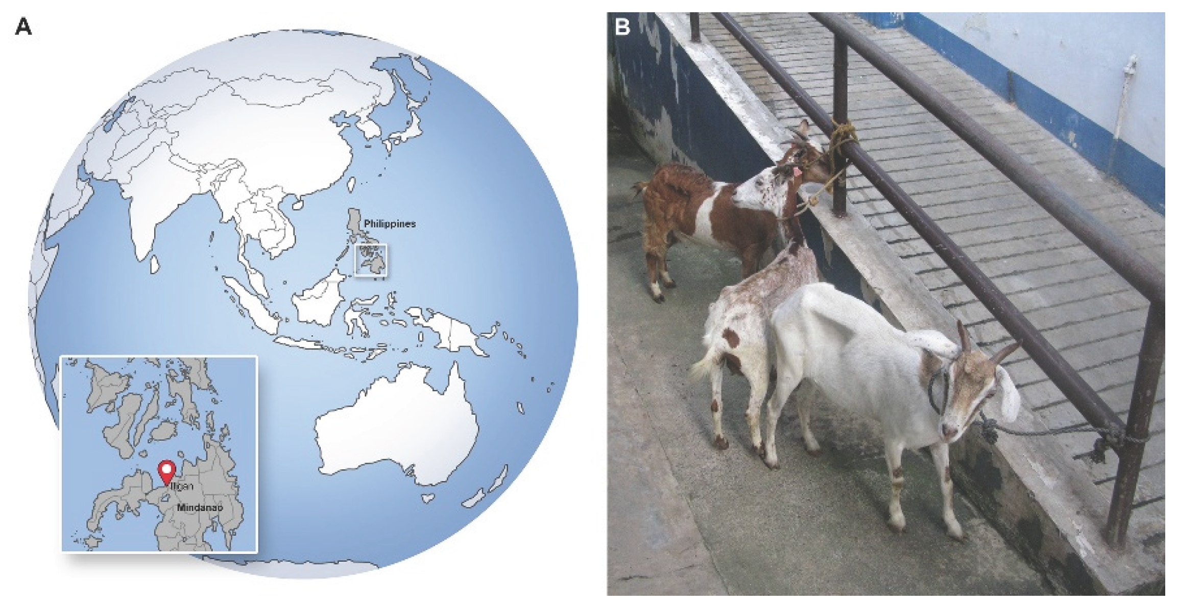
Figure 2.
Scanning electron micrographs illustrating selected morphological features of the male H. contortus recovered from the abomasum of the goat in the Philippines. (A) Lateral view of the cuticular surface, showing the longitudinal ridges of the synlophe; (B) High-magnification view of a cervical papilla (deirid); (C) High-magnification view of a lateral lobe of the copulatory bursa; (D) High-magnification view of the cuticular surface, showing portions of two longitudinal synlophe ridges together with transverse cuticular annulations; (E) Dorso-apical view of the copulatory bursa, showing the spicules, both lateral lobes, and a single asymmetrical dorsal lobe sharing a common base with the left lateral lobe; (F) High-magnification view of the spicules, showing the characteristic terminal inflation and a harpoon-like barb situated slightly proximal to the apex.
Figure 2.
Scanning electron micrographs illustrating selected morphological features of the male H. contortus recovered from the abomasum of the goat in the Philippines. (A) Lateral view of the cuticular surface, showing the longitudinal ridges of the synlophe; (B) High-magnification view of a cervical papilla (deirid); (C) High-magnification view of a lateral lobe of the copulatory bursa; (D) High-magnification view of the cuticular surface, showing portions of two longitudinal synlophe ridges together with transverse cuticular annulations; (E) Dorso-apical view of the copulatory bursa, showing the spicules, both lateral lobes, and a single asymmetrical dorsal lobe sharing a common base with the left lateral lobe; (F) High-magnification view of the spicules, showing the characteristic terminal inflation and a harpoon-like barb situated slightly proximal to the apex.
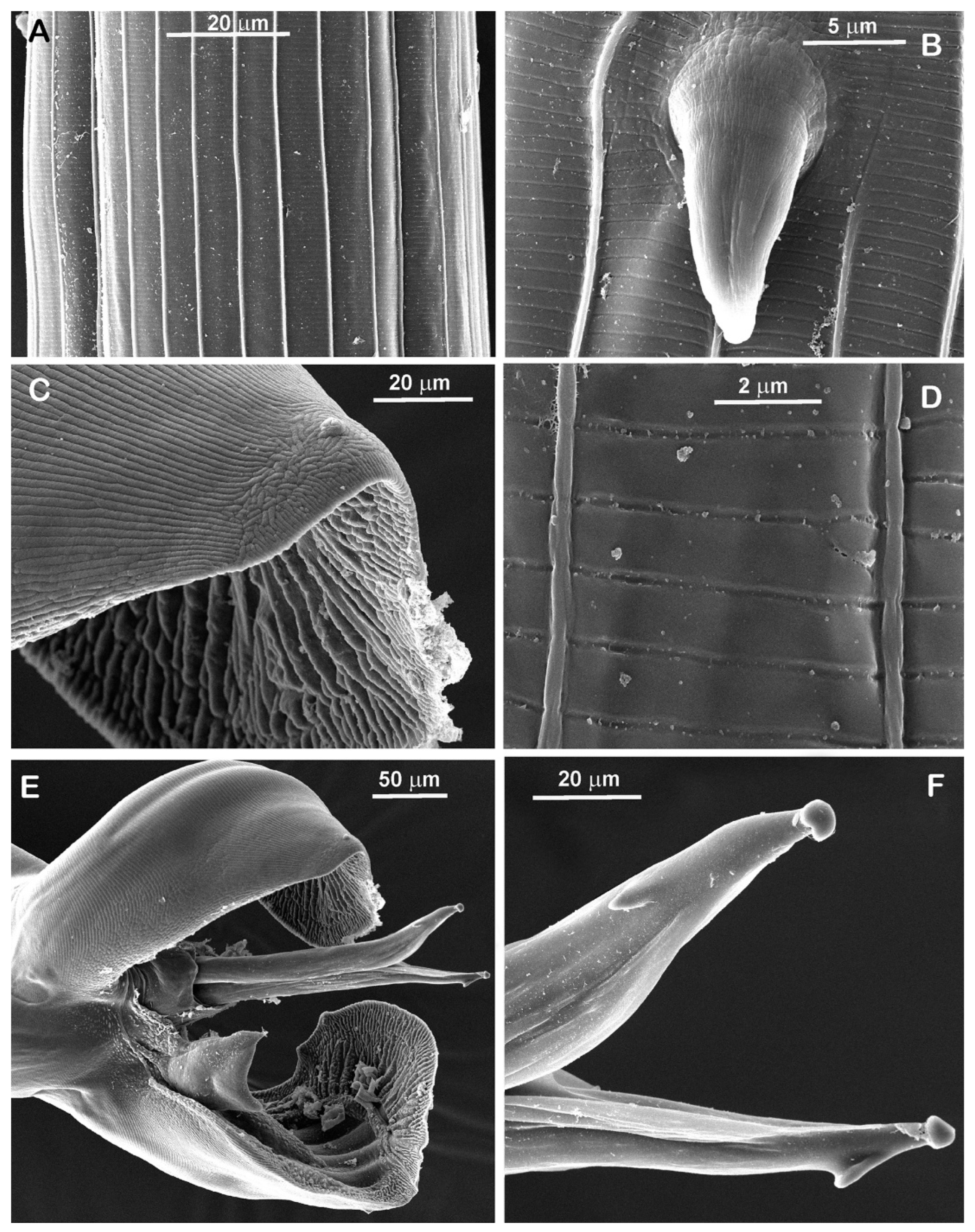
Figure 3.
Bayesian inference phylogenetic tree of H. contortus recovered from the abomasum of the goat in the Philippines based on partial mitochondrial CoxI sequences. H. longistipes was used as the outgroup. Sequences generated in the present study are indicated in bold. Posterior probability values are indicated at the nodes. Circle indicates domestic sheep (Ovis aries), square indicates domestic goat (Capra hircus), triangle indicates cattle (Bos taurus), and inverted triangle indicates water buffalo (Bubalus bubalis).
Figure 3.
Bayesian inference phylogenetic tree of H. contortus recovered from the abomasum of the goat in the Philippines based on partial mitochondrial CoxI sequences. H. longistipes was used as the outgroup. Sequences generated in the present study are indicated in bold. Posterior probability values are indicated at the nodes. Circle indicates domestic sheep (Ovis aries), square indicates domestic goat (Capra hircus), triangle indicates cattle (Bos taurus), and inverted triangle indicates water buffalo (Bubalus bubalis).
Figure 4.
Bayesian inference phylogenetic tree of H. contortus recovered from the abomasum of the goat in the Philippines based on partial nuclear ITS sequences. H. longistipes was used as the outgroup. Sequences generated in the present study are indicated in bold. Posterior probability values are indicated at the nodes. Rhombus indicates European bison (Bison bonasus), hexagon indicates blue wildebeest (Connochaetes taurinus), triangle indicates cattle (Bos taurus), circle indicates domestic sheep (Ovis aries), square indicates domestic goat (Capra hircus), pentagon indicates giraffe (Giraffa camelopardalis), six-pointed star indicates Chinese forest musk deer (Moschus berezovskii), and five-pointed star indicates dromedary (Camelus dromedarius).
Figure 4.
Bayesian inference phylogenetic tree of H. contortus recovered from the abomasum of the goat in the Philippines based on partial nuclear ITS sequences. H. longistipes was used as the outgroup. Sequences generated in the present study are indicated in bold. Posterior probability values are indicated at the nodes. Rhombus indicates European bison (Bison bonasus), hexagon indicates blue wildebeest (Connochaetes taurinus), triangle indicates cattle (Bos taurus), circle indicates domestic sheep (Ovis aries), square indicates domestic goat (Capra hircus), pentagon indicates giraffe (Giraffa camelopardalis), six-pointed star indicates Chinese forest musk deer (Moschus berezovskii), and five-pointed star indicates dromedary (Camelus dromedarius).
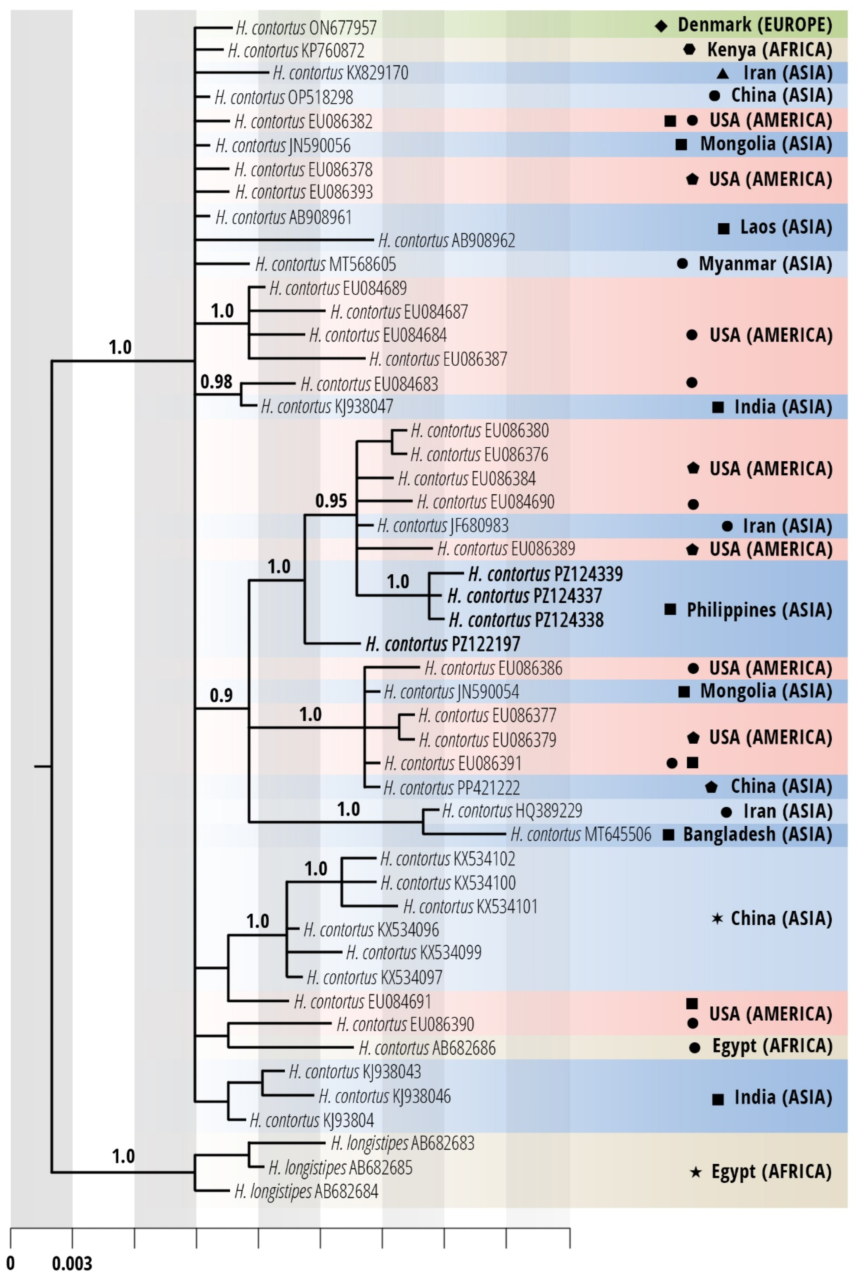
Figure 5.
Median-joining haplotype network of H. contortus based on CoxI sequences. The network was constructed from the same sequence dataset used in Figure 3, excluding H. longistipes, which was included there as an outgroup. A total of 55 sequences, including one generated in the present study, yielded 52 haplotypes; the sequence obtained in this study constituted a distinct haplotype. Circles represent haplotypes, with circle size proportional to haplotype frequency, and hatch marks on the connecting lines indicate mutational steps. The haplotype identified in the present study is indicated by a black arrowhead.
Figure 5.
Median-joining haplotype network of H. contortus based on CoxI sequences. The network was constructed from the same sequence dataset used in Figure 3, excluding H. longistipes, which was included there as an outgroup. A total of 55 sequences, including one generated in the present study, yielded 52 haplotypes; the sequence obtained in this study constituted a distinct haplotype. Circles represent haplotypes, with circle size proportional to haplotype frequency, and hatch marks on the connecting lines indicate mutational steps. The haplotype identified in the present study is indicated by a black arrowhead.
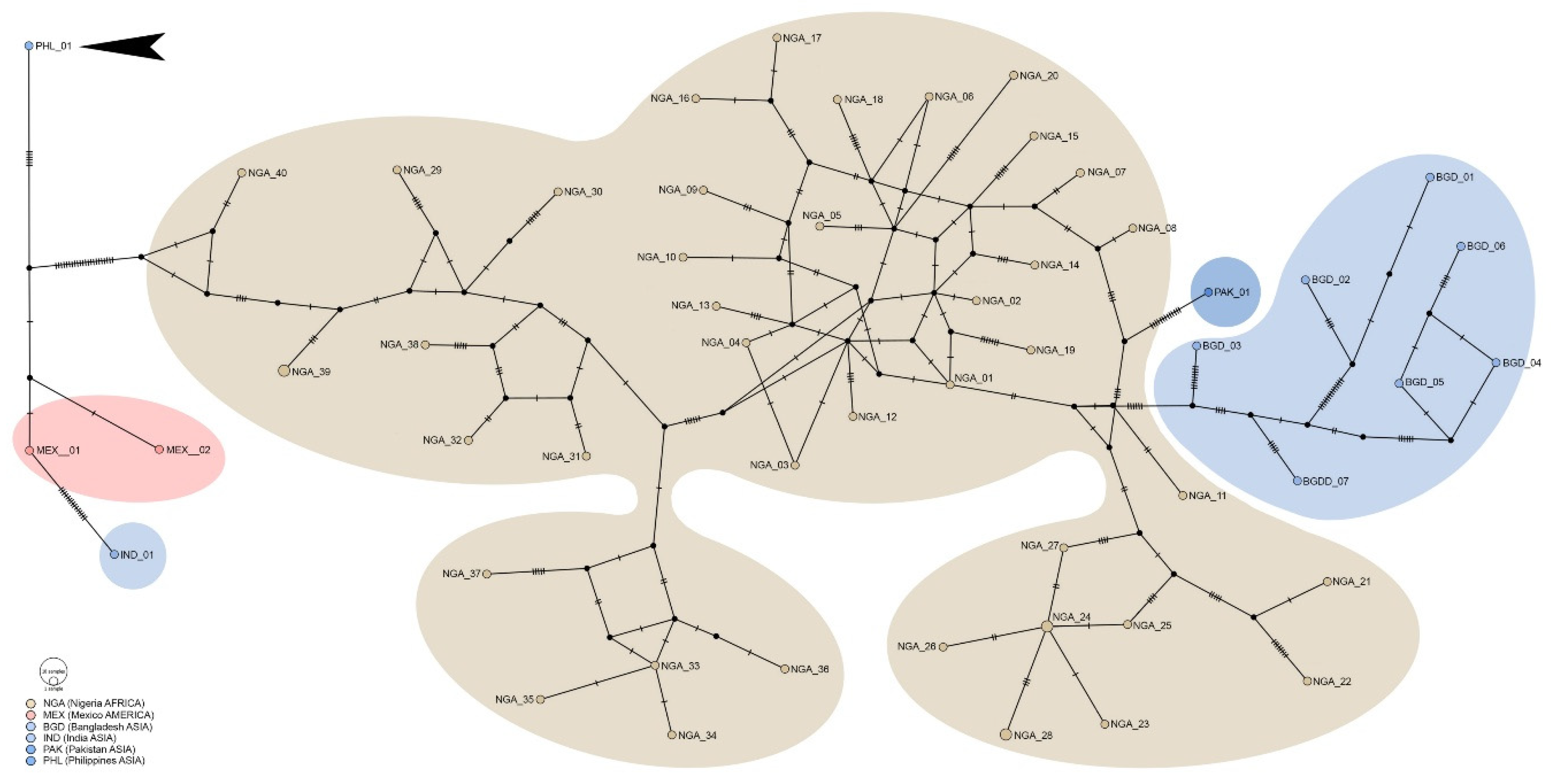
Table 1.
BLAST comparison of ITS and СoxI sequences of two trichostrongylids recovered from the abomasum of the goat in the Philippines with the two closest GenBank matches; query coverage are 100% for all comparisons.
Table 1.
BLAST comparison of ITS and СoxI sequences of two trichostrongylids recovered from the abomasum of the goat in the Philippines with the two closest GenBank matches; query coverage are 100% for all comparisons.
| Species | Locus | Length (bp) | GenBank | Identity, % | Host species | Location | Reference |
|---|---|---|---|---|---|---|---|
| H. contortus | CoxI | 577 | OP785768 | 98.61 | sheep (Ovis aries) | Mexico | [unpublished]* |
| OP785767 | 98.61 | ||||||
| ITS | 583 | JN590053 | 98.80 | goat (Capra hircus) | Mongolia | [34] | |
| EU086392 | 98.64 | sheep (Ovis aries) | USA | [35] | |||
| ITS (1) | 583 | JN590053 | 98.80 | goat (Capra hircus) | Mongolia | [34] | |
| EU086392 | 98.64 | sheep (Ovis aries) | USA | [35] | |||
| ITS (2) | 586 | EU086392 | 99.32 | sheep (Ovis aries) | USA | [35] | |
| JF680983 | 99.15 | sheep (Ovis aries) | Iran | [36] | |||
| ITS (3) | 586 | EU086392 | 99.32 | sheep (Ovis aries) | USA | [35] | |
| JF680983 | 99.15 | sheep (Ovis aries) | Iran | [36] | |||
| T. axei | CoxI | 689 | NC013824 | 90.86 | sheep (Ovis aries) | Australia | [37] |
| OZ259446 | 89.99 | sheep (Ovis aries) | UK | [38] | |||
| ITS | 817 | ON677948 | 100 | European bison (Bison bonasus) |
Denmark | [39] | |
| ON677949 | 99.88 |
* Submitted by Pacheco-Arjona, R., Torres-Acosta, F., Sandoval-Castro, C., Gonzalez-Pech, P., and Mancilla-Montelongo, G. on 07 November 2022.
Table 2.
Species of trichostrongylids recovered from the abomasum of the goat in the Philippines and identified genetically.
Table 2.
Species of trichostrongylids recovered from the abomasum of the goat in the Philippines and identified genetically.
| Trichostrongylid species | Locus (loci) | Length (bp) | GenBank 1 | Voucher 2 |
|---|---|---|---|---|
| H. contortus | CoxI | 577 | PZ122374 | IPEE_Parasites 14466 |
| ITS1, 5.8S, ITS2 | 583 | PZ122197 | IPEE_Parasites 14466 | |
| ITS1, 5.8S, ITS2 (clone 1) | 583 | PZ124337 | IPEE_Parasites 14472 | |
| ITS1, 5.8S, ITS2 (clone 2) | 586 | PZ124338 | IPEE_Parasites 14472 | |
| ITS1, 5.8S, ITS2 (clone 3) | 586 | PZ124339 | IPEE_Parasites 14472 | |
| T. axei | CoxI | 689 | PZ122159 | IPEE_Parasites 14467 |
| ITS1, 5.8S, ITS2, 28S | 817 | PZ124293 | IPEE_Parasites 14467 |
1 GenBank accession numbers for sequences from the fragments of the adult males of representative species. 2 Voucher specimens with definitive identifications and accession numbers archived in the Museum of Helminthological Collections of the IEE RAS (Moscow, Russia).
Disclaimer/Publisher’s Note: The statements, opinions and data contained in all publications are solely those of the individual author(s) and contributor(s) and not of MDPI and/or the editor(s). MDPI and/or the editor(s) disclaim responsibility for any injury to people or property resulting from any ideas, methods, instructions or products referred to in the content. |
© 2026 by the authors. Licensee MDPI, Basel, Switzerland. This article is an open access article distributed under the terms and conditions of the Creative Commons Attribution (CC BY) license (http://creativecommons.org/licenses/by/4.0/).
Copyright: This open access article is published under a Creative Commons CC BY 4.0 license, which permit the free download, distribution, and reuse, provided that the author and preprint are cited in any reuse.


