

Submitted:
23 May 2026
Posted:
25 May 2026
You are already at the latest version
Abstract
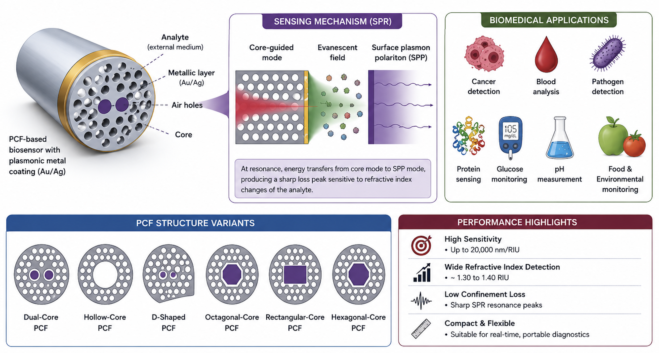
Photonic crystal fiber (PCF)-based biosensors have emerged as highly efficient optical sensing devices because of their superior light confinement capability, structural flexibility, and tunable optical characteristics. This review presents a comprehensive overview of recent developments in PCF-assisted biosensors integrated with surface plasmon resonance (SPR) technology for biomedical applications. The influence of structural parameters such as pitch spacing, air-hole diameter, and plasmonic coating thickness on resonance conditions and sensing performance is critically analyzed. These biosensors have demonstrated remarkable capability in detecting glucose, serum proteins, pathogens, pH levels, cancerous cells, and biochemical analytes in blood, urine, saliva, food, water, and environmental samples. The paper discusses the operating principles, structural classifications, and sensing mechanisms of PCF biosensors while comparing their performance with conventional optical sensing systems. Different PCF configurations including dual-core, hollow-core, D-shaped, rectangular-core, octagonal-core, and hexagonal structures are reviewed based on sensitivity, confinement loss, and refractive index detection range. The findings indicate that SPR-assisted PCF biosensors possess strong potential for future biomedical diagnostics, environmental monitoring, and intelligent healthcare systems because of their compact size, enhanced sensitivity, and fabrication flexibility.
Keywords:
optical biosensor
; photonic crystal fiber
; surface plasmon resonance
; biomedical applications
; PCF sensors
I. Introduction
Photonic crystal fibers (PCFs) are a special class of optical fibers in which the cladding region contains periodically arranged air holes that form photonic crystal structures [1]. These photonic crystals generate photonic bandgap (PBG) effects capable of controlling electromagnetic wave propagation within specific wavelength ranges and directions [2,3]. The photonic bandgap property is related to the local density of electromagnetic states, which determines the number of available optical modes for photon propagation [4,5,6].
Similar to conventional optical fibers, PCFs provide exceptional flexibility in tailoring optical properties such as birefringence, dispersion, confinement loss, nonlinear effects, and refractive index sensitivity through geometrical modification of air holes and pitch spacing. The confinement loss mainly depends on the imaginary part of the effective refractive index. In conventional optical fibers, light propagation occurs through total internal reflection (TIR), where light traveling through a higher refractive index medium undergoes complete reflection at the interface of a lower refractive index material [7].
PCFs are mainly classified into two categories: index-guiding PCFs and photonic bandgap-guiding PCFs. Index-guiding PCFs generally contain solid cores, whereas photonic bandgap fibers usually possess hollow-core structures. Owing to their microstructure architecture, PCFs are also known as microstructure optical fibers or holey fibers [8].
The integration of surface plasmon resonance (SPR) technology with PCFs has significantly improved sensing performance in optical biosensors. SPR occurs when incident electromagnetic waves interact with free electrons present on a metallic surface, resulting in collective electron oscillation at the metal-dielectric interface [9]. This interaction generates a sharp resonance peak that is highly sensitive to refractive index variation in surrounding analytes [10].
Recent developments in nanotechnology and plasmonic materials have accelerated the advancement of highly sensitive PCF biosensors. Two-dimensional materials such as graphene and molybdenum disulfide (MoS2), along with noble metals including gold (Au) and silver (Ag), are widely utilized to improve resonance characteristics and sensing performance. Different SPR fiber configurations including D-shaped fibers, cladding-removed fibers, grating-assisted structures, and fiber-tip sensors have demonstrated promising sensing capability [11].
PCF-based biosensors are currently employed in numerous biomedical applications including cancer detection [12,13,14], blood component analysis, pathogen identification, glucose monitoring, pH measurement, and environmental sensing. Because of their compact structure, high sensitivity, low confinement loss, and tunable optical properties, PCF biosensors are becoming increasingly important in modern biomedical diagnostics and healthcare monitoring systems.
This review paper presents recent advancements in PCF-based biosensors for cancer detection and biomedical applications. Different PCF structures, SPR sensing mechanisms, plasmonic materials, and performance characteristics are critically discussed and compared. The paper also highlights recent biomedical applications, existing challenges, and future research directions for highly sensitive and compact optical biosensing systems.
II. Operating Principles of PCF Based Sensors
The operating principle of PCF-based SPR sensors mainly depends on the interaction between the evanescent optical field and the plasmonic metal surface [15]. When light propagates through the fiber core, a portion of the electromagnetic field penetrates the cladding region and forms an evanescent wave. This evanescent field interacts with the free electrons present on the metallic layer deposited near the sensing surface [16,27,31].
Resonance occurs when the propagation constant of the core-guided mode becomes equal to the propagation constant of the surface plasmon polariton (SPP) mode. Under resonance conditions, maximum optical energy transfers from the guided mode to the plasmonic mode, resulting in a sharp confinement loss peak [17]. The propagation constant of the guided mode is expressed as
where k0 represents the free-space wave number and neff denotes the effective refractive index. The confinement loss is calculated using
where Im(neff) corresponds to the imaginary component of the effective refractive index. The wavelength sensitivity of the sensor is determined by
where Δλpeak represents the resonance wavelength shift and Δna denotes analyte refractive index variation.
Similarly, amplitude sensitivity is given by
where α(λ,na) represents confinement loss at wavelength λ for analyte refractive index na.
Gold and silver are commonly employed as plasmonic materials in SPR biosensors. Gold exhibits excellent chemical stability and biocompatibility, whereas silver provides sharper resonance peaks and higher sensitivity but suffers from oxidation-related degradation [18].
Certain PCF biosensors also utilize interferometric sensing mechanisms in which light is divided into sensing and reference paths. External perturbations such as strain, temperature, or refractive index changes alter the optical phase difference between the two paths, producing measurable interference patterns [19].
III. Structural Classification of PCF-Based Sensors
The structural configuration of PCF-based sensors significantly affects optical confinement, analyte interaction, propagation characteristics, and sensing performance. By modifying geometrical parameters such as air-hole diameter, pitch spacing, core arrangement, and plasmonic layer thickness, researchers can optimize confinement loss, birefringence, and wavelength sensitivity according to specific biomedical applications.
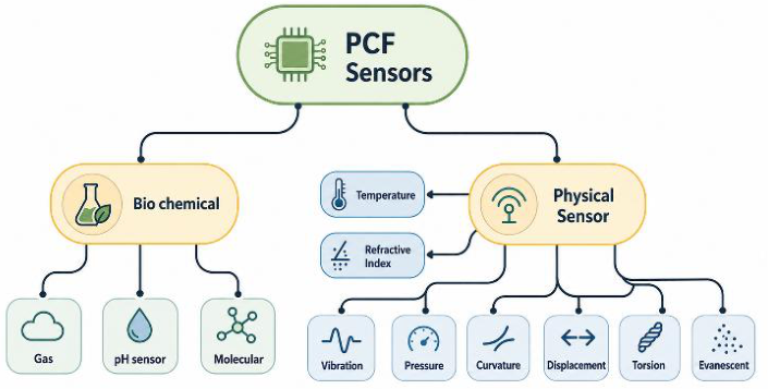
PCF-based sensors are broadly classified into biochemical sensors and physical sensors. Figure 1 illustrates the global classification of PCF-based sensors.
Biochemical PCF sensors are employed for detecting gases, proteins, enzymes, antibodies, glucose, pathogens, and chemical analytes because of their high sensitivity, compact size, and remote sensing capability.
IV. Biochemical Sensor
a. Gas Sensors: Gas sensing is one of the major applications of PCF-based optical sensors. Different gases possess unique absorption spectra and refractive index characteristics, enabling selective gas identification [20]. Hollow-core PCFs are particularly suitable for gas sensing because they allow direct gas filling inside the sensing region, thereby increasing interaction between light and gas molecules.
Figure 1.
Global classification of PCF-based sensors adapted from.
b) Biosensors: PCF biosensors are extensively utilized for detecting biological analytes such as glucose, serum proteins, enzymes, antibodies, DNA samples, and cancerous cells. Integration of SPR technology significantly improves sensing performance because the plasmonic metal layer enhances interaction between the evanescent field and analyte medium [21].
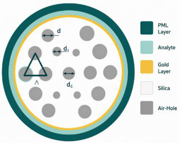
Hasanuzzaman et al. [22] proposed a dual-core gold-coated PCF biosensor based on a hexagonal lattice structure. The sensor utilized fused silica as the background material with pitch spacing Λ = 2 μm, air-hole diameter d = 0.573 μm, and gold layer thickness tAu = 35 nm. The proposed structure operated within the refractive index range of 1.30–1.40 and achieved maximum wavelength sensitivity of 5000 nm/RIU along with amplitude sensitivity of 267.66 RIU−1.
Figure 2.
Cross-sectional geometry of the dual-core gold-coated PCF biosensor adapted from [23].
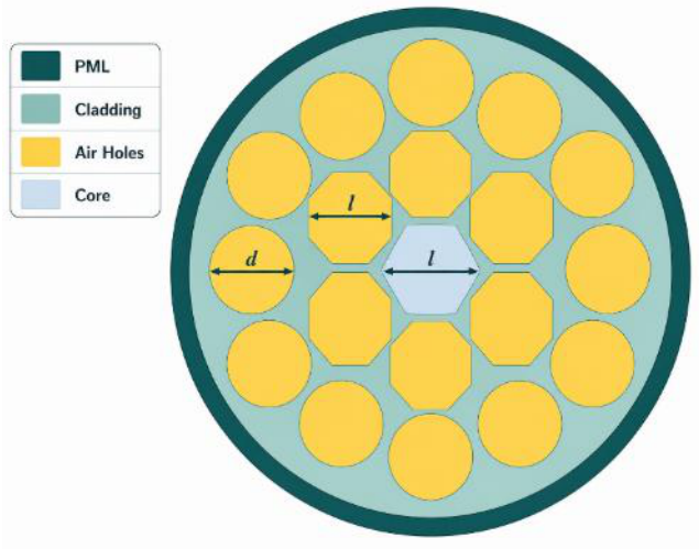
Maidi et al. [24] introduced an octagonal-core PCF biosensor for blood component analysis. The sensor detected RBCs, HB, WBCs, plasma, and water using refractive indices of 1.40, 1.38, 1.36, 1.35, and 1.33, respectively. The proposed structure achieved relative sensitivity values of 99.89%, 99.13%, 97.95%, 97.77%, and 96.68% for RBCs, HB, WBCs, plasma, and water at an operating wavelength of 7 μm.
Figure 3.
Cross-sectional geometry of the octagonal-core PCF biosensor adapted from [24].
Mollah et al. [13] investigated a bi-core PCF biosensor for blood cancer detection applications. The sensor utilized central air-hole diameter d0 = 1.9 μm, cladding air-hole diameter d = 1.5 μm, and pitch spacing Λ = 2 μm. The proposed structure demonstrated coupling lengths of 16.3 mm for cancerous cells and 26.2 mm for normal cells under x-polarized conditions.
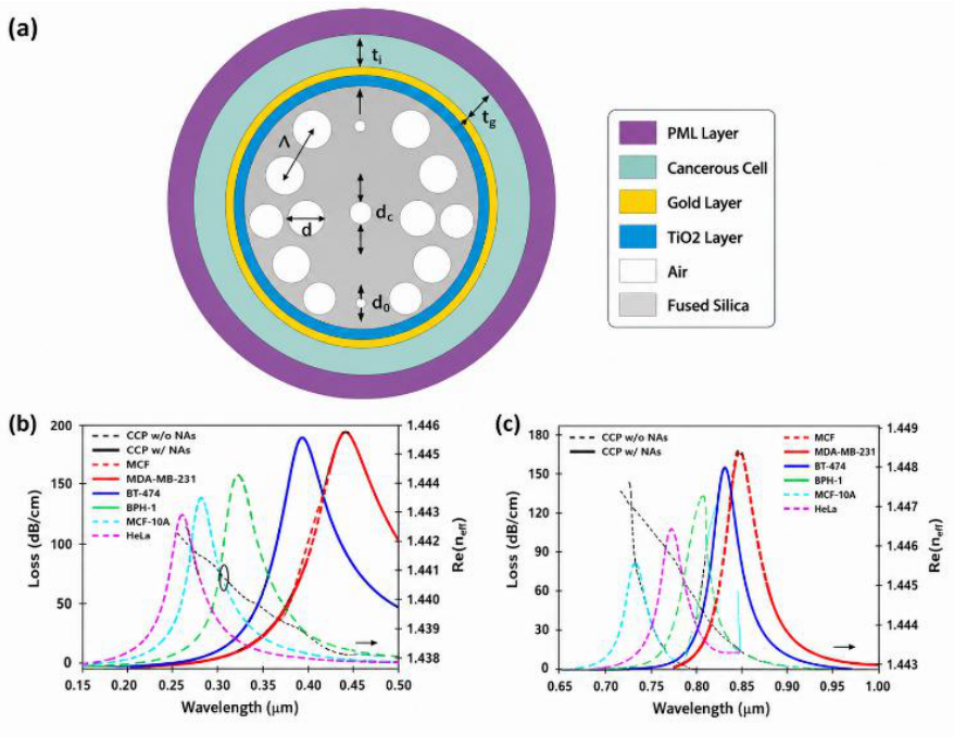
Chaudhary et al. [25] proposed a solid-core SPR-PCF biosensor coated with gold and titanium oxide (TiO2) layers for detecting cancerous cells including HeLa, Jurkat, MCF-7, MDAMB-231, and PC-12 cells. The optimized geometrical parameters were d0 = 0.36 μm, dc = 0.8 μm, d = 1.44 μm, pitch spacing Λ = 1.8 μm, titanium oxide thickness ti = 35 nm, and gold thickness tg = 25 nm.
Figure 4.
(a) Structural geometry of the bi-core PCF sensor and (b) coupling length response for normal and cancerous cells adapted from [13].
Figure 4.
(a) Structural geometry of the bi-core PCF sensor and (b) coupling length response for normal and cancerous cells adapted from [13].
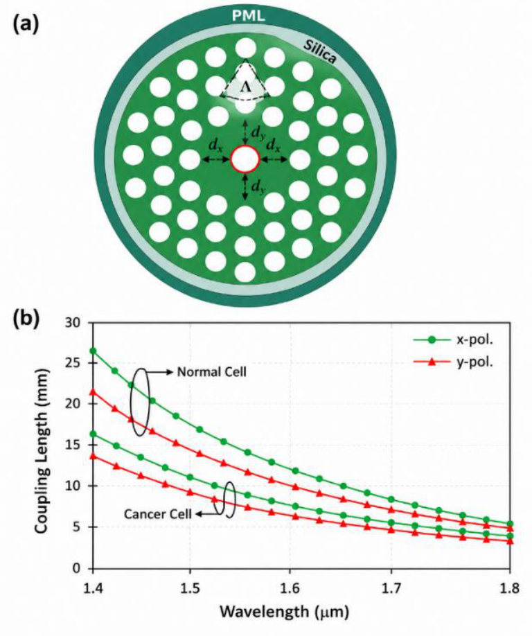
Figure 5.
(a) Cross-sectional structure of the solid-core SPR-PCF biosensor and (b) resonance loss characteristics adapted from [25].
Figure 5.
(a) Cross-sectional structure of the solid-core SPR-PCF biosensor and (b) resonance loss characteristics adapted from [25].
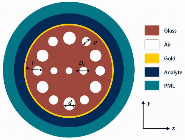
Jahan et al. [26] developed a circular lattice SPR-PCF biosensor for detecting Pseudomonas bacteria. The optimized structure employed pitch spacing p = 1.5 μm, air-hole diameter D = 0.2p μm, secondary hole diameter D1 = 0.75D μm, and gold layer thickness dg = 30 nm. The proposed sensor achieved wavelength sensitivity of 20,000 nm/RIU.
Figure 6.
Two-dimensional circular lattice PCF biosensor structure adapted from [26].
c) pH Sensors: PCF-based pH sensors provide advantages including rapid response time, compact size, electromagnetic immunity, and multiplexing capability. Variations in pH alter the refractive index of the sensing medium, producing measurable resonance wavelength shifts.
V. Physical Sensors
Physical PCF sensors are designed to measure parameters such as temperature, refractive index, pressure, strain, displacement, and magnetic field.
a) Temperature Sensors: PCF temperature sensors are widely employed in biomedical systems, industrial processing, agriculture, and food industries because of their high thermal sensitivity and environmental stability.
Figure 7.
(a) Cross-sectional structure of the hexagonal PCF sensor and (b) loss spectra for different analyte refractive indices adapted from [27].
Figure 7.
(a) Cross-sectional structure of the hexagonal PCF sensor and (b) loss spectra for different analyte refractive indices adapted from [27].
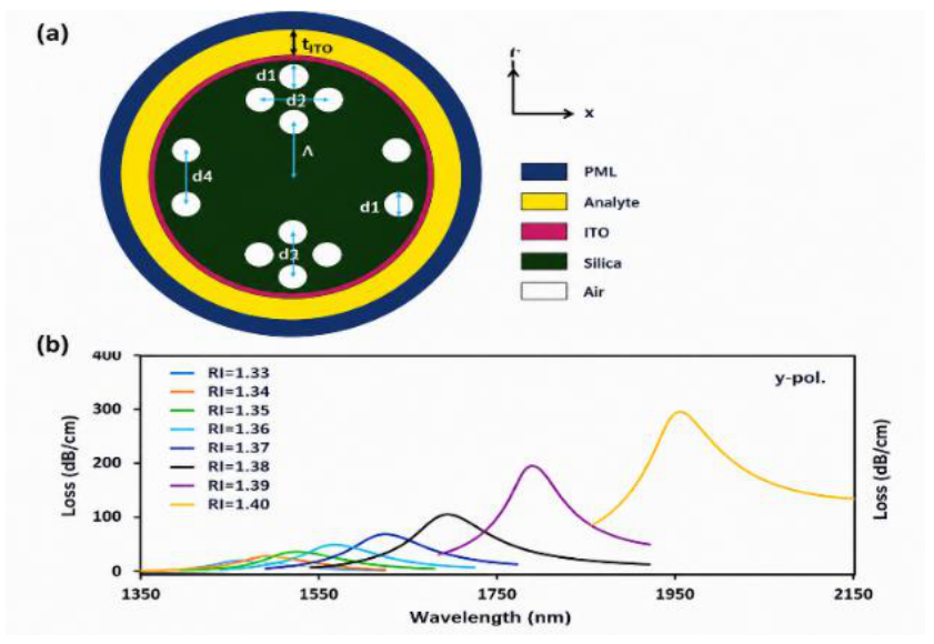
b) Refractive Index Sensors: Rahman et al. [28] proposed a highly sensitive hexagonal PCF-SPR refractive index sensor with geometrical parameters d1 = 1.4 μm, d2 = 2.8 μm, d3 = 2.1 μm, d4 = 2.8 μm, pitch spacing Λ = 2.8 μm, and indium tin oxide thickness tITO = 50 nm. The sensor exhibited strong resonance wavelength shifts as analyte refractive index increased from 1.33 to 1.40.
c) Pressure Sensors: Podder et al. [29] proposed a rectangular-core PCF biosensor operating in the terahertz frequency range for blood component analysis. The sensor utilized TOPAS as the background material and achieved relative sensitivity of 94.38% for RBC detection at 1.08 THz frequency with minimal confinement loss
Figure 8.
(a) Rectangular-core THz-PCF biosensor structure and (b) relative sensitivity variation adapted from [28].
Figure 8.
(a) Rectangular-core THz-PCF biosensor structure and (b) relative sensitivity variation adapted from [28].
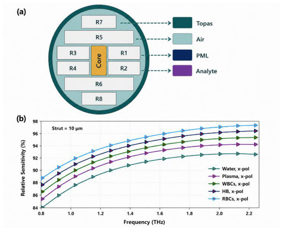
Table 1 presents the comparative performance analysis of different PCF-based biosensors utilized for biomedical sensing applications. Among the reviewed structures, the dual-core gold-coated PCF proposed in [23] demonstrated wavelength sensitivity of 5000 nm/RIU over the refractive index range of 1.30–1.40, indicating strong capability for biochemical sensing. The octagonal-core PCF introduced in [24] achieved relative sensitivity of 99.89% for blood component detection, making it highly suitable for biomedical diagnostics. Similarly, the bi-core PCF sensor reported in [13] exhibited strong coupling response characteristics for cancer detection applications. The solid-core PCF biosensor presented in [25] demonstrated significant resonance wavelength shifts during cancer cell identification because of refractive index variation between normal and infected cells. In addition, the circular lattice PCF proposed in [26] achieved exceptionally high wavelength sensitivity of 20,000 nm/RIU for bacterial detection applications. Podder et al. [29] developed a rectangular-core PCF biosensor for blood analysis with relative sensitivity of 94.38% within the refractive index range of 1.33–1.40. Furthermore, the hollow-core PCF sensor reported in [30] demonstrated wavelength sensitivity of 14,285.71 nm/RIU for malaria detection because of enhanced analyte interaction inside the hollow sensing region. The comparative analysis indicates that optimization of PCF geometry and plasmonic materials significantly improves sensing performance in biomedical applications.
VI. Challenges and Future Directions
Despite the remarkable progress achieved in photonic crystal fiber (PCF)-based biosensors, several challenges still limit their widespread practical implementation in biomedical and healthcare applications. One of the major limitations is the complexity of fabrication processes, particularly for structures containing intricate air-hole geometries, plasmonic coatings, and nanoscale sensing regions. Precise control of pitch spacing, air-hole diameter, and metal layer thickness increases manufacturing difficulty and overall production cost [1,2]. In addition, plasmonic materials such as silver are highly susceptible to oxidation and environmental degradation, which can reduce long-term sensor stability and reliability [8]. Mechanical fragility of microstructure fibers also presents challenges during sensor handling, integration, and practical deployment [12].
Future research should therefore focus on the development of cost-effective and simplified fabrication techniques capable of producing highly sensitive PCF biosensors with improved structural stability. The incorporation of advanced nanomaterials such as graphene, titanium oxide, and transition metal dichalcogenides may further enhance plasmonic interaction, sensing accuracy, and chemical stability [5,14]. Moreover, integration of artificial intelligence and machine learning algorithms with optical sensing systems can enable intelligent signal processing, automated diagnosis, and real-time biomedical monitoring. The development of portable, wearable, and multi-analyte biosensing platforms is also expected to play a significant role in next-generation point-of-care healthcare systems [9,10]. Continuous advancements in nanotechnology, plasmonic, and microfabrication techniques are anticipated to substantially improve the sensitivity, selectivity, and practical applicability of PCF-based biosensors in future biomedical diagnostics and environmental monitoring applications [21,22,23,24,25].
VII. Conclusions
Photonic crystal fiber-based biosensors have emerged as highly promising sensing platforms for biomedical and biochemical applications because of their tunable structural properties, enhanced evanescent field interaction, and compatibility with SPR technology. This review discussed recent developments in different PCF configurations including dual-core, hollow-core, D-shaped, rectangular-core, octagonal-core, and hexagonal structures for applications such as cancer diagnosis, blood analysis, pathogen detection, glucose monitoring, and environmental sensing. Comparative analysis demonstrated that optimization of structural parameters and plasmonic materials significantly improves sensing performance. With continuous advancements in optical engineering and nanotechnology, PCF biosensors are expected to become key components of future biomedical diagnostic and intelligent healthcare systems.
References
- S. M. R. Islam et al., “Advances in Photonic Crystal Fiber Biosensors for Multi-Organ Cancer Diagnostics: A Systematic Review,” in 2026 22nd IEEE International Colloquium on Signal Processing & Its Applications (CSPA), 2026, pp. 102–107. [CrossRef]
- A. Starczewska and M. Kępińska, “Photonic crystal structures for photovoltaic applications,” Materials, vol. 17, no. 5, p. 1196, 2024.
- G. Guida, A. De Lustrac, and A. C. Priou, “An introduction to photonic band gap (PBG) materials,” Progress In Electromagnetics Research, vol. 41, p. 1, 2003.
- S. K. Biswas, S. M. R. Islam, M. Ahmed, A. Halder, M. R. Islam, and M. Hossain, “Analytical model for coupling Whispering gallery mode silica microcavity for sensing application,” in 2017 2nd International Conference on Electrical & Electronic Engineering (ICEEE), IEEE, 2017, pp. 1–4.
- S. K. Biswas, A. al Noman, and S. M. R. Islam, “Nonlinear coupling of whispering gallery mode silica microcavity for sensing application,” in 2017 3rd International Conference on Electrical Information and Communication Technology (EICT), IEEE, 2017, pp. 1–3.
- S. Akter and H. Abdullah, “High sensitivity gold-coated photonic crystal fiber sensor for blood component detection,” Plasmonics, vol. 20, no. 6, pp. 3927–3938, 2025.
- S. K. Biswas, T. Ahmed, S. M. R. Islam, M. R. Islam, M. M. A. Mia, and M. F. Wahid, “Highly Nonlinear Dispersion Compensating Octagonal Photonic Crystal Fiber: Design and Analysis,” in 2019 International Conference on Electrical, Computer and Communication Engineering (ECCE), IEEE, 2019, pp. 1–5.
- M. M. Faruk, M. R. Islam, S. M. R. Islam, M. Ahmed, M. S. Miah, and S. K. Biswas, “Ultra-high Negative Dispersion Compensating Index Guiding Single Mode Octagonal Photonic Crystal Fiber: Design and Analysis,” in 2019 Innovations in Power and Advanced Computing Technologies (i-PACT), IEEE, 2019, pp. 1–5.
- S. K. Biswas, S. M. R. Islam, M. R. Islam, M. M. A. Mia, S. Sayem, and F. Ahmed, “Design of an ultrahigh birefringence photonic crystal fiber with large nonlinearity using all circular air holes for a fiber-optic transmission system,” in Photonics, MDPI, 2018, p. 26.
- B. M. A. Rahman et al., “Optical fiber, nanomaterial, and thz-metasurface-mediated nano-biosensors: A Review,” Biosensors (Basel)., vol. 12, no. 1, p. 42, 2022.
- P. Sangeetha, N. Ayyanar, G. Prabhakar, and S. Rajaram, “Study review of optical biosensors based on 2D materials,” Plasmonics, vol. 20, no. 9, pp. 7155–7169, 2025.
- F. Bray et al., “Global cancer statistics 2022: GLOBOCAN estimates of incidence and mortality worldwide for 36 cancers in 185 countries,” CA Cancer J. Clin., vol. 74, no. 3, pp. 229–263, 2024.
- M. F. Majeed and A. K. Ahmad, “Design and analysis of a dual-core PCF biosensor based on SPR for cancerous cells detection,” Opt. Quantum Electron., vol. 56, no. 6, Jun. 2024. [CrossRef]
- W. Emon, A. Chaki, T. P. Mondal, M. D. F. Nayan, and R. R. Mahmud, “Photonic crystal fiber-based SPR biosensor coated with Ag-TiO2 and Au-TiO2 for the detection of skin cancer: a comparison,” Opt. Quantum Electron., vol. 56, no. 8, Aug. 2024. [CrossRef]
- M. R. Islam, S. M. R. Islam, M. Ahmed, F. Ahmed, M. M. Faruk, and S. K. Biswas, “Analysis of Dispersion and Nonlinear Property in Defected Core Square Photonic Crystal Fiber,” in 2018 9th International Conference on Computing, Communication and Networking Technologies (ICCCNT), IEEE, 2018, pp. 1–5.
- S. K. Biswas, S. M. R. Islam, M. Ahmed, A. Halder, M. R. Islam, and M. Hossain, “Analytical model for coupling Whispering gallery mode silica microcavity for sensing application,” in 2017 2nd International Conference on Electrical & Electronic Engineering (ICEEE), IEEE, 2017, pp. 1–4.
- A. A. Maimun et al., “A Numerical Simulation for the Diagnosis of Cancer Cells Using a Gold-Coated SPR-Boosted PCF Biosensor,” in 2026 22nd IEEE International Colloquium on Signal Processing & Its Applications (CSPA), 2026, pp. 356–359. [CrossRef]
- S. Khani and M. Hayati, “Optical biosensors using plasmonic and photonic crystal band-gap structures for the detection of basal cell cancer,” Sci. Rep., vol. 12, no. 1, p. 5246, 2022.
- S. Das, R. Devireddy, and M. R. Gartia, “Surface plasmon resonance (SPR) sensor for cancer biomarker detection,” Biosensors (Basel)., vol. 13, no. 3, p. 396, 2023.
- R. Yu, Y. Chen, L. Shui, and L. Xiao, “Hollow-core photonic crystal fiber gas sensing,” Sensors, vol. 20, no. 10, p. 2996, 2020.
- O. B. Daramola, R. K. Omole, and B. A. Akinsanola, “Emerging applications of biorecognition elements-based optical biosensors for food safety monitoring,” Discover Sensors, vol. 1, no. 1, p. 3, 2025.
- G. K. M. Hasanuzzaman, T. M. Sakib, and A. K. Paul, “Gold coated surface plasmon resonance based biosensor: An hexagonal photonic crystal Fiber platform,” Sens. Biosensing Res., vol. 42, p. 100582, 2023.
- A. M. Maidi, M. A. Kalam, and F. Begum, “Unsafe food additive sensing through octagonal-core photonic crystal fibre sensor,” Phys. Scr., vol. 98, no. 6, p. 065528, 2023.
- A. Ramola, A. K. Shakya, A. Droby, and A. Bergman, “Numerical study of a novel kagome-inspired photonic crystal fiber-based surface plasmon resonance biosensor for detection of blood components and analytical targets,” Biosensors (Basel)., vol. 15, no. 8, p. 539, 2025.
- V. S. Chaudhary, D. Kumar, and S. Kumar, “Au-TiO2Coated Photonic Crystal Fiber Based SPR Refractometric Sensor for Detection of Cancerous Cells,” IEEE Trans. Nanobioscience, vol. 22, no. 3, pp. 562–569, Jul. 2023. [CrossRef]
- N. Jahan et al., “Photonic crystal fiber based biosensor for pseudomonas bacteria detection: A simulation study,” Ieee Access, vol. 9, pp. 42206–42215, 2021.
- Motakabber, S. M. A., et al. “ADVANCING SYSTEM INTEGRATION: VERILOG-BASED HARDWARE IMPLEMENTATION OF AN ASIC INTERFACE FOR THREE AMBA PROCESSORS.” IIUM Engineering Journal 25.1 (2024): 253-262.
- M. M. Rahman, M. A. Molla, A. K. Paul, M. A. Based, M. M. Rana, and M. S. Anower, “Numerical investigation of a highly sensitive plasmonic refractive index sensor utilizing hexagonal lattice of photonic crystal fiber,” Results Phys., vol. 18, p. 103313, 2020.
- M. E. Haque, N. J. Diya, M. S. Salman, S. M. Salimullah, and R. R. Mahmud, “Ultra-Sensitive LSPR-PCF Design with Dual Resonance for Both Polarizations and Broad Wavelength Range for Next-Generation Sensing,” Sens. Imaging, vol. 26, no. 1, p. 53, 2025.
- S. Sawraj et al., “PCF-based sensors for biomedical applications: A review,” IEEE Trans. Nanobioscience, vol. 24, no. 2, pp. 157–164, 2024.
- Paul, John, et al. “A data mining approach to analyze the role of biomacromolecules-based nanocomposites in sustainable packaging.” International Journal of Biological Macromolecules 265 (2024): 130850.

Table 1.
Performance comparison of PCF-based biosensor.
| Sensor Structure | Application | RI Range | Sensitivity | Ref. |
| Dual-Core Gold-Coated PCF | Biochemical sensing | 1.30–1.40 | 5000 nm/RIU | [23] |
| Octagonal-Core PCF | Blood detection | 1.33–1.40 | 99.89% | [24] |
| Bi-Core PCF | Cancer detection | 1.36–1.40 | High coupling response | [13] |
| Solid-Core PCF | Cancer sensing | 1.36–1.40 | Strong resonance shift | [25] |
| Circular Lattice PCF | Bacterial detection | 1.33–1.39 | 20,000 nm/RIU | [26] |
| Rectangular-Core PCF | Blood analysis | 1.33–1.40 | 94.38% | [28] |
| Hollow-Core PCF | Malaria detection | 1.33–1.40 | 14,285.71 nm/RIU | [29] |
Disclaimer/Publisher’s Note: The statements, opinions and data contained in all publications are solely those of the individual author(s) and contributor(s) and not of MDPI and/or the editor(s). MDPI and/or the editor(s) disclaim responsibility for any injury to people or property resulting from any ideas, methods, instructions or products referred to in the content. |
© 2026 by the authors. Licensee MDPI, Basel, Switzerland. This article is an open access article distributed under the terms and conditions of the Creative Commons Attribution (CC BY) license (http://creativecommons.org/licenses/by/4.0/).
Copyright: This open access article is published under a Creative Commons CC BY 4.0 license, which permit the free download, distribution, and reuse, provided that the author and preprint are cited in any reuse.


