Submitted:
21 May 2026
Posted:
22 May 2026
You are already at the latest version
Abstract
Myocardial infarction (MI) has become one of the leading causes of mortality worldwide, and the prevalence is anticipated to rise considerably in the coming years. Among non-surgical procedures, chemical drugs, including diuretics, vasodilators, calcium channel blockers, ꞵ blockers, angiotensin converting enzyme inhibitors, are now in use to treat MI progression. These drugs often cause side effects and do not focus on mitigating oxidative stress, which has been recently proven to contribute to MI advancement. Naturally occurring antioxidant compounds, on the other hand, are safe, effective due to their multiple molecular targets, and have the potential to be used as lead compounds for finding novel drugs to therapeutically manage MI. Some of them, namely quercetin, puerarin, α-lipoic acid, and curcumin, have already made their way up to clinical trials. To develop and formulate natural antioxidant compounds as drugs against MI, it is crucial to comprehend their underlying mechanisms of cardio-protective activities and structure-activity relationships. This comprehensive review sheds light on the contribution of oxidative stress in the pathogenesis and progression of Myocardial Infarction, and highlights the cardio-protective roles of 51 natural antioxidant compounds along with their mechanistic insights and structure-activity relationships.
Keywords:
myocardial infarction
; reactive oxygen species
; oxidative stress
; antioxidant
; phytochemicals
; structure activity-relationship
1. Introduction
Myocardial infarction (MI) is a common clinical condition in cardiovascular diseases (CVDs) affecting cardiac tissues and ultimately leading to their necrosis due to blood perfusion arrest, failure in supplying oxygen and nutrients, and toxic substance accumulation (Frangogiannis, 2015; Fuentes et al., 2019). It is categorized into distinct classes based on pathological, clinical, and prognostic characteristics, as well as treatment options, such as type 1, type 2, type 3, type 4, and type 5 (Thygesen et al., 2018). Type 1 MI results from atherothrombotic coronary artery disease (CAD) (Thygesen et al., 2018), whereas Type 2 myocardial infarction (T2MI) occurs when the heart’s demand for oxygen or supply of oxygen rises without a sudden disruption in atherothrombotic plaque (Sandoval & Jaffe, 2019). In type 2 myocardial infarction, the metabolic needs of myocardial cells are also greater than the amount of oxygen in blood (Musher et al., 2019). Although Type 3 MI is rare (as reflected by a study that found an annual incidence below 10 cases per 100,000 persons and a frequency of 3% to 4% among all MI types), it frequently causes sudden death even prior to the obtaining of biomarkers or ECG confirmation (Anderson & Morrow, 2017; Thygesen et al., 2018). Among other types, type 4 MI results from PCI (percutaneous coronary intervention) procedure and thrombosis of a coronary stent, while Type 5 MI is caused by coronary artery bypass grafting (CABG) (Anderson & Morrow, 2017) .
A key pathological hallmark of MI is oxidative stress, which describes a condition in which there is a mismatch between oxidants and antioxidants in the body (Navarro-Yepes et al., 2014). Oxidative stress is caused by an imbalance between the production of reactive oxygen and nitrogen species (ROS/RNS) and the amounts of enzymatic and non-enzymatic antioxidants. ROS and RNS are the two primary classes of free radicals involved in cellular redox signaling processes (Tern et al., 2021). Oxidative stress (OxS) and molecular damage are mainly caused by the overabundance of these free radicals, which are also the two most significant prooxidants. While the ROS encompass superoxide anion, hydroxyl radicals, and hydrogen peroxide, members of the RNS family include peroxynitrite and nitrosoperoxycarbonate. Peroxynitrite develops due to the reaction between superoxide and nitric oxide (NO), whereas nitrosoperoxycarbonate is created when peroxynitrite reacts with carbon dioxide (Chowdhury et al., 2015). Even though ROS play physiological roles possibly by controlling cardiomyocyte survival and death, their uncontrolled abundance deteriorates cardiac pathological conditions by inducing oxidative stress (Wu & Liu, 2022).
To defend against oxidative stress, which damages every component of the cell, cells depend mostly on antioxidant enzymes and non-enzymatic antioxidants, among others (Gulcin, 2025; Kozlov et al., 2024). Antioxidant enzymes, also termed as metalloproteins, fall under the class of proteins that catalyze the conversion of reactive oxygen species (ROS) and possibly their byproducts into more stable, generally less hazardous species. In pathological conditions like MI, where endogenous defenses are inadequate, there is a therapeutic need for exogenous antioxidants, and plant-produced natural antioxidants, including flavonoids, phenolics, and polyphenolics (hydrolysable and condensed tannins), have this therapeutic potential as they have been shown to prevent reactive oxygen species-induced oxidative stress (Thygesen et al., 2018). These natural bioactive compounds may lower oxidative stress in humans by getting rid of free radicals, slowing down lipid peroxidation, and stopping nitrosation reactions (Smit et al., 2020). In order to eliminate the free radicals that are produced as a byproduct of oxidative metabolism and to keep the redox balance in its proper place, natural antioxidants can work either independently or in conjunction with one another. Although current medications for treating MI include chemical drugs, their side effects on prolonged use and lack of ability to deal with the deeper root causes render the treatment less effective. Natural bioactive compounds, on the other hand, provide a safe, multi-targeted strategy. Therefore, scientific interest has grown in these compounds for developing safer and more effective drugs to fight the war against MI.
To enhance the understanding and optimization of these natural antioxidant compounds, it is imperative to study their SAR or structure-activity relationship. SAR unveils the impact of the structure of the molecule on its therapeutic activity. For example, the quantity and location of hydroxyl groups in flavonoids determine their ability to scavenge radicals, and the conjugated double bonds in carotenoids enable them to be better at quenching singlet oxygen. Moreover, during the journey of drug discovery, phytochemicals have to undergo critical scrutiny to be developed as valid therapeutic agents for MI. Such scrutiny looks at their structural attributes, SAR, and underlying mechanism of action to make sure that they are safe and effective. Thus, insights from SAR and mechanistic studies help promote potential compounds to clinical use from preliminary studies. Although there are mounting scientific data regarding the cardio-protective activity of these natural bioactive compounds, there is still a dearth of systematic investigations that link SAR to their cardio-protective action pathway in MI. To address this gap, this review aims to provide a concise summary of the role of oxidative stress in the development and progression of MI, highlight the identified natural antioxidant compounds that alleviate MI and its associated complications, and finally explore the SAR of some of the most promising compounds to understand how their structural features contribute to their cardio-protective activity.
2. Method
In order to garner relevant literature, well-known scientific databases, including Google Scholar, Springer Link, Science Direct, PubMed, Scopus, Web of Science, and the Wiley Online Library, were accessed. The search procedure was led by writing specific keywords such as “myocardial infarction”, “oxidative stress in myocardial infarction”, “natural antioxidant compounds”, “structure-activity relationship”, and “myocardial infarction mechanism”. Only publications published in the English language were considered, as articles in other languages could not be evaluated due to linguistic constraints. This review covered research studies on the following topics: association of oxidative stress with myocardial infarction, plant-derived anti-oxidant compounds with cardio-protective effects to alleviate MI, along with their underlying mechanisms of effects, and their SAR that contributes to these activities.
3. Oxidative Stress in MI Development
Free radicals are regarded as reactive chemical species that possess a single unpaired electron in the outermost orbital (Chandimali et al., 2025; Riley, 1994). They are capable of either donating or accepting electron species, therefore can act as both oxidant and reductant (Chaudhary et al., 2023; Cheeseman & Slater, 1993). The odd-numbered radical attacks molecules in its vicinity in order to attain stability. As a result, the attacked molecule loses its electron and converts into a radical family, initiating a reaction cascade that ultimately leads to cell death. The majority of free radicals that are engaged in impairing the biological system are known as oxygen free radicals; more particularly, “Reactive Oxygen Species” (ROS). These ROS induces myocardial damage during ischemia or reperfusion (G. Gong et al., 2025). Free radicals have been proven both deleterious and beneficial for the biological system. On the one side, at low concentrations, they are required for the maturation of cellular structures and the regulation of the host defense mechanism. Free radicals are released by the body’s phagocytes (neutrophils, macrophages, monocytes) and aid in destroying and invading pathogenic agents. The production of ROS and RNS by non-phagocytic NADPH oxidase isoforms plays a vital role in modulating intracellular signaling cascades in various types of cells, including fibroblasts, vascular smooth muscle cells, endothelial cells, cardiac myocytes, and thyroid cells. Free radical nitric oxides and superoxide play a significant role in the biological regulatory system. Nitric oxide radical is a crucial intracellular messenger for regulating blood flow, thrombosis, and neural activities (Pacher et al., 2007). On the other side, free radicals have an influence on a number of pathological conditions like diabetes, cancer, rheumatoid arthritis, inflammation, asthma, cardiovascular disease, muscular dystrophy, intestinal tract disease, progerias, atherosclerosis, parkinson’s disease, cataract, ischemia, and so forth (Phaniendra et al., 2015; Reddy, 2023). Overproduction or excessive accumulation of free radicals alters homeostasis, which subsequently gives rise to pathological conditions. For example, hydroxyl radical and peroxynitrite in excess can damage cell membranes and lipoproteins by lipid peroxidation that leads to the formation of malondialdehyde (MDA) and conjugated diene compounds, which are mutagenic and cytotoxic.
Oxidative stress occurs due to the imbalance between free radical generation and antioxidant defensive mechanisms. In other words, the term “oxidative stress” refers to the disruption between reduced to oxidized glutathione or (GSH/GSSG) or NADPH/NADP+ ratios (Kurian et al., 2016). It can be a consequence of either- due to the impairment of the antioxidant system caused by malnutrition (inadequate consumption of vitamin C, vitamin E, sulphur-containing amino acids) or by the excessive production of free radicals, including ROS and RNS (due to external or internal stimulus). Cells can bear up mild oxidative stress because there is an upregulation of the antioxidant system in response to that mild rise, so that cells can cope with the raised situation. For example, if experimental rats are continuously exposed to higher level of oxygen, they develop better oxygen tolerance in comparison to control rats due to antioxidant system elevation in lungs (Halliwell, 2012). But when this adaptive capacity of the cell exceeds, it starts to experience oxidative stress. Oxidative stress asserts to play role in plethora of diseases including atherosclerosis, inflammatory diseases (arthritis, glumerulonephritis, vasculitis, lupus erythematous, respiratory distress syndrome), ageing process, fibromyalgia, AIDS, emphysema, hemochromatosis, hypertension, preeclampsia, gastric ulcers, neurological disorders (Alzheimer’s disorder, parkinson’s disease, muscular dystrophy), and so on (Islam et al., 2025; Lobo et al., 2010). Oxidative stress manifests cellular derangement like DNA strand breakage, elevation in cytosolic free calcium, cell membranes and transport proteins damage, and release of transition metal ions that finally results in cell death (Halliwell, 2012).
3.1. Pathophysiology of Oxidative Stress Associated MI:
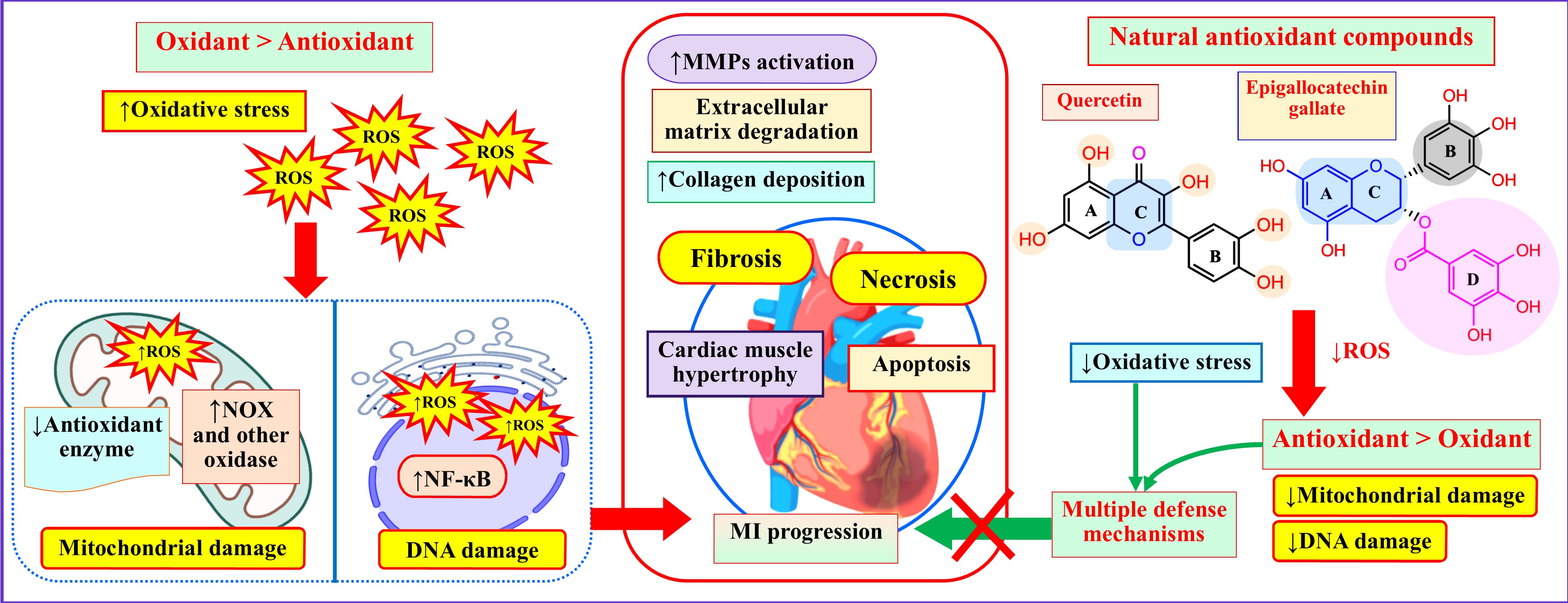
There is growing evidence which indicates that oxidative stress has an important role in the pathophysiology of myocardial infarction (MI) (Neri et al., 2015; van der Pol et al., 2019). A contemporary hot subject is the oxidative stress theory, which holds that excessive production and accumulation of ROS cause cardiac damage (Wen et al., 2021). The multifaceted roles of oxidative stress in MI pathogenesis are summarized and presented in Figure 1. MI occurs in a series of events after the supply of oxygen in cardiovascular tissue is acutely hampered, mostly due to coronary thrombosis resulting from atherosclerotic plaque rapture, a condition known as ischemia. This shifts cellular respiration to anaerobic, and also increases intracellular calcium level that results in cell swelling, rupture, and mostly cell death by both necrosis and apoptosis (Fuentes et al., 2019; Hausenloy & Yellon, 2013). When the supply of oxygen to tissue is reestablished, reactive oxygen species are produced in cardiac muscle cells that contribute to the prognosis of this disease. Mitochondria, as well as some other cardiovascular enzymes such as NADPH oxidase (NOX), nitric oxide synthase (when uncoupled), and xanthine oxidase are responsible for the generation of ROS. Though several antioxidant systems are present to counter the deleterious effect of ROS, sometimes a disproportion between pro-oxidant and antioxidant systems occurs, which leads to a situation known as “oxidative stress” (Fuentes et al., 2019; González-Montero et al., 2018; Hori & Nishida, 2009). This disproportion impairs mitochondrial functions. Consequently, even more ROS are produced, increasing the level of inflammatory cytokines, which in turn induces further production of ROS and damages vital cellular structures, including mitochondria. This disastrous loop of mitochondrial failure, increased oxidative stress, and cellular destruction keeps ramping up and eventually expedites MI progression (Cai et al., 2023; Ide et al., 2001). Moreover, ROS and inflammatory cytokines trigger the increased activation of matrix metalloproteins (MMPs) and excessive deposition of collagen. MMPs are enzymes responsible for the degradation of the extracellular matrix components (ECM), including collagen that offers cardiac muscle its structural stability and tensile strength. Even though in normal circumstances MMPs have a crucial role in tissue remodeling, ROS and inflammatory cytokines mediated overactivation of them during the initial stage of MI speeds up ECM and collagen breakdown, leading to diminishing strength of cardiac muscle. This is succeeded by an excess accumulation of collagen, a condition referred to as fibrosis, which leads to an increase in the stiffness of the cardiac muscle. Both processes aid in the alterations of the ventricular remodeling of injured myocardium following MI. Also, high levels of ROS in impaired cardiac muscle cells can lead to substantial oxidative damage to DNA. Consequently, an enzyme called ribonuclease polymerase gets activated, and its hyperactivity hampers regular cell metabolism and triggers the secretion of inflammatory mediators, which are intricately associated with cardiac remodeling and heart (Q. Guo et al., 2024; R. Yang et al., 2018). In addition to DNA-damaging mechanisms, ROS can directly initiate cascades of intracellular signals that exacerbate cardiac damage. For example, Hydrogen peroxide directly stimulates the production of tumor necrosis factor α (TNF-α) via the p38 mitogen-activated protein kinase (MAPK) pathway that mediates myocardial dysfunction and apoptosis (Fuentes et al., 2019; Meldrum et al., 1998). Likewise, in experimental rat models of heart failure, the superoxide anion in the aorta was observed to be markedly increased in the same p38 MAPK pathway in addition to the elevated level of the NOX subunit p47phox (Widder et al., 2004).
Furthermore, in post-myocardial infarction patients, a consistently high level of hydroxyl radical production was reported to be associated with the likelihood of progressing towards heart failure according to a clinical study (Valgimigli et al., 2004). These aforementioned oxidizing species can further oxidize LDL, leading to a rise of oxidized LDL or oxLDL in the circulation. Elevated concentration of Circulating oxLDL is linked with various cardiovascular diseases, including atherosclerosis (S. Gao & Liu, 2017; Poznyak et al., 2021; Zingg et al., 2021). Apart from inflicting direct damage, ROS also function as secondary messengers in cardiac signaling pathways. For instance, Src family of tyrosine kinases, MAPKs, small GTP-binding proteins, and cytokines are all activated by ROS. These subsequently induce the activation of nuclear transcription factors connected to the cellular stress responses, which eventually result in cell hypertrophy and apoptosis (Sia et al., 2002). Activated platelets also produce ROS, which is crucial in controlling platelet response to thrombus development. In addition to established activities that promote the growth of atherothrombotic plaques, changes in platelet hemostatic characteristics may promote the progression of injury at the infarct zone (Hori & Nishida, 2009).
3.2. Impact of Oxidative Stress on MI Complications
Mechanical complications, which are the most common within the first week after myocardial infarction, include left ventricular free wall rupture, ventricular septal rupture with acute ventricular septal defect, papillary muscle rupture with severe mitral regurgitation, pseudo-aneurysm, true aneurysm, and hemodynamically significant right ventricular infarction (F. F. Gong et al., 2020; Reeder, 1995). Pseudoaneurysm, a rare complication of MI, particularly may occur when the visceral, parietal, or both pericardia retain the blood leaked due to the hemorrhagic process resulting from the rupture of the cardiac wall (Ashraf et al., 2021). Some of the other common complications associated with an acute MI are cardiogenic shock, acute pulmonary edema, pericarditis, and heart failure (F. F. Gong et al., 2020; Hubbard, 2003). Cardiac myocyte apoptosis, chronic neurohumoral activation, and ventricular remodeling also occur following MI and ischemia reperfusion (Jenča et al., 2021; Mihalko et al., 2019).
Oxidative stress has a strong impact on MI complications. In myocardial reperfusion injury, the restoration of blood flow to the already existent ischemic zone causes a significant increase in ROS, which quickly and severely damages crucial bio-molecular constituents of the myocardium. Thus, the myocardium loses its structural integrity and functionality to a great extent. This phenomenon is accompanied by several post-MI complications, including lethal reperfusion, no-reflow phenomenon, myocardial stunning, and reperfusion arrhythmias. Post-MI patients who also have diabetes mellitus experience worsened oxidative stress conditions (González-Montero et al., 2018). It is because the constant presence of hyperglycemia is reported to promote oxidative stress succeeding MI, causing endothelial dysfunction and the advancement of atherosclerosis (Kitano et al., 2016). This obstructs the coronary supply of oxygenated blood to the myocardium, thereby further deteriorating the condition of heart cells. So, together, diabetes and myocardial ischemia induce oxidative stress, which causes cardiac tissue to overexpress iNOS and increase NO production. Inhibited NADPH activity may interact with the latter, causing the generation of peroxynitrites and a decrease in NO bioavailability. These responses are closely tied to the rise in cytokine production and apoptosis inside the myocardium, which increases necrosis and disrupts the post-MI recovery of cardiac cells (Filippo et al., 2006).
4. Biomarkers of Oxidative Stress in MI
There are numerous biomarkers of oxidative stress which are present in patients with MI, indicating an association between oxidative stress and MI (Table 1). For example, according to a study by Feng et al., MI patients have higher levels of advanced oxidation protein products (AOPP) in their blood (Feng et al., 2010). Normally, the H2O2-myeloperoxidase (MPO) system uses plasma proteins to react with chlorinated oxidants, such as hypochlorous acid, and produce AOPP. AOPPs are unique indicators of oxidant-mediated protein degradation because they possess di-tyrosine. Albumin is the primary carrier of AOPP in the bloodstream (Melough et al., 2017). Protein carbonyl, a significant biomarker for protein oxidation, is made when proteins react with lipid peroxidation products or when certain amino acid residues oxidize. According to another study, protein carbonyl was found significantly higher in patients with MI than in controls, indicating the ROS-driven oxidative protein damage as a result of MI (Shahzad et al., 2018).
It is worthwhile to mention that ROS causes the oxidation of LDL, which raises the levels of circulating oxidized LDL (oxLDL), a marker that has been linked to the emergence of CVDs. oxLDL stimulates the growth and accumulation of foam cells in atherosclerotic plaques, which may then favor platelet activation and cause a cycle of oxidative damage. This establishes the link between atherosclerosis and platelet oxidative stress (Fuentes et al., 2019). Also, lipid peroxidation products such as MDA, HNE, and F2-isoprostanes which are derived from polyunsaturated arachidonic acid are important biomarkers for oxidative stress linked to cardiovascular complications (Tsikas, 2017). Several studies found that an increased level of MDA and F2-isoprostanes are associated with exacerbation of MI (Babu et al., 2010; Davies & Roberts II, 2011; Kharb & Singh, 2000; Mythiili & Malathi, 2015; Yin et al., 2019).
Several vitamins such as vitamin A, C, and E act as antioxidants and protect the body from harm by neutralizing free radicals. A study revealed that patients with acute MI who were hospitalized to intensive care had significantly lower levels of vitamins E and C, except for vitamin A which did not differ significantly (Ismail et al., 2018). Another study reported that individuals with AMI had relatively low levels of vitamins E and C than controls (Singh et al., 2004). This is in line with the findings of Singh et al. who reported that lipid peroxide levels were much greater in AMI patients compared to controls, whereas vitamins C, E, A, and β-carotene decreased dramatically (Singh et al., 1994).
The term “superoxide dismutase” refers to a protein family that includes Mn-SOD and Cu-Zn-SOD. The spontaneous conversion of superoxide radicals to H2O2 is accelerated by SOD activity. The catalase enzyme may eliminate the final byproduct H2O2 of the dismutation cycle. Glutathione peroxidase (GPx) uses GSH as its substrate to catalyze peroxide reduction, turning it to GSSG or glutathione disulfide (Rodrigo et al., 2013). According to an investigation, patients with MI and ischemic heart disease had considerably lower SOD, CAT, and GPx activity than healthy controls (Hazini et al., 2015; Rodrigo et al., 2013). Additionally, smokers with AMI had significantly lower SOD, CAT, and GPx activity than nonsmokers, making these people more susceptible to oxidative stress (Metta et al., 2015). It has been noted that SOD and CAT activity in the erythrocytes of AMI patients has been decreased due to elevated peroxidation (Anbarasi et al., 2006; Scott et al., 1991). In the erythrocytes of AMI patients, there has been evidence of reduced GPx activity. Because GSH is one of the substrates for GPx, a drop in GSH levels may also result in a decrease in GPx activity. Accordingly, it has been demonstrated that individuals with AMI have considerably lower GSH concentrations in plasma and erythrocytes (Ścibior et al., 2008; Serdar et al., 2006; Tavares et al., 2012). So, levels of SOD, CAT, GPx, and GSH are decreased in MI due to oxidative stress which was confirmed by several studies (Aladag et al., 2021; Freitas et al., 2014; Khan et al., 2013; Shahzad et al., 2018). The amount of zinc, magnesium, and calcium in the body is related to oxidative stress. For instance, zinc levels and oxidative stress have an opposing connection. Zinc deficiency causes many cell lines to produce more ROS. It has been reiterated that zinc supplementation can lower elevated levels of ROS. Superoxide dismutase 1 (SOD 1), an antioxidant enzyme that scavenges superoxide anion, uses zinc as a cofactor. The decrease in serum zinc levels during acute tissue injury, such as MI, has long been known. According to a previous study, MI patients had significantly lower serum zinc levels within the first three days than the control group. In MI patients, Zn may therefore be a helpful oxidative marker (S. Choi et al., 2018). A lack of magnesium also increases the formation of free radicals linked to MI (Kharb & Singh, 2000). It is noteworthy to mention that Mg exhibits antiarrhythmic properties in MI. Acute MI is more likely to be fatal and is related to low serum Mg levels. Mg shortage worsens MI through increasing oxidative stress-induced ischemia damage and mitochondrial dysfunction (M. Liu & Dudley, 2020).
Oxidative stress can lead to cellular abnormalities, including a decrease in the activity of the (Na+K)-ATPase, which in turn increases calcium influx, and suppresses the sarcolemma calcium pump ATPase, hence lowering calcium efflux. Moreover, it has been observed that oxidative stress decreases the sarcoplasmic reticulum calcium pump ATPase, which prevents calcium from being secreted from the cardiomyocytes. Eventually, ROS causes intracellular calcium accumulation and cell death by depressing the calcium-regulating system. Additionally, a rise during ischemia causes xanthine dehydrogenase to change into xanthine oxidase, which then causes an increase in superoxide generation (González-Montero et al., 2018).
5. Natural Antioxidant Products Based Therapeutic Approaches for MI
In recent years, scientists have realized how crucial oxidative stress is to the emergence of MI (W. Guo et al., 2023). Excessive oxidative stress may be a factor in the ischemia/reperfusion injury which accounts for MI-related mortality. This revelation prompts researchers to explore innovative therapeutic approaches that specifically target this particular path in the context of anti-MI drugs (Rhana et al., 2022). Considering this, in this section, we have provided an overview of bioactive natural compounds reported for potential MI management focusing on mitigating oxidative stress. A summary of reported natural bioactive compounds and their mechanistic insights into cardioprotection in MI is also presented in Table 2.
- Scopoletin
Scopoletin, a prominent phytoalexin present in many plant species, particularly those belonging to the Convolvulaceae, Ulmaceae, Solanaceae, Compositae, Oleaceae, Rutaceae, and Aceraceae families, has been revealed to have a strong capacity to scavenge free radicals. A pre-treatment with scopoletin, according to Rong et al. (Rong et al., 2022), significantly decreased heart-to-body weight ratio, cardiac diagnostic markers, MDA, inflammatory indicators, and apoptotic markers, as well as elevated levels of antioxidant enzymes. As a result, histological features similar to inflammation and necrosis were reversed in the MI model.
- Shikimic acid
Shikimic acid (SA), a plant derived phenolic compound, is one of the major intermediate products formed in the shikimate pathway in a number of plants (Bochkov et al., 2012). It exhibits notable pharmacological effects, including antioxidant (M. Estevez & J. Estevez, 2012), anti-inflammatory (Rabelo et al., 2016), anti-platelet, and antithrombotic activities (Veach et al., 2016). SA has been shown to reduce MDA levels and enhance SOD activity, thereby minimizing oxidative stress and cellular damage (Khattak et al., 2025). Moreover, it reduces pro-inflammatory cytokines, such as TNF-α and IL-1β, which contribute to ventricular remodeling, inflammation, and cardiomyocyte apoptosis after myocardial infarction (Rabelo et al., 2016).
- Nerolidol
Nerolidol is a plant derived sesquiterpene alcohol commonly found in lavender, lemongrass, and ginger (Chan et al., 2016). It is reported for a wide number of pharmacological activities like antioxidant, anticancer, antinociceptive, anti-inflammatory activities (Ayhan et al., 2025; Fonsêca et al., 2016; Tousif et al., 2025). A recent research has demonstrated its effectiveness in prevention of myocardial damage when administered for long time in animal models (Asaikumar et al., 2019). Another research reported the cardioprotective role of nerolidol in MI where different oxidative stress markers were found to be altered. It significantly increased SOD activity and reduced lipid peroxidation and carbonyl levels leading to minimizing isoproterenol-induced heart damage (Asaikumar et al., 2019; Gonçalves et al., 2022).
- Biochanin-A
Biochanin-A (BCA) is an O-methylated isoflavonoid found in plants like red clover, soy, alfalfa sprouts, peanuts, and chickpea. Govindasami et al. have studied the impact of BCA on MI which revealed that this compound showed cardioprotection in MI by reducing lipid peroxidation and increasing antioxidant enzyme function (Govindasami et al., 2020).
- Diosmetin
Diosmetin is a citrus fruits derived mono-methoxy flavonoid commonly found in lemons and olives. It is also extracted from medicinal herbs like Rosmarinus officinalis. Diosmetin is reported for inhibiting MDA formation as well as restoring antioxidant enzyme functions in myocardium (Ahmad et al., 2023). In a H9c2 cell line based study on hypoxia-afflicted damaged myocardium, Si et al. reported that Diosmetin reduces the apoptosis of myocardium via the autophagy induction by activating AMPK (Si et al., 2020). This study suggested Diosmetin as a potential drug candidate in the treatment of myocardial infarction.
- 10-gingerol
10-gingerol is a phenolic compound found in ginger, a popular spice of cooking. It has been reported for potential role in diminishing ROS levels in cells. Previous research showed that this phytocompound exhibits protective activity against MI in both in vitro and in vivo experiments. The underlying mechanism is activation of JAK2/STAT3 signaling pathway and prevention of oxidative stress leading to reduction of apoptosis (Han et al., 2022).
- Quercetin
Quercetin is a potent flavonoid commonly found in wide varieties of plants. It is also very commonly found in vegetables, fruits, berries, and teas. This compound is reported to increase the level of nitric oxide in the ischemic myocytes of myocardium (P. G. Li et al., 2012). It also exhibits the potential to inhibit several oxidative enzymes like oxidases and lipoxygenases. More recently, it is known as potent antioxidant medication (Sadik et al., 2003; W. Zhang et al., 2023). A recent study conducted by Kozhukhov et al. has revealed that quercetin aids in protecting the heart by reducing the infarct size and stopping intramyocardial haemorrhage, when administered as intravenous infusion alongside the conventional treatment in patients following the first incidence of ST elevation MI (Kozhukhov et al., 2024).
- Lycopene
An integrated experiment by Guo, Huang and Li provided a theoretical basis for the antioxidant effect of lycopene in both preventing and curing MI (W. Guo et al., 2023). The research demonstrated that lycopene can effectively neutralize free radicals and stimulate the transcription of downstream antioxidant enzymes via activating the Nrf2/HO-1 pathway.
- Berberine (Ber)
Doxorubicin (DOX)-induced MI damage has been acknowledged as a significant side effect of cancer chemotherapy. The goal of an experimental investigation by Y. Wang et al. (Y. Wang et al., 2023) was to determine whether Ber, an isoquinoline alkaloid initially isolated from Chinese goldthread (Coptis chinensis), could offer defense against this problem. Berberine has a wide spectrum of biological roles, including antioxidant function. The study reveals that myocardial damage was prevented by pretreatment with Ber as it reduced the formation of ROS and MDA and raised SOD activity via triggering the Nrf2-mediated pathway.
- Ellagic acid
Ellagic acid (EA) is an antioxidant and anti-inflammatory polyphenol that is mostly present in pomegranates, blackberries, raspberries, strawberries, cranberries, walnuts, pecans, and wolfberries (Costa et al., 2022). Plenty of studies reported that EA treatment prevented lipid peroxidation (Osawa et al., 1987) , oxidant-induced endothelial dysfunction, and atherosclerosis (Y. Ding et al., 2014), in addition to inhibiting the isoproterenol-induced cardiac necrosis in rats (Kannan & Quine, 2011) . Additionally, EA improved the structural and functional recovery of myocardial energy metabolism when paired with indapamide, a thiazide diuretic prescribed for hypertension. Moreover, oral administration of EA reduces ROS production and blocks the CaMKII signaling pathway, which improves left ventricular diastolic dysfunction in rats with ovariectomies. Therefore, EA seems like a viable option for the treatment of clinical conditions like hypertension, heart failure, and MI (Costa et al., 2022) .
- Thymoquinone
Thymoquinone (TQ) is the primary active component of Nigella sativa, giving Nigella sativa its anti-inflammatory and antioxidant potentials. To further understand how TQ affects oxidative stress, inflammation, apoptosis as well as how it protects cardiac mtDNA in an experimentally induced MI model, an experimental study was conducted by Khalifa, Rashad and El-Hadidy (Khalifa et al., 2021). The finding demonstrates that pre- and co-administration of TQ reduced the oxidative stress linked to the occurrence of MI, reversed the changes in histoarchitecture, and retained the amount of cardiac mtDNA. These characteristics enhance the cardiac advantages resulting from using TQ as a natural cardio-protective agent.
- Lupeol
A pentacyclic triterpenoid molecule known as lupeol is abundantly present in edible plants like olive oil, strawberries, etc. According to Moris et al. (Moris et al., 2017), unusual ROS production and NF-κB activation are key determinants of the severity of myocardial damage. Also, Nrf2 has been linked to the control of oxidative stress responses as a key transcription factor. In order to find out whether lupeol has protective benefits against myocardial ischemia/reperfusion injury (I/R) via modulating NF-κB & Nrf2 signaling pathways, Li et al.(J. Li et al., 2022) used myocardial I/R rat models. The results revealed that lupeol dramatically altered NF-κB and Nrf2 signaling, suggesting that it may be an effective treatment for preventing I/R-induced myocardial infarction.
- S-Limonene
S-Limonene (SL) is one of the most prevalent monoterpenes discovered in lemon and orange peels. To assess the potential therapeutic use of SL in an isoproterenol-induced MI animal model, an investigation was conducted by Rhana et al. (Rhana et al., 2022). The findings demonstrated that s-limonene supports cardio-protection against MI injury, most likely via decreasing elevated Ca2+ and reducing oxidative stress via CaMKII. This makes it a potentially effective choice for the treatment of MI.
- Ferulic acid
Ferulic acid (FA), a polyphenol generated from plants, is widely present in cereals, vegetables, and fruits. FA is well known for its anti-inflammatory properties. According to Pandi et al.(Pandi et al., 2022), FA’s cardio protective effects mainly involve modulating cellular antioxidants, ROS-mediated oxidative stress, apoptosis, inflammation, and autophagy. FA primarily modulates PI3K/Akt dependant Nrf2 signaling pathway.
- Kaempferol
Kaempferol is an active phytoconstituent of Amaranthus viridis. An ISO-induced rat model was used by Krishna et al. to study the cardioprotective effects of kaempferol (Krishna et al., 2023). The study reported that kaempferol can improve antioxidant response by reducing myocardial oxidative stress via the Nrf-2/Ho-1 signaling pathway, which is principally responsible for its cardioprotective effects against ISO-Induced MI in rats.
- Icariin
Icariin is the primary bioactive medicinal ingredient identified from Epimedii herba. Inhibition of oxidative stress, attenuation of DNA damage, prevention of mitochondrial oxidative damage, and reduction of cardiomyocyte apoptosis are just a few of the many potential pathways that could underlie icariin’s cardioprotective benefits. An in silico study by Ke et al. (Ke et al., 2023) hypothesized that, suppressed oxidative stress may be related to icariin’s protective effects against MI. This protective characteristic makes icariin a candidate for treating MI.
- Liensinine
The Nelumbo nucifera Gaertn. seed embryo contains a naturally occurring bisbenzylisoquinoline alkaloid liensinine (LSN), which has been shown to have several antioxidant and cardiovascular potentials. A research conducted by Shen et al. assessed the cardioprotective activities of LSN following MI which demonstrated its novel cardioprotective function. This compound has significant potential to protect myocytes from oxidative stress induced damage in MI by downregulating Wnt/β-catenin signaling pathway (F. Shen et al., 2023). This finding supports the novel therapeutic role of LSN in ischemic heart diseases as well.
- Taraxerol
Taraxerol is a plant derived naturally occurring pentacyclic triterpenoid with diverse medicinal properties. Recent studies investigated it’s possible cardioprotective activity to prevent MI which revealed that it increases antioxidant enzymes like SOD and reduces pro-inflammatory cytokines leading to protect heart from ISO-induced damage (Aodah et al., 2023).
- Dioscin
Dioscin is a natural phytocompound present in numerous medicinal plants belonging to Dioscorea and Liliaceae families. Several studies have shown that dioscin can lower oxidative stress and inflammation in rats with cardiac ischemia-reperfusion. Dioscin showed significant potential to prevent MI by controlling inflammation and oxidative stress that are mediated by BMP4/NOX1 (Z. Zhang et al., 2022).
- Salvianolic acid B
Salvianolic acid B (Sal B), derived from the herb Salvia miltiorrhiza has numerous health benefits. It works against oxidative stress, inflammation, liver fibrosis, and cancer. Numerous in vitro and in vivo studies suggest that in myocardial damage models, Sal B inhibits ferroptosis by activating the Nrf2 signaling pathway, thereby offering protection against MI (Y. Shen et al., 2022).
- Hinokitiol (β-thujaplicin)
Hinokitiol (β-thujaplicin), a plant derived biocompound resembling to tropolone, obtained from cupressaceous plant’s wood, exhibits bioactivities like antibacterial, anticancer, anti-inflammatory, and antioxidant activities (Hachlafi et al., 2021). A research done by Xiao et al. stated that this compound reduces apoptosis by downregulating autophagy flux (H. Xiao et al., 2022).
- Saprirearine
Saprirearine, is a diterpenoid derived from Salvia prionitis. Recent research on this compound explored that it possesses cardioprotective activity on hypoxic/re-oxygenated H9c2 cardiomyocytes model of MI. The underlying mechanism involves enhancing mitochondrial dysfunction and preventing oxidative stress, thereby preventing mitochondria-mediated cell death. Apart from that, it also activates Nrf2 which synergistically fosters cardioprotection(G. Zhang et al., 2022).
- Auraptene
Auraptene is a citrus peel-derived component, and has a variety of pharmacological properties that makes it useful in treating MI. Sunagawa et al. found that auraptene drastically reduced MI-induced systolic dysfunction in MI-affected rats and improved posterior wall thickness (Sunagawa et al., 2022). Auraptene therapy was used to counteract MI-induced reductions in the expression of PPARα-dependent genes.
- Notoginsenoside R1
In China, cardiovascular problems are frequently prevented and treated using Panax notoginseng, a well-known traditional Chinese medication. One of P. notoginseng’s main active ingredients, notoginsenoside R1 (NGR1), possesses anti-inflammatory, antioxidant, and anti-apoptotic activities (J. Xiao et al., 2018). According to Zhu and Wan, NGR1 produces superior therapeutic effects on MI (Zhu & Wan, 2023). The reporting of Xu et al. suggests that NGR1 controls the JAK2/STAT3 signaling pathway in hypoxia/reoxygenation (H/R)-treated H9C2 cells to enhance proliferation and prevent apoptosis (H. Xu et al., 2022). NGR1 also slowed the in vivo course of MI.
- Salidroside (SAL)
SAL has several pharmacological properties that boost immunity, improve tolerance to hypoxia, and reduce free radical induced damage. A thorough investigation by Fan et al. into SAL’s ability to prevent harm has revealed that it can act in a variety of ways to fight oxidation, inflammation, and apoptosis (Fan et al., 2022). In that study, SAL greatly improved the remodeling process of MI and decreased cardiac damage by reducing oxidative and inflammatory effects via the PI3K/AKT/Nrf2/HO-1 pathway. This suggests that SAL could be used to treat MI effectively.
- Psoralidin (PSO)
Adriamycin (ADR) is an effective and widely used broad-spectrum anticancer medication. However, ADR’s cumulative and dose-dependent cardiotoxicity severely restricts its clinical use. A natural phenolic coumarin called psoralidin (PSO), which was reported to have reduced ADR-induced cardiotoxicity, is extracted from the seeds of the medicinal plant Psoralea corylifolia L. PSO demonstrates a variety of biological actions, including anti-inflammatory, anti-allergic, antibacterial, antidepressant, and antioxidant properties. According to Liang et al., PSO significantly decreased ADR-induced excessive ROS buildup in cardiomyocytes (Liang et al., 2022). It also increased the levels of Nrf2, NQO1, and HO-1, suggesting that it may prevent oxidative stress and ultimately lessen the cardiotoxicity caused by ADR.
- Calycosin
Calycosin is one of the Radix astragali’s main active ingredients, having cardioprotective qualities. In order to figure out if calycosin can reduce oxidative stress and oxidative stress-induced cardiac apoptosis in newborn cardiomyocytes (NCMs) via activation of aldehyde dehydrogenase 2 (ALDH2), calycosin has been studied in vivo and in vitro by Ding et al.(W. jun Ding et al., 2023). The results strongly support the potential therapeutic application of calycosin in MI by strongly indicating that it lowers oxidative stress and oxidative stress-induced apoptosis through the control of ALDH2 signaling.
- Emodin
It has been demonstrated that the principal active ingredient in traditional Chinese medicine rhubarb, an anthraquinone derivative, emodin possesses anticancer, antibacterial, immunomodulatory, and antioxidant properties. To generate fresh concepts for the clinical management of post-MI heart failure (MI HF), the effect of emodin on myocardial cells was investigated in an experimental rat model by Liu and Ning (J. Liu & Ning, 2021). According to the research, emodin has the potential to be a therapeutic option because it can considerably enhance energy metabolism, lower the pace at which myocardial tissues undergo apoptosis, and improve cardiac function in post-MI HF rats.
- Nuciferine
Nuciferine, an aporphine alkaloid that is mostly found in Nelumbo nucifera leaves, has been linked to anti-cancer, anti-oxidant, anti-tumor, and insulin secretor actions. An investigation was carried out by HarishKumar and Selvaraj to assess the cardioprotective effects of nuciferine (HarishKumar & Selvaraj, 2022). There, isoproterenol (ISO) was first used to cause MI in Wistar rats, and then nuciferine was administered orally as a pretreatment. In rats with MI, the pretreatment with nuciferine greatly raised the amount of endogenous antioxidants and reduced lipid peroxidation. In a different study on acute myocardial infarction mice model, Xie et al. found that regulation of PI3K/AKT pathway is responsible for the cardioprotective function of nuciferine (Xie et al., 2024). In light of this, nuciferine may be employed as plant-based cardioprotective drugs.
- Gallic acid
Gallic acid, a phenolic substance present in many plants, such as gallnuts, sumac, witch hazel, tea leaves, oak bark, and grape seeds, has shown impressive outcomes in the treatment of ailments caused by oxidative stress. Several studies explored that the isoprenaline (ISO)-induced myocardial infarction (MI) in rat model can possibly be improved by gallic acid (Abdelhalim et al., 2021; Shackebaei et al., 2022). The findings provide evidence that gallic acid reduces the oxidative damage induced by ISO, activates endogenous natural antioxidant enzymes, and guards the heart against MI.
- Diosgenin
Diosgenin is a bioactive steroidal sapogenin molecule. It is found largely in Rhizoma polgonati, Smilax china, and Trigonella foenum-graecum along with other plants (Semwal et al., 2022). Liu and his team evaluated the cardioprotective effects of diosgenin molecule on MI mice models (X. Liu et al., 2024). The study revealed that diosgenin minimizes oxidative stress in myocardial infarction-damaged cardiac tissue of rat. Diosgenin also has cholesterol-lowering traits, which may assist to prevent and control cardiovascular disease (Sun et al., 2021). Another study on MI model of wister rat exhibited that diosgenin along with exercise show cardio-protective activity because it has antioxidant and free radical scavenging potential (Salimeh et al., 2013).
- Puerarin
Puerarin is the principal isoflavone obtained from Radix puerariae. It has anti-inflammatory, anti-oxidative, and anti-apoptotic properties. An experimental model is developed and examined to find out if puerarin pretreatment enhances heart function in AMI (F. Chen et al., 2021). The outcomes demonstrated that pretreatment with puerarin in AMI can significantly enhance cardiac function by preventing myocardial apoptosis. The PI3K/Akt pathway in cardiomyocytes may be activated in order to mediate the molecular mechanism of this protective action.
- Bakuchiol
Bakuchiol (BAK) is a bioactive compound extracted from the seeds of the Psoralea corylifolia plant. It is known to have preventive effects against cardiac complications due to its anti-oxidant and anti-inflammatory capabilities. BAK has shown cardio-protective activity by alleviating hyperglycemia-induced diabetic cardiomyopathy, improving aortic banding-induced pathological cardiac hypertrophy, and attenuating MIRI-induced mitochondrial oxidative damage in isolated heart cells. BAK maintains cardiac function following MI and also protects the heart from harmful remodeling. It most likely acts by inhibiting the gene expression of ERK2 and TGF-β1 (Duan et al., 2021).
- β-Sitosterol
β-Sitosterol is a phytosterol, isolated from Nepeta deflersiana. There are mounting evidences establishing β-sitosterol’s protective effects on cardiovascular illnesses. In a recent mechanistic investigation by Wong et al., it was discovered that β-sitosterol effectively increased cellular glutathione redox cycling, thereby reducing oxidative damage in rat cardiomyocytes (Wong et al., 2014). Additionally, rat cardiac damage induced by isoproterenol, was prevented by β-sitosterol. Moreover, β-sitosterol demonstrated protective effects against carbon tetrachloride generated hepatotoxicity in rats via enhancing mitochondrial glutathione redox cycling. Another study by Lin et al. found that β-sitosterol provided protection against the damage that hypoxia/reoxygenation-induced cardiomyocyte injury and cardiac I/R injury caused to cells in culture. Also, the regulation of PPARc/NF-κB signaling after myocardial I/R injury may contribute to the β-sitosterol-mediated cardioprotective effects (Lin et al., 2020).
- Allicin
Allicin, a very effective natural antimicrobial compound found in garlic, prevents the growth of numerous pathogens (Choo et al., 2020). Gao et al. demonstrated that allicin could alleviate myocardial infarction in I/R by exerting a cardioprotective effect via promoting SHP2 axis to inhibit p-PERK-mediated oxidative stress (T. Gao et al., 2024). Allicin was utilized to treat rats using a myocardial infarction rat model in order to study the effects of allicin on myocardial infarction (W. Xu et al., 2020). It demonstrated how allicin works with oxidative stress markers, apoptosis-related proteins, and the JNK signaling cascade to minimize oxidative stress damage and cardiomyocyte death in a rat model of myocardial infarction. Hence, the therapeutic impact of allicin on myocardial infarction should be further investigated using a large sample size of clinical patients.
- α-lipoic acid
α-lipoic acid (ALA) is a powerful antioxidant that occurs in nature and has a wide range of antioxidant potential that may increase its effectiveness in disease states where oxidative stress plays a vital role in cardiovascular pathophysiology. In a mouse model of AMI, the cardioprotective potential of ALA was investigated by Yang et al. (Z. Yang et al., 2020). The study showed that daily administration of ALA not only decreased oxidative stress during both the acute phase immediately following an AMI and the chronic remodeling phase that followed, but also protected the heart from future attacks and helped maintain cardiac structure and function throughout the progression of LVR post-AMI.
- Taxifolin
Taxifolin (TAX) is a naturally occurring flavonoid that is mostly found in olive oil, grapes, and onions. It has a number of pharmacological effects, including anti-inflammatory and radical-scavenging capabilities. TAX protects against ISO-induced acute myocardial injury through the activation of the Nrf2/HO-1 signaling pathway, attenuation of oxidative tissue injury, attenuation of key regulators of the inflammatory response and apoptosis. Thus, TAX may be of use to find a novel cardioprotective treatment for acute MI. However, it requires additional investigation in prospective human trials (Obeidat et al., 2022).
- Nootkatone
Nootkatone (NKT), a naturally occurring bioactive sesquiterpene which is widely present in grapefruit, is one of many well-known plant-derived substances. It has gained attention for its potential health advantages and pharmacological activities. Meeran et al. looked into NKT’s ability to protect against MI in the heart of rats (Meeran et al., 2021). The results showed that, in ISO-induced MI, NKT’s protective actions occur by activating the PI3K/Nrf2/Akt signaling cascades and moderating aberrant TLR4/NF-κB/MAPK signaling, which in turn reduced oxidative stress, inflammation, and apoptosis.
- Formononetin
It has been widely reported that the isoflavone formononetin (FN), which is found in many plants but is particularly abundant in Trifolium pratense (red clover) and the Chinese herbal supplement Astragalus membranaceus, has a variety of pharmacological properties, including antioxidant, cardioprotective and neuroprotective activity, vasorelaxant, anticancer, anti-inflammatory, and antiviral activity. Wang et al. designed an experimental investigation to better understand how FN affects myocardial ischemia/reperfusion injury (MIRI) (D. S. Wang et al., 2020). The results demonstrated that FN alleviated MIRI in rats and reduced the activation of the NLRP3 inflammasome through the regulation of ROS-TXNIP-NLRP3 pathway.
- Curcumin
Curcumin is a polyphenolic compound commonly found in turmeric. It is reported to prevent myocardial injury by reducing oxidative stress, inflammation, apoptosis, and fibrosis. Pretreatment with curcumin alleviates MIRI induced mitochondrial oxidative damage (Mokhtari-Zaer et al., 2019). Xiao et al. reported that curcumin prevents myocardial fibrosis most possibly via the activation of SIRT1(J. Xiao et al., 2016). Therefore, it can be used alongside the conventional therapy for the better management of myocardial fibrosis following MI.
- Baicalein
Baicalein, the primary bioactive component found in the roots of Scutellaria baicalensis and Scutellaria lateriflora, has a wide range of pharmacological activities, including anti-inflammatory, anti-cancer, anxiolytic, antidepressant, and antioxidant action. Kumar et al. assessed the effects of baicalein pretreatment on ISO-induced MI and looked into potential causes (Kumar et al., 2016). The results of their investigation suggest that pretreatment with baicalein protects the rat myocardium from ISO-induced myocardial infarction. Additionally, Baicalein preserves the cardiac enzyme functioning, sustains the integrity of the myocardium, inhibits oxidative-nitrosative stress, boosts the antioxidant defense system, and suppresses NF-κB protein production, which minimizes necrosis or/and cell death in rats with myocardial infarction.
- Luteolin
Luteolin is a falconoid substance with anti-inflammatory, antioxidant, and anti-tumor properties. It can be obtained from traditional Chinese remedies. Recently, it has been used in clinical treatment for the prevention of IHD and MIRI. Luteolin markedly reduces the elevated ROS generation due to its ability to balance out oxidant/antioxidant system. According to Yu et al. in vivo and in vitro myocardial ischemia/reperfusion has been successfully treated with luteolin (Yu et al., 2015). The ROS system and MAPK pathways may be involved in mediating the effect, which improves mitochondrial function by inhibiting the phosphorylation of JNK and p38 MAPK, facilitating the activation of ERK1/2, increasing the transcription of Mn-SOD’s mRNA. This advantage, together with luteoline’s safety, made it a viable treatment option for MIRI.
- Brucine
MI-related harms can be mitigated and significant advantages can be achieved by combining antioxidant and anti-inflammatory functions. In this context, the major bioactive alkaloid from Strychnos nux-vomica seeds, brucine, which has anti-inflammatory and antioxidant characteristics, could be quite effective in treating MI. An extensive study by Liu et al. showed that oral brucine preparation protected rat myocardium from ISO-induced myocardial damage (B. Liu et al., 2021). Brucine successfully reduced the extent of the infarct by boosting endogenous antioxidants, lowering the status of the marker enzymes TBARS and LOOH, and ameliorating histopathological damage.
- Sinapic acid
Sinapic acid (SA) is a widely distributed, orally accessible phytochemical with anti-inflammatory and peroxynitrite-scavenging properties. It is present in various foods, including cereals, citrus, berry fruits, vegetables, cereal grains, and oilseed crops. SA was observed to show an altered lipid profile, reduced myocardial infarct size, and improved lysosomal membrane damage in ISO exposed rats. A research was conducted to divulge the protective function of SA on cardiac mitochondrial damage in rats with ISO-induced myocardial infarction (Stanely Mainzen Prince et al., 2020). It was found that pretreatment and cotreatment with SA elevated the levels of antioxidants and protected heart mitochondria in ISO-treated rats by reducing oxidative stress.
- Tanshinone IIA
Tanshinone IIA (TIIA) is a natural compound obtained from the roots of Salvia miltiorrhiza Bunge. Several researches have shown that TIIA has antioxidant, anti-cancer, and anti-inflammatory properties (R. Guo et al., 2020). TIIA is utilized to treat cardiovascular disorders, because it can improve myocardial metabolic dysfunction caused by hypoxia. It can also improve coronary blood flow. It is reported that TIIA therapy can lessen infarction and boost myocardial regeneration and contractility (X. Zhang et al., 2019). Moreover, CHEN et al. found that, TIIA might alleviate cardiac dysfunction and fibrosis in heart failure (HF) of MI-rats by preventing oxidative stress (CHEN et al., 2021). The imbalance between antioxidant and oxidant levels was also rectified by TIIA.
- Rosmarinic acid
Rosmarinic acid (RA) is a bioactive phytochemical isolated from the Rosmarinus officinalis plant. It has particularly drawn scientific interest due to its antioxidant, anti-inflammatory, anti-depressive, and anti-proliferative properties. RA is able to treat MI associated depression because it has multidimensional pharmacological effects. Verma et al. investigated the possible benefits of RA on comorbidly depressed MI rat models (Verma et al., 2022). The study was designed in a way that the depression (maternal stress) affected the severity of MI. The study divulged that the anti-inflammatory, anti-oxidative, anti-depressive, and brain-derived neurotrophic factor modulatory capabilities of RA provide cardio-protection to test animals against MI.
- Swertiamarin
Swertiamarin is a naturally occurring plant glycoside. It is found abundantly in Enicostemma littorale Blume (E. littorale) along with variety of other plant species. Swertiamarin is reported for its anti-diabetic, antinociceptive, antilipidemic, hepatoprotective, anti-obesity, anti-malarial, anti-leprosy, antioxidant, and anti-inflammatory activities (Leong et al., 2016). The cardioprotective effect of swertiamarin was investigated by Wang et al. which demonstrated that pre-treatment with swertiamarin reduced oxidative stress while increasing levels of cellular antioxidants in MI rat models (T. Wang et al., 2022). The underlying mechanism could be attributed to its ability to lower ROS by scavenging ROS radicals and by simultaneously activating the genes which encode antioxidant/antioxidant enzymes. The study also found decreased levels of the pro-inflammatory cytokines TNF-α and IL-6 and a significant protection against histological changes in rats with ISO-induced MI.
- Fraxetin
Fraxetin is a coumarin derived from the Fraxinus rhynchophylla plant. Dual antioxidative action of fraxetin against metals and free radicals has been proven by Thuong et al.(Thuong et al., 2009). According to that study, fraxetin is a more effective free radical scavenger than other antioxidants such as esculetin and caffeic acid. Among numerous studies, Yin et al. looked into the cardioprotective abilities of fraxetin (Fx) in experimental rats with myocardial infarction (Yin et al., 2022). The study showed that, besides improving antioxidant activity and decreasing free radicals, fraxetin also reduces cardiac tissue inflammation and damage, supporting its cardioprotective effects in rats following MI.
- Ginsenoside Rg1
Ginsenoside Rg1 (Rg1) is a saponin of the protopanaxtriol type. It has the ability to provide protection to the mitochondria and ischemic myocardium. Rg1 has several functions, including the prevention of apoptosis and oxidative damage, as well as the stimulation of blood vessel and cardiomyocytes regeneration. An analysis by Yang, Jiang and Xing on the protective effect of Rg1 on myocardial ischemic injury in rats after AMI revealed that, Rg1 may increase the rate of recovery of cardiomyocytes, lower the rate of cardiomyocyte apoptosis, lessen myocardial infarction area, elevate microvessel density in infract area, and preserve myocardial cells after AMI by means of antioxidant damage mechanisms (C. Yang et al., 2022). It is therefore a potential therapeutic candidate for MI due to these attributes.
- Lutein
Lutein (LU) is a naturally occurring plant-derived oxygenated carotenoid. It is found in a variety of fruits and vegetables, particularly in spinach and egg yolks. LU has been reported to show numerous pharmacological properties, such as anti-inflammatory, antioxidant, and anti-apoptotic actions (Fuad et al., 2020). A study designed by Abdelmonem et al. investigated the possible preventative role of LU against ISO-induced MI (Abdelmonem et al., 2021). The study concluded that, LU may lessen the severity of the condition in rats by improving endogenous antioxidant defense and regulating the MIAT/miR200a/Nrf2 pathway.
- α-Bisabolol
α-Bisabolol is a sesquiterpene alcohol which is present in the essential oils of several different plants, such as chamomile (Chamomilla recutita L.), salvia (Salvia runcinata), Plinia cerrocampanensis, and wood of candeia (Eremanthus erythropappus) (Kamatou & Viljoen, 2009). It has been demonstrated that bisabolol has anti-inflammatory, antimutagenic, gastroprotective, antispasmodic, and antioxidant activities (Eddin et al., 2022). As free radical-mediated oxidative stress is crucial for MI development, Meeran et al. carried out an investigation to see how α-Bisabolol affects lipid peroxidation, nonenzymatic antioxidants, and hemodynamics in ISO-induced MI (Meeran et al., 2018). The in vivo and in vitro hemodynamic and biochemical data were obtained from that study. The study revealed that, bisabolol protects the myocardium of ISO-induced MI rat models due to its powerful anti-lipid and antioxidant effects. The reversal of altered lipid peroxidation and nonenzymatic antioxidant status was observed following the treatment with α-Bisabolol.
6. Reported SAR of Natural Antioxidant Compounds Used as Therapeutics for MI
6.1. Quercetin
Quercetin is a phenolic compound, specifically a flavonol. It contains a specialized polyphenolic structure composed of two benzene rings linked by a pyrone ring. It contains five hydroxyl groups (-OH) responsible for donating hydrogen atoms, which help in the neutralization of reactive oxygen species (ROS), resulting in antioxidant defense in MI (Figure 2). In myocytes, -OH groups in ring A and B function as free radical scavengers and facilitate the relaxation of vascular tissues. The C pyrone ring containing C2=C3 and 4-carbonyl group is responsible for binding to matrix metalloproteinase 9 (MMP-9), leading to enhancing recovery of myocardial tissue and lessening post-MI remodeling. Bioavailability is directly impacted by O-glycosylation substitutions. Derivatives, such as quercetin-3-O-glucoside, exhibit tissue-specific targeting and altered bioavailability (Alizadeh & Ebrahimzadeh, 2022; Magar & Sohng, 2020).
6.2. Resveratrol
A stilbenoid polyphenol, resveratrol, is made up of two benzene rings joined by an ethylene (1,2-diphenylethylene) backbone. Three hydroxyl groups (-OH) are joined to the benzene rings at particular locations to form the core structure, which is trans-3,5,4’-trihydroxystilbene. Stilbene backbone, along with its arrangement, is mandatory to exert cardioprotective activity in MI (Figure 3). Modification of this core leads to a lack of inhibition of endothelin-1, which enhances myocardial necrosis and arrhythmogenesis, thereby worsening the MI. Apart from that, the number and position of –OH groups are directly related to endothelin-1 inhibition activity. Modification is possible only in the case of –OH groups. For example, 2 –OH groups in the 4 and 4′ position form 4,4′-dihydroxy-trans-stilbene, which is reported to show more potent cardioprotective activity than resveratrol. The antioxidant activity of this compound is also due to the presence of multiple –OH groups (Szekeres et al., 2010).
6.3. Ellagic Acid
Ellagic acid, a natural polyphenolic compound, is reported to show effectiveness in MI treatment due to its process antioxidant and anti-inflammatory activities. The presence of multiple hydroxyl groups (-OH) at positions 3, 3′, 4, and 4′ enhances its radical scavenging capacity, reducing oxidative stress and lipid peroxidation in myocytes. Oral bioavailability of this compound is affected due to the polar –OH moieties. Besides, the polar moieties are also responsible for extensive phase-II metabolism and rapid clearance. Cellular signaling for inflammation, as well as optimal interaction with molecular target, is dependent on conjugated (C=O) structure (Figure 4). Apart from that, the presence of two lactone rings is essential for structural stability as well as bonding to target sites (Y. Chen et al., 2020; Mari Kannan & Darlin Quine, 2011; Yamasan et al., 2021).
6.4. Biochanin A
Biochanin A is an O-methylated isoflavone containing a core isoflavone scaffold attached with a number of substituents. Isoflavone scaffold, also known as 3-phenylchromen-4-one, is essential for maintaining structural rigidity and pertaining affinity to the target site for vasodilation during MI. In ring A, the 5,7-dihydroxy groups (-OH) are essential for antioxidant activity, leading to the prevention of oxidative damage of myocytes during MI. In B ring, the 4′-methoxy group (C-O-C) facilitates oral absorption due to its lipophilic nature, thus increasing oral bioavailability (Figure 5). Apart from that, it is essential for blocking cytokine expression and preventing inflammation(Sarfraz et al., 2020; J. Zhang et al., 2010).
6.5. 10-Gingerol
10-Gingerol, a phenolic compound, is a beta-hydroxy ketone, specifically a 5-hydroxydecan-3-one substituted by a 4-hydroxy-3-methoxyphenyl moiety at position 1. The presence of aromatic ring in the compound is essential for stabilization of the structure and molecular binding. It exhibits cardioprotective activity in MI by potent antioxidant and anti-inflammatory properties. The presence of phenolic hydroxyl group (-OH) is responsible for the strong antioxidant activity (Figure 6). The C-3 ketone group (C=O) and the extended aliphatic 10 chained hydrocarbon side chain add the lipophilic behavior, thus facilitating bioavailability and intracellular signaling process (H. Choi et al., 2017).
6.6. Epigallocatechin Gallate
Epigallocatechin gallate, a 3-flavanol, is basically a catechin esterified with gallic acid. It contains a pyran ring, C, which is esterified at position 3 with gallate and is linked to three aromatic rings, A, B, and D. It shows strong cardioprotective activity in MI due to its unique polyphenolic structure with multiple –OH groups. The –OH groups and the gallate molecule are crucial for high antioxidant activity. The B-ring is essential for chelation of metal and excellent antioxidant activity (Figure 7). The A-ring and the attached pyran ring, C, are essential for molecular interaction with inflammatory signaling targets (Renzetti et al., 2020; Zuhra et al., 2022).
6.7. Scopoletin
Scopoletin, also known as 7-hydroxy-6-methoxycoumarin, is a natural coumarin derivative reported for cardioprotective properties in MI due to its anti-inflammatory, antioxidant, and vasorelaxant activities. It contains core coumarin scaffold, which is key to showing antioxidant and anti-inflammatory activities. The 7-hydroxyl group (7-OH) is responsible for free radical scavenging and antioxidant activity. The 6-methoxy group (6-OCH3) is responsible for oral absorption and bioavailability as well as vasorelaxation. Substitution on the aromatic ring produces different types of analogs (Figure 8). Any analog containing nucleophilic moiety shows better antioxidant activity but easily gets metabolized. Analog with electrophiles shows opposite effects (Luo et al., 2018).
7. Clinical Trial
Numerous plant-derived compounds which were reported to undergo clinical trials for the treatment of MI and oxidative stress include quercetin, puerarine, curcumin, α-lipoic acid, and lycopene (Figure 9). Among them, Quercetin underwent a multicenter randomized controlled trial where its cardioprotective potential was assessed in ST-elevated MI patients (Kozhukhov et al., 2024). This study reported that quarcetine is able to decrease the infarct size and prevent the intramyocardial hemorrhage when administered intravenously along with standard therapy. In another clinical trial, puerarin was investigated to examine whether it is effective in treating acute MI (L. Xiao et al., 2005). The study concluded that puerarin is capable of reducing infarct size. Moreover, to check if curcumin supplementation combined with piperine is able to mitigate myocardial injury succeeding acute MI, a randomized double blind clinical trial was conducted, which stated that supplementation with curcumin markedly diminishes HbA1C, LDL, ALT, and ALP, while increasing HDL level in acute MI patients (Tabaee et al., 2021). However, no influence was noticed on ejection fraction and cardiac troponin 1 level. Also, post non-Q MI patients who have type 2 diabetes mellitus went through a separate clinical trial where they were administered α-lipoic acid for a duration of 4 months in addition to oral antidiabetic medication and basic cardiovascular drugs (Altunina et al., 2020). The trial observed a reduction in CRP, IL-6, and TNF-α. Therefore, the investigation suggested the use of α-lipoic acid for the above-mentioned patients due to its antioxidant potential, along with vasorelaxation and anti-inflammatory properties. Furthermore, a clinical trial involving lycopene-enriched tomatoes was carried out (Burton-Freeman et al., 2012). The trial came to the conclusion that consumption of lycopene enrich tomatoes could reduce postprandial oxidative stress and inflammatory response.
8. Conclusions
This review summarizes the evidence that confirms the contribution of oxidative stress in the pathogenesis and progression of myocardial infarction. It also sheds light on the underlying cardio-protective mechanisms of bioactive 51 natural antioxidant compounds, along with the reported structure-activity relationships of 7 of them. Considering the vast number of naturally occurring bioactive antioxidant compounds, the number of antioxidant compounds that have been investigated to treat myocardial infarction is comparable to the tip of the iceberg. The structure-activity relationship regarding the cardio-protective activity of these compounds reported is even too little. Although plenty of researchers have focused on finding the potential cardio-protective activity of natural antioxidant compounds against MI, the overwhelming majority of them have used animal models. Moreover, a single, naturally occurring, unmodified antioxidant compound hasn’t fulfilled all the requirements to be a novel drug to date. Therefore, this review recommends the continuation of more work regarding the structural modification of the already explored compounds to find a more potent and safer drug for the purpose of bringing them from laboratory to bedside. To facilitate the structural modification, this review also suggests the conduction of newer works exploring the structure-activity relationship of natural antioxidant compounds with a focus on their development and formulation as drugs.
Author Contributions
Md. Ashraful Alam: Conceptualization, Writing – original draft, Methodology, Visualization, Writing – review and editing; Asma Aktar: Writing – original draft, Visualization; Ayesha Begum: Writing – original draft; Md. Liakot Ali: Writing – original draft; Fariha Sultana Etu: Writing – original draft; S. M. Naim Uddin: Conceptualization, Writing – review and editing; Koichi Fukase: Writing – review and editing, Funding Acquisition; Mohammed Kamrul Hossain: Writing – review and editing, Software, Resources, Supervision; Kishor Mazumder: Conceptualization, Writing – review and editing, Project administration, Supervision.
Conflicts of Interest
The authors declare that they have no known competing financial interests or personal relationships that could have appeared to influence the work reported in this paper.
References
- Abdelhalim, A.; Mahmoud, S.; Nur, N.; Shaban, M.; Mansour, S.; Ibrahim, S. Cardioprotective Effects of Gallic Acid on an Isoprenaline-Induced Myocardial Infarction Rat Model. Int. J. Nutr. Pharmacol. Neurol. Dis. 2021, 11, 174–179. [Google Scholar] [CrossRef]
- Abdelmonem, M.; Ibrahim, S. M.; Essam, R. M.; Amin, H. A. A.; Abd-Elmawla, M. A. Lutein exerts its cardioprotective effect against the experimental model of isoprenaline-induced myocardial infarction via MIAT/miR-200a/Nrf2/TXINP pathway. In Journal of Biochemical and Molecular Toxicology; REQUESTEDJOURNAL:JOURNAL:10990461; SUBPAGE:STRING:ACCESS, 2021; p. 35. [Google Scholar] [CrossRef]
- Ahmad, T.; Khan, T.; Kirabo, A.; Shah, A. J. Antioxidant Flavonoid Diosmetin Is Cardioprotective in a Rat Model of Myocardial Infarction Induced by Beta 1-Adrenergic Receptors Activation. Curr. Issues Mol. Biol. 2023, 45, 4675–4686. [Google Scholar] [CrossRef] [PubMed]
- Aladag, N.; Asoglu, R.; Ozdemir, M.; Asoglu, E.; Atabey, R. D.; Demir, C.; Demir, H. Oxidants and antioxidants in myocardial infarction (mi); investigation of ischemia modified albumin, malondialdehyde, superoxide dismutase and catalase in individuals diagnosed with st elevated myocardial infarction (stemi) and non-stemi (nstemi). J. Med. Biochem. 2021, 40, 286–294. [Google Scholar] [CrossRef]
- Alizadeh, S. R.; Ebrahimzadeh, M. A. Quercetin derivatives: Drug design, development, and biological activities, a review. Eur. J. Med. Chem. 2022, 229, 114068. [Google Scholar] [CrossRef] [PubMed]
- Altunina, N. V.; Lizogub, V. G.; Bondarchuk, O. M. Alpha-Lipoic Acid as a Means of Influence on Systemic Inflammation in Type 2 Diabetes Mellitus Patients with Prior Myocardial Infarction. J. Med. Life 2020, 13, 32–36. [Google Scholar] [CrossRef]
- Anbarasi, K.; Vani, G.; Balakrishna, K.; Devi, C. S. S. Effect of bacoside A on brain antioxidant status in cigarette smoke exposed rats. Life Sci. 2006, 78, 1378–1384. [Google Scholar] [CrossRef] [PubMed]
- Anderson, J. L.; Morrow, D. A. Acute Myocardial Infarction. N. Engl. J. Med. 2017, 376, 2053–2064. [Google Scholar] [CrossRef]
- Aodah, A. H.; Devi, S.; Alkholifi, F. K.; Yusufoglu, H. S.; Foudah, A. I.; Alam, A. Effects of Taraxerol on Oxidative and Inflammatory Mediators in Isoproterenol-Induced Cardiotoxicity in an Animal Model. Molecules 2023, 28, 1–14. [Google Scholar] [CrossRef]
- Asaikumar, L.; Vennila, L.; Akila, P.; Sivasangari, S.; Kanimozhi, K.; Premalatha, V.; Sindhu, G. Preventive effect of nerolidol on isoproterenol induced myocardial damage in Wistar rats: Evidences from biochemical and histopathological studies. Drug Dev. Res. 2019, 80, 814–823. [Google Scholar] [CrossRef]
- Ashraf, H.; Sadatnaseri, A.; Aminorroaya, A.; Kuhi, Z.; Zandi, N.; Saleh, S. K. Left ventricular pseudoaneurysm as a complication of myocardial infarction; a case series and review of the literature. Front. Emerg. Med. 2021, 5. [Google Scholar] [CrossRef]
- Ayhan, İ.; Turkmen, N. B.; Taslidere, A.; Aydin, M.; Ciftci, O. Protective Effect of Nerolidol on Paclitaxel-Induced Reproductive Toxicity in Rats: Oxidative Stress and Inflammation. In Basic and Clinical Pharmacology and Toxicology; PAGE:STRING:ARTICLE/CHAPTER, 2025; Volume 136, p. e14126. [Google Scholar] [CrossRef]
- Babu, S.; Shetty, J. K.; Prakash, M. Total thiols and MDA levels in patients with acute myocardial infarction before and after reperfusion therapy. Online J. Health Allied Sci. 2010, 9, 1–4. [Google Scholar]
- Bochkov, D. V.; Sysolyatin, S. V.; Kalashnikov, A. I.; Surmacheva, I. A. Shikimic acid: Review of its analytical, isolation, and purification techniques from plant and microbial sources. J. Chem. Biol. 2012, 5, 5–17. [Google Scholar] [CrossRef]
- Burton-Freeman, B.; Talbot, J.; Park, E.; Krishnankutty, S.; Edirisinghe, I. Protective activity of processed tomato products on postprandial oxidation and inflammation: A clinical trial in healthy weight men and women. Mol. Nutr. Food Res. 2012, 56, 622–631. [Google Scholar] [CrossRef] [PubMed]
- Cai, S.; Zhao, M.; Zhou, B.; Yoshii, A.; Bugg, D.; Villet, O.; Sahu, A.; Olson, G. S.; Davis, J.; Tian, R. Mitochondrial dysfunction in macrophages promotes inflammation and suppresses repair after myocardial infarction. J. Clin. Investig. 2023, 133. [Google Scholar] [CrossRef]
- Chan, W. K.; Tan, L. T. H.; Chan, K. G.; Lee, L. H.; Goh, B. H. Nerolidol: A sesquiterpene alcohol with multi-faceted pharmacological and biological activities. Molecules 2016, 21, 529. [Google Scholar] [CrossRef]
- Chandimali, N.; Bak, S. G.; Park, E. H.; Lim, H. J.; Won, Y. S.; Kim, E. K.; Park, S. I.; Lee, S. J. Free radicals and their impact on health and antioxidant defenses: a review. Cell Death Discov. 2025, 11. [Google Scholar] [CrossRef] [PubMed]
- Chaudhary, P.; Janmeda, P.; Docea, A. O.; Yeskaliyeva, B.; Abdull Razis, A. F.; Modu, B.; Calina, D.; Sharifi-Rad, J. Oxidative stress, free radicals and antioxidants: Potential crosstalk in the pathophysiology of human diseases. Front. Chem. 2023, 11, 1158198. [Google Scholar] [CrossRef] [PubMed]
- Cheeseman, K. H.; Slater, T. F. An introduction to free radical biochemistry. Br. Med. Bull. 1993, 49, 481–493. [Google Scholar] [CrossRef]
- Chen, F.; Chen, Z. Q.; Wang, H.; Zhu, J. J. Puerarin pretreatment inhibits myocardial apoptosis and improves cardiac function in rats after acute myocardial infarction through the PI3K/Akt signaling pathway. Adv. Clin. Exp. Med. 2021, 30, 255–261. [Google Scholar] [CrossRef]
- CHEN, R.; CHEN, W.; HUANG, X.; RUI, Q. Tanshinone IIA attenuates heart failure via inhibiting oxidative stress in myocardial infarction rats. Mol. Med. Rep. 2021, 23, 1–10. [Google Scholar] [CrossRef]
- Chen, Y.; Xu, L.; Wang, Y.; Chen, Z.; Zhang, M.; Panichayupakaranant, P.; Chen, H. Study on the active polyphenol constituents in differently colored Rubus Chingii Hu and the structure-activity relationship of the main ellagitannins and ellagic acid. Lwt-Food Sci. Technol. 2020, 121, 108967. [Google Scholar] [CrossRef]
- Choi, H.; Ham, S. Y.; Cha, E.; Shin, Y.; Kim, H. S.; Bang, J. K.; Son, S. H.; Park, H. D.; Byun, Y. Structure-Activity Relationships of 6- and 8-Gingerol Analogs as Anti-Biofilm Agents. J. Med. Chem. 2017, 60, 9821–9837. [Google Scholar] [CrossRef] [PubMed]
- Choi, S.; Liu, X.; Pan, Z. Zinc deficiency and cellular oxidative stress: Prognostic implications in cardiovascular diseases review-article. Acta Pharmacol. Sin. 2018, 39, 1120–1132. [Google Scholar] [CrossRef] [PubMed]
- Choo, S.; Chin, V. K.; Wong, E. H.; Madhavan, P.; Tay, S. T.; Yong, P. V. C.; Chong, P. P. Review: antimicrobial properties of allicin used alone or in combination with other medications. Folia Microbiol. 2020, 65, 451–465. [Google Scholar] [CrossRef]
- Chowdhury, R.; Alam, D. S.; Fakir, I. I.; Adnan, S. D.; Naheed, A.; Tasmin, I.; Monower, M. M.; Hossain, F.; Hossain, F. M.; Rahman, M. M.; Afrin, S.; Roy, A. K.; Akter, M.; Sume, S. A.; Biswas, A. K.; Pennells, L.; Surendran, P.; Young, R. D.; Spackman, S. A.; Di Angelantonio, E. The Bangladesh Risk of Acute Vascular Events (BRAVE) study: Objectives and design. Eur. J. Epidemiol. 2015, 30, 577–587. [Google Scholar] [CrossRef]
- Costa, B. M.; Mengal, V.; Brasil, G. A.; Peluso, A. A.; Treebak, J. T.; Endlich, P. W.; de Almeida, S. A.; de Abreu, G. R. Ellagic acid prevents myocardial infarction-induced left ventricular diastolic dysfunction in ovariectomized rats. J. Nutr. Biochem. 2022, 105, 108990. [Google Scholar] [CrossRef]
- Davies, S. S.; Roberts, L. J., II. F2-isoprostanes as an indicator and risk factor for coronary heart disease Sean. Free Radic. Biol. Med. 2011, 50, 559–566. [Google Scholar] [CrossRef]
- Ding, W. jun; Chen, G. hong; Deng, S. hui; Zeng, K. feng; Lin, K. li; Deng, B.; Zhang, S. wei; Tan, Z. Bin; Xu, Y. cai; Chen, S.; Chen, J. bang; Chen, T. fang; Tan, Y. zhen; Zhou, Y. chun; Zhang, J. zhi; Liu, B. Calycosin protects against oxidative stress-induced cardiomyocyte apoptosis by activating aldehyde dehydrogenase 2. Phyther. Res. 2023, 37, 35–49. [Google Scholar] [CrossRef]
- Ding, Y.; Zhang, B.; Zhou, K.; Chen, M.; Wang, M.; Jia, Y.; Song, Y.; Li, Y.; Wen, A. Dietary ellagic acid improves oxidant-induced endothelial dysfunction and atherosclerosis: Role of Nrf2 activation. Int. J. Cardiol. 2014, 175, 508–514. [Google Scholar] [CrossRef]
- Duan, Y.-J.; Shen, Z.-X.; Huang, T.; Gu, H.-H.; Wu, Y.-T.; Sun, Y.-M.; Wang, J. Bakuchiol protects against adverse cardiac remodeling after myocardial infarction. Ethiop. J. Health Dev. 2021, 35, 208–219. [Google Scholar]
- Eddin, L. B.; Jha, N. K.; Goyal, S. N.; Agrawal, Y. O.; Subramanya, S. B.; Bastaki, S. M. A.; Ojha, S. Health Benefits, Pharmacological Effects, Molecular Mechanisms, and Therapeutic Potential of α-Bisabolol. Nutrients 2022, 14, 1370. [Google Scholar] [CrossRef]
- Fan, Y.; Wang, W.; Tian, L.; Yin, J. Salidroside induced repair of myocardial infarction through Nrf2/HO-1. Braz. J. Pharm. Sci. 2022, 58, 1–9. [Google Scholar] [CrossRef]
- Feng, Y.; Shen, C.; Ma, G.; Wang, J.; Chen, Z.; Dai, Q.; Zhi, H.; Yang, C.; Fu, Q.; Shang, G.; Guan, Y. Prolonged pain to hospital time is associated with increased plasma advanced oxidation protein products and poor prognosis in patients with percutaneous coronary intervention for ST-elevation myocardial infarction. Heart Vessel. 2010, 25, 374–378. [Google Scholar] [CrossRef]
- Di Filippo, C.; Cuzzocrea, S.; Rossi, F.; Marfella, R.; D’Amico, M. Oxidative stress as the leading cause of acute myocardial infarction in diabetics. Cardiovasc. Drug Rev. 2006, 24, 77–87. [Google Scholar] [CrossRef]
- Fonsêca, D. V.; Salgado, P. R. R.; de Carvalho, F. L.; Salvadori, M. G. S. S.; Penha, A. R. S.; Leite, F. C.; Borges, C. J. S.; Piuvezam, M. R.; Pordeus, L. C. de M.; Sousa, D. P.; Almeida, R. N. Nerolidol exhibits antinociceptive and anti-inflammatory activity: involvement of the GABAergic system and proinflammatory cytokines. Fundam. Clin. Pharmacol. 2016, 30, 14–22. [Google Scholar] [CrossRef]
- Frangogiannis, N. G. Pathophysiology of Myocardial Infarction. Compr. Physiol. 2015, 5, 1841–1875. Available online: https://onlinelibrary.wiley.com/doi/abs/10.1002/j.2040-4603.2015.tb00664.x. [CrossRef]
- Freitas, F.; Brucker, N.; Durgante, J.; Bubols, G.; Bulcão, R.; Moro, A.; Charão, M.; Baierle, M.; Nascimento, S.; Gauer, B.; Sauer, E.; Zimmer, M.; Thiesen, F.; Castro, I.; Saldiva, P.; Garcia, S. C. Urinary 1-hydroxypyrene is associated with oxidative stress and inflammatory biomarkers in acute myocardial infarction. Int. J. Environ. Res. Public Health 2014, 11, 9024–9037. [Google Scholar] [CrossRef]
- FU, W.; ZENG, M.; CHEN, J.; FENG, G.; GUAN, P.; ZHONG, C. Study on Mechanism of Epigallocatechin Gallate Alleviating Myocardial Ischemia-reperfusion Injury by Inhibiting Cardiomyo- cyte Apoptosis. China Pharm. 2019, 12, 2187–2192. [Google Scholar]
- Fuad, N. I. N.; Sekar, M.; Gan, S. H.; Lum, P. T.; Vaijanathappa, J.; Ravi, S. Lutein: A comprehensive review on its chemical, biological activities and therapeutic potentials. Pharmacogn. J. 2020, 12, 1769–1778. [Google Scholar] [CrossRef]
- Fuentes, E.; Moore-Carrasco, R.; de Andrade Paes, A. M.; Trostchansky, A. Role of Platelet Activation and Oxidative Stress in the Evolution of Myocardial Infarction. J. Cardiovasc. Pharmacol. Ther. 2019, 24, 509–520. [Google Scholar] [CrossRef]
- Gao, S.; Liu, J. Association between circulating oxidized low-density lipoprotein and atherosclerotic cardiovascular disease. Chronic Dis. Transl. Med. 2017, 3, 89–94. [Google Scholar] [CrossRef]
- Gao, T.; Liu, M.; Fu, D.; Xue, Y.; Liao, J.; Yang, P.; Li, X. Allicin treats myocardial infarction in I/R through the promotion of the SHP2 axis to inhibit p-PERK-mediated oxidative stress. Aging 2024, 16, 5207. [Google Scholar] [CrossRef]
- Gonçalves, M. S. S.; Silva, E. A. P.; Santos, D. M.; Santana, I. R.; Souza, D. S.; Araujo, A. M.; Heimfarth, L.; Vasconcelos, C. M. L.; Santos, V. C. O.; Santos, M. R. V.; de S. S. Barreto, R.; Quintans Júnior, L. J.; Barreto, A. S. Nerolidol attenuates isoproterenol-induced acute myocardial infarction in rats. Naunyn-Schmiedeberg’s Arch. Pharmacol. 2022, 395, 353–363. [Google Scholar] [CrossRef]
- Gong, F. F.; Vaitenas, I.; Malaisrie, S. C.; Maganti, K. Mechanical Complications of Acute Myocardial Infarction: A Review. JAMA Cardiol. 2020, 6, 341–349. [Google Scholar] [CrossRef]
- Gong, G.; Wan, W.; Zhang, X.; Chen, X.; Yin, J. Management of ROS and Regulatory Cell Death in Myocardial Ischemia – Reperfusion Injury. Mol. Biotechnol. 2025, 67, 1765–1783. [Google Scholar] [CrossRef]
- González-Montero, J.; Brito, R.; Gajardo, A. I.; Rodrigo, R. Myocardial reperfusion injury and oxidative stress: Therapeutic opportunities. World J. Cardiol. 2018, 10, 74–86. [Google Scholar] [CrossRef]
- Govindasami, S.; Uddandrao, V. V. S.; Raveendran, N.; Sasikumar, V. Therapeutic Potential of Biochanin-A Against Isoproterenol-Induced Myocardial Infarction in Rats. Cardiovasc. Hematol. Agents Med. Chem. 2020, 18, 31–36. [Google Scholar] [CrossRef]
- Gulcin, İ. Antioxidants: a comprehensive review. Arch. Toxicol. Vol. 99(Issue 5). [CrossRef]
- Guo, Q.; Wang, J.; Ni, C.; Pan, J.; Zou, J.; Shi, Y.; Sun, J.; Zhang, X.; Wang, D.; Luan, F. Research progress on the natural products in the intervention of myocardial infarction. Front. Pharmacol. 2024, 2025(15(August)), 1–32. [Google Scholar] [CrossRef]
- Guo, R.; Li, L.; Su, J.; Li, S.; Duncan, S. E.; Liu, Z.; Fan, G. Pharmacological activity and mechanism of tanshinone iia in related diseases. Drug Des. Dev. Ther. 2020, 14, 4735–4748. [Google Scholar] [CrossRef]
- Guo, W.; Huang, D.; Li, S. Lycopene alleviates oxidative stress-induced cell injury in human vascular endothelial cells by encouraging the SIRT1/Nrf2/HO-1 pathway. Clin. Exp. Hypertens. 2023, 45. [Google Scholar] [CrossRef]
- El Hachlafi, N.; Lakhdar, F.; Khouchlaa, A.; Bakrim, S.; El Omari, N.; Balahbib, A.; Shariati, M. A.; Zengin, G.; Fikri-Benbrahim, K.; Orlando, G.; Ferrante, C.; Meninghi, L.; Bouyahya, A. Health Benefits and Pharmacological Properties of Hinokitiol. Processes 2021, 9, 1680. [Google Scholar] [CrossRef]
- Halliwell, B. Free radicals and antioxidants: Updating a personal view. Nutr. Rev. 2012, 70, 257–265. [Google Scholar] [CrossRef]
- Han, X.; Qi, J.; Yang, Y.; Zheng, B.; Liu, M.; Liu, Y.; Li, L.; Guan, S.; Jia, Q.; Chu, L. Protective mechanisms of 10-gingerol against myocardial ischemia may involve activation of JAK2/STAT3 pathway and regulation of Ca2+ homeostasis. Biomed. Pharmacother. 2022, 151, 113082. [Google Scholar] [CrossRef]
- HarishKumar, R.; Selvaraj, C. I. Nuciferine from Nelumbo nucifera Gaertn. attenuates isoproterenol-induced myocardial infarction in Wistar rats. Biotechnol. Appl. Biochem. 2022, 69, 1176–1189. [Google Scholar] [CrossRef]
- Hausenloy, D. J.; Yellon, D. M. Myocardial ischemia-reperfusion injury: A neglected therapeutic target. J. Clin. Investig. 2013, 123, 92–100. [Google Scholar] [CrossRef]
- Hazini, A.; Cemek, M.; Işildak, I.; Alpdaʇtaş, S.; Önül, A.; Şenel, Ü.; Kocaman, T.; Dur, A.; Iraz, M.; Uyarel, H. Investigation of ischemia modified albumin, oxidant and antioxidant markers in acute myocardial infarction. Adv. Interv. Cardiol. W Kardiol. Interwencyjnej 2015, 11, 298–303. [Google Scholar] [CrossRef]
- Hori, M.; Nishida, K. Oxidative stress and left ventricular remodelling after myocardial infarction. Cardiovasc. Res. 2009, 81, 457–464. [Google Scholar] [CrossRef]
- Hubbard, J. Complications associated with myocardial infarction. Nurs. Times 2003, 99, 28–29. Available online: https://europepmc.org/article/med/12733287.
- Ide, T.; Tsutsui, H.; Hayashidani, S.; Kang, D.; Suematsu, N.; Nakamura, K. I.; Utsumi, H.; Hamasaki, N.; Takeshita, A. Mitochondrial DNA damage and dysfunction associated with oxidative stress in failing hearts after myocardial infarction. Circ. Res. 2001, 88, 529–535. [Google Scholar] [CrossRef]
- Islam, M. A.; Alam, M. A.; Ahmed, S.; Rakib, A.; Uddin, M. G.; Islam, M. N.; Etu, F. S.; Masud, M. I. U.; Uddin, S. M. N. The Relationship Between Lipid Peroxidation, Serum Antioxidant Vitamin, Inflammatory Biomarker, Trace Elements, and Macro-Minerals With Bangladeshi Fibromyalgia Patients: A Case-Control Study. In Health Science Reports; PAGE:STRING:ARTICLE/CHAPTER, 2025; Volume 8, p. e71222. [Google Scholar] [CrossRef]
- Ismail, M. K.; Samera, M. Y.; Abid, S. K. Oxidative stress markers and antioxidant activity in patients admitted to Intensive Care Unit with acute myocardial infarction. Int. J. Health Sci. 2018, 12, 14–19. [Google Scholar]
- Jenča, D.; Melenovský, V.; Stehlik, J.; Staněk, V.; Kettner, J.; Kautzner, J.; Adámková, V.; Wohlfahrt, P. Heart failure after myocardial infarction: incidence and predictors. ESC. Heart Fail. 2021, 8, 222–237. [Google Scholar] [CrossRef]
- Jiang, H.; Zhou, Y.; Nabavi, S. M.; Sahebkar, A.; Little, P. J.; Xu, S.; Weng, J.; Ge, J. Mechanisms of Oxidized LDL-Mediated Endothelial Dysfunction and Its Consequences for the Development of Atherosclerosis. Front. Cardiovasc. Med. 2022, 9, 1–11. [Google Scholar] [CrossRef]
- Kamatou, G. P. P.; Viljoen, A. M. A Review of the Application and Pharmacological Properties of α-Bisabolol and α-Bisabolol-Rich Oils. J. Am. Oil Chem.’ Soc. 2009, 87, 1–7. [Google Scholar] [CrossRef]
- Kannan, M. M.; Quine, S. D. Ellagic acid ameliorates isoproterenol induced oxidative stress: Evidence from electrocardiological, biochemical and histological study. Eur. J. Pharmacol. 2011, 659, 45–52. [Google Scholar] [CrossRef]
- Ke, Z.; Wang, Y.; Silimu, G.; Wang, Z.; Gao, A. Pharmacology-Based Prediction of the Targets and Mechanisms for Icariin against Myocardial Infarction. Medicina 2023, 59. [Google Scholar] [CrossRef]
- Khalifa, A. A.; Rashad, R. M.; El-Hadidy, W. F. Thymoquinone protects against cardiac mitochondrial DNA loss, oxidative stress, inflammation and apoptosis in isoproterenol-induced myocardial infarction in rats. Heliyon 2021, 7, e07561. [Google Scholar] [CrossRef]
- Khan, H. A.; Alhomida, A. S.; Sobki, S. H.; Habib, S. S.; Al Aseri, Z.; Khan, A. A.; Al Moghairi, A. Serum markers of tissue damage and oxidative stress in patients with acute myocardial infarction. Biomed. Res. 2013, 24, 15–20. [Google Scholar]
- Kharb, S.; Singh, V. Magnesium deficiency potentiates free radical production associated with myocardial infarction. J. Assoc. Physicians India 2000, 48, 484–485. Available online: https://europepmc.org/article/med/11273138.
- Khattak, M.; Khan, I. A.; Shah, N.; Abdulsamad, S. A.; Naeem, A. A.; Shah, A. J. Shikimic acid, a phenolic acid reverses isoproterenol-induced myocardial infarction in rat model. Phytomed. Plus 2025, 5, 100788. [Google Scholar] [CrossRef]
- Kitano, D.; Takayama, T.; Nagashima, K.; Akabane, M.; Okubo, K.; Hiro, T.; Hirayama, A. A comparative study of time-specific oxidative stress after acute myocardial infarction in patients with and without diabetes mellitus. BMC Cardiovasc. Disord. 2016, 16, 1–6. [Google Scholar] [CrossRef]
- Kozhukhov, S.; Parkhomenko, A.; Lutay, Y.; Dovganych, N. Impact of quercetin in patients with myocardial infarction. A multicenter, randomized, and open-label pilot study. Hell. J. Cardiol. 2024, 76, 68–74. [Google Scholar] [CrossRef] [PubMed]
- Kozlov, A. V.; Javadov, S.; Sommer, N. Cellular ROS and Antioxidants: Physiological and Pathological Role. Antioxidants 2024, 13, 1–11. [Google Scholar] [CrossRef]
- Krishna, P. S.; Ramesh Kumar, N.; Swathi; Rani, S.; Roja Rani, A. Amaranthus viridis methanolic extract and its active compound kaempferol ameliorate myocardial infarction induced by isoproterenol through decreasing oxidative stress and cell death via Nrf-2/HO-1 and MMP/Bax/Bcl-2/TLR-4 pathways in rats. Comp. Clin. Pathol. 2023, 32, 661–670. [Google Scholar] [CrossRef]
- Kumar, M.; Kasala, E. R.; Bodduluru, L. N.; Dahiya, V.; Lahkar, M. Baicalein protects isoproterenol induced myocardial ischemic injury in male Wistar rats by mitigating oxidative stress and inflammation. Inflamm. Res. 2016, 65, 613–622. [Google Scholar] [CrossRef] [PubMed]
- Kurian, G. A.; Rajagopal, R.; Vedantham, S.; Rajesh, M. The Role of Oxidative Stress in Myocardial Ischemia and Reperfusion Injury and Remodeling: Revisited. In Oxidative Medicine and Cellular Longevity; 2016. [Google Scholar] [CrossRef]
- Leong, X. Y.; Thanikachalam, P. V.; Pandey, M.; Ramamurthy, S. A systematic review of the protective role of swertiamarin in cardiac and metabolic diseases. Biomed. Pharmacother. 2016, 84, 1051–1060. [Google Scholar] [CrossRef] [PubMed]
- Li, J.; Ma, X.; Yang, J.; Wang, L.; Huang, Y.; Zhu, Y. Lupeol Alleviates Myocardial Ischemia-Reperfusion Injury in Rats by Regulating NF-κB and Nrf2 Pathways. Am. J. Chin. Med. 2022, 50, 1269–1280. [Google Scholar] [CrossRef]
- Li, P. G.; Sun, L.; Han, X.; Ling, S.; Gan, W. T.; Xu, J. W. Quercetin induces rapid eNOS phosphorylation and vasodilation by an Akt-independent and PKA-dependent mechanism. Pharmacology 2012, 89(3–4), 220–228. [Google Scholar] [CrossRef]
- Liang, Z.; Chen, Y.; Wang, Z.; Wu, X.; Deng, C.; Wang, C.; Yang, W.; Tian, Y.; Zhang, S.; Lu, C.; Yang, Y. Protective effects and mechanisms of psoralidin against adriamycin-induced cardiotoxicity. J. Adv. Res. 2022, 40, 249–261. [Google Scholar] [CrossRef]
- Lin, F.; Xu, L.; Huang, M.; Deng, B.; Zhang, W.; Zeng, Z.; Yinzhi, S. β-Sitosterol Protects against Myocardial Ischemia/Reperfusion Injury via Targeting PPARγ/NF-κB Signalling. Evid.-Based Complement. Altern. Med. 2020, 2020, 2679409. [Google Scholar] [CrossRef]
- Liu, B.; Zhang, Y.; Wu, Q.; Wang, L.; Hu, B. Alleviation of isoprenaline hydrochloride induced myocardial ischemia injury by brucine through the inhibition of Na+/K+-ATPase. Exp. Gerontol. 2021, 149. [Google Scholar] [CrossRef] [PubMed]
- Liu, J.; Ning, L. Protective role of emodin in rats with post-myocardial infarction heart failure and influence on extracellular signal-regulated kinase pathway. Bioengineered 2021, 12, 10246–10253. [Google Scholar] [CrossRef]
- Liu, M.; Dudley, S. C. Magnesium, Oxidative Stress, Inflammation, and Cardiovascular Disease. Antioxidants 2020, 9, 907. [Google Scholar] [CrossRef] [PubMed]
- Liu, M.; Samuel, C.; Dudley, J.; Liu, M.; Samuel, C.; Dudley, J. Magnesium, Oxidative Stress, Inflammation, and Cardiovascular Disease. Antioxidants 2020, 9, 1–31. [Google Scholar] [CrossRef]
- Liu, X.; Shen, D.; Liu, L.; Peng, Y.; Lu, Q. Diosgenin improves post-myocardial infarction cardiac function via HAND2-induced angiogenesis. Biochem. Biophys. Res. Commun. 2024, 712–713, 149941. [Google Scholar] [CrossRef]
- Lobo, V.; Patil, A.; Phatak, A.; Chandra, N. Free radicals, antioxidants and functional foods: Impact on human health. Pharmacogn. Rev. 2010, 4, 118. [Google Scholar] [CrossRef]
- Luo, J.; Lai, T.; Guo, T.; Chen, F.; Zhang, L.; Ding, W.; Zhang, Y. Synthesis and Acaricidal Activities of Scopoletin Phenolic Ether Derivatives: QSAR, Molecular Docking Study and in Silico ADME Predictions. Molecules 2018, 23, 995. [Google Scholar] [CrossRef]
- M. Estevez, A.; J. Estevez, R. A Short Overview on the Medicinal Chemistry of (—)-Shikimic Acid. Mini-Rev. Med. Chem. 2012, 12, 1443–1454. [Google Scholar] [CrossRef]
- Magar, R. T.; Sohng, J. K. A Review on Structure, Modifications and Structure-Activity Relation of Quercetin and Its Derivatives. J. Microbiol. Biotechnol. 2020, 30, 11–20. [Google Scholar] [CrossRef]
- Maneewong, K.; Mekrungruangwong, T.; Luangaram, S.; Thongsri, T.; Kumphune, S. Combinatorial determination of ischemia modified albumin and protein carbonyl in the diagnosis of NonST-elevation myocardial infarction. Indian J. Clin. Biochem. 2011, 26, 389–395. [Google Scholar] [CrossRef]
- Mari Kannan, M.; Darlin Quine, S. Pharmacodynamics of ellagic acid on cardiac troponin-T, lyosomal enzymes and membrane bound ATPases: Mechanistic clues from biochemical, cytokine and in vitro studies. Chem.-Biol. Interact. 2011, 193, 154–161. [Google Scholar] [CrossRef]
- Meeran, M. F. N.; Azimullah, S.; Adeghate, E.; Ojha, S. Nootkatone attenuates myocardial oxidative damage, inflammation, and apoptosis in isoproterenol-induced myocardial infarction in rats. Phytomedicine 2021, 84, 153405. [Google Scholar] [CrossRef]
- Meeran, M. F. N.; Laham, F.; Al-Taee, H.; Azimullah, S.; Ojha, S. Protective effects of α-bisabolol on altered hemodynamics, lipid peroxidation, and nonenzymatic antioxidants in isoproterenol-induced myocardial infarction: In vivo and in vitro evidences. J. Biochem. Mol. Toxicol. 2018, 32, 1–6. [Google Scholar] [CrossRef]
- Meldrum, D. R.; Dinarello, C. A.; Cleveland, J.; Cain, B. S.; Shames, B. D.; Meng, X.; Harken, A. H.; Zwischenberger, J. B.; Billiar, T. R. Hydrogen peroxide induces tumor necrosis factor α-mediated cardiac injury by a P38 mitogen-activated protein kinase-dependent mechanism. Surgery 1998, 124, 291–297. [Google Scholar] [CrossRef]
- Melough, M. M.; Sun, X.; Chun, O. K. The role of AOPP in age-related bone loss and the potential benefits of berry anthocyanins. Nutrients 2017, 9. [Google Scholar] [CrossRef]
- Metta, S.; Basalingappa, D. R.; Uppala, S.; Mitta, G. Erythrocyte antioxidant defenses against cigarette smoking in ischemic heart disease. J. Clin. Diagn. Res. 2015, 9, BC08–BC11. [Google Scholar] [CrossRef]
- Mihalko, E. P.; Huang, K.; Sproul, E. P.; Cheng, K.; Brown, A. C. Targeted treatment of ischemic and fibrotic complications of myocardial infarction using a dual-delivery microgel therapeutic. Transactions of the Annual Meeting of the Society for Biomaterials and the Annual International Biomaterials Symposium 2019, 40, 440. [Google Scholar] [CrossRef]
- Minuz, P.; Fava, C.; Lechi, A. Lipid peroxidation, isoprostanes and vascular damage. Pharmacol. Rep. 2006, 58, 57–68. [Google Scholar]
- Mohd Sabri, N. A.; Lee, S. K.; Murugan, D. D.; Ling, W. C. Epigallocatechin gallate (EGCG) alleviates vascular dysfunction in angiotensin II-infused hypertensive mice by modulating oxidative stress and eNOS. Sci. Rep. 2022, 12, 1–11. [Google Scholar] [CrossRef]
- Mokhtari-Zaer, A.; Marefati, N.; Atkin, S. L.; Butler, A. E.; Sahebkar, A. The protective role of curcumin in myocardial ischemia–reperfusion injury. J. Cell. Physiol. 2019, 234, 214–222. [Google Scholar] [CrossRef]
- Moris, D.; Spartalis, M.; Spartalis, E.; Karachaliou, G. S.; Karaolanis, G. I.; Tsourouflis, G.; Tsilimigras, D. I.; Tzatzaki, E.; Theocharis, S. The role of reactive oxygen species in the pathophysiology of cardiovascular diseases and the clinical significance of myocardial redox. Ann. Transl. Med. 2017, 5, 1–11. [Google Scholar] [CrossRef]
- Musher, D. M.; Abers, M. S.; Corrales-Medina, V. F. Acute Infection and Myocardial Infarction. N. Engl. J. Med. 2019, 380, 171–176. [Google Scholar] [CrossRef]
- Mythiili, S.; Malathi, N. Diagnostic markers of acute myocardial infarction. Biomed. Rep. 2015, 3, 743–748. [Google Scholar] [CrossRef]
- Navarro-Yepes, J.; Burns, M.; Anandhan, A.; Khalimonchuk, O.; Del Razo, L. M.; Quintanilla-Vega, B.; Pappa, A.; Panayiotidis, M. I.; Franco, R. Oxidative stress, redox signaling, and autophagy: Cell death versus survival. Antioxid. Redox Signal. 2014, 21, 66–85. [Google Scholar] [CrossRef]
- Neri, M.; Fineschi, V.; Paolo, M.; Pomara, C.; Riezzo, I.; Turillazzi, E.; Cerretani, D. Cardiac Oxidative Stress and Inflammatory Cytokines Response after Myocardial Infarction. Curr. Vasc. Pharmacol. 2015, 13, 26–36. [Google Scholar] [CrossRef]
- Obeidat, H. M.; Althunibat, O. Y.; Alfwuaires, M. A.; Aladaileh, S. H.; Algefare, A. I.; Almuqati, A. F.; Alasmari, F.; Aldal’in, H. K.; Alanezi, A. A.; Alsuwayt, B.; Abukhalil, M. H. Cardioprotective Effect of Taxifolin against Isoproterenol-Induced Cardiac Injury through Decreasing Oxidative Stress, Inflammation, and Cell Death, and Activating Nrf2/HO-1 in Mice. Biomolecules 2022, 12, 1–16. [Google Scholar] [CrossRef]
- Osawa, T.; Ide, A.; de Su, J.; Namiki, M. Inhibition of in vitro lipid peroxidation by ellagic acid. J. Agric. Food Chem. 1987, 35, 808–812. [Google Scholar] [CrossRef]
- Pacher, P.; Beckman, J. S.; Liaudet, L. Nitric oxide and peroxynitrite in health and disease. Physiol. Rev. 2007, 87, 315–424. [Google Scholar] [CrossRef]
- Pandi, A.; Raghu, M. H.; Chandrashekar, N.; Kalappan, V. M. Cardioprotective effects of Ferulic acid against various drugs and toxic agents. Beni-Suef Univ. J. Basic Appl. Sci. 2022, 11. [Google Scholar] [CrossRef]
- Phaniendra, A.; Jestadi, D. B.; Periyasamy, L. Free Radicals: Properties, Sources, Targets, and Their Implication in Various Diseases. Indian J. Clin. Biochem. 2015, 30, 11–26. [Google Scholar] [CrossRef]
- Poznyak, A. V.; Nikiforov, N. G.; Markin, A. M.; Kashirskikh, D. A.; Myasoedova, V. A.; Gerasimova, E. V.; Orekhov, A. N. Overview of OxLDL and Its Impact on Cardiovascular Health: Focus on Atherosclerosis. Front. Pharmacol. 2021, 11(January), 1–11. [Google Scholar] [CrossRef]
- Rabelo, T. K.; Guimarães, A. G.; Oliveira, M. A.; Gasparotto, J.; Serafini, M. R.; de Souza Araújo, A. A.; Quintans-Júnior, L. J.; Moreira, J. C. F.; Gelain, D. P. Shikimic acid inhibits LPS-induced cellular pro-inflammatory cytokines and attenuates mechanical hyperalgesia in mice. Int. Immunopharmacol. 2016, 39, 97–105. [Google Scholar] [CrossRef]
- Reddy, V. P. Oxidative Stress in Health and Disease. Biomedicines 2023, 11. [Google Scholar] [CrossRef]
- Reeder, G. S. Identification and Treatment of Complications of Myocardial Infarction. Mayo Clin. Proc. 1995, 70, 880–884. [Google Scholar] [CrossRef]
- Renzetti, A.; Betts, J. W.; Fukumoto, K.; Rutherford, R. N. Antibacterial green tea catechins from a molecular perspective: Mechanisms of action and structure-activity relationships. Food Funct. 2020, 11, 9370–9396. [Google Scholar] [CrossRef]
- Rhana, P.; Barros, G. M.; Santos, V. C. de O.; Costa, A. D.; dos Santos, D. M.; Fernandes-Braga, W.; Durço, A. O.; Santos, M. R. V.; Roman-Campos, D.; de Vasconcelos, C. M. L.; Cruz, J. S.; Souza, D. S. S-limonene protects the heart in an experimental model of myocardial infarction induced by isoproterenol: Possible involvement of mitochondrial reactive oxygen species. Eur. J. Pharmacol. 2022, 930. [Google Scholar] [CrossRef]
- Riccioni, G.; D’Orazio, N.; Salvatore, C.; Franceschelli, S.; Pesce, M.; Speranza, L. Carotenoids and vitamins C and E in the prevention of cardiovascular disease. Int. J. Vitam. Nutr. Res. 2012, 82, 15–26. [Google Scholar] [CrossRef]
- Riley, P. A. Free radicals in biology: Oxidative stress and the effects of ionizing radiation. In International Journal of Radiation Biology; WEBSITE:WEBSITE:TFOPB;REQUESTEDJOURNAL:JOURNAL:IRAB20;PAGEGROUP; STRING:PUBLICATION, 1994; Volume 65, pp. 27–33. [Google Scholar] [CrossRef]
- Rodrigo, R.; Libuy, M.; Feliú, F.; Hasson, D. Oxidative Stress-Related Biomarkers in Essential Hypertension and Ischemia-Reperfusion Myocardial Damage. Dis. Markers 2013, 35, 773–790. [Google Scholar] [CrossRef]
- Rong, N.; Yang, R.; Ibrahim, I. A. A.; Zhang, W. Cardioprotective Role of Scopoletin on Isoproterenol-Induced Myocardial Infarction in Rats. Appl. Biochem. Biotechnol. 2022, 195, 919–932. [Google Scholar] [CrossRef]
- Sadik, C. D.; Sies, H.; Schewe, T. Inhibition of 15-lipoxygenases by flavonoids: structure–activity relations and mode of action. Biochem. Pharmacol. 2003, 65, 773–781. [Google Scholar] [CrossRef]
- Salimeh, A.; Mohammadi, M.; Rashidi, B. Preconditioning with diosgenin and treadmill exercise preserves the cardiac toxicity of isoproterenol in rats. J. Physiol. Biochem. 2013, 69, 255–265. [Google Scholar] [CrossRef]
- Sandoval, Y.; Jaffe, A. S. Type 2 Myocardial Infarction: JACC Review Topic of the Week. J. Am. Coll. Cardiol. 2019, 73, 1846–1860. [Google Scholar] [CrossRef]
- Sarfraz, A.; Javeed, M.; Shah, M. A.; Hussain, G.; Shafiq, N.; Sarfraz, I.; Riaz, A.; Sadiqa, A.; Zara, R.; Zafar, S.; Kanwal, L.; Sarker, S. D.; Rasul, A. Biochanin A: A novel bioactive multifunctional compound from nature. Sci. Total Environ. 2020, 722, 137907. [Google Scholar] [CrossRef]
- Ścibior, D.; Skrzycki, M.; Podsiad, M.; Czeczot, H. Glutathione level and glutathione-dependent enzyme activities in blood serum of patients with gastrointestinal tract tumors. Clin. Biochem. 2008, 41, 852–858. [Google Scholar] [CrossRef]
- Scott, M. D.; Lubin, B. H.; Zuo, L.; Kuypers, F. A. Erythrocyte defense against hydrogen peroxide: Preeminent importance of catalase. J. Lab. Clin. Med. 1991, 118, 7–16. [Google Scholar] [CrossRef]
- Semwal, P.; Painuli, S.; Abu-Izneid, T.; Rauf, A.; Sharma, A.; Daştan, S. D.; Kumar, M.; Alshehri, M. M.; Taheri, Y.; Das, R.; Mitra, S.; Emran, T. Bin; Sharifi-Rad, J.; Calina, D.; Cho, W. C. Diosgenin: An Updated Pharmacological Review and Therapeutic Perspectives. Oxidative Med. Cell. Longev. 2022. [Google Scholar] [CrossRef]
- Serdar, Z.; Aslan, K.; Dirican, M.; Sarandöl, E.; Yeşilbursa, D.; Serdar, A. Lipid and protein oxidation and antioxidant status in patients with angiographically proven coronary artery disease. Clin. Biochem. 2006, 39, 794–803. [Google Scholar] [CrossRef]
- Shackebaei, D.; Hesari, M.; Ramezani-Aliakbari, S.; Hoseinkhani, Z.; Ramezani-Aliakbari, F. Gallic acid protects against isoproterenol-induced cardiotoxicity in rats. Hum. Exp. Toxicol. 2022, 41, 1–10. [Google Scholar] [CrossRef]
- Shahzad, S.; Hasan, A.; Faizy, A. F.; Mateen, S.; Fatima, N.; Moin, S. Elevated DNA Damage, Oxidative Stress, and Impaired Response Defense System Inflicted in Patients With Myocardial Infarction. Clin. Appl. Thromb. 2018, 24, 780–789. [Google Scholar] [CrossRef]
- Shen, F.; Wu, C.; Zhong, X.; Ma, E.; Peng, J.; Zhu, W.; Wo, D.; Ren, D. ni. Liensinine prevents ischemic injury following myocardial infarction via inhibition of Wnt/β-catenin signaling activation. Biomed. Pharmacother. 2023, 162. [Google Scholar] [CrossRef]
- Shen, Y.; Shen, X.; Wang, S.; Zhang, Y.; Wang, Y.; Ding, Y.; Shen, J.; Zhao, J.; Qin, H.; Xu, Y.; Zhou, Q.; Wang, X.; Shen, J. Protective effects of Salvianolic acid B on rat ferroptosis in myocardial infarction through upregulating the Nrf2 signaling pathway. Int. Immunopharmacol. 2022, 112, 109257. [Google Scholar] [CrossRef]
- Si, Q.; Shi, Y.; Huang, D.; Zhang, N. Diosmetin alleviates hypoxia-induced myocardial apoptosis by inducing autophagy through AMPK activation. Mol. Med. Rep. 2020, 22, 1335–1341. [Google Scholar] [CrossRef]
- Sia, Y. T.; Parker, T. G.; Liu, P.; Tsoporis, J. N.; Adam, A.; Rouleau, J. L. Improved post-myocardial infarction survival with probucol in rats: Effects on left ventricular function, morphology, cardiac oxidative stress and cytokine expression. J. Am. Coll. Cardiol. 2002, 39, 148–156. [Google Scholar] [CrossRef]
- Singh, R. B.; Niaz, M. A.; Sharma, J. P.; Kumar, R.; Bishnoi, I.; Begom, R. Plasma levels of antioxidant vitamins and oxidative stress in patients with acute myocardial infarction. Acta Cardiol. 1994, 49, 441–452. Available online: https://europepmc.org/article/med/7839763.
- Singh, R. B.; Pella, D.; Neki, N. S.; Chandel, J. P.; Rastogi, S.; Mori, H.; Otsuka, K.; Gupta, P. Mechanisms of acute myocardial infarction study (MAMIS). Biomed. Pharmacother. 2004, 58, S111–S115. [Google Scholar] [CrossRef]
- Smit, M.; Coetzee, A. R.; Lochner, A. The Pathophysiology of Myocardial Ischemia and Perioperative Myocardial Infarction. J. Cardiothorac. Vasc. Anesth. 2020, 34, 2501–2512. [Google Scholar] [CrossRef]
- Stanely Mainzen Prince, P.; Dey, P.; Roy, S. J. Sinapic acid safeguards cardiac mitochondria from damage in isoproterenol-induced myocardial infarcted rats. J. Biochem. Mol. Toxicol. 2020, 34, 2–7. [Google Scholar] [CrossRef]
- Sun, F.; Yang, X.; Ma, C.; Zhang, S.; Yu, L.; Lu, H.; Yin, G.; Liang, P.; Feng, Y.; Zhang, F. The effects of diosgenin on hypolipidemia and its underlying mechanism: A review. Diabetes Metab. Syndr. Obes. 2021, 14, 4015–4030. [Google Scholar] [CrossRef]
- Sunagawa, Y.; Kawaguchi, S.; Miyazaki, Y.; Katanasaka, Y.; Shimizu, K.; Shimizu, S.; Hamabe-horiike, T.; Kawase, Y.; Mori, K.; Murakami, A.; Hasegawa, K.; Morimoto, T.; Hospital, N.; Hospital, S. G.; Pharmacology, C.; Hospital, S. G. Auraptene, a Citrus Peel-derived Natural Product, Prevents Myocardial Infarction- Induced Heart Failure by Activating PPARα in Rats. Phytomedicine 2022, 107, 154457. [Google Scholar] [CrossRef]
- Szekeres, T.; Fritzer-Szekeres, M.; Saiko, P.; Jäger, W. Resveratrol and resveratrol analogues-structure-activity relationship. Pharm. Res. 2010, 27, 1042–1048. [Google Scholar] [CrossRef]
- Tabaee, S.; Sahebkar, A.; Aghamohammadi, T.; Pakdel, M.; Dehabeh, M.; Sobhani, R.; Alidadi, M.; Majeed, M.; Mirhafez, S. R. The Effects of Curcumin Plus Piperine Supplementation in Patients with Acute Myocardial Infarction: A Randomized, Double-Blind, and Placebo-Controlled Trial. Adv. Exp. Med. Biol. 2021, 1328, 199–211. [Google Scholar] [CrossRef]
- Tanita, A.; Namiuchi, S.; Onodera, K.; Sunamura, S.; Ogata, T.; Noda, K.; Takii, T. Serum zinc concentration in patients with myocardial infarction: a retrospective study. BMC Cardiovasc. Disord. 2024, 24, 107. [Google Scholar] [CrossRef]
- Tavares, A. M. V.; Da Rosa Araujo, A. S.; Llesuy, S.; Khaper, N.; Rohde, L. E.; Clausell, N.; Belló-Klein, A. Early loss of cardiac function in acute myocardial infarction is associated with redox imbalance. Exp. Clin. Cardiol. 2012, 17, 263–267. [Google Scholar]
- Tern, P. J. W.; Ho, A. K. H.; Sultana, R.; Ahn, Y.; Almahmeed, W.; Brieger, D.; Chew, D. P.; Fong, A. Y. Y.; Hwang, J.; Kim, Y.; Komuro, I.; Maemura, K.; Mohd-Ali, R.; Quek, D. K. L.; Reid, C.; Tan, J. W. C.; Wan-Ahmad, W. A.; Yasuda, S.; Yeo, K. K. Comparative overview of ST-elevation myocardial infarction epidemiology, demographics, management, and outcomes in five Asia-Pacific countries: A meta-analysis. Eur. Heart J.-Qual. Care Clin. Outcomes 2021, 7, 6–17. [Google Scholar] [CrossRef]
- Thuong, P. T.; Pokharel, Y. R.; Lee, M. Y.; Kim, S. K.; Bae, K. H.; Su, N. D.; Oh, W. K.; Kang, K. W. Dual anti-oxidative effects of fraxetin isolated from Fraxinus rhinchophylla. Biol. Pharm. Bull. 2009, 32, 1527–1532. [Google Scholar] [CrossRef]
- Thygesen, K.; Alpert, J. S.; Jaffe, A. S.; Chaitman, B. R.; Bax, J. J.; Morrow, D. A.; White, H. D.; Corbett, S.; Chettibi, M.; Hayrapetyan, H.; Roithinger, F. X.; Aliyev, F.; Sujayeva, V.; Claeys, M. J.; Smajić, E.; Kala, P.; Iversen, K. K.; El Hefny, E.; Marandi, T.; Parkhomenko, A. Fourth Universal Definition of Myocardial Infarction (2018). J. Am. Coll. Cardiol. 2018, 72, 2231–2264. [Google Scholar] [CrossRef]
- Tousif, M.; Nadeem, M.; Tabassum, M.; Rizvi, M. M. A.; Haque, S. E. Anticancer efficacy of nerolidol, cyclophosphamide, and their combination against breast cancer cell line MCF-7. Med. Oncol. 2025, 42, 1–10. [Google Scholar] [CrossRef]
- Tsikas, D. Assessment of lipid peroxidation by measuring malondialdehyde (MDA) and relatives in biological samples: Analytical and biological challenges. Anal. Biochem. 2017, 524, 13–30. [Google Scholar] [CrossRef]
- Valgimigli, M.; Merli, E.; Malagutti, P.; Soukhomovskaia, O.; Cicchitelli, G.; Antelli, A.; Canistro, D.; Francolini, G.; Macrì, G.; Mastrorilli, F.; Paolini, M.; Ferrari, R. Hydroxyl radical generation, levels of tumor necrosis factor-alpha, and progression to heart failure after acute myocardial infarction. J. Am. Coll. Cardiol. 2004, 43, 2000–2008. [Google Scholar] [CrossRef]
- van der Pol, A.; van Gilst, W. H.; Voors, A. A.; van der Meer, P. Treating oxidative stress in heart failure: past, present and future. Eur. J. Heart Fail. 2019, 21, 425–435. [Google Scholar] [CrossRef]
- Veach, D.; Hosking, H.; Thompson, K.; Santhakumar, A. B. Anti-platelet and anti-thrombogenic effects of shikimic acid in sedentary population. Food Funct. 2016, 7, 3609–3616. [Google Scholar] [CrossRef]
- Verma, H.; Bhattacharjee, A.; Shivavedi, N.; Nayak, P. K. Evaluation of rosmarinic acid against myocardial infarction in maternally separated rats. Naunyn-Schmiedeberg’s Arch. Pharmacol. 2022, 395, 1189–1207. [Google Scholar] [CrossRef]
- Wang, D. S.; Yan, L. Y.; Yang, D. Z.; Lyu, Y.; Fang, L. H.; Wang, S. B.; Du, G. H. Formononetin ameliorates myocardial ischemia/reperfusion injury in rats by suppressing the ROS-TXNIP-NLRP3 pathway. Biochem. Biophys. Res. Commun. 2020, 525, 759–766. [Google Scholar] [CrossRef]
- Wang, I. C.; Lin, J. H.; Lee, W. Sen; Liu, C. H.; Lin, T. Y.; Yang, K. T. Baicalein and luteolin inhibit ischemia/reperfusion-induced ferroptosis in rat cardiomyocytes. Int. J. Cardiol. 2023, 375, 74–86. [Google Scholar] [CrossRef]
- Wang, T.; Wu, S.; Ibrahim, I. A. A.; Fan, L. Cardioprotective Role of Swertiamarin, a Plant Glycoside Against Experimentally Induced Myocardial Infarction via Antioxidant and Anti-inflammatory Functions. Appl. Biochem. Biotechnol. 2022, 195, 5394–5408. [Google Scholar] [CrossRef]
- Wang, Y.; Liao, J.; Luo, Y.; Li, M.; Su, X.; Yu, B.; Teng, J.; Wang, H.; Lv, X. Berberine Alleviates Doxorubicin-Induced Myocardial Injury and Fibrosis by Eliminating Oxidative Stress and Mitochondrial Damage via Promoting Nrf-2 Pathway Activation. Int. J. Mol. Sci. 2023, 24. [Google Scholar] [CrossRef]
- Wen, Z. J.; Xin, H.; Wang, Y. C.; Liu, H. W.; Gao, Y. Y.; Zhang, Y. F. Emerging roles of circRNAs in the pathological process of myocardial infarction. Mol. Ther. Nucleic Acids 2021, 26(December), 828–848. [Google Scholar] [CrossRef]
- Widder, J.; Behr, T.; Fraccarollo, D.; Hu, K.; Galuppo, P.; Tas, P.; Angermann, C. E.; Ertl, G.; Bauersachs, J. Vascular endothelial dysfunction and superoxide anion production in heart failure are p38 MAP kinase-dependent. Cardiovasc. Res. 2004, 63, 161–167. [Google Scholar] [CrossRef]
- Wong, H. S.; Chen, N.; Leong, P. K.; Ko, K. M. β-Sitosterol enhances cellular glutathione redox cycling by reactive oxygen species generated from mitochondrial respiration: Protection against oxidant injury in H9c2 cells and rat hearts. Phyther. Res. 2014, 28, 999–1006. [Google Scholar] [CrossRef]
- Wu, T.; Liu, W. Functional hydrogels for the treatment of myocardial infarction. NPG Asia Mater. 2022, 14. [Google Scholar] [CrossRef]
- Xiao, H.; Liang, S.; Cai, Q.; Liu, J.; Jin, L.; Yang, Z.; Chen, X. Hinokitiol Protects Cardiomyocyte from Oxidative Damage by Inhibiting GSK3 β-Mediated Autophagy. Oxidative Med. Cell. Longev. 2022. [Google Scholar] [CrossRef]
- Xiao, J.; Sheng, X.; Zhang, X.; Guo, M.; Ji, X. Curcumin protects against myocardial infarction-induced cardiac fibrosis via SIRT1 activation in vivo and in vitro. Drug Des. Dev. Ther. 2016, 10, 1267–1277. [Google Scholar] [CrossRef]
- Xiao, J.; Zhu, T.; Yin, Y. zhang; Sun, B. Notoginsenoside R1, a unique constituent of Panax notoginseng, blinds proinflammatory monocytes to protect against cardiac hypertrophy in ApoE-/- mice. Eur. J. Pharmacol. 2018, 833, 441–450. [Google Scholar] [CrossRef]
- Xiao, L.; Gao, L.; Ma, S. Comparative study on effects of puerarin and granulocyte colony-stimulating factor in treating acute myocardial infarction. Chin. J. Integr. Tradit. West. Med. 2005, 25, 210–213. [Google Scholar]
- Xie, W.; Chen, S.; Wang, W.; Qin, X.; Kong, C.; Wang, D. Nuciferine reduces vascular leakage and improves cardiac function in acute myocardial infarction by regulating the PI3K/AKT pathway. Sci. Rep. 2024, 14 14, 1–11. [Google Scholar] [CrossRef] [PubMed]
- Xu, H.; Zhang, X.; Shi, Y.; Yu, K.; Jiang, Y. Notoginsenoside R1 relieves the myocardial infarction via activating the JAK2/STAT3 signaling pathway in vivo and in vitro. Bioengineered 2022, 13, 5653–5662. [Google Scholar] [CrossRef]
- Xu, W.; Li, X. P.; Li, E. Z.; Liu, Y. F.; Zhao, J.; Wei, L. N.; Ma, L. Protective Effects of Allicin on ISO-Induced Rat Model of Myocardial Infarction via JNK Signaling Pathway. Pharmacology 2020, 105, 505–513. [Google Scholar] [CrossRef]
- Yamasan, B. E.; Mercan, T.; Erkan, O.; Ozdemir, S. Ellagic Acid Prevents Ca2+ Dysregulation and Improves Functional Abnormalities of Ventricular Myocytes via Attenuation of Oxidative Stress in Pathological Cardiac Hypertrophy. Cardiovasc. Toxicol. 2021, 21, 630–641. [Google Scholar] [CrossRef]
- Yang, C.; Jiang, G.; Xing, Y. Protective Effect of Ginsenosides Rg1 on Ischemic Injury of Cardiomyocytes After Acute Myocardial Infarction. Cardiovasc. Toxicol. 2022, 22, 910–915. [Google Scholar] [CrossRef]
- Yang, R.; Chang, Q.; Meng, X.; Gao, N.; Wang, W. Prognostic value of Systemic immune-inflammation index in cancer: A meta-analysis. J. Cancer 2018, 9, 3295–3302. [Google Scholar] [CrossRef]
- Yang, Z.; Tian, Y.; Berr, S. S.; French, B. A. Therapeutic Efficacy of Alpha-Lipoic Acid against Acute Myocardial Infarction and Chronic Left Ventricular Remodeling in Mice. Cardiol. Res. Pract. 2020, 2020, 1–8. [Google Scholar] [CrossRef]
- Yin, Y.; Han, W.; Cao, Y. Association between activities of SOD, MDA and Na+-K+-ATPase in peripheral blood of patients with acute myocardial infarction and the complication of varying degrees of arrhythmia. Hell. J. Cardiol. 2019, 60, 366–371. [Google Scholar] [CrossRef]
- Yin, Y.; Wang, L.; Chen, G.; You, H. Effect of Fraxetin on Oxidative Damage Caused by Isoproterenol-Induced Myocardial Infarction in Rats. Appl. Biochem. Biotechnol. 2022, 194, 5666–5679. [Google Scholar] [CrossRef]
- Yu, D.; Li, M.; Tian, Y.; Liu, J.; Shang, J. Luteolin inhibits ROS-activated MAPK pathway in myocardial ischemia/reperfusion injury. Life Sci. 2015, 122, 15–25. [Google Scholar] [CrossRef]
- Zhang, G.; Zhang, D.; Zhang, X.; Yu, K.; Jiang, A. Saprirearine protects H9c2 cardiomyocytes against hypoxia/ reoxygenation-induced apoptosis by activating Nrf2. Acta Biochim. Pol. 2022, 69, 429–436. [Google Scholar] [CrossRef]
- Zhang, J.; Du, F.; Peng, B.; Lu, R.; Gao, H.; Zhou, Z. Structure, electronic properties, and radical scavenging mechanisms of daidzein, genistein, formononetin, and biochanin A: A density functional study. J. Mol. Struct. THEOCHEM 2010, 955, 1–6. [Google Scholar] [CrossRef]
- Zhang, W.; Zheng, Y.; Yan, F.; Dong, M.; Ren, Y. Research progress of quercetin in cardiovascular disease. Front. Cardiovasc. Med. 2023, 10, 1–17. [Google Scholar] [CrossRef]
- Zhang, X.; Wang, Q.; Wang, X.; Chen, X.; Shao, M.; Zhang, Q.; Guo, D.; Wu, Y.; Li, C.; Wang, W.; Wang, Y. Tanshinone IIA protects against heart failure post-myocardial infarction via AMPKs/mTOR-dependent autophagy pathway. Biomed. Pharmacother. 2019, 112, 108599. [Google Scholar] [CrossRef]
- Zhang, Z.; Zhao, X.; Gao, M.; Xu, L.; Qi, Y.; Wang, J.; Yin, L. Dioscin alleviates myocardial infarction injury via regulating BMP4/NOX1-mediated oxidative stress and inflammation. Phytomedicine 2022, 103, 154222. [Google Scholar] [CrossRef]
- Zhu, T.; Wan, Q. Pharmacological properties and mechanisms of Notoginsenoside R1 in ischemia-reperfusion injury. Chin. J. Traumatol. 2023, 26, 20–26. [Google Scholar] [CrossRef]
- Zingg, J. M.; Vlad, A.; Ricciarelli, R. Oxidized ldls as signaling molecules. Antioxidants 2021, 10, 1184. [Google Scholar] [CrossRef]
- Zuhra, K.; Petrosino, M.; Gupta, B.; Panagaki, T.; Cecconi, M.; Myrianthopoulos, V.; Schneiter, R.; Mikros, E.; Majtan, T.; Szabo, C. Epigallocatechin gallate is a potent inhibitor of cystathionine beta-synthase: Structure-activity relationship and mechanism of action. Nitric Oxide 2022, 128, 12–24. [Google Scholar] [CrossRef]
Figure 1.
Impact of natural compounds on alleviating oxidative stress associated MI progression.

Figure 2.
Structure activity relationship of Quercetin.

Figure 3.
Structure activity relationship of Resveratrol.

Figure 4.
Structure activity relationship of Ellagic Acid.

Figure 5.
Structure activity relationship of Biochanin A.

Figure 6.
Structure activity relationship of 10-Gingerol.

Figure 7.
Structure activity relationship of Epigallocatechin Gallate.

Figure 8.
Structure activity relationship of Scopoletin.

Figure 9.
Plant-derived compounds under clinical trial for MI and oxidative stress management.


Table 1.
Biomarkers of oxidative stress in Myocardial infarction.
| Biomarker | Role in MI pathophysiology | Reference |
|---|---|---|
| Advanced oxidation protein products (AOPPs) | Advanced oxidation protein products (AOPP), indicative of extensively oxidized proteins, were significantly elevated in acute MI patients experiencing acute hyperglycemia. These pro-inflammatory mediators directly disrupt HDL metabolism and may play a crucial role in the progression of cardiovascular disease. | (Feng et al., 2010) |
| Protein carbonyl (PC) content | The protein carbonyl (PC) content, a widely recognized marker of protein oxidation, was found to be elevated in acute MI patients, suggesting protein damage as a result of acute MI. | (Maneewong et al., 2011) |
| Oxidized LDL (oxLDL) | Oxidized LDL (oxLDL) promotes the proliferation and buildup of foam cells within atherosclerotic plaques, potentially triggering platelet activation and perpetuating a cycle of oxidative damage. This further confirms connection between atherosclerosis and platelet oxidative stress. | (Jiang et al., 2022) |
| Lipid peroxidation products | Lipid peroxidation products (MDA, HNE, and F2-isoprostanes) derived from arachidonic acid as serve as biomarkers of oxidative stress related to CVD. Increased levels of MDA and F2-isoprostanes are linked to the worsening of MI, reported by multiple investigations. | (Minuz et al., 2006) |
| SOD, CAT, and GPx | In MI or IHD patient, SOD, CAT, and GPx levels are significantly low compared to healthy subjects. Here, elevated peroxidation plays important role. | (Aladag et al., 2021) |
| Antioxidant Vitamins |
AMI had notably lower levels of vitamins C, E, A, and β-carotene compared to the control group. | (Riccioni et al., 2012; Singh et al., 1994) |
| Zinc and magnesium | Low serum zinc levels during acute tissue injury such as MI are observed. Similarly, magnesium deficiency Increases ROS generation associated with MI. Magnesium usually plays a preventive role in MI. Hence, lower serum Mg levels are linked to a higher fatality risk in AMI. | (M. Liu et al., 2020; Tanita et al., 2024) |
Table 2.
Reported natural bioactive compounds and their mechanistic insights into cardioprotection in MI.
Table 2.
Reported natural bioactive compounds and their mechanistic insights into cardioprotection in MI.
| Compound | Source | Assay | Model | Effect & mechanism | Application | Reference |
| Scopoletin | Convovulaceae (convolvulus tricolor), Ulmaceae, Solanaceae | In vivo | Albino Sprague-Dawley male rats | Prevents oxidative stress by reducing MDA level & by increasing the levels of antioxidant enzymes (SOD and CAT) | MI | (Rong et al., 2022) |
| Shikimic acid | Illicium verum, Liquidambar styraciflua, Pinus sylvestris, Malus pumila | In vitro, in vivo | Sprague-Dawley rats | Decreases MDA level and increases SOD activity, and thereby decreasing the cellular damage caused by free radicals | MI | (Khattak et al., 2025) |
| Nerolidol | lavender, lemon grass, and ginger | In vivo | male Wistar normotensive rats | Increases SOD activity and reduces lipid peroxidation and carbonyl levels | MI | (Gonçalves et al., 2022) |
| Biochanin A | Red Clover, Soy, Alfalfa Sprouts, Peanuts, And Chickpea | In vivo | Male Wistar rats | ⅰ) Protects from oxidative stress by enhancing the levels of GPx, GST, GSH, and GRD ⅱ) Increases activities of enzymatic antioxidants, such as (SOD) and (CAT) & reduces MDA levels | (Govindasami et al., 2020) | |
| Diosmetin | Lemon peel and Citrus Fruit (Olea europaea L) | In vivo, In vitro |
Rat model, H9c2 cell line |
ⅰ) Protects against oxidative stress by inhibiting the generation of MDA and restoring the antioxidant enzymes (SOD, GPX, CAT). ⅱ) Activates ꞵ 1-adrenergic receptors ⅲ) Reduces apoptosis of myocardium via the autophagy induction by activating AMPK |
MI | (Ahmad et al., 2023; Si et al., 2020) |
| 10-gingerol | Ginger | In vivo | Sprague-Dawley (SD) rats | Minimizes oxidative stress by reducing ROS & MDA & increasing SOD, CAT, and GSH levels; Activates JAK2/STAT3 pathway | MI | (Han et al., 2022) |
| Quercetin | Apple, Onion, Tea | Clinical trial | Human | May Increase in the content of nitric oxide & metabolism of leukotriene and thereby decreasing oxidative stress & inflammation |
ST elevated MI | (Kozhukhov et al., 2024) |
| Lycopene | Tomato | In vitro | HMEC-1 cell,ECV-304 cell | Minimizes oxidative stress by declining ROS & MDA levels, restores GSH,GCLC,GCLM expression & activates the SIRT1/Nrf2/HO-1 pathway | MI | (W. Guo et al., 2023) |
| Berberine | Chinese goldthread (Coptis chinensis) | In vivo | Wild-type male SD rats | Inhibits oxidative stress and mitochondrial injury by up-regulating the expression of Nrf2, HO1, TFAM, as well as decreasing MDA & increasing SOD levels | Myocardial injury | (Y. Wang et al., 2023) |
| Ellagic acid | Pomegranates, Blackberries, Raspberries, Strawberries, Cranberries, Walnuts, Pecans, Wolfberry | In vivo | Female Wistar rats | Increases SOD activity and reduces ROS & CaMKII phosphorylation | MI induced LVDD | (Costa et al., 2022) |
| Thymoquinone | Nigella Sativa | In vivo | Male albino rats | Decreases MDA & ROS, & significantly elevates GSH, GPx,SOD, CAT, as well as preserves the cardiac mtDNA content | MI | (Khalifa et al., 2021) |
| Epigallocatechin Gallate | Green tea (Camellia sinensis) | In vitro, in vivo | H9C2 cardiomyocytes, male C57BL/6 mice | ⅰ) MDA levels & the expression of Bax & p-PI3K/PI3K, p-Akt/Akt are significantly decreased, and the activity of SOD & expression of Bcl-2 are significantly increased ⅱ) Antihypertensive effect by reducing oxidative stress through down-regulation of vascular NADPH oxidase activity |
Myocardial Ischemia-reperfusion Injury(MIRI) |
(FU et al., 2019; Mohd Sabri et al., 2022) |
| Lupeol | Olive Oil, Strawberry | In vivo | SD rats | Suppresses the ROS & MDA expression & increases the level of SOD, GSH, GPx, as well as enhances the expression of Nrf2/HO-1 | MIRI | (J. Li et al., 2022) |
| S-limonene | Lemons and Oranges peel | In vivo | Wistar rats | Inhibits increased Ca2+ and attenuates oxidative stress via CaMKII, as well as causes the restoration of SOD, GPx activity | MI | (Rhana et al., 2022). |
| Ferulic acid | Grain bran, Whole grain foods, Citrus fruits, Banana, Coffee, Cabbage, Celery and Carrots | In vitro, in vivo | H9c2 cell, C5/BL/6 J mice | Reduces oxidative stress by alleviating MDA, Restores SOD, CAT, GPx, GST & GSH levels, as well as minimizes apoptosis and activates Nrf2 signaling pathway | Arrhythmia, MI, Myocardial Hypertrophy | (Pandi et al., 2022) |
| Kaempferol | Amaranthus viridis | In vivo | Male wistar rats | Attenuates oxidative stress by elevating GSH, and decreasing lipid peroxidation, along with suppressing ROS & NOx, in addition to restoring the activity of SOD, CAT, GPx, GR, GST, and up-regulating the Nrf2/HO1 pathway | MI & post MI damage | (Krishna et al., 2023) |
| Icariin | Epimedii Herba | In silico | — | Potential targets are EGFR, AKT1, TP53, JUN, ESR1, PTGS2, TNF, RELA, HSP90AA1, BCL2L1 & protective effect is associated with inhibited oxidative stress | MI | (Ke et al., 2023) |
| Liensinine | Nelumbo nucifera Gaertn | In vitro, in vivo | Human AC16, rat H9c2 cells, male C57BL/6 mice | Protects against ischemic and oxidative stress-induced DNA damage via inhibiting Wnt/β-catenin signaling pathway activation | MI induced ischemic injury | (F. Shen et al., 2023) |
| Taraxerol | Asteraceae family (Taraxacum officinale) | In vivo | Male SD rats | Decreases MDA levels & increases the activity of SOD and GPx | MI | (Aodah et al., 2023) |
| Dioscin | Dioscorea nipponica Makino | In vitro, in vivo | HL-1 cells, C57BL/6 mice | Increases the viability of HL-1 cells and inhibits ROS level along with decreasing LDH,CK-MB, cTnI, MDA & increasing SOD by down-regulating BMP4/NOX1-mediated oxidative stress |
MI injury | (Z. Zhang et al., 2022) |
| Salvianolic acid B | Salvia miltiorrhiza | In vivo | SPF-grade male rats | Inhibits MI-induced ferroptosis by alleviating iron overload and oxidative stress through activating the Nrf2 signaling pathway | MI | (Y. Shen et al., 2022) |
| Hinokitiol (β-thujaplicin) | Cupressaceae (Chamaecyparis obtusa) | In vitro | Human AC16 cells | Protects cardio-myocytes by increasing p21 expression through GSK3β/p21 signaling | MIRI | (H. Xiao et al., 2022) |
| Saprirearine | Salvia prionitis | In vitro | Rat H9c2 | Oxidative state is reversed by decreasing ROS & MDA & increasing the activity of SOD and CAT. It also activates Nrf2 Pathway | MI | (G. Zhang et al., 2022) |
| Auraptene | Citrus hassaku | In vitro, in vivo | Rat Neonatal cardiomyocytes, SD rats | Represses MI-induced cardiac hypertrophy & left ventricular systolic dysfunction by activating PPARα | MI | (Sunagawa et al., 2022) |
| Notoginsenoside R1 | Panax notoginseng | In vitro, in vivo | Rat H9C2 cell, male SD rats | Regulates the proliferation and apoptosis of H9C2 cells & attenuates MI by activating the JAK/STAT3 signaling pathway. | MI | (H. Xu et al., 2022) |
| Salidroside | Rhodiola rosea L. | In vivo | Male wild-type SD rats | Increases SOD and CAT activities and decreases MDA and LDH content, along with significantly promoting p-PI3k,p-AKT,Nrf2 and HO-1 expression | MI | (Fan et al., 2022) |
| Psoralidin | Psoralea corylifolia L | In vitro, in vivo | HL-1 cells, male BALB/c mice | Upregulates expression of Bcl2/Bax,Nrf2,NQO1,HO-1,NRF1,TFAM,UCP2, and inhibits oxidative stress by upregulating the expression of Nrf2,HO-1,NQO1 |
Cardiotoxicity (MI, HF) | (Liang et al., 2022) |
| Calycosin | Radix Astragali | In vitro In vivo |
Neonatal cardiomyocytes. C57BL/6 male mice | Inhibits cell apoptosis and oxidative stress by reducing ROS,4-HNE & MDA levels via activating ALDH2 | MI | (W. jun Ding et al., 2023) |
| Emodin | Rhubarb | In vivo | Male SD rats | Declines levels of cTnI and PGC-1 & the expressions of complex I and p-ERK in myocardial tissues | Post MI HF |
(J. Liu & Ning, 2021) |
| Nuciferine | Nelumbo nucifera | In vivo, in silico | Male Wistar rats | Mitigates oxidative stress by decreasing ROS & MDA levels along with increasing SOD,CAT,GSH, and enhances the up-regulation of Bcl-2, in addition to the down-regulation of Bax,Cas-9 & Cas-3 | MI | (HarishKumar & Selvaraj, 2022) |
| Gallic acid | Gallnuts, Sumac, Witch hazel, Tea leaves, Oak bark, and Grape seeds | In vivo | Male albino rats | Attenuates the oxidative stress by restoring the activity of SOD, GSH & decreases MDA level. | MI | (Abdelhalim et al., 2021; Shackebaei et al., 2022) |
| Diosgenin | Rhizoma polgonati, Smilax china, Trigonella foenum-graecum | In vivo | Mice, male albino wistar rats | Minimizes oxidative stress by increasing activity of SOD, CAT, GSH, GPx, GST & reducing ROS formation | MI | (X. Liu et al., 2024) (Semwal et al., 2022) (Sun et al., 2021) (Salimeh et al., 2013) |
| Puerarin | Radix puerariae | In vivo | Male SD rats | Activates the PI3K/Akt signaling pathway | AMI | (F. Chen et al., 2021) |
| Bakuchiol | Psoralea Corylifolia | In vivo | Wild-Type C57/BL 6N Mice | Produces cardioprotective effects by inhibiting the gene expression of ERK2 and TGF-β1. | MI | (Duan et al., 2021) |
| β-Sitosterol | Nepeta deflersiana | In vitro | H9c2 cells | Reduces oxidative stress and promotes mitochondrial function by inhibiting cell apoptosis and ROS production, and increasing MMP | MIRI | (Lin et al., 2020) |
| Allicin | Garlic | In vivo | Male SD rats | Increases SOD, CAT, GSH-Px activities and decreases MDA levels, along with significantly regulating the JNK signaling pathway. | MI | (W. Xu et al., 2020) |
| Alpha-Lipoic Acid | Broccoli | In vivo | C57Bl/6 mice | Reduces oxidative stress by decreasing ROS & MDA & plasma nitrate/nitrite & Myeloperoxidase | AMI | (Z. Yang et al., 2020) |
| Taxifolin | Olive Oil, Grapes, Onions | In vivo | Swiss albino mice | Attenuates cardiac injury by CK-MB, cTnI, LDH, and protects against oxidative stress via reduction in MDA, protein carbonyl, and NO contents, and increase in SOD,CAT,GSH, as well as activates cardiac Nrf2/HO-1 signaling pathway | MI Other CVDs |
(Obeidat et al., 2022) |
| Nootkatone | Grapefruit | In vivo | Male wistar albino rats | Protects myocardium by inhibiting the release of CK, LDH, & troponin-T into the serum, Minimizes oxidative stress by increasing in the activities of SOD, CAT, GSH, vitamin-C,vitamin-E via modulation of PI3K/Akt/Nrf2 signaling pathway. | MI | (Meeran et al., 2021) |
| Formononetin | Trifolium pratense (red clover) | In vitro, in vivo | NCMs, male SD rats | Mitigates the elevation of ROS & inhibits the activation of NLRP3 inflammasome via the suppression of ROS-TXNIP-NLRP3 pathway | MIRI | (D. S. Wang et al., 2020) |
| Curcumin | Turmeric (Curcuma longa) | In vitro, in vivo | Wistar rat’s cardiac fibroblasts, wild-type male mice | Alleviates cardiac fibrosis by regulating collagen deposition, ECM degradation, and CFs’ proliferation and migration, as well as produces antioxidative stress effects by attenuating the down-regulation of SIRT1 |
MI-induced cardiac fibrosis | (J. Xiao et al., 2016) |
| Baicalein | Scutellaria baicalensis, Scutellaria lateriflora | In vivo | Male Wistar albino rats | ⅰ) Exerts significant antioxidant activity by declining nitric oxide and lipid peroxidation (MDA) and enhancing SOD, catalase and GSH activity ⅱ) Suppresses ferroptosis in cardiomyocytes via reducing ROS and MDA |
MIRI MI |
(Kumar et al., 2016) (I. C. Wang et al., 2023) |
| Luteolin | Carrots, Olive Oil, Peppers, Rosemary | In vitro, in vivo | H9c2 cell, male SD rats | ⅰ) Increases T-SOD activity, and decreases LDH, CK, MDA level, along with decreasing the ROS generation and modulation of MAPK pathway ⅱ) Suppresses ferroptosis in cardiomyocytes via reducing ROS and MDA |
MIRI MI |
(Yu et al., 2015) (I. C. Wang et al., 2023) |
| Brucine | Strychnos nux-vomica seeds | In vivo | Male wistar rats | Increases enzymatic antioxidants activities such as SOD, CAT, GPx, GSH, GSSG, GSH/GSSG ratio, and elevates the Na+/K+-ATPase activity | MI | (B. Liu et al., 2021) |
| Sinapic acid | Citrus and Berry fruits, Vegetables, Cereals and Oilseed | In vivo | Male Albino wistar rats | Prevents oxidative stress by reducing the level of TBARS, & improving SOD, GPx, and GSH levels, as well as protects the heart mitochondria for its anti-lipid peroxidation effect | MI | (Stanely Mainzen Prince et al., 2020) |
| Tanshinone IIA | Salvia miltiorrhiza Bunge | In vivo | Male SD rats | Attenuates oxidative stress by decreasing MDA, superoxide anions and Nox activity levels & by increasing SOD activity | MI | (CHEN et al., 2021) |
| Rosmarinic acid | Rosmarinus officinalis | In vivo | Rats | Exerts potent cardio-protective effects by increasing BDNF, IL-10, GSH, and SOD activity | Depression induced MI | (Verma et al., 2022) |
| Swertiamarin | Enicostemma littorale Blume | In vivo | Albino male wistar rats | Alleviates oxidative stress by reducing MDA, PC levels, and raising GSH, SOD, CAT, GPx, GST, GR, and TAC levels | MI | (T. Wang et al., 2022) |
| Fraxetin | Fraxinus rhynchophylla | In vivo | Male wistar rats | Reduces oxidative stress by preventing the rise of MDA levels & elevating the levels of SOD, GSH, CAT, and GPx, along with blocking lipid peroxidation & increasing Na+/K+ATPase levels | MI | (Yin et al., 2022) |
| Ginsenosides Rg1 | Panax ginseng | In vivo | Wistar rats | Scavenges oxygen free radicals by SOD and GSH-Px contents | MI | (C. Yang et al., 2022) |
| Lutein | Spinach and Egg Yolks | In vivo | Male wistar rats | Reduces ROS by Nrf2 activation, leading to the down-regulation in TXINP expression that increases thioredoxin, and GSH content | MI | (Abdelmonem et al., 2021) |
| α-bisabolol |
Chamomilla recutita L, Salvia runcinata, Plinia cerrocampanens, Eremanthus erythropappus | In vivo | Male albino wistar rats | Exerts free radical scavenging effect by reducing the concentrations of lipid peroxidation products & restoring the concentrations of GSH and vitamin-C | MI | (Meeran et al., 2018) |
Disclaimer/Publisher’s Note: The statements, opinions and data contained in all publications are solely those of the individual author(s) and contributor(s) and not of MDPI and/or the editor(s). MDPI and/or the editor(s) disclaim responsibility for any injury to people or property resulting from any ideas, methods, instructions or products referred to in the content. |
© 2026 by the authors. Licensee MDPI, Basel, Switzerland. This article is an open access article distributed under the terms and conditions of the Creative Commons Attribution (CC BY) license (http://creativecommons.org/licenses/by/4.0/).
Copyright: This open access article is published under a Creative Commons CC BY 4.0 license, which permit the free download, distribution, and reuse, provided that the author and preprint are cited in any reuse.


