

Submitted:
19 May 2026
Posted:
20 May 2026
You are already at the latest version
Abstract
15% of all soft tissue tumors are located on the hand and wrist. Only 2% of lesions are malignant. Considering that most masses are benign, if clinical evaluation and ul-trasound show frankly benign features for lesions smaller than 3 cm, a primary exci-sional biopsy is justified. If the history and clinical evaluation or imaging show fea-tures suggestive of malignancy, a second-level imaging method is indicated. Treatment in a non-oncology center including unplanned excision does not influence the progno-sis. The primary objective of the study is to evaluate the effectiveness of our treatment method for wrist and hand tumors smaller than 3 cm in size, with primary excisional biopsy performed as an outpatient surgery procedure, following a clinical and anam-nestic assessment and ultrasound imaging, in terms of functional and aesthetic benefit and reduction of any painful symptoms. The secondary objectives are to demonstrate the advantages of treating these tu-mors with our procedure in limiting the overload of specialist oncology centers and to evaluate the outcomes of the excisional biopsy in terms of prognosis in patients who tested positive for malignant disease at histological examination. In the present study, we retrospectively analyzed 209 specimens from surgical ex-cision of masses with a diameter of less than 3 cm involving the wrist and hand from 01/01/2015 to 01/06/2024, from a patient aged over 18 years. In our clinic, during the first outpatient visit, the patient's medical history is collected, a clinical evaluation is performed, first-level tests such as X-rays are performed, and any ultrasound exami-nations are acquired. If no risk factors or red flags for malignant pathologies emerge, the patient is offered excision under outpatient surgery. On the day of surgery, ultrasound examinations are acquired, local anesthesia is performed with the WALANT technique, and the mass is excised. At the end of the procedure, the specimen is sent to pathology for histological examination. The most frequent histological diagnosis was ganglion cyst (44,98%) the second diagnosis in frequence was giant-cell tumor of the synovial sheath (15,32%). Only one case (0,48%) had a diagnosis of malignant disease. According to our method, in accordance with the literature, tumors < 3 cm in size can be treated with excisional biopsy, without necessarily overloading specialized on-cology centers for such tumors.
Keywords:
surgery
; hand
; WALANT
; soft tissue
; tumors
1. Introduction
15% of all soft tissue tumors are located on the hand and wrist. 95% of these are benign. Pseudotumor lesions constitute 50 to 70% of all neoplastic lesions. Only 2% of lesions are malignant [1,2,3,4]. Considering that most lesions are benign, if clinical evaluation and ultrasound show frankly benign characteristics for lesions smaller than 3 cm, a primary excisional biopsy is justified [5,6,7]. Treatment in a non-oncological center including un-planned excision does not influence the prognosis [8].
The WALANT (Wide Awake Local Anesthesia No Torniquet) technique, i.e. tumescent anesthesia with local anesthetic (in most cases lidocaine or mepivacaine) and adrenaline, was first described by Prof. Donald Lalonde in 2005 [9] and has become increasingly used through year in hand surgery [10,11].
The local anesthetic used is mixed with adrenaline, which allows for hemostasis and prolongs the absorption time of the anesthetic and therefore its effect. The technique appears advantageous as it does not require preoperative tests, fasting or interruption in administration of drugs such as metformin, anticoagulants, which must be suspended in traditional anesthesiologic technique. The patient does not suffer the discomfort of the hemostatic tourniquet, nor the side effects of sedation. The technique also allows the patient's active participation in the procedure and the reduction of operating room times and is low cost [10,11,12,13].
Due to its advantages confront with classic anesthesia, and its low risks, the WALANT technique is an excel-lent tool that every hand surgeon should own, and it has shown to be safe and effective in the most common elective pathologies.
In our clinic since 2015 we have started to progressively use the WALANT technique in small outpatient surgical procedures such as cases of carpal ligament lysis for carpal tunnel syndrome, A1 pulley lysis for trigger finger and the removal of cysts of the wrist and hand. Its application in oncology for tumor lesions of the hand and wrist remains poorly explored. Potential advantages include improved intraoperative visualization, the ability to assess nerve function during surgery, and reduced costs and recovery times.
Current literature documents a limited number of studies on the application of the WALANT technique for the surgical treatment of masses of the wrist and hand. Most publications focus on isolated clinical cases or small case series.
Given the low number of studies, we believe that by presenting our experience we can provide an important tool in the further evaluation of this anesthesiologic technique.
The main aim of the study is to evaluate the efficacy of our treatment method for wrist and hand masses smaller than 3 cm in size, with primary excisional biopsy performed under outpatient surgery, following clinical and anamnestic assessment and ultrasound imaging, in terms of functional and aesthetic benefit and reduction of any painful symptoms.
Secondary objectives are to demonstrate the advantages of treating these masses with our procedure in limiting the overload of specialist oncology centers and to evaluate the outcomes of the excisional biopsy in terms of prognosis in patients who tested positive for malignant disease at histological examination.
2. Materials and Methods
In the present study, we retrospectively analyzed 209 specimens from surgical excision of a mass with a diameter of less than 3 cm involving the wrist and hand from 01/01/2015 to 01/06/2024, from patients aged over 18 years. We excluded patients with a history of malignant tumor, with mass larger than 3 cm in diameter. Patients with masses of the soft tissues of the hand or wrist with a diameter ≤ 3 cm operated on an outpatient basis with the WALANT technique were retrospectively included in the study.
In our clinic, during the first outpatient visit, the patient history is collected, clinical evaluation is performed, first-level tests such as X-ray are performed, and based on case we asked for second level imaging tests such as ultrasound (US) examinations.
If the patient's history shows red flags or the clinical examination shows signs of malignancy, we ask the patient for more specific tests to characterize the lesion such as Magnetic Resonance Imaging (MRI). MRI or and the patient is sent to interventional radiology to perform US or Computed tomography (CT) guided needle biopsy, and, in case of positivity for malignant lesion, the patient is sent to a clinic specialized in the treatment of malignant lesions.
If no risk factors or red flags for malignancy emerge, the patient is offered excision in an outpatient surgery regime. On the day of the operation, US or MRI exams are acquired, local anesthesia is performed with the WALANT technique, and the mass is excised.
Before the incision, the patients receive subcutaneous infiltrations of 1% mepivacaine with adrenaline (1:100,000) at the surgical site, in a volume sufficient to create a tumescent effect (typically 5–10 mL, variable based on the size of the lesion). Approximately 20–30 minutes after the infiltrate, the mass is excised with a simple sterile operating field. No tourniquet is applied, nor is additional sedation administered.
The masses are excised whole, finding a cleavage plane between the capsule or pseudocapsule and the surrounding noble tissues. Each sample is sent to pathology for histological examination. After approximately 2 weeks the patient is seen again for a follow-up visit for removal of stitches and for a clinical examination (Figure 1).
We collected from the review of medical records demographic data, lesion characteristics (histological type, location), intra- and postoperative adverse events, histological outcome, and clinical follow-up.
3. Results
A total of 205 patients (209 lesions) were retrospectively analyzed with respect to disease characteristics, surgical approach, and both immediate and long-term outcomes. The mean age was 53,07 ± 16.54 years (range: 17-88 years; median: 54 years). The cohort included 131 women and 78 men, yielding a female-to-male ratio of 1,68:1. The mean age was 53.4 ± 16.98 years for women (range: 17-87 years; median: 54 years) and 52.53 ± 15.78 years for men (range: 25-88 years; median: 52 years).
The most frequent histological type was ganglion cysts (44,98%), the second in term of frequency was tenosynovial giant cell tumor of the tendon sheath [14] (TGCT) (15,32%), among these, 1 case was a recurrence localized on the distal phalanx of the 1st finger.
We grouped lesions with an incidence of less than 1% as "other." Only one case of malignant neoplasia was highlighted. Among the lesions with an incidence of less than 1%, the most frequent in terms of incidence were chronic hyperplastic synovitis (2 cases) and neurofibroma (2 cases).
Only 1 case concerns a malignant lesion (Figure 3).
The patient was a 63-year-old female presenting a swelling on the palmar side of the hand of about 3 cm, between the first and second metacarpal. The lesion was considered suspicious for TGCT. After WALANT anesthesia it was excised together with 1-2 cm of the tendon sheath of the superficial flexor of the second finger, preserving the tendon. A fragment of gray-brownish tissue measuring 3 x 2 x1.2 cm presenting a whitish nodule of tense-elastic consistency of 1.6 x 1.5 x 0.8 cm was extracted and subjected to histological examination. The case was concluded to be a mesenchymal neoplasm predominantly with spindle cells, undifferentiated, with a low grade of malignancy. The margins of excision were wide.
4. Discussion
The clinical presentation of hand masses in most cases begins with the incidental finding of a subcutaneous mass, almost always less than 4 cm in diameter. US has broad indications in the characterization of the mass, allowing us to distinguish the solid or cystic content, homogeneous or heterogeneous, the supra- or sub-fascial location and the relationships with the surrounding soft tissues. Considering that most lesions are benign, in the case in which the clinical evaluation and ultrasound show frankly benign characteristics for lesions less than 3 cm, it is justified to perform a primary excisional biopsy.
If the history and clinical evaluation or imaging show features suggestive of malignancy such as fascial edema, skin thickening, skin contact, hemorrhage, and necrosis [15], a second level imaging modality such as MRI, which represents the gold standard, and the performance of a fine needle biopsy are indicated [5,6,7].
4.1. TCGT
TGCT is the most common solid mass found in the hand [16]. They behave more aggressively than ganglion cysts and are relatively more common in our case series, therefore they deserve particular attention.
Recurrence rate is around 9 and 10% [17,18]. Lower recurrence rates are associated with margins wider than 1 cm [19], thus higher risk of involvement of noble structures which necessitate reconstruction [20].
There are unclear indications in the literature regarding locally aggressive lesions such as TGCT. A recent consensus, which considers various forms of TCGT in different body area [21], proposes MRI as diagnostic tool of choice in detection and characterization of TGCT and to detect recurrence with level of evidence IV, A. Furthermore, it states that they should be managed within expert centers or reference networks, by a dedicated, experienced sarcoma multidisciplinary treatment team, including a pathologist, radiologist, orthopedic surgeon, pain specialist, surgical, radiation and medical oncologists III B.
In our series, we recorded 32 cases of TGCT (Figure 4).
The lesions were excised with wide margins whenever possible (Figure 5).
We did not require second-level imaging tests such as MRI, and the patients were successfully treated in an outpatient setting.
We did not record any complications due to the need to sacrifice important structures or accidental lesions.
Among the TGCTs recorded, we recorded 1 case of recurrence. This discrepancy with the literature data may be explained by losses to follow-up or insufficient sample size.
4.2. WALANT
The WALANT is now well-established in hand surgery. Over the past twenty years, it has demonstrated advantages in terms of costs, efficiency, patient satisfaction, and clinical outcomes [22].
Initially used for simple procedures (such as carpal tunnel syndrome and trigger finger), WALANT has been extended to more complex procedures, including fractures and joint reconstructions [23]. For soft tissue masses, although large-scale randomized trials are lacking, several reports suggest good results in the lysis of ganglia, removal of lipomas, and excision of skin tumors of the upper limb, with reduced pain, anxiety, and recovery times [24,25,26].
In this type of pathology WALANT gives the possibility to intraoperatively perform functional evaluation: the patient, awake and cooperative, can be invited to move fingers, wrist and intrinsic muscles at the end of the excision to immediately verify the preservation of function. This is particularly useful in interventions near nervous or tendon structures, improving the precision of the resection and the prevention of postoperative functional impairments.
Furthermore, reduction of time and costs: the outpatient set-up in minor surgery (field sterility) leads to shorter preparation times and significant economic savings.
Recently, there has been increased interest in the environmental impact of surgical procedures. Expanding the indications for outpatient treatment of hand and wrist masses can reduce both material and nonmaterial waste—energy consumption, sterilization techniques, device reprocessing, patient transportation, production of surgical supplies, anesthesia, and sanitation [27,28,29].
However, there are limitations: unlike the tourniquet, the hemostatic effect of WALANT is not absolute: although after about 30 minutes the visibility is normally optimal [22], in some cases modest residual bleeding may remain which slightly reduces the clarity of the operating field. In our experience this has never compromised the outcome but requires attention in correspondence with small lesions or thin margins.
Furthermore, in case of oncological lesions WALANT is indicated for masses with low suspicion of malignancy. In the case of malignant tumors or uncertain diagnosis, the oncological principle of wide margins remains primary, independent of the anesthesiologic technique. There is no evidence that WALANT negatively influences oncological radicality. However, it should be considered that, currently, there are no specific clinical studies available that directly compare WALANT and traditional anesthesia in oncological excisions of the hand. Therefore, in the presence of a suspected malignant lesion, it is appropriate to evaluate each case by case.
Our major concern is related to cases of small malignant lesions that do not show red flags in their history or clinical examination. In our series of over 205 patients, only one patient had a lesion of this type.
The mass did not show suspicious characteristics that according to the currently available literature would indicate a second level imaging exam.
The clinical examination and US led us to a suspicion of TGCT, while intraoperative inspection of the tumor led us to adopt wide margins of resection.
The apparently benign behavior in our patient's anamnesis is presumably related to the low degree of malignancy; in the case of more aggressive lesions, we believe it is probable that red flags will emerge in the anamnesis that can guide clinical suspicion.
The vasoconstriction due to adrenaline in the WALANT technique allowed us a relatively bloodless operating field allowing us to obtain wide margins with maximum respect for the surrounding anatomical structures, furthermore, since the lesion was small, it has never been necessary to sacrifice noble structures of the hand to widen the resection margins.
After the low-grade malignancy result at histological examination, the patient continued the therapeutic process in a specialized center, and the prognosis was not compromised.
It is likely that the indications we have proposed allow us to reserve primary excision for lesions of relatively low malignancy.
5. Conclusions
Our experience suggests that a hand surgery unit, even if not specialized in oncology, can perform the excision of small masses in the hand and wrist while maintaining acceptable surgical standards if the patient history, clinical examination and preliminary imaging studies do not show red flags for malignancy.
Most excised tumors were benign, consistent with existing literature suggesting a high prevalence of benign tumors in this anatomic region. The case of a malignant tumor highlights the importance of thorough clinical evaluation and imaging to guide surgical decisions. Our approach allows for the safe excision of tumors while minimizing unnecessary referrals to specialized cancer centers, thus optimizing the use of healthcare resources.
The masses to be sent for observation to specialize oncology centers are limited to lesions that are larger than 3 cm, fixed on deep planes or that cause skin retractions or ulcers, reducing the load on specialized oncology centers. Since this type of lesion represents a very small percentage of the total, it is interesting to reduce the costs of periprocedural examinations by limiting second-level examinations only to suspicious lesions and, at the same time, limiting the load for specialized oncology centers.
WALANT technique has proven to be a valuable approach for the excision of small tumors in the wrist and hand, particularly those less than 3 cm in size. Our experience with 205 patients demonstrates that this method not only facilitates effective surgery but also improves patient comfort and participation during the procedure. In addition, the WALANT technique has been shown to improve intraoperative visualization and allow assessment of nerve function during surgery, which are crucial aspects in hand surgery. The reduction in recovery time and associated costs represents an additional benefit for patients and the healthcare system.
We believe that our experience can contribute to a greater awareness and further evaluation of the WALANT technique in the surgical treatment of tumorous lesions of the hand and wrist. It is essential to continue collecting data and conducting larger studies to confirm the efficacy and safety of this approach, especially in oncological settings.
Overall, the proposed indications seem appropriate to reserve WALANT for lesions of low suspicion of malignancy, maintaining caution in doubtful cases.
The main limitation of our study lies in its retrospective nature and the absence of a control group subjected to traditional anesthesia. Prospective studies, preferably randomized and multicenter, will be necessary to confirm these results, optimize the selection criteria and define shared guidelines for the outpatient management of soft tissue neoplasms in the hand and wrist.
Author Contributions
Conceptualization, F.P. and G.D.; methodology, G.D.; validation, F.P., G.D. and A.R.; formal analysis, G.D., F.S. and V.L.; investigation, V.C.; data curation, G.D. and V.L.; writing—original draft preparation, G.D., F.S. and L.L.; writing—review and editing, G.D., A.R., and F.S.; supervision, F.P. All authors have read and agreed to the published version of the manuscript.
Funding
This research received no external funding.
Institutional Review Board Statement
This study was approved by the local Ethical Review Board.
Informed Consent Statement
Written informed consent has been obtained from the patients to publish this paper.
Data Availability Statement
The datas reported in this article were extracted from patient medical records. Access is limited for privacy reasons. Patients gave their informed consent for their data to be used anonymously for the purpose of this study.
Conflicts of Interest
The authors declare no conflicts of interest.
Abbreviations
The following abbreviations are used in this manuscript:
| WALANT | Wide Awake Local Anesthesia No Torniquet |
| US | Ultrasound |
| MRI | Magnetic Resonance Imaging |
| CT | Computed Tomography |
| TGCT | Tenosynovial Giant Cell tumor of the Tendon sheath |
References
- Nepal, P.; Songmen, S.; Alam, S.I.; Gandhi, D.; Ghimire, N.; Ojili, V. Common Soft Tissue Tumors Involving the Hand with Histopathological Correlation. J. Clin. Imaging Sci. 2019, 9, 1–10. [Google Scholar] [CrossRef]
- Johnson, J.; Kilgore, E.; Newmeyer, W. Tumorous Lesions of the Hand. J. Hand Surg. 1985, 10, 284–286. [Google Scholar] [CrossRef]
- Campanacci, M. Bone and Soft Tissue Tumors, 2nd ed.; Springer-Verlag, Piccin Nuova Libraria S.p.A.: New York, Padova, 1999; ISBN 88-299-1141-0. [Google Scholar]
- Capanna, R.; Campanacci, D. Management and Surgery of Soft Tissue Tumors. Orthopaedics 1070–1077.
- Errani, C.; Traina, F.; Perna, F.; Calamelli, C.; Faldini, C. Current Concepts in the Biopsy of Musculoskeletal Tumors. Sci. World J. 2013, 2013, 538152. [Google Scholar] [CrossRef]
- Lazerges, C. Soft Tissue Sarcomas of the Forearm, Wrist and Hand. Hand Surg. Rehabil. 2017, 36, 233–243. [Google Scholar] [CrossRef]
- Traina, F.; Errani, C.; Toscano, A.; Pungetti, C.; Fabbri, D.; Mazzotti, A.; Donati, D.; Faldini, C. Current Concepts in the Biopsy of Musculoskeletal Tumors: AAOS Exhibit Selection. J. Bone Jt. Surg. 2015, 97, e7. [Google Scholar] [CrossRef] [PubMed]
- Lans, J.; Yue, K.-L.C.; Castelein, R.M.; Chen, N.C.; Lozano-Calderon, S.A. Soft Tissue Sarcoma of the Hand: Is Unplanned Excision a Problem? Eur. J. Surg. Oncol. 2019, 45, 1281–1287. [Google Scholar] [CrossRef]
- Lalonde, D.; Bell, M.; Benoit, P.; Sparkes, G.; Denkler, K.; Chang, P. A Multicenter Prospective Study of 3,110 Consecutive Cases of Elective Epinephrine Use in the Fingers and Hand: The Dalhousie Project Clinical Phase. J. Hand Surg. 2005, 30, 1061–1067. [Google Scholar] [CrossRef] [PubMed]
- Kurtzman, J.S.; Etcheson, J.I.; Koehler, S.M. Wide-Awake Local Anesthesia with No Tourniquet: An Updated Review. Plast. Reconstr. Surg.-Glob. Open 2021, 9, e3507. [Google Scholar] [CrossRef] [PubMed]
- Moscato, L.; Helmi, A.; Kouyoumdjian, P.; Lalonde, D.; Mares, O. The Impact of WALANT Anesthesia and Office-Based Settings on Patient Satisfaction after Carpal Tunnel Release: A Patient Reported Outcome Study. Orthop. Traumatol. Surg. Res. 2023, 109, 103134. [Google Scholar] [CrossRef]
- Ilicki, J. Safety of Epinephrine in Digital Nerve Blocks: A Literature Review. J. Emerg. Med. 2015, 49, 799–809. [Google Scholar] [CrossRef]
- Lalonde, D.; Martin, A. Epinephrine in Local Anesthesia in Finger and Hand Surgery: The Case for Wide-Awake Anesthesia. J. Am. Acad. Orthop. Surg. 2013, 21, 443–447. [Google Scholar] [CrossRef]
- Lans, J.; Yue, K.-L.C.; Castelein, R.M.; Suster, D.I.; Petur Nielsen, G.; Chen, N.C.; Lozano-Calderon, S.A. Benign Hand Tumors (Part II): Soft Tissue Tumors. HAND 2022, 17, 519–528. [Google Scholar] [CrossRef]
- Calleja, M.; Dimigen, M.; Saifuddin, A. MRI of Superficial Soft Tissue Masses: Analysis of Features Useful in Distinguishing between Benign and Malignant Lesions. Skelet. Radiol. 2012, 41, 1517–1524. [Google Scholar] [CrossRef] [PubMed]
- Teh, J.; Shahabpour, M.; Drape, J.-L.; Feydy, A.; Sudoł-Szopińska, I.; Vanhoenacker, F.M. Hand Masses. Semin. Musculoskelet. Radiol. 2021, 25, 216–231. [Google Scholar] [CrossRef] [PubMed]
- Monacelli, G.; Rizzo, M.I.; Spagnoli, A.M.; Pardi, M.; Valesini, L.; Irace, S. Tumore a cellule giganti tenosinoviale della mano e del polso: diagnosi precoce e trattamento chirurgico.
- Linney, L.S.; Al-Hassani, F.; Pikturnaite, J.; Mathew, B.; Thornton, D.; Wade, R.G.; Pinder, R.M. Tenosynovial Giant Cell Tumours of the Hand: A Multicentre Case-Control Study. J. Plast. Reconstr. Aesthet. Surg. 2019, 72, 918–923. [Google Scholar] [CrossRef]
- Monaghan, H.; Salter, D.M.; Al-Nafussi, A. Giant Cell Tumour of Tendon Sheath (Localised Nodular Tenosynovitis): Clinicopathological Features of 71 Cases. J. Clin. Pathol. 2001, 54, 404–407. [Google Scholar] [CrossRef]
- Glowacki, K.A. Giant Cell Tumors of Tendon Sheath. J. Am. Soc. Surg. Hand 2003, 3, 100–107. [Google Scholar] [CrossRef]
- Stacchiotti, S.; Dürr, H.R.; Schaefer, I.-M.; Woertler, K.; Haas, R.; Trama, A.; Caraceni, A.; Bajpai, J.; Baldi, G.G.; Bernthal, N.; et al. Best Clinical Management of Tenosynovial Giant Cell Tumour (TGCT): A Consensus Paper from the Community of Experts. Cancer Treat. Rev. 2023, 112, 102491. [Google Scholar] [CrossRef] [PubMed]
- Degreef, I.; Lalonde, D.H. WALANT Surgery of the Hand: State of the Art. EFORT Open Rev. 2024, 9, 349–356. [Google Scholar] [CrossRef]
- Connors, K.M.; Guerra, S.M.; Koehler, S.M. Current Evidence Involving WALANT Surgery. J. Hand Surg. Glob. Online 2022, 4, 452–455. [Google Scholar] [CrossRef]
- Khurana, A.; Singh, J.P.; Preeti; Littlefield, Z.; Young, S.; Shah, A. Demystifying Giant Cell Tumours of Tendon Sheath (GCTTS): A Case Series of 18 Cases with Review of Literature. Indian J. Orthop. 2023, 57, 1858–1873. [Google Scholar] [CrossRef] [PubMed]
- Seretis, K.; Boptsi, A.; Boptsi, E.; Lykoudis, E.G. The Efficacy of Wide-Awake Local Anesthesia No Tourniquet (WALANT) in Common Plastic Surgery Operations Performed on the Upper Limbs: A Case–Control Study. Life 2023, 13, 442. [Google Scholar] [CrossRef] [PubMed]
- Rhee, P.C.; Fischer, M.M.; Rhee, L.S.; McMillan, H.; Johnson, A.E. Cost Savings and Patient Experiences of a Clinic-Based, Wide-Awake Hand Surgery Program at a Military Medical Center: A Critical Analysis of the First 100 Procedures. J. Hand Surg. 2017, 42, e139–e147. [Google Scholar] [CrossRef]
- Tevlin, R.; Panton, J.A.; Fox, P.M. Greening Hand Surgery: Targeted Measures to Reduce Waste in Ambulatory Trigger Finger and Carpal Tunnel Decompression. HAND 2025, 20, 634–641. [Google Scholar] [CrossRef]
- Van Demark, R.E.; Smith, V.J.S.; Fiegen, A. Lean and Green Hand Surgery. J. Hand Surg. 2018, 43, 179–181. [Google Scholar] [CrossRef] [PubMed]
- Bravo, D.; Gaston, R.G.; Melamed, E. Environmentally Responsible Hand Surgery: Past, Present, and Future. J. Hand Surg. 2020, 45, 444–448. [Google Scholar] [CrossRef]
Figure 1.
Intraoperative images showing (a) WALANT anesthesia, (b) incision, (c) and (d) Wide excision of the tumour. (e) Postoperative specimen.
Figure 1.
Intraoperative images showing (a) WALANT anesthesia, (b) incision, (c) and (d) Wide excision of the tumour. (e) Postoperative specimen.
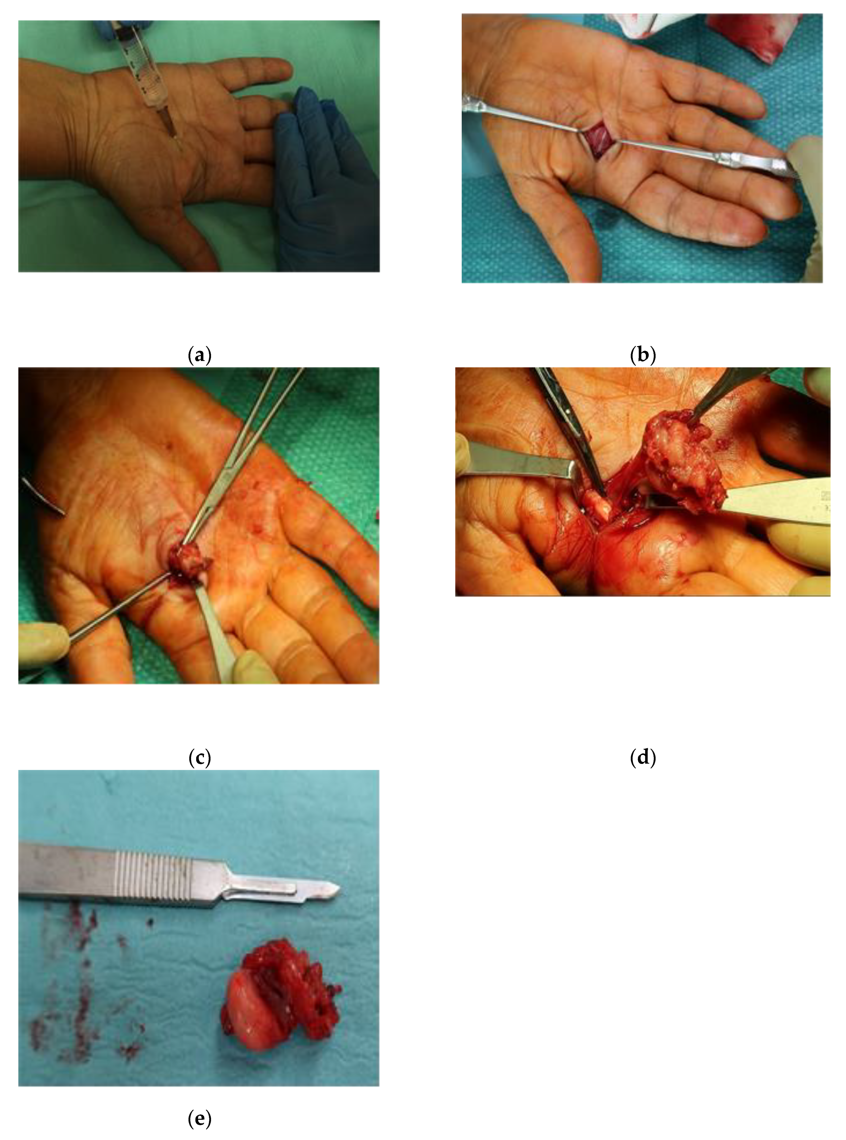
Figure 2.
Frequency of histological types in excised lesions. Ganglion cyst (44,98 %) the second diagnosis in frequence was Tenosynovial giant-cell tumor of the synovial sheath (15,32%). (0,48%) malignant disease (exploded).
Figure 2.
Frequency of histological types in excised lesions. Ganglion cyst (44,98 %) the second diagnosis in frequence was Tenosynovial giant-cell tumor of the synovial sheath (15,32%). (0,48%) malignant disease (exploded).
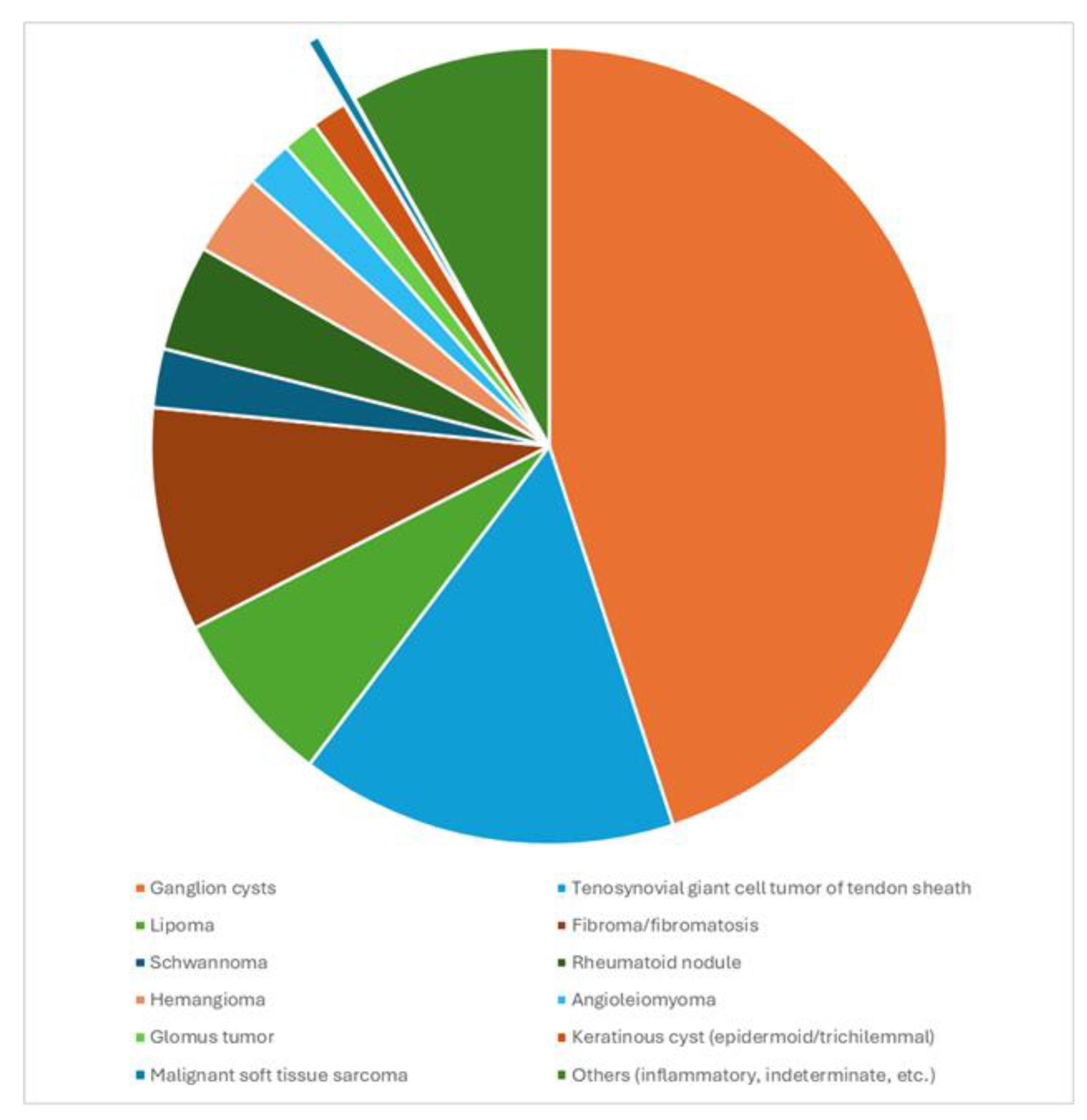
Figure 3.
Histological report: Mesenchymal sarcoma spindle-cell.
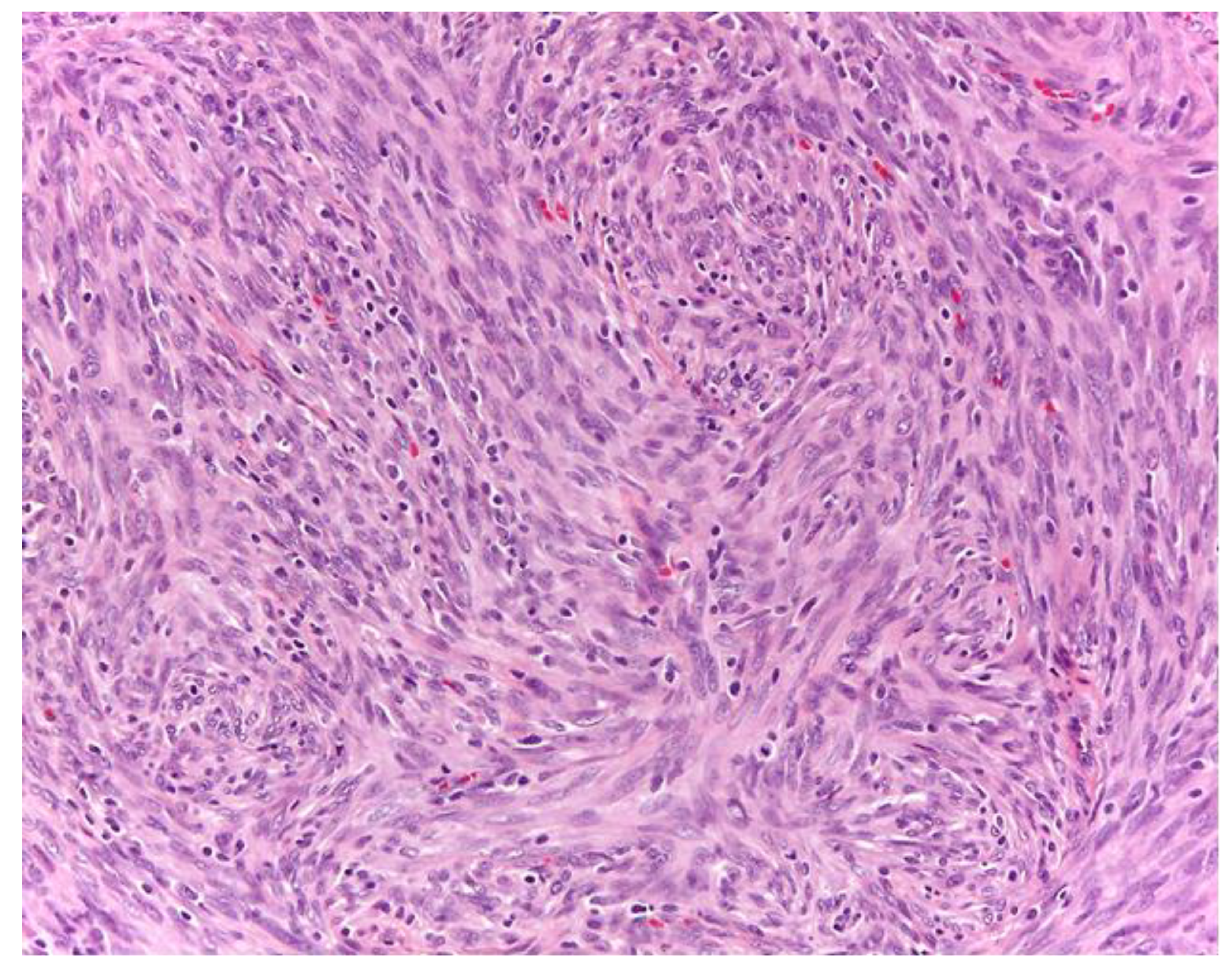
Figure 4.
Histological report: tenosynovial giant cell tumor.
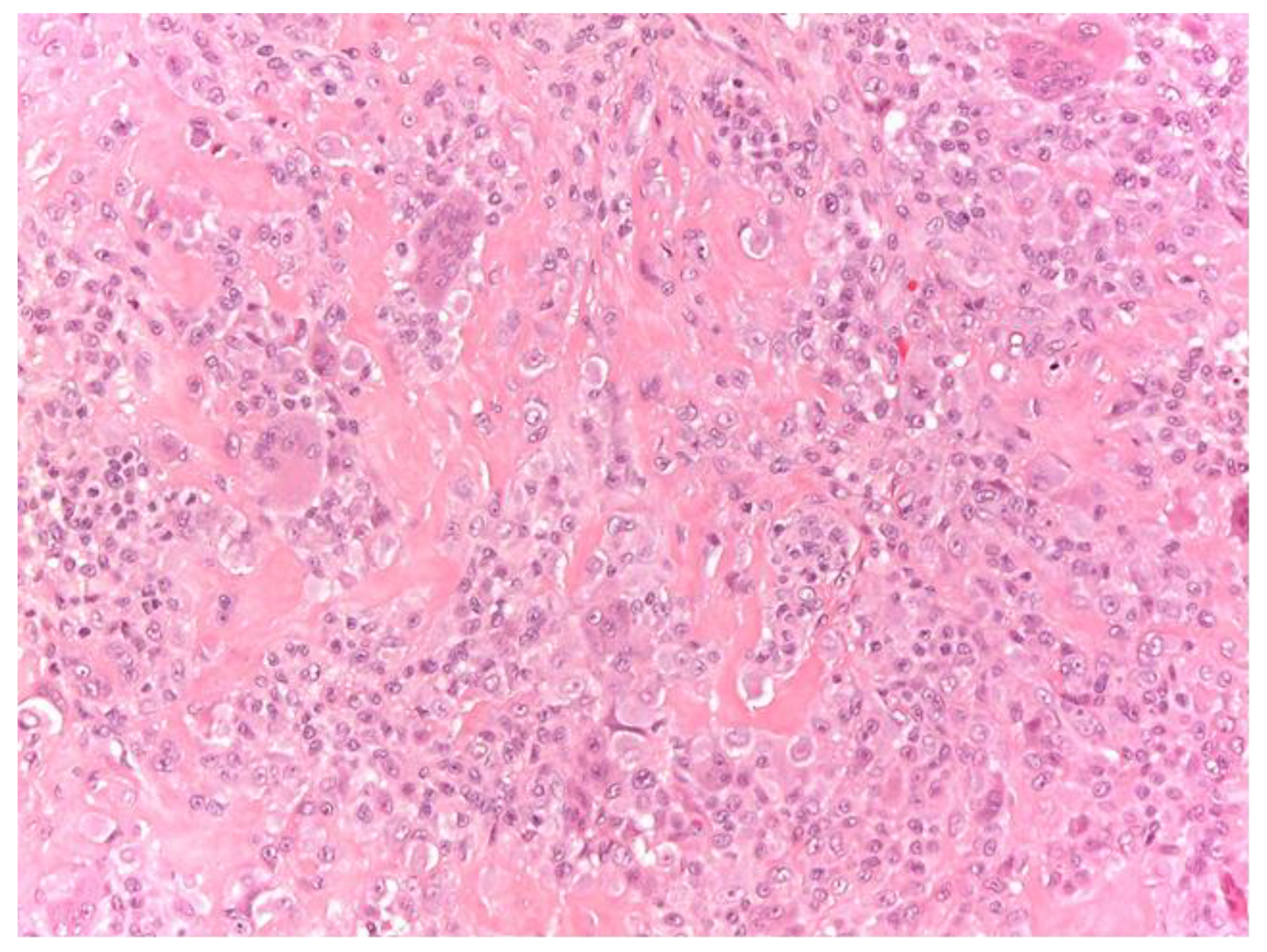
Figure 5.
Female, 50 years old. Onset of volar-radial swelling at the wrist and paresthesias in the first 3 fingers of the hand. Ultrasound: oval, regular lesion, heterogeneous content, avascular, about 2 cm. Excision in WALANT. Histological report: tenosynovial giant cell tumor.
Figure 5.
Female, 50 years old. Onset of volar-radial swelling at the wrist and paresthesias in the first 3 fingers of the hand. Ultrasound: oval, regular lesion, heterogeneous content, avascular, about 2 cm. Excision in WALANT. Histological report: tenosynovial giant cell tumor.
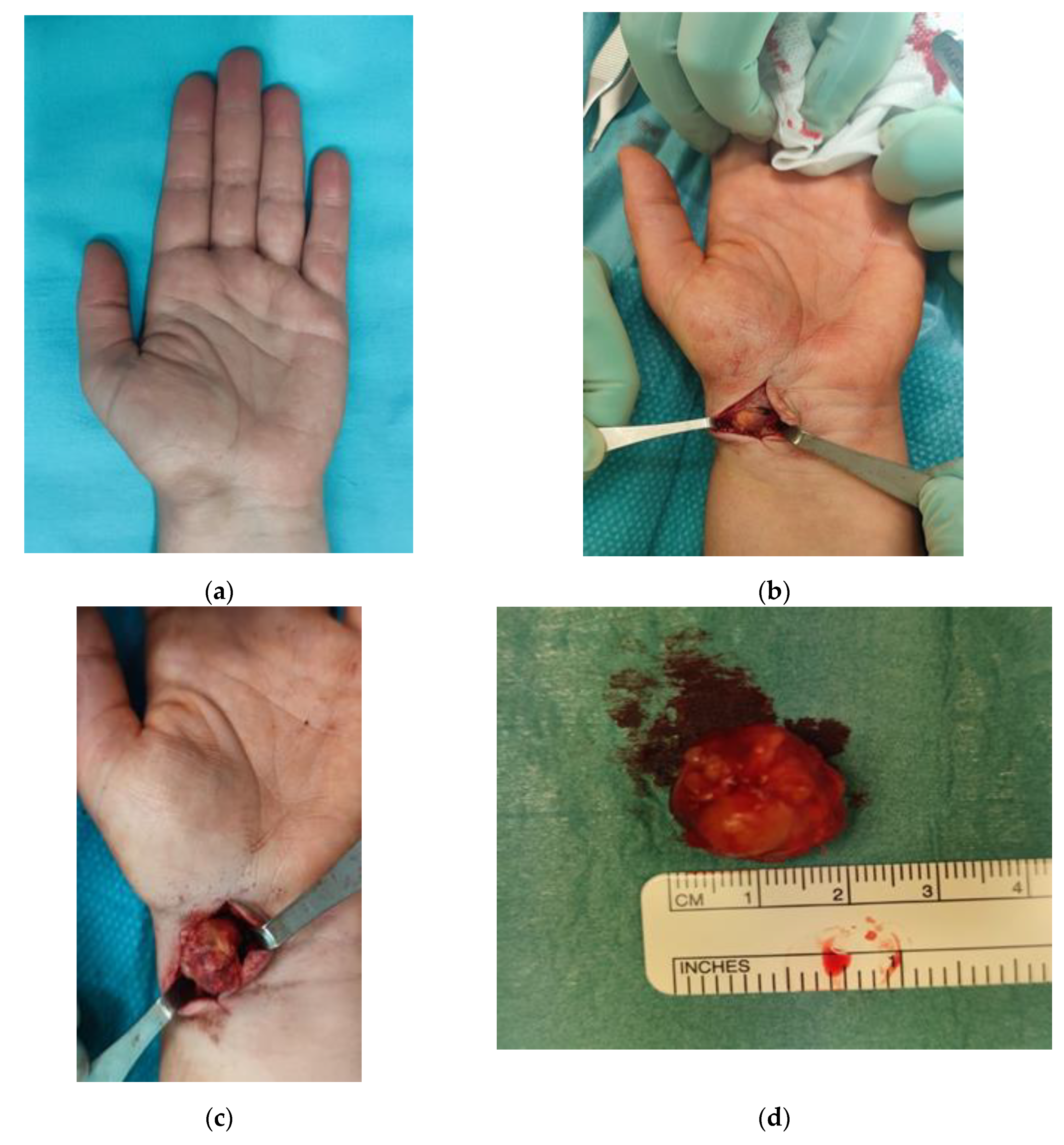

Table 1.
Histological type of 209 lesions removed from the wrist and hand.
| Lesion type | Total per lesion | Total | N° of Male | N° of Female | Age | Lesion size (major axis) |
|---|---|---|---|---|---|---|
| Ganglion cysts | 0 | 94 | 31 | 63 | 51,42±18.86 | 1.31±0.55 |
| TGCT | 15,32 | 32 | 9 | 23 | 57.55±10.61 | 1.98±1.01 |
| Lipoma | 7,18 | 15 | 7 | 8 | 54.57±10.43 | 2.13±1.09 |
| Fibroma/fibromatosis | 9,09 | 19 | 9 | 10 | 50.07±16.59 | 1.02±1.06 |
| Schwannoma | 2,39 | 5 | 4 | 1 | 43.40±8.71 | 1.87±0.05 |
| Rheumatoid nodule | 4,31 | 9 | 5 | 4 | 66.33±9.72 | 1.27±0.43 |
| Hemangioma | 3,35 | 7 | 3 | 4 | 59.25±8.17 | 1.37±0.41 |
| Angioleiomyoma | 1,91 | 4 | 0 | 4 | 65.33±7.76 | 1.77±0.98 |
| Glomus tumor | 1,44 | 3 | 1 | 2 | 49.67±19.26 | 0.60±0.28 |
| Keratinous cyst | 1,44 | 3 | 3 | 0 | 51.33±4.64 | 1.28±0.57 |
| Malignant soft tissue sarcoma | 0,48 | 1 | 0 | 1 | 63±00 | 1,60±0.00 |
| Others | 8,14 | 17 | 7 | 10 | 48.78±14.23 | 1.52±0.95 |
| Total | 100.03 | 209 |
*Values are reported as percentage and absolute number of lesions. Lesions with an incidence lower than 1% were grouped as "others" except mesenchymal neoplasm with spindle cells with a low grade of malignancy. Age is expressed as average ± standard deviation. Lesion sizes are expressed in cm (average ± standard deviation). Results are rounded to two decimal places.
Disclaimer/Publisher’s Note: The statements, opinions and data contained in all publications are solely those of the individual author(s) and contributor(s) and not of MDPI and/or the editor(s). MDPI and/or the editor(s) disclaim responsibility for any injury to people or property resulting from any ideas, methods, instructions or products referred to in the content. |
© 2026 by the authors. Licensee MDPI, Basel, Switzerland. This article is an open access article distributed under the terms and conditions of the Creative Commons Attribution (CC BY) license (http://creativecommons.org/licenses/by/4.0/).
Copyright: This open access article is published under a Creative Commons CC BY 4.0 license, which permit the free download, distribution, and reuse, provided that the author and preprint are cited in any reuse.


