

Submitted:
11 May 2026
Posted:
12 May 2026
You are already at the latest version
Abstract
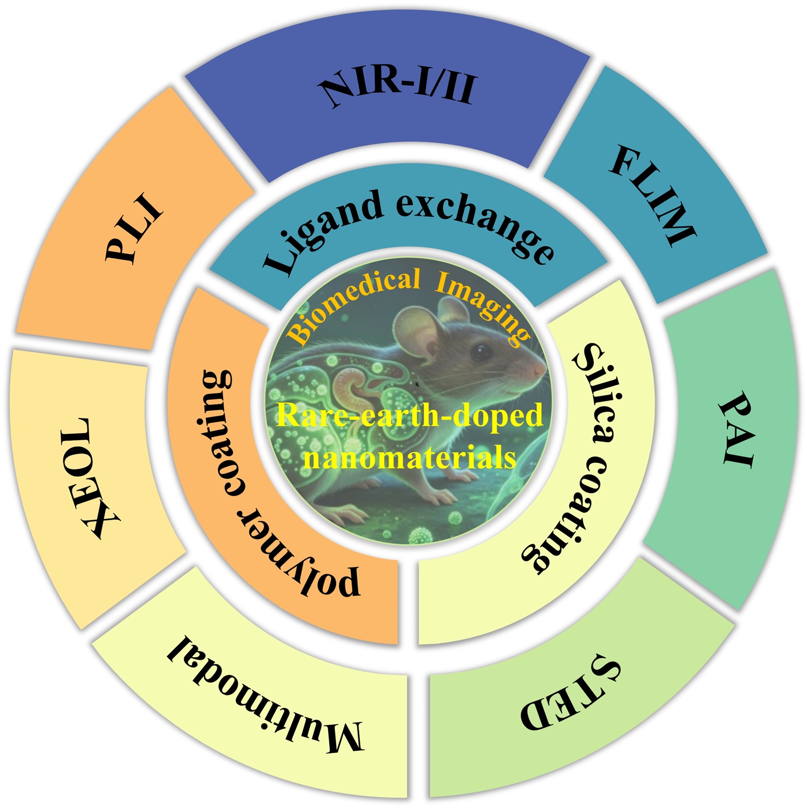
Benefiting from tunable emission from ultraviolet to near-infrared windows, long luminescence lifetimes, and exceptional photostability, rare-earth-doped nanomaterials overcome the limitations of conventional dyes and quantum dots, enabling deep-tissue, high-resolution, and low-background imaging. As multifunctional fluorescent probes, rare-earth-doped nanomaterials are driving the development of next-generation biomedical imaging. This review summarizes recent advances in the structural design of rare earth-doped nanomaterials, surface engineering for biocompatibility, and targeting strategies for improved performance, and highlights their integration into advanced imaging modalities, including NIR-I/II fluorescence, FLIM, PAI, super-resolution STED, multimodal FL/MRI/CT, X-ray-excited luminescence, and persistent luminescence. Meanwhile, mechanistic insights, material innovations, and comparative advantages are discussed. Furthermore, challenges related to quantum yield, scalable synthesis, imaging resolution, and clinical translation are considered, while future directions—centered on multifunctional probe design, NIR-II imaging, and AI-assisted data analysis—are proposed, offering a versatile platform for precise multimodal imaging with significant potential to advance early diagnosis, personalized therapy, and clinical applications.
Keywords:
rare-earth-doped nanomaterials
; surface engineering
; fluorescent probes
; biomedical imaging
; near infrared
1. Introduction
Optical bioimaging has evolved into an indispensable cornerstone of modern biomedical research and clinical translational medicine, enabling non-invasive, real-time visualization of biological structures, dynamic cellular processes, and pathological events at molecular, cellular, and in vivo levels.[1,2,3,4,5,6] Over the past few decades, continuous advances in fluorescence probe design, optical system engineering, and image processing algorithms have propelled optical imaging from simple qualitative observation to high-resolution, quantitative, and multi-dimensional dynamic analysis, profoundly reshaping our understanding of life sciences and driving innovations in disease diagnosis, drug development, and precision therapy.[7,8] However, conventional optical bioimaging still faces persistent bottlenecks in complex biological environments, including strong tissue autofluorescence interference, limited photon penetration depth, severe photobleaching of fluorescent probes, potential biotoxicity of nanomaterials, and unsatisfactory signal-to-noise ratio and spatial resolution, which severely restrict its application in deep-tissue imaging, long-term dynamic tracking, and high-sensitivity biomarker detection.[2,9,10]
Against this backdrop, the rational design and surface engineering of nano-fluorescent probes have emerged as a pivotal breakthrough in addressing the above challenges.[11,12] Among a diverse library of nanoscale optical materials, rare-earth-doped nanoparticles (RENPs) have stood out as a transformative and preeminent platform for next-generation optical bioimaging, owing to their unique and superior photophysical properties that outperform traditional organic fluorophores, semiconductor quantum dots (QDs), and carbon-based nanomaterials.[13,14] Distinguished by large anti-Stokes shifts that eliminate excitation-emission spectral crosstalk, ultra-narrow and symmetric emission bands ideal for multiplexed detection, extraordinary resistance to photobleaching and photoblinking, long luminescence lifetimes suitable for time-gated background rejection, and tunable excitation/emission profiles covering ultraviolet-visible to near-infrared (NIR) biological transparency windows, RENPs effectively overcome the inherent limitations of conventional fluorescent probes, such as autofluorescence interference, photodegradation, and heavy-metal toxicity, thereby enabling non-invasive, high-contrast, and long-term visualization of biological processes in live cells and deep tissues.[15,16,17,18]
The performance of RENPs in biomedical applications is fundamentally governed by two core aspects: the rational design of nanomaterial matrices and doping systems, and the multifunctional surface engineering that ensures biocompatibility and enhances performance in biological environments.[19] The as-synthesized RENPs are typically hydrophobic and lack biocompatibility and targeting ability, necessitating surface modification as an prerequisite for their in vitro and in vivo applications. Various surface functionalization strategies, including ligand exchange, amphiphilic polymer coating, silica coating, ligand oxidation, and ligand-free methods, have been developed to impart hydrophilicity, biocompatibility, reduced non-specific interactions, and controllable bioconjugation capabilities to RENPs, thereby laying a solid foundation for targeted imaging and responsive detection in complex biological systems.[20] Meanwhile, the unique 4f-4f electronic transitions of RE ions endow RENPs with versatile luminescence mechanisms, enabling their adaptation to a broad range of advanced optical imaging modalities, including NIR-I/NIR-II imaging, fluorescence lifetime imaging (FLIM), photoacoustic imaging (PAI), stimulated emission depletion (STED) super-resolution imaging, multimodal imaging, X-ray excited optical luminescence (XEOL) imaging, and persistent luminescence imaging (PLI).[21,22,23] These diversified modalities integrate the advantages of high sensitivity, deep tissue penetration, super-resolution, and background-free detection, thereby expanding the application boundaries of optical bioimaging—from superficial cellular observation to deep-tissue dynamic tracking and clinical precise diagnosis.
In recent years, the integration of RE-doped nano-fluorescent probes with advanced optical imaging technologies has witnessed rapid progress, with continuous breakthroughs in core-shell structures, multi-modal signal coupling, responsive probe design, and in vivo clinical translation.[19,24,25,26,27] Nevertheless, several critical challenges remain for the practical clinical application of these nanoprobes, including the long-term biosafety and metabolic clearance of nanomaterials in vivo, precise control of surface ligand density and orientation, development of biodegradable coating materials, and improvement of imaging resolution and penetration depth. This review systematically summarizes the design principles, material classification, and surface engineering strategies of RE-doped nano-fluorescent probes, elaborates on their working mechanisms and application progress across seven representative advanced optical imaging modalities, and discusses future directions and clinical translation potential. This paper serves as a comprehensive reference on RE-doped nanomaterials for researchers in materials science, biomedical engineering, and clinical medicine, and is expected to promote the innovation and practical application of high-performance nano-fluorescent probes in optical bioimaging technologies.
2. Design and Surface Engineering of Nano-Fluorescent Probes
The foundation of high-performance optical bioimaging lies in the rational design of nano-fluorescent probes, which directly determines imaging resolution, signal-to-noise ratio, tissue penetration depth, and long-term stability in complex biological environments. Among the diverse library of nanoscale optical materials developed over the past two decades, RENPs have emerged as a preeminent and transformative choice for cutting-edge bioimaging applications. This prominent status stems from their exceptional and unique photophysical properties that outperform conventional fluorescent probes: large anti-Stokes shifts that eliminate excitation-emission spectral crosstalk, ultra-narrow and symmetric emission bands ideal for multiplexed detection, extraordinary resistance to photobleaching and photoblinking, long luminescence lifetimes enabling time-gated background rejection, and tunable excitation/emission profiles spanning the ultraviolet–visible to NIR biological transparency windows.
Unlike organic fluorophores and semiconductor QDs that suffer from autofluorescence interference, photodegradation, or potential heavy-metal toxicity, RENPs enable non-invasive, high-contrast, and long-term tracking of biological processes in live cells and deep tissues. This section systematically categorizes the major types of nanomaterials employed for advanced optical imaging, with a primary focus on RENPs including their representative host lattices and core–shell structural engineering strategies. Following this material classification, we further elaborate on the indispensable and multifaceted roles of surface engineering and ligand functionalization. Specifically, we highlight how rational surface design endows these nanoprobes with physiological stability, favorable biocompatibility, reduced non-specific interactions, and controllable bioconjugation capabilities—ultimately enabling reliable, targeted, and context-responsive performance in real-world biomedical imaging scenarios.
2.1. Types of Fluorescent Nanomaterials for Advanced Bioimaging
To achieve high-sensitivity and high-resolution fluorescence imaging in the biological window, a variety of nanomaterials have been developed, including organic dyes, QDs, carbon-based nanomaterials (e.g., modified graphene oxide and carbon dots), and RENPs. Each class of materials possesses inherent advantages and limitations that determine their suitability for specific bioimaging applications.
QDs are semiconductor nanocrystals known for their high quantum yield, size-tunable emission, and narrow emission spectra. These properties make them bright and color-tunable probes for multiplexed imaging. The development of QDs has progressed through several key milestones (Figure 1).[28,29,30,31,32,33,34,35] The quantum size effect was first experimentally observed in CuCl nanocrystals by Ekimov in 1981, followed by Brus's independent discovery of size-dependent optical shifts in colloidal CdS crystallites in 1983. A major breakthrough came in 1993 when Bawendi and colleagues introduced the “hot-injection” synthesis method, enabling the production of high-quality, monodisperse QDs with precisely tunable optical properties. Subsequently, the development of core/shell structures, particularly CdSe/ZnS QDs reported in 1996, significantly enhanced photoluminescence quantum yield to approximately 50% after addressing surface defect-related quenching. Since then, further advances have enabled near-unity quantum yield QDs, heavy-metal-free alternatives, and perovskite QDs, culminating in the 2023 Nobel Prize in Chemistry awarded to Ekimov, Brus, and Bawendi. Despite these remarkable advances, the biomedical application of QDs remains limited by their inherent toxicity due to heavy metal ions (e.g., cadmium, lead) and chemical instability, leading to controversial biosafety profiles that restrict their in vivo use.[36,37] This long-standing toxicity concern has motivated the search for cadmium-free alternatives such as InP, CuInS2, carbon, and silicon QDs.
Organic fluorescent probes offer advantages such as well-defined chemical structures, easy functionalization, and good biocompatibility. They are widely used in commercial assays and clinical diagnostics.[38] Nevertheless, they are often plagued by complex synthesis procedures, poor photostability leading to rapid photobleaching, and broad absorption/emission spectra. These shortcomings severely limit their performance in long-term imaging and multiplexed detection applications.[37,39] Carbon-based nanomaterials exhibit unique photoluminescence properties, including emission in the NIR-II window, which enables deeper tissue penetration. However, their complex synthesis, poor solubility in physiological solutions, and potential long-term toxicity pose considerable challenges for routine biological use.[40]
RENPs stand out as the most promising candidates for advanced bioimaging, effectively addressing many of the limitations discussed above. From the first conceptualization of infrared quantum counters in 1959 to the establishment of ETU mechanisms (1966), the identification of NaYF4 as the optimal host (1972), the first size-controlled synthesis of RENPs (2004), and recent breakthroughs in NIR-triggered therapy and imaging (2011-2026), the field has evolved from fundamental physics to promising biomedical applications (Figure 2).[41,42,43,44,45,46,47,48,49,50] Typical RENPs are composed of an inorganic host matrix (e.g., hexagonal-phase NaYF4, known for its low phonon energy of ~350 cm-1 and high upconversion efficiency), co-doped with sensitizer (e.g., Yb3+, Nd3+) and activator (e.g., Er3+, Tm3+, Ho3+) ions.[36,51] RENPs offer several decisive advantages that overcome the drawbacks of other nanomaterials.
(1) Unique anti-stokes optical properties: Unlike QDs and organic dyes that require high-energy UV or visible excitation, RENPs convert low-energy NIR excitation (e.g., 980 nm or 808 nm) into higher-energy visible or UV emission. This eliminates background autofluorescence from biological tissues, a persistent problem with other probes, thereby providing exceptionally high signal-to-noise ratios for in vivo imaging.
(2) Superior photostability: While organic dyes suffer from rapid photobleaching, RENPs possess inherent non-photobleaching and non-photoblinking properties. This allows for long-term and repetitive imaging with high temporal resolution without signal degradation, a critical advantage for tracking biological processes over extended periods.
(3) Tunable emission and long luminescence lifetime: By selecting different RE dopants, the emission spectra of RENPs can be precisely tuned from UV to NIR-II (1000-1700 nm), enabling multi-color imaging.[52] Additionally, the parity-forbidden f-f electronic transitions endow them with long luminescence lifetimes (microsecond to millisecond scale), which is orders of magnitude longer than the nanosecond-scale autofluorescence of tissues. This property is highly advantageous for time-gated imaging and multiplexed detection using FLIM, a capability not readily available with QDs or organic dyes.[53]
(4) Core-shell engineering: To address the surface quenching effect that plagues all nanomaterials due to their high surface-to-volume ratio, advanced core-shell structures have been developed for RENPs. An inert shell (e.g., NaYF4 or NaGdF4) can be epitaxially grown on the UCNP core to spatially confine RE dopants away from the surface. This strategy dramatically enhances luminescence intensity by suppressing surface-induced quenching of excitation energy, a challenge that is particularly acute for small-sized nanoparticles.[40]
In summary, RENPs, particularly RENPs with engineered core-shell architectures, uniquely combine deep-tissue NIR excitation, high photostability, tunable multi-color emission, and long luminescence lifetimes, positioning them as the most versatile and powerful nanoplatform for next-generation biomedical imaging.
2.2. Surface Engineering of Renps
As-synthesized RENPs are typically coated with hydrophobic ligands (e.g., oleylamine, oleic acid) to prevent aggregation and ensure colloidal stability during high-temperature synthesis. While these hydrophobic ligands are essential for controlling nanoparticle size and morphology, they render the nanoparticles only dispersible in non-polar organic solvents such as cyclohexane or chloroform.[54] This hydrophobic surface presents a critical barrier for biomedical applications, as biological environments—ranging from cell culture media to blood and interstitial fluids—are inherently aqueous. In a cellular environment, the surface properties of nanoparticles directly dictate their interactions with biological systems. Hydrophobic nanoparticles introduced into aqueous biological media tend to aggregate via hydrophobic interactions, leading to large particle clusters that can occlude blood vessels, trigger unwanted immune responses, and be rapidly cleared by the reticuloendothelial system (RES) before reaching target cells or tissues.[55] Furthermore, the exposed hydrophobic surface can non-specifically adsorb serum proteins, forming a “protein corona” that alters the nanoparticle's size, surface charge, and biological identity. This protein corona often leads to enhanced uptake by macrophages, reduced targeting specificity, and potential immunogenicity.[56,57] Therefore, rendering RENPs hydrophilic is an indispensable prerequisite for any in vitro or in vivo application.
Beyond achieving colloidal stability in aqueous media, surface ligand engineering is crucial for two additional objectives essential for cellular-level applications. The surface coating must not only confer hydrophilicity but also be non-toxic and biocompatible at the cellular level. For instance, the oleic acid (OA) ligands, while safe in organic solvents, can be cytotoxic if not properly removed or shielded. Common biocompatible coatings include polyethylene glycol (PEG), which creates a “stealth” layer that resists protein adsorption and reduces RES uptake, thereby prolonging blood circulation time and minimizing non-specific interactions with healthy cells.[58,59] Studies have shown that appropriate surface coatings, such as thick silica shells, can significantly reduce the leakage of toxic fluoride and RE ions from degrading RENPs, dramatically improving their biosafety profile in cellular environments.[60,61] After establishing a hydrophilic and biocompatible surface, the next critical step is to introduce reactive functional groups (e.g., -COOH, -NH2, -SH, or -CHO) that serve as anchoring points for covalent conjugation with biomolecules.[62] These functional groups enable the attachment of specific targeting ligands, such as antibodies, peptides, folic acid, or aptamers, which can recognize and bind to receptors overexpressed on the surface of diseased cells (e.g., cancer cells).[63,64] This active targeting strategy enhances cellular uptake via receptor-mediated endocytosis, increases the local concentration of the nanoprobe at the target site, and improves imaging contrast while reducing off-target effects on healthy tissues.[55]
To address these critical requirements, a variety of surface modification strategies have been developed, including ligand exchange, amphiphilic polymer coating, silica coating, ligand oxidation, and the ligand-free approach. Each strategy offers distinct advantages in terms of hydrophilicity, colloidal stability, conjugation flexibility, and preservation of the luminescent properties of RENPs. Figure 3 provides an overview of these representative approaches.
2.2.1. Ligand Exchange
Ligand exchange is one of the most widely employed strategies for rendering hydrophobic RENPs water-dispersible. This method involves replacing the original hydrophobic ligands on the nanoparticle surface with hydrophilic ligands that have a higher affinity for the RE ions. The controlled synthesis of RE-doped NaYF4 nanocrystals with tunable size, morphology, and crystal phase laid the foundation for subsequent surface engineering strategies.[69,70] The process typically requires an excess of the incoming ligand to drive the equilibrium toward complete ligand substitution.[62]
Poly(acrylic acid) (PAA) is a linear polymer containing multiple carboxyl (-COOH) groups along its backbone that can coordinate to RE ions on the nanoparticle surface, displacing the original OA ligands (Figure 3a).[59] The abundant terminal and pendant carboxyl groups also serve as versatile anchoring points for subsequent bioconjugation with targeting ligands or therapeutic agents via standard carbodiimide chemistry (EDC/NHS coupling).[59,71] This dual functionality—simultaneously conferring hydrophilicity and providing reactive handles—makes PAA an attractive choice for theragnostic applications. Short-chain PEGylated ligands, such as thiol-terminated methoxypolyethylene glycol (mPEG-SH) or PEG-phosphates, are also commonly used in ligand exchange. The PEG chains create a hydration layer around the nanoparticle through hydrogen bonding with water molecules, effectively shielding the nanoparticle surface from protein adsorption.[72] This “stealth” property reduces recognition and uptake by the reticuloendothelial system, prolonging blood circulation time and enhancing the probability of tumor accumulation via the enhanced permeability and retention (EPR) effect.[55,73,74] An innovative extension of this concept involves in vivo assembly, where a pre-administered nanoparticle “anchor” accumulates in tumors via the EPR effect and subsequently captures a separately injected small-molecule imaging agent, combining rapid tumor influx with prolonged retention.[75] Compared to PAA, PEGylated ligands generally provide superior colloidal stability in complex biological media. Other functional ligands can also be exchanged onto the RENP surface depending on the specific application. For example, a particularly innovative approach involves the use of DNA-protein conjugates as ligands for one-step bioconjugation, where the DNA phosphate backbone chelates to the RE surface while presenting functional proteins for subsequent recognition (Figure 3b).[65] Alternatively, small molecules bearing thiol (-SH) or amine (-NH2) groups, such as 3-mercaptopropionic acid or 6-aminocaproic acid, can serve dual roles as both surface-capping agents and hydrophilic ligands, as demonstrated in one-step hydrothermal synthesis approaches.[76] Citrate ions, while providing weaker surface binding, can rapidly confer water dispersibility for applications requiring small hydrodynamic diameters.
The primary advantage of ligand exchange is its simplicity and versatility. However, several limitations should be noted. First, complete removal of the original hydrophobic ligands is often difficult to achieve. Second, the exchanged ligands may desorb over time, particularly under physiological conditions or upon dilution, leading to nanoparticle aggregation.[77] Third, the surface density of functional groups can be difficult to precisely control, which may affect the reproducibility of subsequent bioconjugation. Despite these challenges, ligand exchange remains a straightforward and effective method for rapidly producing hydrophilic RENPs for preliminary biological evaluations.
2.2.2. Amphiphilic Polymer Coating
Amphiphilic polymer coating represents one of the most widely adopted strategies for rendering hydrophobic RENPs water-dispersible while simultaneously imparting “stealth” properties essential for in vivo applications. This method is particularly advantageous because it does not require the removal of the original hydrophobic ligands, thereby preserving the nanoparticle's intrinsic optical properties.[58,62]
This strategy utilizes amphiphilic block copolymers, most notably DSPE-PEG, to create a stable lipid bilayer around the nanoparticle core. The hydrophobic lipid tails intercalate with the original OA ligand layer, while the hydrophilic PEG segments orient outward into the aqueous environment. The outward-facing PEG chains create a dense hydration layer that minimizes opsonization, and reduces recognition and clearance by the RES. By evading RES uptake, DSPE-PEG-coated nanoparticles exhibit significantly extended circulation half-lives, enhancing their probability of accumulating at target tissues via the EPR effect.
Crucially, the amphiphilic polymer coating method preserves the nanoparticle’s upconversion luminescence (UCL). As illustrated in Figure 3c, the core-shell structure consists of a PEO-b-PCL shell, DiR molecules, and LNP cores.[66] Unlike ligand exchange, which can introduce surface defects, this approach leaves the original OA ligand layer intact, maintaining the passivation that prevents non-radiative energy losses at the particle surface.[58,62] Studies have demonstrated that DSPE-PEG-coated RENPs retain bright luminescence in aqueous media, enabling highly sensitive early tumor detection with NIR-II emissive nanoparticles. The amphiphilic polymer coating strategy also provides a versatile platform for additional functionalization. The terminal groups of the PEG chains can be readily conjugated to targeting ligands such as antibodies or peptides, enabling active targeting and theranostic applications. Furthermore, photosensitizers or nanozymes can be incorporated for combination therapy.[78]
2.2.3. Silica Coating
Coating RENPs with a silica shell represents one of the most versatile strategies for surface functionalization, particularly for biomedical applications requiring high stability, biocompatibility, and multifunctionality. The broader context of RERE-doped upconversion nanomaterials in biological molecular imaging has been comprehensively reviewed, underscoring their significance across diagnostic and therapeutic applications.
Silica coating is typically achieved through two classical methods: the Stöber method and the reverse microemulsion method. The Stöber method, first reported in 1968, involves the base-catalyzed hydrolysis and condensation of TEOS in a mixture of water, alcohol, and ammonia, producing uniform, non-porous silica spheres.[79] However, obtaining monodisperse particles with diameters smaller than 100 nm remains challenging. The reverse microemulsion method offers a more versatile alternative, where water droplets containing TEOS are entrapped within reverse micelles formed by surfactants in a non-polar continuous phase. This method enables precise control over shell thickness and the synthesis of mesoporous silica structures, with particle size depending critically on the water-to-surfactant molar ratio and ammonia concentration.[80] As illustrated in Figure 3d, four representative UCNP@SiO2 nanostructures have been developed: UCNP@dSiO2 (1), UCNP@mSiO2 (2), UCNP@dSiO2@mSiO2 (3), and UCNP@hmSiO2 (4), highlighting the architectural versatility of this platform.[67] Optimization studies have shown how reactant concentrations and surfactant types affect the final particle characteristics.[81]
Importantly, silica coating methods are mild enough to preserve the intrinsic UCL of the RENP core. Studies have shown that mesoporous silica coating can even enhance certain emission bands; for instance, the red emission at 650 nm in NaYF4:Yb3+/Er3+ RENPs exhibits longer luminescence lifetimes and higher emission intensity after coating.[82] The silica shell offers several critical advantages for medical applications.
(1) Biocompatibility and optical transparency: silica is inherently biocompatible, chemically inert in physiological environments, and optically transparent across the UV-visible-NIR spectrum. And, silica-coated RENPs are non-toxic to mammalian cells, eliciting only minor, cell type-dependent stress responses.
(2) Facile surface functionalization: the silica surface is rich in silanol groups, which can be readily functionalized with various silane coupling agents to introduce amine, carboxyl, or thiol groups for subsequent bioconjugation. Surface functionalization with targeting ligands such as folic acid enables receptor-mediated endocytosis, enhancing cellular uptake by cancer cells. The successful functionalization can be confirmed by FTIR and elemental analysis. This functionalization versatility has enabled the development of sophisticated theranostic platforms combining multiple imaging modalities with therapeutic functions.
(3) Enhanced biosafety: a critical concern for UCNP clinical translation is the potential release of toxic fluoride and RE ions during degradation in biological environments.[83] The dissolution is particularly problematic under high dilution conditions commonly used in biological applications. Thick silica coatings provide an effective physical barrier, dramatically reducing ion leakage. Direct comparisons have shown that while bare RENPs released 297.37 μM of fluoride ions after 48 hours in water, thick silica-coated RENPs released only 4.42 μM.[60] Similarly, thick silica-coated RENPs maintained fluoride and yttrium ion leakage at near-zero levels for over 70 hours.[61]
Despite these advantages, silica coating has limitations including increased hydrodynamic diameter and slow biodegradation of pure inorganic Si-O-Si frameworks. Current research has shifted toward organic/inorganic hybrid mesoporous organosilica nanoparticles incorporating disulfide bonds to improve biodegradability and enable multi-responsive drug release.[84]
2.2.4. Other Strategies
Ligand oxidation uses oxidizing agents to convert the terminal alkene groups of OA ligands into carboxyl groups without removing the ligands from the nanoparticle surface. The Lemieux-von Rudloff reaction selectively oxidizes carbon-carbon double bonds, rendering the nanoparticles water-dispersible while preserving the inorganic core. The newly formed carboxyl groups can be directly used for bioconjugation.
Simultaneous synthesis and functionalization represent another elegant strategy. Wang et al. developed a facile one-step hydrothermal synthesis technique through which water-soluble NaYF4:Yb3+/Er3+ nanoparticles with various functional groups (including 3-mercaptopropionic acid, 6-aminocaproic acid, and PEG) on their surface can be directly prepared without any further surface treatment.[76] This approach eliminates the need for post-synthesis modification steps and was demonstrated to produce NPs suitable for both in vitro cell imaging and in vivo nude mouse imaging.
The ligand-free approach offers an even simpler alternative through acid treatment that protonates the carboxylate headgroups of OA ligands, releasing them from the nanoparticle surface. The resulting naked nanoparticles are stabilized in aqueous solution by electrostatic repulsion, as predicted by the classical DLVO theory.[85] This approach is extremely simple and produces nanoparticles with a clean, unmodified surface highly accessible for further functionalization. This process is visually summarized in the schematic from Nyk et al., where the left panel depicts ligand removal and the right panel shows the resulting naked particle surface with exposed RE ions (Figure 3e).[68] Recent improvements have achieved reaction yields up to 96% by carefully controlling acid concentration and reaction time.[86] However, the long-term colloidal stability of ligand-free nanoparticles in complex biological media may be limited. The absence of a protective organic layer makes them susceptible to aggregation under physiological ionic strength conditions and more prone to non-specific protein adsorption. Additionally, water itself acts as an influential agent for RE-doped luminescent nanoparticles, affecting their colloidal stability, luminescence quenching, and optical penetration depth.
3. Advanced Imaging Technologies Based on Fluorescent Renps
Benefiting from the unique 4f–4f electronic transitions of RE ions, RENPs exhibit abundant and controllable luminescence behaviors, which can perfectly match the working principles of diverse advanced optical imaging modalities. By rationally designing the host lattice, doping components, core–shell structure and surface functionalization of RENPs, the luminescence output, energy transfer path, response mode and signal conversion efficiency can be precisely regulated, so as to adapt to the technical requirements of different imaging modes. These imaging technologies not only retain the high sensitivity and non-invasiveness of optical imaging, but also break through the bottlenecks of traditional fluorescence imaging in tissue penetration, spatial resolution, background interference and dynamic monitoring. In this section, we systematically elaborate seven representative advanced optical imaging modalities driven by RENPs, including NIR-I/II imaging, FLIM, PAI, STED super-resolution imaging, multimodal imaging, XEOL imaging and PLI. For each modality, we focus on the luminescence mechanism matching with RENPs, material design strategies, typical application cases and comparative advantages, so as to clarify the key role of RE-doped nano-fluorescent probes in promoting the performance upgrade of biomedical optical imaging. The seven imaging modalities discussed above are enabled by distinct photophysical mechanisms of RENPs (Table 1).
3.1. Near-Infrared I and Ii Imaging
3.1.1. Mechanism
The mechanism of NIR imaging using RE-doped nanoparticles primarily relies on downshifting (Stokes) emission. In this process, RENPs are excited by a shorter-wavelength NIR-I laser (e.g., 808 nm) and emit longer-wavelength NIR-II photons (1000–1700 nm). The NIR-II window offers reduced photon scattering, minimal tissue autofluorescence, and deeper penetration depth compared to visible and NIR-I windows.[112]
For downshifting, sensitizer ions such as Nd3+ or Yb3+ absorb excitation photons and transfer energy to activator ions (e.g., Nd3+ itself, Er3+, or Tm3+), which subsequently emit at NIR-II wavelengths. The energy transfer follows the general pathway: sensitizer (absorption) → energy migration → activator (emission).[113] To avoid back energy transfer from activators to sensitizers, core-shell architectures have been developed where sensitizers and activators are spatially separated into different layers. For example, in NaYF4:Yb3+/Er3+@NaYF4:Yb3+@NaNdF4:Yb3+ multi-shell nanoparticles, energy transfer occurs directionally from Nd3+ → Yb3+ → Er3+, effectively eliminating Er3+ → Nd3+ back transfer and resulting in over 10-fold enhancement of UCL upon 800 nm excitation.[96,113] This mechanism underlies high-contrast deep-tissue NIR-II imaging of vasculature, lymph nodes, and tumors.
3.1.2. Application
Fluorescence bioimaging within NIR biological windows has emerged as an indispensable tool for noninvasive deep-tissue visualization and clinical diagnosis. The conventional NIR-I window (700–900 nm) provides limited tissue penetration and is severely hindered by strong photon scattering, high tissue autofluorescence, and low signal-to-noise ratio, which restrict its capacity for high-resolution imaging in deep biological tissues. In comparison, the NIR-II window (1000–1700 nm), particularly the NIR-IIb subwindow (1500–1700 nm), exhibits unprecedented advantages for in vivo imaging. These include significantly reduced photon scattering, negligible background autofluorescence, deep tissue penetration up to 3–6 cm, and remarkably enhanced signal-to-noise ratio, enabling high-fidelity, high-resolution imaging that is inaccessible with NIR-I fluorescence.
Among various fluorescent probes developed for NIR-IIb imaging, RENPs stand out as a highly promising platform compared with QDs and organic fluorescent probes. In contrast to QDs and carbon nanomaterials that may raise concerns regarding heavy-metal toxicity or complicated surface functionalization, RENPs possess excellent biocompatibility, outstanding photostability, and ultra-narrow, tunable emission peaks in the NIR-IIb region. Organic probes commonly suffer from photobleaching and broad emission bands, whereas RENPs maintain long-lasting luminescence and minimal spectral crosstalk. Moreover, RENPs allow flexible excitation–emission modulation and multifunctional integration, making them superior candidates for high-resolution deep-tissue NIR-IIb imaging and image-guided theranostic applications.
In terms of practical biomedical applications, RE-doped upconversion nanoparticles have demonstrated exceptional performance in high-resolution deep-tissue NIR-IIb imaging and tumor theranostics. As reported by Liu et al., magnetically responsive RENPs/Fe3O4 superparticles enable real-time NIR-IIb tumor imaging under 808 nm excitation (Figure 4a), and magnetic targeting significantly enhances nanoparticle accumulation at tumor sites, providing precise guidance for on-demand photodynamic therapy.[89] As demonstrated by Zhang et al., RE co-doped upconversion-downshifting nanoparticles exhibit intense dual-wavelength NIR-IIb luminescence (Figure 4b), supporting high-contrast in vivo tumor visualization while achieving synergistic photodynamic-chemodynamic therapy through Au-catalyzed reactive oxygen species amplification, thus greatly improving antitumor therapeutic outcomes.[90] Furthermore, as illustrated by Song et al., dumbbell-shaped core–shell–shell RE nanocrystals present excitation-switchable visible and NIR-IIb emissions, and NIR-IIb signals remain clearly identifiable through 3 mm-thick phantom tissue.[107] When integrated with deep learning algorithms, such multimodal luminescence can convert blurred visible images into high-resolution NIR-IIb-equivalent deep-tissue images (Figure 4c,d), further boosting imaging fidelity in complex biological environments.[107] Collectively, these studies highlight RE-based upconversion nanoprobes as powerful tools for high-resolution deep-tissue NIR-IIb imaging, precise tumor theranostics, and next-generation intelligent biomedical optical applications.
3.2. Fluorescence Lifetime Imaging
3.2.1. Mechanism
FLIM exploits the exceptionally long excited-state lifetimes of RE ions (μs to ms range), which are orders of magnitude longer than the ns-scale autofluorescence of biological tissues. This temporal distinction allows for time-gated detection that effectively eliminates background signals.[114,115]
The mechanism of FLIM is based on time-domain measurements. Upon excitation with a pulsed laser, the luminescence decay follows an exponential function: I(t) = I₀ e(-t/τ), where τ is the luminescence lifetime. This lifetime is an intrinsic property of the RE ion that is independent of probe concentration, excitation power, and tissue penetration depth.[116] Moreover, τ can be modulated by Förster resonance energy transfer (FRET) when an acceptor is brought into close proximity, as described by τdonor = τ₀/(1+(R0/r)6), where τ₀ is the donor lifetime in the absence of acceptor, R0 is the Förster distance, and r is the donor-acceptor distance.[117] For sensing applications, RENPs are conjugated with dye molecules whose absorption overlaps with the emission of the RENPs. Upon interaction with an analyte (e.g., hypochlorous acid, reactive oxygen species, or pH changes), the dye is cleaved or modified, disrupting FRET and recovering the original luminescence lifetime.[118] This mechanism enables quantitative and background-free detection of biomarkers in complex biological environments.
3.2.2. Application
FLIM records the exponential decay time of luminescence after excitation pulse removal, providing intensity-independent information about the local microenvironment. Lifetime scales are categorized by emission mechanism: nanosecond (ns) for conventional fluorescence (organic dyes, QDs); microsecond (μs) for phosphorescence, triplet emission, and UCL; and millisecond (ms) for persistent luminescence and X-ray scintillation. Unlike intensity-based imaging, lifetime signals remain unaffected by probe concentration, excitation power, or tissue attenuation, enabling quantitative and interference-free detection in complex biological matrices.
RENPs exhibit microsecond-scale lifetimes (100–1000 μs) originating from parity-forbidden 4f–4f electron transitions, which are orders of magnitude longer than tissue autofluorescence (ns scale). This large lifetime difference enables time-gated detection that fully rejects background noise, drastically boosting signal-to-background ratio in deep tissues. Furthermore, the long lifetime can be precisely tailored by doping, shell engineering, and energy transfer regulation, making RENPs ideal for multiplexed lifetime encoding and quantitative biosensing.
Benefiting from the unique microsecond lifetime and anti-interference capability, upconversion nanoparticles have been extensively explored in diverse biomedical scenarios. Mahmoud Al-Salihi et al. developed a rapid lifetime determination method based on triple adjustable gates, which realized high-speed FLIM of mouse brain vasculature and in situ pH mapping (Figure 5a,b); this technique enables clear cerebrovascular visualization without removing the skull, exhibiting outstanding deep-tissue.[92] Building on this lifetime-responsive design concept, Yingjie Chai et al. constructed a hypochlorous acid sensor by quenching the excited state of Yb3+ sensitizers, in which the lifetime variation at 540 nm could serve as a stable quantitative signal, and the feasibility of in vivo detection was verified in an arthritis model.[93] Beyond structural imaging and biomarker detection, Pang et al. extended the lifetime imaging strategy to virus tracking, employing time-gated and time-resolved modes to realize high-contrast in vivo visualization of influenza A virus; notably, two different viruses could be clearly distinguished in the same tissue relying on their distinctive lifetime characteristics (Figure 5c).[94] Collectively, these studies demonstrate the considerable value of upconversion lifetime imaging in deep-tissue observation, physiological detection, and biological process tracing.
3.3. Photoacoustic Imaging
3.3.1. Mechanism
PAI relies on the conversion of absorbed light into heat and subsequent generation of ultrasound waves, a process distinct from fluorescence emission. When RENPs are irradiated with a pulsed laser, the absorbed photon energy is converted into heat through non-radiative relaxation pathways (vibrational relaxation and phonon emission). This localized heating causes rapid thermoelastic expansion of the surrounding medium, generating a pressure wave that propagates as an ultrasound signal detectable by transducers.[119,120]
The photoacoustic signal amplitude (P₀) is given by P₀ = Γ·ηₜₕ·μₐ·F, where Γ is the Grüneisen parameter (thermoelastic conversion efficiency), ηₜₕ is the thermal conversion efficiency, μₐ is the optical absorption coefficient, and F is the local optical fluence.[121] For efficient PAI, RENPs are often combined with strong NIR-absorbing dyes such as indocyanine green (ICG) or gold nanoparticles, which accept energy from the RENPs via FRET to enhance photothermal conversion.[122] Alternatively, the intrinsic UCL of RENPs can be quenched in aqueous environments, redirecting the energy into heat generation. For example, α-cyclodextrin-coated NaYF4:Yb3+/Er3+ nanoparticles exhibit significant luminescence quenching in water, which enhances photothermal conversion and enables real-time PAI of mouse kidneys under 980 nm excitation.[95] This mechanism allows for imaging depths of up to several centimeters, overcoming the penetration limitations of pure optical imaging.
3.3.2. Application
PAI is an emerging non-ionizing biomedical modality that uniquely combines the high contrast of optical imaging with the deep penetration and high spatial resolution of ultrasound imaging. The fundamental principle of PAI is based on the photoacoustic effect: when biological tissues are irradiated with a short-pulsed laser, absorbed light energy is converted into heat through non-radiative relaxation, inducing transient thermoelastic expansion and subsequent generation of broadband acoustic waves. Unlike pure optical imaging techniques that suffer from strong light scattering in biological tissues, PAI enables deep tissue imaging up to several centimeters while maintaining diffraction-limited spatial resolution. The NIR-II window offers deeper penetration owing to reduced scattering and minimal tissue autofluorescence, making it particularly attractive for in vivo deep-tissue PAI. Despite the intrinsic contrast provided by endogenous chromophores such as hemoglobin, the overall sensitivity of PAI is often limited by weak endogenous signals and high background noise. The development of exogenous contrast agents with strong NIR absorption has become a critical strategy to enhance PAI contrast. Various nanoprobes have been explored, including carbon nanotubes, gold nanostructures, QDs, and organic dyes. However, issues such as toxicity, poor photostability, and rapid clearance have motivated the search for alternative materials.
RENPs have emerged as particularly promising candidates due to their unique optical properties, low toxicity, and excellent photostability. Maji and co-workers first demonstrated RENPs as PAI contrast agents using α-cyclodextrin-coated NaYF4:Yb3+/Er3+ nanoparticles (UC-α-CD).[95] As shown in Figure 6a, UC-α-CD in water generated strong PA signals under 980 nm pulsed laser excitation, arising from solvent-induced luminescence quenching (approximately 62%, Figure 6b) that promotes non-radiative relaxation and localized heating. In vivo PAI, clearly visualized UC-α-CD accumulation in the mouse kidney within 35 minutes post-injection, confirming the feasibility of UCNP-based PAI with excellent biocompatibility.[95] Liu and colleagues developed a reversibly photoswitchable nanoprobe integrating RENPs with photochromic diarylethene molecules, enabling photoacoustic photoswitching imaging (PAPSI). This strategy suppressed background signals from hemoglobin solutions, achieving a detection limit as low as 0.05 nM. In living mice, PAPSI successfully imaged as few as 104 subcutaneously implanted HeLa cells (Figure 6c), an improvement of two orders of magnitude over conventional methods.[98] Yang and colleagues achieved orthogonal activation of PAI and photodynamic therapy (PDT) using a single 980 nm laser by exploiting the temporal dynamics of upconversion. Figure 6d shows real-time PA imaging of RENPs-DI in tumor-bearing mice, with the PA signal intensity at the tumor site reaching 18.7-fold enhancement over RENPs alone at 12 hours post-injection, enabling safe long-term imaging guidance without phototoxicity.[99]
3.4. Stimulated Emission Depletion Super-Resolution Imaging
3.4.1. Mechanism
STED nanoscopy with RENPs operates through a photon-avalanche-like mechanism that differs from conventional dye-based STED.[123] In traditional STED, a donut-shaped depletion beam de-excites fluorophores via stimulated emission, requiring high laser intensities (MW·cm-2) that can cause photobleaching and phototoxicity.[100]
In heavily Tm3+ doped RENPs (e.g., NaYF4:Yb3+/Tm3+ with 8–10 mol% Tm3+), a high doping concentration facilitates intense cross-relaxation (CR) processes between adjacent Tm3+ ions, particularly 3H4 + 3H6 → 3F4 + 3H4 (CR1).[100] This CR process suppresses the synergistic upconversion enhancement effect induced by the 810 nm laser and enhances the STED efficiency. When an 810 nm donut-shaped depletion laser resonant with the 1D2 → 3F2 transition of Tm3+ is applied, it induces efficient STED of the 455 nm UCL. The depletion efficiency reaches up to 96%, with a saturation intensity (Iₛₐₜ) as low as ~0.85 MW·cm-2, nearly two orders of magnitude lower than that required for organic dyes in conventional CW-STED microscopy. This mechanism enables super-resolution imaging with a lateral resolution of ~66 nm (and ~82 nm in fixed cells) using continuous-wave low-power NIR lasers.[100] Furthermore, by utilizing cascade amplified depletion—where de-exciting a common sensitizer (e.g., Nd3+) universally quenches higher-order upconversion emissions from multiple activators—single-wavelength-pair STExD (stimulated-emission induced excitation depletion) nanoscopy has been achieved. With a 740 nm Gaussian excitation beam and a 1064 nm donut-shaped depletion beam, multi-chromatic probes can be imaged with resolutions down to 34 nm, greatly simplifying the optical setup for multi-color super-resolution imaging.[102]
3.4.2. Application
STED microscopy breaks the optical diffraction limit by employing a donut-shaped depletion beam to quench peripheral fluorophores via stimulated emission, leaving only a sub-diffraction central volume for photon emission. This mechanism imposes stringent requirements on fluorescent probes: high brightness, exceptional photostability, and a low saturation intensity (Isat) for efficient depletion. Traditional organic dyes often suffer from rapid photobleaching and require high depletion powers (typically tens to hundreds of milliwatts), limiting their biocompatibility. RENPs have emerged as promising alternatives due to their unique multiphoton upconversion process, large anti-Stokes shifts, and excellent photostability. As shown in Figure 7a, the heterochromatic nonlinear optical responses of single RENPs under a single doughnut beam enable distinct emission PSFs from different excited states, providing a powerful platform for point-scanning super-resolution imaging.[103] The saturation behavior of RENPs, characterized by a sharp nonlinear response to excitation power, further reduces the required depletion intensity, making them ideal probes for low-power STED nanoscopy.
Recent studies have systematically evaluated the performance of RENPs in STED microscopy. Zhan et al. demonstrated that cross-relaxation between Tm3+ ions in highly doped NaYF4:18% Yb3+/10% Tm3+ nanoparticles achieves a depletion efficiency of 96% for the 455-nm emission with a saturation intensity of only 849 kW/cm2, significantly lower than that required for conventional STED fluorophores.[100] Figure 7b shows the cascade amplified depletion effect in a Nd3+ sensitized system, where higher-order emissions are depleted more efficiently than lower-order ones, with the three-photon emission achieving 99.3% depletion and an ultra-low Isat of 23.8 kW/cm2.[102] This STED strategy using a single pair of fixed-wavelength NIR lasers (740 nm excitation, 1064 nm depletion) can deplete emissions from multiple RE emitters (Nd3+, Er3+, Ho3+, Pr3+, Eu3+, Tm3+, Tb3+), demonstrating the versatility of UCNP-based probes.[102] Figure 7c (right) presents the Fourier-domain heterochromatic fusion deconvolution result. This method leverages the distinct nonlinear optical responses of RENPs from different excited states (e.g., two-photon 2H4 → 3H6 at 800 nm and four-photon 1D2 → 3H4 at 740 nm), which exhibit different power-dependent saturation behaviors. By fusing the spectral information of the saturated Gaussian-like point spread function and the doughnut point spread function, this approach achieves a resolution of 106.7 nm with significantly fewer artifacts compared to confocal imaging (Figure 7c (left)).[103] These results establish RENPs as highly sensitive probes for STED, capable of operating at power densities orders of magnitude lower than traditional probes.
Building on their favorable photophysical properties, RENPs have enabled several advanced STED nanoscopy modalities. For deep-tissue applications, Chen et al. developed NIR emission saturation (NIRES) nanoscopy using a 980-nm doughnut beam and 800-nm detection, achieving sub-50 nm resolution through 93-um-thick mouse liver tissue.[104] Figure 7d displays NIRES images of single RENPs at different tissue depths (0 to 93 um), demonstrating consistent resolution below 50 nm even through thick scattering media. Mechanistically, Liu et al. revealed that dynamic cross-relaxation (CR3) in highly Tm3+-doped RENPs allows population control of intermediate energy states, enabling a resolution down to 33 nm (lambda/30) with only 1 mW excitation power.[101] Figure 7e shows the super-resolution image of 39-nm RENPs under optimized conditions, achieving a FWHM of 33.6 nm. Critically, these probes have been successfully applied to biological imaging. Using antibody-conjugated RENPs, Zhan et al. achieved super-resolution imaging of immunostained desmin filaments in fixed HeLa cells with a lateral resolution of ~82 nm.[100] Additionally, two-color super-resolution imaging using a single pair of excitation/depletion beams has been demonstrated, as shown in Figure 7f, where NaYF4:Nd/Ce and NaYF4:Nd/Yb/Ho nanoparticles are simultaneously resolved in different emission channels.[102] Collectively, these advances demonstrate that UCNP-based STED nanoscopy is a powerful, biocompatible, and versatile tool for high-resolution imaging in life sciences.
3.5. Multimodal Imaging
3.5.1. Mechanism
The mechanism of multimodal imaging using RENPs relies on integrating multiple functional RE ions into a single nanoparticle host.[124] For computed tomography (CT), high-atomic-number (high-Z) RE elements such as Yb (Z = 70) and Lu (Z = 71) show strong X-ray attenuation because the X-ray attenuation coefficient (μ) follows the relationship μ∝ρZ4/AE3, where ρ is density, Z is atomic number, A is atomic mass, and E is X-ray energy.[125] For magnetic resonance imaging (MRI), paramagnetic Gd3+ provides efficient T₁-weighted contrast by shortening the longitudinal relaxation time of water protons, owing to its seven unpaired 4f electrons and large magnetic moment.[126] When doped into NaGdF4 shells, Gd3+ yields a high r1 relaxivity of up to ~8.15 mM-1·s-1. Luminescent RE ions further supply stable optical contrast for fluorescence or UCL imaging.[96] By assembling these functional ions into rationally designed core–shell nanostructures, these independent contrast mechanisms can be combined without mutual interference, enabling high-performance multimodal imaging.[127]
The key to successful multimodal imaging is spatial separation of different RE ions into distinct layers to prevent deleterious energy transfer and cross-relaxation.[128] For example, sandwich-structured NaYF4:Yb3+/Er3+@NaYF4:Yb3+@NaNdF4:Yb3+@NaYF4@NaGdF4 nanoparticles contain Er3+ activators in the core, Yb3+ sensitizers in the inner shell, Nd3+ sensitizers in the middle shell, and Gd3+ in the outermost shell. [96] This design eliminates Er3+ → Nd3+ back energy transfer, enhances upconversion efficiency, and provides T1-MRI contrast simultaneously. These multimodal probes enable the fusion of high-sensitivity molecular imaging (fluorescence) with high-resolution anatomical imaging (CT/MRI), facilitating precise tumor diagnosis and surgical guidance.
3.5.2. Application
The design of RENPs for multimodal imaging relies on their unique ability to convert NIR excitation into higher-energy visible or ultraviolet emission through sequential photon absorption. This anti-Stokes process effectively eliminates tissue autofluorescence and enables deep-tissue excitation. The host matrix, typically NaYF4, NaLuF4, or Gd2O3, can be doped with multiple RE ions (e.g., Yb3+ as sensitizer, Er3+/Tm3+ as activators) to achieve tunable emission from visible to the NIR-II window. More importantly, the same nanoparticle platform can integrate additional imaging modalities by exploiting the intrinsic properties of the host lattice or by surface functionalization with complementary contrast agents. For instance, a multi-shell structure (NaYF4:Yb/Er@NaYF4:Yb@NaNdF4:Yb@NaYF4@NaGdF4) has been developed to spatially separate sensitizer and activator ions, preventing back energy transfer while introducing Gd3+ for magnetic resonance imaging (Figure 8a).[96] In another approach, shape-controlled Gd2O3 nanoplates doped with Er3+/Yb3+ or Eu3+/Tb3+ have been shown to exhibit both UCL and XEOL (Figure 8b).[129] These strategies share a common principle: decoupling the generation of different imaging signals within a single nanoplatform through multi-shell architecture or selective RE doping.
Significant progress has been made in developing RE-based nanoprobes that integrate two or more imaging modalities within a single platform, offering complementary advantages such as high sensitivity, deep tissue penetration, and high spatial resolution. Among the representative examples, CS2 RENPs with an outer NaGdF4 shell have been synthesized, which not only enhance UCL by minimizing surface quenching but also provide T1-weighted MRI contrast with a longitudinal relaxivity (r1) of 8.152 mM-1s-1.[96] In vivo experiments confirmed the superiority of the proposed multimodal imaging strategy: CS2-ICG enabled positive-contrast MR imaging of both tumor sites via intratumoral injection (Figure. 8d (left)) and liver accumulation via intravenous injection (Figure 8d (right)), while UCL imaging independently provided tumor visualization via intratumoral injection (Figure 8c), together demonstrating the versatility of the nanocomposite across different imaging modalities.[96] Shape-controlled Gd2O3 tripodal and triangular nanoplates have also been reported to exhibit characteristic green and red upconversion emission under 980 nm excitation upon doping with Er3+/Yb3+.[129] The paramagnetic properties of Gd3+ enabled MRI relaxometry at high magnetic fields (9.4 and 14.1 T), with r1 values of 1.41 and 0.89 mM-1s-1, respectively. Beyond FL/MRI combinations, a temperature-feedback nanoplatform (UCILA) was constructed by encapsulating NaLuF4:Yb/Er@NaLuF4 RENPs and an NIR-II dye (IR-1048) into an aptamer-modified lipid shell.[129] The high atomic number of Lu endowed the RENPs with strong X-ray attenuation, enabling concentration-dependent CT imaging, and in vivo experiments showed clear CT contrast enhancement at the tumor site with optimal signal observed at 4 hours post-injection. Simultaneously, the RENPs provided UCL under 980 nm excitation, while IR-1048 enabled NIR-II fluorescence imaging, demonstrating that Lu-based RENPs can serve as a single component for both FL and CT. Other strategies have also been explored, including bubble-enhanced RE-doped platforms for dual-modal photoacoustic and NIR-II fluorescence imaging,[97] NdVO4/Au heterojunction nanocrystals for photothermal/PAI guided phototherapy,[130] and NIR-II quinolinium cyanine-based nanoprobes for multimodal imaging.[87] Taken together, these studies illustrate the versatility of RE-doped nanoprobes, which can be tailored to achieve FL/MRI, FL/CT, or even more advanced multimodal imaging capabilities depending on the choice of host matrix and dopant ions.
3.6. X-Ray Excited Optical Luminescence Imaging
3.6.1. Mechanism
The mechanism of XEOL involves the interaction of high-energy X-ray photons with the high-Z RE atoms within the nanoparticle. When X-rays (typically 10–500 keV) penetrate biological tissues and impinge on RENPs, three primary interactions occur: the photoelectric effect, Compton scattering, and the Auger process.[131] In the photoelectric effect, which dominates at lower X-ray energies (10–50 keV), an X-ray photon transfers its full energy to an inner-shell electron of a RE atom, ejecting it as a photoelectron. This creates a core hole, which is subsequently filled by an outer-shell electron, releasing energy as characteristic X-ray fluorescence or Auger electrons. These secondary electrons, along with the photoelectrons, undergo thermalization and energy cascades, generating numerous low-energy electron-hole pairs. These charge carriers migrate through the crystal lattice and are captured by RE activator ions (e.g., Tb3+, Eu3+, or Er3+), exciting them to higher energy states. The subsequent 4f-4f radiative transitions produce visible or NIR luminescence.
The light yield (LY) of a nanoscintillator is given by LY = E/(β·Eg)·S·Q, where E is the incident X-ray energy, Eg is the bandgap, β is a phenomenological parameter (typically 2–3), S is the transport quantum efficiency, and Q is the luminescence quantum efficiency. For Ce3+ or Tb3+ doped fluoride nanoscintillators, light yields approaching or even exceeding that of the commercial CsI (Tl) single crystal (approximately 54,000 photons·MeV-1) have been reported.[108] The incorporation of Ce3+ as a co-dopant introduces a 5d state that competes with trap states for secondary electrons, significantly reducing afterglow and improving image contrast in dynamic X-ray imaging.
3.6.2. Application
The interaction of X-ray photons with RE-doped fluoride nanoparticles initiates a cascade of energy conversion processes that ultimately lead to optical luminescence. When X-rays penetrate the nanocrystal lattice, high-energy photons interact primarily with heavy RE ions through the photoelectric effect and Compton scattering, generating hot electrons and deep holes. These primary charge carriers subsequently undergo electron-electron scattering and Auger processes, producing a large number of secondary electrons with lower kinetic energies. These secondary electrons can be captured by RE activator ions, leading to the population of their excited states and subsequent XEOL emission.[108] In parallel, the interaction of high-momentum X-ray photons with fluoride ions can create Frenkel defects, including fluoride vacancies and interstitials, which serve as electron and hole traps responsible for X-ray excited persistent luminescence (XEPL).[88,106] Understanding these fundamental mechanisms is crucial for designing nanoscintillators with optimized XEOL intensity and controlled afterglow properties.
RE-doped fluoride nanoparticles exhibit rich optical phenomena under different excitation conditions. In Ce3+/Tb3+ codoped systems, the introduction of Ce3+ sensitizers significantly reduces afterglow by introducing a competitive electron-capture pathway through the Ce3+ 5d state, which transfers captured electrons to surface quenchers via the Gd3+ sublattice (Figure 9a) .[108] A critical advancement in the synthesis of these nanomaterials is the suppression of cation intermixing during core-shell growth, which has long been a challenge for achieving predictable optical properties. By employing a successive shell growth method with excess sodium oleate precursors, cation intermixing is greatly inhibited, resulting in superior multi-optical performances including upconversion, downshifting, XEOL, and XEPL (Figure 9b).[132] This improved synthesis strategy enables precise control over dopant distribution, as demonstrated by the significantly enhanced NIR-II emission (over 100-fold) in optimized core-shell nanoparticles and the tunable emission color from pink to yellow in Ce/Eu-codoped systems (Figure 9c).[132]
Significant progress has been made in developing RE-doped fluoride nanoparticles for high-performance X-ray imaging. The modulation transfer function (MTF) analysis revealed a spatial resolution of 18.6 lp mm-1 for the NaGdF4:Ce/Tb nanoscintillator film (Figure 9d).[108] The film successfully visualized electronic circuit boards, screws inside opaque capsules, and biological tissues such as chicken claws, demonstrating the potential for practical X-ray imaging applications. Beyond conventional imaging, Frenkel defect-responsive upconversion nanoparticles have been employed for long-term information storage, where X-ray irradiation suppresses upconversion emission, and the stored information can be visualized even after 50 days.[88] Furthermore, Er3+/Mn2+ codoped core-shell nanoparticles demonstrated dual-mode emission for advanced anti-counterfeiting, where green XEPL under X-ray and red upconversion under 1532 nm laser can be temporally combined to achieve time-dependent color evolution.[106] Collectively, these advances highlight the versatility of RE-doped fluoride nanoparticles for multifaceted applications ranging from high-resolution X-ray imaging and information storage to anti-counterfeiting and delayed imaging.
3.7. Persistent Luminescence Imaging
3.7.1. Mechanism
PLI, also known as afterglow imaging, eliminates the need for real-time external illumination, thereby completely avoiding tissue autofluorescence and enabling high-contrast imaging.[133,134] The mechanism involves the storage of excitation energy in lattice defects (traps) followed by slow, thermally or optically stimulated release.[135] Upon irradiation with UV light or X-rays, electrons in the LnNP host are promoted to the conduction band. Some of these photogenerated electrons are captured by defect states in the crystal lattice—primarily anion Frenkel defects (fluoride vacancies and interstitials) induced by thermal dissociation during high-temperature synthesis or X-ray exposure.[110] The trapped electrons remain in these metastable states with characteristic trap depths (Et), typically ranging from 0.5 to 1.2 eV.
After cessation of excitation, these trapped electrons are gradually released via thermal perturbation following the Arrhenius relation: release rate ∝ exp(-Et/kBT).[136,137] The released electrons re-enter the conduction band and subsequently recombine with holes at the luminescence centers (activator ions), generating long-lasting emission that can persist for minutes to hours. The PersL intensity decays with time following I(t) = I₀ exp[-(t/τPersL)ᵝ], where τPersL is the PersL decay time and β is a dispersion parameter (0<β<1). To enhance PersL, core-shell engineering is employed to spatially separate trap-forming elements (e.g., Gd3+ in the shell) from luminescent centers (e.g., Er3+ in the core).[135,138] This design prevents deleterious cross-relaxation while maintaining a high density of charge traps. For NIR-II PersL (>1000 nm), optimal activators include Er3+ (1525 nm), Nd3+ (1064 nm), Ho3+ (1180 nm), and Tm3+ (1475 nm). These PersL nanoparticles can be recharged through deep tissue using low-dose X-rays (e.g., 50 mGy), enabling long-term imaging of tumors, vasculature, and ureters with signal-to-noise ratios up to fourfold higher than conventional NIR-II fluorescence imaging.[109]
3.7.2. Application
Persistent luminescence has long been investigated as a unique optical phenomenon, in which phosphors store excitation energy and exhibit sustained photon emission after excitation ceases. Early developments focused on bulk phosphors with limited structural tunability and short emission wavelengths, restricting their in vivo utility. In recent years, nanoscale persistent luminescence systems have undergone revolutionary progress, driven by advanced colloidal synthesis and core–shell engineering. The working mechanism involves the capture of charge carriers in lattice defects or dopant-associated trap states under excitation, followed by slow energy release to luminescent centers for long-lasting afterglow. Core–shell nanostructures effectively suppress surface quenching, optimize trap depth, and enhance luminescence intensity and persistence. A key advantage for bioimaging is the complete removal of real-time in situ excitation, which eliminates tissue autofluorescence and light-induced photodamage, enabling ultrahigh-contrast deep-tissue imaging.[39]
Compared with conventional fluorescence and upconversion probes, persistent luminescence nanoprobes offer remarkable superiority. They fully suppress background autofluorescence from endogenous biomolecules, drastically improving signal-to-noise ratio and imaging reliability. Rational core–shell design yields uniform, monodisperse hexagonal nanoparticles with precisely tunable and long-lived NIR-II persistent luminescence, as directly visualized by transmission electron microscopy and luminescence characterization. As illustrated in Figure 10a, the X-ray-activated mechanism enables energy storage in RE-doped nanoparticles (Ln-PLNPs) followed by slow release after excitation ceases. The uniform hexagonal morphology and well-defined core–shell structure are confirmed by transmission electron microscopy (Figure 10b). Upon X-ray irradiation, these nanoparticles exhibit tunable persistent luminescence in the NIR-II window, with different RE activators (Nd3+, Ho3+, Tm3+, Er3+) producing distinct emission channels (Figure 10c). The decay curves demonstrate that the NIR-II persistent luminescence lasts for more than 72 hours, while repeated X-ray recharging cycles (Figure 10d) confirm excellent photostability and recyclability, making them highly suitable for long-term in vivo imaging applications.[109] These nanosystems also exhibit exceptional photostability and rechargeability, maintaining stable luminescence over dozens of excitation cycles.[26] Moreover, versatile morphology control and surface functionalization support multimodal imaging and targeted biological applications.[39,111]
Benefiting from decades of fundamental understandings, recent breakthroughs have further pushed the field forward. X-ray-activated RE-doped NIR-II persistent luminescence nanoparticles enabled high-contrast in vivo imaging of blood vessels, tumors, and ureters with greatly improved resolution and sensitivity. Controlled colloidal growth produced core–shell heterostructures with enhanced afterglow and tunable morphologies, expanding structural versatility. Ultralong-lived X-ray nanoscintillators achieved persistent radioluminescence over 30 days and enabled high-resolution, flat-panel-free 3D X-ray luminescence extension imaging on curved and irregular objects (Figure 10e-g).[110] Most recently, NIR-excitable persistent luminescence nanocomposites were developed via upconversion energy transfer, realizing 980 nm laser-triggered NIR-II afterglow for safer and deeper-penetrating bioimaging. Collectively, these advances establish persistent luminescence nanomaterials as a next-generation platform for high-performance biomedical imaging.
4. Current Challenges and Future Perspectives
4.1. Current Challenges
QDs suffer from inherent cytotoxicity derived from heavy metal components (e.g., Cd, Pb), which severely restricts them in vivo biomedical applications. For RE-doped nanofluorescent probes, further optimization is urgently needed to enhance quantum yield and photostability, particularly for small-sized nanoparticles suffering from serious surface quenching effects. Moreover, the scalable synthesis of high-quality nanofluorescent probes with uniform size, morphology, and consistent optical properties remains technically challenging, limiting result reproducibility and industrial translation. A persistent trade-off exists between spatial resolution and tissue penetration depth in deep-tissue imaging; improving one factor often impairs the other, making high-precision imaging at centimeter-scale depth difficult to achieve. In STED super-resolution imaging, high-power depletion lasers induce considerable phototoxicity in biological specimens, restricting applications in long-term dynamic observation of living cells and in vivo tissues. Most RE-doped nanofluorescent probes exhibit poor biodegradability and unclear long-term in vivo metabolic pathways, raising substantial biosafety concerns. Surface modification procedures are complicated, and balancing biocompatibility, targeting capability, and luminescence performance remains difficult. Meanwhile, the absence of standardized imaging protocols (including excitation parameters, data acquisition, and analytical criteria) leads to poor result reproducibility across laboratories, hindering clinical validation and translation.
4.2. Future Perspectives
Further efforts will focus on the precise construction of core–shell structured RE-doped nanofluorescent probes to spatially separate sensitizers and activators, suppress surface quenching and back energy transfer, and substantially enhance luminescence efficiency. Multifunctional integrated nanoprobes will be developed to unify targeting, imaging, and therapeutic functions, enabling real-time imaging-guided precision therapy. Powered by artificial intelligence (AI), high-throughput screening and performance prediction of probe compositions and structures will accelerate research and development cycles while simultaneously optimizing optical properties and biocompatibility. The integration of multiple advanced imaging modalities (e.g., UCL, FLIM, PAI, MRI, CT) will be advanced to combine the strengths of high sensitivity, high resolution, and deep tissue penetration. Intensive development will be devoted to NIR-IIb and longer-wavelength imaging to further reduce tissue scattering and autofluorescence, thereby achieving higher signal-to-noise ratio and deeper penetration. AI algorithms will be employed for accurate image analysis, noise reduction, and multimodal image fusion, improving resolution and signal recognition efficiency to support precise lesion localization and early screening. RE-doped nanofluorescent probes will be extensively applied in the early diagnosis of major diseases (e.g., cancers, neurodegenerative disorders), enabling high-sensitivity detection of early biomarkers. Personalized theranostic strategies will be established based on patient-specific pathological features for precise intervention. The clinical translation of nanofluorescent probe-based imaging will be accelerated, accompanied by the formulation of safety evaluation and quality control standards. Coupled with AI-assisted diagnostic models, intelligent interpretation of imaging results will be realized, driving the large-scale clinical implementation of fluorescence imaging technologies.
5. Conclusions
Recent years have witnessed remarkable progress in nanofluorescent probes in terms of material design, surface engineering, and optical property optimization. Particularly, the advances on RENPs have enabled widespread applications in state-of-the-art optical bioimaging modalities, including NIR-I/NIR-II imaging, FLIM, PAI, STED super-resolution imaging, multimodal imaging, XEOL imaging, and PLI. Unlike conventional organic dyes, QDs, and carbon-based nanomaterials, RE-doped nanofluorescent probes offer unique advantages: large anti-Stokes shifts, ultra-narrow symmetric emission bands, exceptional photostability, long luminescence lifetimes, and favorable biocompatibility achievable through rational surface modification. These merits effectively overcome the bottlenecks of conventional imaging, such as tissue autofluorescence interference, photobleaching, and limited penetration depth. Consequently, such nanoprobes demonstrate irreplaceable value in deep-tissue dynamic visualization, long-term monitoring of biological processes, high-sensitivity biomarker detection, and imaging-guided therapy. Moreover, the integration of multimodal imaging further breaks the limitations of single-modal techniques, achieving synergistic combination of high-sensitivity molecular imaging and high-resolution anatomical imaging.
Despite remaining challenges in material optimization, imaging performance, biosafety assurance, and clinical standardization, continuous advances in precise core–shell engineering, multifunctional probe design, multimodal fusion technologies, and AI integration are expected to resolve these bottlenecks. Looking forward, RE-doped nanofluorescent probes will occupy a central role in early disease diagnosis, personalized precision theranostics, and clinical translational medicine, serving as a pivotal platform to drive the innovation and development of next-generation biomedical imaging.
Funding
This work was supported by State Key Laboratory of Advanced Papermaking and Paper-based Materials (Project Number: 202503).
Institutional Review Board Statement
Not applicable.
Informed Consent Statement
Not applicable.
Data Availability Statement
The original contributions presented in this study are included in the article. Further inquiries can be directed to the corresponding author(s).
Conflicts of Interest
The authors declare no conflict of interest.
References
- Ntziachristos, V. Going deeper than microscopy: The optical imaging frontier in biology. Nat. Methods 2010, 7, 603–614. [Google Scholar] [CrossRef]
- Weissleder, R.; Pittet, M.J. Imaging in the era of molecular oncology. Nature 2008, 452, 580–589. [Google Scholar] [CrossRef] [PubMed]
- Park, J.; Choi, S.; Knieling, F.; Clingman, B.; Bohndiek, S.; Wang, L.V.; Kim, C. Clinical translation of photoacoustic imaging. Nat. Rev. Bioeng. 2024, 3, 193–212. [Google Scholar] [CrossRef]
- Qian, J.; Feng, Z.; Fan, X.; Kuzmin, A.; Gomes, A.S.L.; Prasad, P.N. High contrast 3-D optical bioimaging using molecular and nanoprobes optically responsive to IR light. Phys. Rep. 2022, 962, 1–107. [Google Scholar] [CrossRef]
- Zhang, Y.; Chen, N.; Li, M.; Li, J.; Hu, Z.; Gao, Y.; Cheng, X.; Ji, D.; Sun, Y.; Shi, H. Advanced optical probes assisted biomedical diagnostics. TrAC. Trends Anal. Chem. 2026, 197, 118729. [Google Scholar] [CrossRef]
- Xu, C.; Zhang, Y.; Liang, G.; Pu, K. Molecular afterglow imaging for biomedical applications. Nat. Mater. 2025, 24, 1694–1708. [Google Scholar] [CrossRef]
- Gao, X.; Cui, Y.; Levenson, R.M.; Chung, L.W.K.; Nie, S. In vivo cancer targeting and imaging with semiconductor quantum dots. Nat. Biotechnol. 2004, 22, 969–976. [Google Scholar] [CrossRef] [PubMed]
- Huang, X.; El-Sayed, M.A. Gold nanoparticles: optical properties and implementations in cancer diagnosis and photothermal therapy. J. Adv. Res. 2010, 1, 13–28. [Google Scholar] [CrossRef]
- Resch-Genger, U.; Grabolle, M.; Cavaliere-Jaricot, S.; Nitschke, R.; Nann, T. Quantum dots versus organic dyes as fluorescent labels. Nat. Methods 2008, 5, 763–775. [Google Scholar] [CrossRef]
- Thakur, H.; Mishra, N.K.; Singh, R.K.; Singh, B.P.; Kumar, R.; Gathania, A.K. Emerging trends in rare-earth doped luminescent nanophosphors: Synthesis, advances and applications. Mater. Res. Bull. 2026, 198, 113997. [Google Scholar] [CrossRef]
- Liu, Y.; Huang, Y.; Luo, Y.; Luo, Y.; Jiang, L.; Gallego-Ortega, D.; Cheng, Y.Y.; Gale, P.A.; Bao, G. Lanthanide-Doped nanoprobes for microrna detection. Coord. Chem. Rev. 2025, 536, 216644. [Google Scholar] [CrossRef]
- Pu, G.; Song, J.; Cheng, Z.; Tang, Y.; Kang, C.; Wang, J. Interfacial molecular engineering of rare earth-doped nanocrystals: basic principles, construction strategies, and advanced applications. Laser Photonics Rev. 2025, 19, 2500156. [Google Scholar] [CrossRef]
- Zhang, J.; Du, J.; Jia, S.; Li, Y.; Ågren, H.; Prasad, P.N.; Chen, G. Recent advances in upconversion nanoparticles for therapeutics: From fundamentals to cutting-edge applications. Electron 2025, 3, e70012. [Google Scholar] [CrossRef]
- Chen, G.; Qiu, H.; Prasad, P.N.; Chen, X. Upconversion nanoparticles: Design, nanochemistry, and applications in theranostics. Chem. Rev. 2014, 114, 5161–5214. [Google Scholar] [CrossRef]
- Wang, F.; Liu, X. Recent advances in the chemistry of lanthanide-doped upconversion nanocrystals. Chem. Soc. Rev. 2009, 38, 976. [Google Scholar] [CrossRef]
- Zhou, J.; Liu, Q.; Feng, W.; Sun, Y.; Li, F. Upconversion luminescent materials: advances and applications. Chem. Rev. 2015, 115, 395–465. [Google Scholar] [CrossRef]
- Li, Y.; Jia, S.; Zhang, J.; Chen, G. The size effects of lanthanide upconversion nanoparticles. Adv. Funct. Mater. 2026, e26421. [Google Scholar] [CrossRef]
- Farooq, F.; Shin, S.; Oh, S.J.; Jang, H.S. Advancements in multicolor control of lanthanide-doped upconversion nanoparticles for red/green/blue luminescence. j. Korean ceram. Soc. 2025, 62, 1079–1098. [Google Scholar] [CrossRef]
- Tressaud, A.; Wang, Z.; Tang, Y.; Li, Q. Emerging applications of rare-earth fluoride nanocrystals in medicine and biotechnologies. Responsive Mater. 2025, 3, e70031. [Google Scholar] [CrossRef]
- Anand, P.J.; Jain, R.K.; Sunil, D. Advances in lanthanide-doped upconversion nanomaterials in security inks: a comprehensive review. j. coat. Technol. Res. 2026. [Google Scholar] [CrossRef]
- Fan, Y.; Zhang, F. A new generation of NIR-II probes: lanthanide-based nanocrystals for bioimaging and biosensing. Adv. Opt. Mater. 2019, 7, 1801417. [Google Scholar] [CrossRef]
- Xu, R.; Cao, H.; Lin, D.; Yu, B.; Qu, J. Lanthanide-Doped upconversion nanoparticles for biological super-resolution fluorescence imaging. Cell Rep. Phys. Sci. 2022, 3, 100922. [Google Scholar] [CrossRef]
- Cao, X.; Yao, C.; Jiang, S.; Gunn, J.; Van Namen, A.C.; Bruza, P.; Pogue, B.W. Time-Gated luminescence imaging for background free in vivo tracking of single circulating tumor cells. Opt. Lett. 2020, 45, 3761. [Google Scholar] [CrossRef]
- Lv, R.; Raab, M.; Wang, Y.; Tian, J.; Lin, J.; Prasad, P.N. Nanochemistry advancing photon conversion in rare-earth nanostructures for theranostics. Coord. Chem. Rev. 2022, 460, 214486. [Google Scholar] [CrossRef]
- Wen, F.; Li, H.; Tu, D.; Lian, W.; Yu, S.; Zhang, M.; Liu, Q.; Ma, P.; Chen, X. Lanthanide-Doped photo-stimulated luminescence materials: from trap manipulation to applications. Adv. Funct. Mater. 2026, 36, e29409. [Google Scholar] [CrossRef]
- Zhang, L.; Pang, T.; Zeng, L.; Huang, F.; Chen, D. Recent progress of fluoride core-shell nanocrystals: manipulating the non-steady-state of upconversion luminescence. Chin. J. Struct. Chem. 2025, 44, 100713. [Google Scholar] [CrossRef]
- Kayani, K.F.; Hamarawf, R.F. Lanthanide ion-doped carbon dots (Ln-CDs) as fluorescent nanoprobes: a review. RSC Adv. 2026, 16, 16631–16652. [Google Scholar] [CrossRef]
- Ekimov, A.I.; Onushchenko, A.A. Quantum size effect in three-dimensional microscopic semiconductor crystals. JETP Lett. 2023, 118, S15–S17. [Google Scholar] [CrossRef]
- Rossetti, R.; Nakahara, S.; Brus, L.E. Quantum size effects in the redox potentials, resonance raman spectra, and electronic spectra of CdS crystallites in aqueous solution. J. Chem. Phys. 1983, 79, 1086–1088. [Google Scholar] [CrossRef]
- Murray, C.B.; Norris, D.J.; Bawendi, M.G. Synthesis and characterization of nearly monodisperse CdE (E = sulfur, selenium, tellurium) semiconductor nanocrystallites. J. Am. Chem. Soc. 1993, 115, 8706–8715. [Google Scholar] [CrossRef]
- Protesescu, L.; Yakunin, S.; Bodnarchuk, M.I.; Krieg, F.; Caputo, R.; Hendon, C.H.; Yang, R.X.; Walsh, A.; Kovalenko, M.V. Nanocrystals of cesium lead halide perovskites (CsPbX3, X = Cl, Br, and I): novel optoelectronic materials showing bright emission with wide color gamut. Nano Lett. 2015, 15, 3692–3696. [Google Scholar] [CrossRef]
- Won, Y.-H.; Cho, O.; Kim, T.; Chung, D.-Y.; Kim, T.; Chung, H.; Jang, H.; Lee, J.; Kim, D.; Jang, E. Highly efficient and stable InP/ZnSe/ZnS quantum dot light-emitting diodes. Nature 2019, 575, 634–638. [Google Scholar] [CrossRef]
- Xu, X.; Ray, R.; Gu, Y.; Ploehn, H.J.; Gearheart, L.; Raker, K.; Scrivens, W.A. Electrophoretic analysis and purification of fluorescent single-walled carbon nanotube fragments. J. Am. Chem. Soc. 2004, 126, 12736–12737. [Google Scholar] [CrossRef] [PubMed]
- Hines, M.A.; Guyot-Sionnest, P. Synthesis and characterization of strongly luminescing ZnS-capped CdSe nanocrystals. J. Phys. Chem. 1996, 100, 468–471. [Google Scholar] [CrossRef]
- Kortan, A.R.; Hull, R.; Opila, R.L.; Bawendi, M.G.; Steigerwald, M.L.; Carroll, P.J.; Brus, L.E. Nucleation and growth of CdSe on ZnS quantum crystallite seeds, and vice versa, in inverse micelle media. J. Am. Chem. Soc. 1990, 112, 1327–1332. [Google Scholar] [CrossRef]
- Zheng, K.; Loh, K.Y.; Wang, Y.; Chen, Q.; Fan, J.; Jung, T.; Nam, S.H.; Suh, Y.D.; Liu, X. Recent advances in upconversion nanocrystals: expanding the kaleidoscopic toolbox for emerging applications. Nano Today 2019, 29, 100797. [Google Scholar] [CrossRef]
- Zhu, H.; Ding, X.; Wang, C.; Cao, M.; Yu, B.; Cong, H.; Shen, Y. Preparation of rare earth-doped nano-fluorescent materials in the second near-infrared region and their application in biological imaging. J. Mater. Chem. B 2024, 12, 1947–1972. [Google Scholar] [CrossRef]
- Zhang, Y.; Li, Z.; Du, Z.; Pan, J.; Huang, Y. Multifunctional upconversion nanoparticles transforming photoacoustic imaging: a review. Nanomaterials 2025, 15, 1074. [Google Scholar] [CrossRef]
- Yan, J.; Li, B.; Yang, P.; Lin, J.; Dai, Y. Progress in light-responsive lanthanide nanoparticles toward deep tumor theranostics. Adv. Funct. Mater. 2021, 31, 2104325. [Google Scholar] [CrossRef]
- Zhu, H.; Li, B.; Yu Chan, C.; Low Qian Ling, B.; Tor, J.; Yi Oh, X.; Jiang, W.; Ye, E.; Li, Z.; Jun Loh, X. Advances in single-component inorganic nanostructures for photoacoustic imaging guided photothermal therapy. Adv. Drug Deliv. Rev. 2023, 192, 114644. [Google Scholar] [CrossRef]
- Bloembergen, N. Solid state infrared quantum counters. Phys. Rev. Lett. 1959, 2, 84–85. [Google Scholar] [CrossRef]
- Kaiser, W.; Garrett, C.G.B. Two-Photon excitation in CaF2 : Eu2+. Phys. Rev. Lett. 1961, 7, 229–231. [Google Scholar] [CrossRef]
- Auzel, F. Upconversion and anti-stokes processes with f and d ions in solids. Chem. Rev. 2004, 104, 139–174. [Google Scholar] [CrossRef] [PubMed]
- Menyuk, N.; Dwight, K.; Pierce, J.W. NaYF4 : Yb, Er—an efficient upconversion phosphor. Appl. Phys. Lett. 1972, 21, 159–161. [Google Scholar] [CrossRef]
- Yi, G.; Lu, H.; Zhao, S.; Ge, Y.; Yang, W.; Chen, D.; Guo, L.-H. Synthesis, Characterization, and biological application of size-controlled nanocrystalline NaYF4 : Yb, Er infrared-to-visible up-conversion phosphors. Nano Lett. 2004, 4, 2191–2196. [Google Scholar] [CrossRef]
- Lu, H.; Yi, G.; Zhao, S.; Chen, D.; Guo, L.-H.; Cheng, J. Synthesis and characterization of multi-functional nanoparticles possessing magnetic, up-conversion fluorescence and bio-affinity properties. J. Mater. Chem. 2004, 14, 1336. [Google Scholar] [CrossRef]
- Boyer, J.-C.; Cuccia, L.A.; Capobianco, J.A. Synthesis of colloidal upconverting NaYF4 : Er3+ /Yb3+ and Tm3+/Yb3+ monodisperse nanocrystals. Nano Lett. 2007, 7, 847–852. [Google Scholar] [CrossRef]
- Wang, C.; Tao, H.; Cheng, L.; Liu, Z. Near-Infrared light induced in vivo photodynamic therapy of cancer based on upconversion nanoparticles. Biomaterials 2011, 32, 6145–6154. [Google Scholar] [CrossRef]
- Dai, Y.; Xiao, H.; Liu, J.; Yuan, Q.; Ma, P.; Yang, D.; Li, C.; Cheng, Z.; Hou, Z.; Yang, P.; et al. In vivo multimodality imaging and cancer therapy by near-infrared light-triggered trans -platinum pro-drug-conjugated upconverison nanoparticles. J. Am. Chem. Soc. 2013, 135, 18920–18929. [Google Scholar] [CrossRef]
- Wang, W.; Li, Q.; Yang, Y.; Wang, W.; Jiang, J.; Xiang, M.; Jiang, C.; Zhao, Q.; Cui, X.; Cui, S.; et al. A NIR-IIb-excited capsule endoscope enabling high-contrast fluorescence imaging via spectrally-isolated detection for early-stage gastrointestinal cancer. J. Nanobiotechnology 2026. [Google Scholar] [CrossRef]
- Lei, L.; Wang, Y.; Kuzmin, A.; Hua, Y.; Zhao, J.; Xu, S.; Prasad, P.N. Next generation lanthanide doped nanoscintillators and photon converters. eLight 2022, 2, 17. [Google Scholar] [CrossRef]
- Fan, Y.; Wang, P.; Lu, Y.; Wang, R.; Zhou, L.; Zheng, X.; Li, X.; Piper, J.A.; Zhang, F. Lifetime-Engineered NIR-II nanoparticles unlock multiplexed in vivo imaging. Nat. Nanotechnol. 2018, 13, 941–946. [Google Scholar] [CrossRef]
- Zhao, M.; Li, B.; Wu, Y.; He, H.; Zhu, X.; Zhang, H.; Dou, C.; Feng, L.; Fan, Y.; Zhang, F. A tumor-microenvironment-responsive lanthanide–cyanine fret sensor for NIR-II luminescence-lifetime in situ imaging of hepatocellular carcinoma. Adv. Mater. 2020, 32, 2001172. [Google Scholar] [CrossRef] [PubMed]
- Min, Y.; Li, J.; Liu, F.; Padmanabhan, P.; Yeow, E.; Xing, B. Recent advance of biological molecular imaging based on lanthanide-doped upconversion-luminescent nanomaterials. Nanomaterials 2014, 4, 129–154. [Google Scholar] [CrossRef]
- Petros, R.A.; DeSimone, J.M. Strategies in the design of nanoparticles for therapeutic applications. Nat. Rev. Drug Discov. 2010, 9, 615–627. [Google Scholar] [CrossRef]
- Tenzer, S.; Docter, D.; Kuharev, J.; Musyanovych, A.; Fetz, V.; Hecht, R.; Schlenk, F.; Fischer, D.; Kiouptsi, K.; Reinhardt, C.; et al. Rapid formation of plasma protein corona critically affects nanoparticle pathophysiology. Nat. Nanotechnol. 2013, 8, 772–781. [Google Scholar] [CrossRef]
- Barz, M.; Parak, W.J.; Zentel, R. Concepts and approaches to reduce or avoid protein corona formation on nanoparticles: Challenges and opportunities. Adv. Sci. 2024, 11, 2402935. [Google Scholar] [CrossRef] [PubMed]
- Ren, F.; Ding, L.; Liu, H.; Huang, Q.; Zhang, H.; Zhang, L.; Zeng, J.; Sun, Q.; Li, Z.; Gao, M. Ultra-Small nanocluster mediated synthesis of Nd3+-doped core-shell nanocrystals with emission in the second near-infrared window for multimodal imaging of tumor vasculature. Biomaterials 2018, 175, 30–43. [Google Scholar] [CrossRef] [PubMed]
- Chen, Z.; Chen, H.; Hu, H.; Yu, M.; Li, F.; Zhang, Q.; Zhou, Z.; Yi, T.; Huang, C. Versatile synthesis strategy for carboxylic acid−functionalized upconverting nanophosphors as biological labels. J. Am. Chem. Soc. 2008, 130, 3023–3029. [Google Scholar] [CrossRef]
- Bastos, V.; Oskoei, P.; Andresen, E.; Saleh, M.I.; Rühle, B.; Resch-Genger, U.; Oliveira, H. Stability, dissolution, and cytotoxicity of NaYF4-upconversion nanoparticles with different coatings. Sci. Rep. 2022, 12, 3770. [Google Scholar] [CrossRef]
- Saleh, M.I.; Rühle, B.; Wang, S.; Radnik, J.; You, Y.; Resch-Genger, U. Assessing the protective effects of different surface coatings on NaYF4:Yb3+, Er3+ upconverting nanoparticles in buffer and DMEM. Sci. Rep. 2020, 10, 19318. [Google Scholar] [CrossRef] [PubMed]
- Sedlmeier, A.; Gorris, H.H. Surface modification and characterization of photon-upconverting nanoparticles for bioanalytical applications. Chem. Soc. Rev. 2015, 44, 1526–1560. [Google Scholar] [CrossRef] [PubMed]
- Wang, T.; Li, Y.; Yan, L.; Liang, Q.; Wang, X.; Tao, J.; Yang, J.; Qiu, Y.; Meng, Y.; Mao, B.; et al. Core–Shell NaErF4 @NaYF4 upconversion nanoparticles qualify as a NIR speckle wavemeter for a visible ccd. Nanoscale 2021, 13, 16207–16215. [Google Scholar] [CrossRef] [PubMed]
- Shi, S.; Chen, F.; Goel, S.; Graves, S.A.; Luo, H.; Theuer, C.P.; Engle, J.W.; Cai, W. In vivo tumor-targeted dual-modality PET/optical imaging with a yolk/shell-structured silica nanosystem. Nano-Micro Lett. 2018, 10, 65. [Google Scholar] [CrossRef]
- Lu, J.; Chen, Y.; Liu, D.; Ren, W.; Lu, Y.; Shi, Y.; Piper, J.; Paulsen, I.; Jin, D. One-Step protein conjugation to upconversion nanoparticles. Anal. Chem. 2015, 87, 10406–10413. [Google Scholar] [CrossRef] [PubMed]
- Tao, Z.; Dang, X.; Huang, X.; Muzumdar, M.D.; Xu, E.S.; Bardhan, N.M.; Song, H.; Qi, R.; Yu, Y.; Li, T.; et al. Early tumor detection afforded by in vivo imaging of near-infrared II fluorescence. Biomaterials 2017, 134, 202–215. [Google Scholar] [CrossRef]
- Liu, J.-N.; Bu, W.-B.; Shi, J.-L. Silica coated upconversion nanoparticles: a versatile platform for the development of efficient theranostics. Acc. Chem. Res. 2015, 48, 1797–1805. [Google Scholar] [CrossRef]
- Bogdan, N.; Vetrone, F.; Ozin, G.A.; Capobianco, J.A. Synthesis of ligand-free colloidally stable water dispersible brightly luminescent lanthanide-doped upconverting nanoparticles. Nano Lett. 2011, 11, 835–840. [Google Scholar] [CrossRef]
- Wang, L.; Li, Y. Controlled synthesis and luminescence of lanthanide doped NaYF4 nanocrystals. Chem. Mater. 2007, 19, 727–734. [Google Scholar] [CrossRef]
- Li, Z.; Zhang, Y.; Jiang, S. Multicolor core/shell-structured upconversion fluorescent nanoparticles. Adv. Mater. 2008, 20, 4765–4769. [Google Scholar] [CrossRef]
- Gee, A.; Xu, X. Surface functionalisation of upconversion nanoparticles with different moieties for biomedical applications. Surfaces 2018, 1, 96–121. [Google Scholar] [CrossRef]
- Jokerst, J.V.; Lobovkina, T.; Zare, R.N.; Gambhir, S.S. Nanoparticle PEGylation for imaging and therapy. Nanomed. 2011, 6, 715–728. [Google Scholar] [CrossRef]
- Otsuka, H.; Nagasaki, Y.; Kataoka, K. PEGylated nanoparticles for biological and pharmaceutical applications. Adv. Drug Deliv. Rev. 2003, 55, 403–419. [Google Scholar] [CrossRef]
- Maeda, H.; Wu, J.; Sawa, T.; Matsumura, Y.; Hori, K. Tumor vascular permeability and the EPR effect in macromolecular therapeutics: a review. J. Control. Release 2000, 65, 271–284. [Google Scholar] [CrossRef]
- Perrault, S.D.; Chan, W.C.W. In vivo assembly of nanoparticle components to improve targeted cancer imaging. Proc. Natl. Acad. Sci. 2010, 107, 11194–11199. [Google Scholar] [CrossRef]
- Wang, Z.-L.; Hao, J.; Chan, H.L.W.; Law, G.-L.; Wong, W.-T.; Wong, K.-L.; Murphy, M.B.; Su, T.; Zhang, Z.H.; Zeng, S.Q. Simultaneous synthesis and functionalization of water-soluble up-conversion nanoparticles for in-vitro cell and nude mouse imaging. Nanoscale 2011, 3, 2175. [Google Scholar] [CrossRef] [PubMed]
- Wan, J.; Kim, Y.; Mulvihill, M.J.; Tokunaga, T.K. Dilution destabilizes engineered ligand-coated nanoparticles in aqueous suspensions. Environ. Toxicol. Chem. 2018, 37, 1301–1308. [Google Scholar] [CrossRef]
- Liu, F.; Lin, L.; Zhang, Y.; Wang, Y.; Sheng, S.; Xu, C.; Tian, H.; Chen, X. A tumor-microenvironment-activated nanozyme-mediated theranostic nanoreactor for imaging-guided combined tumor therapy. Adv. Mater. 2019, 31, 1902885. [Google Scholar] [CrossRef] [PubMed]
- Stöber, W.; Fink, A.; Bohn, E. Controlled growth of monodisperse silica spheres in the micron size range. J. Colloid Interface Sci. 1968, 26, 62–69. [Google Scholar] [CrossRef]
- Arriagada, F.J.; Osseo-Asare, K. Synthesis of nanosize silica in a nonionic water-in-oil microemulsion: effects of the water/surfactant molar ratio and ammonia concentration. J. Colloid Interface Sci. 1999, 211, 210–220. [Google Scholar] [CrossRef]
- Bagwe, R.P.; Yang, C.; Hilliard, L.R.; Tan, W. Optimization of dye-doped silica nanoparticles prepared using a reverse microemulsion method. Langmuir 2004, 20, 8336–8342. [Google Scholar] [CrossRef] [PubMed]
- Oskoei, P.; Afonso, R.; Bastos, V.; Nogueira, J.; Keller, L.-M.; Andresen, E.; Saleh, M.I.; Rühle, B.; Resch-Genger, U.; Daniel-da-Silva, A.L.; et al. Upconversion nanoparticles with mesoporous silica coatings for doxorubicin targeted delivery to melanoma cells. Molecules 2025, 31, 74. [Google Scholar] [CrossRef] [PubMed]
- Labrador-Páez, L.; Kostiv, U.; Widengren, J.; Liu, H. Water: An influential agent for lanthanide-doped luminescent nanoparticles in nanomedicine. Adv. Opt. Mater. 2023, 11, 2200513. [Google Scholar] [CrossRef]
- Wang, X.; Bi, Z.; Liao, Y.; Tang, C.-Y.; Li, X.; Law, W.-C. Multi-Responsive upconversion/organic porous silicon nanocomposite for controlled drug release via NIR irradiation and tumor microenvironment stimuli. RSC Adv. 2026, 16, 5168–5178. [Google Scholar] [CrossRef] [PubMed]
- Salou, M.; Siffert, B.; Jada, A. Study of the stability of bitumen emulsions by application of DLVO theory. Colloids Surf. Physicochem. Eng. Asp. 1998, 142, 9–16. [Google Scholar] [CrossRef]
- Jurga, N.; Przybylska, D.; Kamiński, P.; Grzyb, T. Improvement of ligand-free modification strategy to obtain water-stable up-converting nanoparticles with bright emission and high reaction yield. Sci. Rep. 2021, 11, 18846. [Google Scholar] [CrossRef]
- Xu, M.; Lin, Y.; Li, Y.; Dong, Y.; Guo, C.; Zhou, X.; Wang, L. Nanoprobe based on novel nir-ii quinolinium cyanine for multimodal imaging. Small 2024, 20, 2406879. [Google Scholar] [CrossRef]
- Bian, H.; Qin, X.; Chen, X.; Wang, Y. Frenkel defect responsive upconversion through high-energy radiation. Adv. Opt. Mater. 2023, 11, 2202010. [Google Scholar] [CrossRef]
- Liu, Y.; Liang, Y.; Lei, P.; Zhang, Z.; Chen, Y. Multifunctional superparticles for magnetically targeted NIR-II imaging and photodynamic therapy. Adv. Sci. 2023, 10, 2203669. [Google Scholar] [CrossRef]
- Zhang, R.; Zhou, Y.; Lu, Y.; Sun, X.; Gong, J.; Lv, K.; Fu, X.; Cheng, Y.; Feng, J.; Zhang, H. Au-Reinforced chemo-photodynamic therapy of lanthanide-doped nanoplatforms with upconversion-downshifting luminescence via catalytic amplification. Chin. Chem. Lett. 2026, 37, 111144. [Google Scholar] [CrossRef]
- Zheng, Y.; Kasim, L.; Luo, Y.; Yan, P.; Aihemaiti, A.; Shen, J.; Abdukayum, A. NIR-II afterglow from NIR-excitable nanocomposites for deep-tissue bioimaging. Adv. Opt. Mater. 2026, 14, e03758. [Google Scholar] [CrossRef]
- Al-Salihi, M.; Ghellab, S.E.; Li, Y.; Luo, C.; Kalsoom, U.; Liu, L. Effective rapid fluorescence lifetime imaging of the brain: A novel approach using upconversion photoluminescence lifetime based on gate-width acquisition. Nano Lett. 2024, 24, 14973–14982. [Google Scholar] [CrossRef]
- Chai, Y.; Zhou, X.; Chen, X.; Wen, C.; Ke, J.; Feng, W.; Li, F. Influence on the apparent luminescent lifetime of rare-earth upconversion nanoparticles by quenching the sensitizer’s excited state for hypochlorous acid detection and bioimaging. ACS Appl. Mater. Interfaces 2022, 14, 14004–14011. [Google Scholar] [CrossRef]
- Pang, G.; Zhang, Y.; Wang, X.; Pan, H.; Zhang, X.; Li, Y.; Zhang, S.; Yan, C.; Sun, L.; Wang, H.; et al. In vivo high-contrast visualization of upconversion nanoparticle labeled virus using time-resolved approach. Nano Today 2021, 40, 101264. [Google Scholar] [CrossRef]
- Maji, S.K.; Sreejith, S.; Joseph, J.; Lin, M.; He, T.; Tong, Y.; Sun, H.; Yu, S.W.; Zhao, Y. Upconversion nanoparticles as a contrast agent for photoacoustic imaging in live mice. Adv. Mater. 2014, 26, 5633–5638. [Google Scholar] [CrossRef] [PubMed]
- Liu, Y.; Kang, N.; Lv, J.; Zhou, Z.; Zhao, Q.; Ma, L.; Chen, Z.; Ren, L.; Nie, L. Deep photoacoustic/luminescence/magnetic resonance multimodal imaging in living subjects using high-efficiency upconversion nanocomposites. Adv. Mater. 2016, 28, 6411–6419. [Google Scholar] [CrossRef] [PubMed]
- Xu, Y.; Liang, H.; Zeng, Q.; He, F.; Liu, C.; Gai, S.; Ding, H.; Yang, P. A bubble-enhanced lanthanide-doped up/down-conversion platform with tumor microenvironment response for dual-modal photoacoustic and near-infrared-II fluorescence imaging. J. Colloid Interface Sci. 2024, 659, 149–159. [Google Scholar] [CrossRef] [PubMed]
- Liu, C.; Zheng, X.; Dai, T.; Wang, H.; Chen, X.; Chen, B.; Sun, T.; Wang, F.; Chu, S.; Rao, J. Reversibly photoswitching upconversion nanoparticles for super-sensitive photoacoustic molecular imaging. angew. Chem. Int. Ed. 2022, 61, e202116802. [Google Scholar] [CrossRef] [PubMed]
- Yang, Y.; Huang, J.; Wei, W.; Zeng, Q.; Li, X.; Xing, D.; Zhou, B.; Zhang, T. Switching the NIR upconversion of nanoparticles for the orthogonal activation of photoacoustic imaging and phototherapy. Nat. Commun. 2022, 13, 3149. [Google Scholar] [CrossRef]
- Zhan, Q.; Liu, H.; Wang, B.; Wu, Q.; Pu, R.; Zhou, C.; Huang, B.; Peng, X.; Ågren, H.; He, S. Achieving high-efficiency emission depletion nanoscopy by employing cross relaxation in upconversion nanoparticles. Nat. Commun. 2017, 8, 1058. [Google Scholar] [CrossRef]
- Liu, Y.; Wen, S.; Wang, F.; Zuo, C.; Chen, C.; Zhou, J.; Jin, D. Population control of upconversion energy transfer for stimulation emission depletion nanoscopy. Adv. Sci. 2023, 10, 2205990. [Google Scholar] [CrossRef]
- Guo, X.; Pu, R.; Zhu, Z.; Qiao, S.; Liang, Y.; Huang, B.; Liu, H.; Labrador-Páez, L.; Kostiv, U.; Zhao, P.; et al. Achieving low-power single-wavelength-pair nanoscopy with NIR-II continuous-wave laser for multi-chromatic probes. Nat. Commun. 2022, 13, 2843. [Google Scholar] [CrossRef]
- Chen, C.; Liu, B.; Liu, Y.; Liao, J.; Shan, X.; Wang, F.; Jin, D. Heterochromatic nonlinear optical responses in upconversion nanoparticles for super-resolution nanoscopy. Adv. Mater. 2021, 33, 2008847. [Google Scholar] [CrossRef]
- Chen, C.; Wang, F.; Wen, S.; Su, Q.P.; Wu, M.C.L.; Liu, Y.; Wang, B.; Li, D.; Shan, X.; Kianinia, M.; et al. Multi-Photon near-infrared emission saturation nanoscopy using upconversion nanoparticles. Nat. Commun. 2018, 9, 3290. [Google Scholar] [CrossRef]
- Xu, M.; Xue, B.; Wang, Y.; Wang, D.; Gao, D.; Yang, S.; Zhao, Q.; Zhou, C.; Ruan, S.; Yuan, Z. Temperature-Feedback nanoplatform for NIR-II penta-modal imaging-guided synergistic photothermal therapy and CAR-NK immunotherapy of lung cancer. Small 2021, 17, 2101397. [Google Scholar] [CrossRef]
- Xu, W.; Zheng, Y.; Liu, X.; Zhou, M.; Deng, R.; Yang, Y.; Qiu, J. Manipulation of coupled x-ray-excited persistent luminescence and upconversion in Er3+ doped fluoride nanoparticles for multifaceted applications. Laser Photonics Rev. 2024, 18, 2400147. [Google Scholar] [CrossRef]
- Song, Y.; Lu, M.; Xie, Y.; Sun, G.; Chen, J.; Zhang, H.; Liu, X.; Zhang, F.; Sun, L. Deep learning fluorescence imaging of visible to NIR-II based on modulated multimode emissions lanthanide nanocrystals. Adv. Funct. Mater. 2022, 32, 2206802. [Google Scholar] [CrossRef]
- Ma, J.; Zhu, W.; Lei, L.; Deng, D.; Hua, Y.; Yang, Y.M.; Xu, S.; Prasad, P.N. Highly efficient NaGdF4 :Ce/Tb nanoscintillator with reduced afterglow and light scattering for high-resolution x-ray imaging. ACS Appl. Mater. Interfaces 2021, 13, 44596–44603. [Google Scholar] [CrossRef] [PubMed]
- Pei, P.; Chen, Y.; Sun, C.; Fan, Y.; Yang, Y.; Liu, X.; Lu, L.; Zhao, M.; Zhang, H.; Zhao, D.; et al. X-Ray-Activated persistent luminescence nanomaterials for NIR-II imaging. Nat. Nanotechnol. 2021, 16, 1011–1018. [Google Scholar] [CrossRef]
- Ou, X.; Qin, X.; Huang, B.; Zan, J.; Wu, Q.; Hong, Z.; Xie, L.; Bian, H.; Yi, Z.; Chen, X.; et al. High-Resolution x-ray luminescence extension imaging. Nature 2021, 590, 410–415. [Google Scholar] [CrossRef]
- Huang, K.; Li, Z.; Li, Y.; Yu, N.; Gao, X.; Huang, L.; Lim, S.F.; Han, G. Three-Dimensional colloidal controlled growth of core–shell heterostructured persistent luminescence nanocrystals. Nano Lett. 2021, 21, 4903–4910. [Google Scholar] [CrossRef] [PubMed]
- Hong, G.; Antaris, A.L.; Dai, H. Near-Infrared fluorophores for biomedical imaging. Nat. Biomed. Eng. 2017, 1, 0010. [Google Scholar] [CrossRef]
- Xie, X.; Gao, N.; Deng, R.; Sun, Q.; Xu, Q.-H.; Liu, X. Mechanistic investigation of photon upconversion in Nd3+ -sensitized core–shell nanoparticles. J. Am. Chem. Soc. 2013, 135, 12608–12611. [Google Scholar] [CrossRef] [PubMed]
- Becker, W. Fluorescence lifetime imaging – techniques and applications. J. Microsc. 2012, 247, 119–136. [Google Scholar] [CrossRef]
- Szmacinski, H.; Lakowicz, J.R. Fluorescence lifetime-based sensing and imaging. Sens. Actuators B Chem. 1995, 29, 16–24. [Google Scholar] [CrossRef]
- Del Rosal, B.; Ortgies, D.H.; Fernández, N.; Sanz-Rodríguez, F.; Jaque, D.; Rodríguez, E.M. Overcoming autofluorescence: Long-lifetime infrared nanoparticles for time-gated in vivo imaging. Adv. Mater. 2016, 28, 10188–10193. [Google Scholar] [CrossRef]
- Sen, T.; Patra, A. Resonance energy transfer from rhodamine 6G to gold nanoparticles by steady-state and time-resolved spectroscopy. J. Phys. Chem. C 2008, 112, 3216–3222. [Google Scholar] [CrossRef]
- Wang, M.; Ye, H.; You, L.; Chen, X. A supramolecular sensor array using lanthanide-doped nanoparticles for sensitive detection of glyphosate and proteins. ACS Appl. Mater. Interfaces 2016, 8, 574–581. [Google Scholar] [CrossRef]
- Wang, L.V.; Hu, S. Photoacoustic Tomography: In vivo imaging from organelles to organs. Science 2012, 335, 1458–1462. [Google Scholar] [CrossRef]
- Yao, J.; Wang, L.V. Photoacoustic microscopy. Laser Photonics Rev. 2013, 7, 758–778. [Google Scholar] [CrossRef]
- Cox, B.; Laufer, J.G.; Arridge, S.R.; Beard, P.C. Quantitative spectroscopic photoacoustic imaging: a review. J. Biomed. Opt. 2012, 17, 061202. [Google Scholar] [CrossRef]
- Pu, K.; Shuhendler, A.J.; Jokerst, J.V.; Mei, J.; Gambhir, S.S.; Bao, Z.; Rao, J. Semiconducting polymer nanoparticles as photoacoustic molecular imaging probes in living mice. Nat. Nanotechnol. 2014, 9, 233–239. [Google Scholar] [CrossRef] [PubMed]
- Liu, Y.; Peng, Z.; Peng, X.; Yan, W.; Yang, Z.; Qu, J. Shedding new lights into STED microscopy: emerging nanoprobes for imaging. Front. Chem. 2021, 9, 641330. [Google Scholar] [CrossRef] [PubMed]
- Lee, D.-E.; Koo, H.; Sun, I.-C.; Ryu, J.H.; Kim, K.; Kwon, I.C. Multifunctional nanoparticles for multimodal imaging and theragnosis. Chem. Soc. Rev. 2012, 41, 2656–2672. [Google Scholar] [CrossRef]
- Lusic, H.; Grinstaff, M.W. X-Ray-Computed tomography contrast agents. Chem. Rev. 2013, 113, 1641–1666. [Google Scholar] [CrossRef] [PubMed]
- Caravan, P. Strategies for increasing the sensitivity of gadolinium based MRI contrast agents. Chem. Soc. Rev. 2006, 35, 512. [Google Scholar] [CrossRef]
- Wang, M.; Abbineni, G.; Clevenger, A.; Mao, C.; Xu, S. Upconversion nanoparticles: synthesis, surface modification and biological applications. Nanomed. nanotechnol. Biol. Med. 2011, 7, 710–729. [Google Scholar] [CrossRef]
- Wang, F.; Deng, R.; Wang, J.; Wang, Q.; Han, Y.; Zhu, H.; Chen, X.; Liu, X. Tuning upconversion through energy migration in core–shell nanoparticles. Nat. Mater. 2011, 10, 968–973. [Google Scholar] [CrossRef]
- Paik, T.; Gordon, T.R.; Prantner, A.M.; Yun, H.; Murray, C.B. Designing tripodal and triangular gadolinium oxide nanoplates and self-assembled nanofibrils as potential multimodal bioimaging probes. ACS Nano 2013, 7, 2850–2859. [Google Scholar] [CrossRef]
- Chang, M.; Wang, M.; Shu, M.; Zhao, Y.; Ding, B.; Huang, S.; Hou, Z.; Han, G.; Lin, J. Enhanced photoconversion performance of ndvo4/au nanocrystals for photothermal/photoacoustic imaging guided and near infrared light-triggered anticancer phototherapy. Acta Biomater. 2019, 99, 295–306. [Google Scholar] [CrossRef]
- Song, J.; Xu, F.; Yao, B.; Lu, H.; Zhang, W.; Li, L. Mechanism of x-ray excited optical luminescence in NaGdF4-based nanoparticles. Results Phys. 2024, 58, 107480. [Google Scholar] [CrossRef]
- Xu, W.; Zhang, S.; Wei, A.; Deng, D.; Zhou, S.; Xu, S.; Lei, L. Advancing multi-optical performances in lanthanide-doped fluoride core@shell nanoarchitectures by minimizing cation intermixing. Adv. Funct. Mater. 2025, 35, 2415815. [Google Scholar] [CrossRef]
- le Masne de Chermont, Q.; Chanéac, C.; Seguin, J.; Pellé, F.; Maîtrejean, S.; Jolivet, J.; Gourier, D.; Bessodes, M.; Scherman, D. Nanoprobes with near-infrared persistent luminescence for in vivo imaging. Proc. Natl. Acad. Sci. 2007, 104, 9266–9271. [Google Scholar] [CrossRef] [PubMed]
- Maldiney, T.; Bessière, A.; Seguin, J.; Teston, E.; Sharma, S.K.; Viana, B.; Bos, A.J.J.; Dorenbos, P.; Bessodes, M.; Gourier, D.; et al. The in vivo activation of persistent nanophosphors for optical imaging of vascularization, tumours and grafted cells. Nat. Mater. 2014, 13, 418–426. [Google Scholar] [CrossRef]
- Li, Y.; Gecevicius, M.; Qiu, J. Long Persistent phosphors—from fundamentals to applications. Chem. Soc. Rev. 2016, 45, 2090–2136. [Google Scholar] [CrossRef]
- Clabau, F.; Rocquefelte, X.; Jobic, S.; Deniard, P.; Whangbo, M.-H.; Garcia, A.; Le Mercier, T. Mechanism of phosphorescence appropriate for the long-lasting phosphors Eu2+ -doped SrAl2O4 with codopants Dy3+ and B3+. Chem. Mater. 2005, 17, 3904–3912. [Google Scholar] [CrossRef]
- Van Den Eeckhout, K.; Smet, P.F.; Poelman, D. Persistent luminescence in Eu2+-doped compounds: A review. Materials 2010, 3, 2536–2566. [Google Scholar] [CrossRef]
- Liang, L.; Chen, J.; Shao, K.; Qin, X.; Pan, Z.; Liu, X. Controlling persistent luminescence in nanocrystalline phosphors. Nat. Mater. 2023, 22, 289–304. [Google Scholar] [CrossRef] [PubMed]
Figure 1.
Historical timeline of key milestones in the development of QDs.[28,29,30,31,32,33,34,35].
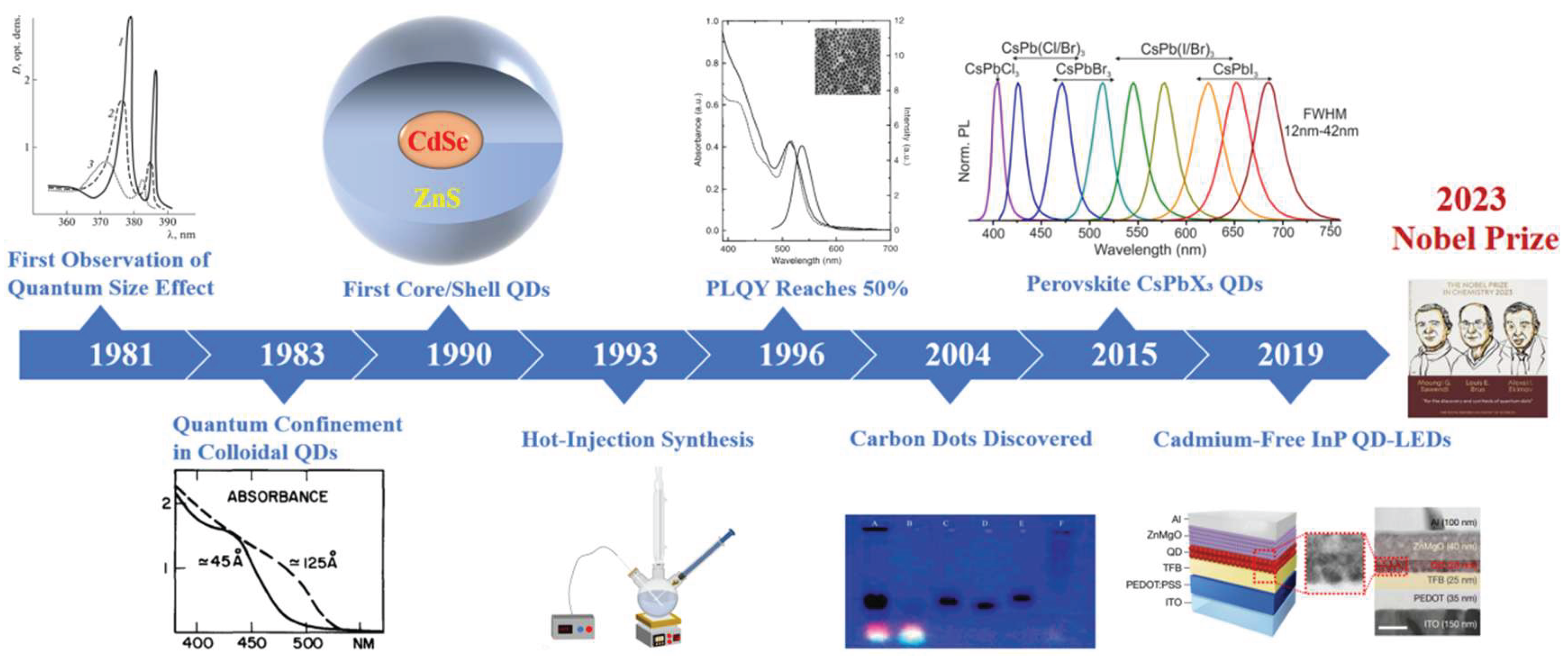
Figure 2.
Historical timeline of key milestones in the development of RENPs.[41,42,43,44,45,46,47,48,49,50].
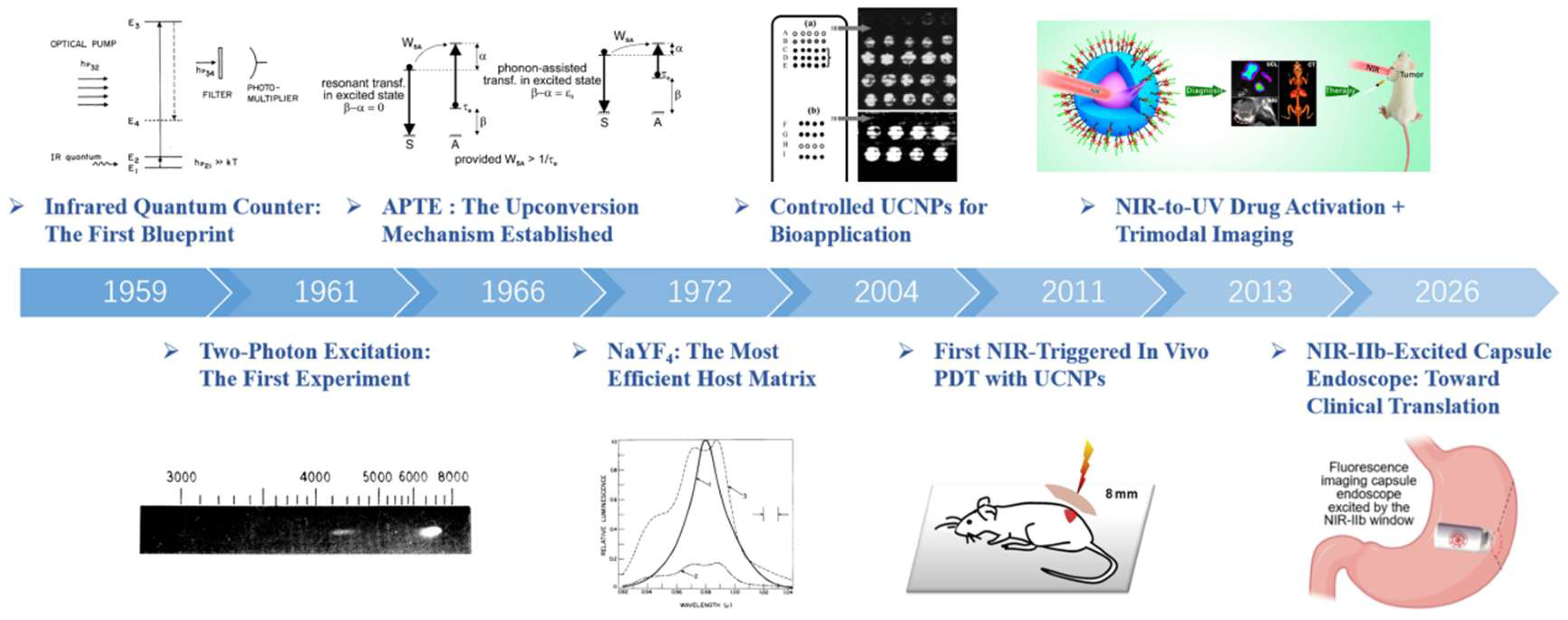
Figure 3.
Representative surface engineering for RENPs. (a) Preparation of carboxylic acid-functionalized RENPs from oleic acid-capped precursors and their FRET system.[59] (b) Schematic diagram of one step bioconjugation method using DNA-protein conjugate (red arrow), compared with the typical conventional multi-steps bioconjugation methods (blue arrow), including: (1) the transformation of hydrophobic RENPs into hydrophilic ones, (2) the introduction of functional ligands, and (3) the protein bioconjugation.[65] (c) Illustration depicting core-shell nanoparticles comprised of PEO-b-PCL diblock copolymers (yellow), the organic NIR-I fluorophore DiR (red), and RENPs (blue).[66] (d) Transmission electron microscopy (TEM) images of various UCNP@SiO2 nanostructures, including UCNP@dSiO2 (1), UCNP@mSiO2 (2), UCNP@dSiO2@mSiO2 (3), and UCNP@hmSiO2 (4).[67] (e) Schematic illustration showing the removal of the oleate capping ligand from the surface of the NaYF4:Er3+/Yb3+ nanoparticles (left) and artistic rendition of the Ln-UCNP surface: purple spheres = Y, orange = F, red = Yb, and yellow = Er. The coordination of the oleate is shown with the Y3+; however, it could also coordinate to Yb3+ and Er3+.[68].
Figure 3.
Representative surface engineering for RENPs. (a) Preparation of carboxylic acid-functionalized RENPs from oleic acid-capped precursors and their FRET system.[59] (b) Schematic diagram of one step bioconjugation method using DNA-protein conjugate (red arrow), compared with the typical conventional multi-steps bioconjugation methods (blue arrow), including: (1) the transformation of hydrophobic RENPs into hydrophilic ones, (2) the introduction of functional ligands, and (3) the protein bioconjugation.[65] (c) Illustration depicting core-shell nanoparticles comprised of PEO-b-PCL diblock copolymers (yellow), the organic NIR-I fluorophore DiR (red), and RENPs (blue).[66] (d) Transmission electron microscopy (TEM) images of various UCNP@SiO2 nanostructures, including UCNP@dSiO2 (1), UCNP@mSiO2 (2), UCNP@dSiO2@mSiO2 (3), and UCNP@hmSiO2 (4).[67] (e) Schematic illustration showing the removal of the oleate capping ligand from the surface of the NaYF4:Er3+/Yb3+ nanoparticles (left) and artistic rendition of the Ln-UCNP surface: purple spheres = Y, orange = F, red = Yb, and yellow = Er. The coordination of the oleate is shown with the Y3+; however, it could also coordinate to Yb3+ and Er3+.[68].
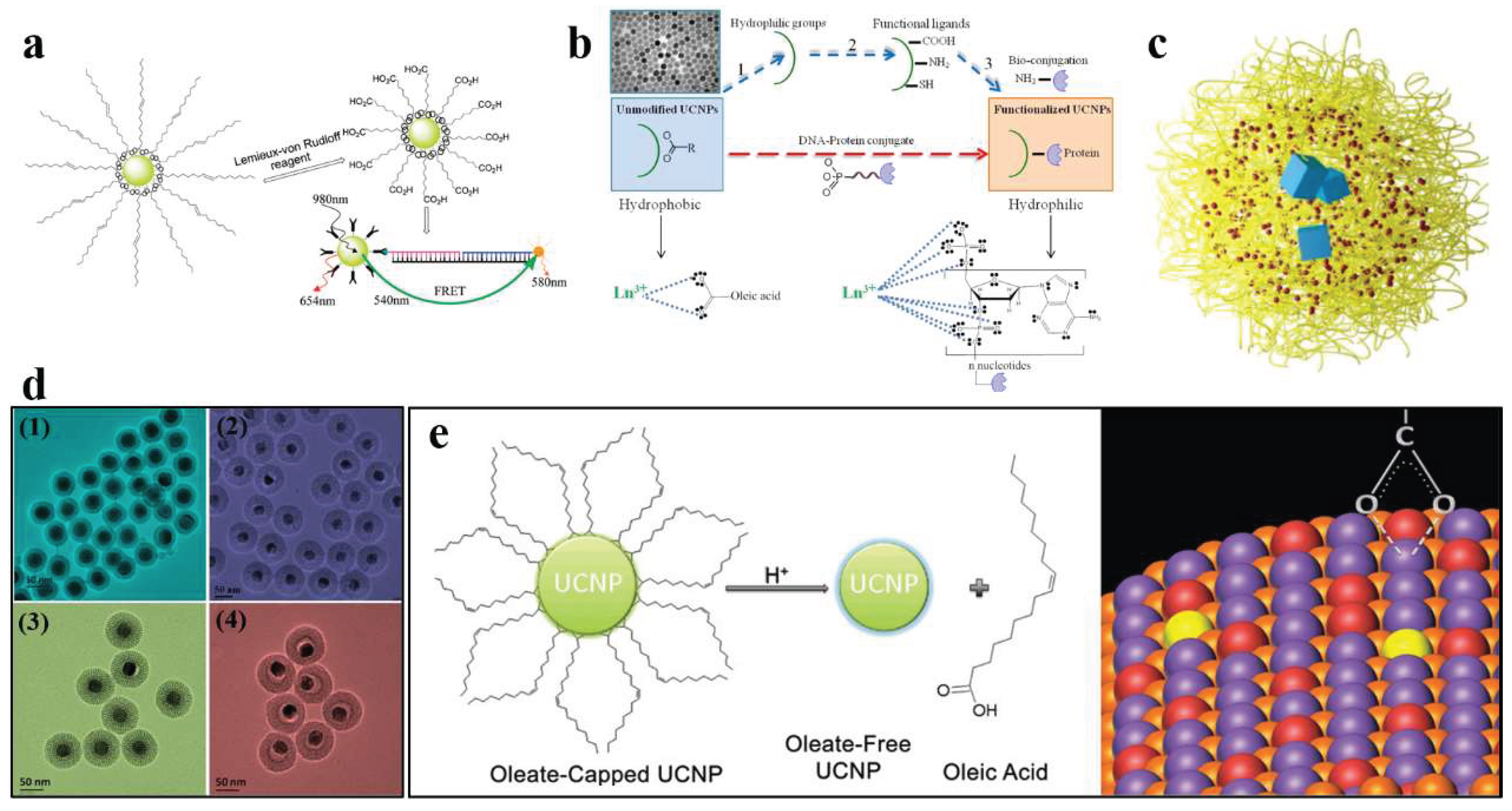
Figure 4.
Schematic illustrations and imaging performance of representative RENPs. (a) Magnetically targeted NIR-II bioimaging and PDT in mice using RENPs/Fe3O4 superparticles.[89] (b) UDNP-MC/Au-FeTA for NIR-II fluorescence imaging and synergistic CDT-PDT of tumors.[90] (c) Fabrication of 2D QR codes using the dumbbell-like nanocrystals (top) and experimental setup for visible and NIR-II imaging through intralipid medium at depths of 0–3 mm, showing superior depth penetration of NIR-II fluorescence (bottom).[107] (d) Dumbbell-like nanocrystal with multimode emissions (visible upconversion and NIR-II downshifting) switchable by 808 nm and 980 nm excitation for deep learning fluorescence imaging.[107].
Figure 4.
Schematic illustrations and imaging performance of representative RENPs. (a) Magnetically targeted NIR-II bioimaging and PDT in mice using RENPs/Fe3O4 superparticles.[89] (b) UDNP-MC/Au-FeTA for NIR-II fluorescence imaging and synergistic CDT-PDT of tumors.[90] (c) Fabrication of 2D QR codes using the dumbbell-like nanocrystals (top) and experimental setup for visible and NIR-II imaging through intralipid medium at depths of 0–3 mm, showing superior depth penetration of NIR-II fluorescence (bottom).[107] (d) Dumbbell-like nanocrystal with multimode emissions (visible upconversion and NIR-II downshifting) switchable by 808 nm and 980 nm excitation for deep learning fluorescence imaging.[107].
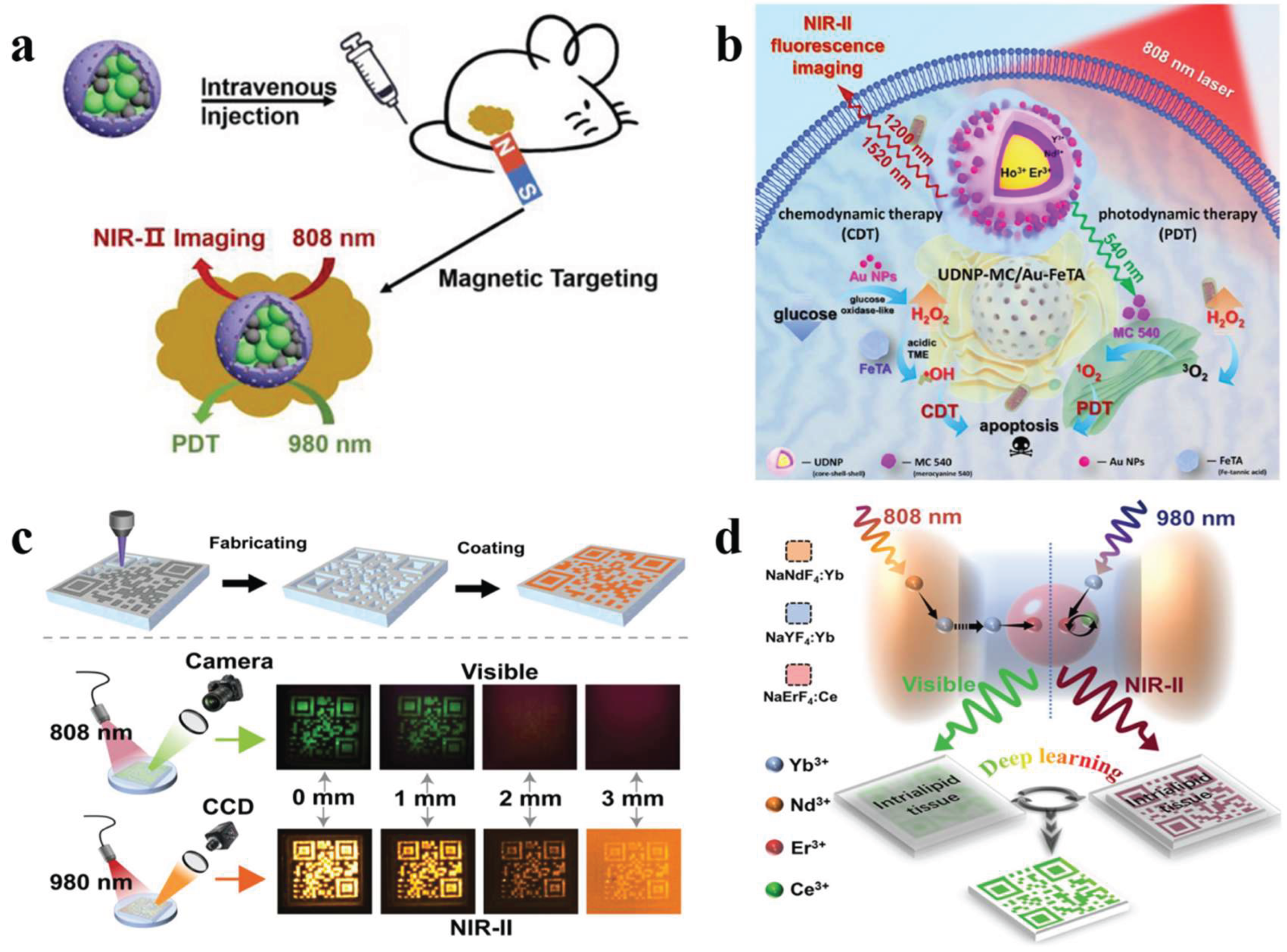
Figure 5.
Fluorescence lifetime imaging of RENPs. (a) Schematic of RENPs@Phospholipid@pHrodo nanoprobe (top) and noninvasive upconversion imaging of mouse brain vasculature through the intact skull (bottom).[92] (b) In vivo fluorescence intensity and lifetime imaging of mouse brain vasculature at different pH levels, enabling quantitative pH mapping via lifetime shifts.[92] (c) Fluorescence intensity (FI) and lifetime imaging (LTI) of mice and lung tissues infected with IAV and Ad5 labeled by RENPs with different luminescence lifetimes.[94].
Figure 5.
Fluorescence lifetime imaging of RENPs. (a) Schematic of RENPs@Phospholipid@pHrodo nanoprobe (top) and noninvasive upconversion imaging of mouse brain vasculature through the intact skull (bottom).[92] (b) In vivo fluorescence intensity and lifetime imaging of mouse brain vasculature at different pH levels, enabling quantitative pH mapping via lifetime shifts.[92] (c) Fluorescence intensity (FI) and lifetime imaging (LTI) of mice and lung tissues infected with IAV and Ad5 labeled by RENPs with different luminescence lifetimes.[94].
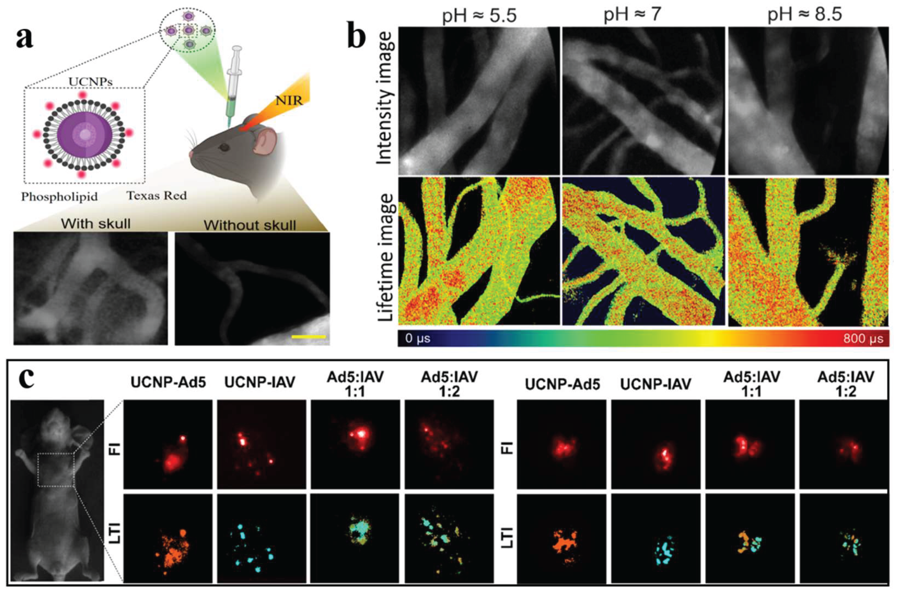
Figure 6.
Modulation of photoacoustic signals using upconversion RENPs. (a) PA signals from UC-α-CD in water, RENPs in cyclohexane, and distilled water under 980 nm excitation.[95] (b) UC luminescence spectra of RENPs in cyclohexane and UC-α-CD in water under 980 nm excitation.[95] (c) PAPSI images of labeled HeLa cells in a transparent plastic tube.[98] (d) Real-time PA images of tumors after injection of RENPs-DI under 980 nm irradiation.[99].
Figure 6.
Modulation of photoacoustic signals using upconversion RENPs. (a) PA signals from UC-α-CD in water, RENPs in cyclohexane, and distilled water under 980 nm excitation.[95] (b) UC luminescence spectra of RENPs in cyclohexane and UC-α-CD in water under 980 nm excitation.[95] (c) PAPSI images of labeled HeLa cells in a transparent plastic tube.[98] (d) Real-time PA images of tumors after injection of RENPs-DI under 980 nm irradiation.[99].
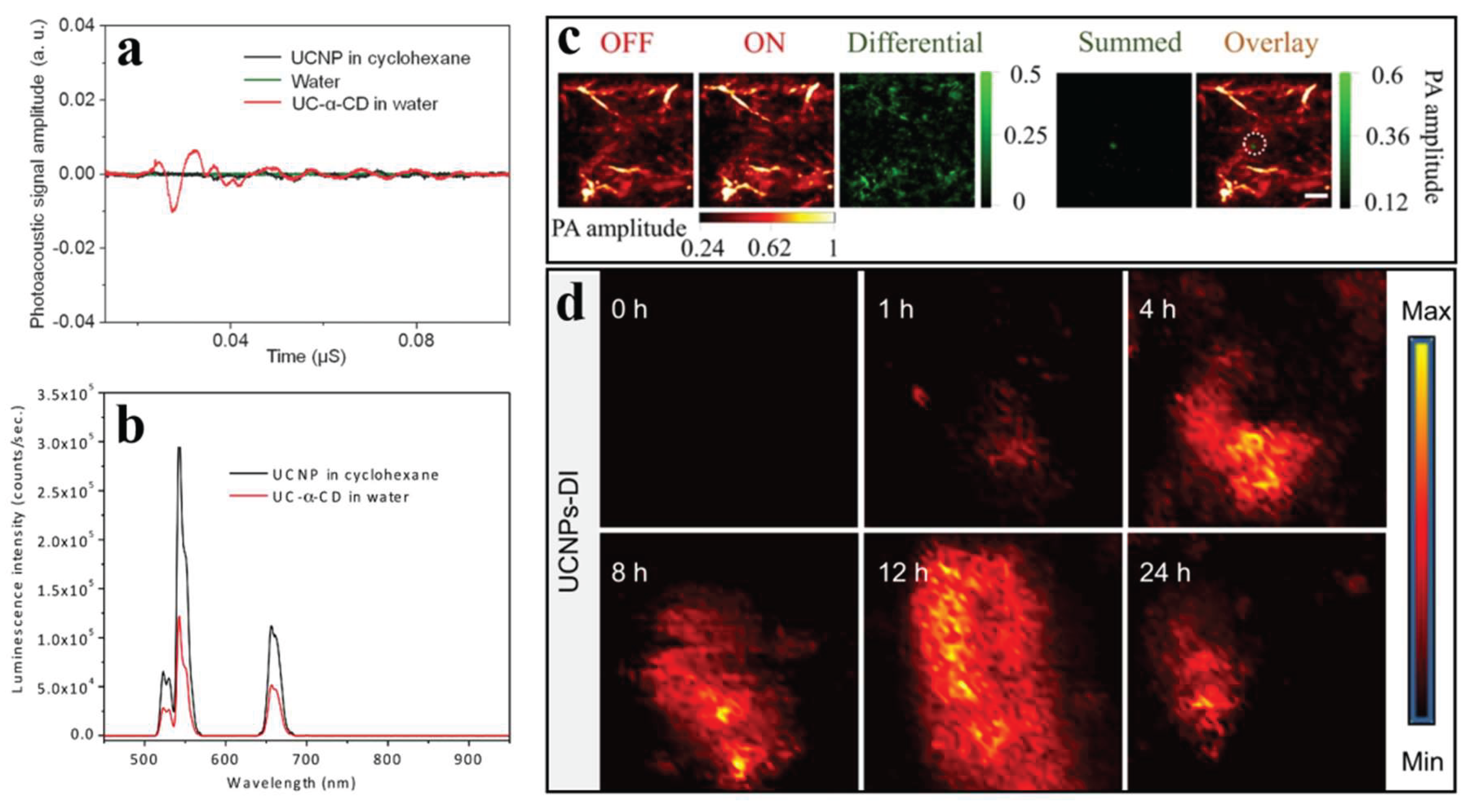
Figure 7.
RENPs probes for stimulated emission depletion super-resolution imaging. (a) One doughnut illumination beam generates two power-dependent emission PSF patterns of Gaussian in red (1) and doughnut in green.[103] (b) Emission intensity of one-photon (867 nm), two-photon (588 nm), and three-photon (450 nm) emissions from NaYF4:Nd nanoparticles as a function of 1064 nm depletion laser intensity, with measured Iₛₐₜ values of 116.2, 42.4, and 23.8 kW cm⁻², respectively.[102] (c) Confocal image (left) and Fourier-domain heterochromatic fusion image (right) of a sunflower pattern, showing improved resolution of fine features.[103] (d) Normalized emission attenuation of 455 nm and 800 nm signals through 93 μm mouse liver tissue, with 800 nm retaining 38.7% and 455 nm retaining 11.3% of the original intensity.[101] (e) STED image of 8% Tm3+ doped RENPs (top) and the corresponding intensity profile (bottom) under 1 mW excitation power.[100] (f) Two-color STED imaging of NaYF4:Nd/Ce (red, 588 nm) and NaYF4:Yb/Ho (green, 645 nm) nanoparticles using a 740 nm excitation beam and a 1064 nm donut-shaped depletion beam.[102].
Figure 7.
RENPs probes for stimulated emission depletion super-resolution imaging. (a) One doughnut illumination beam generates two power-dependent emission PSF patterns of Gaussian in red (1) and doughnut in green.[103] (b) Emission intensity of one-photon (867 nm), two-photon (588 nm), and three-photon (450 nm) emissions from NaYF4:Nd nanoparticles as a function of 1064 nm depletion laser intensity, with measured Iₛₐₜ values of 116.2, 42.4, and 23.8 kW cm⁻², respectively.[102] (c) Confocal image (left) and Fourier-domain heterochromatic fusion image (right) of a sunflower pattern, showing improved resolution of fine features.[103] (d) Normalized emission attenuation of 455 nm and 800 nm signals through 93 μm mouse liver tissue, with 800 nm retaining 38.7% and 455 nm retaining 11.3% of the original intensity.[101] (e) STED image of 8% Tm3+ doped RENPs (top) and the corresponding intensity profile (bottom) under 1 mW excitation power.[100] (f) Two-color STED imaging of NaYF4:Nd/Ce (red, 588 nm) and NaYF4:Yb/Ho (green, 645 nm) nanoparticles using a 740 nm excitation beam and a 1064 nm donut-shaped depletion beam.[102].
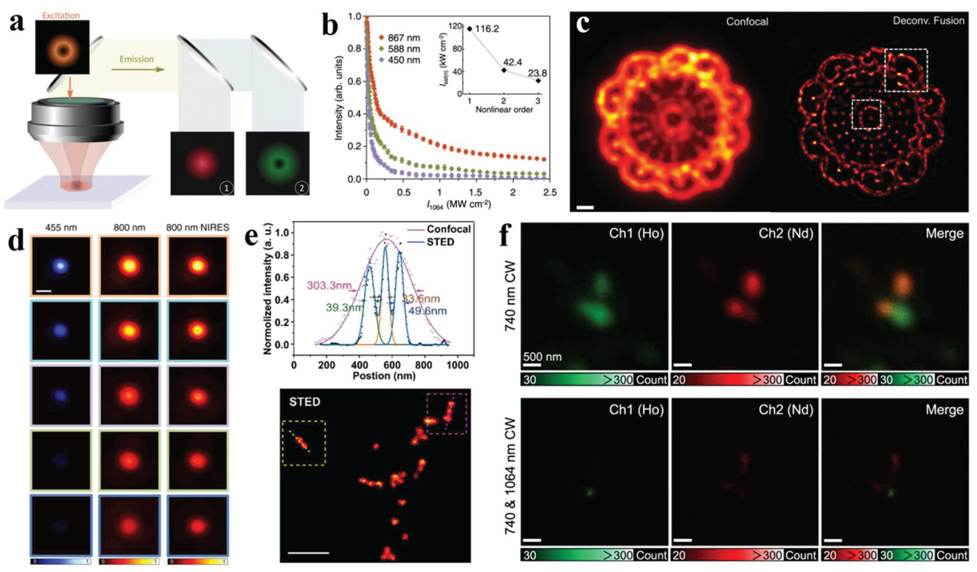
Figure 8.
Multimodal imaging of living subjects using RENPs. (a) Scheme of deep animal imaging using high-efficiency multi-shell RENPs for in vivo PAI, UCL, and MRI.[96] (b) UCL spectra of Er3+/Yb3+ co-doped Gd2O3 nanoplates under 980 nm excitation (top) and XEOL spectra of Eu3+ and Tb3+-doped Gd2O3 nanoplates excited by Cu Kα X-ray irradiation (bottom). [129] (c) In vivo UCL imaging of mouse injected with RENPs at the tumor site.[96] (d) In vivo MRI of mouse injected with CS2-ICG at tumor site (left) and in vivo MRI of mouse acquired before injection and 2 h post-injection (right).[96].
Figure 8.
Multimodal imaging of living subjects using RENPs. (a) Scheme of deep animal imaging using high-efficiency multi-shell RENPs for in vivo PAI, UCL, and MRI.[96] (b) UCL spectra of Er3+/Yb3+ co-doped Gd2O3 nanoplates under 980 nm excitation (top) and XEOL spectra of Eu3+ and Tb3+-doped Gd2O3 nanoplates excited by Cu Kα X-ray irradiation (bottom). [129] (c) In vivo UCL imaging of mouse injected with RENPs at the tumor site.[96] (d) In vivo MRI of mouse injected with CS2-ICG at tumor site (left) and in vivo MRI of mouse acquired before injection and 2 h post-injection (right).[96].
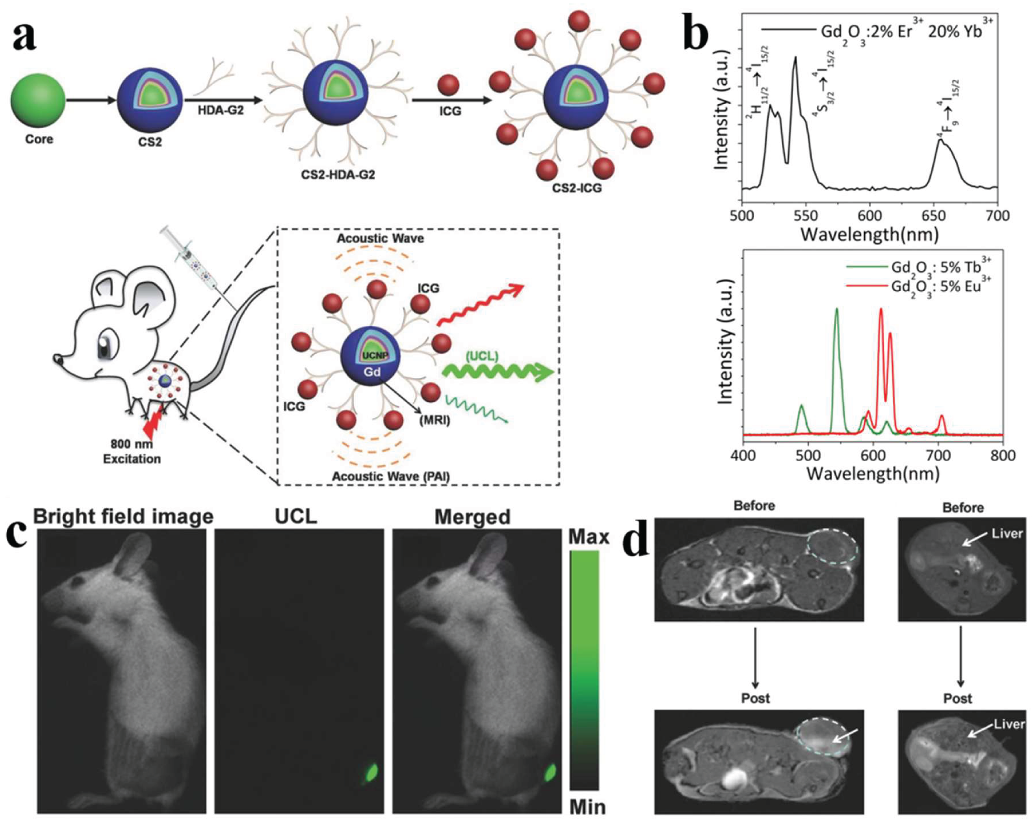
Figure 9.
X-ray-excited optical luminescence properties of RENPs. (a) Proposed energy conversion and transport and luminescence mechanisms in the NaGdF4:Ce/Tb nanoscintillator. ET represents energy transfer. The blue circles represent X-ray produced secondary electrons.[108] (b) Comparison of integral DS intensity ratios (left), XEOL intensity ratios (middle), and XEPL intensity ratios (right) of ICe@Tb/ITb across the four experimental groups.[132] (c) Corresponding digital photographs of DS, XEOL, XEPL/DS. The core/shell NPs were synthesized using two different methods: the Separate method (Group I) and the Successive/NaOA method (Group IV).[132] (d) MTF curve of the XEOL image obtained from the nanoscintillator film.[108].
Figure 9.
X-ray-excited optical luminescence properties of RENPs. (a) Proposed energy conversion and transport and luminescence mechanisms in the NaGdF4:Ce/Tb nanoscintillator. ET represents energy transfer. The blue circles represent X-ray produced secondary electrons.[108] (b) Comparison of integral DS intensity ratios (left), XEOL intensity ratios (middle), and XEPL intensity ratios (right) of ICe@Tb/ITb across the four experimental groups.[132] (c) Corresponding digital photographs of DS, XEOL, XEPL/DS. The core/shell NPs were synthesized using two different methods: the Separate method (Group I) and the Successive/NaOA method (Group IV).[132] (d) MTF curve of the XEOL image obtained from the nanoscintillator film.[108].
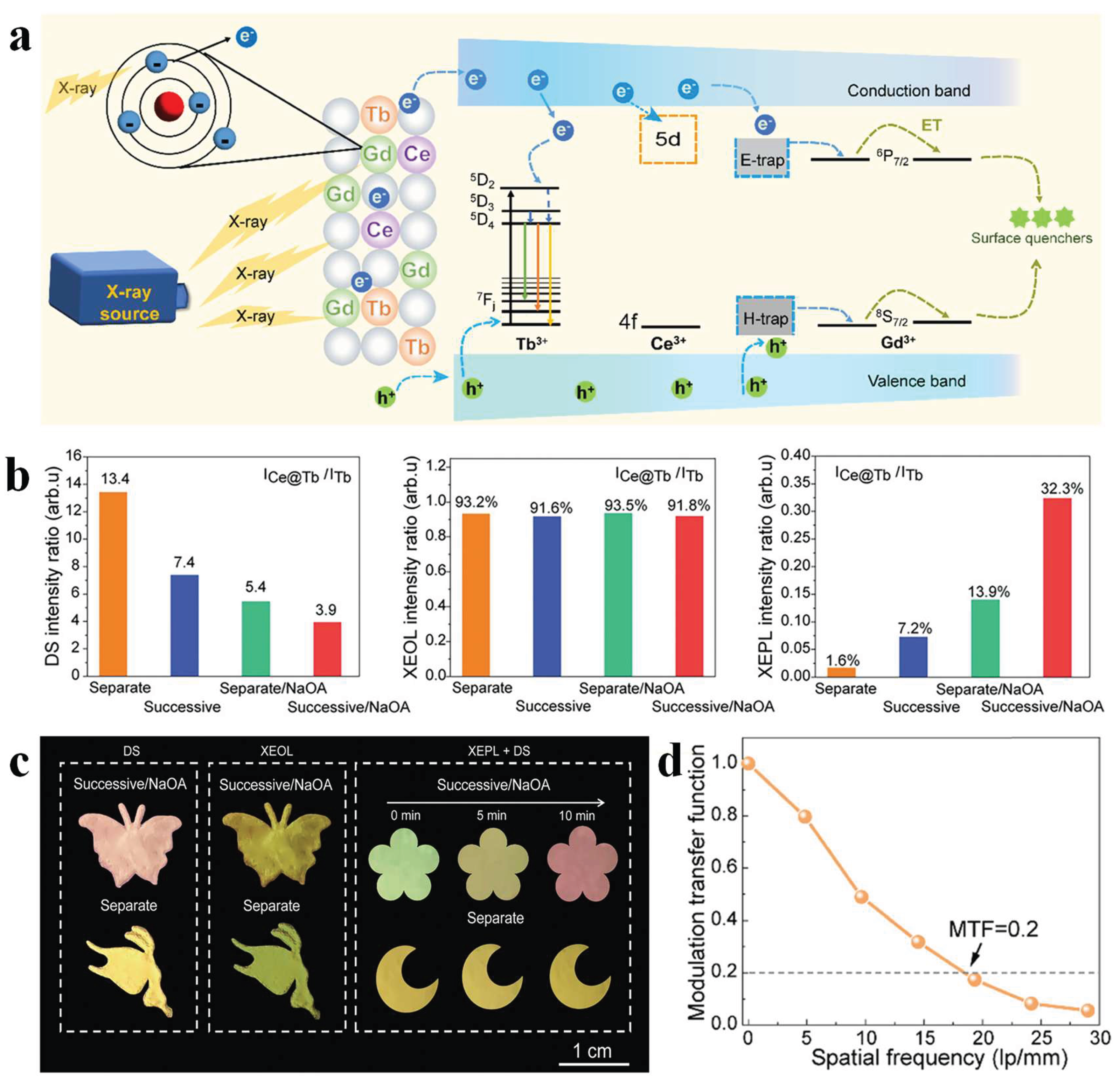
Figure 10.
X-ray-activated persistent luminescence imaging from RENPs. (a) Schematic representation of X-ray-activated PL generated in RE-doped nanoparticles.[109] (b) Low-resolution transmission electron microscopy (TEM) image of the as-synthesized NaYF4:3%Er@NaYF4 nanoparticles. Scale bar, 50 nm. Insets: a high-resolution TEM image of a single NaYF4:3%Er@NaYF4 nanoparticle (top right) and the corresponding electron diffraction pattern (bottom right). Scale bar, 2 nm.[109] (c) NIR-II PL images of Nd-, Ho-, Tm- and Er-PLNPs in centrifuge tubes as a function of time. H, high; L, low.[109] (d) PL decay curves of typical emission bands of Ln-PLNPs (left) and NIR-II PL photostability of Er-PLNPs (right) in the NIR-II window derived from NIR-II PL images. Imaging exposure time, 5 s. a.u., arbitrary units.[109] (e) Radioluminescent intensity of NaLuF4:Tb (15 mol%) and NaLuF4:Tb (15 mol%)@NaYF4 nanocrystals, monitored at 546 nm as a function of time upon cessation of X-rays (left) and comparison of afterglow decay profiles of various phosphors after cessation of X-ray excitation (right).[110] (f) Afterglow photographs of NaLuF4:Tb (15 mol%)@NaYF4 nanocrystals dispersed in 1 ml of cyclohexane. X-ray operation was set at a voltage of 70 kV with a tube current of 1 mA.[110] (g) Radioluminescence and afterglow of NaLuF4 nanocrystals doped with various activators (Nd3+, Tm3+, Dy3+, Tb3+, Er3+, Ho3+, Sm3+ and Pr3+).[110].
Figure 10.
X-ray-activated persistent luminescence imaging from RENPs. (a) Schematic representation of X-ray-activated PL generated in RE-doped nanoparticles.[109] (b) Low-resolution transmission electron microscopy (TEM) image of the as-synthesized NaYF4:3%Er@NaYF4 nanoparticles. Scale bar, 50 nm. Insets: a high-resolution TEM image of a single NaYF4:3%Er@NaYF4 nanoparticle (top right) and the corresponding electron diffraction pattern (bottom right). Scale bar, 2 nm.[109] (c) NIR-II PL images of Nd-, Ho-, Tm- and Er-PLNPs in centrifuge tubes as a function of time. H, high; L, low.[109] (d) PL decay curves of typical emission bands of Ln-PLNPs (left) and NIR-II PL photostability of Er-PLNPs (right) in the NIR-II window derived from NIR-II PL images. Imaging exposure time, 5 s. a.u., arbitrary units.[109] (e) Radioluminescent intensity of NaLuF4:Tb (15 mol%) and NaLuF4:Tb (15 mol%)@NaYF4 nanocrystals, monitored at 546 nm as a function of time upon cessation of X-rays (left) and comparison of afterglow decay profiles of various phosphors after cessation of X-ray excitation (right).[110] (f) Afterglow photographs of NaLuF4:Tb (15 mol%)@NaYF4 nanocrystals dispersed in 1 ml of cyclohexane. X-ray operation was set at a voltage of 70 kV with a tube current of 1 mA.[110] (g) Radioluminescence and afterglow of NaLuF4 nanocrystals doped with various activators (Nd3+, Tm3+, Dy3+, Tb3+, Er3+, Ho3+, Sm3+ and Pr3+).[110].
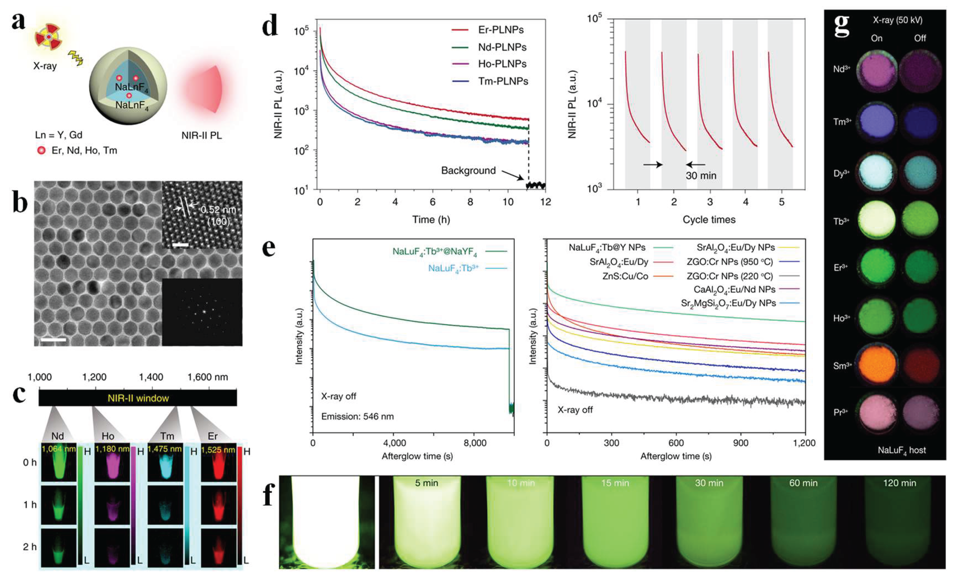

Table 1.
Summary of representative RE-doped nanoparticles for different imaging modality.
| Imaging Modality | Host:dopants | Excitation (nm) | Emission (nm) | Application | Reference |
| Near-infrared imaging | NaYF4:Yb3+/Er3+/Tm3+; NaYF4:Yb3+/Er3+; NaYF4:Yb3+/Ho3+ |
980 | 1566, 1162, 800, 778 | Early tumor detection | [66] |
| NaYF4:Yb3+/Er3+ | 980 | 1039, 1058 | Blood vessels, lymph nodes, and tumor imaging | [87] | |
| NaYF4@NaGdF₄:Yb3+/Tm3+@NaGdF4 | 980 | 450, 475, 650, 800 | Information storage | [88] | |
| NaYF4:Yb3+/Er3+@ NaLuF4:Y3+ | 808 | 1000~ 1600 |
Tumor diagnosis | [89] | |
| NaYF4:Yb3+/Er3+/Ho3+@NaYF4:Yb3+/Nd3+@NaYF4 | 808 | 1200, 1520 | Chemo-photodynamic therapy of breast cancer | [90] | |
| NaGdF4@NaGdF4:Yb3+/Er3+@NaYF4:Yb3+@NaNdF4:Yb3+ | 808 | 1525, 1155 | Quantitative detection of tumor biomarkers (ER, PR, HER2) | [52] | |
| β-NaYF4@NaYF4:Nd3+ | 808 | 1060 | Hepatocellular carcinoma detection | [53] | |
| ZnGa2O4:Ni2⁺@NaErF4:Yb3⁺/ Mn2⁺ |
980 | 1290, 1532 | Deep tissue penetration | [91] | |
| Fluorescence lifetime imaging | NaYbF4@NaYF4:Yb3⁺/Er3⁺/Tm3⁺@NaYbF4 | 980 | 540, 580 | In vivo pH mapping in mouse brain | [92] |
| β-NaYbF4:Er3⁺@NaYF4:Yb3⁺@NaYF4:Nd3⁺/Yb3⁺ | 800 | 540, 980 | Hypochlorous acid (HClO) detection | [93] | |
| β-NaGdF4:Yb3+/Er3+; Cubic-NaYbF4:Er3+@NaYF4; NaYF4:Gd3+/Yb3+/Er3+@NaYF4 | 980 | Green, red |
In vivo high-contrast virus tracking (Influenza A virus and Adenovirus) | [94] | |
| Photoacoustic imaging | NaYF4:Yb3+/Er3+ | 980 | Kidney imaging in live mice | [95] | |
| NaYF4:Yb3+/Er3+@NaYF4:Yb3+@NaNdF4:Yb3+@NaYF4@NaGdF4 | 800 | Tumor blood vessel imaging (depth up to 10 mm) | [96] | ||
| NaYbF4:Ce3+/Er3+@NaYF4 | 980 | Tumor microenvironment-responsive PAI | [97] | ||
| NaYF4:Yb3+/Tm3+@NaYF4 | 980, 680 | Super-sensitive PA molecular imaging | [98] | ||
| NaYF4:Yb3+/Er3+/Tm3+ | 980 | Orthogonal activation of PAI and PDT | [99] | ||
| Stimulated emission depletion super-resolution imaging | NaYF4:Yb3+/Tm3+ | 975, 810 | 455 | Super-resolution imaging (~66 nm resolution) | [100] |
| NaYF4:Yb3+/Tm3+ | 980, 808 | 455 | Low-power STED (resolution down to ~33 nm) | [101] | |
| NaYF4:Nd3+/ RE3+ | 740, 1064 | 450, 588 | NIR-II CW laser STExD nanoscopy (resolution down to 34 nm) | [102] | |
| NaYF4:Yb3+/Tm3+ | 980 | 740, 800 | Fourier domain heterochromatic fusion super-resolution imaging | [103] | |
| NaYF4:Yb3+/Tm3+ | 980 | 800 | Deep tissue NIRES nanoscopy | [104] | |
| Multimodal imaging | NaLuF4:Yb3+Er3+@NaLuF4 | 980, 1064 | 520, 540, 650 | Penta-modal imaging-guided PTT of lung cancer | [105] |
| NaYF4:Yb3+/Er3+@NaYF4:Yb3+@NaNdF4:Yb3+@NaYF4@NaGdF4 | 800 | Tumor blood vessel imaging (depth up to 10 mm) | [96] | ||
| NaYF4:Er3+/Mn2+@NaLuF4 | 1532 | 521, 541, 651 | Dual-modal XEPL/UC imaging for anti-counterfeiting | [106] | |
| NaErF4:Ce3+/Yb3+@NaYF4:Yb3+@NaNdF4:Yb3+ | 808, 980 | 545, 654, 1530 | Deep learning fluorescence imaging combining narrow visible emission peak with deep tissue penetration of NIR-II | [107] | |
| NaYF4:Nd3+@NaGdF4 | 808 | 1060, 1300 | Tumor vasculature during tumorigenesis, growth, and necrosis | [58] | |
| X-ray excited optical luminescence imaging | NaYF4:Er3+@NaLuF4 | X-ray | 521, 541 | Enhanced XEOL/XEPL via heavy atomic shell coating | [106] |
| NaGdF4:Ce3+/Tb3+ | X-ray | 543 | High-resolution X-ray imaging | [108] | |
| NaLuF4:Tb3+@NaYF4 | X-ray | 540 | High-resolution X-ray luminescence extension imaging | [99] | |
| NaYF4:Er3+@NaGdF4 | X-ray | 1525 | Deep-tissue diagnosis | [109] | |
| NaLuF4:Tb3+@NaYF4 | X-ray | 546, 584 | 3D conformal X-ray imaging of curved electronics | [110] | |
| Persistent luminescence imaging | NaLuF4:Tb3+@NaYF4 | X-ray | 540 | High-resolution X-ray luminescence extension imaging | [99] |
| NaYF4:Er3+@NaLuF₄ | X-ray | 521, 541 | Enhanced XEOL/XEPL via heavy atomic shell coating | [106] | |
| CaF2:Dy3+@NaYF4 | UV / X-ray | 480, 575 | UV and X-ray excited persistent luminescence | [111] | |
| NaYF4:Er3+@NaYF4:Nd3+@NaYF4, NaYF4:Ho3+@NaYF4:Tm3+@NaYF4 | X-ray | 1064, 1180, 1475, 1525 | Deep-tissue diagnosis | [109] | |
| NaLuF4:Tb3+@NaYF4 | X-ray | 546, 584 | Flat-panel-free X-ray detector | [110] | |
| ZnGa2O4:Ni2+@NaErF4:Yb3+/Mn2+ | 980 | 1290 | Deep-tissue bioimaging without real-time excitation | [91] |
Disclaimer/Publisher’s Note: The statements, opinions and data contained in all publications are solely those of the individual author(s) and contributor(s) and not of MDPI and/or the editor(s). MDPI and/or the editor(s) disclaim responsibility for any injury to people or property resulting from any ideas, methods, instructions or products referred to in the content. |
© 2026 by the authors. Licensee MDPI, Basel, Switzerland. This article is an open access article distributed under the terms and conditions of the Creative Commons Attribution (CC BY) license (http://creativecommons.org/licenses/by/4.0/).
Copyright: This open access article is published under a Creative Commons CC BY 4.0 license, which permit the free download, distribution, and reuse, provided that the author and preprint are cited in any reuse.