
Preprint
Review

This version is not peer-reviewed.

Beyond Antibodies: How GFAP, NfL, Cytokines, Complement, miRNAs, Metabolomics, and the Microbiome Are Shaping Precision Medicine in NMOSD – A Narrative Review

Submitted:

03 May 2026

Posted:

06 May 2026

You are already at the latest version

Abstract
Neuromyelitis optica spectrum disorder (NMOSD) is a severe autoimmune astrocytopathy of the central nervous system, characterized by recurrent optic neuritis and longitudinally extensive transverse myelitis. Due to overlapping features with multiple sclerosis (MS), misdiagnosis remains common, yet treatment strategies differ fundamentally between these conditions. This narrative review synthesises recent advances in diagnostic and prognostic biomarkers for NMOSD, focusing on serological, cellular, molecular, and microbiological indicators.A targeted literature search was conducted in PubMed, Scopus and Web of Science (January 2015 – June 2025) using keywords related to NMOSD and biomarkers. Of over 500 initially identified articles, 72 key references were selected based on scientific relevance, methodological quality and clinical applicability, consistent with a narrative review approach.Established biomarkers – aquaporin‑4 immunoglobulin G (AQP4‑IgG) and myelin oligodendrocyte glycoprotein antibodies (MOG‑IgG) – remain central to diagnosis and phenotypic classification. Emerging markers, including glial fibrillary acidic protein (GFAP), neurofilament light chain (NfL), cytokines (interleukin‑6, CXCL13, CXCL10), complement components (C3, C4, sC5b‑9), microRNAs, metabolomics profiles, gut microbiome signatures and viral serology (Epstein–Barr virus, human herpesvirus 6), show promise in differentiating NMOSD from MS and MOGAD, predicting relapses, and guiding therapy. Integration of antibody assays with markers of astrocytic and axonal injury, immune activation and microbiological signatures could improve diagnostic accuracy and enable personalised treatment. However, clinical application remains limited by assay variability, lack of standardised cut-offs and small study populations. Future progress requires validated multimarker panels and harmonised measurement platforms across diverse populations.
Keywords: 
;  ;  ;  ;  ;  ;  ;  ;  ;  

1. Introduction

Neuromyelitis optica spectrum disorder (NMOSD) is a rare but severe autoimmune astrocytopathy primarily targeting the optic nerves and spinal cord. It is characterized by recurrent episodes of optic neuritis and longitudinally extensive transverse myelitis, often leading to permanent visual loss and substantial neurological disability [1]. The global prevalence of NMOSD is estimated to range between 0.5 and 4 cases per 100,000 individuals, with a higher incidence reported among non-Caucasian populations, especially in Asia and Africa, where prevalence rates may reach 3 to 10 per 100,000 [2,3]. In Iran, epidemiological data from national registries indicate a prevalence of approximately 1 to 2 per 100,000, with a pronounced female predominance (female-to-male ratio of 9:1) and typical disease onset occurring during the third to fourth decades of life [4]. These observations underscore the under diagnosis of NMOSD in regions with limited access to advanced serological testing [5]. Previously misclassified as a variant of multiple sclerosis (MS), NMOSD is now recognized as a distinct immunopathological disorder, primarily mediated by pathogenic immunoglobulin G antibodies against aquaporin-4 (AQP4-IgG), a water channel protein expressed on astrocytic perivascular end-feet [6]. Despite the development of refined international consensus criteria and heightened clinical awareness, the early and accurate differentiation of NMOSD from MS, myelin oligodendrocyte glycoprotein antibody-associated disease (MOGAD), and other inflammatory central nervous system disorders remains a considerable clinical challenge, particularly in patients who are seronegative for AQP4-IgG or who present with overlapping clinical and radiological features [7,8].
The imperative for reliable biomarkers in neuromyelitis optica spectrum disorder (NMOSD) is critical, as diagnostic errors can lead to significant therapeutic mismanagement [9]. Treatments efficacious in multiple sclerosis (MS), such as interferon-beta or fingolimod, may exacerbate NMOSD relapses by promoting humoral autoimmunity, whereas immunotherapies designed for NMOSD, including rituximab or eculizumab, are either ineffective or contraindicated in MS [10,11]. Therefore, robust biomarkers are indispensable for differential diagnosis, stratification of patients into aquaporin-4 immunoglobulin G (AQP4-IgG)-positive and -negative subgroups, prediction of relapse risk, and the advancement of personalized therapeutic approaches aimed at mitigating disability progression [12,13,14]. AQP4-IgG seropositivity, detectable in approximately 60–90% of cases via cell-based assays, demonstrates near-perfect specificity (>99%) for NMOSD and has substantially influenced diagnostic criteria[15]. Nonetheless, up to 30% of patients remain seronegative, highlighting the need for additional diagnostic modalities [16]. In this context, myelin oligodendrocyte glycoprotein immunoglobulin G (MOG-IgG) has emerged as a critical biomarker for AQP4-IgG-negative phenotypes that clinically mimic NMOSD, thereby distinguishing MOG antibody-associated disease (MOGAD)—which is typically monophasic and responsive to corticosteroids—from the classical relapsing AQP4-IgG-positive NMOSD, which is associated with a poorer prognosis in the absence of aggressive immunosuppressive treatment [17,18]. Beyond autoantibodies, a growing spectrum of biomarkers—including astrocytic injury markers such as glial fibrillary acidic protein (GFAP)[19], axonal injury markers like neurofilament light chain (NFL)[20], soluble cytokines exemplified by interleukin-6 (IL-6), complement activation fragments such as C3a and C5b-9, non-coding RNAs including microRNAs, extracellular vesicles, and microbiome-derived signatures—offers valuable insights into the complex immunopathogenesis of NMOSD and holds significant promise for enhancing diagnostic precision in challenging clinical scenarios [21].
This narrative review offers a comprehensive synthesis of recent findings related to diagnostic and prognostic biomarkers across serological, cerebrospinal fluid (CSF), imaging, molecular, and microbiological fields. The analysis incorporates evidence from clinical trials, observational research, and translational studies conducted between 2015 and 2025. By critically assessing the diagnostic performance of these biomarkers—especially in distinguishing neuromyelitis optica spectrum disorder (NMOSD) from multiple sclerosis (MS) and myelin oligodendrocyte glycoprotein antibody-associated disease (MOGAD)—the review highlights both their strengths, such as the increased levels of glial fibrillary acidic protein (GFAP) during acute attacks (demonstrating approximately 80% sensitivity in aquaporin-4 immunoglobulin G AQP4-IgG-positive NMOSD), and their limitations, including variability in assay methods and differences in threshold values across populations. The review ultimately recommends the adoption of multi-biomarker panels to overcome diagnostic challenges, particularly in seronegative NMOSD cases, emphasizing that early identification through these approaches may prevent up to 50% of relapses by enabling personalized treatment strategies. Additionally, it stresses the importance of conducting further validation studies in diverse populations, with special focus on underrepresented regions such as Iran.

2. Methods

This article is a narrative review designed to provide a comprehensive, critical and qualitative synthesis of recent advances in diagnostic and prognostic biomarkers for neuromyelitis optica spectrum disorder (NMOSD), with emphasis on serological, cellular, molecular and microbiological markers. Unlike systematic reviews, no formal meta-analysis or quantitative quality assessment was performed; instead, a selective, expert-driven approach was used to identify the most relevant and high-impact studies.

Search Strategy

A systematic literature search was conducted in three major biomedical databases – PubMed, Scopus and Web of Science – for articles published between 1 January 2015 and 30 June 2025. The search combined controlled vocabulary and free-text terms using Boolean operators:
(“neuromyelitis optica spectrum disorder” OR “NMOSD” OR “Devic’s disease”) AND (“biomarker*” OR “AQP4” OR “aquaporin-4” OR “MOG” OR “myelin oligodendrocyte glycoprotein” OR “GFAP” OR “glial fibrillary acidic protein” OR “NfL” OR “neurofilament light” OR “cytokine*” OR “chemokine*” OR “microRNA” OR “miRNA” OR “exosome*” OR “metabolom*” OR “microbiome” OR “gut microbiota” OR “complement” OR “viral serology” OR “EBV” OR “HHV-6”).
Additional relevant studies were identified by manually screening the reference lists of retrieved articles, key consensus statements and previous high-quality reviews. The search was limited to human studies and English language publications.

Inclusion and Exclusion Criteria

Studies were considered if they: (1) involved patients with a confirmed diagnosis of NMOSD (2015 IPND criteria or later) or related AQP4-IgG/MOG-IgG-associated disorders; (2) evaluated at least one biomarker in serum, cerebrospinal fluid, imaging or microbiological specimens; and (3) reported diagnostic performance, prognostic value or treatment monitoring utility. Preference was given to original research articles (cohort, case–control, cross-sectional), clinical trials, meta-analyses and high-quality systematic reviews. Exclusion criteria included case reports with fewer than five patients, non-English publications, animal or in vitro studies without human validation, and editorials or conference abstracts without original data.

Study Selection and Reference Management

Titles and abstracts were screened by the authors, and full texts of potentially relevant articles were obtained. Based on relevance, methodological soundness and clinical applicability, a final set of 78 key references was selected for inclusion in this narrative review. This reference list encompasses the most representative studies across all biomarker categories, including diagnostic antibody assays, astrocytic/axonal injury markers, cytokine/chemokine/complement profiles, and emerging molecular and microbiological markers.

Evolving Biomarker Landscape in NMOSD

The biomarker landscape of neuromyelitis optica spectrum disorder (NMOSD) has advanced markedly across serological, astrocytic/neuronal injury, and molecular–microbiological domains. Aquaporin-4 immunoglobulin G (AQP4-IgG) remains the diagnostic and phenotypic cornerstone; by targeting astrocytic AQP4 and driving complement-mediated cytotoxicity, it enables earlier, more specific case identification and facilitates timely deployment of targeted immunotherapies, including complement and IL-6 pathway inhibitors [22,23,24]. In AQP4-IgG–negative patients with optic neuritis, longitudinally extensive transverse myelitis, or brainstem syndromes, myelin oligodendrocyte glycoprotein antibodies (MOG-IgG) are critical to distinguish MOG-antibody–associated disease (MOGAD) from AQP4-positive NMOSD and multiple sclerosis; persistent MOG-IgG positivity correlates with higher relapse risk, whereas seroconversion to negativity suggests a more favorable course. Composite serologic panels that integrate autoantibodies, soluble inflammatory mediators, and complement split products are being explored to increase diagnostic sensitivity in seronegative or atypical cases [23,25].
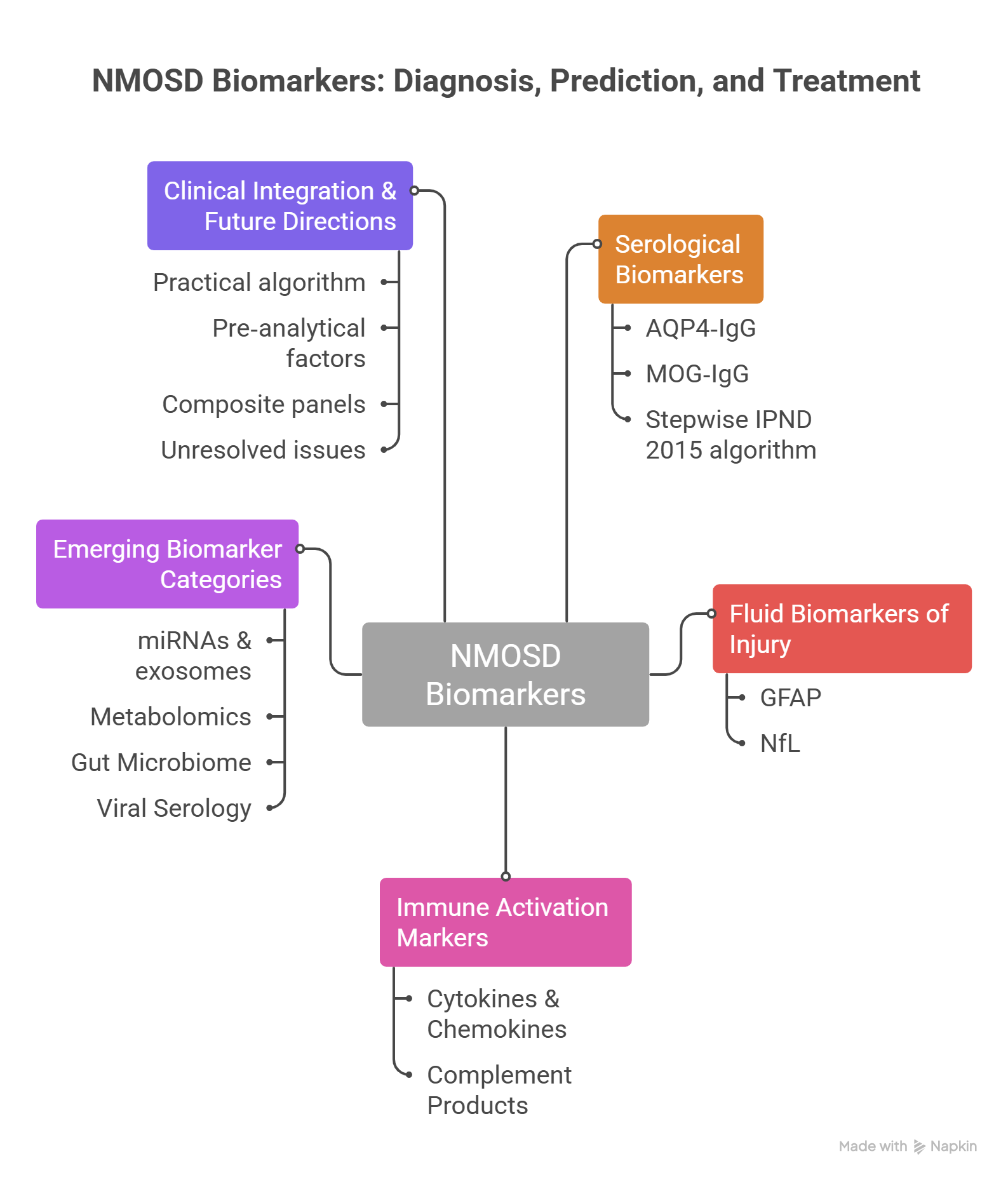
Serum and cerebrospinal fluid (CSF) levels of glial fibrillary acidic protein (GFAP) are typically elevated in AQP4-IgG–positive neuromyelitis optica spectrum disorder (NMOSD). GFAP rises during acute attacks, correlates with disability, and reflects the profound astrocytic injury that characterizes this disease. By contrast, neurofilament light chain (NfL) primarily indexes axonal damage. Although less specific to NMOSD, NFL is useful for monitoring neuroaxonal injury and informing long-term prognosis. Immunologic biomarkers can further refine assessments of disease activity and risk. Increases in interleukin-6 (IL-6), CXCL13, and CXCL10 are associated with clinical activity, B-cell recruitment, and intrathecal inflammation, and may help identify patients at higher risk for relapse or poorer outcomes. Complement pathway readouts provide additional insight and potential therapeutic guidance. Patterns of reduced C3 and C4 with concomitant elevations in C3a, Ba, and C5b-9 underscore the central role of complement activation in NMOSD pathogenesis. These profiles may help clinicians prioritize complement-targeted therapies in appropriate patients [26,27]. Emerging molecular biomarkers are advancing both phenotyping and prognostication in NMOSD. Circulating and exosomal microRNA (miRNA) signatures—some of which rise during clinical relapses—mirror astrocytic dysfunction, cytokine pathway activation, and immune cell signaling, and correlate with measures of disease activity [16,28]. Complementary proteomic and metabolomic panels that index astrocyte injury, oxidative stress, and shifts in energy metabolism can distinguish NMOSD from multiple sclerosis (MS) and MOG antibody–associated disease (MOGAD), thereby sharpening diagnostic confidence and refining risk stratification. Microbiological and microbiome-associated signals are also gaining importance. Serologic evidence of prior exposure to Epstein–Barr virus (EBV) and human herpesvirus 6 (HHV-6), together with observations of relapses following systemic viral infections, lend support to an infectious-trigger model in susceptible hosts [29,30]. In parallel, gut microbiome dysbiosis marked by enrichment of pro-inflammatory taxa and depletion of butyrate-producing, immunoregulatory bacteria appears to shape peripheral and central immune tone via bioactive microbial metabolites [31]. In combination, viral serology and gut microbiome profiling, integrated with established serologic and injury biomarkers, are poised to underpin multidimensional panels that capture both host biology and environmental drivers of NMOSD susceptibility, activity, and long-term course. The spectrum of NMOSD biomarkers from highly specific markers to broader indicators of inflammatory activity is summarized conceptually in Figure 1.

Serological Biomarkers in NMOSD Diagnosis and Classification

AQP4-IgG: The Cornerstone Biomarker

Aquaporin-4 immunoglobulin G (AQP4-IgG) remains the defining serological marker of neuromyelitis optica spectrum disorder (NMOSD) and is integral to diagnosis, phenotyping and prognostication. Cell-based assays (CBAs) using live or fixed cells expressing M23-AQP4 provide superior sensitivity and specificity to ELISA or immunoblot, and are endorsed by consensus criteria [32,33]. CBA-detected AQP4-IgG shows >99% specificity for NMOSD and robust sensitivity in typical phenotypes (optic neuritis, longitudinally extensive transverse myelitis, area postrema syndrome) [32,34,35]. The 2015 International Panel for NMO Diagnosis (IPND) incorporated AQP4-IgG seropositivity as the principal laboratory criterion, allowing diagnosis with a single core clinical feature when imaging supports typical lesion patterns [1]. A screening dilution cutoff of ≥1:100 on CBA balances specificity and sensitivity while mitigating low-titer false positives [36]. AQP4-IgG is directly pathogenic: it binds to astrocytic endfeet, activates complement and triggers astrocytopathy, leading to secondary demyelination, axonal injury and blood–brain barrier disruption [37,38,39]. Higher titres often coincide with relapse activity, and aerostats guides’ treatment selection, as complement- and B-cell-targeted biologics are more effective in AQP4-IgG-positive disease [40,41]. Repeat testing with CBA is advisable when clinical suspicion is high but initial results are negative, especially if previous testing used non-CBA platforms. Serum is preferred over cerebrospinal fluid (CSF) for AQP4-IgG detection; CSF testing adds little when serum CBA is negative [34,42]. Overall, the evidence quality for AQP4-IgG as a diagnostic biomarker is high, based on consistent accuracy across prospective cohorts and reference laboratories [43,44].

MOG-IgG in Seronegative Cases

Myelin oligodendrocyte glycoprotein immunoglobulin G (MOG-IgG) defines a distinct disease – MOG antibody-associated disease (MOGAD) – that can phenocopy NMOSD, particularly in AQP4-IgG-negative patients. Live CBAs using full-length, conformationally intact MOG are the reference method; fixed-CBA and ELISA are inferior due to epitope instability and non-specific binding [34,45]. A screening cutoff of ≥1:32 on live CBA optimises specificity, with higher titres conferring stronger positive predictive value and discriminating true MOGAD from mimics [45,46]. In seronegative NMOSD presentations (optic neuritis, longitudinally extensive myelitis), MOG-IgG testing should be performed systematically when AQP4-IgG is negative or when clinical-radiological features (anterior optic neuritis with disc oedema, conus or cortical/juxtacortical lesions, ADEM-like patterns in children) suggest MOGAD [47]. Prognostically, MOG-IgG titres may fluctuate with disease activity; sustained seropositivity and higher titres are associated with relapse risk, whereas seroreversion correlates with monophasic courses, especially in paediatric cohorts [48,49]. Unlike AQP4-IgG-positive NMOSD, which features primary astrocytopathy, MOGAD pathology reflects primary oligodendrocytopathy with less complement deposition and distinct lesion topography, making accurate serological stratification essential for optimal therapeutic responses [50,51]. The evidence base for MOG-IgG in differential diagnosis is high, although standardization of assay format, reporting units and clinically validated cut-offs remains an active harmonization effort [52].

Integrating Serology into the 2015 IPND Framework

A stepwise serological testing algorithm is recommended: first, serum AQP4-IgG should be measured using a live or high-quality fixed cell-based assay (CBA) with a positivity cutoff of ≥1:100; if the result is negative but clinical suspicion persists, serum MOG-IgG should be tested by live CBA at a cutoff of ≥1:32. Repeat testing during a relapse or after discontinuation of high-dose steroids or plasmapheresis is advised to minimize false negatives. This algorithm reduces misdiagnosis, enables serological stratification, and facilitates early initiation of mechanism-specific therapies — such as complement inhibitors for AQP4-IgG-positive NMOSD — thereby optimising relapse prevention and long-term disability outcomes [42,43,44,45]. Serological biomarkers for NMOSD and MOGAD is summarized in Table 1 and Figure 2.

Astrocytic Injury, Neuroaxonal Damage and Immune Activation Biomarkers in NMOSD

Fluid biomarkers derived from astrocytes, axons and the immune system have become increasingly valuable for diagnosing, monitoring disease activity, predicting relapses and assessing treatment responses in neuromyelitis optica spectrum disorder (NMOSD). In AQP4 IgG positive patients, astrocytopathy is the pathological core; therefore, astrocytic markers such as glial fibrillary acidic protein (GFAP) closely reflect attack biology and disability. By contrast, axonal markers such as neurofilament light chain (NfL) capture secondary axonal injury and offer long term prognostic value but have lower specificity for NMOSD [54,55]. Concurrently, cytokine/chemokine signatures (particularly interleukin 6 (IL 6), CXCL13 and CXCL10) and complement activation products (e.g., soluble C5b 9) map central immunopathology and guide targeted therapies [56,57].

GFAP: Marker of Primary Astrocytopathy

GFAP, an astrocytic intermediate filament protein, is measured in serum (sGFAP) and cerebrospinal fluid (cGFAP) using ultrasensitive platforms such as Simoa. Multicentre studies show marked elevations of sGFAP and cGFAP during NMOSD attacks – often exceeding levels seen in multiple sclerosis (MS) relapses and frequently surpassing those in myelin oligodendrocyte glycoprotein antibody associated disease (MOGAD). This pattern underscores primary astrocytopathy in NMOSD versus predominant oligodendrocytopathy in MS [54,55,58]. Acute phase cGFAP dynamics are more pronounced than sGFAP, but serum assays offer broader clinical utility due to higher accessibility [59].
For disease activity and relapse prediction, sGFAP correlates with attack severity and Expanded Disability Status Scale (EDSS) scores. Elevations have been reported weeks to months before clinical relapses across cohorts, suggesting prognostic utility. Population specific serum thresholds (e.g., >90 pg/mL) have been proposed for imminent attacks, although these vary by platform and cohort; intra individual longitudinal trends outperform absolute cut offs [54,60]. Treatment responses show sGFAP reductions with B cell depletion (rituximab, inebilizumab) and IL 6 axis inhibition (tocilizumab, satralizumab), aligning with attenuated astrocytopathy [61,62]. Practically, results should be interpreted considering sampling timing relative to attacks, recent corticosteroids or plasmapheresis, and inter assay variability [59].

NfL: Tracking Secondary Axonal Injury

NfL, a marker of axonal cytoskeletal integrity, is quantifiable in serum and CSF via Simoa. It rises during NMOSD attacks but has limited differential diagnostic specificity, as elevations also occur in MS and MOGAD [54,63]. Unlike GFAP, serum NfL (sNfL) lacks robust short term relapse prediction but strongly correlates with cumulative disability and long term outcomes, aiding prognostic stratification and monitoring of axonal injury^[64,65]. Age and comorbidity adjusted norms are essential, and combined interpretation of sGFAP and sNfL is recommended to disentangle astrocytic versus axonal contributions [66,67].

Immune Mediators: Cytokines and Chemokines

Soluble inflammatory mediators reveal key pathogenic pathways in NMOSD. Interleukin-6 (IL-6) drives B-cell survival, plasmablast differentiation, blood–brain barrier disruption and AQP4-IgG-mediated pathogenesis. During acute NMOSD attacks, CSF IL-6 levels markedly exceed those seen in multiple sclerosis (MS) and other neuroinflammatory conditions; optimal thresholds of 7–8 pg/mL have been proposed for differentiation. Higher IL-6 levels correlate with severe myelitis or optic neuritis and worse outcomes, aligning with the demonstrated efficacy of IL-6 receptor blockade (satralizumab, tocilizumab) [56,57,62]. Chemokines such as CXCL13 (a B-cell and T follicular helper cell attractant) rise with intrathecal B-cell activity and relapse risk, whereas CXCL10 (involved in Th1 cell trafficking) correlates with active inflammation [68,69]. Cellular indices, including peripheral plasmablast expansion and shifts in B-memory cell subsets in AQP4-IgG-positive NMOSD, normalize with B-cell-targeted therapies [28].

Complement Activation Products

Complement plays a pivotal role in NMOSD through AQP4-IgG-mediated cytotoxicity. Terminal complement products such as soluble C5b-9 (sC5b-9) are elevated during acute attacks and correlate with disease severity; notably, sC5b-9 elevations are often absent in Guillain-Barre syndrome, which enhances specificity for NMOSD [70]. Cleavage fragments (C3a, Ba) indicate classical pathway initiation with alternative pathway amplification, and their levels link to attack intensity and relapse risk. Hypocomplementaemia (low C3 and C4) and reduced levels of the regulatory factor CFH persist into the recovery phase, signaling ongoing regulatory imbalance [71,72]. These complement profiles provide a rationale for terminal complement inhibitors (eculizumab, ravulizumab) and support their pharmacodynamics monitoring [70,71]. Fluid biomarkers of astrocytic injury, neuroaxonal damage and immune activation in NMOSD is illustrated in Table 2 and Figure 3a,b.

Emerging Biomarker Categories in Neuromyelitis Optica Spectrum Disorder (NMOSD)

Recent advances in molecular profiling have uncovered several novel biomarker classes that hold promise for refining diagnosis, predicting relapses, and guiding targeted therapies in neuromyelitis optica spectrum disorder (NMOSD). As summarized in Table 3, these emerging categories include microRNAs (miRNAs), metabolites, gut microbiome signatures, and viral serology markers.
MicroRNAs, such as miR-122-3p and miR-200a-5p, have shown potential as non-invasive indicators of disease activity. Altered expression levels of these small non-coding RNAs correlate with relapse risk and may help stratify patients according to disease severity, thereby informing treatment intensity and monitoring frequency [73].
Metabolomic profiling has identified distinctive changes, including elevated ceramides and disrupted tricarboxylic acid (TCA) cycle intermediates. These metabolites not only differentiate NMOSD from multiple sclerosis (MS) – a common diagnostic challenge – but also enable subtyping of NMOSD patients. Kim and colleagues demonstrated that plasma sphingolipids, including various ceramide species, serve as disease severity biomarkers with distinct characteristics from neurofilament light chain and glial fibrillary acidic protein [74] Such metabolic fingerprints could guide personalized therapeutic decisions and provide insights into underlying pathophysiological pathways.
The gut microbiome represents another promising frontier. Patients with NMOSD exhibit increased abundance of Clostridium species and reduced levels of short-chain fatty acids (SCFAs). Gong et al. reported a striking depletion of fecal SCFAs in NMOSD patients, with acetate and butyrate showing significant negative correlation with disease severity [75]. These alterations may contribute to immune dysregulation via the gut–brain axis, and they offer opportunities for risk assessment. Moreover, microbiome-derived signatures suggest adjunctive therapeutic targets, such as probiotic supplementation, though clinical validation is still needed [76].
Finally, viral serology markers, including Epstein–Barr virus viral capsid antigen (EBV VCA) IgG and human herpesvirus 6 (HHV-6) DNA, have been implicated in triggering relapses. Shi and colleagues found that patients with NMOSD exhibited significantly higher rates of EBV recent infection, as well as increased cytomegalovirus infection, both associated with NMOSD risk [77]. Additionally, recent investigations have explored anti-HHV6-A IgG autoantibody expression in NMOSD patients as a potential biomarker for disease differentiation [78]. Identifying active or recent viral infections in NMOSD patients could help distinguish infection-induced exacerbations from true disease relapses, thereby avoiding unnecessary escalation of immunosuppressive therapy and guiding appropriate antiviral management.
Collectively, these emerging biomarker categories extend beyond conventional antibody-based diagnostics and promise to improve precision medicine approaches in NMOSD. Future prospective studies are required to validate their clinical utility and integrate them into routine practice. Emerging Biomarker Categories in Neuromyelitis Optica Spectrum Disorder (NMOSD) is shown in Table 3 and Figure 4.

Clinical Integration and Future Directions

Clinical Integration and Practical Considerations

A profile combining elevated serum/CSF GFAP, increased IL-6, CXCL13 or CXCL10, and complement activation products (sC5b-9, Ba) favours a diagnosis of NMOSD over multiple sclerosis (MS), particularly in AQP4-IgG-positive patients. In seronegative cases, this biomarker pattern aids diagnosis alongside magnetic resonance imaging (MRI) findings (longitudinally extensive transverse myelitis, optic neuritis) and MOG-IgG testing [54,56]. For relapse prediction, serial serum GFAP trends are most informative, whereas IL-6 and CXCL13 indicate inflammatory propensity and sNfL better reflects cumulative disability risk [65]. Therapy monitoring favours complement signatures for inhibitor selection, IL-6 patterns for receptor blockade, and B-cell-targeted agents for normalisation of GFAP and B-cell subsets [62,70].
Key practical considerations include pre-analytical factors (timing of sampling relative to attack, recent corticosteroids or plasmapheresis, assay variability between Simoa and ELISA, age-dependent changes in sNfL), prioritization of intra-individual longitudinal changes over absolute cut-offs, and serostatus (biomarker responses are stronger in AQP4-IgG-positive patients). CSF offers superior sensitivity for GFAP and IL-6 measurements, but serum is more suitable for repeated monitoring [55].

Clinical Utility

Serum/CSF GFAP robustly tracks acute disease activity, attack severity, relapse risk and responses to B-cell or IL-6-targeted therapies, while differentiating NMOSD from MS and MOGAD [54]. Serum/CSF NfL reflects axonal damage; it has limited utility for short-term relapse prediction but provides strong long-term prognostic value across NMOSD and MOGAD [63,65]. CSF IL-6 distinguishes acute NMOSD from MS, correlates with disease activity and outcomes, and aligns mechanistically with IL-6 receptor blockade [55,62]. CXCL13 and CXCL10 indicate intrathecal B-cell and Th1-cell trafficking, respectively, and are associated with relapse propensity [68]. Complement markers (sC5b-9, Ba, low C3/C4/CFH) match the known pathogenesis, reflect attack severity and help identify candidates for complement inhibitor therapy [70]. Clinical integration and practical considerations is shown in Figure 5.

Emerging Composite Panels and Biomarker Combinations

Although single-analyte serology remains foundational, composite biomarker panels can refine diagnosis and prognosis. Pairing AQP4-IgG or MOG-IgG with markers of astrocytic and axonal injury (GFAP and NfL) may enhance risk stratification by capturing ongoing tissue damage independently of autoantibody titres [35]. CSF or serum GFAP is typically elevated during relapses in AQP4-IgG-positive NMOSD, reflecting astrocytopathy, whereas MOGAD more often shows modest GFAP changes and variable NFL elevations that parallel lesion burden [17,18]. In parallel, standardised cell-based assays (CBAs) with clinically validated cut-offs (≥1:100 for AQP4-IgG; ≥1:32 for MOG-IgG) are being integrated into multiplex platforms to support same-visit decision-making and reduce inter-laboratory variability [4,11,15,16]. As consensus guidelines evolve, composite panels are expected to complement – not replace – high-specificity antibody assays. Current GRADE certainty is high for AQP4-IgG and MOG-IgG, and moderate for GFAP and NfL as prognostic adjuncts [5,17,18]. In conclusion, AQP4-IgG detected by CBA at titres ≥1:100 remains the hallmark of NMOSD and anchors the 2015 IPND diagnostic framework, while MOG-IgG (≥1:32) defines a related yet distinct entity in seronegative presentations. Future work should prospectively validate integrated panels that combine AQP4-IgG and MOG-IgG with GFAP, NfL, and cytokine and complement measures to improve individualized prognosis and therapeutic selection across diverse populations.

Discussion

The pathological hallmark of Neuromyelitis Optica Spectrum Disorder (NMOSD) is a devastating autoimmune attack on the central nervous system, primarily targeting the optic nerves and spinal cord; however, timely diagnosis and differentiation from other neuroinflammatory conditions remain significant clinical challenges. In this context, the comprehensive review by Ghobadzadeh and Ayromlou [2024] provides a critical evaluation of next-generation diagnostic biomarkers, highlighting that while traditional autoantibodies like AQP4-IgG remain foundational, novel markers such as glial fibrillary acidic protein (GFAP) and neurofilament light chain (NFL) demonstrate superior potential for distinguishing relapse activity and monitoring disease progression. Furthermore, the review synthesizes emerging evidence on molecular candidates including microRNAs (e.g., miR-155 and miR-146a), complement proteins, and exosomal metabolites, advocating for the integration of multi-biomarker panels into clinical practice to achieve a comprehensive assessment of disease activity and ultimately optimize management strategies for patients with NMOSD [78].
The present narrative review provides a comprehensive synthesis of diagnostic and prognostic biomarkers in NMOSD, spanning from established serological markers (AQP4-IgG and MOG-IgG) to emerging fluid, cellular, molecular, and microbiological indicators. The key finding is that while AQP4-IgG detected by cell-based assays (CBA) remains the gold standard with >99% specificity for NMOSD [1,32,33,34,35], a substantial proportion of patients (up to 30%) are seronegative, and among them MOG-IgG identifies a distinct disease entity (MOGAD) with different pathophysiology and prognosis [16,45]. This underscores the necessity of a stepwise diagnostic algorithm integrating both antibodies within the 2015 IPND framework [1,34,42,43,45].
Beyond autoantibodies, biomarkers of tissue injury and immune activation have matured to the point of clinical applicability. Glial fibrillary acidic protein (GFAP), measured in serum or CSF, reflects acute astrocytopathy and reliably distinguishes NMOSD attacks from multiple sclerosis (MS) relapses, with sGFAP elevations occurring weeks before clinical events (54, 59, and 60). Neurofilament light chain (NfL), while less specific, provides strong long-term prognostic information regarding cumulative disability [64,65]. The combination of GFAP and NfL thus offers a complementary view of disease activity versus chronic damage, and their serial monitoring is already being incorporated into treatment response evaluation for B-cell-depleting and IL-6-targeted therapies [61,62].
Cytokine and complement biomarkers map directly onto NMOSD pathogenesis. Elevated CSF IL-6 is a hallmark of acute NMOSD, with proposed cutoffs of 7–8 pg/mL differentiating it from MS and correlating with attack severity (56, 57, and 62). CXCL13 and CXCL10 further refine the assessment of intrathecal B-cell and Th1-cell activity, respectively [68,69]. Complement activation products – particularly sC5b-9, C3a, and Ba – are elevated during attacks, and their profiles (low C3/C4 with high split products) provide a mechanistic rationale for using terminal complement inhibitors (eculizumab, ravulizumab) and allow pharmacodynamics monitoring [70,71,72]. Importantly, these immune mediators are not merely correlative; they are directly targetable, and their levels may guide escalation or de-escalation of immunotherapy.
Emerging biomarker classes – microRNAs, metabolomic signatures, gut microbiome composition, and viral serology – are still in the validation phase but hold considerable promise. For instance, miR-122-3p and miR-200a-5p have been associated with relapse risk and may serve as non-invasive severity indicators [1]. Metabolomic panels, including ceramides and TCA cycle intermediates, can distinguish NMOSD from MS and even subtype AQP4-IgG-positive from MOG-IgG-positive patients [2,3]. Gut dysbiosis, characterized by increased Clostridium and decreased short-chain fatty acids, not only correlates with disease activity but also offers a potential therapeutic target (probiotics, faecal microbiota transplantation)[4,5]. Viral serology (EBV, HHV-6) may help identify infection-triggered relapses, preventing unnecessary immunosuppressive intensification [6,7].

Clinical Integration and Unresolved Issues

Despite these advances, several limitations must be acknowledged. First, most candidate biomarkers have been studied in single-centre cohorts with small sample sizes, and assay platforms (Simoa vs. ELISA, live vs. fixed CBA) produce variable cutoffs [34,45,59]. Second, the dynamic range of biomarkers such as GFAP and NfL is strongly influenced by sampling time relative to attack onset, recent use of corticosteroids or plasmapheresis, and age-related changes in NfL baseline [54,59,64]. Third, prospective validation of composite panels (e.g., AQP4-IgG + GFAP + IL-6) is lacking, and the added value over conventional serology remains to be quantified in head-to-head studies. Fourth, while the 2015 IPND criteria have high specificity, they still misclassify some MOGAD patients as seronegative NMOSD; the 2023 MOGAD diagnostic criteria attempt to address this, but their harmonization with NMOSD guidelines is ongoing [47,52].
From a clinical perspective, we propose a practical algorithm: (1) for any patient with a core clinical syndrome (optic neuritis, myelitis, and area postrema syndrome), order serum AQP4-IgG by live CBA (cutoff ≥1:100). (2) If negative but suspicion remains, add serum MOG-IgG by live CBA (cutoff ≥1:32). (3) During acute attacks, measure sGFAP and CSF IL-6 to confirm NMOSD and assess severity; if available, sNfL adds prognostic value for long-term disability. (4) For seropositive patients with frequent relapses or severe attacks, consider complement profile (C3, C4, sC5b-9) to support the use of complement inhibitors. (5) In refractory or atypical cases, explore emerging biomarkers (miRNA, metabolomics, and gut microbiome) within research protocols. This multimarker approach is not yet standard of care but is increasingly feasible with high-sensitivity immunoassays.

Comparison with Other Reviews

Previous reviews have largely focused on single biomarker classes (e.g., GFAP/NfL or cytokines) or on diagnostic antibodies alone (15, 19, and 27). The present review is unique in its breadth, covering serological, and injury, immune, molecular, and microbiological markers within a unified pathogenic framework. Moreover, we explicitly address practical pre-analytical and analytical challenges and provide specific cutoff recommendations where evidence supports them. The inclusion of studies up to June 2025 captures the most recent developments, including the first large-scale validation of MOG-IgG cutoffs in Asian populations [46] and the emerging role of exosomal miRNAs [1].

Limitations of This Review

As a narrative review, we did not perform a quantitative meta-analysis, which limits our ability to pool diagnostic accuracy metrics. The search, although systematic, may have missed grey literature or non-English publications. Heterogeneity in study designs, reference standards, and outcome definitions precluded a formal GRADE assessment for each biomarker. Finally, few studies included ethnically diverse cohorts, and most originated from Europe, East Asia, or North America; data from Middle Eastern (including Iranian) populations are scarce, despite a notable prevalence of NMOSD in this region [4].

Future Directions

The path forward requires: (i) multicenter, prospective validation of multimarker panels using harmonised assays and clinically relevant cutoffs; (ii) development of point-of-care or high-throughput multiplex platforms that can simultaneously measure autoantibodies, GFAP, NfL, IL-6, and complement fragments; (iii) integration of machine-learning algorithms to combine clinical, radiological, and biomarker data for individualized relapse prediction; (iv) larger studies in underrepresented populations, including the Middle East and Africa, to assess ethnic variations in biomarker performance; and (v) randomized trials that use biomarker-guided treatment algorithms (e.g., escalating to complement inhibitors based on persistently elevated sC5b-9) to prove clinical utility.

Conclusions

In conclusion, the biomarker landscape for NMOSD has evolved from a single autoantibody to a multidimensional panel that includes astrocytic injury (GFAP), axonal damage (NfL), immune activation (IL-6, CXCL13, complement products), and emerging molecular signatures (miRNAs, metabolomics, gut microbiome, viral serology). AQP4-IgG and MOG-IgG remain indispensable for initial diagnosis and classification, but fluid biomarkers of tissue injury and inflammation substantially refine relapse prediction, treatment monitoring, and long-term prognostication. The integration of these biomarkers into a stepwise algorithm can reduce misdiagnosis, avoid harmful therapies, and enable personalised immunotherapy. However, standardisation of assays, validation of cutoffs across diverse populations, and prospective interventional trials are urgently needed before composite panels can be recommended for routine clinical practice. Until then, clinicians should use CBA-detected AQP4-IgG and MOG-IgG as the foundation, supplementing with GFAP and IL-6 in acute settings, and consider emerging biomarkers in specialized centres. The future of NMOSD management lies in precision medicine, where biomarker profiles will guide not only diagnosis but also the choice and duration of immunosuppressive therapy, ultimately improving outcomes for this devastating disease.

Acknowledgments

The authors sincerely thank Professor Hormoz Ayromlou for his invaluable guidance and support as our advisor throughout this work. Looking forward, we remain optimistic: despite ongoing challenges in NMOSD diagnosis and treatment, the rapid expansion of biomarkers—from astrocytic injury markers to multi-omic signatures—promises a new era of precision medicine. Individualized biomarker panels can guide real-time treatment, reduce diagnostic delays, prevent irreversible disability, and improve patients’ quality of life. This vision will be realized through continued interdisciplinary collaboration, assay standardisation, and validation across diverse populations.

Conflicts of Interest

The authors declare no competing financial or non-financial interests.

References

  1. Wingerchuk, D.M.; Banwell, B.; Bennett, J.L.; Cabre, P.; Carroll, W.; Chitnis, T.; et al. International consensus diagnostic criteria for neuromyelitis optica spectrum disorders. Neurology 2015, 85(2), 177–89. [Google Scholar] [CrossRef]
  2. Kümpfel, T.; Giglhuber, K.; Aktas, O.; Ayzenberg, I.; Bellmann-Strobl, J.; Häußler, V.; et al. Update on the diagnosis and treatment of neuromyelitis optica spectrum disorders (NMOSD)–revised recommendations of the Neuromyelitis Optica Study Group (NEMOS). Part II: Attack therapy and long-term management. J. Neurol. 2024, 271(1), 141–76. [Google Scholar] [CrossRef]
  3. Chung, Y.H.; Han, K.-D.; Jung, J.H.; Kwon, S.; Cho, E.B.; Park, J.; et al. Migraine Risk in Multiple Sclerosis and Neuromyelitis Optica Spectrum Disorder: A Nationwide Cohort Study in South Korea. Neuroepidemiology 2025, 59(2), 131–9. [Google Scholar] [CrossRef]
  4. Eskandarieh, S.; Nedjat, S.; Azimi, A.R.; Moghadasi, A.N.; Sahraian, M.A. Neuromyelitis optica spectrum disorders in Iran. Mult. Scler. Relat. Disord. 2017, 18, 209–12. [Google Scholar] [CrossRef]
  5. Pimentel Maldonado, D.A.; Amezcua, L. Neuroimmunology. In Achieving Equity in Neurological Practice; Springer: Principles and Pathways, 2024; pp. 195–230. [Google Scholar]
  6. Pittock, S.J.; Lucchinetti, C.F. Neuromyelitis optica and the evolving spectrum of autoimmune aquaporin-4 channelopathies: a decade later. Ann. New York Acad. Sci. 2016, 1366(1), 20–39. [Google Scholar] [CrossRef] [PubMed]
  7. Niino, M.; Isobe, N.; Araki, M.; Ohashi, T.; Okamoto, T.; Ogino, M.; et al. Clinical practice guidelines for multiple sclerosis, neuromyelitis optica spectrum disorder, and myelin oligodendrocyte glycoprotein antibody-associated disease 2023 in Japan. Mult. Scler. Relat. Disord. 2024, 90, 105829. [Google Scholar] [CrossRef] [PubMed]
  8. Butzkueven, H.; Giovannoni, G.; Arkelsten, S.; Comi, G.; Costello, K.; Devlin, M.; et al. Brain Health–Time Matters: Multiple Sclerosis, Neuromyelitis Optica Spectrum Disorder (MOGAD), Myelin Oligodendrocyte Glycoprotein Antibody-Associated Disease (MOGAD) and Related Conditions. 2024 Report. 2024. [Google Scholar]
  9. Li, R.; Wang, J.; Wang, J.; Xie, W.; Song, P.; Zhang, J.; et al. Serum Lipid Biomarkers for the Diagnosis and Monitoring of Neuromyelitis Optica Spectrum Disorder: Towards Improved Clinical Management. J. Inflamm. Res. 2025, 3779–94. [Google Scholar] [CrossRef]
  10. Palace, J.; Leite, M.I.; Nairne, A.; Vincent, A. Interferon Beta treatment in neuromyelitis optica: increase in relapses and aquaporin 4 antibody titers. Arch. Neurol. 2010, 6, 1016–(8)7. [Google Scholar] [CrossRef] [PubMed]
  11. Jacqueline Palace, D.; Leite, M.I.; Nairne, A.; Vincent, A. Interferon Beta Treatment in Neuromyelitis Optica. Arch. Neurol. 2010, 67(8), 1016–7. [Google Scholar] [CrossRef]
  12. Dreher, L.; Kuehl, M.B.; Wenzel, U.O.; Kylies, D. Aortic aneurysm and dissection: complement and precision medicine in aortic disease. Am. J. Physiol.-Heart Circ. Physiol. 2025, 328(4), H814–H29. [Google Scholar] [CrossRef] [PubMed]
  13. Sabet, M.F.; Barman, S.; Beller, M.; Meuth, S.G.; Melzer, N.; Aktas, O.; et al. Myelinating co-culture as a model to study anti-NMDAR neurotoxicity. Int. J. Mol. Sci. 2022, 24(1), 248. [Google Scholar] [CrossRef]
  14. Yang, X.; Yao, S.-Q.; Chan, H.H.-L.; Tan, S. Dynamic characterization of pathological and functional deterioration in a mouse model of optic neuritis related to neuromyelitis optica spectrum disorder. Neural Regen. Res. 2025, 10.4103. [Google Scholar] [CrossRef]
  15. Jarius, S.; Paul, F.; Weinshenker, B.G.; Levy, M.; Kim, H.J.; Wildemann, B. Neuromyelitis optica. Nat. Rev. Dis. Prim. 2020, 6(1), 85. [Google Scholar] [CrossRef] [PubMed]
  16. Reindl, M.; Waters, P. Myelin oligodendrocyte glycoprotein antibodies in neurological disease. Nat. Rev. Neurol. 2019, 15(2), 89–102. [Google Scholar] [CrossRef]
  17. Jurynczyk, M.; Messina, S.; Woodhall, M.R.; Raza, N.; Everett, R.; Roca-Fernandez, A.; et al. Clinical presentation and prognosis in MOG-antibody disease: a UK study. Brain 2017, 140(12), 3128–38. [Google Scholar] [CrossRef]
  18. Cobo-Calvo, A.; Ruiz, A.; Maillart, E.; Audoin, B.; Zephir, H.; Bourre, B.; et al. Clinical spectrum and prognostic value of CNS MOG autoimmunity in adults: the MOGADOR study. Neurology 2018, 90(21), e1858–e69. [Google Scholar] [CrossRef] [PubMed]
  19. Schindler, P.; Aktas, O.; Ringelstein, M.; Wildemann, B.; Jarius, S.; Paul, F.; Ruprecht, K. Glial fibrillary acidic protein as a biomarker in neuromyelitis optica spectrum disorder: a current review. Expert Rev. Clin. Immunol. 2023, 19(1), 71–91. [Google Scholar] [CrossRef]
  20. Disanto, G.; Barro, C.; Benkert, P.; Naegelin, Y.; Schädelin, S.; Giardiello, A.; et al. Serum neurofilament light: a biomarker of neuronal damage in multiple sclerosis. Ann. Neurol. 2017, 81(6), 857–70. [Google Scholar] [CrossRef]
  21. Chen, B.; Qin, C.; Chen, M.; Yu, H.-H.; Tao, R.; Chu, Y.-H.; et al. Dynamic changes in AQP4-igG level and immunological markers during protein-A immunoadsorption therapy for NMOSD: A case report and literature review. Front. Immunol. 2021, 12, 650782. [Google Scholar] [CrossRef]
  22. Wingerchuk, D.M.; Lucchinetti, C.F. Neuromyelitis optica spectrum disorder. N. Engl. J. Med. 2022, 387(7), 631–9. [Google Scholar] [CrossRef] [PubMed]
  23. Chu, F.; Shi, M.; Zhu, J. Differences in clinical phenotype, laboratory, and imaging manifestations between AQP4 IgG positive and AQP4 MOG IgG double negative NMOSD: How to correctly diagnose the two. Autoimmun. Rev. 2025, 103761. [Google Scholar] [CrossRef] [PubMed]
  24. Matsushita, T.; Tateishi, T.; Isobe, N.; Yonekawa, T.; Yamasaki, R.; Matsuse, D.; et al. Characteristic cerebrospinal fluid cytokine/chemokine profiles in neuromyelitis optica, relapsing remitting or primary progressive multiple sclerosis. PLoS ONE 2013, 8(4), e61835. [Google Scholar] [CrossRef]
  25. Ducloyer, J.-B.; Marignier, R.; Wiertlewski, S.; Lebranchu, P. Optic neuritis classification in 2021. Eur. J. Ophthalmol. 2022, 32(2), 754–66. [Google Scholar] [CrossRef]
  26. Yandamuri, S.S.; Jiang, R.; Sharma, A.; Cotzomi, E.; Zografou, C.; Ma, A.K.; et al. High-throughput investigation of molecular and cellular biomarkers in NMOSD. Neurol. Neuroimmunol. Neuroinflammation 2020, 7(5), e852. [Google Scholar] [CrossRef] [PubMed]
  27. Stathopoulos, P.; Dalakas, M.C. The role of complement and complement therapeutics in neuromyelitis optica spectrum disorders. Expert Rev. Clin. Immunol. 2022, 18(9), 933–45. [Google Scholar] [CrossRef]
  28. Marignier, R.; Hacohen, Y.; Cobo-Calvo, A.; Pröbstel, A.-K.; Aktas, O.; Alexopoulos, H.; et al. Myelin-oligodendrocyte glycoprotein antibody-associated disease. Lancet Neurol. 2021, 20(9), 762–72. [Google Scholar] [CrossRef]
  29. Tao, C.; Simpson-Yap, S.; Taylor, B.; Blizzard, L.; Lucas, R.; Ponsonby, A.-L.; et al. Markers of Epstein-Barr virus and Human Herpesvirus-6 infection and multiple sclerosis clinical progression. Mult. Scler. Relat. Disord. 2022, 59, 103561. [Google Scholar] [CrossRef]
  30. Simpson, Jr S; Taylor, B.; Dwyer, D.E.; Taylor, J.; Blizzard, L.; Ponsonby, A.-L.; et al. Anti-HHV-6 IgG titer significantly predicts subsequent relapse risk in multiple sclerosis. Mult. Scler. Journal. 2012, 18(6), 799–806. [Google Scholar] [CrossRef]
  31. Deng, X.; Gong, X.; Zhou, D.; Hong, Z. Perturbations in gut microbiota composition in patients with autoimmune neurological diseases: a systematic review and meta-analysis. Front. Immunol. 2025, 16, 1513599. [Google Scholar] [CrossRef]
  32. Pittock, S.J.; Weinshenker, B.G.; Lucchinetti, C.F.; Wingerchuk, D.M.; Corboy, J.R.; Lennon, V.A. Neuromyelitis optica brain lesions localized at sites of high aquaporin 4 expression. Arch. Neurol. 2006, 63(7), 964–8. [Google Scholar] [CrossRef]
  33. Jiao, Y.; Fryer, J.P.; Lennon, V.A.; Jenkins, S.M.; Quek, A.M.; Smith, C.Y.; et al. Updated estimate of AQP4-IgG serostatus and disability outcome in neuromyelitis optica. Neurology 2013, 81(14), 1197–204. [Google Scholar] [CrossRef] [PubMed]
  34. Waters, P.; Reindl, M.; Saiz, A.; Schanda, K.; Tuller, F.; Kral, V.; et al. Multicentre comparison of a diagnostic assay: aquaporin-4 antibodies in neuromyelitis optica. J. Neurol. Neurosurg. Psychiatry 2016, 87(9), 1005–15. [Google Scholar] [CrossRef] [PubMed]
  35. Sato, D.K.; Callegaro, D.; Lana-Peixoto, M.A.; Waters, P.J.; Jorge, FMdH; Takahashi, T.; et al. Distinction between MOG antibody-positive and AQP4 antibody-positive NMO spectrum disorders. Neurology 2014, 82(6), 474–81. [Google Scholar] [CrossRef]
  36. Bien, C.G. Overinterpretation and overtreatment of low-titer antibodies against contactin-associated protein-2. Front. Immunol. 2018, 9, 703. [Google Scholar] [CrossRef]
  37. Jarius, S.; Wildemann, B. AQP4 antibodies in neuromyelitis optica: diagnostic and pathogenetic relevance. Nat. Rev. Neurol. 2010, 6(7), 38.92–3. [Google Scholar] [CrossRef]
  38. Chan, K.-H.; Lee, C.-Y. Treatment of neuromyelitis optica spectrum disorders. Int. J. Mol. Sci. 2021, 22(16), 8638. [Google Scholar] [CrossRef]
  39. Kinoshita, M.; Okuno, T. Autoimmune-mediated astrocytopathy. Inflamm. Regen. 2023, 43(1), 39. [Google Scholar] [CrossRef]
  40. Jitprapaikulsan, J.; Fryer, J.P.; Majed, M.; Smith, C.Y.; Jenkins, S.M.; Cabre, P.; et al. Clinical utility of AQP4-IgG titers and measures of complement-mediated cell killing in NMOSD. Neurol. Neuroimmunol. Neuroinflammation 2020, 7(4), e727. [Google Scholar] [CrossRef] [PubMed]
  41. Vorasoot, N.; Blackburn, K.M.; Nguyen, L.; Bush, M.L.; Jenkins, S.M.; Jitprapaikulsan, J.; et al. Late Relapse After Autologous Hematopoietic Stem Cell Transplantation in AQP4-IgG–Positive NMOSD. JAMA Netw. Open. 2025, 8(4), e255989–e. [Google Scholar] [CrossRef]
  42. Majed, M.; Fryer, J.P.; McKeon, A.; Lennon, V.A.; Pittock, S.J. Clinical utility of testing AQP4-IgG in CSF: guidance for physicians. Neuroimmunol. Neuroinflammation 2016, 3(3), e231. [Google Scholar] [CrossRef] [PubMed]
  43. Redenbaugh, V.; Montalvo, M.; Sechi, E.; Buciuc, M.; Fryer, J.P.; McKeon, A.; et al. Diagnostic value of aquaporin-4-IgG live cell based assay in neuromyelitis optica spectrum disorders. Mult. Scler. Journal–Experimental Transl. Clin. 2021, 7(4), 20552173211052656. [Google Scholar] [CrossRef]
  44. Ruiz-Gaviria, R.; Baracaldo, I.; Castañeda, C.; Ruiz-Patiño, A.; Acosta-Hernandez, A.; Rosselli, D. Specificity and sensitivity of aquaporin 4 antibody detection tests in patients with neuromyelitis optica: a meta-analysis. Mult. Scler. Relat. Disord. 2015, 4(4), 345–9. [Google Scholar] [CrossRef]
  45. Sechi, E.; Buciuc, M.; Pittock, S.J.; Chen, J.J.; Fryer, J.P.; Jenkins, S.M.; et al. Positive predictive value of myelin oligodendrocyte glycoprotein autoantibody testing. JAMA Neurol. 2021, 78(6), 741–6. [Google Scholar] [CrossRef]
  46. Kwon, Y.N.; Ju, W.; hyun Jeon, J.; Hahm, C.; Sung, J.-J.; Kim, S.-M. Clear-positive cut off determination of flow cytometric MOG-antibody assay in Korea: Alignment with international MOGAD diagnostic standards. J. Neuroimmunol. 2025, 578806. [Google Scholar] [CrossRef]
  47. Forcadela, M.; Rocchi, C.; San Martin, D.; Gibbons, E.L.; Wells, D.; Woodhall, M.R.; et al. Timing of MOG-IgG testing is key to 2023 MOGAD diagnostic criteria. Neurol. Neuroimmunol. Neuroinflammation 2023, 11(1), e200183. [Google Scholar] [CrossRef] [PubMed]
  48. Lui, A.; Chong, J.; Flanagan, E.; Abrams, A.W.; Krysko, K.M.; Arikan, B.; et al. High titers of myelin oligodendrocyte glycoprotein antibody are only observed close to clinical events in pediatrics. Mult. Scler. Relat. Disord. 2021, 56, 103253. [Google Scholar] [CrossRef] [PubMed]
  49. Hennes, E.-M.; Baumann, M.; Schanda, K.; Anlar, B.; Bajer-Kornek, B.; Blaschek, A.; et al. Prognostic relevance of MOG antibodies in children with an acquired demyelinating syndrome. Neurology 2017, 89(9), 900. [Google Scholar] [CrossRef]
  50. Akaishi, T.; Takahashi, T.; Misu, T.; Kaneko, K.; Takai, Y.; Nishiyama, S.; et al. Difference in the source of anti-AQP4-IgG and anti-MOG-IgG antibodies in CSF in patients with neuromyelitis optica spectrum disorder. Neurology 2021, 97(1), e1–e12. [Google Scholar] [CrossRef]
  51. Dauby, S.; Dive, D.; Lutteri, L.; Andris, C.; Hansen, I.; Maquet, P.; Lommers, E. Comparative study of AQP4-NMOSD, MOGAD and seronegative NMOSD: a single-center Belgian cohort. Acta Neurol. Belg. 2022, 122(1), 135–44. [Google Scholar] [CrossRef]
  52. Filippatou, A.G.; Said, Y.; Chen, H.; Vasileiou, E.S.; Ahmadi, G.; Sotirchos, E.S. Validation of the international MOGAD panel proposed criteria: a single-centre US study. J. Neurol. Neurosurg. Psychiatry 2024, 95(9), 870–3. [Google Scholar] [CrossRef] [PubMed]
  53. Miyamoto, K.; Minamino, M.; Kuwahara, M.; Tsujimoto, H.; Ohtani, K.; Wakamiya, N.; et al. Complement biomarkers reflect the pathological status of neuromyelitis optica spectrum disorders. Front. Immunol. 2023, 14, 1090548. [Google Scholar] [CrossRef]
  54. Watanabe, M.; Nakamura, Y.; Michalak, Z.; Isobe, N.; Barro, C.; Leppert, D.; et al. Serum GFAP and neurofilament light as biomarkers of disease activity and disability in NMOSD. Neurology 2019, 93(13), e1299–e311. [Google Scholar] [CrossRef]
  55. Hyun, J.-W.; Kwon, Y.N.; Kim, S.-M.; Lee, H.L.; Jeong, W.K.; Lee, H.J.; et al. Value of area postrema syndrome in differentiating adults with AQP4 vs. MOG antibodies. Front. Neurol. 2020, 11, 396. [Google Scholar] [CrossRef]
  56. Hyun, J.-W.; Jeong, I.H.; Joung, A.; Kim, S.-H.; Kim, H.J. Evaluation of the 2015 diagnostic criteria for neuromyelitis optica spectrum disorder. Neurology 2016, 86(19), 1772–9. [Google Scholar] [CrossRef] [PubMed]
  57. Uzawa, A.; Mori, M.; Kuwabara, S. MOG antibody disorders and AQP4 antibody NMO spectrum disorders share a common immunopathogenesis; BMJ Publishing Group Ltd., 2018; p. 900. [Google Scholar]
  58. Magliozzi, R.; Fadda, G.; Brown, R.A.; Bar-Or, A.; Howell, O.W.; Hametner, S.; et al. “Ependymal-in” gradient of thalamic damage in progressive multiple sclerosis. Ann. Neurol. 2022, 92(4), 670–85. [Google Scholar] [CrossRef]
  59. Aktas, O.; Smith, M.A.; Rees, W.A.; Bennett, J.L.; She, D.; Katz, E.; et al. Serum glial fibrillary acidic protein: a neuromyelitis optica spectrum disorder biomarker. Ann. Neurol. 2021, 89(5), 895–910. [Google Scholar] [CrossRef]
  60. Reddy, N.J.; Benjamin, N.Z.Y.; Reddy, P.H.; Thai, A.; Al Rawashdeh, H.M.; Saravanan, C.; et al. The role and benefits of ketogenic diet in modulating inflammation in multiple sclerosis: A systematic review and meta-analysis. Disease-a-Month 2025, 102013. [Google Scholar] [CrossRef]
  61. Traboulsee, A.; Greenberg, B.M.; Bennett, J.L.; Szczechowski, L.; Fox, E.; Shkrobot, S.; et al. Safety and efficacy of satralizumab monotherapy in neuromyelitis optica spectrum disorder: a randomised, double-blind, multicentre, placebo-controlled phase 3 trial. Lancet Neurol. [CrossRef] [PubMed]
  62. Yamamura, T.; Araki, M.; Fujihara, K.; Okuno, T.; Misu, T.; Guo, Y.-C.; et al. Exploring steroid tapering in patients with neuromyelitis optica spectrum disorder treated with satralizumab in SAkuraSky: a case series. Mult. Scler. Relat. Disord. 2022, 61, 103772. [Google Scholar] [CrossRef]
  63. Mariotto, S.; Gastaldi, M.; Grazian, L.; Mancinelli, C.; Capra, R.; Marignier, R.; et al. NfL levels predominantly increase at disease onset in MOG-Abs-associated disorders. Mult. Scler. Relat. Disord. 2021, 50, 102833. [Google Scholar] [CrossRef]
  64. Khalil, M.; Teunissen, C.E.; Otto, M.; Piehl, F.; Sormani, M.P.; Gattringer, T.; et al. Neurofilaments as biomarkers in neurological disorders. Nat. Rev. Neurol. 2018, 14(10), 577–89. [Google Scholar] [CrossRef]
  65. Barro, C.; Benkert, P.; Disanto, G.; Tsagkas, C.; Amann, M.; Naegelin, Y.; et al. Serum neurofilament as a predictor of disease worsening and brain and spinal cord atrophy in multiple sclerosis. Brain 2018, 141(8), 2382–91. [Google Scholar] [CrossRef]
  66. Kaneko, K.; Takahashi, T.; Misu, T.; Nakashima, I.; Fujihara, K. Myelin oligodendrocyte glycoprotein immunoglobulin G-associated disease. Clin. Exp. Neuroimmunol. 2020, 11, 28–33. [Google Scholar] [CrossRef]
  67. Novakova, L.; Zetterberg, H.; Sundström, P.; Axelsson, M.; Khademi, M.; Gunnarsson, M.; et al. Monitoring disease activity in multiple sclerosis using serum neurofilament light protein. Neurology 2017, 89(22), 2230–7. [Google Scholar] [CrossRef]
  68. Chihara, N.; Aranami, T.; Sato, W.; Miyazaki, Y.; Miyake, S.; Okamoto, T.; et al. Interleukin 6 signaling promotes anti-aquaporin 4 autoantibody production from plasmablasts in neuromyelitis optica. Proc. Natl. Acad. Sci. 2011, 108(9), 3701–6. [Google Scholar] [CrossRef]
  69. Songthammawat, T.; Srisupa-Olan, T.; Siritho, S.; Kittisares, K.; Jitprapaikulsan, J.; Sathukitchai, C.; Prayoonwiwat, N. A pilot study comparing treatments for severe attacks of neuromyelitis optica spectrum disorders: Intravenous methylprednisolone (IVMP) with add-on plasma exchange (PLEX) versus simultaneous ivmp and PLEX. Mult. Scler. Relat. Disord. 2020, 38, 101506. [Google Scholar] [CrossRef] [PubMed]
  70. Pittock, S.J.; Berthele, A.; Fujihara, K.; Kim, H.J.; Levy, M.; Palace, J.; et al. Eculizumab in aquaporin-4–positive neuromyelitis optica spectrum disorder. N. Engl. J. Med. 2019, 381(7), 614–25. [Google Scholar] [CrossRef] [PubMed]
  71. Harder, M.J.; Kuhn, N.; Schrezenmeier, H.; Höchsmann, B.; von Zabern, I.; Weinstock, C.; et al. Incomplete inhibition by eculizumab: mechanistic evidence for residual C5 activity during strong complement activation. Blood J. Am. Soc. Hematol. 2017, 129(8), 970–80. [Google Scholar] [CrossRef] [PubMed]
  72. Wehling, C.; Amon, O.; Bommer, M.; Hoppe, B.; Kentouche, K.; Schalk, G.; et al. Monitoring of complement activation biomarkers and eculizumab in complement-mediated renal disorders. Clin. Exp. Immunol. 2017, 187(2), 304–15. [Google Scholar] [CrossRef]
  73. Teekaput, C.; Thiankhaw, K.; Tanprawate, S.; et al. Alterations in the Expression of Let-7i, miR-21-5p, and miR-30b-5p in Plasma-Derived Extracellular Vesicles as the Possible Prognostic Markers in Central Demyelinating Diseases. Mol. Neurobiol. 2025, 63(1), 267. [Google Scholar] [CrossRef] [PubMed]
  74. Chan, C.; Loh, J.X.Y.; springs, S.L.M.T. Extracellular viral microRNAs as biomarkers of virus infection in human cells. Mol. Ther.-Nucleic Acids 2025, 36(1), 102444. [Google Scholar] [CrossRef] [PubMed]
  75. Shen, X.; Feng, R.; Zhou, R.; et al. Ceramide as a Promising Tool for Diagnosis and Treatment of Clinical Diseases: A Review of Recent Advances. J. Korean Med. Sci. Lab 2025. [Google Scholar] [CrossRef]
  76. Epstein - Barr virus Antibodies to Differentiate Multiple Sclerosis from Other Neuroinflammatory Diseases. JAMA Neurol. 2026.
  77. Yin, Z.; Qiu, Y.; Yang, Y.; et al. Exploring the causal role of gut microbiota and inflammatory proteins in neuromyelitis optica spectrum disorder: A Mendelian randomization study with mediation analysis. Medicine 2025, 104(40), e43967. [Google Scholar] [CrossRef]
  78. Ghobadzadeh, S.; Ayromlou, H. Neuromyelitis Optica Spectrum Disorder (NMOSD): New Generation Medical Laboratory Diagnostic Biomarkers (PP-13). Neurol. Lett. 2024, 3 (Suppl 1). [Google Scholar]
Figure 1. The spectrum of NMOSD biomarkers, from highly specific to broader indicators of disease activity.
Preprints 211667 g001
Figure 2. Serological biomarkers for NMOSD and MOGAD.
Preprints 211667 g002
Figure 3. (a,b) Fluid biomarkers of astrocytic injury, neuroaxonal damage and immune activation in NMOSD.
Preprints 211667 g003aPreprints 211667 g003b
Figure 4. Emerging Biomarker Categories in Neuromyelitis Optica Spectrum Disorder (NMOSD).
Preprints 211667 g004
Figure 5. Clinical integration and practical considerations.
Preprints 211667 g005
Table 1. Serological biomarkers and stepwise diagnostic algorithm for NMOSD and MOGAD.
Section
Biomarker / Method Clinical Application References
AQP4-IgG: The Cornerstone Biomarker AQP4-IgG (detection by CBA, cutoff ≥1:100, preferred specimen: serum) Diagnosis of NMOSD, differentiation from MS, relapse prediction, treatment guidance (complement inhibitors, anti-CD20 therapies) [1,32,33,34,35,36,37,38,39,40,41,42,43,44]
MOG-IgG in Seronegative Cases MOG-IgG (live CBA, cutoff ≥1:32, specimen: serum) Diagnosis of MOGAD in AQP4-IgG–negative patients, differentiation from NMOSD, prognosis (persistent seropositivity indicates relapse risk; seroreversion indicates monophasic course) [34,45,46,47,48,49,50,51,52]
Table 2. Fluid biomarkers of astrocytic injury, neuroaxonal damage and immune activation in NMOSD.
Section Biomarker / Method Clinical Application References
GFAP: primary astrocytopathy GFAP (serum/CSF, Simoa or ELISA) Diagnosis of NMOSD attacks, differentiation from MS/MOGAD, relapse prediction (weeks to months in advance), monitoring treatment response (B-cell or IL-6 targeted therapies) [54,55,58,59,60,61,62]
NfL: secondary axonal injury NfL (serum/CSF, Simoa) Long-term prognostic stratification, cumulative disability assessment, monitoring axonal injury; low specificity for NMOSD vs MS/MOGAD [54,63,64,65,66,67]
Cytokines and chemokines IL-6, CXCL13, CXCL10 (CSF or serum); plasmablast/B-cell subsets Differentiation NMOSD from MS, relapse risk prediction, guiding IL-6 receptor blockade (satralizumab, tocilizumab), monitoring response to B-cell therapies [28,56,57,62,68,69]
Complement activation products sC5b-9, C3a, Ba, C3, C4, CFH (serum/CSF) Diagnosis of acute attacks, specificity for NMOSD vs GBS, correlation with attack severity and relapse risk, rationale and pharmacodynamics monitoring for complement inhibitors (eculizumab, ravulizumab) [70,71,72]
Table 3. Emerging Biomarker Categories in Neuromyelitis Optica Spectrum Disorder (NMOSD).
Category Examples Potential Clinical Application References
miRNAs & exosomal miRNAs MiR-122-3p, miR-200a-5p, miR-30b-5p, Let-7i, miR-21-5p Relapse prediction; severity stratification; differentiation from MS; monitoring treatment response [73,74]
Metabolomics & Lipid omics Ceramides (C16, C18), lactosyl/glucosyl ceramides, sphingomyelins, TCA intermediates. NMOSD vs MS differentiation; disease subtyping (AQP4-IgG vs MOG-IgG); identifying necroptosis pathway; therapeutic targeting [75]
Gut Microbiome Clostridium perfringens, ↑ Streptococcus, ↓ SCFAs (butyrate, propionate) Risk assessment; causal inference via MR studies; adjunctive therapeutic target (probiotics, FMT) [76]
Viral Serology & Viral miRNAs EBV VCA/IgG, EBNA-1 antibodies; HHV-6 DNA; extracellular viral miRNAs (exmiRs). Identifying infection-triggered relapses; differentiating MS from NMOSD/MOGAD; detecting latent vs reactivated infection. [77]
Disclaimer/Publisher’s Note: The statements, opinions and data contained in all publications are solely those of the individual author(s) and contributor(s) and not of MDPI and/or the editor(s). MDPI and/or the editor(s) disclaim responsibility for any injury to people or property resulting from any ideas, methods, instructions or products referred to in the content.
Copyright: This open access article is published under a Creative Commons CC BY 4.0 license, which permit the free download, distribution, and reuse, provided that the author and preprint are cited in any reuse.
Prerpints.org logo

Preprints.org is a free preprint server supported by MDPI in Basel, Switzerland.

Subscribe

Disclaimer

Terms of Use

Privacy Policy

Privacy Settings

© 2026 MDPI (Basel, Switzerland) unless otherwise stated