

Submitted:
30 April 2026
Posted:
04 May 2026
You are already at the latest version
Abstract
The olive ridley turtle (Lepidochelys olivacea) is the second smallest and most abundant of the seven sea turtle species worldwide, currently distributed across approximately 80 countries. However, due to various anthropogenic and environmental pressures, its populations are declining and categorized as "Vulnerable" by the IUCN. This study describes the post-mortem macroscopic and microscopic detection of spirorchiids and associated lesions in six adult female olive ridleys stranded along the Northern and Central-Northern Chilean coast between November 2024 and January 2026. Macroscopic findings primarily revealed alterations in the gastrointestinal, reproductive, and vascular systems, characterized by transmural granulomatous lesions and mucosal thickening. Histopathological analysis confirmed the presence of these cryptic parasites in multiple organs, including the intestine, reproductive tract, vasculature, liver, spleen, and adrenal glands, with varying degrees of severity. These findings confirm the presence of spirorchiidiasis in olive ridley turtle in Chile, which may be directly or contributorily associated with stranding and mortality events. Molecular characterization is essential to determine whether this agent corresponds to a known species or represents a novel cryptic parasite taxon in the South-Eastern Pacific.
Keywords:
spirorchiid
; blood flukes
; Lepidochelys olivacea
; digeneans
; wildlife pathology
1. Introduction
The olive ridley turtle (Lepidochelys olivacea) is a circumtropical sea turtle species currently listed as "Vulnerable" by the International Union for Conservation of Nature (IUCN) [1]. Throughout its distribution, this species faces significant risks that lead to frequent stranding events, including habitat degradation and diverse anthropogenic pressures as highlighted in global and regional recovery assessments [2,3]. Specific threats impacting survival include predation of eggs and hatchlings by birds [4], domestic dogs [5], and large felids [6], as well as infectious diseases such as fibropapillomatosis [7]. Furthermore, environmental stressors like heavy metal accumulation [8], oil pollution [9], vessel collisions [10], and other direct human impacts [11] play a critical role in population declines. Beyond these traumatic and environmental causes, infectious and parasitic diseases are fundamental to the health of these chelonians, often acting as determining or contributory factors in the mortality of stranded individuals [12]. Among the parasitic diseases affecting olive ridley turtles, infections caused by blood flukes of the family Spirorchiidae are recognized as a significant pathological condition and have been associated with mortality in some cases [13,14]. These endoparasites inhabit the cardiovascular system and are responsible for spirorchiidiasis, a multisystemic pathology characterized by the presence of adults in large vessels and the dissemination of eggs toward various vital organs [15]. The Spirorchiidae family comprises approximately 21 genera, 10 of which exclusively parasitize marine turtles, including the olive ridley turtle [16]. Although spirorchiidiasis is considered near-cosmopolitan, it has been documented in only five of the seven marine turtle species, with the pathology and host specificity being topics of active molecular and clinical research [15,17]. The host's response to these embolized eggs could trigger chronic granulomatous inflammation that can severely compromise tissue function in multiple systems [18]. In severe cases, this infection leads to arteritis, thrombosis, and systemic debility, which may cause the direct death of the animal [12]. Despite the importance of this parasitosis, reports in the South-Eastern Pacific are extremely limited. While lesions associated with Spirorchiidae have been described in olive ridley turtle and green turtles (Chelonia mydas) in the South Atlantic (Brazil) [19] and Central America (Costa Rica) [20], there are no previous records documenting the impact of these trematodes on sea turtles in Chile. The inherent nature of these blood flukes inhabiting blood vessels often complicates their detection, particularly in regions where systematic monitoring is limited. The National Fisheries and Aquaculture Service of Chile (SERNAPESCA) conducts constant monitoring of strandings, but the histopathological characterization of internal parasitic diseases remains an area with critical knowledge gaps regarding the prevalence and diversity of these etiological agents [21]. Therefore, the objective of this communication is to report, for the first time, the presence of histopathological lesions associated with spirorchiidiasis in six adult female olive ridley turtle specimens stranded along the Northern and Central-Northen Chilean coast between 2024 and 2026. This study describes the multisystemic distribution of lesions, evaluates the impact of infection on the health and mortality of these individuals, and contributes to the epidemiological knowledge of these parasites in the South Pacific.
2. Materials and Methods
2.1. Stranding Records and Sample Collection
This study included six female adult olive ridley turtles that stranded either alive (n = 4) or dead (n = 2) along the Northern (Atacama) and Central-Northern (Coquimbo, Valparaíso) Chilean coast between 2024 and 2026. These stranding events occurred across three administrative regions: Atacama, Coquimbo, and Valparaíso. All specimens were donated for scientific research under official authorizations issued by the National Fisheries and Aquaculture Service (SERNAPESCA) for the Coquimbo and Atacama regions. Data recorded for each individual included identification number, stranding condition (live or dead), body condition, stranding date, date of death, and geographic location. Body condition score (BCS) was determined following the criteria established by Stacy et al. [12].
2.2. Necropsy and Histopathology
Complete necropsies were performed at the Laboratory of Veterinary Science Research (LiCiVet), Universidad del Alba, La Serena, Chile. A systematic gross examination of all organs was conducted to identify possible causes of death or associated contributory factors. Representative tissue samples from the gastrointestinal tract, liver, reproductive tract, heart, adrenal glands, spleen, kidney, and major vessels were collected and fixed in 10% neutral buffered formalin (DiaPath, Martinengo, Italy). These fixed tissues were submitted to the Veterinary Histopathology Center (VeHiCe) in Puerto Montt, Chile, for routine histological processing. Tissue sections (4 µm) were stained with hematoxylin and eosin (H&E) and subsequently digitized using a Motic Easy Scan system (Motic, Xiamen, China) at magnifications up to 80× for detailed pathological evaluation.
2.3. Lesion Grading and Spirorchiid Impact Rating
Vascular damage, specifically arteritis, was evaluated through integrated gross and microscopic findings and categorized into four distinct grades. Grade 0 was defined by the absence of gross lesions and normal parameters upon microscopic examination. Grade 1 included focal lesions less than 1.5 cm in diameter with histological evidence of minimal inflammation and/or fibrosis. Grade 2 comprised focally extensive lesions between 1.5 and 4 cm in diameter, or a maximum of three lesions, characterized microscopically by moderate inflammation and intimal proliferation. Grade 3 was assigned to focally extensive or multiple lesions greater than 4 cm in dimension or those involving one or more major blood vessels, with histological criteria including thrombi, aneurysm, or medial necrosis. Enteric spirorchiidiasis was similarly graded based on the microscopic identification of distinct egg masses: Grade 0 (no lesions), Grade 1 (1–10 egg masses), Grade 2 (20–50 egg masses), and Grade 3 (more than 50 egg masses). Based on these integrated pathological findings and concurrent clinical data, the final impact rating was classified as none detected, incidental infection (low parasite load and mild inflammation), contributory (significant parasite burden associated with complications like thrombosis or secondary infections), fatal (severe lesions directly linked to mortality), or unknown, where significant damage was present but the definitive cause of death could not be determined due to concurrent factors [12,22].
3. Results
3.1. Stranding Records and Biological Data
A total of six olive ridley turtles were analyzed, all of which were identified as adult females. The strandings occurred between November 2024 and January 2026 across the regions of Atacama (n = 2), Coquimbo (n = 3), and Valparaíso (n = 1). Body condition was classified as intermediate (2/6) or poor (4/6). Two specimens were found dead on arrival and were delivered directly by SERNAPESCA from the Atacama and Coquimbo regions. The four turtles that stranded alive were transferred to the Humboldt Conservation Rescue Center for general clinical examination and quarantine; however, all died within 24 hours of arrival due to their severely deteriorated physiological state (Table 1).
3.2. Postmortem findings
Post-mortem examination of the six specimens revealed significant lesions across multiple organs, predominantly associated with infection by these blood parasites.
3.2.1. Gross findings
All turtles (6/6) exhibited significant gastrointestinal alterations characterized by varying degrees of mucosal thickening and intestinal edema. Multi-focal to coalescing transmural granulomatous lesions were observed in the subserosa, with the largest masses measuring up to 10 × 12 × 7 cm (Figure 1a). In the liver, multifocal nodular granulomas were identified in three specimens (Animal IDs 3, 4, and 6). Specifically, Animal ID 3 presented a large granulomatous lesion (20 × 10 × 6 cm) adjacent to a mature oviduct; upon sectioning, the mass exhibited a friable, whitish-to-yellowish appearance (Figure 2a). Cardiac examination revealed serous atrophy of fat and prominent lymphatic vessels (Figure 3a). Vascular evaluation confirmed the presence of adult trematodes within the lumen of major vessels (Figure 3b). Morphologically, these parasites were consistent with the flukes of family Spirorchiidae, appearing either attached to the vessel wall or forming entangled clusters. Concurrent findings included a severe cranial fracture and predator bite wounds in Animal ID 2, and an embedded fishhook in the esophageal mucosa of Animal ID 3, surrounded by chronic fibrous tissue.
3.2.2. Microscopic findings
Microscopic analysis confirmed multisystemic spirorchiidiasis in all specimens. The most consistent finding was chronic granulomatous inflammation associated with trematode eggs. In the intestine, duodenal villi were markedly thickened due to dense lymphoplasmacytic infiltration in the lamina propria. Numerous elongated eggs compatible with Spirorchiidae family were observed, often surrounded by histiocytes and multinucleated giant cells (MGCs). Multifocal to coalescing granulomas of variable diameter were distributed throughout the submucosa, muscularis, and serosa; these lesions featured necrotic centers containing parasite eggs and, in some cases, the developed miracidium inside them (Figure 1b). Vascular lesions included the formation of parasitic thrombi and emboli. Affected vessels showed significant intimal proliferation, medial necrosis, and adventitial thickening. In Cases 4 and 6, high egg concentrations within the vascular walls triggered severe endarteritis. Extra-vascular involvement included granulomas and MGCs surrounding spirorchiid eggs in the uterus (Figure 2b), spleen (Cases 4 and 6), and adrenal glands (Case 2). Additionally, granulomatous inflammation without visible eggs was identified in the lungs (Case 3) and heart (Case 4). Secondary bacterial aggregates were noted in one individual (Case 3).
4. Discussion
The findings presented in this study constitute the first formal report of lesions associated with spirorchiid trematodes in olive ridley turtles within the South-Eastern Pacific region. While spirorchiidiasis is considered a near-cosmopolitan disease, it has been documented in only five of the seven marine turtle species, with no confirmed reports to date in the leatherback (Dermochelys coriacea) or the flatback (Natator depressus) turtles [15]. Within the American continent, previous reports in olive ridley turtle were restricted to the Atlantic coast of Brazil [19] and the Pacific coast of Costa Rica [20]. Our findings in Chile significantly expand the known geographical distribution of these parasites in the Pacific, suggesting a wider presence in temperate latitudes than previously estimated. The most characteristic presentation of spirorchiidiasis is chronic granulomatous inflammation around embolized eggs, a finding observed in all the cases analyzed in this study. According to Chapman et al. [15], the organs most frequently affected by egg-associated granulomas include the lungs, spleen, gastrointestinal tract, and thyroid gland. Our results align with these observations, particularly regarding the high prevalence of pulmonary and splenic involvement. While the thyroid gland is often reported as a common site of infection in loggerheads (Caretta caretta) and green turtles, the documentation of granulomas in the adrenal glands (Animal ID 2) and uterus (Animal ID 3) in the present study is a notable finding. The presence of eggs in the adrenal glands could interfere with the host's endocrine response to stress, while uterine involvement might impact reproductive health, highlighting the high invasive potential of these parasites in olive ridley turtle [12,18]. Genus Hapalotrema, previously recorded from the South Atlantic, has been associated with severe arterial damage, including aneurysms and endarteritis [23]. Evaluating the impact of spirorchiidiasis on stranding events requires distinguishing between incidental, contributory, and fatal infections. In this study, infection was classified as fatal in Animal ID 6 due to the severity of vascular occlusion and subsequent multiorgan failure. In other individuals (Animal IDs 3 and 4), spirorchiidiasis was considered a contributory factor alongside anthropogenic stressors, such as fishhook ingestion. This cumulative stress model suggests that chronic infections by these cryptic parasites cause systemic debility, increasing host vulnerability to traumatic events or secondary infections [15].
Histologically, the granulomatous lesions observed must be differentiated from other causes of chronic systemic inflammation, such as systemic fungal or bacterial diseases [15]. The presence of secondary bacterial aggregates in Animal ID 3 underscores the importance of considering concurrent opportunistic infections in cases of severe tissue damage, which may exacerbate the primary parasitic pathology. For definitive confirmation and species-level identification of these cryptic parasites, molecular techniques remain essential due to the high level of diversity within the family Spirorchiidae [17]. To contextualize these findings within the regional and global conservation of sea turtles, it is relevant to consider the ecology of these species in Chile [24]. Furthermore, these results complement pathological records from other regions such as Indonesia [25], Australia [26], Taiwan [27], and Brazil [28]. The pathological patterns observed here are consistent with lesions described in other species, such as green turtles in the Caribbean [29] and the Mediterranean [30], where specialized qualitative and quantitative methods are increasingly utilized to estimate parasite burden and health impact [22]. Future research in the South-Eastern Pacific should prioritize the integration of these advanced diagnostic tools with systematic necropsies to clarify the species composition, transmission dynamics, and true clinical significance of spirorchiids in this region.
5. Conclusions
This study provides the first evidence of spirorchiidiasis in olive ridley turtles in the South-Eastern Pacific, significantly extending the known distribution of these parasites in this host species from temperate latitudes. The documentation of multisystemic lesions, particularly the involvement of the adrenal glands and uterus, underscores the high invasive potential of these trematodes and suggests that their impact on sea turtle health may be more complex than previously recognized, potentially affecting endocrine stability and reproductive success. Our findings demonstrate that spirorchiidiasis is a significant factor in sea turtle strandings along the Northern and Central-Northern Chilean coast, acting either as a primary cause of death or as a severe contributory stressor in individuals already compromised by anthropogenic factors. Given the lesions observed in the analyzed specimens, it is imperative to integrate systematic histopathological and molecular screening into national stranding response protocols to better monitor these and other parasites. Future research should prioritize the use of advanced diagnostic tools, such as a molecular approach, to clarify the species diversity within the family Spirorchiidae. Such knowledge is essential for developing effective, evidence-based conservation strategies for these vulnerable marine populations in the South Pacific.
Author Contributions
Conceptualization, C.A.F.O., P.O. and C.R.L.; methodology, C.A.F.O. and C.R.L.; validation, C.S.H. and P.O.-R.; formal analysis, J.P.R.; investigation, C.A.F.O., C.R.L. and J.P.R.; resources, G.C.G., S.M.C., T.P.D. and G.T.M.; writing—original draft preparation, C.A.F.O and C.R.L.; writing—review and editing, C.S.H.; C.A.F.O., P.O.-R. and C.S.H. All authors have read and agreed to the published version of the manuscript.
Funding
This research was funded by Universidad del Alba.
Institutional Review Board Statement
All analyses were performed under authorization and supervision of SERNAPESCA, and within the framework of a collaboration agreement between the Regional Directorate of Coquimbo and Atacama of SERNAPESCA and Universidad del Alba, La Serena campus, Exempt Resolution No. Coq-0061/2025.
Informed Consent Statement
Not applicable.
Data Availability Statement
The data presented in this study are available on request from the corresponding author due to legal reasons and privacy regulations.
Acknowledgments
We thank the National Fisheries and Aquaculture Service (SERNAPESCA), Coquimbo; School of Veterinary Medicine, La Serena campus, Rubí Gatica; Universidad del Alba, Cecilia Echeverría; and Research and Innovation of Universidad del Alba, María Raquel Ibáñez, for their support and contribution to this publication. We thank Yuvisa Torres and Nareth Allende from “Conservación Humboldt” for her support in performing the necropsies. During the preparation of this manuscript, the authors used ChatGPT 5.2 (paid version, OpenAI) to improve language clarity and correct grammatical errors. No AI tool was used to generate original scientific content or analyze data. The authors have reviewed and edited the output and take full responsibility for the content of this publication.
Conflicts of Interest
The authors declare no conflicts of interest.
Abbreviations
The following abbreviations are used in this manuscript:
| IUCN | International Union for Conservation of Nature |
References
- Abreu-Grobois, A. Plotkin, P. (IUCN SSC Marine Turtle Specialist Group). Lepidochelys olivacea. The IUCN Red List of Threatened Species. 2008; p. e.T11505A3289028. [Google Scholar] [CrossRef]
- Turtles, S.E.E. Sea Turtle Threats. 2021. Available online: https://www.seeturtles.org/sea-turtles-threats (accessed on accedido el 12 de abril de 2026).
- National Oceanic and Atmospheric Administration (NOAA). Recovery Plan for U.S. Pacific Populations of the Olive Ridley Turtle (Lepidochelys olivacea); National Marine Fisheries Service: Silver Spring, MD, USA, 2013. [Google Scholar]
- Burger, J.; Gochfeld, M. Wood storks (Mycteria americana) prey on eggs and hatchlings of olive ridley sea turtles. Waterbirds 2013, 36, 358–363. [Google Scholar] [CrossRef]
- Sarlin, P.J.; Heeralal, S. Record of fatal stray dog attack on nesting olive ridley sea turtle. J. Bombay Nat. Hist. Soc. 2021, 118, 94–95. [Google Scholar]
- Guilder, J.; Barca, B.; Arroyo-Arce, S.; Gramajo, R.; Salom-Pérez, R. Jaguars increase kill utilization rates in response to nesting green turtle abundance. Mamm. Biol. 2015, 80, 65–72. [Google Scholar] [CrossRef]
- Álvarez-Varas, R.; Petit-Marty, N.; Berríos-Amesquita, V.; Rojas-Hernández, N.; Azócar-Arias, J.; Véliz, D.; Medina-Vogel, G. First report of fibropapillomatosis in an olive ridley turtle from the southeastern Pacific. Dis. Aquat. Org. 2019, 133, 157–162. [Google Scholar] [CrossRef]
- Ross, D.A.; Wood, D.; Bonnett, B.A.; Dutton, P.H.; Higgins, B.M. Metal contents of marine turtle eggs and the implications for human health. J. Environ. Sci. Health Part B 2016, 51, 675–687. [Google Scholar] [CrossRef] [PubMed]
- National Research Council. Oil in the Sea III: Inputs, Fates, and Effects; National Academies Press: Washington, DC, USA, 2003. [Google Scholar] [CrossRef]
- Foley, A.M.; Stacy, B.A.; Hardy, R.F.; Shea, C.P.; Minch, K.E.; Schroeder, B.A. Characterizing watercraft-related mortality of sea turtles in Florida. J. Wildl. Manag. 2019, 83, 1057–1072. [Google Scholar] [CrossRef]
- Lutcavage, M.E.; Plotkin, P.; Witherington, B.; Lutz, P.L. Human impacts on sea turtle survival. In The biology of sea turtles; Wyneken, J., Lohmann, K.J., Musick, J.A., Eds.; CRC Press: Boca Raton, FL, USA, 2017; Vol 1, pp. 45–68. [Google Scholar]
- Stacy, B.A.; Foley, A.M.; Greiner, E.; Herbst, L.H.; Bolten, A.B.; Klein, P.A.; Manire, C.A.; Jacobson, E.R. Spirorchiidiasis in stranded loggerhead Caretta caretta and green turtles Chelonia mydas in Florida (USA): host pathology and significance. Dis. Aquat. Org. 2010, 89, 237–259. [Google Scholar] [CrossRef]
- Gordon, A.N.; Kelly, W.R.; Cribb, T.H. Lesions caused by cardiovascular flukes (Digenea: Spirorchiidae) in stranded green turtles (Chelonia mydas). Vet. Pathol. 1998, 35, 21–30. [Google Scholar] [CrossRef] [PubMed]
- Jerdy, H.; Werneck, M.; Goldberg, D.; Baldassin, P.; Feriolli, R.; Maranho, A.; Ribeiro, R.; Bianchi, M.; Ribeiro, L.; Shimoda, E.; Carvalho, E. Ocular spirorchiidiosis in sea turtles from Brazil. J. Helminthol. 2020, 94, e56. [Google Scholar] [CrossRef]
- Chapman, P.A.; Cribb, T.H.; Flint, M.; Traub, R.J.; Blair, D.; Kyaw-Tanner, M.T.; Mills, P.C. Spirorchiidiasis in marine turtles: The current state of knowledge. Dis. Aquat. Org. 2019, 133, 217–245. [Google Scholar] [CrossRef]
- Santoro, M.; Di Nocera, F.; Iaccarino, D.; Lawton, S.P.; Cerrone, A.; degli Uberti, B.; D’Amore, M.; Affuso, A.; Hochscheid, S.; Maffucci, F.; Galiero, G. Pathology and molecular analysis of Hapalotrema mistroides (Digenea: Spirorchiidae) infecting a Mediterranean loggerhead turtle Caretta caretta. Dis. Aquat. Org. 2017, 124, 101–108. [Google Scholar] [CrossRef] [PubMed]
- Stacy, B.A.; Chapman, P.A.; Foley, A.M.; Greiner, E.C.; Herbst, L.H.; Bolten, A.B.; Klein, P.A.; Manire, C.A.; Jacobson, E.R. Evidence of diversity, site, and host specificity of sea turtle blood flukes (Digenea: Schistosomatoidea: "Spirorchiidae"): A molecular prospecting study. J. Parasitol. 2017, 103, 756–767. [Google Scholar] [CrossRef]
- Jerdy, H.; Barreto, B.; Werneck, M.; Hauser-Davis, R.A.; Baldassin, P.; Gabriel, P.; Souza, A.L.M.; Silva, M.A.; Felix, A.; Rodrigues, R.R.; Bianchi, M.; Barbosa, C.; Vieira, G.G.; Ribeiro, L.; Petronetto, B.; Souza, A.; Silveira, R.L.; Carvalho, E. Serious lesions in Green turtles (Chelonia mydas) afflicted by fatal Spirorchiidiasis found stranded in south and southeastern Brazil. Int. J. Parasitol. Parasites Wildl. 2023, 20, 73–78. [Google Scholar] [CrossRef]
- Jerdy, H.; Ribeiro, R.B.; Silva, M.A.; Medina, R.M.; Werneck, M.R.; Carvalho, E.C.Q. Spirorchiid Infection in Olive Ridley Turtle, Lepidochelys olivacea (Eschscholtz, 1829) (Testudines: Cheloniidae), from Brazil. J. Parasitol. 2016, 102, 290–292. [Google Scholar] [CrossRef]
- Santoro, M.; Morales, J.A. Some digenetic trematodes of the olive ridley sea turtle, Lepidochelys olivacea (Testudines, Cheloniidae) in Costa Rica. Helminthologia 2007, 44, 25–28. [Google Scholar] [CrossRef]
- Servicio Nacional de Pesca y Acuicultura (SERNAPESCA). Informe de Varamiento 2024. Valparaíso, Chile, 2024. [Google Scholar]
- Marchiori, E.; Cassini, R.; Ricci, I.; Marcer, F. Qualitative and quantitative methods for estimating Spirorchiidiasis burden in sea turtles. Int. J. Parasitol. Parasites Wildl. 2018, 7, 409–414. [Google Scholar] [CrossRef] [PubMed]
- Werneck, M.R.; Souza, G.B.; Berger, B.C.; Trazzi, A.; Ribeiro, R.B.; Silva, M.A.; Leandro, H.J.; Carvalho, E.C.Q. Pathological changes by Hapalotrema postorchis Rao 1976 (Digenea: Spirorchiidae) in a green turtle Chelonia mydas Linnaeus 1758 (Testudines, Cheloniidae) from Brazil. Helminthologia 2015, 52, 148–154. [Google Scholar] [CrossRef]
- Sarmiento-Devia, R.A.; Harfush-Melendez, M.; Herrera-Correal, J.; Lopez-Mendilaharsu, M. Ecology and conservation of sea turtles in Chile. Chelonian Conserv. Biol. 2015, 14, 21–33. [Google Scholar] [CrossRef]
- Adnyana, W.; Ladds, P.W.; Blair, D. Prevalence and Clinico-Pathological Studies on Trematodiasis in Marine Turtles in Indonesia. Int. J. Sci. Res. Publ. 2020, 10, 274–295. [Google Scholar] [CrossRef]
- Chapman, P.A.; Owen, H.; Flint, M.; Soares Magalhães, R.J.; Traub, R.J.; Cribb, T.H.; Kyaw-Tanner, M.T.; Mills, P.C. Molecular epidemiology and pathology of spirorchiid infection in green sea turtles (Chelonia mydas). Int. J. Parasitol. Parasites Wildl. 2017, 6, 39–47. [Google Scholar] [CrossRef] [PubMed]
- Chen, H.C.; Kuo, R.J.; Chang, T.C.; Hus, C.K.; Bray, R.A.; Cheng, I.J. Fluke (Spirorchiidae) Infections in Sea Turtles Stranded on Taiwan: Prevalence and Pathology. J. Parasitol. 2012, 98, 437–439. [Google Scholar] [CrossRef]
- Goldberg, D.W.; Stahelin, G.D.; Cegoni, C.T.; Wanderlinde, J.; Paes e Lima, E.; Medina, R.M.; Ribeiro, R.B.; Silva, M.A.; Carvalho, E.C.Q. Case report: Lung Spirorchidiasis in a Green Turtle (Chelonia mydas) in Southern Brazil. Mar. Turt. Newsl. 2013, 139, 14–15. [Google Scholar]
- Santoro, M.; Morales, J.A.; Rodríguez-Ortíz, B. Spirorchiidiosis (Digenea: Spirorchiidae) and lesions associated with parasites in Caribbean green turtles (Chelonia mydas). Vet. Rec. 2007, 161, 482–486. [Google Scholar] [CrossRef] [PubMed]
- Santoro, M.; Marchiori, E.; Cassini, R.; Drigo, M.; Iaccarino, D.; Di Nocera, F.; Degli Uberti, B.; De Luca, G.; D’Amore, M.; Centelleghe, C.; Pietrobelli, M.; Marcer, F. Epidemiology of blood flukes (Digenea: Spirorchiidae) in sea turtles from Tyrrhenian and Adriatic Seas, off Italy. Parasit. Vectors 2020, 13, 52. [Google Scholar] [CrossRef] [PubMed]
Figure 1.
Duodenal lesions associated with spirorchiids in adult female olive ridley turtle stranded in Chile (Animal ID 3). (a) Gross: Thickened, indurated duodenal walls with multiple transmural granulomatous lesions (white arrows) and thickened, indurated blood and lymphatic vessels (black arrow) Bar= 4 cm. (b) Micrograph of thickened duodenal walls with multiple granulomatous lesions (black arrows) in the mucosa, submucosa, muscularis, and adjacent tissue. Bar= 1.600 µm. Inset: Large-caliber blood vessel with a wide lumen containing abundant trematode eggs and miracidios. (H&E). Bar= 100 µm.
Figure 1.
Duodenal lesions associated with spirorchiids in adult female olive ridley turtle stranded in Chile (Animal ID 3). (a) Gross: Thickened, indurated duodenal walls with multiple transmural granulomatous lesions (white arrows) and thickened, indurated blood and lymphatic vessels (black arrow) Bar= 4 cm. (b) Micrograph of thickened duodenal walls with multiple granulomatous lesions (black arrows) in the mucosa, submucosa, muscularis, and adjacent tissue. Bar= 1.600 µm. Inset: Large-caliber blood vessel with a wide lumen containing abundant trematode eggs and miracidios. (H&E). Bar= 100 µm.
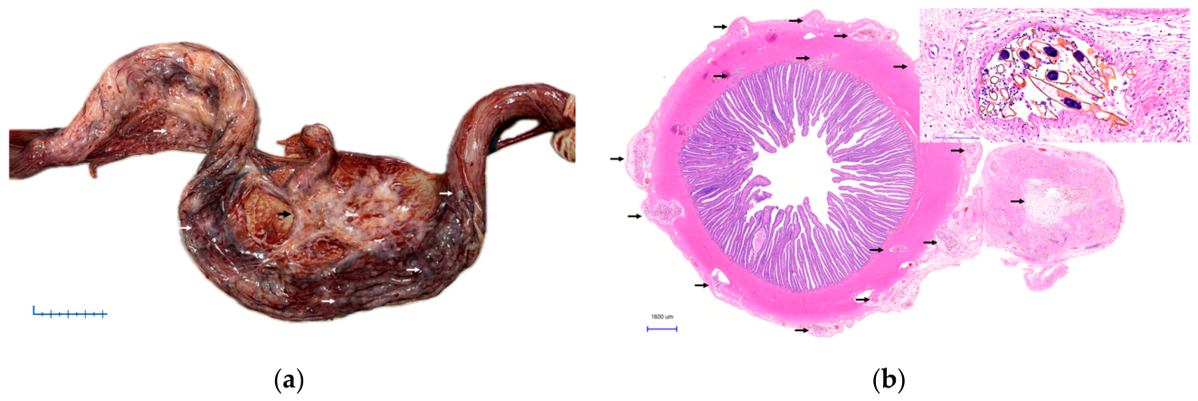
Figure 2.
Uterine lesions associated with spirorchiids in adult female olive ridley turtles stranded in Chile (Animal ID 3). (a) Gross: Uterus showing granulomatous lesions in the mature oviduct adjacent to follicles and corpora albicans. The section reveals a granulomatous appearance with a whitish-to-yellowish center. Bar= 2 cm. (b) Micrograph of the uterus with a granulomatous lesion (black arrow). Inset: Trematode eggs surrounded by lymphocytic inflammatory aggregates and multiple multinucleated giant cells (H&E). Bar= 200 µm.
Figure 2.
Uterine lesions associated with spirorchiids in adult female olive ridley turtles stranded in Chile (Animal ID 3). (a) Gross: Uterus showing granulomatous lesions in the mature oviduct adjacent to follicles and corpora albicans. The section reveals a granulomatous appearance with a whitish-to-yellowish center. Bar= 2 cm. (b) Micrograph of the uterus with a granulomatous lesion (black arrow). Inset: Trematode eggs surrounded by lymphocytic inflammatory aggregates and multiple multinucleated giant cells (H&E). Bar= 200 µm.
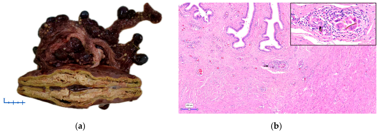
Figure 3.
Heart and great vessels of an adult female olive ridley turtle stranded in Chile (Animal ID 2) showing lesions associated with spirorchiids. (a) Heart with serous atrophy of fat and dilated lymphatic vessels. Bar= 2 cm. (b) Presence of adult spirorchiids (arrows) within the lumen of a large-caliber artery.
Figure 3.
Heart and great vessels of an adult female olive ridley turtle stranded in Chile (Animal ID 2) showing lesions associated with spirorchiids. (a) Heart with serous atrophy of fat and dilated lymphatic vessels. Bar= 2 cm. (b) Presence of adult spirorchiids (arrows) within the lumen of a large-caliber artery.
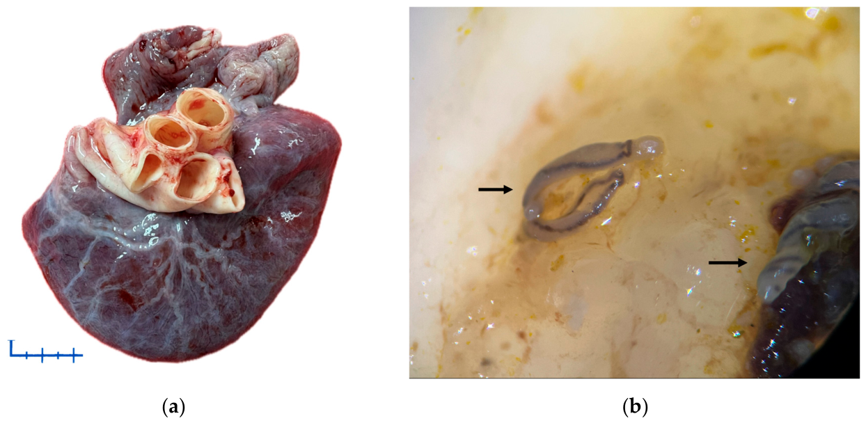

Table 1.
Data from female adult olive ridley turtles stranded along the Northern and Central-Northern Chilean coast between November 2024 and January 2026 with evidence of spirorchiidiasis.
Table 1.
Data from female adult olive ridley turtles stranded along the Northern and Central-Northern Chilean coast between November 2024 and January 2026 with evidence of spirorchiidiasis.
| Animal ID | Stranding Report | Stranding condition | Body Condition Score | Stranding Date | Date of death | Geographical Location | Region |
|---|---|---|---|---|---|---|---|
| 1 | 13066 | Alive | Intermediated | 9/21/24 | 9/22/24 | 29°55'29"S S, 71°16'43" O | Coquimbo |
| 2 | 14911 | Dead | Poor | 7/3/25 | Dead on arrival | 28°09'54"S S, 71°10'09" O | Atacama |
| 3 | 14427 | Alive | Intermediated | 11/3/25 | 11/4/25 | 26°27'50"S S, 70°41'12" O | Atacama |
| 4 | 14395 | Alive | Poor | 11/4/25 | 11/5/25 | 29°55'12"S S, 70°16'37" O | Coquimbo |
| 5 | 14521 | Alive | Poor | 12/19/25 | 12/20/25 | 32°50'19"S S, 71°31'18" O | Valparaíso |
| 6 | 14613 | Dead | Poor | 1/2/26 | Dead on arrival | 30°08'35"S S, 71°22'23" O | Coquimbo |
Disclaimer/Publisher’s Note: The statements, opinions and data contained in all publications are solely those of the individual author(s) and contributor(s) and not of MDPI and/or the editor(s). MDPI and/or the editor(s) disclaim responsibility for any injury to people or property resulting from any ideas, methods, instructions or products referred to in the content. |
© 2026 by the authors. Licensee MDPI, Basel, Switzerland. This article is an open access article distributed under the terms and conditions of the Creative Commons Attribution (CC BY) license (http://creativecommons.org/licenses/by/4.0/).
Copyright: This open access article is published under a Creative Commons CC BY 4.0 license, which permit the free download, distribution, and reuse, provided that the author and preprint are cited in any reuse.