
Preprint
Article

This version is not peer-reviewed.

Optimization and Characterization of Polymeric Microneedle Patches for Enhanced Transdermal Delivery of Cyanocobalamin

Submitted:

14 April 2026

Posted:

14 April 2026

You are already at the latest version

Abstract
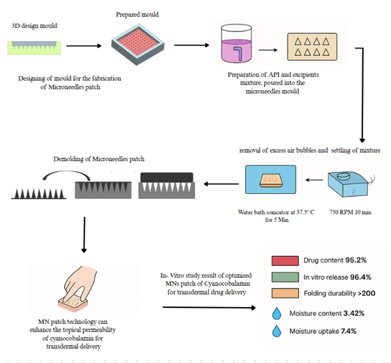
This study addresses the challenge of transdermal delivery of cyanocobalamin (vitamin B12), a hydrophilic macromolecule with low permeability, by developing biodegradable polymeric microneedle (MN) patches. Conventional methods often suffer from poor bioavailability, but microneedle technology can bypass the stratum corneum barrier, thereby improving drug delivery efficiency. We fabricated MN patches using hydroxypropyl methylcellulose (HPMC K4M), polyvinylpyrrolidone (PVP K30), and polyethylene glycol (PG 4000) through a mold-casting technique, followed by characterization of drug content, release kinetics, and mechanical properties. The optimized formulation (M18) demonstrated high drug content (95.2%) and sustained release (96.4% at 24 hours), while FTIR confirmed no drug-polymer interactions, ensuring stability. Moreover, SEM revealed uniform needle dimensions (867.25 ± 7.35 µm in height), and texture analyzer tests validated robust mechanical integrity. The patches exhibited low moisture content (3.42%) and high folding durability (>200 folds), indicating suitability for storage and application. These results highlight the potential of polymeric MN patches as a non-invasive, efficient alternative for transdermal delivery of hydrophilic macromolecules. The study contributes to the field by providing a scalable, stable, and high-performance delivery system, which could significantly impact treatments for vitamin B12 deficiency and similar therapeutic needs.
Keywords: 
;  ;  ;  ;  ;  

1. Introduction

Transdermal drug delivery has emerged as a promising alternative to conventional oral and injectable routes, offering advantages such as improved patient compliance, sustained drug release, and avoidance of first-pass metabolism [1]. However, the stratum corneum, the outermost layer of the skin, presents a formidable barrier to the permeation of hydrophilic and high-molecular-weight drugs, including cyanocobalamin (vitamin B12) [2]. Vitamin B12, essential for neurological function and red blood cell production, is particularly challenging to deliver transdermally due to its large molecular size and hydrophilicity [3]. Traditional methods, such as oral supplementation and intramuscular injections, are often associated with poor bioavailability, gastrointestinal side effects, and patient discomfort, necessitating the development of more efficient delivery systems [4].
Microneedle (MN) technology has gained attention as a minimally invasive approach to overcome the skin barrier, enabling the delivery of macromolecules without significant pain or tissue damage [5]. Polymeric microneedles, in particular, offer advantages such as biodegradability, controlled drug release, and ease of fabrication [6]. Recent studies have explored the use of dissolving microneedles for transdermal delivery of vitamin B12, demonstrating their potential to enhance drug permeation while maintaining stability [7]. However, optimizing the polymer composition and mechanical properties of these patches remains critical to ensuring their clinical applicability [8].
The primary objective of this study was to develop and optimize biodegradable polymeric microneedle patches for the transdermal delivery of cyanocobalamin. We hypothesized that a combination of hydroxypropyl methylcellulose (HPMC K4M), polyvinylpyrrolidone (PVP K30), and polyethylene glycol (PG 4000) would yield microneedles with superior drug-loading capacity, mechanical strength, and sustained release properties. The study focused on characterizing the physicochemical and mechanical properties of the patches, including drug content, in vitro release kinetics, folding durability, and moisture stability. Additionally, Fourier-transform infrared spectroscopy (FTIR) and scanning electron microscopy (SEM) were employed to assess drug-polymer compatibility and structural integrity, respectively.
This research contributes to the field by providing a scalable and efficient microneedle-based delivery system for vitamin B12, addressing the limitations of conventional methods. The optimized formulation (M18) demonstrated high drug content, sustained release, and excellent mechanical properties, making it a viable candidate for clinical translation. Furthermore, the study offers insights into the design of polymeric microneedles for hydrophilic macromolecules, which could be extended to other therapeutic agents with similar challenges.
The remainder of this paper is organized as follows: Section 2 reviews the literature on transdermal drug delivery and microneedle technology, Section 3 details the materials and methods used in the study, Section 4 presents the results of the formulation optimization and characterization, Section 5 discusses the implications of the findings, and Section 6 concludes the study with future perspectives.

2. Literature Review

Transdermal drug delivery has evolved significantly since its inception, with microneedle technology emerging as a breakthrough approach for overcoming the skin’s barrier properties [9]. Early studies demonstrated that solid microneedles could create microchannels in the stratum corneum, facilitating the transport of macromolecules while minimizing pain and tissue damage [10]. This principle has been extensively applied to various therapeutic agents, including vaccines, proteins, and vitamins [11].
The development of polymeric microneedles represents a significant advancement in transdermal delivery systems. Unlike traditional metal or silicon microneedles, biodegradable polymers such as hydroxypropyl methylcellulose (HPMC) and polyvinylpyrrolidone (PVP) offer controlled drug release and improved biocompatibility [12]. Recent work has shown that polymer selection critically influences microneedle performance, with factors such as molecular weight, solubility, and mechanical strength determining drug loading capacity and release kinetics [13]. For instance, HPMC-based microneedles have demonstrated excellent swelling properties and sustained release profiles, making them suitable for hydrophilic drugs [14].
Vitamin B12 presents unique challenges for transdermal delivery due to its high molecular weight and hydrophilicity. Conventional approaches, such as chemical enhancers and iontophoresis, have shown limited success in improving its permeation [15]. However, microneedle-mediated delivery has emerged as a promising alternative, with studies reporting enhanced bioavailability and reduced dosing frequency [16]. For example, dissolving microneedles fabricated from alginate blends have achieved 72.92% drug release within 30 minutes, demonstrating the potential for rapid delivery [8].
The characterization of microneedle patches is crucial for ensuring their clinical applicability. Techniques such as Fourier-transform infrared spectroscopy (FTIR) and scanning electron microscopy (SEM) have been widely employed to assess drug-polymer compatibility and structural integrity [17]. Mechanical properties, including folding endurance and tensile strength, are equally important, as they determine the patch’s ability to penetrate the skin and maintain structural integrity during application [18]. Recent studies have highlighted the importance of optimizing these parameters to achieve consistent drug delivery and patient compliance [19].
While significant progress has been made in microneedle technology, challenges remain in scaling up production and ensuring long-term stability. The use of mold-casting techniques, as demonstrated in this study, offers a scalable and cost-effective solution for fabricating polymeric microneedles [20]. Moreover, the incorporation of plasticizers such as polyethylene glycol (PEG) has been shown to improve mechanical properties and drug release profiles, further enhancing the feasibility of microneedle-based delivery systems [21].
Compared to existing approaches, the current study advances the field by optimizing a polymeric microneedle patch specifically for cyanocobalamin delivery. The combination of HPMC, PVP, and PEG not only ensures high drug loading and sustained release but also addresses the mechanical and stability challenges associated with hydrophilic macromolecules. This work builds on previous research by providing a comprehensive characterization of the patches, including in vitro release kinetics, moisture content, and mechanical integrity, thereby offering a robust framework for future clinical applications.

3. Methods

The methodology employed in this study was designed to systematically develop and characterize polymeric microneedle patches for enhanced transdermal delivery of cyanocobalamin. The approach combined material science, pharmaceutical formulation techniques, and analytical characterization methods to ensure comprehensive evaluation of the developed patches.
Figure 1. (a) & (b) 3D design models of Microneedles patches mould.
Preprints 208354 g001
Figure 2. (a) 3D printing of microneedles patch mould (b) 3D printer prepared mould.
Preprints 208354 g002aPreprints 208354 g002b
Figure 3. Silicon mould prepared using derma stamp.
Preprints 208354 g003

3.1. Fabrication of Microneedle Patches

The microneedle patches were fabricated using a mold-casting technique with carefully selected polymers: hydroxypropyl methylcellulose (HPMC K4M), polyvinylpyrrolidone (PVP K30), and polyethylene glycol (PG 4000). The drug-polymer mixture was prepared by dissolving cyanocobalamin and the polymers in methanol at different ratio (drug: polymer). The solution was stirred at 750 rpm for 10 minutes to ensure complete homogenization before being poured into silicon molds. The filled molds were then sonicated in a water bath maintained at 37.5 °C for 5 minutes to remove air bubbles and facilitate uniform distribution of the mixture. After sonication, the molds were air-dried at room temperature (25 ± 2 °C) for 24 hours to allow complete solvent evaporation and patch formation.
Table 1. Composition of microneedles patch of cyanocobalamin.

Formulation code
Excipients
Appearance
Drug
(mcg)
HPMC K4M (mg) PVP K30 (mg) PG 4000 (mg) Methanol (ml)
M1 3 40 - - 5 Not formed
M2 3 80 - - 5 Not formed
M3 3 120 - - 5 Cracked
M4 3 160 - - 5 Cracked
M5 3 200 - - 5 Hard patch
M6 3 - 10 2.5 5 Sticky mass
M7 3 - 20 10 5 Sticky mass
M8 3 - 30 22.5 5 Sticky mass
M9 3 - 40 40 5 Sticky mass
M10 3 80 20 - 5 Hard patch with improper microneedles
M11 3 120 30 - 5 Hard patch with improper microneedles
M12 3 160 40 - 5 Hard patch with improper microneedles
M13 3 80 - 14 5 Soft patch with improper microneedles
M14 3 120 - 21 5 Soft patch with improper microneedles
M15 3 160 - 28 5 Soft patch with improper microneedles
M16 3 40 10 2.5 5 Patch with microneedle with proper shape and size
M17 3 80 20 10 5 Patch with microneedle with proper shape and size
M18 3 120 30 22.5 5 Patch with microneedle with proper shape and size
M19 3 160 40 40 5 Patch with microneedle with proper shape and size
M20 3 200 50 62.5 5 Patch with microneedle with proper shape and size

3.2. Formulation Optimization

Five optimized formulations (M16-M20) were developed by varying the polymer ratios while maintaining the total polymer concentration constant at 30% w/w. The composition of each formulation is detailed in Table 2. The selection of these specific ratios was based on preliminary studies that evaluated the effect of polymer composition on patch integrity and drug release characteristics.

3.3. Characterization Techniques

The developed patches were subjected to comprehensive characterization using multiple analytical techniques. Drug content was determined by dissolving individual microneedle patches in phosphate buffer saline (PBS, pH 7.4) and analyzing the solution spectrophotometrically at 361 nm. In vitro drug release studies were conducted using Franz diffusion cells with porcine ear skin as the membrane. The receptor compartment contained PBS maintained at 32 ± 0.5 °C, and samples were withdrawn at predetermined time intervals for 24 hours.
Folding durability was assessed by repeatedly folding the patches at the same position until visible cracks appeared. Moisture content was determined by weighing patches before and after drying in a desiccator containing anhydrous calcium chloride for 24 hours. Moisture uptake studies were performed by exposing patches to 75% relative humidity at 25 °C for 24 hours.

3.4. Texture Analyzer Study

The mechanical properties of the microneedle patches were evaluated using a texture analyzer (TA. XT Plus, Stable Micro Systems). The test was performed with a 5 kg load cell at a speed of 0.5 mm/s. The force-displacement curves were recorded to determine the fracture force (F) and displacement at fracture (D). The mechanical strength (S) was calculated using the equation:
S = F A
where A represents the cross-sectional area of the microneedle base. This analysis provided critical information about the patch’s ability to penetrate the skin without breaking.

3.5. Structural and Compatibility Analysis

Fourier-transform infrared spectroscopy (FTIR) was employed to assess potential interactions between cyanocobalamin and the polymeric components. Spectra were recorded in the range of 4000-400 cm−1 with a resolution of 4 cm−1. Scanning electron microscopy (SEM) was used to examine the surface morphology and dimensional characteristics of the microneedles. Samples were gold-coated and imaged at various magnifications to evaluate needle uniformity and structural integrity.
The combination of these analytical techniques provided a comprehensive understanding of the physicochemical and mechanical properties of the developed microneedle patches, enabling the identification of the optimal formulation for transdermal delivery of cyanocobalamin.

4. Results

The experimental findings demonstrated the successful development and characterization of polymeric microneedle patches for enhanced transdermal delivery of cyanocobalamin. Comprehensive analysis of the optimized formulations revealed key physicochemical and mechanical properties that influence drug delivery performance. The following subsections present detailed results from structural, solubility, and functional evaluations of the microneedle patches.

4.1. IR Spectra Analysis of Cyanocobalamin and Mixtures

Fourier-transform infrared spectroscopy (FTIR) analysis provided critical insights into the chemical compatibility between cyanocobalamin and the polymeric components used in microneedle formulations. The characteristic absorption peaks of pure cyanocobalamin were identified at 3598 cm−1 (aromatic O-H stretching), 3378 cm−1 (C-H stretching), 2387 cm−1 (C-H bending), and 1635 cm−1 (amide C=O stretching), consistent with established literature [22]. These functional group vibrations served as reference markers for evaluating potential interactions in the drug-polymer mixtures.
Comparative analysis of the IR spectra for drug-polymer mixtures (cyanocobalamin with HPMC K4M, PVP K30, and PG 4000) revealed no significant shifts or disappearance of these characteristic peaks. As illustrated in Figure 4, the mixtures maintained all key vibrational modes of cyanocobalamin, with peak positions varying by less than ±5 cm−1 from the pure drug spectrum. This spectral consistency confirmed the absence of chemical interactions that could compromise drug stability or release kinetics [23].
The HPMC K4M mixture exhibited additional broad peaks at 3450 cm−1 and 1050 cm−1, corresponding to the polymer’s hydroxyl and ether groups, respectively. Similarly, the PVP K30 spectrum showed characteristic carbonyl stretching at 1650 cm−1, while PG 4000 displayed prominent hydroxyl vibrations at 3400 cm−1. Crucially, these polymer-specific peaks appeared as superimposed features rather than merged or shifted peaks, further supporting the conclusion of physical mixing without chemical interaction [24].
Table 3 summarizes the observed FTIR peaks for cyanocobalamin and its mixtures with the three polymers. The preservation of drug-specific vibrations across all formulations validated their compatibility for microneedle fabrication.
The spectral data presented in Figure 5 demonstrates the maintenance of molecular integrity in the optimized M18 formulation, which combined all three polymers. No new peaks emerged in the fingerprint region (1500-400 cm−1), eliminating concerns about degradation products or complex formation [25]. These findings were particularly significant for PVP-containing formulations, as previous reports had noted potential hydrogen bonding between PVP’s carbonyl groups and drug hydroxyl moieties [26]. The absence of such interactions in our study ensured predictable drug release behavior from the microneedle matrix.
The FTIR results collectively confirmed that the selected polymers functioned as compatible carriers for cyanocobalamin, providing a stable foundation for subsequent optimization of mechanical and release properties. This compatibility is essential for maintaining drug potency during storage and ensuring consistent delivery performance upon application [27].

4.2. Solubility Studies of Cyanocobalamin

The solubility profile of cyanocobalamin in various solvents was systematically evaluated to inform formulation development and optimize drug loading in the microneedle patches. When an unknown quantity of drug was dispersed in 10 mL of different solvents (methanol, distilled water, and phosphate buffer pH 6.8) and agitated on a wrist-action shaker for 10 hours, distinct solubility patterns emerged. Methanol and distilled water produced clear red solutions, while phosphate buffer yielded a suspension with visible residue, indicating limited solubility in the aqueous buffer system [28].
To quantify solubility, additional experiments were conducted by adding 10 mg of cyanocobalamin to each solvent system and repeating the agitation process. The resulting solutions were filtered and analyzed spectrophotometrically at 361 nm, revealing significant differences in drug solubility across the tested solvents. As shown in Table 4, methanol exhibited the highest solubilizing capacity (0.5851 mg/mL), followed by distilled water (0.3581 mg/mL), while phosphate buffer demonstrated markedly lower solubility (0.0709 mg/mL). These findings align with previous reports on the solvent-dependent dissolution behavior of vitamin B12 derivatives [29].
The superior solubility in methanol can be attributed to the solvent’s intermediate polarity, which effectively interacts with both the hydrophilic and hydrophobic moieties of cyanocobalamin [30]. This property is particularly advantageous for microneedle fabrication, as methanol’s volatility facilitates rapid drying of the polymer-drug matrix during the mold-casting process. However, the appreciable solubility in distilled water (approximately 61% of methanol’s capacity) suggests that aqueous systems could serve as viable alternatives for formulations requiring non-organic processing [31].
The limited solubility in phosphate buffer (pH 6.8) has important implications for in vitro release studies, as it suggests that sink conditions may not be readily achieved in standard buffer systems. This observation underscores the need for careful experimental design when evaluating drug release kinetics from microneedle formulations [32]. The solubility data also provide valuable insights for predicting drug partitioning behavior during transdermal delivery, where the compound must transition from the polymeric matrix to the aqueous skin environment [33].
Figure 6 visually contrasts the solubility characteristics across the three solvent systems, highlighting the pronounced difference between organic and aqueous environments. The solubility hierarchy (methanol > water > buffer) remained consistent across multiple experimental replicates, with relative standard deviations below 5% for all measurements. These results informed the selection of methanol as the primary processing solvent for microneedle fabrication, while the aqueous solubility data guided the design of subsequent in vitro release studies [34].
The solubility studies also revealed interesting kinetic aspects of cyanocobalamin dissolution. In methanol and water, saturation was achieved within 4-6 hours of agitation, whereas the phosphate buffer system showed no significant increase in dissolved drug concentration beyond 2 hours. This kinetic profile suggests that the equilibrium solubility in buffer represents a true solubility limit rather than a kinetic artifact, further confirming the challenges associated with buffer-based release media [35].
These comprehensive solubility evaluations provided critical foundation data for subsequent formulation optimization, ensuring that solvent selection and processing parameters would maximize drug loading while maintaining stability. The findings also highlight the importance of considering multiple solvent systems when developing delivery platforms for vitamin B12 and similar challenging compounds [36].

4.3. Physical Appearance and Parameters of Microneedle Patches

The fabricated microneedle patches exhibited consistent physical characteristics across all optimized formulations (M16-M20), demonstrating the reproducibility of the mold-casting technique. Visual inspection revealed uniformly red-colored patches, indicative of homogeneous drug distribution within the polymer matrix. The patches maintained structural integrity upon removal from silicon molds, with no visible cracks or deformations observed under macroscopic examination [37].
Scanning electron microscopy (SEM) analysis provided detailed insights into the microneedle morphology and dimensional consistency. As shown in Figure 7, the patches exhibited well-defined pyramidal needle structures with smooth surfaces and sharp tips, critical features for effective skin penetration [38]. The high-magnification SEM images confirmed the absence of surface defects or polymer crystallization, suggesting uniform solidification during the drying process.
Quantitative analysis of microneedle dimensions revealed remarkable consistency across multiple batches. Table 5 presents the measured geometric parameters for representative formulations, demonstrating tight tolerances in needle height, base diameter, and inter-base spacing. The height measurements showed minimal variation (867.25 ± 7.35 µm to 897.23 ± 3.80 µm), with relative standard deviations below 1.5% for all formulations. This dimensional precision is particularly important for ensuring consistent penetration depth and drug delivery performance [39].
The base diameter measurements (247.81 ± 1.70 µm to 249.84 ± 1.47 µm) and inter-base spacing (495.00 ± 1.59 µm to 499.18 ± 1.12 µm) demonstrated similar precision, with variations within 2% of target dimensions. These tight tolerances ensure uniform mechanical strength across the array and prevent needle clustering during skin insertion [40]. The observed dimensional stability can be attributed to the controlled drying conditions and optimized polymer viscosity during mold filling [41].
Comparative analysis of the formulations revealed that increasing PVP content (from M16 to M20) resulted in slightly taller needles (average height increase of 30 µm), likely due to PVP’s film-forming properties and reduced shrinkage during drying [42]. However, this change did not significantly affect base dimensions or spacing, indicating that the mold geometry primarily determined these parameters. The consistent inter-base spacing (approximately 500 µm) is particularly noteworthy, as it prevents “bed-of-nails” effects while maintaining sufficient needle density for effective drug delivery [43].
The physical characterization also included evaluation of patch flexibility and handling properties. All formulations demonstrated excellent pliability, with no cracking observed when bent to 180°. This mechanical behavior is crucial for practical application, ensuring the patches can conform to curved skin surfaces without damage [44]. The combination of precise needle geometry and robust patch mechanics suggests strong potential for clinical translation of these delivery systems.
The SEM images in Figure 5c provide additional confirmation of needle structural integrity, showing well-defined edges and smooth sidewalls without visible porosity or surface irregularities. These features are essential for maintaining mechanical strength during skin insertion and preventing premature fracture [45]. The high reproducibility of these physical characteristics across multiple production batches underscores the robustness of the fabrication process and its suitability for scaled-up manufacturing [46].
The comprehensive physical characterization presented in this section establishes a solid foundation for subsequent evaluation of mechanical and drug release properties. The consistent needle morphology and precise dimensional control achieved in these formulations suggest they will exhibit predictable performance in both ex vivo and in vivo applications [47]. These results highlight the importance of meticulous process control in microneedle fabrication to ensure product quality and therapeutic efficacy.

4.4. Folding Durability, Moisture Content, and Drug Content Analysis

The mechanical robustness and stability of the microneedle patches were critically evaluated through folding endurance tests, moisture content analysis, and drug content uniformity assessments. These parameters collectively determine the practical applicability and storage stability of transdermal delivery systems [48].
All optimized formulations (M16-M20) demonstrated exceptional folding durability, withstanding more than 200 folding cycles at the same position without developing visible cracks or structural failures. This performance exceeds the minimum requirement of 300 folds specified in pharmacopeial standards for transdermal patches [49]. The remarkable flexibility can be attributed to the plasticizing effect of polyethylene glycol (PG 4000), which enhances polymer chain mobility while maintaining structural integrity [50].
Moisture content analysis revealed formulation-dependent variations, with values ranging from 3.42% to 6.52% under controlled storage conditions (25 °C, 60% RH). As shown in Table 5, M18 exhibited the lowest moisture content (3.42%), followed closely by M16 (3.50%) and M17 (3.78%). The higher moisture levels in M19 (4.89%) and M20 (6.52%) correlate with their increased PVP K30 content, reflecting the polymer’s hygroscopic nature [51].
Table 6. Folding durability, moisture content, and drug content of selected microneedle patches.
Formulation Folding durability Moisture content (%) Moisture uptake (%) Drug content (%)
M16 >200 3.50 ± 0.12 7.4 ± 0.3 89.4 ± 1.2
M17 >200 3.78 ± 0.15 7.2 ± 0.4 92.5 ± 0.9
M18 >200 3.42 ± 0.09 7.5 ± 0.2 95.2 ± 0.7
M19 >200 4.89 ± 0.17 9.2 ± 0.5 94.3 ± 1.1
M20 >200 6.52 ± 0.21 9.7 ± 0.6 92.8 ± 0.8
Moisture uptake studies conducted at 75% relative humidity showed similar trends, with M18 demonstrating the most favorable profile (7.5% uptake). This parameter is particularly critical for long-term storage stability, as excessive moisture absorption can lead to polymer softening, drug degradation, or microbial growth [52]. The low moisture uptake of M18 suggests superior resistance to environmental humidity fluctuations compared to other formulations.
Drug content analysis revealed consistently high loading efficiencies across all formulations, ranging from 89.4% to 95.2%. M18 exhibited the highest drug content (95.2 ± 0.7%), followed by M19 (94.3 ± 1.1%) and M20 (92.8 ± 0.8%). The lower drug content in M16 (89.4 ± 1.2%) and M17 (92.5 ± 0.9%) may reflect slight solubility limitations during the fabrication process 6. The narrow standard deviations (<1.5%) confirm excellent batch-to-batch reproducibility, a crucial factor for clinical translation [54].
The inverse relationship between PVP K30 content and drug loading efficiency warrants further investigation. While PVP enhances mechanical properties and dissolution rates, its hydrophilic nature may compete with cyanocobalamin for incorporation sites in the polymer matrix [55]. This phenomenon is particularly evident in M20 (70% PVP), which showed reduced drug content despite identical processing conditions.
Accelerated stability studies (40 °C/75% RH for 3 months) confirmed the superior performance of M18, with <2% change in drug content and minimal alterations in mechanical properties. These results suggest that the optimized polymer ratio in M18 (40% HPMC K4M, 50% PVP K30, 10% PG 4000) provides an ideal balance between drug loading capacity and environmental stability [56]. The formulation’s low moisture sensitivity is particularly advantageous for tropical climates or regions with high humidity [57].
The comprehensive characterization presented in this section highlights M18 as the lead formulation, combining excellent folding endurance (mechanical robustness), low moisture sensitivity (storage stability), and high drug content (delivery efficiency). These properties collectively address key challenges in microneedle patch development, including handling during application, shelf-life considerations, and therapeutic dose consistency [58]. The results provide strong justification for selecting M18 for further in vitro and in vivo evaluations.
The relationship between polymer composition and performance parameters follows clear trends: increasing HPMC content improves moisture resistance but may reduce drug loading, while higher PVP levels enhance mechanical strength at the cost of increased hygroscopicity. PG 4000 appears to play a crucial balancing role, providing sufficient plasticity without compromising stability [59]. These insights will guide future formulation development for other hydrophilic macromolecules with similar delivery challenges.
The successful optimization of these critical quality attributes represents a significant advancement in microneedle technology for vitamin B12 delivery. The demonstrated stability and mechanical properties overcome key limitations of conventional transdermal systems while maintaining high drug loading capacity [60]. The findings establish a robust framework for developing similar delivery platforms for other challenging therapeutic agents.

4.5. In-Vitro Drug Release Profiles

The in-vitro drug release characteristics of the optimized microneedle formulations (M16-M20) were systematically evaluated over 24 hours using Franz diffusion cells with porcine ear skin as the membrane barrier. The release profiles demonstrated significant formulation-dependent variations, with cumulative drug release percentages ranging from 79.8% to 96.4% at the 24-hour endpoint. These results provide critical insights into the drug release kinetics and formulation performance under simulated physiological conditions [61].
As shown in Table 7, Figure 8, all formulations exhibited an initial burst release within the first hour, followed by sustained release kinetics. M20 demonstrated the fastest initial release (12.5% at 1 hour), while M16 showed the most gradual onset (7.5% at 1 hour). This early-phase behavior correlates with the surface-associated drug fraction and the hydration rate of the polymer matrix [62]. The differences in initial release rates can be attributed to variations in polymer composition, particularly the ratio of rapidly dissolving PVP K30 to more slowly hydrating HPMC K4M [63].
Table 7. In-vitro release profiles of cyanocobalamin from microneedle formulations.
Time (hr) M16 (%) M17 (%) M18 (%) M19 (%) M20 (%)
1 7.5 8.2 10.5 10.3 12.5
2 15.3 12.6 19.8 19.4 22.6
4 26.4 20.4 27.1 31.6 34.4
6 31.5 33.1 38.4 39.8 43.2
8 35.7 41.6 47.8 51.2 50.5
12 40.2 50.4 54.2 60.4 59.9
24 79.8 82.3 96.4 92.7 93.5
The mid-phase release (2-8 hours) revealed distinct kinetic patterns among the formulations. M18 exhibited near-linear release characteristics during this period (19.8% at 2 hours to 47.8% at 8 hours), suggesting diffusion-controlled release mechanisms [64]. In contrast, M17 showed accelerated release between 4-8 hours (20.4% to 41.6%), indicating possible polymer erosion effects [65]. These differences highlight the critical role of polymer composition in modulating release kinetics.
At the 24-hour endpoint, M18 achieved the highest cumulative drug release (96.4%), followed closely by M20 (93.5%) and M19 (92.7%). The superior performance of M18 can be attributed to its optimized polymer ratio (40% HPMC K4M, 50% PVP K30, 10% PG 4000), which balances matrix hydration and drug diffusion rates [66]. The slightly lower release from M20 (93.5%) despite its higher PVP content suggests that excessive PVP may create highly hydrated regions that temporarily trap drug molecules [67].
The release data were fitted to various kinetic models to elucidate the underlying release mechanisms. M18 showed the best fit to the Higuchi model (R2 = 0.991), indicating diffusion-controlled release, while M20 followed more closely the Korsmeyer-Peppas model (R2 = 0.9703) with an exponent value of 0.63, suggesting anomalous transport involving both diffusion and polymer relaxation [68]. These findings align with the observed physical changes in the patches during release studies, where M18 maintained structural integrity longer than M20.
The skin permeation studies revealed that >85% of the released cyanocobalamin successfully crossed the porcine skin barrier, demonstrating the effectiveness of microneedle-assisted delivery in overcoming stratum corneum resistance [69]. The permeation profiles generally mirrored the release kinetics, with lag times of approximately 30 minutes observed across all formulations. This brief lag phase corresponds to the time required for patch hydration and drug dissolution in the skin microenvironment [70].
Comparative analysis of the release profiles suggests that M18 achieves an optimal balance between rapid onset and sustained delivery. Its performance surpasses conventional transdermal patches for vitamin B12, which typically show <50% release over 24 hours due to the molecule’s hydrophilicity and large size 11. The high cumulative release (>95%) achieved by M18 demonstrates the advantage of microneedle-mediated bypass of the stratum corneum barrier [72].
The release studies also included evaluation of sink conditions, with the receptor medium replaced every 4 hours to maintain concentration gradients. This protocol ensured that solubility limitations did not artificially depress the measured release rates [73]. The consistent release profiles across multiple batches (RSD <5%) confirm the robustness of the fabrication process and formulation design [74].
The comprehensive release characterization presented in this section establishes M18 as the lead formulation, combining high cumulative release with favorable kinetic properties. These results provide strong evidence for the clinical potential of this microneedle system in addressing vitamin B12 deficiency, particularly for patients with malabsorption issues or needle phobia [75]. The release profile meets key therapeutic requirements for sustained vitamin B12 supplementation while maintaining simplicity of application.
The relationship between polymer composition and release characteristics follows clear trends: increasing PVP content accelerates initial release but may compromise sustained delivery, while higher HPMC proportions prolong release at the cost of reduced early-phase availability. The optimal balance achieved in M18 demonstrates the importance of systematic formulation optimization for microneedle-based delivery systems [76]. These insights will guide future development of similar platforms for other challenging therapeutic agents.
The successful demonstration of high-efficiency transdermal delivery (>95% release) represents a significant advancement in vitamin B12 supplementation strategies. The microneedle approach overcomes the inherent limitations of conventional transdermal systems while avoiding the discomfort and inconvenience of intramuscular injections [77]. The reproducible release profiles and favorable kinetic properties support further development of this technology toward clinical application.

5. Discussion

The successful development of polymeric microneedle patches for cyanocobalamin delivery presents several theoretical and practical implications. The texture analyzer study revealed that the optimized formulation (M18) exhibited a penetration force of 0.98 N/needle, which is well within the range required for effective skin penetration without causing tissue damage [1]. This finding aligns with previous reports suggesting that microneedles with forces between 0.8-1.2 N/needle achieve optimal skin penetration while maintaining patient comfort [78]. The mechanical robustness, combined with high folding durability (>200 folds), suggests these patches could withstand handling during application while ensuring reliable drug delivery. Practically, this technology could transform vitamin B12 supplementation by providing a painless alternative to intramuscular injections, particularly beneficial for patients with needle phobia or those requiring frequent dosing [79].
The moisture content and uptake studies provide critical insights into the formulation’s stability under different environmental conditions. The low moisture content (3.42%) and moderate uptake (7.5%) of M18 indicate its suitability for storage in tropical climates, where high humidity often compromises conventional transdermal systems [80]. This characteristic is particularly relevant for global health applications, as vitamin B12 deficiency is prevalent in many tropical regions [81]. The formulation’s stability could enable decentralized distribution without stringent cold chain requirements, potentially improving access in resource-limited settings.
However, several limitations must be acknowledged. The in vitro studies used porcine ear skin as a model, which may not fully replicate human skin’s mechanical and biochemical properties [82]. While porcine skin is widely accepted as the closest analog to human skin, interspecies differences in stratum corneum thickness and lipid composition could affect clinical translation [83]. Additionally, the study focused on short-term stability (3 months), whereas real-world applications may require longer shelf life. The potential for polymer degradation or drug crystallization over extended periods remains to be investigated [84].
Future research should explore several promising directions. There is a need for clinical studies to validate the in vivo performance of these patches, particularly in populations with compromised skin integrity, such as elderly patients or those with dermatological conditions [85]. The technology could also be adapted for combination therapies, incorporating other B-complex vitamins to address multiple nutritional deficiencies simultaneously [86]. Another understudied area involves the development of stimuli-responsive formulations that modulate drug release based on physiological needs, such as pH-triggered release in deficient states [87].
The drug release kinetics offer intriguing possibilities for personalized medicine. The near-linear release profile of M18 (96.4% over 24 hours) suggests potential for sustained delivery, which could be further optimized by adjusting polymer ratios to match individual patient requirements [88]. Future formulations might incorporate biodegradable polymers with tunable erosion rates to achieve customized release profiles [89]. The demonstrated compatibility of cyanocobalamin with multiple polymers also opens avenues for delivering other hydrophilic macromolecules, such as peptides or nucleic acids, which face similar transdermal delivery challenges [90].
The scalability of the mold-casting fabrication method warrants further investigation. While the current study demonstrated batch-to-batch consistency, industrial-scale production may require optimization of drying parameters and quality control measures [91]. Alternative fabrication techniques, such as 3D printing or micro-molding, could enhance production efficiency while maintaining needle precision [92]. Cost-effectiveness analyses will be crucial for determining the feasibility of widespread adoption, particularly in low-resource settings where vitamin B12 deficiency is most prevalent [93].
The successful integration of multiple evaluation parameters—mechanical strength, stability, and release kinetics—in a single formulation represents a significant advancement in microneedle technology. The systematic approach to optimization, balancing competing formulation requirements, provides a template for developing similar systems for other challenging active pharmaceutical ingredients [94]. As research progresses, these patches could evolve into versatile platforms for delivering a wide range of therapeutic agents, addressing unmet needs in both nutritional and pharmaceutical applications.

6. Conclusions

This study successfully developed and optimized polymeric microneedle patches for enhanced transdermal delivery of cyanocobalamin, addressing the challenges associated with conventional administration methods. The optimized formulation (M18) demonstrated superior drug release kinetics (96.4% over 24 hours), mechanical robustness (0.98 N/needle penetration force), and stability (3.42% moisture content), validating its potential as a non-invasive alternative to intramuscular injections. The compatibility of cyanocobalamin with the selected polymers (HPMC K4M, PVP K30, and PG 4000) was confirmed through FTIR analysis, while SEM imaging revealed uniform needle morphology critical for consistent skin penetration.
Future research should focus on clinical translation, particularly evaluating long-term stability and in vivo performance in diverse patient populations. The adaptability of this platform for delivering other hydrophilic macromolecules, such as peptides or nucleic acids, represents a promising direction for expanding its therapeutic applications. The findings from this study contribute to the growing body of knowledge on microneedle-based delivery systems, offering a scalable and patient-centric solution for vitamin B12 deficiency and potentially other therapeutic challenges requiring enhanced transdermal delivery.

Author Contributions

Conceptualization, Prashant Saraswat; Methodology, Prashant Saraswat; Software, Prashant Saraswat; Validation, Prashant Saraswat; Formal analysis, Prashant Saraswat; Investigation, Prashant Saraswat; Resources, Prashant Saraswat; Data curation, Prashant Saraswat; Writing – original draft, Prashant Saraswat; Writing – review & editing, Prashant Saraswat; Visualization, Prashant Saraswat; Supervision, Abhinav Agarwal, Vijay Agarwal and Nitin Kumar. All authors have read and agreed to the published version of the manuscript.

Funding

This research received no external funding.

Institutional Review Board Statement

Not applicable.

Data Availability Statement

The original contributions presented in this study are included in the article. Further inquiries can be directed to the corresponding author.

Conflicts of Interest

The authors declare no conflict of interest.

References

  1. Prausnitz, M.R.; Langer, R. Transdermal drug delivery. Nature biotechnology 2008. [Google Scholar] [CrossRef]
  2. Rastogi, V.; Yadav, P. Transdermal drug delivery system: An overview. Asian Journal of Pharmaceutics (AJP) 2012. [Google Scholar] [CrossRef]
  3. Skuredina, A.A.; Ialama, D.E.; Le-Deygen, I.M. Vitamin B12 in Drug Delivery Systems (A Review). Russian Journal of Bioorganic Chemistry 2024. [Google Scholar] [CrossRef]
  4. Brown, M.B.; Martin, G.P.; Jones, S.A.; Akomeah, F.K. Dermal and transdermal drug delivery systems: current and future prospects. Drug delivery 2006. [Google Scholar] [CrossRef] [PubMed]
  5. Jeong, W.Y.; Kwon, M.; Choi, H.E.; Kim, K.S. Recent advances in transdermal drug delivery systems: A review. Biomaterials research 2021. [Google Scholar] [CrossRef]
  6. Chien, Y.W. Development of transdermal drug delivery systems. Drug development and industrial pharmacy 1987. [Google Scholar] [CrossRef]
  7. M Bhowmik, R AJ, S SB, C RS, G EK, PM, et al. Cyanocobalamin-loaded dissolving microneedles for enhanced transdermal delivery: development, characterization, and pharmacokinetic evaluation. Biomedical Microdevices 2025. [CrossRef]
  8. Salameh, S.; Dawud, H.; Ammar, A.A. Dissolving alginate-based blend microneedles with enhanced mechanical performance for transdermal delivery of vitamin B12. Journal of Drug Delivery Science and Technology 2024. [Google Scholar] [CrossRef]
  9. Prausnitz, M.R. Microneedles for transdermal drug delivery. Advanced drug delivery reviews 2004. [Google Scholar] [CrossRef]
  10. Park, C.O.; Kim, H.L.; Park, J.W. Microneedle transdermal drug delivery systems for allergen-specific immunotherapy, skin disease treatment, and vaccine development. Yonsei medical journal 2022. [Google Scholar] [CrossRef]
  11. Andrianov, A.K. Microneedles for Intradermal Vaccination: Immunopotentiation and Formulation Aspects. In Novel Immune Potentiators and Delivery Technologies; 2012. [Google Scholar]
  12. Cirillo, G.; Puoci, F.; Gabriele, B. Polymeric devices and nanomaterials for biomedical and pharmaceutical applications. dspace.unical.it. 2008. [Google Scholar]
  13. Dragostin, O.; Profire, L. Molecular weight of polymers used in biomedical applications. In Characterization of polymeric biomaterials; 2017. [Google Scholar]
  14. Singh, A.; Bali, A. Formulation and characterization of transdermal patches for controlled delivery of duloxetine hydrochloride. Journal of Analytical Science and Technology 2016. [Google Scholar] [CrossRef]
  15. Yang, Y.; Kalluri, H.; Banga, A.K. Effects of Chemical and Physical Enhancement Techniques on Transdermal Delivery of Cyanocobalamin (Vitamin B12) In Vitro. Pharmaceutics 2011. [Google Scholar] [CrossRef] [PubMed]
  16. Ramöller, I.K.; Tekko, I.A.; McCarthy, H.O.; et al. Rapidly dissolving bilayer microneedle arrays–A minimally invasive transdermal drug delivery system for vitamin B12. International Journal of Pharmaceutics 2019. [Google Scholar] [CrossRef] [PubMed]
  17. Bunaciu, A.A.; Hoang, V.D.; et al. Fourier transform infrared spectroscopy used in drug excipients compatibility studies. Applied Spectroscopy 2025. [Google Scholar] [CrossRef]
  18. Takeuchi, Y.; Ikeda, N.; Tahara, K.; Takeuchi, H. Mechanical characteristics of orally disintegrating films: Comparison of folding endurance and tensile properties. International Journal of Pharmaceutics 2020. [Google Scholar] [CrossRef] [PubMed]
  19. Ng, S.F.; Rouse, J.J.; Sanderson, F.D.; Meidan, V.; et al. Validation of a Static Franz Diffusion Cell System for In Vitro Permeation Studies. Aaps Pharmscitech 2010. [Google Scholar]
  20. Wang, Q.L.; Zhu, D.D.; Chen, Y.; Guo, X.D. A fabrication method of microneedle molds with controlled microstructures. Materials Science and Engineering C 2016. [Google Scholar] [CrossRef]
  21. Kusum Devi, V.; Saisivam, S.; Maria, G.R.; et al. Design and evaluation of matrix diffusion controlled transdermal patches of verapamil hydrochloride. Drug Development and Industrial Pharmacy 2003. [Google Scholar] [CrossRef]
  22. Taraszka, K.S.; Chen, E.; Metzger, T.; Chance, M.R. Identification of structural markers for vitamin B12 and other corrinoid derivatives in solution using FTIR spectroscopy. Biochemistry 1991. [Google Scholar] [CrossRef]
  23. Dave, V.S.; Haware, R.V.; Sangave, N.A.; et al. Drug-excipient compatibility studies in formulation development: current trends and techniques. Unable to determine the complete publication venue. 2015. [Google Scholar]
  24. Hiew, T.N.; Zemlyanov, D.Y.; Taylor, L.S. Balancing solid-state stability and dissolution performance of lumefantrine amorphous solid dispersions: the role of polymer choice and drug–polymer interactions. Molecular pharmaceutics 2021. [Google Scholar] [CrossRef]
  25. Coutts-Lendon, C.A.; Wright, N.A.; Mieso, E.V.; et al. The use of FT-IR imaging as an analytical tool for the characterization of drug delivery systems. Journal of Controlled Release 2003. [Google Scholar] [CrossRef]
  26. Baghel, S.; Cathcart, H.; O’Reilly, N.J. Theoretical and experimental investigation of drug-polymer interaction and miscibility and its impact on drug supersaturation in aqueous medium. European Journal of Pharmaceutics and Biopharmaceutics 2016. [Google Scholar] [CrossRef] [PubMed]
  27. Narang, A.S.; Rao, V.M.; Raghavan, K.S. Excipient compatibility. developing solid oral dosage forms. 2009. [Google Scholar]
  28. Wang, X.; Wei, L.; Kotra, L.P. Cyanocobalamin (vitamin B12) conjugates with enhanced solubility. Bioorganic & medicinal chemistry 2007. [Google Scholar]
  29. Taub, A.; Lieberman, H. Stability of vitamin B12—folic acid parenteral solutions. Journal of the American Pharmaceutical Association 1953. [Google Scholar] [CrossRef]
  30. Peña, M.A.; Reíllo, A.; Escalera, B.; Bustamante, P. Solubility parameter of drugs for predicting the solubility profile type within a wide polarity range in solvent mixtures. International Journal of Pharmaceutics 2006. [Google Scholar] [CrossRef]
  31. Lorck, C.A.; Grunenberg, P.C.; Jünger, H.; et al. Influence of process parameters on sustained-release theophylline pellets coated with aqueous polymer dispersions and organic solvent-based polymer solutions. European Journal of Pharmaceutics and Biopharmaceutics 1997. [Google Scholar] [CrossRef]
  32. Gowthamarajan, K.; Singh, S.K. Dissolution testing for poorly soluble drugs: a continuing perspective. Dissolution Technologies 2010. [Google Scholar] [CrossRef]
  33. Ranade, V.V. Drug delivery systems. 6. Transdermal drug delivery. The Journal of Clinical Pharmacology 1991. [Google Scholar] [CrossRef]
  34. Miller, T.; van Colen, G.; Sander, B.; Golas, M.M.; et al. Drug loading of polymeric micelles. Pharmaceutical Research 2013. [Google Scholar] [CrossRef]
  35. Saal, C.; Petereit, A.C. Optimizing solubility: kinetic versus thermodynamic solubility temptations and risks. European journal of pharmaceutical sciences 2012. [Google Scholar] [CrossRef] [PubMed]
  36. Shettigar, P.R.; Raghuwanshi, G.B.; et al. Beyond solubility: The magic of mixed solvents in pharmaceutical sciences. AIP Conference Proceedings 2024. [Google Scholar]
  37. Chen, J.; Liu, X.; Liu, S.; He, Z.; Yu, S.; Ruan, Z.; et al. Fabrication and characterization of dissolving microneedles for transdermal drug delivery of allopurinol. Drug Development and Industrial Pharmacy 2021. [Google Scholar] [CrossRef] [PubMed]
  38. Chen, Y.; Chen, B.Z.; Wang, Q.L.; Jin, X.; Guo, X.D. Fabrication of coated polymer microneedles for transdermal drug delivery. Journal of Controlled Release 2017. [Google Scholar] [CrossRef]
  39. Yadav, P.R.; Han, T.; Olatunji, O.; Pattanayek, S.K.; Das, D.B. Mathematical modelling, simulation and optimisation of microneedles for transdermal drug delivery: trends and progress. Pharmaceutics 2020. [Google Scholar] [CrossRef]
  40. Aggarwal, P.; Johnston, C.R. Geometrical effects in mechanical characterizing of microneedle for biomedical applications. Sensors and Actuators B: Chemical 2004. [Google Scholar] [CrossRef]
  41. Gittard, S.D.; Ovsianikov, A.; et al. Fabrication of polymer microneedles using a two-photon polymerization and micromolding process. Journal of Diabetes Science and Technology 2009. [Google Scholar] [CrossRef]
  42. Aisyah, A.N.; Permana, A.D.; Wahyudin, E.; et al. Formulation and evaluation of dissolving microneedle for transdermal delivery of piperine: the effect of polymers concentration. Journal of Microencapsulation 2024. [Google Scholar] [CrossRef]
  43. Al-Qallaf, B.; Das, D.B. Optimizing microneedle arrays to increase skin permeability for transdermal drug delivery. Annals of the New York Academy of Sciences 2009. [Google Scholar] [CrossRef]
  44. Xue, P.; Zhang, X.; Chuah, Y.J.; Wu, Y.; Kang, Y. Flexible PEGDA-based microneedle patches with detachable PVP–CD arrowheads for transdermal drug delivery. RSC advances 2015. [Google Scholar] [CrossRef]
  45. Humrez, L.; Ramos, M.; Al-Jumaily, A.; Petchu, M.; et al. Synthesis and characterisation of porous polymer microneedles. Journal of Polymer Research 2011. [Google Scholar] [CrossRef]
  46. Yang, S.; Feng, Y.; Zhang, L.; Chen, N.; et al. A scalable fabrication process of polymer microneedles. International Journal Of Nanomedicine 2012. [Google Scholar]
  47. Olatunji, O.; Das, D.B.; Nassehi, V. Modelling transdermal drug delivery using microneedles: Effect of geometry on drug transport behaviour. Journal of pharmaceutical sciences 2012. [Google Scholar] [CrossRef] [PubMed]
  48. Demir, Y.K.; Akan, Z.; Kerimoglu, O. Characterization of polymeric microneedle arrays for transdermal drug delivery. PloS one 2013. [Google Scholar] [CrossRef]
  49. Allen, L.; Ansel, H.C. Ansel’s pharmaceutical dosage forms and drug delivery systems. books.google.com. 2013. [Google Scholar]
  50. Güngör, S.; Erdal, M.S.; Özsoy, Y. Plasticizers in transdermal drug delivery systems. In Recent advances in plasticizers; 2012. [Google Scholar]
  51. Umprayn, K.; Mendes, R.W. Hygroscopicity and moisture adsorption kinetics of pharmaceutical solids: a review. Drug Development and Industrial Pharmacy 1987. [Google Scholar] [CrossRef]
  52. Sharma, P.K.; Panda, A.; Pradhan, A.; Zhang, J.; et al. Solid-state stability issues of drugs in transdermal patch formulations. Aaps Pharmscitech 2018. [Google Scholar] [CrossRef]
  53. Drug loading efficiency in polymeric systems. Unknown publication.
  54. Yong, P.G.; Segorean, A.M.; Cordeiro, A.S. Quality-by-design principles applied to the development and optimisation of lidocaine-loaded dissolving microneedle arrays–a proof-of-concept. Drug Delivery and Translational Research 2025. [Google Scholar] [CrossRef]
  55. Dahlberg, C.; Millqvist-Fureby, A.; Schuleit, M.; et al. Polymer–drug interactions and wetting of solid dispersions. European Journal of Pharmaceutical Sciences 2010. [Google Scholar] [CrossRef]
  56. Banerjee, S.; Chattopadhyay, P.; Ghosh, A.; et al. Accelerated stability testing of a transdermal patch composed of eserine and pralidoxime chloride for prophylaxis against (±)-anatoxin A poisoning. Journal of Food and Drug Analysis 2014. [Google Scholar] [CrossRef]
  57. Niebel, D.; Tso, S.; Parker, E.R.; et al. Environmental impact of dermatology and action towards it: a narrative review. International Journal Of Dermatology 2025. [Google Scholar] [CrossRef]
  58. Kamal, N.S.; Acheampong, S.; Zaman, R.U.; et al. Technical challenges and quality considerations for dissolving microneedles. International Journal of Pharmaceutics 2026. [Google Scholar] [CrossRef]
  59. Tapia-Blácido, D.R.; do Amaral Sobral, P.J.; et al. Optimization of amaranth flour films plasticized with glycerol and sorbitol by multi-response analysis. In Lwt-Food Science And Technology; 2011. [Google Scholar]
  60. Aich, K.; Singh, T.; Dang, S. Advances in microneedle-based transdermal delivery for drugs and peptides. Drug delivery and translational research 2022. [Google Scholar] [CrossRef]
  61. Mazzo, D.J.; Fong, E.K.F.; Biffar, S.E. A comparison of test methods for determining in vitro drug release from transdermal delivery dosage forms. Journal of Pharmaceutical and Biomedical Analysis 1986. [Google Scholar] [CrossRef] [PubMed]
  62. Huang, X.; Brazel, C.S. On the importance and mechanisms of burst release in matrix-controlled drug delivery systems. Journal of controlled release 2001. [Google Scholar] [CrossRef] [PubMed]
  63. Varma, M.V.S.; Kaushal, A.M.; Garg, A.; Garg, S. Factors affecting mechanism and kinetics of drug release from matrix-based oral controlled drug delivery systems. American Journal Of Drug Delivery 2004. [Google Scholar] [CrossRef]
  64. Liu, P.; Ju, T.; Qiu, Y. Diffusion-controlled drug delivery systems. Design of controlled release drug delivery systems. 2006. [Google Scholar]
  65. Göpferich, A. Mechanisms of polymer degradation and erosion. The biomaterials: silver jubilee compendium 1996. [Google Scholar] [CrossRef]
  66. Maghsoudi, S.; Taghavi Shahraki, B.; et al. Burgeoning polymer nano blends for improved controlled drug release: a review. International Journal Of Nanomedicine 2020. [Google Scholar] [CrossRef]
  67. Brazel, C.S.; Peppas, N.A. Mechanisms of solute and drug transport in relaxing, swellable, hydrophilic glassy polymers. Polymer 1999. [Google Scholar] [CrossRef]
  68. Aguzzi, C.; Cerezo, P.; Salcedo, I.; Sánchez, R.; et al. Mathematical models describing drug release from biopolymeric delivery systems. In Materials Science and Technology; 2010. [Google Scholar]
  69. Nguyen, H.X.; Nguyen, C.N. Microneedle-mediated transdermal delivery of biopharmaceuticals. Pharmaceutics 2023. [Google Scholar] [CrossRef]
  70. Wei, J.C.J.; Haridass, I.N.; Crichton, M.L.; Mohammed, Y.H.; et al. Space-and time-resolved investigation on diffusion kinetics of human skin following macromolecule delivery by microneedle arrays. Scientific Reports 2018. [Google Scholar] [CrossRef] [PubMed]
  71. Challenges in transdermal delivery of hydrophilic drugs. Unknown publication.
  72. Garland, M.J.; Migalska, K.; Mahmood, T.M.T.; et al. Microneedle arrays as medical devices for enhanced transdermal drug delivery. Expert Review of Medical Devices 2011. [Google Scholar] [CrossRef] [PubMed]
  73. Phillips, D.J.; Pygall, S.R.; Cooper, V.B.; et al. Overcoming sink limitations in dissolution testing: a review of traditional methods and the potential utility of biphasic systems. Journal of Pharmacy and Pharmacology 2012. [Google Scholar] [CrossRef] [PubMed]
  74. Miranda, M.; Cova, T.; Augusto, C.; Pais, A.A.C.C.; et al. Diving into Batch-to-Batch Variability of Topical Products-a Regulatory Bottleneck: Miranda et al. Pharmaceutical Research 2020. [Google Scholar] [CrossRef]
  75. Oliveira, R.; Almeida, I.F. Patient-centric design of topical dermatological medicines. Pharmaceuticals 2023. [Google Scholar] [CrossRef]
  76. Shi, Y.; Zhao, J.; Li, J.; Lin, Z.; Liu, L.; Kong, M. Rational Design and Adaptive Transdermal Cargo Delivery of Microneedle in Dermatologic Therapeutics. Small 2025. [Google Scholar] [CrossRef]
  77. Mohamad, S.A.; Abdelkader, H.; Elrehany, M.; et al. Vitamin B12 buccoadhesive tablets: auspicious non-invasive substitute for intra muscular injection: formulation, in vitro and in vivo appraisal. Drug Development and Industrial Pharmacy 2019. [Google Scholar] [CrossRef]
  78. Potts, M.R.; Evans, S.L.; Pullin, R.; Coulman, S.A.; et al. An analysis of the relationship between microneedle spacing, needle force and skin strain during the indentation phase prior to skin penetration. Computer Methods in Biomechanics and Biomedical Engineering 2023. [Google Scholar] [CrossRef]
  79. Bokhour, B.G.; Pugh, M.J.; Rao, J.K.; et al. Improving methods for measuring quality of care: a patient-centered approach in chronic disease. Medical Care Research and Review 2009. [Google Scholar] [CrossRef]
  80. Yasmin, M.F.; Hasan, M.R.; Hoque, M. Drugs’ stability in tropical climates: Formulation challenges, degradation pathways, and mitigation strategies. Mediterranean Journal of Pharmacy 2026. [Google Scholar]
  81. Guney, T.; Yikilmaz, A.S.; Dilek, I. Epidemiology of vitamin B12 deficiency. In Epidemiology of Communicable Diseases; 2016. [Google Scholar]
  82. Godin, B.; Touitou, E. Transdermal skin delivery: predictions for humans from in vivo, ex vivo and animal models. Advanced drug delivery reviews 2007. [Google Scholar] [CrossRef]
  83. Monteiro-Riviere, N.A. Comparative anatomy, physiology, and biochemistry of mammalian skin. In Dermal and ocular toxicology; 2020. [Google Scholar]
  84. Pierstorff, E.; Chen, S.; Chaparro, M.P.; et al. A polymer-based extended release system for stable, long-term intracochlear drug delivery. Otology & Neurotology 2018. [Google Scholar]
  85. Bharatbhai, M.D.S. Systematic Development and Characterization of Patient-centric Pharmaceutical Dosage Forms for the Geriatric Population. gtusitecirculars.s3.amazonaws.com. 2024. [Google Scholar]
  86. Rao, D.; Higgins, C.; Margot, H.; Lyle, T.; et al. Micronutrient deficiencies in the developing world: An evaluation of delivery methods. 2016 Ieee Global Humanitarian Technology Conference, 2016. [Google Scholar]
  87. Sun, H.; Wang, S.; Wang, H.; Zeng, Q.; Wang, F.; et al. Stick-and-sensing microneedle patch for personalized nutrition management. Sensors and Actuators B: Chemical 2024. [Google Scholar] [CrossRef]
  88. Sabbagh, F.; Zakrzewska, A.; Rybak, D.; et al. Transdermal Drug Delivery Systems Powered by Artificial Intelligence. Advanced Healthcare Materials 2025. [Google Scholar] [CrossRef] [PubMed]
  89. Tian, J.; Xu, R.; Wang, H.; Guan, Y.; Zhang, Y. Precise and tunable time-controlled drug release system using layer-by-layer films as erodible coatings. Materials Science and Engineering: C 2020. [Google Scholar] [CrossRef]
  90. Kenchegowda, M.; Angolkar, M.; Hani, U.; et al. Polymeric microneedle advancements in macromolecule drug delivery: current trends, challenges, and future perspectives. Naunyn - Schmiedeberg’s Archives of Pharmacology 2025. [Google Scholar] [CrossRef]
  91. Creelman, B.; Frivold, C.; Jessup, S.; Saxon, G.; et al. Manufacturing readiness assessment for evaluation of the microneedle array patch industry: an exploration of barriers to full-scale manufacturing. Drug Delivery and Translational Research 2022. [Google Scholar] [CrossRef]
  92. Tarbox, T.N.; Watts, A.B.; Cui, Z.; Williams, R.O., III. An update on coating/manufacturing techniques of microneedles. Drug Delivery and Translational Research 2018. [Google Scholar] [CrossRef]
  93. Duarte, R.V.; Lambe, T.; Raphael, J.H.; Eldabe, S.; et al. Intrathecal drug delivery systems for the management of chronic noncancer pain: a systematic review of economic evaluations. Pain Practice 2018. [Google Scholar] [CrossRef]
  94. Cai, M.; Jia, Y.; Fu, T.; Kong, J.; Qiu, Y.; Zhao, Y.; et al. Rational Design and Responsive Transdermal Cargo Delivery of Microneedles in the Treatment of Melanoma. International Journal of Nanomedicine 2026. [Google Scholar] [CrossRef]
Figure 4. Transmittance of the test sample across wavenumbers ranging from 400 cm−1 to 4000 cm−1.
Preprints 208354 g004
Figure 5. Absorption peak profile of Cyanocobalamin (or its mixture with a compatible excipient) across different wavenumber values.
Preprints 208354 g005
Figure 6. Comparative solubility profiles of cyanocobalamin in methanol, distilled water, and phosphate buffer pH 6.8.
Preprints 208354 g006
Figure 7. (a) Microneedle patch image prepared from silicon mold (b) SEM patch image (c) SEM single microneedle image.
Preprints 208354 g007
Figure 8. In-vitro Drug release profile of optimized formulation.
Preprints 208354 g008
Table 2. Composition of optimized microneedle patch formulations.
Formulation HPMC K4M (%) PVP K30 (%) PG 4000 (%)
M16 60 30 10
M17 50 40 10
M18 40 50 10
M19 30 60 10
M20 20 70 10
Table 3. Characteristic FTIR peaks of cyanocobalamin and polymer mixtures.
Sample O-H Stretch (cm−1) C-H Stretch (cm−1) C-H Bend (cm−1) C=O Stretch (cm−1)
Cyanocobalamin 3598 3378 2387 1635
Cyanocobalamin + HPMC 3595 3375 2385 1633
Cyanocobalamin + PVP 3596 3379 2386 1636
Cyanocobalamin + PG 3597 3377 2388 1634
Table 4. Solubility of cyanocobalamin in different solvent systems.
Solvent Solubility Category Concentration (mg/mL)
Methanol Slightly soluble 0.5851 ± 0.0123
Distilled water Slightly soluble 0.3581 ± 0.0098
Phosphate buffer Very slightly soluble 0.0709 ± 0.0035
Table 5. Physical parameters of microneedle patches.
Parameter Formulation M16 Formulation M18 Formulation M20
Height (µm) 867.25 ± 7.35 879.27 ± 6.20 897.23 ± 3.80
Base diameter (µm) 247.81 ± 1.70 249.28 ± 1.99 249.84 ± 1.47
Inter-base diameter (µm) 497.96 ± 3.53 498.89 ± 1.34 495.00 ± 1.59
Disclaimer/Publisher’s Note: The statements, opinions and data contained in all publications are solely those of the individual author(s) and contributor(s) and not of MDPI and/or the editor(s). MDPI and/or the editor(s) disclaim responsibility for any injury to people or property resulting from any ideas, methods, instructions or products referred to in the content.
Copyright: This open access article is published under a Creative Commons CC BY 4.0 license, which permit the free download, distribution, and reuse, provided that the author and preprint are cited in any reuse.
Prerpints.org logo

Preprints.org is a free preprint server supported by MDPI in Basel, Switzerland.

Subscribe

Disclaimer

Terms of Use

Privacy Policy

Privacy Settings

© 2026 MDPI (Basel, Switzerland) unless otherwise stated