Submitted:
09 April 2026
Posted:
10 April 2026
You are already at the latest version
Abstract
Copper is an indispensable trace element for maintaining metabolic homeostasis; however, the dysregulation and subsequent accumulation of Cu²⁺ are critically linked to neurodegenerative pathologies, including Alzheimer’s disease in humans. Consequently, the development of robust analytical tools for Cu²⁺ monitoring is of paramount importance. Here, we report a 2,2′-dipicolylamine porphyrin (DPAP) based fluorescent sensor designed for the precision detection of metal cations. Photophysical investigations reveal that DPAP operates via a rapid turn-off fluorescence mechanism, achieving high-performance sensing in the parts-per-million range. Notably, the probe demonstrates exceptional sensitivity with a detection limit of 30.3 nM for Cu²⁺ and 34.8 nM for Ni²⁺. Interference studies further confirm the superior selectivity of DPAP for Cu²⁺ over a broad spectrum of competing metal ions. These findings highlight DPAP as a simple, yet highly sensitive and selective probe for environmental monitoring and biomedical diagnostics involving copper ions.
Keywords:
porphyrin sensor
; fluorescence
; 2
; 2′-dipicolylamine
; metal cation
; copper(II) ion
1. Introduction
Heavy and transition metals are significant environmental pollutants, yet they are essential for the vital functions of many organisms. However, by exposure to these metals both excess and deficiency can disrupt metabolic homeostasis or lead to environmental toxicity [1,2,3,4]. Due to their inherent toxicity, the precise detection and quantification of these metals are crucial. Fluorescence-based sensors have emerged as highly sensitive and efficient probes for this purpose; consequently, various chemosensors for metal cations have been developed [1,3,5,6,7,8,9,10,11,12,13,14,15,16,17]. Copper (Cu) is one of the most pervasive transition-metal contaminants. As a key micronutrient and the third most abundant transition metal in the human body [18], approximately 40 μg/L is required for normal metabolism [1,2,8]. Conversely, high concentrations of Cu can induce vomiting, nausea, diarrhea, hepatic or renal damage, and even fatality. Furthermore, Cu is suspected of causing liver damage in infants, and elevated Cu2+ levels are implicated in the pathogenesis of Alzheimer’s disease [19]. Therefore, the quantification of trace Cu2+ ions is of paramount importance. Highly sensitive fluorescence-based methods have attracted significant attention for the detection of metal trace [9,18]. Various fluorophores, such as BODIPY [8,20], rhodamine [21,22], coumarin [23], benzimidazole [24,25,26], and naphthalimide [27,28], have been utilized. Among them, porphyrins having a π-conjugated macrocyclic ring and a planar conformation show intense visible light absorption and fluorescence emission. Porphyrins currently are widely applied in photodynamic therapy, organic solar cells, catalysis, and chemosensors [30,31,32,33].
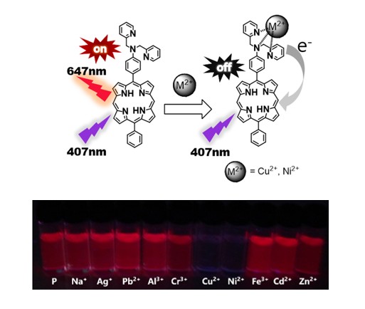
In this study, we report the synthesis of a novel fluorescence-based probe, 2,2′-dipicolylamine porphyrin (DPAP), designed for the detection of metal ions, particularly Cu2+. This sensor features a porphyrin fluorophore substituted with 2,2′-dipicolylamine (DPA) at the meso position. In the DPAP system, the porphyrin acts as the signaling unit, while the DPA moiety serves as the metal-recognition unit. Notably, the high selectivity of DPA for Cu2+, achieved through direct N–metal interactions, results in fluorescence quenching via intramolecular charge transfer (ICT) [5,29].
2. Results and Discussion
2.1. Synthesis and Characterization
To achieve high-performance fluorescence sensing, the fluorophores must have high quantum efficiency and photostability after metal binding. In addition, to overcome photon attenuation, fluorophores with long emission wavelengths, especially in the near-infrared (NIR) region, are preferred. Porphyrins have been used as fluorophores in fluorescence sensors for many metal ions. Crucially, porphyrin fluorophores have large Stokes shifts between the absorption (approximately 400 nm) and red/NIR emission [33,34,35]. Further, the DPA ligand forms stable complexes with many metal cations, resulting in high sensitivity [36]. In particular, we selected DPA because it can efficiently ligate metals in tridentate binding mode. Further, we assumed that, after coordination with a metal, the lone pair on nitrogen would not be available for conjugation, and fluorescence would be quenched. A previous study showed that DPA attached to a TPP moiety selectively reacted with Cd2+; however, we expected a different result from the DPP-based DPAP sensor because it has a twisted structure, unlike that of TPP [12].
The synthesis of compounds 1–3 is shown in Scheme 1 and follows a literature method [30,31]. DPAP (3) was synthesized by Lindsey cross-coupling condensation, combining two aldehydes and DPM, followed by oxidation [37]. The synthesis of DPAP was confirmed by MALDI-TOF MS and 1H-NMR spectroscopy (Figures S1 and S2).
2.2. Optical Properties
A stock solution of DPAP was prepared in THF, and all photochemical experiments were carried out at a concentration of 1.5 × 10⁻⁵ M. Free DPAP produced a prominent absorption band peak maximum at 407 nm (Soret band), attributed to S0–S2 absorption, and four Q bands at 506, 546, 583, and 639 nm, attributed to S0–S1 electronic transitions. In addition, DPAP yielded fluorescence emissions at 647 and 706 nm when excited at 407 nm in THF [29,38]. Regarding the DPP reference, without DPA, DPP exhibited a Soret band at 405 nm and four Q bands at 501, 534, 576 m, and 633 nm. DPP showed fluorescence emissions at 635 and 700 nm when excited at 405 nm in THF solution.
At the same concentration in THF, the absorption spectra of DPP and DPAP differed (Figure 1(a)): the Soret band of DPAP was broadened, and the peak was red-shifted compared with those of DPP, possible because of the electron-donating ability of the DPA moiety toward the porphyrin ring.
2.3. Effect of Metal Ions on the Absorption and Emission of DPAP
To observe the photophysical response to various metal cations, the UV–vis and fluorescence spectra were measured in the presence of perchlorate salts of Na+, Ag+, Cu2+, Ni2+, Cr3+, Pb2+, Al3+, Fe2+, Cd2+, and Zn2+.
As shown in Figure 2(a), the Soret band is sharpened, and a hyperchromic shift occurs on metal-ion binding. The spectra for each ion are presented in order of peak intensity. The slight blue-shift suggests that M2+/DPA coordination decreased the electron-donating ability of DPA, reducing the efficiency of ICT from the DPA subunits to the porphyrin fluorophore. In addition to this electron-withdrawing effect induced by the cations, M2+/DPA coordination via the nitrogen might distort the porphyrin ring, decreasing the conjugation of the DPA moiety and fluorophore.
Next, the emission spectra of the sensors were measured at an excitation wavelength of 407 nm. Figure 2(b) shows that the fluorescence intensity is reduced by the coupling of the porphyrin sensor with various metals. The spectra for each ion are presented in order of peak intensity. The metal ions are chelated by the DPA functional group, and this depends on the size of the ion and its binding affinity.
In Figure 2(a), there remain four distinct Q bands, indicating that the metal ions do not bind to the porphyrin ring, and this is consistent with their high affinity for DPP. As shown in Figure S3, the UV–vis and fluorescence spectra of DPP showed no changes in the presence of metal cations, indicating that the DPA groups are the binding sites.
In the case of fluorescence, a slight metal-ion-dependent change was observed; however, a significant quenching was observed in the presence of Cu2+, and Ni2+. This is because the bite angle of DPAP is commensurate with the size of these metal ions. Figure 2(c) shows visible-light images of DPAP under UV light (λem = 365 nm), revealing considerable fluorescence quenching in the presence of Cu2+, and Ni2+. Therefore, we focused on the use of the DPAP-based sensor for Cu2+, and Ni2+ detection.
The fluorescence quantum yields of DPAP after the addition of Cu2+, and Ni2+ were calculated using the reference standard TPP (QY = 0.11). The quantum yield of DPAP before the addition of metal cations was 0.1018, and after the addition of Cu2+, and Ni2+ the corresponding quantum yields of DPAP-Cu2+, and DPAP-Ni2+ were 0.0016, and 0.0033 respectively. Further, the fluorescence quenching percentages of DPAP after the addition of Cu2+, and Ni2+ were 99.1%, and 98.5% respectively. In terms of quenching and quantum yield changes, DPAP exhibited the highest reactivity towards Cu and Ni ions.
The titration of DPAP with increasing concentrations of Cu2+ led to a decrease in the emission intensity, as shown in Figure 3. No further changes were observed beyond the addition of 0.5 eq. The titration of DPAP with Ni2+ led to fluorescence quenching (Figure S4).
The LOD of DPAP for the two metal ions in THF was calculated from a linear fit of the maximum emission intensity at 647 nm and the quencher concentration (Figures S5–S6). The calculated LODs are 26.3, and 34.8 nM (≈ 2.0 ppb, R2 = 0.98), respectively, for Cu2+, and Ni2+ based on Eq. (1).
2.4. Binding Stoichiometry and Binding Affinity
To understand the binding behavior and determine the stoichiometry of the DPAP-Cu2+ complex, Job’s plot analysis was carried out [6] (Figures S7–S8). The changes in the emission intensity (I0 − I) at 647 nm versus the mole fraction of Cu2+ were measured while maintaining the concentrations of Cu2+ and DPAP at 1.5 × 10⁻⁵ M. The maximum change in emission intensity was observed at a molar fraction of approximately 0.5–0.6. In addition, in the case of Ni2+, the results show that the DPA-based sensor for Ni2+ metal ion form 1:1 metal–ligand complexes.
As shown in Figure 4, and Figure S9 near-linear Stern–Volmer plots were obtained at low quencher (metal ion) concentrations; these plots were used to investigate the “kinetics” of the quencher binding affinity. Linear fitting yielded a coefficient of determination (R2) of > 0.99, and the obtained Stern–Volmer constants were 1.39 × 10⁶, and 7.71 × 10⁵ M-1, respectively, for Cu2+, and Ni2+ indicating an increase in binding affinity in that order.
Next, interference (matrix) effects were investigated using metal cations proven to bind to the DPA moiety, such as Zn ions [12,13,39,40,41,42,43,44,45,46]. A comparative study was carried out by measuring at a low concentration (1 eq) mixture of Cu2+, and Ni2+ and a high concentration (10 eq) of the interfering metal mixture. As shown in Figure 5, Cu2+ completely quenched the fluorescence, unlike the other metals, and no interference was observed. Although Ni2+ quenched the fluorescence, interference was observed in the presence of the metal mixture, and the fluorescence intensity increased again because of its low binding affinities. These results clearly demonstrate the high specificity of our sensor for Cu2+, even in a complex matrix of metal ions.
To investigate the interference effect of each metal ions and metal ion mixtures on the detection ability of DPAP for Cu2+, and Ni2+ the fluorescence response was measured in the presence of other metal ions (Figure 5 and Figures S10–S11). Exposure of DPAP to a mixed metal-ion solution containing Na+, Ag2+, Pb2+, Cr3+, Al3+, Fe2+, Zn2+, and Cd2+ (10 equivalents each) does not significantly alter the fluorescence intensity, which remains comparable to that of the free DPAP. In contrast, addition of Cu2+ to the mixed-metal system, quenched the fluorescence of DPAP, highlighting the high selectivity of DPAP for Cu2+ even under competitive conditions. The addition of Ni2+ to the mixed-metal system results in partial quenching. In addition, DPAP has greater selectivity for Cu2+ than for Ni2+ ions as shown in Figures S10 and S11. Taken together these results highlight the Cu2+selective fluorescence quenching exhibited by DPAP, underscoring its potential utility as a highly selective fluorescent probe for Cu2+ detection.
3. Experimental
3.1. Materials
All chemicals for synthesis were purchased from commercial suppliers and used as received. Aniline, phosphorus(V) oxychloride, and all metal salts were purchased from Sigma–Aldrich. Hexadecyltrimethylammonium chloride, benzaldehyde, dipyromethane (DPM), and chloranil were purchased from TCI. Picolyl chloride hydrochloride was purchased from Acros Organics. N,N′-Dimethylformamide (DMF) and dichloromethane (DCM) were purchased from Samchun Company. Trifluoroacetic acid (TFA) was purchased from Daejung Corporation. Triethylamine (TEA) was purchased from Duksan Corporation.
3.2. Measurements
1H-NMR spectra were recorded at 600 MHz and are reported using the standard abbreviations: s: singlet, d: doublet, t: triplet, m: multiplet, and the coupling constants, J, are given in hertz. The chemical shifts are reported in parts-per-million (ppm), using tetramethylsilane (0 ppm) and CDCl3 as standards. UV−vis absorption and fluorescence emission spectra were recorded using a Shimadzu UV-2450 spectrometer and Hitachi F-7000 fluorescence spectrophotometer, respectively. The slit size for excitation and emission was 5 mm. The emission spectra were measured with excitation at 407 nm. All spectra were recorded at room temperature (r.t.) in a quartz cuvette having a path length of 10 mm.
A stock solution of DPAP was prepared in tetrahydrofuran (THF) at a concentration of 1.5 × 10⁻⁵ M for excitation and emission measurements and stored in a cold, dark place before use. To test the sensing performance and interference effects, various metal-ion solutions (Na+, Ag+, Cu2+, Ni2+, Cr3+, Pb2+, Al3+, Fe2+, Cd2+ and Zn2+ as perchlorate salts) were prepared in water. Column chromatography was performed on Merck silica gel (230–400 mesh).
3.3. DPAP Synthesis
Compounds 1 and 2 were synthesized following literature procedures [30,31]. Compound 2 (DPA aldehyde) (0.519 g, 1.712 mmol), benzaldehyde (0.18 mg, 1.712 mmol), and DPM (0.5 g, 3.424 mmol) were dissolved in dichloromethane (DCM; 600 mL), and the mixture was degassed for 10 min by sparging the solution with N2. After the addition of TFA (0.234 g, 0.6 eq), the resulting solution was stirred at room temperature for 6 h in the dark. To this reaction mixture, chloranil (1.05 g, 4.28 mmol) was added, and the reaction mixture was stirred for another 1 h at r.t. After reaction completion, the mixture was neutralized with TEA (4 mL), filtered through a Büchner funnel filled with silica gel, and washed with chloroform, completing the work-up process. The crude compound 3 (i.e., DPAP) was purified by silica-gel column chromatography using chloroform (0.5% v/v methanol) as the eluent, yielding a magenta solid in 18.8% yield. 1H-NMR (600 MHz, chloroform-d) δ = 10.281 (s, 2H, meso), 9.374 (d, 4H, J = 4.677 Hz, β), 9.179 (d, 2H, J = 4.402 Hz, β), 9.058 (d, 2H, J = 4.677 Hz, β), 8.7045 (d, 2H, J = 4.402 Hz, aromatic), 8.274−8.259 (m, 2H, aromatic), 8.083(d, 2H, J = 8.528 Hz, aromatic), 7.844−7.794 (m, 5H, aromatic), 7.5975 (d, 2H, J = 7.703Hz, aromatic), 7.297–7.285(m, 2H, aromatic), 7.161 (d, 2H, J = 8.528 Hz, aromatic), 5.126 (s, 4H, aliphatic), and -3.047 (s, 2H, NH).
Matrix-assisted laser desorption/ionization–time-of-flight mass spectrometry (MALDI-TOF-MS) measurements were conducted, and the weight of the target molecule (m/z) was calculated for C44H33N7 [M+ H]+: 660.283; found: 660.471.
3.4. Limit of Detection
The limit of detection (LOD) was calculated using fluorescence titration with Eq. (1).
LOD = 3σ/k
Here, σ is the standard deviation of the emission intensity of DPAP, and k is the slope of the emission intensity plotted against the concentration. To determine the signal-to-noise ratio, the emission intensity of pristine DPAP without metal ions was measured four times, and the standard deviation of the blank measurements was determined to be 0.221736. Three independent duplicate measurements of the emission intensity were performed in the presence of Ni2+, and Cu2+ and the average value of each intensity was plotted as a function of cation concentration to determine the slope.
3.5. Quantum Yield Measurements
The quantum yield was calculated using Eq. (2).
Here, QY and QYR are the quantum yields of the sample and reference, respectively, A and AR are the absorbances of the sample and reference, respectively, and I and IR are the areas of the emission peaks for the sample and reference, respectively. Tetraphenyl porphyrin (TPP), which has a quantum yield of 0.11, was used as a reference.
3.6. Binding Constants and Stoichiometry
The binding (Ksv) and quenching constants for the interaction of the metal cations with the probe were determined using Stern–Volmer plots, Eq. (3) (Keizer, 1983) [32].
Here, I0 is the initial emission intensity of DPAP before the addition of the quencher, I is the emission intensity at a given concentration of quencher Q ([Q]), and Ksv is the Stern–Volmer constant.
In addition, Job’s plots were constructed to determine the stoichiometry of the complexation process between the metal ions and dye ligand [6,9,32]. In the plots, the x-axis represents the mole fraction, which is the ratio of the cation concentration to the total concentration of DPAP and cations. The total molar concentrations of the probe and metal ion were constant (1.5 × 10⁻⁵ M), and the difference in the emission intensity difference (I0 − I) between DPAP and the DPAP–M2+ complex at 647 nm versus the mole fraction was measured.
4. Conclusions
A novel fluorescence-based sensor, DPAP, was successfully synthesized by coupling a DPA metal-recognition moiety to a porphyrin fluorophore for the detection of transition metals, with a primary focus on Cu2+. The sensor shows ‘turn-off’ fluorescence, where the addition of less than 1.0 molar equivalent amount of Cu2+ ions was sufficient to induce complete fluorescence quenching. The sensor achieved a limit of detection (LOD) of approximately 2.0 ppb for both Cu2+ and Ni2+, demonstrating its potential for trace-level monitoring. Our results from quantum yield and Stern–Volmer analyses revealed the quenching efficiency of Ni2+ < Cu2+, suggesting the high affinity of the DPAP probe for Cu2+ ions. Interestingly, DPAP exhibited selectivity for Cu2+ even in a mixture of metal ions. Altogether, our findings reveal that our porphyrin-based DPAP sensor is not only a robust tool for determining Cu2+ traces but is also a useful probe for detecting Cu2+ in complex aqueous environments.
Supplementary Materials
The following supporting information can be downloaded at the website of this paper posted on Preprints.org, Figure S1: MALDI-TOF MS of DPAP; Figure S2: 1H-NMR spectrum of DPAP; Figure S3: Absorption and emission spectra of DPP in the presence of various metal ions; Figures S4: Absorption and emission spectra of DPAP titration in the presence of Ni2+ ion; Figures S5–S6: LODs of DPAP; Figures S7–S8: Job’s plots of DPAP; Figures S9: Stern–Volmer plot of DPAP; Figures S10 and S11: Interference effects of different metal ions (10 eq).
Author Contributions
So-Hyun Shin: Methodology, Investigation and Synthesis. Jihyun Kim: Investigation and Synthesis. Hyungkyu Moon: Investigation and Synthesis. T. Sheshashena Reddy: Investigation, Characterization & Writing. Myung-Seok Choi: Conceptualization, Data curation, Funding acquisition, Resources, Project administration, Writing, Review, & Editing.
Funding
This work was supported by the Technology Innovation Program [No. 20026593 & No.00418987] funded by the Ministry of Trade, Industry & Energy (MOTIE, Korea). This paper was supported by Konkuk University Researcher Fund in 2024.
Institutional Review Board Statement
Not applicable.
Informed Consent Statement
Not applicable.
Data Availability Statement
All data and material described in this work are available in this article or in the Supplementary Materials.
Conflicts of Interest
The authors declare no conflicts of interest.
References
- Sie, Y.W.; Li, C.L.; Wan, C.F.; Yan, H.; Wu, A.T. A novel fluorescence sensor for dual sensing of Hg2+ and Cu2+ ions. J. Photochem. Photobiol. A Chem. 2018, 353, 19–25. [Google Scholar] [CrossRef]
- Nekouei, F.; Nekouei, S. Determination of copper, nickel and cobalt in water and food samples by FAAS after separation and preconcentration using multiwalled carbon nanotubes modified by methyl-(2-pyridyl) ketone oxime. J. Anal. Chem. 2014, 8, 138–145. [Google Scholar]
- Neupane, L.N.; Oh, E.T.; Park, H.J.; Lee, K.H. Selective and sensitive detection of heavy metal ions in 100% aqueous solution and cells with a fluorescence chemosensor based on peptide using aggregation-induced emission. Anal. Chem. 2016, 88, 3333–3340. [Google Scholar] [CrossRef]
- Momidi, B.K.; Tekuri, V.; Trivedi, D.R. Multi-signaling thiocarbohydrazide-based colorimetric sensors for selective recognition of heavy metal ions in aqueous medium. Spectrochim. Acta A 2017, 180, 175–182. [Google Scholar] [CrossRef]
- Zhou, X. A highly selective fluorescent sensor for distinguishing cadmium from zinc ions based on a quinoline platform. Inorg. Chem. 2012, 51, 9226–9231. [Google Scholar] [CrossRef] [PubMed]
- Zhang, X.; Wang, R.; Fan, C.; Liu, G.; Pu, S. A highly selective fluorescent sensor for Cd2+ based on a diarylethene with a 1,8-naphthyridine unit. Dyes Pigments 2017, 139, 208–217. [Google Scholar] [CrossRef]
- Cheng, T. Red-emission fluorescent probe sensing cadmium and pyrophosphate selectively in aqueous solution. Org. Lett. 2011, 13, 3656–3659. [Google Scholar] [CrossRef]
- Baslak, C.; Kursunlu, A.N. A naked-eye fluorescent sensor for copper(II) ions based on a naphthalene-conjugated BODIPY dye. Photochem. Photobiol. Sci. 2018, 17, 1091–1097. [Google Scholar] [CrossRef]
- Chae, J.B. Highly sensitive dansyl-based chemosensor for detection of Cu2+ in aqueous solution and zebrafish. ACS Omega 2019, 4, 12537–12543. [Google Scholar] [CrossRef]
- Tian, Z.; Cui, S.; Pu, S. A highly selective fluorescent sensor for dual detection of Zn2+ and F− based on a diarylethene. Tetrahedron Lett. 2016, 57, 2703–2707. [Google Scholar] [CrossRef]
- Chen, X. Aggregation-induced emission enhancement-based ratiometric fluorescent sensor for detecting trace uranyl ion and application in living-cell imaging. J. Lumin. 2017, 186, 301–306. [Google Scholar] [CrossRef]
- Huang, W.B. A porphyrin-based fluorescent probe for optical detection of toxic Cd2+ ion in aqueous solution and living cells. Dyes Pigments 2017, 143, 427–435. [Google Scholar] [CrossRef]
- Diana, R. Data on a real-time tripodal colorimetric/fluorescence sensor for multiple target metal ions. Data Brief 2018, 19, 2119–2125. [Google Scholar] [CrossRef]
- Zhao, Q. A highly selective on/off fluorescence sensor for cadmium(II). Inorg. Chem. 2011, 50, 10041–10046. [Google Scholar] [CrossRef] [PubMed]
- Surjeet, S. 2-(2,2-Bis-benzylamino-1-cyano-vinyl)-benzonitrile: A Selective Turn-off Fluorescent Cu2+ Sensor. Chemistry Select 2016, 1, 2576–2580. [Google Scholar]
- Li, Z.X. Fluoranthene-based pyridine as a fluorescent chemosensor for Fe3+. Inorg. Chem. Commun. 2011, 14, 1656–1658. [Google Scholar] [CrossRef]
- Han, A.; Liu, X.; Prestwich, G.D.; Zang, L. Fluorescent sensor for Hg2+ detection in aqueous solution. Sens. Actuators B 2014, 198, 274–277. [Google Scholar] [CrossRef]
- Narayanaswamy, N.; Govindaraju, T. Aldazine-based colorimetric sensors for Cu2+ and Fe3+. Sens. Actuators B 2012, 161, 304–310. [Google Scholar] [CrossRef]
- Bagheri, S.; Squitti, R.; Haertlé, T.; Siotto, M.; Saboury, A.A. Role of copper in the onset of Alzheimer’s disease compared to other metals. Front. Aging Neurosci. 2018, 9, 1–15. [Google Scholar] [CrossRef] [PubMed]
- He, X. A BODIPY-based colorimetric and fluorometric dual-mode chemosensor for Hg2+ and Cu2+. Sens. Actuators B 2014, 192, 29–35. [Google Scholar] [CrossRef]
- Kang, H.; Fan, C.; Xu, H.; Liu, G.; Pu, S. A highly selective fluorescence switch for Cu2+ and Fe3+ based on a diarylethene with a triazole-linked rhodamine 6G unit. Tetrahedron 2018, 74, 4390–4399. [Google Scholar] [CrossRef]
- Jin, X. Dual-functional probe based on rhodamine for sequential Cu2+ and ATP detection in vivo. Spectrochim. Acta A 2018, 204, 657–664. [Google Scholar] [CrossRef]
- Shinde, R.G. Fluorescence off–on signalling of esculetin in the presence of copper and thiol: implications in cellular thiol sensing. Photochem. Photobiol. Sci. 2018, 17, 1197–1205. [Google Scholar] [CrossRef]
- Tang, L. Rapid and highly selective relay recognition of Cu(II) and sulfide ions by a benzimidazole-based fluorescent sensor in water. Sens. Actuators B 2013, 185, 188–194. [Google Scholar] [CrossRef]
- Hu, S.; Zhang, S.; Hu, Y.; Tao, Q.; Wu, A. A selective pyrazoline-based fluorescent chemosensor for Cu2+ in aqueous solution. Dyes Pigments 2013, 96, 509–515. [Google Scholar] [CrossRef]
- Tang, L. Relay recognition of Cu2+ and S2− in water by a benzimidazole-derived fluorescent sensor via ESIPT modulation. Spectrochim. Acta A 2014, 122, 656–660. [Google Scholar] [CrossRef]
- Fan, J. A fluorescent ratiometric chemodosimeter for Cu2+ based on TBET and its application in living cells. Org. Lett. 2013, 15, 492–495. [Google Scholar] [CrossRef] [PubMed]
- Fu, Y.; Fan, C.; Liu, G.; Pu, S. A colorimetric and fluorescent sensor for Cu2+ and F− based on a diarylethene with a naphthalimide Schiff-base unit. Sens. Actuators B 2017, 239, 295–303. [Google Scholar] [CrossRef]
- Frangioni, J.V. In vivo near-infrared fluorescence imaging. Curr. Opin. Chem. Biol. 2003, 7, 626–634. [Google Scholar] [CrossRef] [PubMed]
- Sujatha, V. Use of tetra-ammonium tetrakis(4-sulphonatophenyl) porphyrin for Pseudomonas and Bacillus cell imaging. Int. J. Anal. Chem. 2010, 697528. [Google Scholar]
- Guo, B. Decoration of porphyrin with tetraphenylethene: converting aggregation-caused quenching to aggregation-induced emission enhancement. J. Mater. Chem. B 2016, 4, 4690–4698. [Google Scholar] [CrossRef] [PubMed]
- Shiraishi, Y.; Matsunaga, Y.; Hirai, T. Selective colorimetric sensing of Co(II) in aqueous media using a spiropyran-amide-dipicolylamine linkage. Chem. Commun. 2012, 48, 5485–5487. [Google Scholar] [CrossRef]
- Ahn, S.H. Novel cobalt(II) complexes containing N,N-di(2-picolyl)amine-based ligands: synthesis, characterization and application in methyl methacrylate polymerisation. J. Mol. Struct. 2016, 1113, 24–31. [Google Scholar] [CrossRef]
- Song, Y. Cadmium(II) complexes containing N′-substituted N,N-di(2-picolyl)amine ligands. J. Organomet. Chem. 2015, 783, 55–63. [Google Scholar] [CrossRef]
- Jeong, K. Diisopropyl fluorophosphate degradation activity using transition-metal dipicolylamine complexes. Appl. Organomet. Chem. 2018, 32, e4041. [Google Scholar] [CrossRef]
- Bussey, K.A. Synthesis, X-ray crystallography and catalytic activity of bis(2-pyridylmethyl)amine copper complexes in ATRA reactions. Polyhedron 2016, 114, 256–267. [Google Scholar] [CrossRef]
- Götzke, L. Nickel(II) and zinc(II) complexes of N-substituted di(2-picolyl)amine derivatives. Polyhedron 2011, 30, 708–714. [Google Scholar] [CrossRef]
- Milaeva, E.R. Redox-active metal complexes with dipicolylamine-ferrocenyl ligands. J. Organomet. Chem. 2017, 839, 60–70. [Google Scholar] [CrossRef]
- Azuma, Y. Dipicolylamine as a structural switching element for helical peptides. Org. Biomol. Chem. 2012, 10, 6062–6068. [Google Scholar] [CrossRef] [PubMed]
- Praktikum, P.; Quenching, F. Fluorescence quenching studies. Phys. Prakt. 2016, 1, 1–14. [Google Scholar]
- Kumari, N. Selective ppb-level detection of Cu2+ and Hg2+ using a micellar medium and application in cell imaging. ChemPlusChem 2014, 79, 1643–1652. [Google Scholar] [CrossRef]
- Zhang, L. Fluorescent binary ensemble based on pyrene derivative and SDS assemblies for discriminating metal ions. ACS Sens. 2017, 2, 1821–1830. [Google Scholar] [CrossRef]
- Ncube, P. Fluorescent sensing and determination of mercury(II) ions in water. Water SA 2014, 40, 175–182. [Google Scholar] [CrossRef]
- Milaeva, E.R. Metal complexes with functionalised dipicolylamine ligands containing antioxidant moieties. Dalton Trans. 2013, 42, 6817–6828. [Google Scholar] [CrossRef]
- Lindsey, J.S. Rothemund and Adler–Longo reactions revisited: synthesis of tetraphenylporphyrins under equilibrium conditions. J. Org. Chem. 1987, 52, 827–836. [Google Scholar] [CrossRef]
- Quantum Bioapplications, An introduction to fluorescence spectroscopy. Chem 2007, 312.
Scheme 1.
Synthesis of DPAP (3). Reaction conditions: (a) Hexadecyltrimethylammonium chloride, water, 5 N NaOH/r.t, 24 h; (b) POCl3, DMF/90 ℃, 2 h; (c) DCM, TFA/r.t, 6 h; chloranil/1 h; TEA/15 min.
Scheme 1.
Synthesis of DPAP (3). Reaction conditions: (a) Hexadecyltrimethylammonium chloride, water, 5 N NaOH/r.t, 24 h; (b) POCl3, DMF/90 ℃, 2 h; (c) DCM, TFA/r.t, 6 h; chloranil/1 h; TEA/15 min.

Figure 1.
Absorption (a) and emission (b) (λex = 407 nm) spectra of DPP and DPAP in THF (1.5 × 10⁻⁵ M).
Figure 1.
Absorption (a) and emission (b) (λex = 407 nm) spectra of DPP and DPAP in THF (1.5 × 10⁻⁵ M).

Figure 2.
Absorption (a) and emission (b) spectra (λex = 407 nm) and images (c) under UV light (λem = 365 nm) of DPAP (1.5 × 10⁻⁵ M) in the presence of various metal ions (1.5 × 10⁻⁴ M, 10 eq).
Figure 2.
Absorption (a) and emission (b) spectra (λex = 407 nm) and images (c) under UV light (λem = 365 nm) of DPAP (1.5 × 10⁻⁵ M) in the presence of various metal ions (1.5 × 10⁻⁴ M, 10 eq).

Figure 3.
Absorption (a) and emission (b) (λex = 407 nm) spectra of DPAP (1.5 × 10⁻⁵ M) with increase in Cu2+ concentration (1 eq = 1.5 × 10⁻⁵ M).
Figure 3.
Absorption (a) and emission (b) (λex = 407 nm) spectra of DPAP (1.5 × 10⁻⁵ M) with increase in Cu2+ concentration (1 eq = 1.5 × 10⁻⁵ M).

Figure 4.
Stern–Volmer plot of the quenching emission at 647 nm on the interaction of DPAP with Cu2+ (Ksv = 1.39 × 10⁶ M-1).
Figure 4.
Stern–Volmer plot of the quenching emission at 647 nm on the interaction of DPAP with Cu2+ (Ksv = 1.39 × 10⁶ M-1).

Figure 5.
Fluorescence emission response of DPAP (1.5 × 10⁻⁵M) in the presence of Cu2+, and Ni2+ (1 eq) and interference effects in the presence of a metal mixture (10 eq).
Figure 5.
Fluorescence emission response of DPAP (1.5 × 10⁻⁵M) in the presence of Cu2+, and Ni2+ (1 eq) and interference effects in the presence of a metal mixture (10 eq).

Figure 6.
Emission spectra (λex = 407 nm) of the DPAP-Cu2+ complex (1.5 × 10⁻⁵ M) in the presence of EDTA.
Figure 6.
Emission spectra (λex = 407 nm) of the DPAP-Cu2+ complex (1.5 × 10⁻⁵ M) in the presence of EDTA.

Disclaimer/Publisher’s Note: The statements, opinions and data contained in all publications are solely those of the individual author(s) and contributor(s) and not of MDPI and/or the editor(s). MDPI and/or the editor(s) disclaim responsibility for any injury to people or property resulting from any ideas, methods, instructions or products referred to in the content. |
© 2026 by the authors. Licensee MDPI, Basel, Switzerland. This article is an open access article distributed under the terms and conditions of the Creative Commons Attribution (CC BY) license (http://creativecommons.org/licenses/by/4.0/).
Copyright: This open access article is published under a Creative Commons CC BY 4.0 license, which permit the free download, distribution, and reuse, provided that the author and preprint are cited in any reuse.


