Submitted:
08 April 2026
Posted:
13 April 2026
You are already at the latest version
Abstract
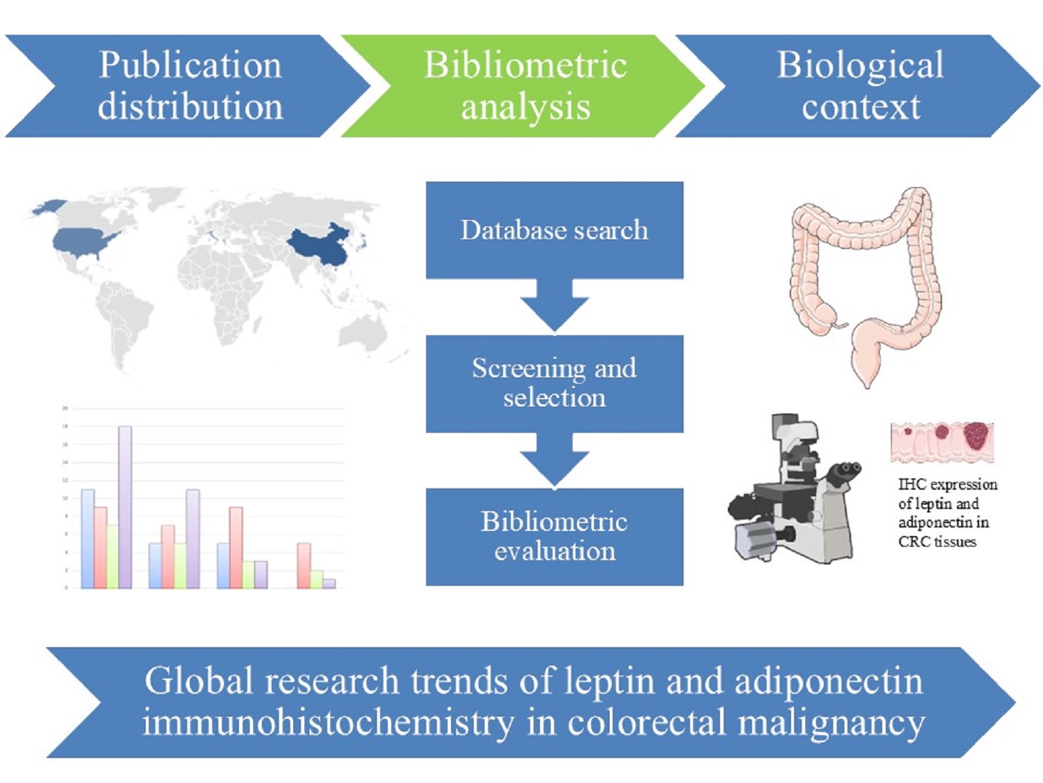
Background: Leptin and adiponectin play a key role in obesity-associated malignancy, particularly in colorectal cancer (CRC). Immunohistochemical (IHC) evaluation of these adipokines may offer valuable prognostic insights in CRC. This study aimed to analyze global publication trends and summarize current knowledge on the potential of these hormones as IHC biomarkers in CRC. Methods: A problem-oriented bibliometric analysis, including publications from 2000 to 2025 was performed across MEDLINE and Scopus databases. In parallel, a literature review was conducted to present the biological and clinical relevance of these adipokines in CRC. Results and Discussion: A total of 101 publications were identified. Scopus indexed substantially more studies than MEDLINE. The journals Cancer Research, Journal of BUON, Cells, BMC Cancer, and Asian Pacific Journal of Cancer Prevention were identified as the core journals publishing on this topic over the 25-year period. Leading countries were China and USA. A review of the literature showed that adiponectin is a promising prognostic marker, while leptin appears to be a better indicator of disease progression. Conclusions: IHC research on leptin and adiponectin in CRC is a promising but still underexplored area. Their integration with routine molecular assessment has the potential to improve patient stratification.
Keywords:
immunohistochemical biomarkers
; colorectal cancer
; leptin
; adiponectin
; publication trends
1. Introduction
Colorectal cancer (CRC) is the third most commonly diagnosed malignancy globally [1,2] and the second leading cause of cancer-related deaths worldwide in 2022 [2]. The global burden of CRC is projected to increase by 60% by 2030 [3]. Moreover, results from the Global Burden of Disease Study, 2021 demonstrate that during the period from 1990 to 2021, there is an increase of 130.97% in disability-adjusted life years and of 130.24% in deaths associated with early-onset colorectal cancer as well as 133.32% and of 139.71% associated with late-onset colorectal cancer, respectively [4].
Data from the Global Cancer Observatory (GLOBOCAN) database demonstrates that in 2022, there are 1 926 425 global CRC incident cases and 904 019 deaths [5]. If CRC incidence rates remain unchanged, the global new cases are projected to increase by 22.51% and to reach 2.36 million by 2050. This alarming trend underlines the emergent needs of identifying new biomarkers for reliable and precise diagnosis of this highly heterogeneous malignancy. Adipokines, particularly leptin and adiponectin, are well known master modulators of colon carcinogenesis [6], especially tumorigenesis that is closely linked to obesity [7]. Leptin possesses robust proliferative, pro-inflammatory and proangiogenic activities promoting tumorigenesis [8], while adiponectin usually exerts strong anti- tumorigenic characteristics [9]. In addition, the leptin/adiponectin ratio is a promising indicator for the colorectal pathogenesis and progression [2]. All these findings suggest that dysregulation in the expression of these two adipokines in colorectal tissue samples could be a promising and reliable biomarker for CRC.
Currently, TNM classification is the basis for predicting patient prognosis in CRC [10]. For better patients’ stratification, a decade ago a transcriptome-based system was invented, the consensus molecular subtype (CMS) classification. It classifies colorectal tumors into four subtypes with different molecular and genetic characteristics: CMS1 (immune), CMS2 (canonical), CMS3 (metabolic), and CMS4 (mesenchymal) [11]. CMS2 subtype defines the best overall survival among the patients while CMS4 subtype determines the worst prognosis. This classification is the most popular transcriptome- based system [12], but it is too expensive for routine clinical applications. That is why alternative and clinically useful immunohistochemical (IHC) methods are needed. One such IHC classifier using five biomarkers, FRMD6, ZEB1, HTR2B, and CDX2, was proposed by Trinh et al. [13]. The main limitation of this classifier was that it was unable to distinguish CMS2 tumors from CMS3 tumors. Thus, in 2021, Lee and his colleagues proposed the inclusion of β-catenin staining in this IHC panel which allowed the differentiation of these two subtypes [14]. All these findings demonstrate the potential clinical utility of IHC-based biomarkers for better patient stratification.
Despite these advances in molecular classification, the clinical implementation of transcriptome-based subtyping remains restricted by cost and technical complexity, underscoring the continuing demand for accessible IHC-based prognostic tools applicable to formalin-fixed, paraffin-embedded (FFPE) tissue. In this context, adipokines, leptin and adiponectin in particular, have attracted growing interest as candidate IHC biomarkers, given their mechanistic involvement in obesity-driven colorectal carcinogenesis and the feasibility of their detection using standard IHC protocols. To date, however, no comprehensive synthesis of the available evidence on the clinicopathological correlates and prognostic significance of leptin and adiponectin IHC expression in CRC has been published. An illustrative example of such approach can be found in the emerging field of adipobiology.The results from a retrospective problem-oriented search in MEDLINE and EMBASE databases available via OVID between 1980 and 2007 using several essential terms as key words such adipose tissue, adipokine(s), adipocytokine(s), adiponectin, and leptin reveal that 11 papers only under the term of ‘adipobiology’ have been abstracted during this period [15]. Some aspects of the dynamic growth of the world publication output in basic, transitional and clinical adipobiology as compared with some other recently emerging topics are demonstrated and certain elements of adipology future development from a bibliometric point of view are traced.
Another example illustrating the application of bibliometric methodology is provided by studies on breast cancer immunohistochemistry. The bibliometric analysis of the publications abstracted inWoS, MEDLINE and BIOSIS as well as in Scopus during the period between 2003 and 2018 within a retrospective problem-oriented based search reveals several essential patterns of the dynamic science institutionalization on breast cancer IHC [16]. There are 1187 publications in 288 journals abstracted in WoS, 776 publications in 140 journals abstracted in BIOSIS, 711 publications in 156 journals abstracted in Scopus, and 616 publications in 234 journals abstracted in MEDLINE. The complex bibliometric methodology presenting with the capacities of the constellation of bibliometric indicators can be purposefully used for integrated assessment of science institutionalization under the conditions of internationalization and for timely identification of the essential patterns of scientific advances in hot topics.
In addition, a retrospective, problem-oriented search was performed in June 2017 to assess the institutionalization of research on colorectal tumour markers [17]. The search covered the period 1987–2016 and included the WoS, Scopus, MEDLINE, and BIOSIS databases. A total of 497 publications were identified in Web of Science, 427 in Scopus, 370 in BIOSIS, and 368 in MEDLINE. The retrieved articles were published in multiple languages across 136–197 journals. Furthermore, they were authored by researchers from 37–50 countries, depending on the database. All these findings provide a broad overview of the global research activity in this field and may be of particular interest to coloproctologists, science policy managers, and journal editors, especially in smaller countries seeking greater international visibility.
The objective of the present study was to present some essential results from the publication trends devoted to the application of IHC for the assessment of leptin and adiponectin as prognostic biomarkers in CRC. Our goal was to synthesize current data on adipokines as potential prognostic markers in CRC applicable through IHC, using an integrated approach involving descriptive bibliographic analysis and literature review.
2. Materials and Methods
2.1. Databases
The bibliometric analysis data in the current research was retrieved from MEDLINE and Scopus databases. These databases were selected because of their extensive coverage of peer-reviewed literature related to IHC studies in oncology, which ensures a comprehensive data set for evaluating the diagnostic and prognostic implications of cancer-related biomarkers.
2.2. Search Strategy, Inclusion and Exclusion Criteria
In October 2025, a retrospective search focused on the subject was performed among articles published between 2000 and 2025, using the MEDLINE and Scopus databases. The publications dealing with these two adipokines, leptin and adiponectin as IHC prognostic biomarkers in CRC patients were included in the analysis. Both databases were searched independently using the following Boolean search string: (“immunohistochemistry OR “immunohistochemical biomarkers” OR “IHC markers”) AND (“colorectal cancer” OR “colorectal malignancy” OR “colorectal carcinoma” OR “CRC”) AND (“leptin” OR “adiponectin” OR “adipokines” OR “adipocytokines|). The search was applied across title, abstract, and keyword fields, with no additional database-specific field restrictions. Results were limited to English-language publications and to original research articles, excluding reviews, editorials, letters, conference abstracts, book chapters, and non-full-text records. The inclusion criteria required: peer-reviewed original research articles reporting IHC assessment of leptin and/or adiponectin in human CRC tissue samples. The exclusion criteria comprised: animal or in vitro studies; studies reporting serum or plasma biomarker levels without tissue IHC data; review articles and meta-analyses; conference abstracts and non-peer-reviewed records; and duplicate entries identified across databases.
The low number of identified publications (n = 101) reflects the focused scope of the search strategy. It was deliberately designed to include studies reporting IHC assessment of leptin and adiponectin specifically in colorectal cancer tissues. This specificity results in a smaller pool compared to broader searches for adipokines.
Citations per article were also retrieved from each database. The following bibliometric parameters were comparatively assessed across both databases: (i) annual and cumulative publication output; (ii) distribution of publications across journal titles; and (iii) geographic distribution of contributing authors by country of affiliation. Citation counts per article were additionally retrieved where available.
2.3. Literature Search
Scopus and Medline datasets were searched between September and December 2025 for English full-text articles (2000–2025) on the correlation between IHC expression of leptin and adiponectin and colorectal cancer in patients. Key words include “colorectal cancer,” “adipokines,” “adipocykines,” “immunohistochemistry,” “adiponectin,” “leptin,” “obesity,” and “biomarkers”. This research was focused on the association of protein expression and clinicopathological parameters.
3. Results
3.1. Annual Publication Output
The distribution of publications during the investigation period shows a growing interest in IHC studies of leptin and adiponectin in CRC. 45 publications on leptin were retrieved in Scopus and 20 in MEDLINE. In Scopus, the largest number of articles were published in 2021–2025 (40.0%, n=18), while the entries in MEDLINE are most prevalent in 2011–2015 (45.0%, n=9) (Table 1).
For adiponectin, 28 publications were indexed in Scopus and 8 in MEDLINE, with the largest percentage again in 2021–2025 (39.28%, n=11) in Scopus, while publications in MEDLINE reached their peak in 2011–2015 (62.5%, n=5) (Table 2). In general, the results indicate a relatively recent increase in scientific publications, particularly in the literature indexed in Scopus.
The publication output demonstrated a generally increasing trend, indicating a growing scientific interest in adipokine-related biomarkers in colorectal cancer research. An initial phase characterized by several sporadic publications is observed during the early years (2000-2010). After 2021 the publication activity related to IHC studies of leptin and adiponectin in CRC increased significantly. This finding aligns with the expanding interest in the role of obesity in malignant transformation. The most productive period was 2021-2025. It is characterized by 33 publications associated with the current topic, accounting for 32.67% of the total output. The highest publication activity is observed in the nearest period (2021–2025). This trend is particularly notable for studies related to leptin in Scopus. Publications related to adiponectin show a similar but less prominent trend. In contrast, MEDLINE indexes fewer studies, with most publications concentrated between 2001 and 2015. The combined annual dynamics of publications in Scopus and MEDLINE are illustrated in Figure 1.
The number and relative proportion of publications on leptin and adiponectin abstracted in Scopus were 2.25-fold and 3.5-fold higher, respectively, than those indexed in MEDLINE. These findings highlight substantial differences in database coverage.
3.2. “Core” Journals Publishing on Leptin in Colorectal Malignancy
A total of 45 publications indexed in Scopus and 20 publications indexed in MEDLINE were identified for leptin-related IHC studies in CRC. The distribution of articles across journals demonstrated a marked dispersion pattern. In the Scopus dataset, three journals, Cancer Research, Journal of BUON, and Cells represent 4.44% of the total output per journal (Table 3). Collectively, these three journals accounted for 6 of 45 publications (13.33%). In MEDLINE, two journals, Cancer Research and Journal of BUON correspond to 10.00% of the indexed corpus per journal and 20.00% cumulatively. The journal Cells did not appear among MEDLINE-indexed records.No journal published more than two articles in either database, indicating the absence of a dominant publication outlet. There is an overlap of Cancer Research and Journal of BUON between Scopus and MEDLINE datasets.
3.3. “Core” Journals Publishing on Adiponectin in Colorectal Malignancy
For adiponectin, 28 publications were identified in Scopus and 8 in MEDLINE. The distribution across journals was more dispersed than for leptin (Table 4). In Scopus, BMC Cancer was the only journal contributing two publications (7.14%). In MEDLINE, Asian Pacific Journal of Cancer Prevention accounted for two publications, representing 25.00% of the indexed literature. No journal overlap was observed between databases for adiponectin-related studies.The higher proportional share observed in MEDLINE reflects the smaller corpus size rather than a stronger concentration effect.
Comparison between Scopus and MEDLINE reveals differences in the centrality patterns of journals. Publications related to leptin show partial agreement between the databases, while studies related to adiponectin show a complete difference in the identified core journals. In addition, all identified core journals are oncology related. The leading journals represent translational cancer research (Cancer Research), clinical oncology (Journal of BUON), molecular and cellular biology (Cells), and cancer focused investigations (BMC Cancer and Asian Pacific Journal of Cancer Prevention)
3.4. Geographic Distribution of Leptin and Adiponectin Related Publications
The geographic distribution of authors contributing to leptin-related immunohistochemical studies in CRC is presented in Table 5.
In Scopus dataset, China ranked first with 10 publications (22.22%), followed by USA with 7 publications (15.56%), and Japan with 5 publications (11.11%). The UK contributed 4 publications (8.89%), while Italy and South Korea each accounted for 3 publications (6.67%). Together, the top three countries (China, USA, Japan) generated 48.89% of the total Scopus indexed output, indicating moderate geographic concentration. In MEDLINE, China and South Korea shared first position with 3 publications each (15.00%), followed by France (6.67%) and Montenegro (10.00%). These findings are illustrated in Figure 2.
The distribution of countries contributing to adiponectin-related studies (Table 6) reveals similar but slightly more regionally concentrated patterns.
In Scopus, China again ranked first with 7 publications (25.00%), followed by the USA with 5 publications (17.86%) and Japan with 4 publications (14.29%). Italy contributed 3 publications (10.71%). Collectively, the top three countries accounted for 57.15% of total Scopus indexed output, indicating stronger concentration compared to leptin-related studies. In MEDLINE, South Korea and Turkey each contributed 2 publications (25.00%), while Greece accounted for 14.29%. This data is illustrated in Figure 3.
Notably, adiponectin-related research demonstrates a pronounced contribution from East Asia and parts of Southern Europe. Unlike leptin research, Western European countries and the United Kingdom are less prominently represented in this subset.
3.5. Literature Review on IHC Biomarkers in CRC with Focus on Leptin and Adiponectin
CRC is a heterogeneous disease that is strongly affected by metabolic and genetic factors. Genetic factors have been well explored over the years. To emphasize the association between metabolic dysregulation and its role in initiation, progression, and prognosis in CRC, we summarized current knowledge into next subsections.
3.5.1. Mechanistic Role of Leptin in CRC
Recent work links obesity-related hyperleptinemia to several cancer “hallmarks” in colorectal cancer (CRC), but data also show context-dependent effects. Numerous studies have reported that leptin and its receptor (LEPR/ObR) are frequently overexpressed in CRC tissue and adenomas, driving mitogenic and anti-apoptotic effects via PI3K/AKT/mTOR, JAK2/STAT3 and MAPK/ERK pathways [18,19,20,21]. In addition, increased ObR and STAT3 activation appear in adenomas and are more pronounced in cancers, supporting a role in early and late carcinogenesis [22]. Leptin promotes proliferation, survival, and anchorage-independent growth of colorectal tumor stem cells, implying support of a cancer stem cell compartment. Moreover, recent study indicates that Wnt signaling upregulates ObR. This leads to active leptin–ObR communication that supports tumor growth via STAT3 without altering β-catenin localization [23]. Such activation usually is detected mainly after tumor initiation.
Furthermore, leptin enhances VEGF/VEGFR2 expression and angiogenesis through PI3K, JAK2/STAT3 and ERK1/2, and induces cytokines (CXCL1, CCL2, TGF-β), COX-2, and lipid droplet formation in intestinal epithelial cells via mTOR, linking obesity-driven inflammation to CRC promotion [21]. In addition, obesity-related insulin resistance, hyperinsulinemia, oxidative stress, and altered leptin/adiponectin balance converge on JAK/STAT and PI3K/AKT/mTOR, remodeling CRC signaling [24].
3.5.2. Mechanistic Role of Adiponectin in CRC
In colorectal carcinoma, adiponectin reduces proliferation, adhesion, and invasion, largely via AMPK activation, with downstream effects on mTOR and ERK1/2 signaling and caspase-3–mediated apoptosis [25]. Similar AMPK/mTOR-mediated anti-proliferative effects are emphasized as central to its antineoplastic role in CRC [26]. In addition, adiponectin alters cancer metabolism by downregulating glycolysis-related genes, and by reshaping lipid metabolism, limiting energy supply for tumor growth [25]. Moreover, recent data indicates that low adiponectin promotes insulin resistance and increased IGF-1, favoring proliferation and VEGF upregulation. At the same time, optimal levels of adiponectin inhibit the PI3K/AKT signaling pathway, thereby reducing carcinogenesis, adhesion, and migration [27]. Adiponectin also generally suppresses NF-κB–driven pro-inflammatory cytokines and VEGF, contributing to anti-angiogenic and anti-tumor actions [28].
Data from epidemiologic studies have shown that higher levels of circulating adiponectin are generally associated with lower CRC risk, strengthening its protective profile [24]. In is important to mention that current data also show context-dependent and even opposing effects of this adipokine in CRC. Supporting this, recent research reported that adiponectin may reprogram tumor-associated macrophages toward an M2-like phenotype and increase IL-8 expression, promoting angiogenesis, tumor growth, and recurrence in CRC [29]. In addition, it was reported that in specific colon cancer cell contexts, adiponectin can stimulate growth and inflammatory cytokine secretion via cAMP/PKA, and its effects may vary depending on glucose availability [26]. These opposing effects underpin the bifurcated role of adiponectin in CRC, influenced by tumor stage, microenvironmental cytokines, and metabolic status.
3.5.3. Immunohistochemical Expression Patterns of Leptin and Adiponectin in CRC
Current studies have demonstrated that altered tissue expression by IHC methods are being explored as diagnostic and prognostic markers in CRC. IHC and molecular studies show that several adipokines, especially leptin, its receptors, and adiponectin and its receptors, are differentially expressed between normal mucosa, adenomas, and CRC. Moreover, the specific expression patterns of these two adipokines show significant correlation with tumor grade, stage and overall survival of the patients. Data reported in the last 10 years was summarized in Table 7 and Table 8.
Data presented in the tables show that multiple IHC studies from recent years have reported that both LEP and LEPR were present at high intensity in cancerous tissues while normal adjacent mucosa demonstrated weak staining [33,35]. Regarding adiponectin and its receptors (AdipoR1, AdipoR2 and T-cadherin) expression patterns in most of the studies are documented significantly reduced levels in CRC tissues compared to the normal colon tissue [44,46]. Furthermore, in 2024 Parmesh and colleagues reported that positive leptin receptor expression and negative ADIPORs have shown strong correlation with larger tumor size, lymph node metastasis, and advanced stage. So, they proposed positive leptin combined with negative adiponectin receptor staining as a marker of metastatic risk [38].
In summary, IHC studies in CRC consistently show frequent and stronger leptin/leptin receptor expression in tumor tissue compared with normal mucosa, often associated with larger, more advanced, and node-positive tumors. In contrast, loss or low adiponectin receptor expression is linked to nodal and distant metastasis and higher stage and grade, supporting an adverse pattern of leptin up-regulation and adiponectin pathway down-regulation in more aggressive colorectal cancers.
All these findings indicate that leptin acts primarily as a factor in progression and metastasis, while adiponectin demonstrates a protective role in tumorigenesis. In summary, adiponectin and its receptors, AdipoRs expression patterns particularly support their use as prognostic and possibly therapeutic targets, while leptin and its receptors, LEPRs, are more promising as progression markers.
3.5.4. Comparison with Other IHC Biomarkers in CRC
The clinical utility of adipokine-based biomarkers should be considered in the context of established IHC and molecular classification systems. Current CRC diagnostics rely on markers such as Ki-67, p53, β-catenin, VEGF and CDX2, as well as transcriptomic classifications such as the consensus molecular subtypes (CMS).
Recent advances in the field of clinically relevant IHC biomarkers in CRC are summarized in Table 9.
4. Discussion
The present study combines descriptive bibliometric analysis with a focused literature review to provide a comprehensive overview of IHC studies investigating leptin and adiponectin in CRC. This dual approach enables both quantitative assessment of research activity and qualitative interpretation of the biological and clinical relevance of adipokine-related biomarkers. To the best of our knowledge, this is the first bibliometric analysis specifically addressing studies based on IHC evaluation of these two adipokines in CRC. Several parameters, such as annual publication output, core journals, and leading countries, have been analyzed. In addition, a detailed overview on the role of these hormones in CRC pathogenesis was presented.
The bibliometric findings indicate that research on leptin and adiponectin in CRC remains relatively limited in volume but demonstrates a clear upward trend, particularly after 2021. This increase reflects the growing recognition of metabolic and particularly obesity-related signaling pathways as critical factors in CRC development and progression [62]. It is well known fact that adipokines mediate obesity-induced inflammation in colorectal carcinogenesis [63].
It is noteworthy to mention that there is a significant difference in the number of publications between the Scopus and MEDLINE databases. The higher number of records indexed in Scopus suggests that bibliometric results in this field depend primarily on the choice of database, which could potentially lead to a systematic error in the assessment of scientific productivity. Therefore, future bibliometric studies should consider approaches involving multiple databases to ensure a more comprehensive representation of the scientific landscape.
Studies related to leptin showed a larger pool of publications and a slightly larger cluster of journals compared to studies related to adiponectin. This indicates that leptin has been investigated more extensively as a potential biomarker in CRC.
Additionally, geographical comparison of leptin and adiponectin datasets reveal several consistent patterns. China as the leading contributor in both research areas, particularly in Scopus-indexed literature. There is a strong representation from East Asia (China, Japan, and South Korea) across both adipokines. Substantial contribution from the USA also is observed, especially in Scopus. Increased international collaboration may therefore improve the robustness and external validity of future investigations.
However, no single country exceeds one-quarter of total output in larger datasets (Scopus), indicating that the field does not exhibit extreme geographic monopolization. Instead, the research landscape can be characterized as internationally distributed but regionally anchored in East Asia. In conclusion, the geographic bibliometric profile supports the idea that IHC research on leptin and adiponectin in CRC is internationally active. Regional concentration has been identified, but without any significant geographical centralization.
In summary, the overall bibliometric profile is characterized by a moderate research volume, high journal dispersion and oncology-centered publication landscape. All these findings indicate that research on the IHC expression of leptin and adiponectin in CRC remains highly specialized and fragmented field. There is no considerable consolidation in a small set of dedicated journals.
Beyond bibliometric patterns, the comprehensive literature review presented in this study demonstrated that leptin and adiponectin IHC-expression in CRC tissue may have potential prognostic value, especially in association with obesity and chronic inflammation. Both adipokines often present opposing biological roles in CRC [64,65].
Leptin is consistently associated with pro-tumorigenic effects, including enhanced proliferation, angiogenesis, and metastatic potential. This adipokine acts primarily through signaling pathways such as JAK/STAT, PI3K/AKT/mTOR, and MAPK/ERK [18,19,20,21,22]. In contrast, adiponectin generally exerts anti-proliferative, anti-inflammatory, and pro-apoptotic effects. It operates via AMPK activation and downstream inhibition of mTOR signaling [25,26]. However, emerging evidence also indicates context-dependent and potentially pro-tumorigenic effects of adiponectin under specific microenvironmental and metabolic conditions [26,29]. All of this data underscores the complexity of adipokine signaling in CRC [63].
From a clinicopathological perspective, IHC studies demonstrate that increased expression of leptin and its receptor is frequently associated with advanced tumor stage, lymph node involvement, and poorer prognosis [30,33,34]. Conversely, reduced expression of adiponectin receptors correlates with more aggressive disease features, including metastasis and higher tumor grade [40,42]. These findings support the concept of a dysregulated leptin–adiponectin axis in CRC progression, where leptin upregulation and adiponectin pathway suppression jointly contribute to tumor aggressiveness [38]. Importantly, this complementary pattern suggests that combined assessment of these markers may provide greater prognostic value than evaluation of either adipokine alone [38].
When compared to established biomarkers for IHC analysis such as Ki-67, p53, CDX2, and mismatch repair (MMR) proteins in CRC, adipokines appear to provide additional insight into the metabolic aspects of tumor biology. While traditional markers primarily assess proliferation, differentiation, and genetic instability, leptin and adiponectin may detect microenvironmental changes associated with obesity and chronic inflammation [63]. Therefore, their inclusion in existing diagnostic panels could improve stratification of the patients.
Despite these promising aspects, several limitations should be considered. The main limitation of the current study is that the total number of identified publications was relatively low. This limits the robustness of trend analyses. In addition, the analysis was restricted to Scopus and MEDLINE, while publications indexed exclusively in other databases like Embase, Google Scholar etc. were not fully included. This may have led to potential underestimation of total research output. Additionally, few studies addressed standardized IHC scoring systems or combined leptin and adiponectin evaluation in large, multicenter cohorts. Future studies incorporating broader database coverage are warranted to provide a more comprehensive assessment of the evolving research landscape.
5. Conclusions
In summary, the results of this study suggest that IHC research on leptin and adiponectin in colorectal malignancy represents a promising but still relatively underexplored scientific area. The increasing publication activity observed in recent years illustrates growing interest in the role of metabolic factors in CRC.
Combining these two adipokines with the routine assessment of molecular status would provide more precise diagnosis and stratification of patients. Standardization and larger prospective series will be needed before these adipokines become routine IHC markers in colorectal pathology.
Author Contributions
Conceptualization, S.S and G.Y.; methodology, T.D.; validation, S.S., D.C. and G.Y.; formal analysis, D.V.; investigation, K.M.; resources, D.C.; data curation, D.V.; writing—original draft preparation, S.S.; writing—review and editing, Y.K., E.K., G.Y; visualization, S.S.; supervision, G.Y. All authors have read and agreed to the published version of the manuscript.
Funding
This research received no external funding.
Acknowledgments
The Medical University of Varna „Prof. Dr. Paraskev Stoyanov” supported this research. During the preparation of this work, the authors used ChatGPT 4.0 in order to optimize the methodology and to streamline the workflow. After using this tool, the authors reviewed and edited the content and take full responsibility for the content of the published article.
Conflicts of Interest
The authors declare no conflicts of interest.
References
- Nguyen, M. L. T.; Bui, L. A.; Pham, C.; Nham, P. L. T.; Can, V. M.; Doan, T. D. H.; Bozko, P.; Bui, K. C.; Nguyen, L. T. Adiponectin receptor agonist suppresses human colorectal cancer. Anticancer Res. 2025, 45, 3059–3068. [Google Scholar] [CrossRef]
- Vahed, I. E.; Moshgelgosha, M.; Kor, A.; Minadi, M.; Ebrahimi, F.; Azhdarian, A.; Arjmandi, M.; Alamdar, A.; Zare, M.; Shabani, N.; Soltaninejad, H.; Rahmanian, M. The role of adiponectin and leptin in colorectal cancer and adenoma: a systematic review and meta-analysis. BMC Cancer 2025, 25, 968. [Google Scholar] [CrossRef] [PubMed]
- Zhang, X.; Han, Q. Y.; Zhang, J. G.; Lin, J.; Yan, W. H.; Lin, A. Human leukocyte antigen-G isoform HLA-G2/6, but not HLA-G1/4/5, is an independent indicator of poor survival in patients with colorectal cancer. Front. Immunol. 2025, 16, 1672144. [Google Scholar] [CrossRef] [PubMed]
- Yue, B.; Lu, Z.; Zong, D.; Hu, Y.; Yang, Z. Global, regional, and national burden of early-onset and late-onset colorectal cancer attributable to high body-mass index from 1990 to 2021: a trend analysis and forecasts up to 2040 based on the Global Burden of Disease Study 2021. BMC Gastroenterol. 2025, 25, 816. [Google Scholar] [CrossRef] [PubMed]
- Wu, S.; Zhang, Y.; Lin, Z.; Wei, M. Global burden of colorectal cancer in 2022 and projections to 2050: incidence and mortality estimates from GLOBOCAN. BMC Cancer 2025, 25, 1770. [Google Scholar] [CrossRef]
- Guadagni, F.; Roselli, M.; Martini, F.; Spila, A.; Riondino, S.; D’Alessandro, R.; Del Monte, G.; Formica, V.; Laudisi, A.; Portarena, I.; Palmirotta, R.; Ferroni, P. Prognostic significance of serum adipokine levels in colorectal cancer patients. Anticancer Res. 2009, 29, 3321–3327. [Google Scholar]
- Pham, D. V.; Park, P. H. Tumor metabolic reprogramming by adipokines as a critical driver of obesity-associated cancer progression. Int. J. Mol. Sci. 2021, 22, 1444. [Google Scholar] [CrossRef]
- Endo, H.; Hosono, K.; Uchiyama, T.; Sakai, E.; Sugiyama, M.; Takahashi, H.; Nakajima, N.; Wada, K.; Takeda, K.; Nakagama, H.; Nakajima, A. Leptin acts as a growth factor for colorectal tumours at stages subsequent to tumour initiation in murine colon carcinogenesis. Gut 2011, 60, 1363–1371. [Google Scholar] [CrossRef]
- Otani, K.; Ishihara, S.; Yamaguchi, H.; Murono, K.; Yasuda, K.; Nishikawa, T.; Watanabe, T. Adiponectin and colorectal cancer. Surgery today 2017, 47, 151–158. [Google Scholar] [CrossRef]
- Chen, K.; Collins, G.; Wang, H.; Toh, J. W. T. Pathological features and prognostication in colorectal cancer. Current Oncology 2021, 28, 5356–5383. [Google Scholar] [CrossRef]
- Guinney, J.; Dienstmann, R.; Wang, X.; de Reyniès, A.; Schlicker, A.; Soneson, C.; Marisa, L.; Roepman, P.; Nyamundanda, G.; Angelino, P.; Bot, B. M.; Morris, J. S.; Simon, I. M.; Gerster, S.; Fessler, E.; De Sousa E Melo, F.; Missiaglia, E.; Ramay, H.; Barras, D.; Homicsko, K.; Maru, D.; Manyam, G. C.; Broom, B.; Boige, V.; Perez-Villamil, B.; Laderas, T.; Salazar, R.; Gray, J. W.; Hanahan, D.; Tabernero, J.; Bernards, R.; Friend, S. H.; Laurent-Puig, P.; Medema, J. P.; Sadanandam, A.; Wessels, L.; Delorenzi, M.; Kopetz, S.; Vermeulen, L.; Tejpar, S. The consensus molecular subtypes of colorectal cancer. Nat. Med. 2015, 21, 1350–1356. [Google Scholar] [CrossRef]
- Valdeolivas, A.; Amberg, B.; Giroud, N.; Richardson, M.; Gálvez, E. J. C.; Badillo, S.; Julien-Laferrière, A.; Túrós, D.; Voith von Voithenberg, L.; Wells, I.; Pesti, B.; Lo, A. A.; Yángüez, E.; Das Thakur, M.; Bscheider, M.; Sultan, M.; Kumpesa, N.; Jacobsen, B.; Bergauer, T.; Saez-Rodriguez, J.; Rottenberg, S.; Schwalie, P. C.; Hahn, K. Profiling the heterogeneity of colorectal cancer consensus molecular subtypes using spatial transcriptomics. NPJ Precis. Oncol. 2024, 8, 10. [Google Scholar] [CrossRef]
- Trinh, A.; Trumpi, K.; De Sousa E Melo, F.; Wang, X.; de Jong, J. H.; Fessler, E.; Kuppen, P. J.; Reimers, M. S.; Swets, M.; Koopman, M.; Nagtegaal, I. D.; Jansen, M.; Hooijer, G. K.; Offerhaus, G. J.; Kranenburg, O.; Punt, C. J.; Medema, J. P.; Markowetz, F.; Vermeulen, L. Practical and robust identification of molecular subtypes in colorectal cancer by immunohistochemistry. Clin. Cancer Res. 2017, 23, 387–398. [Google Scholar] [CrossRef]
- Li, X.; Larsson, P.; Ljuslinder, I.; Ling, A.; Löfgren-Burström, A.; Zingmark, C.; Edin, S.; Palmqvist, R. A modified protein marker panel to identify four consensus molecular subtypes in colorectal cancer using immunohistochemistry. Pathol. Res. Pract. 2021, 220, 153379. [Google Scholar] [CrossRef]
- Tomov, D. T. Adipobiology: a bibliometric view to its increasing significance. Biomed. Rev. 2006, 17, 113–118. [Google Scholar] [CrossRef]
- Yaneva, G.; Dimitrova, Ts.; Ivanov, D.; Ingilizova, G.; Slavov, S. Institutionalization patterns in breast cancer immunohistochemistry. J. IMAB 2022, 28, 4211–4216. [Google Scholar] [CrossRef]
- Tomov, D.; Ivanov, K.; Kolev, N.; Donev, I. Dynamic institutionalization of research in the field of colorectal tumour markers. J. Gastroenterol. Its Complications 2018, 2, 9. [Google Scholar]
- Uddin, S.; Hussain, A. R.; Khan, O. S.; Al-Kuraya, K. S. Role of Dysregulated Expression of Leptin and Leptin Receptors in Colorectal Carcinogenesis. Tumour Biol. 2014, 35, 871–879. [Google Scholar] [CrossRef] [PubMed]
- Ghasemi, A.; Saeidi, J.; Azimi-Nejad, M.; Hashemy, S. I. Leptin-Induced Signaling Pathways in Cancer Cell Migration and Invasion. Cell. Oncol. 2019, 42, 243–260. [Google Scholar] [CrossRef]
- Cao, D.; Luo, Y.; Qin, S.; et al. Metallopanstimulin-1 (MPS-1) Mediates the Promotion Effect of Leptin on Colorectal Cancer through Activation of JNK/c-Jun Signaling Pathway. Cell Death Dis. 2019, 10, 655. [Google Scholar] [CrossRef] [PubMed]
- Socol, C. T.; Chira, A.; Martinez-Sanchez, M. A.; Nuñez-Sanchez, M. A.; Maerescu, C. M.; Mierlita, D.; Rusu, A. V.; Ruiz-Alcaraz, A. J.; Trif, M.; Ramos-Molina, B. Leptin Signaling in Obesity and Colorectal Cancer. Int. J. Mol. Sci. 2022, 23, 4713. [Google Scholar] [CrossRef]
- Uchiyama, T.; et al. Role of the Long Form Leptin Receptor and of the STAT3 Signaling Pathway in Colorectal Cancer Progression. Int. J. Oncol. 2011, 39, 935–940. [Google Scholar] [CrossRef]
- Endo, H.; et al. Leptin Acts as a Growth Factor for Colorectal Tumours at Stages Subsequent to Tumour Initiation in Murine Colon Carcinogenesis. Gut 2011, 60, 1363–1371. [Google Scholar] [CrossRef] [PubMed]
- Taguri, M.; Kuchiba, A.; Yamaji, T.; Sawada, N.; Goto, A.; Iwasaki, M.; Tsugane, S. Importance of Circulating Leptin and Adiponectin in the Causal Pathways Between Obesity and the Development of Colorectal Cancer in Japanese Men. J. Epidemiol. 2024, 34, 563–569. [Google Scholar] [CrossRef]
- Kafeel, S.; et al. Unravelling the Adiponectin Hallmark and Exploring the Therapeutic Potential of Its Receptor Agonists in Cancer Metabolic Reprogramming. Biomolecules 2025, 15, 820. [Google Scholar] [CrossRef]
- Chakraborty, D.; Jin, W.; Wang, J. The Bifurcated Role of Adiponectin in Colorectal Cancer. Life Sci. 2021, 278, 119524. [Google Scholar] [CrossRef] [PubMed]
- Jones, A. N.; et al. Obesity and Inflammatory Factors in the Progression of Early-Onset Colorectal Cancer. Cancers 2024, 16, 1403. [Google Scholar] [CrossRef]
- Kounatidis, D.; Vallianou, N.G.; Karampela, I.; Rebelos, E.; Kouveletsou, M.; Dalopoulos, V.; Koufopoulos, P.; Diakoumopoulou, E.; Tentolouris, N.; Dalamaga, M. Anti-Diabetic Therapies and Cancer: From Bench to Bedside. Biomolecules 2024, 14, 1479. [Google Scholar] [CrossRef] [PubMed]
- Scheurlen, K. M. The Role of Obesity in Macrophage-Mediated Mechanisms Promoting Early-Onset Colon Cancer. 2022. [Google Scholar]
- Koda, M.; Sulkowska, M.; Koda, K.; Sobaniec-Lotowska, M.; Wolczynski, S.; Sulkowski, S. Overexpression of the obesity hormone leptin in human colorectal cancer. J. Clin. Pathol. 2007, 60, 902–906. [Google Scholar] [CrossRef]
- Paik, S. S.; Jang, S. M.; Jang, K. S.; Lee, K. H.; Choi, D.; Jang, S. J. Leptin expression correlates with favorable clinicopathologic phenotype and better prognosis in colorectal adenocarcinoma. Ann. Surg. Oncol. 2009, 16, 297–303. [Google Scholar] [CrossRef]
- Jeong, W. K.; Baek, S. K.; Kim, M. K.; Kwon, S. Y.; Kim, H. S. Prognostic significance of tissue leptin expression in colorectal cancer patients. Ann. Coloproctol. 2015, 31, 222–227. [Google Scholar] [CrossRef]
- Al-Maghrabi, J. A.; Qureshi, I. A.; Khabaz, M. N. Expression of leptin in colorectal adenocarcinoma showed significant different survival patterns associated with tumor size, lymphovascular invasion, distant metastasis, local recurrence, and relapse of disease in the western province of Saudi Arabia. Medicine (Baltimore) 2018, 97, e12052. [Google Scholar] [CrossRef]
- Li, C.; Quan, J.; Wei, R.; Zhao, Z.; Guan, X.; Liu, Z.; Zou, S.; Wang, X.; Jiang, Z. Leptin overexpression as a poor prognostic factor for colorectal cancer. Biomed. Res. Int. 2020, 2020, 7532514. [Google Scholar] [CrossRef] [PubMed]
- Al-Shibli, S. M.; Harun, N.; Ashour, A. E.; Mohd Kasmuri, M. H. B.; Mizan, S. Expression of leptin and leptin receptors in colorectal cancer — an immunohistochemical study. PeerJ 2019, 7, e7624. [Google Scholar] [CrossRef] [PubMed]
- Mahmoudi-Nesheli, M.; Alizadeh-Navaei, R.; Vahedi, L.; Amjadi, O.; Taghvaei, T.; Maleki, I.; Shekarriz, R.; Kazemi, A.; Omrani-Nava, V.; Alizadeh-Foroutan, M. Evaluation of circulating leptin and its receptor (Ob-R) tissue expression in colorectal cancer, a report from North of Iran. Iran. J. Pathol. 2023, 18, 299–305. [Google Scholar] [CrossRef] [PubMed]
- Chludzińska-Kasperuk, S.; Lewko, J.; Sierżantowicz, R.; Krajewska-Kułak, E.; Reszeć-Giełażyn, J. The effect of serum leptin concentration and leptin receptor expression on colorectal cancer. Int. J. Environ. Res. Public Health 2023, 20, 4951. [Google Scholar] [CrossRef]
- Parmesh, P.; Dinesh, U. S.; Khandagale, A. S.; Bapu, A. B.; Sadashiv, R.; Reddy, P. Correlation of leptin and adiponectin receptor expression with clinicopathological parameters in colorectal carcinoma — a cross-sectional prospective study. Arq. Gastroenterol. 2024, 61, e24016. [Google Scholar] [CrossRef]
- Hanafy, S. M.; Hegazy, A. A.; Elmigdadi, F.; Eladl, I. M.; Embaby, A.; Zaitoun, M. A.; Gobran, M. A. Evaluation of the prognostic role of leptin and cyclin D1 expression in colorectal cancer. Middle East J. Cancer 2025, 16. [Google Scholar]
- Williams, C. J.; Mitsiades, N.; Sozopoulos, E.; Hsi, A.; Wolk, A.; Nifli, A. P.; Mantzoros, C. S. Adiponectin receptor expression is elevated in colorectal carcinomas but not in gastrointestinal stromal tumors. Endocr. Relat. Cancer 2008, 15, 289–299. [Google Scholar] [CrossRef]
- Barresi, V.; Tuccari, G.; Barresi, G. Adiponectin immunohistochemical expression in colorectal cancer and its correlation with histological grade and tumour microvessel density. Pathology 2009, 41, 533–538. [Google Scholar] [CrossRef]
- Byeon, J. S.; Jeong, J. Y.; Kim, M. J.; Lee, S. M.; Nam, W. H.; Myung, S. J.; Yang, S. K.; Kim, J. H.; Kim, K. J.; Ye, B. D.; Cho, Y. K.; Chung, Y. H.; Lee, Y. S.; Suh, D. J. Adiponectin and adiponectin receptor in relation to colorectal cancer progression. Int. J. Cancer 2010, 127, 2758–2767. [Google Scholar] [CrossRef] [PubMed]
- Nakajima, T. E.; Yamada, Y.; Hamano, T.; Furuta, K.; Matsuda, T.; Fujita, S.; Kato, K.; Hamaguchi, T.; Shimada, Y. Adipocytokines as new promising markers of colorectal tumors: adiponectin for colorectal adenoma, and resistin and visfatin for colorectal cancer. Cancer Sci. 2010, 101, 1286–1291. [Google Scholar] [CrossRef]
- Gialamas, S. P.; Petridou, E. T.; Tseleni-Balafouta, S.; Spyridopoulos, T. N.; Matsoukis, I. L.; Kondi-Pafiti, A.; Mantzoros, C. S. Serum adiponectin levels and tissue expression of adiponectin receptors are associated with risk, stage, and grade of colorectal cancer. Metabolism 2011, 60, 1530–1538. [Google Scholar] [CrossRef] [PubMed]
- An, W.; Bai, Y.; Deng, S.-X.; Gao, J.; Ben, Q.-W.; Cai, Q.-C.; Zhang, H.-G.; Li, Z.-S. Adiponectin levels in patients with colorectal cancer and adenoma: a meta-analysis. Eur. J. Cancer Prev. 2012, 21, 126–133. [Google Scholar] [CrossRef] [PubMed]
- Canhoroz, M.; Kanat, O.; Saraydaroglu, O.; Buluc, E.; Avci, N.; Cubukcu, E.; Olmez, O. F.; Manavoglu, O. Clinical significance of adiponectin expression in colon cancer patients. J. Cancer Res. Ther. 2014, 10, 347–353. [Google Scholar] [CrossRef]
- Vetvik, K. K.; Sonerud, T.; Lindeberg, M.; Lüders, T.; Størkson, R. H.; Jonsdottir, K.; Bukholm, I. Globular adiponectin and its downstream target genes are up-regulated locally in human colorectal tumors: ex vivo and in vitro studies. Metabolism 2014, 63, 672–681. [Google Scholar] [CrossRef]
- Wei, T.; Ye, P.; Peng, X.; Wu, L. L.; Yu, G. Y. Circulating adiponectin levels in various malignancies: an updated meta-analysis of 107 studies. Oncotarget 2016, 7, 48671–48691. [Google Scholar] [CrossRef]
- Zhou, L.; Zhang, H. F.; Ning, W.; Song, X.; Liu, X.; Liu, J. X. Associations of adiponectin receptor 2 (AdipoR2) gene polymorphisms and AdipoR2 protein expression levels with the risk of colorectal cancer: a case-control study. Mol. Med. Rep. 2017, 16, 2511–2518. [Google Scholar] [CrossRef]
- Lugli, A.; Tzankov, A.; Zlobec, I.; Terracciano, L. M. Differential Diagnostic and Functional Role of the Multi-Marker Phenotype CDX2/CK20/CK7 in Colorectal Cancer Stratified by Mismatch Repair Status. Mod. Pathol. 2008, 21, 1403–1412. [Google Scholar] [CrossRef]
- Ilieva, N.; Tashkova, D.; Staykov, D.; et al. Immunohistochemical Expression of CK20, CK7, and CDX2 in Colorectal Carcinoma in Correlation with Pathomorphological Characteristics. Folia Med. (Plovdiv) 2022, 64, 214–220. [Google Scholar] [CrossRef] [PubMed]
- Werling, R. W.; Yaziji, H.; Bacchi, C. E.; Gown, A. M. CDX2, a Highly Sensitive and Specific Marker of Adenocarcinomas of Intestinal Origin: An Immunohistochemical Survey of 476 Primary and Metastatic Carcinomas. Am. J. Surg. Pathol. 2003, 27, 303–310. [Google Scholar] [CrossRef]
- Choi, H. B.; et al. Diagnostic and Prognostic Roles of CDX2 Immunohistochemical Expression in Colorectal Cancer: A Meta-Analysis. Diagnostics (Basel) 2022, 12, 757. [Google Scholar] [CrossRef]
- Loughrey, M. B.; McGrath, J.; Coleman, H. G.; Bankhead, P.; Maxwell, P.; McGready, C.; Bingham, V.; Humphries, M. P.; Craig, S. G.; McQuaid, S.; Salto-Tellez, M.; James, J. A. Identifying Mismatch Repair-Deficient Colon Cancer: Near-Perfect Concordance between Immunohistochemistry and Microsatellite Instability Testing in a Large, Population-Based Series. Histopathology 2021, 78, 401–413. [Google Scholar] [CrossRef]
- Wu, E. Y.; Resnick, M. Predictive and Prognostic Implications of Microsatellite Instability and Mismatch Repair Deficiency in Carcinomas of the Gastrointestinal Tract, Liver, and Pancreas. Precision Cancer Med. 2019, 2. [Google Scholar] [CrossRef]
- Tashkova, D.; Ilieva, N.; Serteva, D.; Feodorova, Y.; Mehterov, N.; Mollova, A.; Bachurska, S. Immunohistochemical Phenotype of Colorectal Carcinoma in Patients with KRAS Mutation and Mismatch Repair Status. Folia Med. (Plovdiv) 2023, 65, 378–383. [Google Scholar] [CrossRef]
- Sun, Q.; Li, Q.; Gao, F.; Wu, H.; Fu, Y.; Yang, J.; Fan, X.; Cui, X.; Pu, X. HER2 Overexpression/Amplification Status in Colorectal Cancer: A Comparison between Immunohistochemistry and Fluorescence In Situ Hybridization Using Five Different Immunohistochemical Scoring Criteria. J. Cancer Res. Clin. Oncol. 2023, 149, 579–592. [Google Scholar] [CrossRef] [PubMed]
- Zeng, D. T.; Li, M. J.; Lin, R.; Huang, W. J.; Li, S. D.; Huang, W. Y.; Li, B.; Li, Q.; Chen, G.; Jiang, J. S. Prognostic Role of Ki-67 in Colorectal Carcinoma: Development and Evaluation of Machine Learning Prediction Models. World J. Clin. Oncol. 2025, 16, 107306. [Google Scholar] [CrossRef] [PubMed]
- Yang, C.; Wei, C.; Wang, S.; Shi, D.; Zhang, C.; Lin, X.; Dou, R.; Xiong, B. Elevated CD163+/CD68+ Ratio at Tumor Invasive Front Is Closely Associated with Aggressive Phenotype and Poor Prognosis in Colorectal Cancer. Int. J. Biol. Sci. 2019, 15, 984–998. [Google Scholar] [CrossRef]
- Balasubramanian, S.; Priyathersini, N.; Johnson, T. Expression of Vascular Endothelial Growth Factor (VEGF) in Colorectal Adenoma and Carcinoma in a Tertiary Care Center. Cureus 2022, 14, e31393. [Google Scholar] [CrossRef]
- Lee, S. H.; Pankaj, A.; Rickelt, S.; Ting, D.; Ferrone, C.; Patil, D. T.; Yilmaz, O.; Berger, D.; Deshpande, V.; Yilmaz, O. β2-Microglobulin Expression Is Associated with Aggressive Histology, Activated Tumor Immune Milieu, and Outcome in Colon Carcinoma. Am. J. Clin. Pathol. 2024, 162, 500–508. [Google Scholar] [CrossRef]
- Fosam, A.; Perry, R. J. Current mechanisms in obesity and tumor progression. Curr. Opin. Clin. Nutr. Metab. Care 2020, 23, 395–403. [Google Scholar] [CrossRef] [PubMed]
- Riondino, S.; Roselli, M.; Palmirotta, R.; Della-Morte, D.; Ferroni, P.; Guadagni, F. Obesity and colorectal cancer: role of adipokines in tumor initiation and progression. World J. Gastroenterol. 2014, 20, 5177. [Google Scholar] [CrossRef]
- Fenton, J. I.; Birmingham, J. M.; Hursting, S. D.; Hord, N. G. Adiponectin Blocks Multiple Signaling Cascades Associated with Leptin-Induced Cell Proliferation in ApcMin/+ Colon Epithelial Cells. Int. J. Cancer 2008, 122, 2437–2445. [Google Scholar] [CrossRef] [PubMed]
- Yamaji, T.; Iwasaki, M.; Sasazuki, S.; Tsugane, S. Interaction between Adiponectin and Leptin Influences the Risk of Colorectal Adenoma. Cancer Res. 2010, 70, 5430–5437. [Google Scholar] [CrossRef] [PubMed]
Figure 1.
Combined annual dynamics of publications in Scopus and MEDLINE focused on leptin and adiponectin.
Figure 1.
Combined annual dynamics of publications in Scopus and MEDLINE focused on leptin and adiponectin.

Figure 2.
Leading Countries in Leptin Publications Indexed in Scopus and MEDLINE.

Figure 3.
Leading Countries in Adiponectin Publications Indexed in Scopus and MEDLINE.


Table 1.
Annual dynamics of the number and relative share of the publications on leptin abstracted in Scopus and in MEDLINE.
Table 1.
Annual dynamics of the number and relative share of the publications on leptin abstracted in Scopus and in MEDLINE.
| Year span | Scopus | MEDLINE | ||
| n | % | n | % | |
| 2000- 2010 | 11 | 24,44 | 5 | 25,00 |
| 2011-2015 | 9 | 20,00 | 9 | 45,00 |
| 2016-2020 | 7 | 15,56 | 3 | 15,00 |
| 2021-2025 | 18 | 40,00 | 3 | 15,00 |
| total | 45 | 100,00 | 20 | 100,00 |
Table 2.
Annual dynamics of the publications on adiponectin abstracted in Scopus and in MEDLINE.
| Year span | Scopus | MEDLINE | ||
| n | % | n | % | |
| 2000-2010 | 5 | 17,86 | 0 | 0 |
| 2011-2015 | 7 | 25,00 | 5 | 62,50 |
| 2016-2020 | 5 | 17,86 | 2 | 25,00 |
| 2021-2025 | 11 | 39,28 | 1 | 12,50 |
| total | 28 | 100,00 | 8 | 100,00 |
Table 3.
“Core” journals on leptin abstracted in Scopus and in MEDLINE.
| Journal title | Scopus | MEDLINE | ||
| n | % | n | % | |
| Cancer Research | 2 | 4,44 | 2 | 10,00 |
| Journal of BUON | 2 | 4,44 | 2 | 10,00 |
| Cells | 2 | 4,44 | - | - |
Table 4.
“Core” journals on adiponectin abstracted in Scopus and in MEDLINE.
| Journal title | Scopus | MEDLINE | ||
| n | % | n | % | |
| BMC Cancer | 2 | 7,14 | - | - |
| Asian Pacific Journal of Cancer Prevention | - | - | 2 | 25,00 |
Table 5.
Leading countries of authors of papers on leptin abstracted in Scopus and in MEDLINE.
| Country | Scopus | MEDLINE | |||
| n | % | Country | n | % | |
| China | 10 | 22,22 | China | 3 | 15,00 |
| USA | 7 | 15,56 | South Korea | 3 | 15,00 |
| Japan | 5 | 11,11 | Montenegro | 2 | 10,00 |
| UK | 4 | 8,89 | |||
| France | 3 | 6,67 | |||
| Italy | 3 | 6,67 | |||
| South Korea | 3 | 6,67 | |||
Table 6.
Leading countries of authors of papers on adiponectin abstracted in Scopus and in MEDLINE.
| Country | Scopus | MEDLINE | |||
| n | % | Country | n | % | |
| China | 7 | 25,00 | South Korea | 2 | 25,00 |
| USA | 5 | 17,86 | Turkey | 2 | 25,00 |
| Japan | 4 | 14,29 | |||
| Greece | 4 | 14,29 | |||
| Italy | 3 | 10,71 | |||
Table 7.
Summary of IHC studies evaluating leptin in CRC patients.
| Reference |
N (Patients) |
Method/Scoring (IHC) | Main Findings |
| Koda et al., 2007 [30] |
166 | IHC for leptin; expression was scored as 0 (<10% positive cells), 1+ (10–50%), or 2+ (>50%) | Leptin overexpressed in CRC is associated with poorer differentiation and metastasis |
| Paik et al., 2009 [31] |
437 | IHC; expression graded neg/weak/moderate/strong | High leptin expression is associated with better survival of colorectal cancer patients. |
| Jeong et al., 2015 [32] |
146 | IHC; expression graded neg/weak/moderate/strong and grouped for analysis | Strong leptin expression observed in subset; inversely associated with nodal stage in this cohort |
| Al-Maghrabi et al., 2018 [33] |
155 | Tissue microarray; IHC positive/negative; intensity stratified | Leptin positive in most tumors (≈93.5%); higher expression associated with several adverse features (distant metastasis, recurrence) in subgroup analyses |
| Li et al., 2020 [34] |
407 | IHC; high vs low leptin expression; survival analyses, supported by TCGA data | Leptin overexpression correlated with metastasis stage and lymph node involvement |
| Al-Shibli et al., 2019 [35] |
44 | IHC; intensity scoring for leptin and LEPR | Tumors showed high intensity of LEP and LEPR versus adjacent normal mucosa |
| Mahmoudi-Nesheli et al., 2023 [36] |
90 | IHC for leptin and LEPR; ELISA for serum leptin | Reported tissue expression patterns for leptin and LEPR; no significant difference in leptin and LEPR expression between CRC patients and healthy controls. |
| Chludzińska-Kasperuk et al., 2023 [37] |
61 | IHC for leptin and leptin receptor (LEPR) in tissue; ELISA for serum leptin | Higher expression of leptin and LEPR are linked to obesity, suggesting they may influence obesity-associated cancer progression |
| Parmesh et al., 2024 [38] |
60 |
IHC for leptin and adiponectin receptor; expression was scored as 0 (no positive cells), 1 (<10% positive cells), 2 (10–50%), 3 (>51-80%) or 4(˃80% positive cells) | Leptin positivity significantly correlated with larger tumor size, lymph node and distant metastasis, distant metastasis and advanced TNM stage |
| Hanafy et al., 2024 [39] |
60 | IHC for leptin and cyclin D1; scoring by intensity | Significantly increased leptin and cyclin D1 expression in CRC; elevated leptin and cyclin D1 are associated with advanced stage, lymph node and distant metastasis. |
Table 8.
Summary of IHC studies evaluating adiponectin in CRC patients.
| Reference |
N (Patients) |
Method/Scoring (IHC) | Main Findings |
| Williams et al.,2008 [40] |
40 | Semiquantitative IHC intensity/percentage, categorized as negative, positive, strongly positive for AdipoR1/R2 | Marked increase of both receptors in carcinomas vs normal, suggesting CRC-specific up-regulation |
| Barresi et al., 2009 [41] |
45 | IHC using adiponectin antibody | Decreased adiponectin expression in tumor tissue compared to adjacent normal mucosa |
| Byeon et al., 2010 [42] |
100 | IHC graded by staining intensity/extent; cases grouped by grade of AdipoR1/R2 immunostaining | Expression of both receptors is inversely related to T stage; lowest expression in poorly differentiated adenocarcinoma; |
| Nakajima et al., 2010 [43] |
119 | IHC in CRC tissues; intensity & % scoring | Low adiponectin expression correlated with advanced TNM stage and poor differentiation |
| Gialamas et al., 2011 [44] |
104 | IHC staining | Low adiponectin expression correlated with lymph node metastasis |
| An et al., 2012 [45] |
92 | IHC staining in paraffin-embedded CRC tissue; H-score | Reduced adiponectin expression in tumor tissue compared to adjacent normal mucosa |
| Canhoroz et al., 2014 [46] |
53 | IHC; scoring system for staining intensity | Adiponectin expression is reduced in CRC |
| Vetvik et al., 2014 [47] |
60 | IHC for globular adiponectin (gAd) and ADIPOR1; relative staining compared between tumor and mucosa | Adiponectin (mRNA and globular protein) and AdipoR1, were significantly higher in tumors vs adjacent mucosa |
| Wei et al., 2015 [48] |
156 | IHC; semi-quantitative scoring (intensity and extent) | Adiponectin expression decreased in high-grade and advanced stage tumors |
| Zhou et al., 2017 [49] |
281 | IHC for AdipoR2; cases classified as positive vs negative expression | AdipoR2 protein positivity associated with degree of differentiation, tumor infiltration and lymphatic metastasis |
| Parmesh et al., 2024 [38] |
60 | IHC for adiponectin receptors AdipoR1,R2; semi-quantitative scoring; cases grouped as positive vs negative | Negative adiponectin receptor expression significantly associated with advanced tumor stage, lymph node and distant metastasis; combined pattern of positive leptin and negative adiponectin receptor predicted higher metastatic risk |
Table 9.
Key IHC markers in CRC and their clinical significance.
| Marker | Biological Role | Diagnostic Utility | Prognostic Value | Clinical Implications | References |
| CK20/CK7 | Cytokeratins | CK20+/CK7− supports colorectal origin | Limited | Differentiates CRC from other primary tumors | [50] [51] |
| CDX2 | Transcription factor | Sensitive/specific for colorectal origin | Loss leads to worse prognosis | Supports diagnosis | [50] [51] [52] [53] |
| MMR (MLH1, MSH2, MSH6, PMS2) | DNA repair | Loss → MSI/dMMR | Favorable early stage | Guides Lynch + immunotherapy | [54] [55] |
| BRAF V600E | MAPK mutation | Detects mutant CRC | Poor prognosis | Targeted therapy relevance | [56] |
| HER2 | Growth receptor | Identifies subset | Variable | HER2-targeted therapy | [57] |
| Ki-67 | Proliferation | Adjunct | High = aggressive | Supports grading | [58] |
| CD68/CD163 | Macrophage markers | Immune profiling | Emerging | Immunotherapy relevance | [59] |
| VEGF | Angiogenesis | Limited | Investigational | Anti-angiogenic relevance | [60] |
| β2-microglobulin | Immune-related | Refines profiling | Investigational | Immunotherapy context | [61] |
Disclaimer/Publisher’s Note: The statements, opinions and data contained in all publications are solely those of the individual author(s) and contributor(s) and not of MDPI and/or the editor(s). MDPI and/or the editor(s) disclaim responsibility for any injury to people or property resulting from any ideas, methods, instructions or products referred to in the content. |
© 2026 by the authors. Licensee MDPI, Basel, Switzerland. This article is an open access article distributed under the terms and conditions of the Creative Commons Attribution (CC BY) license (http://creativecommons.org/licenses/by/4.0/).
Copyright: This open access article is published under a Creative Commons CC BY 4.0 license, which permit the free download, distribution, and reuse, provided that the author and preprint are cited in any reuse.


