Preprint
Review

This version is not peer-reviewed.

Environmental Xenobiotics and Lysosomal Membrane Permeabilization: Mechanisms of Disruption and Cellular Consequences

Submitted:

07 April 2026

Posted:

08 April 2026

You are already at the latest version

Abstract
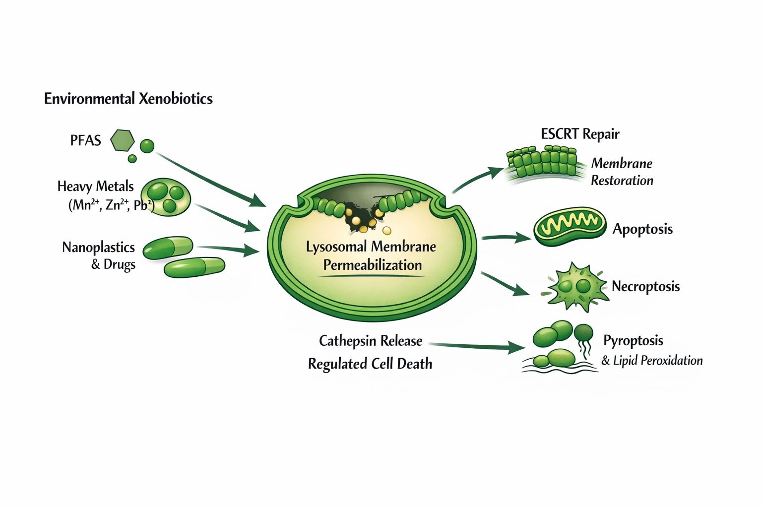
Lysosomes are acidic organelles central to cellular degradation, nutrient sensing, and regulated cell death. The stability of the lysosomal membrane is essential for cell viability, yet it is highly vulnerable to disruption by diverse environmental and endogenous stressors. This review synthesizes current understanding of the molecular mechanisms driving lysosomal membrane permeabilization (LMP) and lysosomal membrane rupture (LMR) in response to xenobiotics, including cationic amphiphilic drugs, perfluorinated alkyl substances (PFAS), and redox‑active heavy metals. We review lysosomal injury, namely intralysosomal ion trapping, metal‑catalyzed Fenton chemistry, and lipid peroxidation, which converges to generate oxidative membrane pores. We also examine cellular membrane‑repair systems, particularly the Endosomal Sorting Complex Required for Transport (ESCRT)-III machinery and ER–lysosome lipid transfer pathways, that act to restore lysosomal integrity. Failure of these protective mechanisms initiates distinct regulated cell death programs, including apoptosis, ferroptosis, and pyroptosis. Finally, we discuss the dual role of LMP in human health: both as a mediator of environmental toxicant‑induced injury and as a promising therapeutic target for overcoming multidrug resistance in cancer. By integrating findings from emerging non‑mammalian model systems and advanced imaging modalities, this review provides a unified framework for understanding lysosomal membrane dynamics under chemical stress.
Keywords: 
;  ;  ;  ;  ;  ;  ;  ;  

1. Introduction

Lysosomes are highly specialized acidic organelles essential for cellular homeostasis, integrating macromolecular degradation, metabolic recycling, nutrient sensing, immune regulation, and multiple forms of regulated cell death. More than sixty distinct hydrolases, including proteases, lipases, nucleases, phosphatases, and sulfatases, operate optimally within the lysosomal lumen at a pH ranging from approximately 4.5 to 5.0. This acidic environment is maintained through coordinated activity of the vacuolar ATPase (v-ATPase) and counter-ion transport systems that preserve luminal charge balance and ionic stability [1,2].
Central to lysosomal function is the lysosomal limiting membrane, a single lipid bilayer enriched in cholesterol, sphingolipids, and lysosome-associated membrane proteins (LAMPs). This membrane serves a dual role: it provides structural reinforcement against mechanical stress while isolating hydrolytic enzymes and sequestered xenobiotics from the cytosol. Even subtle perturbations of this barrier can disrupt luminal pH, impair autophagic flux, and initiate signaling cascades that direct cells toward adaptive repair or regulated cell death [3].
Lysosomes exhibit a unique vulnerability to chemical, oxidative, mechanical, and metabolic stressors, rendering them frequent targets of xenobiotic injury. A growing body of evidence demonstrates that environmental toxicants, cationic amphiphilic drugs, heavy metals, nanoparticles, and persistent organic pollutants preferentially accumulate within lysosomes following endocytic uptake or ion trapping. These agents can induce varying degrees of lysosomal membrane permeabilization (LMP), characterized by nanoscale pore formation that permits selective leakage of luminal contents, or lysosomal membrane rupture (LMR), defined by catastrophic bilayer failure and uncontrolled enzyme release [3,4].
Importantly, LMP and LMR represent mechanistically and functionally distinct injury states. Partial and transient LMP often activates apoptotic signaling pathways mediated by cathepsin release and mitochondrial crosstalk, whereas extensive or sustained membrane disruption overwhelms repair mechanisms and precipitates necrotic, inflammatory, or ferroptotic forms of cell death [4,5]. The magnitude and kinetics of lysosomal membrane damage therefore act as decisive determinants of cellular fate.
Because lysosomes function as primary sequestration hubs for xenobiotics, including pharmaceuticals, environmental pollutants, metals, and misfolded proteins, they occupy a central position at the interface between chemical exposure, redox biology, and cell death signaling.The major routes by which xenobiotics accumulate within lysosomes and initiate intralysosomal stress, including ion trapping, metal sequestration, and particulate uptake, are summarized in Figure 1.
This convergence renders lysosomes critical both for understanding toxicant-induced pathology and for developing therapeutic strategies that exploit lysosomal vulnerabilities, particularly in cancer cells that rely on expanded and stressed lysosomal compartments for survival and drug resistance [6].
In recent years, advances in lysosomal biology have highlighted that lysosomal damage does not inevitably result in cell death. Instead, cells deploy sophisticated membrane repair and regeneration systems that actively restore lysosomal integrity following injury. These pathways include rapid ESCRT-III–mediated membrane sealing, lipid reinforcement via ER–lysosome contact sites, and full organelle regeneration through autophagic remodeling. Failure or exhaustion of these protective mechanisms transforms lysosomes from adaptive stress responders into executioners of regulated cell death [7].
This review examines the molecular mechanisms by which environmental xenobiotics destabilize lysosomal membranes, the cellular programs that attempt to counteract this damage, and the pathological consequences that ensue when repair fails. Particular attention is given to redox-driven lipid peroxidation, metal-catalyzed oxidative chemistry, and emerging repair pathways that shape cell fate decisions under chemical stress. An integrated conceptual model linking environmental xenobiotics, lysosomal membrane damage, repair responses, and downstream cell fate decisions is presented in Figure 2.

2. Classes of Lysosomotropic Xenobiotics

Lysosomes are uniquely susceptible to chemical stress because many xenobiotics preferentially accumulate within their acidic lumen. This selective enrichment reflects the physicochemical properties of xenobiotics—particularly ionization state, lipophilicity, and molecular size—combined with intrinsic lysosomal functions such as macromolecular degradation, autophagy, and metal catabolism. Once sequestered, xenobiotics may exert hydrophobic, osmotic, oxidative, or redox-cycling effects that destabilize lysosomal membranes and compromise cellular homeostasis [8]. Distinct physicochemical mechanisms by which lysosomotropic xenobiotics induce lysosomal membrane permeabilization are schematically illustrated in Figure 3.
Lysosomotropic xenobiotics can be broadly categorized into weak-base cationic amphiphilic compounds, redox-active and non-redox metals, and particulate or nanoscale materials. Although these classes differ chemically, they converge mechanistically on lysosomal membrane stress through ion trapping, reactive oxygen species (ROS) generation, and disruption of lipid organization.

2.1. Weak-Base Sequestration and Ion Trapping

A large fraction of lysosomotropic agents are cationic amphiphilic drugs (CADs), characterized by hydrophobic regions and weakly basic amine groups. These compounds readily diffuse across cellular membranes in their unprotonated form and preferentially accumulate within acidic compartments such as lysosomes. Upon entry into the lysosomal lumen, protonation renders them membrane-impermeable, a phenomenon known as ion trapping [9].
As a result, CAD concentrations within lysosomes can exceed cytosolic concentrations by several orders of magnitude. Although this sequestration may initially function as a protective detoxification mechanism by isolating xenobiotics from sensitive cytosolic targets, sustained accumulation induces osmotic swelling, elevation of intralysosomal pressure, destabilization of luminal pH, and progressive membrane tension [10]. Over time, these biophysical stresses promote lysosomal membrane permeabilization and impair autophagic flux.
In cancer cells, ion trapping contributes directly to multidrug resistance by diverting chemotherapeutic agents away from their intended targets, such as nuclear DNA or cytosolic kinases. Expansion of the lysosomal compartment and upregulation of lysosomal transporters further reinforce this compartmentalized resistance mechanism, enabling malignant cells to survive otherwise lethal drug exposures [10,11].

2.2. Metal Sequestration and Redox

Cycling Lysosomes also serve as central hubs for intracellular metal handling, as they degrade metalloproteins, metal-binding complexes, and iron-storage proteins delivered through autophagy. This role places lysosomes at the intersection of metal detoxification and redox biology, rendering them especially vulnerable to metal-induced oxidative injury. Table 1 shows several metals that are sequestered in lysosomes.

2.2.1. Iron

Iron is delivered to lysosomes primarily through NCOA4-mediated ferritinophagy, during which ferritin complexes are degraded and ferric iron (Fe³⁺) is reduced to ferrous iron (Fe²⁺) within the acidic lumen. Ferrous iron acts as a potent catalyst for Fenton chemistry, reacting with hydrogen peroxide to generate hydroxyl radicals (•OH), among the most reactive and damaging ROS in biological systems [12].
These radicals initiate lipid peroxidation of lysosomal membranes, creating oxidative pores that promote LMP and sensitize cells to ferroptotic and apoptotic death pathways. The combination of high iron availability, acidic pH, and continuous ROS exposure makes lysosomal membranes particularly susceptible to iron-driven destabilization [12,13].

2.2.2. Copper

Copper overload induces lysosomal injury through mechanisms that extend beyond localized ROS generation. Excess copper disrupts endoplasmic reticulum (ER)–lysosome contact sites that are essential for lipid transfer, sterol homeostasis, and membrane repair. Studies indicate that copper interferes with the function of lysosomal adaptor proteins such as LAPTM4B, thereby impairing vesicular trafficking and ER-derived lipid delivery [14].
This disruption compromises the ability of lysosomes to repair oxidative membrane damage, rendering them increasingly fragile and prone to catastrophic rupture. Copper-induced lysosomal dysfunction has been observed across mammalian and non-mammalian systems, underscoring its conserved cytotoxic potential [15].

2.2.3. Cadmium

Cadmium typically enters lysosomes bound to metallothioneins or endocytosed protein complexes. Unlike redox-active metals, cadmium does not directly generate ROS via Fenton chemistry but instead destabilizes lysosomes by disrupting protein folding, inhibiting antioxidant defenses, and impairing autophagic flux. Accumulation of cadmium within lysosomes rapidly induces LMP, leading to cytosolic release of cathepsins and activation of apoptosis-like death pathways [16].
These effects are particularly pronounced in hepatic and phagocytic cells, which exhibit high rates of cadmium uptake and lysosomal turnover, highlighting a tissue-specific vulnerability to cadmium toxicity.

2.3. Nanoparticles and Environmental Particulates

Engineered nanoparticles and environmental fine particulate matter represent an expanding class of lysosomotropic stressors. Following endocytic uptake through clathrin-mediated endocytosis, macropinocytosis, or phagocytosis, these particulates accumulate within lysosomes where they resist enzymatic degradation. Physical characteristics such as size, shape, surface charge, and crystallinity critically influence their lysotoxicity.
Once sequestered, particulates may mechanically damage lysosomal membranes, generate localized ROS bursts, alter luminal pH, and interfere with autophagic degradation. Carbon-based nanoparticles, including carbon quantum dots, have been shown to induce predictable alterations in lysosomal physiology, including membrane stress and bioenergetic imbalance [17]. Similarly, vehicle exhaust particulates and airborne pollutants modify the acidic microenvironment of lysosomes, destabilizing hydrolase activity and promoting LMP [18].
Persistent particulate accumulation has been linked to impaired autophagic clearance, metabolic dysregulation, and inflammatory signaling, particularly in cells with high endocytic capacity. These findings position lysosomes as primary targets of nanoparticle toxicity and underscore their importance in environmental health and nanomedicine safety assessment

3. PFAS Accumulation and Lysosomal Membrane Stress

Per- and polyfluoroalkyl substances (PFAS) constitute a chemically distinct and environmentally persistent class of xenobiotics that pose a unique threat to lysosomal integrity. Unlike classical lysosomotropic weak-base compounds, whose accumulation is driven primarily by ion trapping, PFAS interact directly with lipid membranes and amphipathic protein surfaces due to their fluorinated carbon backbones and exceptional hydrophobicity. These properties promote progressive enrichment of PFAS within endolysosomal compartments, where clearance is inefficient and long-term retention amplifies cellular stress [19].
Recent studies increasingly recognize lysosomal dysfunction as a central mechanistic feature of PFAS toxicity. PFAS accumulation within lysosomes destabilizes membrane architecture, impairs autophagic flux, disrupts redox balance, and sensitizes cells to secondary oxidative or metabolic insults. These effects position lysosomes as critical mediators linking PFAS exposure to cellular injury and long-term pathology. Table 2 illustrates PFAS substances that interact with lysosomes.

3.1. Chain-Length Dependency of PFAS Toxicity

A defining determinant of PFAS-induced lysosomal stress is fluorocarbon chain length. Experimental studies using hepatic cell models have demonstrated that PFAS molecules with longer perfluorinated chains exhibit greater lysosomal retention and higher cytotoxic potency than shorter-chain analogs. As chain length increases, PFAS display enhanced affinity for lipid bilayers and intracellular membranes, promoting preferential localization to lysosomal and endolysosomal compartments [20].
In HepaRG and HepG2 cells, long-chain PFAS upregulate amino acid and xenobiotic transporters, induce compensatory metabolic responses, and alter lysosomal ion homeostasis. These adaptations appear to reflect cellular attempts to mitigate sustained lysosomal stress but ultimately contribute to chronic dysfunction. In contrast, shorter-chain PFAS are cleared more efficiently and induce comparatively modest lysosomal perturbations, supporting a positive correlation between hydrophobic chain length, lysosomal retention, and toxicity [20].

3.2. Sequestration and Lipid Binding Within Lysosomes

Once internalized, PFAS exhibit pronounced co-localization with lysosomal markers, reflecting their preferential accumulation within acidic organelles. PFAS enter cells bound to serum albumin, phospholipid carriers, and lipid-shuttling proteins, facilitating uptake through endocytic pathways. Within the lysosome, PFAS interact directly with membrane phospholipids, inserting their fluorinated tails into the lipid bilayer and displacing endogenous lipids [21].
Comparative analyses reveal that PFAS behave similarly to, yet more persistently than, classical lysosomotropic drugs. Unlike weak-base compounds that may eventually redistribute or efflux, PFAS display slow clearance kinetics, resulting in sustained lysosomal membrane stress and progressive impairment of autophagic degradation. This prolonged sequestration amplifies susceptibility to membrane permeabilization, particularly under conditions of oxidative or metabolic challenge [21,22].

3.3. Oxidative Stress and Lysosome-Centered Toxicity

PFAS exposure has been linked to increased intracellular ROS generation, a process that critically exacerbates lysosomal vulnerability. Subcellular imaging studies demonstrate that PFAS-induced oxidative stress is not uniformly distributed but instead concentrates within lysosomal regions, where redox-active metal pools and acidic conditions favor lipid peroxidation reactions [23].
Within the lysosome, ROS initiate peroxidation of polyunsaturated fatty acids embedded in the limiting membrane, compromising lipid packing density and generating nanoscale oxidative pores. These membrane defects promote lysosomal membrane permeabilization, leading to leakage of hydrolases and disruption of catabolic function. As oxidative injury accumulates, cells transition from adaptive stress responses toward regulated cell death pathways [24].
Collectively, these findings indicate that PFAS toxicity is not merely a consequence of global metabolic disruption but rather involves targeted, lysosome-centered injury driven by persistent membrane association, redox imbalance, and impaired organelle repair. The lysosome thus emerges as a critical nexus through which PFAS exert long-term cytotoxic effects, with implications for environmental health, toxicology, and disease risk assessment.

4. Mechanisms of Lysosomal Membrane Repair

Lysosomes are no longer viewed as passive victims of membrane injury. Instead, they are dynamic organelles equipped with highly regulated repair and recovery mechanisms that preserve integrity under chemical, mechanical, and oxidative stress. In response to membrane damage, cells deploy a hierarchical set of defense pathways that operate across distinct spatial and temporal scales, ranging from rapid sealing of nanoscale pores to membrane reinforcement and full organelle regeneration. The engagement and success of these pathways ultimately determine whether lysosomal stress is reversible or transitions into lysosomal membrane rupture (LMR) and cell death. The rapid recruitment of the ESCRT machinery to damaged lysosomal membranes and its role in restoring membrane integrity are illustrated in Figure 4.

4.1. ESCRT-III–Mediated Sealing of Lysosomal Membrane Lesions

The Endosomal Sorting Complex Required for Transport (ESCRT) machinery constitutes the first-line response to limited lysosomal membrane damage. Minor disruptions in membrane integrity permit transient efflux of Ca²⁺ from the lysosomal lumen into the cytosol, serving as an immediate signal that recruits ESCRT adaptor proteins ALIX and TSG101 to the damaged membrane surface [25]. These adaptors recognize regions of altered membrane curvature and exposed anionic phospholipids, initiating rapid assembly of ESCRT-III components at the lesion site.
The core ESCRT-III subunit CHMP4B polymerizes into curved filaments that constrict membrane defects from the cytosolic face. These filamentous structures form spirals or dome-like assemblies that progressively narrow and seal nanoscale pores [26]. Accessory ESCRT-III proteins, including CHMP2 and CHMP3, modulate filament curvature and stability, allowing the repair machinery to adapt to variations in damage geometry.
Disassembly and recycling of ESCRT-III polymers are driven by the ATP-dependent AAA-ATPase VPS4. VPS4-mediated remodeling is essential not only for reutilization of ESCRT components but also for completion of membrane closure [26]. This repair pathway operates on a timescale of seconds and is highly effective at resolving oxidative pores and early-stage lysosomal membrane permeabilization (LMP) induced by reactive oxygen species, nanoparticles, or transient chemical stress. However, ESCRT-III repair capacity is finite and becomes insufficient when lesions are large, persistent, or recurrent. Dynamic regulation of ESCRT assembly and VPS4-mediated disassembly at sites of membrane damage is depicted in Figure 5.

4.2. ER–Lysosome Contact Sites and Lipid-Mediated Membrane Reinforcement

When membrane damage exceeds the sealing capacity of ESCRT-III, cells engage a secondary repair pathway centered on endoplasmic reticulum (ER)–lysosome contact sites. These specialized inter-organelle junctions enable direct lipid transfer from the ER to reinforce damaged lysosomal membranes. A key mediator of this process is the oxysterol-binding protein–related protein ORP1L, which transports cholesterol to lysosomal membranes in response to damage-associated lipid imbalances [27].
Cholesterol enrichment increases membrane rigidity, restores lipid packing density, and counteracts excessive membrane fluidity caused by oxidative lipid peroxidation. This lipid remodeling stabilizes compromised membrane regions, reduces further permeabilization, and limits leakage of cathepsins into the cytosol [27,28]. Formation and regulation of ER–lysosome contact sites are controlled by luminal pH changes, Ca²⁺ signaling, local lipid depletion, and mechanical strain, ensuring spatially targeted repair responses [29].
Disruption of ER-mediated lipid transfer sensitizes lysosomes to chemical injury and promotes progression from reversible LMP to catastrophic membrane rupture. Conversely, in cancer cells, attenuation of ER–lysosome lipid repair has been shown to enhance susceptibility to lysosome-destabilizing therapies, underscoring this pathway’s dual role in cytoprotection and therapeutic resistance.

4.3. Lysosomal Regeneration and Organelle Renewal Pathways

When lysosomal damage becomes extensive or irreparable, cells may abandon membrane repair and instead initiate organelle regeneration pathways. One prominent mechanism involves autophagic lysosomal reformation (ALR), in which residual lysosomal membranes are remodeled to generate new, functional lysosomes. The Rab7 GTPase-activating protein TBC1D15 plays a central role in this process by regulating Rab7 signaling and redistributing membrane components following severe injury [30].
In parallel, a distinct regeneration pathway mediated by TECPR1 becomes critical under conditions of metabolic or energetic stress. When ATP availability is limited and energy-dependent ESCRT turnover is compromised, TECPR1 drives membrane tubulation from damaged lysosomes, reorganizing remaining membrane fragments into partially functional intermediates [31]. This pathway operates independently of canonical ESCRT activity and provides an energetically resilient mechanism for maintaining lysosomal populations during cellular stress.
Together, ESCRT-mediated sealing, ER-driven lipid reinforcement, and lysosomal regeneration form a tiered defense system that preserves lysosomal integrity across escalating levels of membrane damage. Failure or exhaustion of these repair mechanisms marks a critical threshold, beyond which lysosomes transition from adaptive stress sensors to potent initiators of regulated cell death.

5. Cellular Consequences of Lysosomal Failure

The major regulated cell death pathways activated downstream of lysosomal membrane permeabilization are summarized in Figure 6.
When lysosomal damage exceeds the capacity of membrane repair and regeneration mechanisms, cells transition from adaptive stress responses to irreversible lysosomal failure. The progression from limited lysosomal membrane permeabilization (LMP) to complete lysosomal membrane rupture (LMR) represents a critical inflection point in cell fate determination. The nature, extent, and kinetics of lysosomal damage dictate whether cells undergo apoptosis, ferroptosis, necroptosis, or inflammatory forms of cell death.

5.1. Cathepsin-Mediated Apoptosis

Partial and regulated LMP permits controlled release of lysosomal proteases, particularly cathepsins B and D, into the cytosol. In the neutral cytosolic environment, these proteases retain sufficient activity to initiate apoptotic signaling cascades. One of the central mechanisms involves cathepsin-mediated cleavage of BID into truncated BID (tBID), which translocates to mitochondria and promotes mitochondrial outer membrane permeabilization (MOMP), culminating in cytochrome c release and caspase activation [34,35].
Alterations in lysosomal positioning, membrane composition, and vesicular trafficking strongly influence susceptibility to LMP-driven apoptosis. In cancer cells, dysregulation of vesicular trafficking machinery and ESCRT components promotes lysosomal instability and amplifies cathepsin release, thereby lowering the apoptotic threshold [32,33]. Additionally, accumulation of redox-active metals and impaired antioxidant defenses intensify intralysosomal oxidative stress, further sensitizing lysosomal membranes to permeabilization [37].
Thus, cathepsin-mediated apoptosis represents a primary pathway by which xenobiotics and intracellular stressors translate lysosomal injury into mitochondrial-dependent cell death.

5.2. Ferroptosis and the Iron Axis

The lysosome plays a central role in ferroptosis due to its function in iron storage, mobilization, and redox regulation. Iron delivery to lysosomes occurs predominantly through NCOA4-mediated ferritinophagy, releasing ferrous iron (Fe²⁺) into the acidic lumen. Under conditions of oxidative stress or xenobiotic exposure, elevated intralysosomal Fe²⁺ fuels Fenton chemistry, generating hydroxyl radicals that initiate lipid peroxidation of lysosomal membranes [36,37].
Lipid peroxidation compromises membrane integrity by disrupting acyl-chain packing and forming reactive aldehydes that propagate oxidative damage. As lysosomal membranes become increasingly permeabilized, iron and reactive lipid species leak into the cytosol, amplifying ferroptotic signaling. Xenobiotics and pharmacological agents that exacerbate lysosomal iron accumulation or suppress lysophagic clearance accelerate this process, driving cells toward ferroptotic death [36].
Recent evidence demonstrates that modulation of lysosomal integrity critically determines ferroptotic sensitivity, positioning LMP as both an upstream trigger and an executioner of iron-dependent cell death pathways.

5.3. Necroptosis and Pyroptosis

When lysosomal damage is rapid, extensive, or occurs in contexts where apoptotic machinery is compromised, cells may bypass apoptosis in favor of necroptotic or pyroptotic death programs. These inflammatory forms of regulated cell death are typically associated with catastrophic LMR rather than partial LMP.
In cells deficient in caspase-8 activity, lysosomal damage promotes a switch from apoptosis to necroptosis. Under these conditions, LMP-induced release of cathepsins and reactive oxygen species suppresses residual apoptotic signaling while facilitating assembly of the RIPK1–RIPK3 necrosome. Activation and oligomerization of mixed-lineage kinase domain-like protein (MLKL) subsequently disrupts the plasma membrane, resulting in cell lysis and release of damage-associated molecular patterns (DAMPs) [39].
Pyroptosis is similarly linked to extensive LMR and is driven by activation of the NLRP3 inflammasome. Cytosolic leakage of lysosomal cathepsin B acts as a potent danger signal that promotes inflammasome assembly, caspase-1 activation, and maturation of interleukin-1β and interleukin-18. Pharmacological agents that exacerbate lysosomal instability, such as triamterene, amplify cathepsin release and inflammasome signaling, enhancing pyroptotic cell death [38].
Together, necroptosis and pyroptosis underscore the lysosome’s role as a decisive regulator of inflammatory cell death. When lysosomal repair pathways fail, the organelle becomes a central amplifier of immune-activating signals that shape tissue damage and disease progression.

6. Lysosomes in Cancer and Drug Resistance

Malignant cells undergo extensive lysosomal remodeling to survive metabolic stress, evade therapy, and maintain proliferative capacity. This remodeling includes expansion of lysosomal volume, altered lipid composition, enhanced autophagic flux, and upregulation of lysosome-associated transporters. While these adaptations enhance stress tolerance, they also fundamentally alter lysosomal membrane stability, positioning the lysosome as both a mediator of chemoresistance and a therapeutic vulnerability.

6.1. Sequestration-Mediated Drug Resistance

Lysosomal drug sequestration represents a major, yet often underappreciated, mechanism of chemoresistance across diverse malignancies. Many chemotherapeutic agents, see Table 3 below, are lipophilic weak bases that undergo protonation within the acidic lysosomal lumen, leading to ion trapping and accumulation at concentrations far exceeding those in the cytosol. This sequestration effectively diverts drugs away from their intended molecular targets, including nuclear DNA, cytosolic kinases, and apoptotic regulators [40].
Cancer cells frequently exploit this process by expanding their lysosomal compartment and increasing lysosomal number, thereby creating an intracellular sink that neutralizes cytotoxic drug exposure. Lysosomal hypertrophy and altered vesicular trafficking are hallmark features of multidrug-resistant phenotypes and correlate with poor therapeutic response [41]. In addition to passive trapping, malignant cells actively reinforce sequestration through upregulation of lysosomal transporters such as P-glycoprotein (P-gp) and LAPTM4B, further reducing cytosolic drug bioavailability.

6.2. Anthracyclines and Lysosomal Entrapment

Anthracyclines, particularly doxorubicin, exemplify clinically relevant lysosomotropic chemotherapeutics. Due to its amphipathic weak-base structure, doxorubicin rapidly accumulates within lysosomes following cellular uptake. Protonation in the lysosomal lumen prevents redistribution to the nucleus, where the drug must interact with topoisomerase II to induce DNA damage and apoptosis [42].
Sustained doxorubicin sequestration promotes lysosomal swelling, remodeling of membrane lipids, and activation of compensatory autophagy, all of which enhance cancer cell survival under therapeutic pressure. Experimental and clinical studies demonstrate that lysosome-trapped doxorubicin not only loses cytotoxic efficacy but also selects for subpopulations with increased lysosomal capacity and reinforced stress tolerance [43]. These adaptations contribute directly to the emergence of resistant disease.

6.3. Tyrosine Kinase Inhibitors and Rapid Lysosomal Trapping

Small-molecule tyrosine kinase inhibitors (TKIs), including sunitinib, sorafenib, and related agents, undergo some of the most rapid and efficient lysosomal accumulation observed among anticancer drugs. Their physicochemical properties—optimal pKa, lipophilicity, and membrane permeability—favor swift lysosomal ion trapping, dramatically reducing the pool of pharmacologically active drug available to interact with kinase targets at the plasma membrane or in the cytosol [44].
Chronic TKI exposure reinforces resistance by selecting cancer cells with elevated expression of lysosomal efflux transporters and stress-adaptation pathways. Notably, LAPTM4B plays an integrative role in this process by promoting drug sequestration while simultaneously regulating lysosomal biogenesis and membrane stress responses [45]. Advanced imaging approaches, including stimulated Raman scattering microscopy, have provided direct, real-time evidence of TKI accumulation within live-cell lysosomes, confirming sequestration as a major pharmacokinetic barrier to TKI efficacy [46].

6.4. Lysosomal Fragility as a Therapeutic Opportunity

Paradoxically, the same lysosomal adaptations that support drug resistance—hypertrophy, altered lipid composition, and enhanced acidification—also render cancer cell lysosomes intrinsically fragile. Enlarged lysosomes experience increased membrane tension, heightened oxidative stress, and elevated susceptibility to lipid peroxidation, lowering the threshold for lysosomal membrane permeabilization (LMP) [40,41].
This vulnerability has inspired therapeutic strategies that intentionally target lysosomal integrity to induce selective cancer cell death. Lysosomotropic agents, natural products, and synthetic compounds have been shown to exploit cancer-specific differences in lysosomal physiology to trigger controlled LMP and cathepsin-mediated apoptosis [47]. Unlike traditional chemotherapeutics, these approaches initiate cell death upstream of mitochondrial signaling, thereby bypassing common resistance mechanisms.
Triptolide and its derivatives represent well-characterized examples of lysosome-targeting agents capable of inducing LMP and lysosome-dependent programmed cell death, even in apoptosis-resistant cancer cells [48]. More recently, combinatorial strategies pairing lysosomal destabilizers with apoptosis-sensitizing agents, such as BH3 mimetics or CDK4/6 inhibitors, have demonstrated synergistic cytotoxicity by dismantling multiple survival axes simultaneously [49,50].
Collectively, these findings highlight a dual role for lysosomes in cancer: as facilitators of chemoresistance through drug sequestration and as exploitable vulnerabilities when membrane stability is selectively compromised. Targeting lysosomal fragility represents a promising avenue for overcoming multidrug resistance and improving therapeutic outcomes in refractory malignancies.

7. Nanoparticles and Environmental Particulates

Engineered nanoparticles and environmental particulates represent a rapidly expanding class of lysosomotropic stressors with significant implications for both human health and environmental toxicology. Due to their size, surface chemistry, and resistance to enzymatic degradation, these materials are preferentially internalized through endocytic and phagocytic pathways and ultimately accumulate within lysosomes. Once sequestered, nanoparticles exert sustained mechanical, chemical, and oxidative stress on the lysosomal compartment, often overwhelming cellular repair capacities.
A key determinant of nanoparticle-induced lysosomal injury is the physicochemical diversity of particulate materials. Size, shape, surface charge, crystallinity, and chemical composition critically influence intracellular trafficking, lysosomal retention time, and toxicity. Sharp-edged or crystalline particles can physically disrupt lysosomal membranes, while chemically reactive surfaces catalyze local reactive oxygen species (ROS) generation within the acidic lysosomal lumen [51].

7.1. Cell-Type-Specific Lysosomal Vulnerability

Lysosomal responses to particulate accumulation are highly cell-type dependent, reflecting differences in endocytic capacity, metabolic demand, and adaptive stress responses. Hepatocytes, for example, generally tolerate moderate nanoparticle accumulation by activating transcription factor EB (TFEB)–dependent lysosomal biogenesis and autophagic pathways, thereby maintaining degradative capacity and organelle turnover [52].
In contrast, professional phagocytes such as macrophages, Kupffer cells, microglia, and dendritic cells internalize particulates at much higher rates. This disproportionate burden rapidly saturates lysosomal degradation pathways, leading to luminal alkalinization, impaired hydrolase activity, and accelerated lysosomal membrane permeabilization (LMP). In hepatic models, Kupffer cells exposed to carbon-based nanoparticles exhibit pronounced lysosomal overload, defective TFEB activation, and heightened susceptibility to inflammatory cell death compared with neighboring hepatocytes [53].
These differences underscore a central principle of particulate toxicology: lysosomal fragility is not uniform across tissues. Cells specialized for clearance and immune surveillance are disproportionately sensitive to nanoparticle-induced lysosomal stress, contributing to organ-specific toxicity and inflammation.

7.2. Environmental Particulates and Lysosomal Dysfunction

Ambient environmental particulates, including vehicle exhaust particles, microplastics, and airborne pollutants, exhibit similar lysosomal accumulation profiles despite their heterogeneous composition. Upon internalization, these materials persist within lysosomes and alter the acidic microenvironment required for enzymatic activity. Studies in airway epithelial cells demonstrate that vehicle exhaust particulates elevate lysosomal pH, destabilize membrane integrity, and impair autophagic flux, thereby promoting progressive LMP [54].
Persistent environmental particulate exposure has been linked to chronic lysosomal dysfunction, metabolic dysregulation, and sustained inflammatory signaling. In immune cells, lysosomal damage induced by particulates can activate inflammasome pathways, amplifying tissue-level inflammation and contributing to respiratory, hepatic, and cardiovascular pathologies.

7.3. Sentinel Organisms and Lysosome-Related Organelles Beyond Mammalian Systems, Non-Mammalian Model Organisms Provide Valuable Insight into the Conserved Nature of Lysosomal Vulnerability Under Environmental Stress. Organisms Possessing Lysosome-Related Organelles (LROs), Such as Caenorhabditis elegans and Spodoptera litura, Function as Sensitive Sentinel Models for Assessing Sublethal Toxicant Effects

In these systems, exposure to environmental toxins and nanoparticulates induces alterations in LRO signaling, immune activation, and metabolic reprogramming that mirror lysosomal dysfunction in higher organisms. For example, exposure to Bt-Cry1Ab in S. litura results in distinct ultrastructural lysosomal changes, indicating that LRO disruption represents an evolutionarily conserved response to environmental stress [55].
Collectively, studies across cell types and species demonstrate that nanoparticles and environmental particulates exert their toxicity largely through lysosome-centered mechanisms. Persistent lysosomal accumulation, compounded by limited degradability, positions the lysosome as a primary determinant of particulate-induced cellular injury. These findings have broad implications for environmental risk assessment, nanomaterial design, and the development of safer therapeutic delivery platforms that minimize unintended lysosomal damage.

8. Adaptive Lysosomal Stress Responses

Cells possess an extensive network of adaptive responses that are mobilized when lysosomes experience chronic or sublethal stress. Rather than immediately triggering cell death, lysosomal membrane perturbation, luminal alkalinization, or impaired degradative capacity often initiates compensatory pathways designed to restore organelle function, maintain metabolic homeostasis, and prevent catastrophic lysosomal membrane rupture (LMR). These responses include transcriptional reprogramming, selective autophagic clearance of damaged lysosomes, and membrane remodeling processes that preserve the integrity of the lysosomal network.
Failure or exhaustion of these adaptive mechanisms represents a critical turning point, after which lysosomal dysfunction transitions from a reversible stress state to an irreversible trigger of regulated cell death.

8.1. TFEB-Mediated Lysosomal Biogenesis and Functional Recovery

Transcription Factor EB (TFEB) functions as the master regulator of lysosomal biogenesis and stress adaptation. Under basal conditions, TFEB is phosphorylated by mTORC1 and retained in the cytosol. Lysosomal stress—including membrane permeabilization, luminal pH disruption, nutrient scarcity, or xenobiotic accumulation—suppresses mTORC1 activity, permitting TFEB dephosphorylation and nuclear translocation [56].
Once in the nucleus, TFEB activates the CLEAR (Coordinated Lysosomal Expression and Regulation) gene network, inducing expression of genes encoding lysosomal hydrolases, v-ATPase subunits, lysosomal membrane proteins (LAMPs), and autophagy-related machinery. This transcriptional program expands lysosomal number, enhances degradative capacity, and reinforces membrane integrity, thereby compensating for damaged or dysfunctional lysosomes [57].
TFEB activation has been documented across multiple stress contexts, including nanoparticle exposure, oxidative injury, heavy-metal toxicity, and xenobiotic overload. In many cases, TFEB-dependent lysosomal expansion provides a critical cytoprotective buffer that allows cells to survive prolonged stress that would otherwise initiate apoptotic or necrotic pathways [58].

8.2. Lysophagy: Selective Clearance of Damaged Lysosomes

When lysosomal damage exceeds the capacity of membrane repair mechanisms, cells activate lysophagy, a selective form of macroautophagy dedicated to the removal of compromised lysosomes. The lysophagy pathway is initiated by the exposure of luminal glycan structures on the cytosolic face of damaged lysosomes following LMP. These glycans are recognized by galectins, particularly galectin-3, which act as molecular danger sensors [59].
Galectin recruitment triggers assembly of autophagy receptors, including p62/SQSTM1 and NDP52, which link damaged lysosomes to LC3-positive autophagosomal membranes. The compromised organelle is subsequently engulfed, delivered to intact lysosomes, and degraded. Through this process, lysophagy prevents uncontrolled release of hydrolases and limits propagation of lysosomal damage throughout the endolysosomal system [60].
Lysophagy plays a central role in maintaining lysosomal quality control during chronic stress, particularly in highly specialized or metabolically active cells. Impairment of lysophagic flux leads to accumulation of defective lysosomes, heightened oxidative stress, and increased susceptibility to inflammatory and apoptotic cell death.

8.3. Microautophagy and Membrane Remodeling

In addition to lysophagy, cells employ microautophagy as a complementary strategy for maintaining lysosomal integrity. Microautophagy involves direct invagination or budding of the lysosomal membrane, allowing the selective removal of damaged membrane microdomains, oxidized lipids, and aggregated proteins from the limiting membrane itself.
This process is regulated by phosphoinositide signaling pathways, particularly those involving PIKfyve, which generates phosphatidylinositol-3,5-bisphosphate required for endolysosomal membrane dynamics [61]. Components of the ESCRT machinery also participate in sculpting membrane topology during microautophagy, facilitating controlled membrane turnover without triggering large-scale permeabilization.
Together, lysophagy and microautophagy provide layered quality-control mechanisms that preserve lysosomal fidelity under sustained stress. These pathways enable cells to selectively remove damaged components while maintaining overall organelle function and autophagic flux.
Collectively, TFEB-driven biogenesis, lysophagy, and microautophagy constitute a highly integrated adaptive network that stabilizes lysosomal populations during xenobiotic, metabolic, and environmental stress. When these safeguards succeed, lysosomal dysfunction remains reversible; when they fail, lysosomes shift from adaptive stress responders to central executioners of cell death.

9. Therapeutic and Translational Implications

Advances in lysosomal biology have reframed the lysosome from a passive degradative compartment into a central regulator of cell fate, xenobiotic response, and therapeutic sensitivity. This paradigm shift has profound translational implications, as lysosomal membrane integrity can now be viewed both as a vulnerability to be exploited in cancer and as a barrier to be preserved in degenerative disease and environmental toxicology. Therapeutic strategies targeting lysosomal function must therefore be context-specific, balancing destabilization versus reinforcement depending on disease state.

9.1. Targeted Lysosomal Membrane Permeabilization in Oncology

Cancer cells are characterized by enlarged, metabolically stressed lysosomes that support high autophagic flux, nutrient recycling, and drug sequestration. These adaptations, while advantageous for tumor survival, generate a state of intrinsic lysosomal fragility. Enlarged lysosomal volume, altered lipid composition, and elevated oxidative stress collectively lower the threshold for lysosomal membrane permeabilization (LMP), creating a therapeutic window for selective lysosomal targeting [62].
Multiple classes of anticancer agents now exploit this vulnerability by inducing controlled LMP. Lysosomotropic detergents, natural products, and synthetic amphiphilic compounds preferentially accumulate in cancer lysosomes and destabilize the limiting membrane, triggering cathepsin-mediated apoptosis or non-apoptotic cell death pathways upstream of mitochondrial signaling [63]. Importantly, this mechanism bypasses common resistance pathways that disable caspase activation or mitochondrial priming.
Triptolide and its derivatives remain among the most extensively studied lysosome-targeting anticancer agents. These compounds accumulate within lysosomes, disrupt intraluminal pH homeostasis, impair autophagy, and promote lysosomal collapse even in apoptosis-resistant tumor cells [64]. Recent advances in molecular engineering, including aptamer-functionalized and nanoparticle-encapsulated triptolide, have further improved tumor selectivity and reduced systemic toxicity by enhancing lysosomal delivery [65].
Therapeutic efficacy may be further enhanced through rational combination strategies. Pairing LMP-inducing agents with BH3 mimetics such as venetoclax increases mitochondrial priming and amplifies apoptotic responses following lysosomal destabilization [66]. Similarly, pharmacological inhibition of CDK4/6 signaling has been shown to sensitize cancer cells to lysosomotropic agents by compromising metabolic resilience and lysosomal repair capacity [67]. These combinatorial approaches exemplify a new generation of therapies that dismantle redundant survival circuits by targeting both lysosomal and mitochondrial axes.

9.2. Reinforcing Lysosomal Integrity in Degenerative Disease and Toxicology

In contrast to oncology, therapeutic goals in neurodegenerative disorders, chronic liver disease, and environmental toxicant exposure emphasize preservation of lysosomal integrity. In these contexts, lysosomal dysfunction contributes to protein aggregation, impaired autophagic clearance, sustained inflammation, and progressive cell death. Premature or excessive LMP exacerbates disease pathology and accelerates tissue degeneration [68].
Pharmacological strategies, see Table 4) aimed at reinforcing lysosomal stability include the use of molecular chaperones that stabilize lysosomal enzymes and membrane-associated proteins. Agents that enhance activity of acid sphingomyelinase (ASM) or heat shock protein 70 (HSP70) have demonstrated protective effects against oxidative stress–induced lysosomal damage, preserving hydrolase activity and autophagic flux [69]. Such approaches may be particularly valuable in lysosomal storage disorders and neurodegenerative conditions characterized by chronic lysosomal stress.
Modulation of lysosomal repair pathways also represents a promising protective strategy. Enhancement of ESCRT-III recruitment or function could improve resilience to mechanical or oxidative membrane injury, while stabilization of ER–lysosome contact sites and ORP1L-mediated lipid transfer may fortify lysosomal membranes against redox-driven destabilization [70]. Importantly, these interventions aim to preserve lysosomal function without globally suppressing autophagy or immune signaling.

9.3. Emerging Biological Models and Imaging Technologies

The increasing complexity of lysosome-centered toxicology and therapy underscores the need for physiologically relevant biological models and advanced imaging tools. Sentinel organisms possessing lysosome-related organelles (LROs), including Caenorhabditis elegans and insect models, enable rapid, scalable assessment of sublethal lysosomal injury induced by environmental xenobiotics [71]. These systems provide sensitive readouts of lysosomal stress, immune activation, and metabolic adaptation that often precede overt toxicity in mammalian models.
In parallel, advances in imaging technologies have revolutionized visualization of lysosomal dysfunction. Stimulated Raman scattering (SRS) microscopy and other vibrational spectroscopic approaches enable label-free, real-time detection of lipid accumulation, peroxidation, and membrane remodeling within live-cell lysosomes [72]. These methods provide unprecedented insight into the spatiotemporal dynamics of lysosomal injury and repair, facilitating mechanistic toxicology studies and accelerating therapeutic development.
Together, these translational advances reinforce the central role of lysosomal biology in disease pathogenesis and therapy. Precision modulation of lysosomal membrane stability—either destabilizing or reinforcing—represents a powerful and increasingly tractable strategy across oncology, neurodegeneration, and environmental health sciences.

10. Conclusion

The lysosomal limiting membrane represents a decisive regulatory boundary that determines whether cells adapt to chemical stress or progress toward regulated cell death. Throughout this review, we have shown that a broad spectrum of xenobiotics, including cationic amphiphilic drugs, redox-active metals, per- and polyfluoroalkyl substances (PFAS), engineered nanoparticles, and environmental particulates, exploit the lysosome’s acidic and degradative environment to accumulate and exert highly localized toxicity. These agents destabilize lysosomal membranes through convergent mechanisms that include ion trapping, osmotic stress, metal-catalyzed Fenton chemistry, oxidative lipid peroxidation, and disruption of ER–lysosome lipid transfer.
Importantly, lysosomal injury does not inherently result in cell death. Cells deploy an intricate hierarchy of protective responses—ESCRT-mediated membrane sealing, ER-driven lipid reinforcement, autophagic lysosomal reformation, TFEB-dependent biogenesis, lysophagy, and microautophagy—that collectively preserve lysosomal integrity and cellular homeostasis. The balance between damage and repair determines whether lysosomal membrane permeabilization remains reversible or escalates into catastrophic lysosomal membrane rupture, triggering apoptosis, ferroptosis, necroptosis, or pyroptosis. Lysosomes thus function not merely as passive degradation sites but as dynamic sensors and integrators of chemical, metabolic, and redox stress.
The dual role of lysosomes in human health emerges as a central theme. In toxicology and degenerative disease, reinforcement of lysosomal membrane stability is paramount, as chronic lysosomal dysfunction drives protein aggregation, inflammation, and tissue degeneration. In contrast, in oncology, malignant cells adapt their lysosomal compartment to survive metabolic stress and chemotherapeutic exposure, rendering lysosomes both facilitators of drug resistance and intrinsic vulnerabilities. Therapeutic strategies that selectively destabilize cancer lysosomes or disable lysosomal repair pathways hold considerable promise for overcoming multidrug resistance and inducing tumor-selective cell death.
Looking forward, the integration of lysosomal biology into environmental risk assessment, drug development, and disease modeling will be essential. Emerging non-mammalian sentinel systems, alongside advanced imaging modalities capable of resolving lysosomal lipid dynamics and membrane integrity in real time, are poised to deepen mechanistic understanding of lysosome-centered toxicity. A more precise delineation of the molecular thresholds governing lysosomal resilience versus collapse will enable rational design of interventions that either exploit lysosomal fragility in cancer or reinforce lysosomal integrity in degenerative and toxicological contexts.
In summary, the lysosome occupies a pivotal position at the crossroads of xenobiotic exposure, cellular stress adaptation, and regulated cell death. Appreciating lysosomal membrane dynamics as an active determinant of cell fate transforms our understanding of chemical toxicity and therapeutic vulnerability, establishing the lysosome as a central target in both environmental health sciences and precision medicine.

References

  1. Zhang, R.; Vooijs, M.A.; Keulers, T.G. Key Mechanisms in Lysosome Stability, Degradation and Repair. Mol. Cell. Biol. 2025, 45, 212–224. [Google Scholar] [CrossRef]
  2. Yamashima, T. Lysosomal Membrane-Permeabilization (LMP) and -Rupture (LMR) Are Distinct for Cell Death. Front. Cell Death 2025, 4, 1669955. [Google Scholar] [CrossRef]
  3. Tan, J.X.; Finkel, T. A Phosphoinositide Signalling Pathway Mediates Rapid Lysosomal Repair. Nature 2022, 609, 815–821. [Google Scholar] [CrossRef] [PubMed]
  4. Radulovic, M.; Wenzel, E.M.; Gilani, S.; Holland, L.K.; Lystad, A.H.; Phuyal, S.; Olkkonen, V.M.; Brech, A.; Jäättelä, M.; Maeda, K.; et al. Cholesterol Transfer via Endoplasmic Reticulum Contacts Mediates Lysosome Damage Repair. EMBO J. 2022, 41, e112677. [Google Scholar] [CrossRef] [PubMed]
  5. Wang, F.; Gómez-Sintes, R.; Boya, P. Lysosomal Membrane Permeabilization and Cell Death. Traffic 2018, 19*, 918–931. [Google Scholar] [CrossRef]
  6. Boya, P.; Kroemer, G. Lysosomal Membrane Permeabilization in Cell Death. Oncogene 2008, 27*, 6434–6451. [Google Scholar] [CrossRef]
  7. Ahearn, G.A.; Mandal, P.K.; Cassano, G. Mechanisms of Heavy Metal Transport in Crustacea. Comp. Biochem. Physiol. C Toxicol. Pharmacol. 2004, 138*, 369–381. [Google Scholar]
  8. Li, D.; Shao, R.; Wang, N.; Zhou, N.; Du, K.; Shi, J.; Wang, Y.; Zhao, Z.; Ye, X.; Zhang, X.; et al. Sulforaphane Activates a Lysosome-Dependent Transcriptional Program to Mitigate Oxidative Stress. Autophagy 2021, 17*, 872–887. [Google Scholar] [CrossRef] [PubMed]
  9. Serrano-Puebla, A.; Boya, P. Lysosomal Membrane Permeabilization in Cell Death: New Evidence and Implications for Health and Disease. Ann. N. Y. Acad. Sci. 2016, 1371, 30–44. [Google Scholar] [CrossRef]
  10. Hirata, Y.; Hashimoto, T.; Ando, K.; Kamatari, Y.O.; Takemori, H.; Furuta, K. Structural Features Localizing the Ferroptosis Inhibitor GIF-2197-r to Lysosomes. RSC Adv. 2023, 13, 32276–32281. [Google Scholar] [CrossRef]
  11. Yong, J.; Villalta, J.E.; Vu, N.; Kukurugya, M.A.; Olsson, N.; López, M.P.; Lazzari-Dean, J.R.; Hake, K.; McAllister, F.E.; Bennett, B.D.; et al. Impairment of Lipid Homeostasis Causes Lysosomal Accumulation of Endogenous Protein Aggregates through ESCRT Disruption. Elife 2024, 12, e86194. [Google Scholar] [CrossRef] [PubMed]
  12. Sanchez Garcia, A.; Ramos, J.L.; van Breemen, R.B. Binding of Perfluorinated Alkyl Substances to Serum Albumin and Its Effect on Cellular Uptake. Chem. Res. Toxicol. 2018, 31*, 1234–1241. [Google Scholar]
  13. Zoncu, R.; Perera, R.M. Built to Last: Lysosome Remodeling and Repair in Health and Disease. Trends Cell Biol. 2022, 32*, 597–610. [Google Scholar] [CrossRef]
  14. Trybus, W.; Trybus, E.; Król, T. Lysosomes as a Target of Anticancer Therapy. Int. J. Mol. Sci. 2023, 24*, 2176. [Google Scholar] [CrossRef]
  15. Dharmasivam, M.; Kaya, B. Lysosome as a Chemical Reactor. Int. J. Mol. Sci. 2025, 26*, 11581. [Google Scholar] [CrossRef]
  16. Vietri, M.; Miranda, M.R.; Amodio, G.; Ciaglia, T.; Bertamino, A.; Campiglia, P.; Remondelli, P.; Vestuto, V.; Moltedo, O. The Link between Endoplasmic Reticulum Stress and Lysosomal Dysfunction under Oxidative Stress in Cancer Cells. Biomolecules 2025, 15, 930. [Google Scholar] [CrossRef]
  17. Rafiq, S.; McKenna, S.L.; Muller, S.; Tschan, M.P.; Humbert, M. Lysosomes in Acute Myeloid Leukemia: Potential Therapeutic Targets? Leukemia 2021, 35, 2759–2770. [Google Scholar] [CrossRef]
  18. Sanchez-Garcia, A.; et al. PFAS Sequestration in Acidic Organelles. Toxicol. Sci. 2018, 164, 210–222. [Google Scholar]
  19. Kakuda, K.; Ikenaka, K.; Kuma, A.; Doi, J.; Aguirre, C.; Wang, N.; Ajiki, T.; Choong, C.-J.; Kimura, Y.; Badawy, S.M.M.; et al. Lysophagy Protects against Propagation of α-Synuclein Aggregation through Ruptured Lysosomal Vesicles. Proc. Natl. Acad. Sci. U. S. A. 2024, 121, e2312306120. [Google Scholar] [CrossRef]
  20. Ferrari, V.; Tedesco, B.; Cozzi, M.; Chierichetti, M.; Casarotto, E.; Pramaggiore, P.; Cornaggia, L.; Mohamed, A.; Patelli, G.; Piccolella, M.; et al. Lysosome Quality Control in Health and Neurodegenerative Diseases. Cell. Mol. Biol. Lett. 2024, 29, 116. [Google Scholar] [CrossRef] [PubMed]
  21. Klein, T.; et al. Stress-induced Microautophagy of Lysosomes. Nature Communications 2024, 15, 442. [Google Scholar]
  22. Alhowyan, A.A.; Harisa, G.I. From Molecular Therapies to Lysosomal Transplantation and Targeted Drug Strategies: Present Applications, Limitations, and Future Prospects of Lysosomal Medications. Biomolecules 2025, 15, 327. [Google Scholar] [CrossRef]
  23. Manivannan, M.S.; Yang, X.; Patel, N.; Peters, A.; Johnston, J.B.; Gibson, S.B. Lysosome-Disrupting Agents in Combination with Venetoclax Increase Apoptotic Response in Primary Chronic Lymphocytic Leukemia (CLL) Cells. Cells 2024, 13, 1041. [Google Scholar] [CrossRef] [PubMed]
  24. Li, S.-C.; Shao, S.-Z.; Zhang, Y.-H.; Zhou, Y.; Shang, W.-T.; Gao, Y.; He, Q.-B.; Guo, Q.-L.; Guo, C.-Y.; Zhang, X.-B. Wogonin Derivative V8 Enhances Bortezomib Efficacy in Gastric Carcinoma. World J. Gastroenterol. 2026, 32, 113299. [Google Scholar] [CrossRef]
  25. Ferret, L.; Pol, J.G.; Sauvat, A.; Stoll, G.; Alvarez-Valadez, K.; Muller, A.; Le Naour, J.; Peyre, F.; Anagnostopoulos, G.; Martins, I.; et al. Lysosomal Membrane Permeabilization Enhances the Anticancer Effects of POLR1 Transcription Inhibitors. Autophagy 2025, 21, 2246–2265. [Google Scholar] [CrossRef]
  26. Qing, Y.; Guo, Y.; Zhao, Q.; Hu, P.; Li, H.; Yu, X.; Zhu, M.; Wang, H.; Wang, Z.; Xu, J.; et al. Targeting Lysosomal HSP70 Induces Acid Sphingomyelinase-Mediated Disturbance of Lipid Metabolism. Clin. Transl. Med. 2023, 13, e1229. [Google Scholar] [CrossRef]
  27. Nguyen, V.-N.; Li, H. Recent Development of Lysosome-Targeted Organic Fluorescent Probes for Reactive Oxygen Species. Molecules 2023, 28*, 6650. [Google Scholar] [CrossRef]
  28. Pang, X.; et al. Carbon Quantum Dots Trigger Selective LMP in Hepatocytes. Journal of Nanobiotechnology 2022, 20, 112. [Google Scholar]
  29. ChemistrySelect2023, Special Edition: RSS Probes.
  30. Analytical Chemistry2022, BODIPY Viscosity Probes.
  31. Yamashita, S.; et al. p53 Regulation of Lysosomal Membrane Stability. Cell Death & Differentiation 2022, 29, 1880–1892. [Google Scholar]
  32. Hoyer, N.; et al. Elimination of Damaged Lysosomes via Selective Exocytosis. Current Biology 2022, 32, 4105–4118. [Google Scholar]
  33. Henn, A.; et al. Lysosomal Quality Control in Aging. Ageing Research Reviews 2025, 74, 101550. [Google Scholar]
  34. Domingues, P.; et al. Connexin43-mediated Exocytosis of Damaged Lysosomes. Cell Reports 2024, 43, 113820. [Google Scholar]
  35. Yousef, M.; et al. Sub-cellular Sequestration Kinetics of Alkaline Drugs. Journal of Pharmaceutical Sciences 2023, 112, 1445–1456. [Google Scholar]
  36. Dutt, S.; et al. Molecular Mechanisms of Iron and Heme Metabolism in Lysosomes. Blood Reviews 2022, 52, 100900. [Google Scholar]
  37. Shang, W.; et al. Copper-Induced ER-Lysosome Interplay Disorders in Avian Models. Science of the Total Environment 2025, 912, 168900. [Google Scholar]
  38. Wang, Y.; et al. Tissular Distribution and Lysosomal Response to Chemical Stressors. Environmental Pollution 2024, 340, 122800. [Google Scholar]
  39. Tandon, R.; et al. Machine Learning Identification of Lysosomotropic Compounds. Bioinformatics 2024, 40, btad112. [Google Scholar]
  40. Locquet, G.; et al. Caspase-8 Deficiency and the Switch to Lysosomal Death. Cell Death Discovery 2021, 7, 154. [Google Scholar]
  41. Shang, W.-T.; et al. Copper Disrupts the ER–Lysosome Interplay. Science of the Total Environment 2025, 902, 166500. [Google Scholar]
  42. Onodera, K.; et al. Vehicle Exhaust Particulates and Lysosomal pH. Toxics 2023, 11, 445. [Google Scholar]
  43. Lim, H.; et al. Chain-Length Dependency of PFAS Lysosomal Stress. Toxicology in Vitro 2022, 82, 105370. [Google Scholar]
  44. Wang, H.; et al. Sublethal Effects of Bt-Cry1Ab in Spodoptera litura. Pest Management Science 2024, 80, 2110–2122. [Google Scholar]
  45. Hajdú, B.; et al. C. elegans Stress-Induced Lysosomal Immunity. Nature Communications 2023, 14, 1560. [Google Scholar]
  46. Park, J.; et al. Triamterene-Induced Autophagic Degradation. Scientific Reports 2021, 11, 8900. [Google Scholar]
  47. Yuan, X.; et al. Stimulated Raman Imaging of Drug-Induced Lipid Accumulation. Analytical Chemistry 2023, 95, 5560–5570. [Google Scholar]
  48. Halaby, R. Lysosomal Dysfunction and Survival Signaling. Cancers 2021, 13, 4120. [Google Scholar]
  49. Messina, A.; Halaby, R. Lysosomal-Mediated Apoptosis in Cancer Cells. Cancer Letters 2011, 300, 1–10. [Google Scholar]
  50. Feng, L.; et al. Triptolide Induction of Cancer Cell Death: A Comprehensive Overview. Phytomedicine 2024, 125, 155300. [Google Scholar] [CrossRef]
  51. Chen, Y.; et al. Aptamer-Functionalized Triptolide for TNBC. Advanced Healthcare Materials 2024, 13, 230400. [Google Scholar]
  52. Carbo, N.; et al. Tetracyclic Compounds for Leukemia Therapy. European Journal of Medicinal Chemistry 2023, 250, 115200. [Google Scholar]
  53. Guo, X.; et al. St-N Reverses Docetaxel Resistance via Lysosomal Remodeling. Journal of Experimental & Clinical Cancer Research 2024, 43, 12. [Google Scholar]
  54. Wang, Z.; et al. Polyphyllin D Targets ASM to Induce LMP. Biochemical Pharmacology 2023, 210, 115450. [Google Scholar]
  55. Tandon, R.; et al. ML Prediction of Lysosomal Sequestration. Journal of Cheminformatics 2024, 16, 22. [Google Scholar]
  56. Yousef, M.; et al. Sunitinib Sequestration Kinetics. Molecular Pharmaceutics 2023, 20, 1100–1112. [Google Scholar]
  57. Messina, A.; Halaby, R. Triptolide-Induced LMP in MCF-7 Cells. Cell Biology International 2011, 35, 101–108. [Google Scholar]
  58. Kim, S.; et al. CuCl2-Induced Neuronal Death via Autophagy Flux Dysregulation. NeuroToxicology 2024, 101, 45–56. [Google Scholar]
  59. Eskandari, N.; et al. TRPA1-Mediated Nrf2 Degradation. Free Radical Biology and Medicine 2025, 195, 220–232. [Google Scholar]
  60. Ning, X.; et al. Autophagy and the Nrf2 Antioxidant Pathway. Antioxidants 2023, 12, 880. [Google Scholar]
  61. Qian, H.; et al. Autophagy in Liver Diseases. Journal of Hepatology 2021, 74, 1200–1215. [Google Scholar]
  62. Wang, Y.; et al. Fallback Strategies for Lysosomal Survival. Cell Discovery 2025, 11, 5. [Google Scholar]
  63. Hoyer, N.; et al. Clearance of Damage Lysosomes. Current Biology 2022, 32, 4105. [Google Scholar]
  64. Radulovic, M.; et al. ER-Lysosome Contact Sites in Membrane Repair. EMBO Journal 2022, 41, e110000. [Google Scholar]
  65. Zhu, L.; et al. Iron-Mediated Dual-Induction of Cell Death. Nature Communications 2025, 16, 3017. [Google Scholar] [CrossRef] [PubMed]
  66. Wang, H.; et al. Sublethal effects of Bt-Cry1Ab on lysosomal integrity in Spodoptera litura. Pest Manag. Sci. 2024, 80, 2110–2122. [Google Scholar]
  67. Hajdú, B.; et al. Caenorhabditis elegans stress-induced lysosomal immunity and organelle-related signaling. Nat. Commun. 2023, 14, 1560. [Google Scholar]
  68. Park, J.; et al. Triamterene-induced autophagic degradation and lysosomal stress responses. Sci. Rep. 2021, 11, 8900. [Google Scholar]
  69. Yuan, X.; et al. Stimulated Raman imaging of drug-induced lipid accumulation and peroxidation. Anal. Chem. 2023, 95, 5560–5570. [Google Scholar]
  70. Feng, L.; et al. Triptolide induction of cancer cell death: A comprehensive overview of lysosomal pathways. Phytomedicine 2024, 125, 155300. [Google Scholar]
  71. Chen, Y.; et al. Aptamer-functionalized triptolide for targeted lysosomal disruption in triple-negative breast cancer. Adv. Healthc. Mater. 2024, 13, 230400. [Google Scholar]
  72. Carbo, N.; et al. Tetracyclic compounds for leukemia therapy: Targeting the lysosomal breach. Eur. J. Med. Chem. 2023, 250, 115200. [Google Scholar]
  73. Guo, X.; et al. St-N reverses docetaxel resistance via lysosomal remodeling and TFEB activation. J. Exp. Clin. Cancer Res. 2024, 43, 12. [Google Scholar]
  74. Locquet, G.; et al. Caspase-8 deficiency and the switch to lysosomal-dependent cell death. Cell Death Discov. 2021, 7, 154. [Google Scholar]
  75. Onodera, K.; et al. Vehicle exhaust particulates and their impact on lysosomal pH and stability. Toxics 2023, 11, 445. [Google Scholar]
  76. Wang, Z.; et al. Polyphyllin D targets acid sphingomyelinase to induce lysosomal membrane permeabilization. Biochem. Pharmacol. 2023, 210, 115450. [Google Scholar]
  77. Eskandari, N.; et al. TRPA1-mediated Nrf2 degradation and lysosomal oxidative stress. Free Radic. Biol. Med. 2025, 195, 220–232. [Google Scholar]
  78. Ning, X.; et al. The interplay between autophagy and the Nrf2 antioxidant pathway in xenobiotic stress. Antioxidants 2023, 12, 880. [Google Scholar]
  79. Lim, H.; et al. Structural features of PFAS determining lysosomal membrane sequestration. Toxicol. In Vitro 2022, 82, 105370. [Google Scholar]
Figure 1. Lysosomal uptake of environmental and pharmacological stressors. A schematic representation of a lysosome illustrating molecular fluxes that contribute to lysosomal stress. The lysosomal membrane contains the V-ATPase (H⁺ pump), which maintains an acidic lumen (pH 4.5–5.0). Nanoplastics, PFAS, and heavy metal ions enter the lysosome through membrane-associated or endolysosomal transport pathways. Weakly basic compounds diffuse into the lumen, become protonated, and undergo ion trapping, leading to intralysosomal accumulation and membrane stress.
Preprints 207005 g001
Figure 2. Environmental xenobiotics converge on lysosomal membrane damage to determine cell fate. Environmental xenobiotics, including PFAS, heavy metals, nanoplastics, and lysosomotropic drugs, accumulate within lysosomes and induce membrane damage followed by lysosomal membrane permeabilization (LMP). Sublethal damage can be resolved by ESCRT-mediated membrane repair, whereas sustained or extensive LMP promotes regulated cell death, including apoptosis, necroptosis, and pyroptosis.
Preprints 207005 g002
Figure 3. Mechanisms of lysosomal membrane permeabilization. Three pathways contribute to lysosomal membrane damage: (1) ion trapping and osmotic swelling caused by accumulation and protonation of weak bases in acidic lysosomes; (2) ROS-driven lipid peroxidation initiated by redox-active metals and oxidative stress; and (3) direct structural disruption of the membrane by environmental stressors such as PFAS and nanoplastics. These mechanisms converge on lysosomal membrane permeabilization (LMP).
Preprints 207005 g003
Figure 4. ESCRT-mediated repair of lysosomal membrane damage. Localized lysosomal membrane damage triggers rapid recruitment of the ESCRT machinery to sites of injury. ESCRT-III components assemble at membrane lesions to constrict and seal nanoscale pores, restoring membrane integrity and limiting cytosolic leakage of lysosomal contents. .
Preprints 207005 g004
Figure 5. CHMP4B polymerization and VPS4-mediated disassembly during lysosomal repair. Following recruitment to damaged lysosomal membranes, CHMP4B polymerizes into curved filaments that promote membrane budding and sealing. The AAA-ATPase VPS4 drives disassembly and recycling of ESCRT components, completing membrane repair and maintaining organelle homeostasis.
Preprints 207005 g005
Figure 6. Lysosomal membrane permeabilization as a central trigger of regulated cell death. Lysosomal membrane permeabilization (LMP) results in the release of cathepsins and other luminal factors into the cytosol, initiating regulated cell death signaling. Downstream pathways include apoptosis through mitochondrial dysfunction, necroptosis associated with plasma membrane rupture, and pyroptosis involving inflammasome activation and lipid peroxidation.
Preprints 207005 g006
Table 1. Heavy Metal Sequestration and Redox Toxicity.
Heavy Metal Primary Transporter Mechanism of Interaction Toxicological Consequence References
Cadmium (Cd) ZnT2 / DMT1 Cd–metallothionein complex endocytosis LMP; autophagy disruption [8,45,50]
Mercury (Hg) Direct binding Interaction with thiol groups Increased proton permeability; LMR [15,38]
Lead (Pb) ATP13A2 Sequestration in acidic vacuoles Impaired autophagy–lysosomal flux [7,40]
Copper (Cu) DMT1 Disrupts ER–lysosome contact sites Impaired lipid replenishment [14,41]
Iron (Fe) NCOA4 (cargo-mediated) Ferritinophagy-derived Fe²⁺ pool Fenton chemistry; lipid peroxidation [12,65]
Table 2. PFAS Lysosomal Accumulation and Membrane Impact.
PFAS Compound Class Mechanism of Lysosomal Entry Functional Impact on Membrane References
PFOS Long-chain PFSA PFAS–lipid complex endocytosis High accumulation; induces membrane stress [12,18]
PFOA Long-chain PFCA Lipid association/trafficking Moderate accumulation [18,43]
PFHxA Short-chain PFCA Weak lipid association Minimal uptake; chain-length dependent [43,79]
PFBS Short-chain PFSA Minimal lipid association Negligible lysosomal accumulation [43,79]
Table 3. Chemotherapeutic Accumulation and LMP Induction.
Drug / Agent Class Mechanism of Accumulation Role in Cell Fate References
Doxorubicin Anthracycline Ion trapping; DNA intercalation Sequestration-mediated resistance [14,55,70]
Sunitinib Tyrosine kinase inhibitor Rapid proton trapping Promotes multidrug resistance [56,65]
Siramesine Lysosomotropic agent Direct membrane destabilization Catastrophic LMP induction [6,22]
Triptolide Natural product Cathepsin-mediated signaling Lysosome-dependent apoptosis [48,65]
Vincristine Vinca alkaloid Luminal sequestration Sensitization to secondary LMP [14,73]
Table 4. Therapeutic Strategies and Emerging Biological Models for Lysosomal Stress.
Strategy / Model Mechanism of Action Application / Significance References
ESCRT-III enhancers Ca²⁺-dependent CHMP4B/VPS4 recruitment Rapid sealing of membrane nanolesions [3,17,32]
Lipid transfer (ORP1L) ER–lysosome contact site cholesterol delivery Reinforces oxidatively damaged membranes [4,59]
HSP70–ASM axis Stabilization of acid sphingomyelinase Therapeutic lysosomal destabilization in cancer [26,69]
Antioxidant defense Nrf2-mediated transcription (NQO1, HO-1) Limits lipid peroxidation-driven LMP [59,60]
Autophagic flux modulators TFEB-driven lysosomal biogenesis Lysophagy and organelle renewal [57,61]
Caenorhabditis elegans Lysosome-related organelle signaling Sentinel model of stress-induced immunity [45,67]
Spodoptera litura Bt-Cry1Ab toxin sequestration Environmental pesticide toxicity model [44,66]
Vibrational spectroscopy Stimulated Raman scattering (SRS) Real-time lipid peroxidation mapping [47,69]
Disclaimer/Publisher’s Note: The statements, opinions and data contained in all publications are solely those of the individual author(s) and contributor(s) and not of MDPI and/or the editor(s). MDPI and/or the editor(s) disclaim responsibility for any injury to people or property resulting from any ideas, methods, instructions or products referred to in the content.
Copyright: This open access article is published under a Creative Commons CC BY 4.0 license, which permit the free download, distribution, and reuse, provided that the author and preprint are cited in any reuse.
Prerpints.org logo

Preprints.org is a free preprint server supported by MDPI in Basel, Switzerland.

Subscribe

Disclaimer

Terms of Use

Privacy Policy

Privacy Settings

© 2026 MDPI (Basel, Switzerland) unless otherwise stated