

Submitted:
07 April 2026
Posted:
09 April 2026
You are already at the latest version
Abstract
Autism spectrum conditions have been associated with alterations in synaptic transmission and excitatory–inhibitory balance across distributed neural circuits. Converging evidence from genetic, electrophysiological, and animal models suggests that dysregulated activity-dependent synaptic plasticity—particularly altered long-term potentiation or long-term depression within hippocampal-cortical, cortico-striatal, and cerebellar networks—may contribute to reduced cognitive flexibility, repetitive behaviors, and difficulties in social and communicative adaptation. From this perspective, core behavioral features of autism may reflect circuit-level persistence of previously-formed neural patterns and altered updating of new information, rather than global neural dysfunction. Here we propose that modulating activity-dependent plasticity and excitatory–inhibitory dynamics may represent a plausible strategy for supporting cognitive flexibility in autism. Cannabinoids and terpenes derived from Cannabis sativa interact with multiple neural signaling systems—including CB1 receptors, GPR55, TRP channels, voltage-gated ion channels, serotonergic pathways, and endocannabinoid metabolism—that are known to influence synaptic transmission and plasticity. By engaging these convergent mechanisms, interactions among multiple botanical compounds may influence circuit-level excitability and synaptic plasticity processes implicated in autism. We consider multi-target phytocompound interactions with neural signaling pathways implicated in autism, particularly those regulating synaptic plasticity and excitatory–inhibitory balance. Within this framework, cannabinoid–terpene interactions may influence circuit-level dynamics underlying cognitive flexibility.
Keywords:
autism
; synaptic plasticity
; excitatory-inhibitory balance
; cognitive flexibility
; cannabinoids
; terpenes
; neural circuit dynamics
Background
Individuals on the autism spectrum often experience repetitive behaviors and challenges with communication and social interaction, frequently accompanied by sleep disturbances, seizures, and anxiety [1]. Many on the spectrum also experience aggression, violent outbursts, and self-injurious behaviors [2]. There are currently no U.S. Food and Drug Administration (FDA)-approved medications for many of the core symptoms of autism, leaving patients with few effective treatment options. Drugs like Risperidone and Aripiprazole may be effective for irritability and aggression [3], but these synthetic drugs can come with serious metabolic and movement-related side-effects [4,5] - while behavioral therapies also have limited success [6], perhaps because they cannot adequately address the underlying changes in neurobiology and brain chemistry that may lead to autism symptoms. Since many children and adults do not respond well to standard behavioral and medical treatments, parents and patients are seeking help through non-traditional methods, such as medicinal cannabis [7]. Cannabinoids derived from Cannabis sativa have been reported in some clinical and observational studies to improve certain core and associated symptoms of autism [8,9,10,11,12,13,14,15,16]. A study looking at sixty children with autism reported that whole-plant extracts containing the two major cannabinoids—cannabidiol (CBD) and Δ9-THC (in a 20:1 ratio)—caused significant improvements in behavior, anxiety, and communication problems [11]. An even larger study, with 188 children with autism, observed significant improvements in a majority of patients while they were using cannabis oil containing CBD and Δ9-THC [12]. Several studies showed CBD was beneficial for seizures, attention-deficit/hyperactivity disorder, sleep disorders, communication, and social interaction in children with autism [13,14]. There were also significant improvements in self-injury, aggression, and rage attacks [15,16]. Importantly, these studies observed cannabis-based treatments were generally associated with mild and infrequent side effects like restlessness, sleepiness, and dry mouth. Recent peer-reviewed clinical trials further support the therapeutic potential of cannabinoid-based interventions in autism. In a randomized, double-blind, placebo-controlled trial, a CBD-rich cannabis extract improved social interaction, anxiety, and psychomotor agitation in children with autism while producing few serious adverse effects [10].
The safety and efficacy of cannabinoid-based therapeutics are highlighted by several FDA-approved drugs, including Epidiolex for seizures [17]. Epidiolex consists of purified CBD, which is safe and well tolerated even at relatively high doses [18]. However, purified CBD reflects only a single component of a chemically complex plant; more than 100 phytocannabinoids have been identified in Cannabis sativa, alongside hundreds of terpenes and other secondary metabolites that are being investigated for their biological activity and influence on neural signaling [19,20]. Numerous scientific sources suggest that full-spectrum extracts containing multiple cannabinoids and terpenes may produce effects that differ from those of purified CBD alone[21,22,23,24,25]. Raphael Mechoulam, widely regarded as the ‘father of cannabinoid research,’ was among the first to suggest that interactions among endogenous and plant-derived cannabinoids may give rise to effects beyond those of isolated compounds [26]. While Mechoulam was elucidating the structures of CBD and THC for the first time and isolating both THC and the endocannabinoid anandamide, he postulated that multiple cannabinoids and terpenes could enhance endogenous endocannabinoid signaling [27]. A mixture of botanical compounds could also target a diverse array of ion channels and G-proteins in the brain [24,25,26,27,28]. Together, these mechanisms suggest that combinations of cannabinoids and terpenes may produce biological effects greater than the sum of their parts—a concept commonly referred to as the “entourage effect”. We consider a set of 12 bioactive cannabinoids and terpenes as a theoretical multi-compound system that may leverage synergetic interactions among phytocompounds, which emerging research suggests could support neural function. Since it is largely unknown how combinations of cannabinoids and terpenes may influence autism-related neural dysfunction, we propose a mechanistic framework through which such interactions could modulate neural pathways relevant to autism.
Neurobiological Basis of Phytocompound Modulation in Autism
Part 1: Potential Role of Aberrant Synaptic Plasticity
Every time we learn something new, thousands of neurons in our brain subtly, yet intricately, change the shape of their dendrites or axon terminals, adjusting the strength of electrochemical signals that flow between connected neurons [29]. This process is called synaptic plasticity and it is thought to be the main mechanism responsible for memory formation and learning [29] - especially when it happens in the hippocampus and downstream cortical areas. Synaptic plasticity in the hippocampus is thought to be essential for learning social skills [30], language [31], and the flexible processing of linguistic information [32], while plasticity within motor circuits—including the cortex, cerebellum, and basal ganglia—is critical for the acquisition of rhythmic and sequential motor movements [33,34]. Disruptions in these forms of neural plasticity may therefore contribute to several cognitive and behavioral characteristics associated with autism.
Several studies suggest autism could involve alterations in synaptic transmission between connected neurons in the brain and the synaptic plasticity between them [35,36,37,38,39,40,41,42,43,44,45,46,47]. Long-term potentiation (LTP) is a major form of synaptic plasticity and can be described as a persistent increase in the strength of connections between specific groups of communicating neurons [29]. Multiple electrophysiological studies in animal models associated with autism-linked genes and environmental models of autism show enhanced LTP in hippocampal [48,49,50,51,52] and cortical circuits [53], as well as hyper-plasticity in cerebellar circuits [54], consistent with the idea that overactive excitatory synaptic strengthening is involved with autism. Human neurophysiology studies using non-invasive LTP-like paradigms also find differences in plasticity responses in individuals on the spectrum of autism [55].
If long-term potentiation (LTP) is overactive during adolescent learning, neural connections involved in memory formation, language acquisition, social learning, and rhythm may become over-strengthened. Rather than remaining dynamically adaptable, neural networks may become biased toward reinforcing neural connections that were strengthened during and after exposure to novel environments, salient stimuli, new individuals, or early social experiences. In this scenario, neural engrams—ensembles of neurons whose coordinated activity represents a memory—may be reactivated with increased persistence, reducing the network’s capacity to flexibly update or integrate new information.
If such dysregulated synaptic plasticity occurs within the hippocampus and interconnected cortical regions, it could impair the encoding of new memories and the adaptive modification of existing ones, thereby interfering with language development, semantic associations, communication, and social learning. Behavioral manifestations may reflect excessive stabilization of previously-formed neural patterns, leading to repetitive recall dynamics, reduced cognitive flexibility, and context-inappropriate responses. Similarly, if over-strengthening occurs within motor-related circuits of the basal ganglia or cerebellum, the stabilization of early motor programs may bias these networks toward persistent, rhythmic, or repetitive movements. In this framework, core features of autism could arise from activity-dependent over-consolidation of specific circuit representations rather than from global neural dysfunction.
Alterations to long-term depression (LTD) are also implicated in autism and may further disrupt the balance of synaptic strengthening and weakening required for adaptive learning. Electrophysiological studies in autism-linked models have demonstrated impaired endocannabinoid-mediated LTD (eCB-LTD) within cortico-striatal circuits of the basal ganglia, a form of synaptic weakening that normally constrains action selection and habit formation [56]. Interestingly, the authors noted that enhanced excitatory synaptic transmission was associated with increased motor learning, supporting the idea that dysregulated plasticity within basal ganglia circuits may bias neural networks toward over-stabilization of previously performed action patterns, thereby reinforcing repetitive behaviors at the expense of cognitive flexibility. In another mouse model of autism, corticostriatal synapses showed impaired eCB-LTD, and stimulation protocols that normally induced LTD instead produced potentiation-like responses, indicating a shift in activity-dependent plasticity rules [57]. Because cortico–striatal–pallidal circuits govern action selection by facilitating desired motor programs while suppressing competing alternatives [58], impaired corticostriatal LTD may reduce the adaptive weakening of previously reinforced cortical inputs. This shift in plasticity balance could bias basal ganglia networks toward persistence of established action patterns, contributing to repetitive behaviors and reduced behavioral flexibility. In parallel, hippocampal studies in Fragile X mouse models have revealed exaggerated metabotropic glutamate receptor (mGluR)-dependent LTD, indicating that activity-dependent synaptic weakening can also become excessive in autism-associated conditions [38]. Together, these findings suggest that autism-related phenotypes may emerge not from uniformly increased or decreased plasticity, but from disrupted coordination of long-term potentiation and long-term depression processes across distributed hippocampal–cortical and cortico-striatal networks.
Adaptive learning requires that initial behavioral templates—such as those supporting language acquisition, rhythmic movement, and early social interaction—remain modifiable as new experiences accumulate. Neurotypical individuals encode these early foundations as flexible frameworks that can be elaborated and reorganized across development. In autism, however, early representations may become excessively stabilized through dysregulated plasticity, limiting the incorporation of new experiences over time. One might imagine this as spending a lifetime reinforcing the first few levels of a would-be skyscraper rather than allowing additional levels to be built upon it. In this skyscraper analogy, the upper floors represent increasingly complex behaviors—such as nuanced communication, adaptive social interaction, and coordinated motor output—that may be difficult to attain when early neural patterns are disproportionately consolidated (such that the structure stalls before reaching greater height).
Neurons in the brains of individuals with autism have been reported to exhibit altered synaptic architecture, consistent with over-strengthened incoming excitatory inputs [36,37]. A single neuron can receive hundreds of thousands of inputs from other neurons. These inputs terminate on receiving structures called dendrites. Dendritic spine growth is highly correlated with synaptic plasticity [59] and represents one mechanism by which neurons can ‘change their shape’ during learning. Dendritic spines have been found at higher density on neurons in those with autism compared to neurotypical controls [36,37]. Because dendritic spines are structural correlates of excitatory synapses and are closely linked to synaptic plasticity, elevated spine density may reflect alterations in synaptic strengthening, pruning, or maturation [60]. If early synaptic connections are strengthened or maintained beyond their adaptive window, ensembles encoding initial experiences—such as early social encounters, motor routines, or salient and novel environmental stimuli—may become disproportionately reinforced. Dopaminergic signaling, which is engaged by novelty [61,62] and facilitates long-term synaptic plasticity in regions such as the hippocampus [63,64,65], could further bias consolidation of early experiences. Dopamine dysregulation has also been implicated in autism [66] and in perseverative behavior [67,68], linking neuromodulatory imbalance to reduced cognitive flexibility. Within this framework, excessive stabilization of early activity patterns may limit the flexible updating of neural networks required for learning and adaptation.
Synaptic plasticity deficits and critical period abnormalities have been observed in mouse models of Fragile X syndrome [38,39], Rett syndrome [40,41], and tuberous sclerosis [42,43], all of which are thought to give rise to autism. Defective synapse maturation and enhanced synaptic plasticity have also been observed in other models of autism such as Shank2 knockout mice [44]. In a mouse model of autism/Angelman syndrome researchers found a deficit in synaptic plasticity associated with critical period remodeling of cortical visual circuits [35]. In vitro analysis revealed that cortical synapses were immature and unable to incorporate changes in new sensory experience. Taken together, such findings are consistent with the possibility that neural activity established during previous learning may be excessively stabilized across development, restricting the flexible reorganization of circuits in response to subsequent input.
Autism-related neural phenotypes may also reflect an impaired capacity to extinguish or remodel previously reinforced behavioral patterns. Adaptive development requires not only the strengthening of relevant synapses, but also the weakening or refinement of earlier-established representations so that more complex and context-appropriate behaviors can emerge. When this balance is disrupted, networks may become biased toward persistence of previously learned action patterns, limiting cognitive flexibility and the incorporation of new experiences. Some of this over-stabilization may occur during sleep-dependent consolidation processes, such as hippocampal sharp-wave ripple replay events, which normally strengthen recently acquired memories [69]. If activity-dependent consolidation is excessive or poorly constrained, early-life or novelty-associated neural representations could become disproportionately reinforced, potentially restricting the flexible updating of neural networks as environments become familiar. These mechanisms are consistent with numerous studies which link critical period dysfunction and altered synaptic plasticity to autism [38,39,40,41,42,43,44,45,46,47].
Emerging evidence suggests that dysregulated activity-dependent plasticity and altered excitatory–inhibitory dynamics may represent central circuit-level mechanisms contributing to reduced cognitive flexibility and behavioral persistence in autism. If maladaptive stabilization of synaptic connections underlies aspects of the phenotype, then biological systems capable of modulating synaptic transmission and plasticity warrant careful mechanistic consideration.
Cannabinoids and terpenes derived from Cannabis sativa interact with multiple neural signaling pathways known to influence excitability, synaptic strength, and network balance. The following section examines how these phytocompounds may influence synaptic transmission and plasticity mechanisms implicated in autism.
Part 2: Cannabinoid–Terpene Interactions with Plasticity and Excitatory-Inhibitory Systems
If dysregulated synaptic plasticity contributes to circuit-level rigidity in autism, then exogenous compounds capable of modulating the properties of ion channels and G-proteins that influence synaptic transmission and plasticity represent promising targets for mechanistic investigation. Phytocannabinoids are known to interact with multiple such targets—including type-1 cannabinoid receptors (CB1Rs), G-protein-coupled receptor 55 (GPR55), transient receptor potential (TRP) channels, voltage-gated ion channels, and endocannabinoid metabolism—each of which may modulate neuronal excitability and plasticity. For example, a major mechanism of action of cannabidiol (CBD) is thought to be the interference of LPI-GPR55 signaling in the hippocampus [70,71]. LPI, or lysophosphatidylinositol, is a lipid signaling molecule that activates the G-protein coupled receptor, GPR55. LPI-GPR55 signaling induces calcium influx and presynaptic excitatory neurotransmitter release from pyramidal neurons in the hippocampus [70], neurons which can initiate and undergo many forms of synaptic plasticity (along with their post-synaptic partners) [72,73]. By blocking LPI–GPR55 signaling, CBD could reduce glutamate release and limit its binding to receptor subtypes that initiate long-term plasticity, including AMPA, NMDA, and extra-synaptic mGluR receptors [74,75]. This could disrupt both pre- and post-synaptic plasticity, thereby modulating hyperexcitability and synaptic dynamics implicated in autism. In this way, CBD may reduce the persistence of overactive plasticity states, potentially mitigating excessively strengthened patterns of neural activity, memory, and behavior. Since GPR55 is expressed in neurons across multiple brain regions—including the hippocampus, cortex, basal ganglia, and cerebellum [76,77]—it may be strategically positioned to influence overactive excitatory synaptic plasticity linked to neurodevelopmental conditions.
Autism is also thought to be the result of an imbalance in excitatory and inhibitory neural signaling in the brain [78]. This model is in line with autism being brought about by overactive synaptic plasticity, since overactive synaptic plasticity of excitatory connections or disrupted plasticity of inhibitory connections could increase the ratio of excitation to inhibition and lead to hyper-excitable neural circuits (Figure 1). CBD could potentially contribute to modulation of excitatory–inhibitory balance by interfering with LPI-GPR55 signaling, or by interacting with several other G-proteins and ion channels (Figure 2). CBD can inhibit transient receptor potential (TRP) channels by stimulating and then desensitizing them [79,80,81], which may help put a brake on excitotoxic calcium entry into neurons, excitatory depolarization, and TRP-mediated synaptic plasticity. CBD also functions as an agonist of 5HT1A serotonin receptors [82], which can evoke inhibitory actions in neurons via its G-protein signaling cascades. CBD can further act as an agonist of glycine receptors [83], which mediate fast inhibitory synaptic transmission between neurons. Through these mechanisms CBD may contribute to the regulation of neuronal excitability and synaptic activity. CBD is an antagonist of TRPM8 receptors [84] and inhibits voltage-gated Ca[2]+ channels [85] and voltage-gated Na+ channels [86]. The inhibitory action of CBD on depolarizing ion channels could potentially protect against excitotoxicity and excessive glutamate release from synaptic terminals. CBD’s ability to reduce neuronal excitability by enhancing K+ channel currents may also contribute to its ability to promote neural balance [87]. CBD has been reported to enhance oxytocin and vasopressin release, which could theoretically aid in healthy social interactions [88]. By inhibiting both the uptake of the endocannabinoid anandamide and the enzyme that breaks it down (fatty acid amide hydrolase), CBD can increase extracellular anandamide levels (as Mechoulam previously suggested) [80,89,90]. Since endocannabinoids like anandamide can bind CB1Rs and dampen excitatory activity, this could also help restore excitatory–inhibitory balance in the brain. Because the LPI–GPR55 signaling pathway can also function to reduce expression of a major inhibitory receptor, the ionotropic GABAA receptor, and weaken inhibitory synaptic transmission [71], CBD’s interference with this pathway could also help preserve inhibitory tone within hyperexcitable hippocampal circuits. For these reasons, higher relative concentrations of CBD may be required to effectively engage these pathways.
Δ9-THC represents another key phytocannabinoid within this framework. As an agonist of type-1 cannabinoid receptors (CB1Rs)—the most abundantly expressed G-protein–coupled receptors in the brain [91]—Δ9-THC is well positioned to modulate synaptic transmission and activity-dependent plasticity [92]. Both endogenous cannabinoids (like anandamide) and exogenous cannabinoids (like THC) could protect the brain against hyperexcitability, by dampening excitatory neural transmission and plasticity via CB1Rs [92,93]. For example, when THC or the endocannabinoid 2-arachadonyl-glycerol (2-AG) bind to CB1Rs on excitatory neurons in the hippocampus, those neurons will decrease the amount of glutamate they release. Postsynaptic AMPA and NMDA receptor-mediated strengthening of synapses may be reduced via decreased glutamate release, further disrupting overactive excitatory synaptic plasticity associated with autism. Given CB1Rs are expressed by excitatory neurons throughout the hippocampus [94] (such as by excitatory pyramidal cells and mossy cells which undergo robust forms of long-term potentiation [95]) they may be well-positioned to curb the overactive synaptic plasticity associated with generating over-persistent patterns of neural activity. CB1Rs are also highly expressed throughout the cortex, basal ganglia, and cerebellum, where they may similarly have the potential to protect against over-excitation. Δ9-THC can also transiently activate and desensitize TRPA1, TRPV1, and TRPV2 [79,96,97]. By desensitizing TRP channels, Δ9-THC could potentially protect against neuronal hyperexcitability and calcium-mediated neurotoxicity. Δ9-THC also has potent anti-inflammatory and antioxidant properties and is neuroprotective against toxicity in multiple biological models [70,98,99,100,101,102,103,104,105]. Given the central role of CB1 receptor signaling in regulating excitatory neurotransmission, Δ9-THC may represent an important contributor to modulation of excitatory–inhibitory balance.
Cannabidivarin (CBDV) represents another prominent phytocannabinoid within this framework. Studies in animal models of autism suggest that this cannabinoid may show beneficial effects towards neurological and motor impairments as well as cognitive deficits [106,107,108]. There is also evidence from rodent models of autism—including prenatal valproic acid (VPA) exposure models—that CBDV administration can reduce repetitive behaviors, improve social interaction deficits, attenuate hyperactivity, and normalize alterations in synaptic plasticity and excitatory–inhibitory balance [108,109,110]. In some studies, CBDV has also been reported to ameliorate deficits in cognitive flexibility and reduce seizure susceptibility in models characterized by network hyperexcitability [109,110] suggesting a potential role in modulating circuit-level dysfunction associated with autism-like phenotypes. Preliminary, unpublished clinical investigations are evaluating CBDV in individuals with autism, and early findings are promising, with the lead researcher on the study noting decreases in irritability symptoms, temper tantrums, explosive episodes, as well as improvements in repetitive behaviors [111]. Some of these positive effects may be tied to CBDV’s ability to modulate excitation–inhibition dynamics in the human brain. In a single-dose, placebo-controlled magnetic resonance spectroscopy study in adults with and without autism, CBDV altered glutamate and GABA signaling in key cortical regions implicated in autism, including areas involved in social cognition and sensory processing [112]. Notably, CBDV shifted neurochemical markers of excitatory–inhibitory balance toward normalization in individuals with autism, supporting the hypothesis that this cannabinoid may influence neural systems relevant to synaptic plasticity and circuit-level regulation [112]. Studies in humans with epilepsy suggest CBDV is safe, therapeutic, and well-tolerated [113], supporting its potential suitability for further investigation in neurodevelopmental conditions such as autism.
Another phytocannabinoid of interest is the neuroprotective cannabinoid cannabigerol (CBG), which may complement the effects of other compounds by interacting with multiple neurotransmitter systems, ion channels, receptors, and signaling pathways [114]. CBG has been shown to inhibit voltage-gated sodium channels in central neurons, a mechanism capable of reducing neuronal excitability and modulating synaptic signaling within neural circuits [115]. CBG also modulates several transient receptor potential (TRP) ion channels involved in calcium signaling and neuronal excitability, as well as GPCR targets including α2-adrenergic and serotonergic receptors that regulate neurotransmission and synaptic activity [116,117]. Experimental studies further indicate that CBG can protect neurons against oxidative stress and neuroinflammatory damage, with evidence from animal models demonstrating reduced inflammatory signaling and improved neuronal survival in neurodegenerative disease models [118,119]. Through these interactions, CBG may influence excitatory–inhibitory signaling balance and exert antioxidant and neuroprotective effects in neural tissue, potentially complementing the actions of other cannabinoids and terpenes.
Cannabinol (CBN) is also relevant, due to its reported neuroprotective properties in models of seizures and epilepsy [120,121]. In addition to its anticonvulsant activity, CBN has been reported to interact with CB1 and CB2 receptors [122] and to reduce neural excitability via the modulation of voltage-gated Na+ channels, interactions that could contribute to the stabilization of neuronal network activity and suppression of hyperexcitability [123]. CBN has also been reported to exert anti-inflammatory effects by modulating cytokine signaling and immune cell activity, mechanisms that may contribute to neuroprotection and stabilization of neural circuits under conditions of inflammatory stress [124]. Through these combined receptor-mediated and anti-inflammatory actions, CBN may help protect neural tissue from excitotoxic damage. Cannabichromene (CBC) is reported to exhibit anti-inflammatory, analgesic, and antidepressant properties, and may also enhance endogenous endocannabinoid signaling [125,126]. CBC has also been shown to activate transient receptor potential (TRP) ion channels such as TRPA1 and TRPV1, molecular targets that regulate calcium signaling, nociception, and neuronal excitability, suggesting that CBC may contribute to modulation of neural signaling pathways involved in inflammation and sensory processing [127].
Cannabinoids exert their biological effects through a broad and convergent network of molecular targets that collectively regulate synaptic transmission, neuronal excitability, and activity-dependent plasticity (Figure 2). Across diverse receptor systems—including CB1 receptors, GPR55, transient receptor potential (TRP) channels, voltage-gated ion channels, and serotonergic and glycinergic pathways—phytocannabinoids have been shown to modulate both excitatory and inhibitory signaling in a manner that tends to stabilize network activity. In the context of autism, where dysregulated synaptic plasticity and altered excitatory–inhibitory dynamics may contribute to reduced cognitive flexibility and persistence of previously reinforced neural representations, these effects are particularly relevant. Rather than acting through a single dominant mechanism, different cannabinoids appear to engage complementary pathways that converge on shared circuit-level processes, including the regulation of glutamate and GABA signaling, calcium dynamics, and endocannabinoid signaling tone. Multi-compound cannabinoid formulations may therefore provide a means of simultaneously targeting multiple nodes within these systems, increasing the likelihood of restoring balanced synaptic plasticity and enabling more adaptive updating of neural circuits. Through this multi-target modulation, cannabinoid combinations may help shift neural networks away from rigid, over-stabilized activity patterns toward a more flexible and dynamically regulated state.
Building on these cannabinoid effects, additional plant-derived terpenes may further extend this framework by engaging complementary molecular pathways that influence neural excitability and synaptic plasticity. Linalool has been associated with neuroprotective, antidepressant, and anxiolytic properties [128,129,130], while caryophyllene and pinene have been reported to exhibit anticonvulsant, anti-inflammatory, and neuroprotective effects [131,132]. Limonene [133,134] and myrcene [135] have also been shown to possess antioxidant, anti-inflammatory, and anticancer activity, while pinene demonstrates acetylcholinesterase-inhibitory activity that may support cholinergic signaling, memory formation, and cognitive flexibility [136,137]. Experimental studies suggest that monoterpenes such as linalool, limonene, pinene, and myrcene can influence voltage-gated ion channels and ligand-gated receptors, including GABAergic and glutamatergic systems, mechanisms that may help stabilize excitatory–inhibitory balance in neural circuits [138].
More specifically, linalool has been reported to inhibit glutamatergic transmission in models of glutamate-mediated excitability, with effects consistent with modulation of NMDA receptor–dependent signaling [139] and can also inhibit voltage-gated calcium channels [140], both of which could reduce excitatory drive and calcium-dependent synaptic strengthening. Given that NMDA receptor activation initiates several forms of long-term potentiation [29], linalool may help protect against excessive synaptic strengthening by reducing NMDA receptor activity. β-Caryophyllene contributes anti-inflammatory and neuroprotective effects and has demonstrated the ability to modulate neural circuit stability in preclinical models, including reductions in pathological hyperexcitability and attenuation of blood–brain barrier disruption under conditions of excessive network activity [131,141]. As a selective agonist of CB2 receptors [142], β-caryophyllene engages Gi/o-coupled signaling pathways that regulate neuroimmune interactions and inflammatory tone within the central nervous system. Through these mechanisms, β-caryophyllene may influence synaptic transmission indirectly by reducing inflammation-driven alterations in excitatory and inhibitory signaling, thereby supporting the restoration of balanced network activity and more adaptive regulation of circuit dynamics.
α-Pinene potentiates GABAA responses through direct binding [143], and both α-pinene and β-pinene can modestly increase inhibitory currents [144]. Together, these effects may strengthen inhibition in circuits prone to runaway excitatory activity. Pinene’s ability to inhibit acetylcholinesterase activity [145] could further contribute to restoring circuit balance in a context-dependent manner by enhancing cholinergic tone, which can preferentially recruit inhibitory interneurons [145], shift glutamate/GABA balance toward inhibition [146], and promote desynchronization of hypersynchronous network activity [147].
Collectively, these convergent actions on ion channels, GPCRs, and neurotransmitter systems suggest that terpene components may reinforce cannabinoid-mediated regulation of excitatory–inhibitory balance and synaptic plasticity. Within the context of autism, where dysregulated plasticity and excessive stabilization of neural representations may underlie reduced cognitive flexibility, such multi-target modulation may help rebalance circuit dynamics and support more adaptive updating of neural activity patterns.
Since theories on autism suggest it can result from excessive inflammation during development [148], it is worth noting that inflammation-mediated pathways (like those initiated by cytokines) can also cause disrupted and excessive neuroplasticity and synaptic strengthening [149,150,151,152], which may further function to hyper-encode early life memories in those with autism. Since cannabinoids and terpenes possess anti-inflammatory, antioxidant, and neuroprotective properties [89,90,91,92,93,94,95,96,97,98,99,100,101,102,103,104,105], they may influence neural balance in autism through modulation of neural and inflammatory pathways. Post-traumatic stress disorder (PTSD) may also involve overactive synaptic plasticity (that hyper-encodes traumatic context in memories [153,154,155,156]) and emerging evidence suggests that abnormalities in plasticity–particularly within cortico-striatal circuits–may contribute to tic generation in Tourette syndrome [157,158]. It is thus feasible that multi-compound cannabinoid–terpene combinations could be beneficial for these and other conditions (like obsessive-compulsive ‘disorder’[159,160]) that may be generated, in part, by dysregulated synaptic plasticity or hyperactivity in distinct brain circuits.
Conclusion
This framework is hypothesis-generating and integrates evidence from genetic, electrophysiological, and preclinical studies; however, direct evidence that multi-compound cannabinoid–terpene combinations modulate circuit-level plasticity and cognitive flexibility in humans remains limited and warrants rigorous clinical investigation. It also yields several testable predictions: that such combinations may modulate synaptic plasticity within hippocampal–cortical and cortico-striatal networks, enhance cognitive flexibility, and reduce behavioral rigidity—potentially more effectively than single-compound approaches.
More broadly, this perspective raises the possibility that complex, multi-compound systems may harness synergetic interactions among phytocompounds to produce effects that extend beyond those of isolated constituents. Like individual musical notes converging in harmony to form a “secret chord,” coordinated interactions among phytocompounds may give rise to emergent properties that support neural stability, refine functional plasticity, and improve adaptive cognitive function. Together, these considerations position multi-compound cannabinoid–terpene formulations as a mechanistically grounded and experimentally testable approach for modulating neural circuit dynamics in autism and related conditions.
This article presents a hypothesis-generating perspective based on existing scientific literature. The content is intended for educational and research purposes only and should not be interpreted as medical advice or as evidence of clinical efficacy. Clinical application requires rigorous evaluation in controlled human studies.
Abbreviations
AMPA, α-amino-3-hydroxy-5-methyl-4-isoxazolepropionic acid receptor; Ca2+, calcium; CB1R, cannabinoid receptor type 1; CBD, cannabidiol; CBDV, cannabidivarin; Δ9-THC, Δ9-tetrahydrocannabinol; E/I balance, excitatory–inhibitory balance; GABAAR, γ-aminobutyric acid type A receptor; GPR55, G protein-coupled receptor 55; iGluR, ionotropic glutamate receptor; KV, voltage-gated potassium channel; LPI, lysophosphatidylinositol; NaV, voltage-gated sodium channel; NMDA, N-methyl-D-aspartate receptor; TRP, transient receptor potential; VGCC, voltage-gated calcium channel.
Acknowledgments
The author thanks Dr. Zaven Kaprielian for helpful discussions and feedback on the manuscript.
References
- Hirota, T.; King, B. H. Autism Spectrum Disorder: A Review. JAMA 2023, 329(2), 157–168. [Google Scholar] [CrossRef] [PubMed]
- Maskey, M.; Warnell, F.; Parr, J. R.; Le Couteur, A.; McConachie, H. Emotional and behavioural problems in children with autism spectrum disorder. Journal of autism and developmental disorders 2013, 43(4), 851–859. [Google Scholar] [CrossRef]
- McCracken, J. T.; McGough, J.; Shah, B.; Cronin, P.; Hong, D.; Aman, M. G.; Arnold, L. E.; Lindsay, R.; Nash, P.; Hollway, J.; McDougle, C. J.; Posey, D.; Swiezy, N.; Kohn, A.; Scahill, L.; Martin, A.; Koenig, K.; Volkmar, F.; Carroll, D.; Lancor, A. Research Units on Pediatric Psychopharmacology Autism Network (2002). Risperidone in children with autism and serious behavioral problems. The New England journal of medicine 347(5), 314–321. [CrossRef] [PubMed]
- Correll, C. U.; Manu, P.; Olshanskiy, V.; Napolitano, B.; Kane, J. M.; Malhotra, A. K. Cardiometabolic risk of second-generation antipsychotic medications during first-time use in children and adolescents. JAMA 2009, 302(16), 1765–1773. [Google Scholar] [CrossRef] [PubMed]
- McPheeters, M. L.; Warren, Z.; Sathe, N.; Bruzek, J. L.; Krishnaswami, S.; Jerome, R. N.; Veenstra-Vanderweele, J. A systematic review of medical treatments for children with autism spectrum disorders. Pediatrics 2011, 127(5), e1312–e1321. [Google Scholar] [CrossRef]
- Adler, B. A.; Wink, L. K.; Early, M.; Shaffer, R.; Minshawi, N.; McDougle, C. J.; Erickson, C. A. Drug-refractory aggression, self-injurious behavior, and severe tantrums in autism spectrum disorders: a chart review study. Autism: the international journal of research and practice 2015, 19(1), 102–106. [Google Scholar] [CrossRef]
- Höfer, J.; Hoffmann, F.; Bachmann, C. Use of complementary and alternative medicine in children and adolescents with autism spectrum disorder: A systematic review. Autism: the international journal of research and practice 2017, 21(4), 387–402. [Google Scholar] [CrossRef]
- Bilge, S.; Ekici, B. CBD-enriched cannabis for autism spectrum disorder: an experience of a single center in Turkey and reviews of the literature. Journal of cannabis research 2021, 3(1), 53. [Google Scholar] [CrossRef]
- Silva, E. A. D.; Junior; Medeiros, W. M. B.; Torro, N.; Sousa, J. M. M.; Almeida, I. B. C. M.; Costa, F. B. D.; Pontes, K. M.; Nunes, E. L. G.; Rosa, M. D. D.; Albuquerque, K. L. G. D. Cannabis and cannabinoid use in autism spectrum disorder: a systematic review. Trends in psychiatry and psychotherapy 44 2022, e20200149. [Google Scholar] [CrossRef]
- Silva, E. A. D.; Junior; Medeiros, W. M. B.; Santos, J. P. M. D.; Sousa, J. M. M.; Costa, F. B. D.; Pontes, K. M.; Borges, T. C.; Espínola, C.; Neto Segundo; Andrade E Silva, A. H.; Nunes, E. L. G.; Alves, N. T.; Rosa, M. D. D.; Albuquerque, K. L. G. D. Evaluation of the efficacy and safety of cannabidiol-rich cannabis extract in children with autism spectrum disorder: randomized, double-blind, and placebo-controlled clinical trial. Trends in psychiatry and psychotherapy 2024, 46, e20210396. [Google Scholar] [CrossRef]
- Aran, A.; Cassuto, H.; Lubotzky, A.; Wattad, N.; Hazan, E. Brief Report: Cannabidiol-Rich Cannabis in Children with Autism Spectrum Disorder and Severe Behavioral Problems- A Retrospective Feasibility Study. Journal of autism and developmental disorders 2019, 49(3), 1284–1288. [Google Scholar] [CrossRef] [PubMed]
- Bar-Lev Schleider, L.; Mechoulam, R.; Saban, N.; Meiri, G.; Novack, V. Real life Experience of Medical Cannabis Treatment in Autism: Analysis of Safety and Efficacy. Scientific reports 2019, 9(1), 200. [Google Scholar] [CrossRef] [PubMed]
- Fleury-Teixeira, P.; Caixeta, F. V.; Ramires da Silva, L. C.; Brasil-Neto, J. P.; Malcher-Lopes, R. Effects of CBD-Enriched Cannabis sativa Extract on Autism Spectrum Disorder Symptoms: An Observational Study of 18 Participants Undergoing Compassionate Use. Frontiers in neurology 10 2019, 1145. [Google Scholar] [CrossRef] [PubMed]
- Barchel, D.; Stolar, O.; De-Haan, T.; Ziv-Baran, T.; Saban, N.; Fuchs, D. O.; Koren, G.; Berkovitch, M. Oral Cannabidiol Use in Children With Autism Spectrum Disorder to Treat Related Symptoms and Co-morbidities. Frontiers in pharmacology 2019, 9, 1521. [Google Scholar] [CrossRef]
- Mostafavi, M.; Gaitanis, J. Autism Spectrum Disorder and Medical Cannabis: Review and Clinical Experience. Seminars in pediatric neurology 35 2020, 100833. [Google Scholar] [CrossRef]
- Fusar-Poli, L.; Cavone, V.; Tinacci, S.; Concas, I.; Petralia, A.; Signorelli, M. S.; Díaz-Caneja, C. M.; Aguglia, E. Cannabinoids for People with ASD: A Systematic Review of Published and Ongoing Studies. Brain sciences 2020, 10(9), 572. [Google Scholar] [CrossRef]
- Abu-Sawwa, R.; Scutt, B.; Park, Y. Emerging Use of Epidiolex (Cannabidiol) in Epilepsy. The journal of pediatric pharmacology and therapeutics: JPPT: the official journal of PPAG 2020, 25(6), 485–499. [Google Scholar] [CrossRef]
- Velayudhan, L.; McGoohan, K.; Bhattacharyya, S. Safety and tolerability of natural and synthetic cannabinoids in adults aged over 50 years: A systematic review and meta-analysis. PLoS medicine 2021, 18(3), e1003524. [Google Scholar] [CrossRef]
- ElSohly, M. A.; Radwan, M. M.; Gul, W.; Chandra, S.; Galal, A. Phytochemistry of Cannabis sativa L. Progress in the chemistry of organic natural products 2017, 103, 1–36. [Google Scholar] [CrossRef]
- Hanuš, L. O.; Meyer, S. M.; Muñoz, E.; Taglialatela-Scafati, O.; Appendino, G. Phytocannabinoids: a unified critical inventory. Natural product reports 2016, 33(12), 1357–1392. [Google Scholar] [CrossRef]
- Rosenberg, E. C.; Tsien, R. W.; Whalley, B. J.; Devinsky, O. Cannabinoids and Epilepsy. Neurotherapeutics: the journal of the American Society for Experimental NeuroTherapeutics 2015, 12(4), 747–768. [Google Scholar] [CrossRef]
- McPartland, J. M.; Russo, E. B. Cannabis and cannabis extracts: Greater than the sum of their parts? Journal of Cannabis Therapeutics 2001, 1(3–4), 103–132. [Google Scholar] [CrossRef]
- Russo, E. B. The Case for the Entourage Effect and Conventional Breeding of Clinical Cannabis: No “Strain,” No Gain. Frontiers in plant science 2019, 9, 1969. [Google Scholar] [CrossRef] [PubMed]
- Pamplona, F. A.; da Silva, L. R.; Coan, A. C. Potential Clinical Benefits of CBD-Rich Cannabis Extracts Over Purified CBD in Treatment-Resistant Epilepsy: Observational Data Meta-analysis. Frontiers in neurology 2018, 9, 759. [Google Scholar] [CrossRef] [PubMed]
- Maayah, Z. H.; Takahara, S.; Ferdaoussi, M.; Dyck, J. R. B. The molecular mechanisms that underpin the biological benefits of full-spectrum cannabis extract in the treatment of neuropathic pain and inflammation. Biochimica et biophysica acta. Molecular basis of disease 2020, 1866(7), 165771. [Google Scholar] [CrossRef] [PubMed]
- Mechoulam, R.; Ben-Shabat, S. From gan-zi-gun-nu to anandamide and 2-arachidonoylglycerol: the ongoing story of cannabis. Natural product reports 1999, 16(2), 131–143. [Google Scholar] [CrossRef]
- Ben-Shabat, S.; Fride, E.; Sheskin, T.; Tamiri, T.; Rhee, M. H.; Vogel, Z.; Bisogno, T.; De Petrocellis, L.; Di Marzo, V.; Mechoulam, R. An entourage effect: inactive endogenous fatty acid glycerol esters enhance 2-arachidonoyl-glycerol cannabinoid activity. European journal of pharmacology 1998, 353(1), 23–31. [Google Scholar] [CrossRef]
- Carlini, E. A.; Karniol, I. G.; Renault, P. F.; Schuster, C. R. Effects of marihuana in laboratory animals and in man. British journal of pharmacology 1974, 50(2), 299–309. [Google Scholar] [CrossRef]
- Mayford, M.; Siegelbaum, S. A.; Kandel, E. R. Synapses and memory storage. Cold Spring Harbor perspectives in biology 2012, 4(6), a005751. [Google Scholar] [CrossRef]
- Hitti, F. L.; Siegelbaum, S. A. The hippocampal CA2 region is essential for social memory. Nature 2014, 508(7494), 88–92. [Google Scholar] [CrossRef]
- Davis, M. H.; Gaskell, M. G. A complementary systems account of word learning: neural and behavioural evidence. Philosophical transactions of the Royal Society of London. Series B, Biological sciences 2009, 364(1536), 3773–3800. [Google Scholar] [CrossRef] [PubMed]
- Duff, M. C.; Brown-Schmidt, S. The hippocampus and the flexible use and processing of language. Frontiers in Human Neuroscience 6 2012, 69. [Google Scholar] [CrossRef] [PubMed]
- Karni, A.; Meyer, G.; Jezzard, P.; Adams, M. M.; Turner, R.; Ungerleider, L. G. Functional MRI evidence for adult motor cortex plasticity during motor skill learning. Nature 1995, 377(6545), 155–158. [Google Scholar] [CrossRef] [PubMed]
- Doyon, J.; Benali, H. Reorganization and plasticity in the adult brain during learning of motor skills. Current opinion in neurobiology 2005, 15(2), 161–167. [Google Scholar] [CrossRef]
- Yashiro, K.; Riday, T. T.; Condon, K. H.; Roberts, A. C.; Bernardo, D. R.; Prakash, R.; Weinberg, R. J.; Ehlers, M. D.; Philpot, B. D. Ube3a is required for experience-dependent maturation of the neocortex. Nature neuroscience 2009, 12(6), 777–783. [Google Scholar] [CrossRef]
- Hutsler, J. J.; Zhang, H. Increased dendritic spine densities on cortical projection neurons in autism spectrum disorders. Brain research 1309 2010, 83–94. [Google Scholar] [CrossRef]
- Martínez-Cerdeño, V. Dendrite and spine modifications in autism and related neurodevelopmental disorders in patients and animal models. Developmental neurobiology 2017, 77(4), 393–404. [Google Scholar] [CrossRef]
- Huber, K. M.; Gallagher, S. M.; Warren, S. T.; Bear, M. F. Altered synaptic plasticity in a mouse model of fragile X mental retardation. Proceedings of the National Academy of Sciences of the United States of America 2002, 99(11), 7746–7750. [Google Scholar] [CrossRef]
- Harlow, E. G.; Till, S. M.; Russell, T. A.; Wijetunge, L. S.; Kind, P.; Contractor, A. Critical period plasticity is disrupted in the barrel cortex of FMR1 knockout mice. Neuron 2010, 65(3), 385–398. [Google Scholar] [CrossRef]
- Tropea, D.; Giacometti, E.; Wilson, N. R.; Beard, C.; McCurry, C.; Fu, D. D.; Flannery, R.; Jaenisch, R.; Sur, M. Partial reversal of Rett Syndrome-like symptoms in MeCP2 mutant mice. Proceedings of the National Academy of Sciences of the United States of America 2009, 106(6), 2029–2034. [Google Scholar] [CrossRef]
- Asaka, Y.; Jugloff, D. G.; Zhang, L.; Eubanks, J. H.; Fitzsimonds, R. M. Hippocampal synaptic plasticity is impaired in the Mecp2-null mouse model of Rett syndrome. Neurobiology of disease 2006, 21(1), 217–227. [Google Scholar] [CrossRef]
- Ehninger, D.; Han, S.; Shilyansky, C.; Zhou, Y.; Li, W.; Kwiatkowski, D. J.; Ramesh, V.; Silva, A. J. Reversal of learning deficits in a Tsc2+/- mouse model of tuberous sclerosis. Nature medicine 2008, 14(8), 843–848. [Google Scholar] [CrossRef]
- von der Brelie, C.; Waltereit, R.; Zhang, L.; Beck, H.; Kirschstein, T. Impaired synaptic plasticity in a rat model of tuberous sclerosis. The European journal of neuroscience 2006, 23(3), 686–692. [Google Scholar] [CrossRef] [PubMed]
- Wegener, S.; Buschler, A.; Stempel, A. V.; Kang, S. J.; Lim, C. S.; Kaang, B. K.; Shoichet, S. A.; Manahan-Vaughan, D.; Schmitz, D. Defective Synapse Maturation and Enhanced Synaptic Plasticity in Shank2 Δex7-/- Mice. eNeuro 2018, 5(3), ENEURO.0398–17. [Google Scholar] [CrossRef] [PubMed]
- Morimura, N.; Yasuda, H.; Yamaguchi, K.; Katayama, K. I.; Hatayama, M.; Tomioka, N. H.; Odagawa, M.; Kamiya, A.; Iwayama, Y.; Maekawa, M.; Nakamura, K.; Matsuzaki, H.; Tsujii, M.; Yamada, K.; Yoshikawa, T.; Aruga, J. Autism-like behaviours and enhanced memory formation and synaptic plasticity in Lrfn2/SALM1-deficient mice. Nature communications 8 2017, 15800. [Google Scholar] [CrossRef] [PubMed]
- LeBlanc, J. J.; Fagiolini, M. Autism: a “critical period” disorder? In Neural plasticity, 2011; 2011; p. 921680. [Google Scholar] [CrossRef]
- Chung, L.; Bey, A. L.; Jiang, Y. H. Synaptic plasticity in mouse models of autism spectrum disorders. The Korean journal of physiology & pharmacology: official journal of the Korean Physiological Society and the Korean Society of Pharmacology 2012, 16(6), 369–378. [Google Scholar] [CrossRef]
- Muellerleile, J.; Blistein, A.; Rohlmann, A.; Scheiwe, F.; Missler, M.; Schwarzacher, S. W.; Jedlicka, P. Enhanced LTP of population spikes in the dentate gyrus of mice haploinsufficient for neurobeachin. Scientific reports 2020, 10(1), 16058. [Google Scholar] [CrossRef]
- Rigby, M. J.; Orefice, N. S.; Lawton, A. J.; Ma, M.; Shapiro, S. L.; Yi, S. Y.; Dieterich, I. A.; Frelka, A.; Miles, H. N.; Pearce, R. A.; Yu, J. P. J.; Li, L.; Denu, J. M.; Puglielli, L. Increased expression of SLC25A1/CIC causes an autistic-like phenotype with altered neuron morphology. Brain: a journal of neurology 2022, 145(2), 500–516. [Google Scholar] [CrossRef]
- Simões de Oliveira, L.; O’Leary, H. E.; Nawaz, S.; Loureiro, R.; Davenport, E. C.; Baxter, P.; Louros, S. R.; Dando, O.; Perkins, E.; Peltier, J.; Trost, M.; Osterweil, E. K.; Hardingham, G. E.; Cousin, M. A.; Chattarji, S.; Booker, S. A.; Benke, T. A.; Wyllie, D. J. A.; Kind, P. C. Enhanced hippocampal LTP but normal NMDA receptor and AMPA receptor function in a rat model of CDKL5 deficiency disorder. Molecular autism 2024, 15(1), 28. [Google Scholar] [CrossRef]
- Mohammadkhani, R.; Ghahremani, R.; Salehi, I.; Safari, S.; Karimi, S. A.; Zarei, M. Impairment in social interaction and hippocampal long-term potentiation at perforant pathway-dentate gyrus synapses in a prenatal valproic acid-induced rat model of autism. Brain communications 2022, 4(5), fcac221. [Google Scholar] [CrossRef]
- Rinaldi, T.; Kulangara, K.; Antoniello, K.; Markram, H. Elevated NMDA receptor levels and enhanced postsynaptic long-term potentiation induced by prenatal exposure to valproic acid. Proceedings of the National Academy of Sciences of the United States of America 2007, 104(33), 13501–13506. [Google Scholar] [CrossRef] [PubMed]
- Martin, H. G.; Manzoni, O. J. Late onset deficits in synaptic plasticity in the valproic acid rat model of autism. Frontiers in cellular neuroscience 2014, 8, 23. [Google Scholar] [CrossRef] [PubMed]
- Soda, T.; Mapelli, L.; Locatelli, F.; Botta, L.; Goldfarb, M.; Prestori, F.; D’Angelo, E. Hyperexcitability and Hyperplasticity Disrupt Cerebellar Signal Transfer in the IB2 KO Mouse Model of Autism. The Journal of neuroscience: the official journal of the Society for Neuroscience 2019, 39(13), 2383–2397. [Google Scholar] [CrossRef] [PubMed]
- Oberman, L.; Eldaief, M.; Fecteau, S.; Ifert-Miller, F.; Tormos, J. M.; Pascual-Leone, A. Abnormal modulation of corticospinal excitability in adults with Asperger’s syndrome. The European journal of neuroscience 2012, 36(6), 2782–2788. [Google Scholar] [CrossRef]
- Benthall, K. N.; Cording, K. R.; Agopyan-Miu, A. H. C. W.; Wong, C. D.; Chen, E. Y.; Bateup, H. S. Loss of Tsc1 from striatal direct pathway neurons impairs endocannabinoid-LTD and enhances motor routine learning. Cell reports 2021, 36(6), 109511. [Google Scholar] [CrossRef]
- Wang, W.; Li, C.; Chen, Q.; van der Goes, M. S.; Hawrot, J.; Yao, A. Y.; Gao, X.; Lu, C.; Zang, Y.; Zhang, Q.; Lyman, K.; Wang, D.; Guo, B.; Wu, S.; Gerfen, C. R.; Fu, Z.; Feng, G. Striatopallidal dysfunction underlies repetitive behavior in Shank3-deficient model of autism. The Journal of clinical investigation 2017, 127(5), 1978–1990. [Google Scholar] [CrossRef]
- Mink, J. W. The basal ganglia: focused selection and inhibition of competing motor programs. Progress in neurobiology 1996, 50(4), 381–425. [Google Scholar] [CrossRef]
- Gipson, C. D.; Olive, M. F. Structural and functional plasticity of dendritic spines - root or result of behavior? Genes, brain, and behavior 2017, 16(1), 101–117. [Google Scholar] [CrossRef]
- Sala, C.; Segal, M. Dendritic spines: the locus of structural and functional plasticity. Physiological reviews 2014, 94(1), 141–188. [Google Scholar] [CrossRef]
- Feenstra, M. G.; Botterblom, M. H.; van Uum, J. F. Novelty-induced increase in dopamine release in the rat prefrontal cortex in vivo: inhibition by diazepam. Neuroscience letters 1995, 189(2), 81–84. [Google Scholar] [CrossRef]
- Yamasaki, M.; Takeuchi, T. Locus Coeruleus and Dopamine-Dependent Memory Consolidation. Neural plasticity 2017, 2017, 8602690. [Google Scholar] [CrossRef]
- Tritsch, N. X.; Sabatini, B. L. Dopaminergic modulation of synaptic transmission in cortex and striatum. Neuron 2012, 76(1), 33–50. [Google Scholar] [CrossRef]
- Takeuchi, T.; Duszkiewicz, A. J.; Sonneborn, A.; Spooner, P. A.; Yamasaki, M.; Watanabe, M.; Smith, C. C.; Fernández, G.; Deisseroth, K.; Greene, R. W.; Morris, R. G. Locus coeruleus and dopaminergic consolidation of everyday memory. Nature 2016, 537(7620), 357–362. [Google Scholar] [CrossRef] [PubMed]
- Jay, T. M. Dopamine: a potential substrate for synaptic plasticity and memory mechanisms. Progress in neurobiology 2003, 69(6), 375–390. [Google Scholar] [CrossRef] [PubMed]
- Pavăl, D. A Dopamine Hypothesis of Autism Spectrum Disorder. Developmental neuroscience 2017, 39(5), 355–360. [Google Scholar] [CrossRef] [PubMed]
- Klanker, M.; Feenstra, M.; Denys, D. Dopaminergic control of cognitive flexibility in humans and animals. Frontiers in neuroscience 2013, 7, 201. [Google Scholar] [CrossRef]
- van Holstein, M.; Aarts, E.; van der Schaaf, M. E.; Geurts, D. E.; Verkes, R. J.; Franke, B.; van Schouwenburg, M. R.; Cools, R. Human cognitive flexibility depends on dopamine D2 receptor signaling. Psychopharmacology 2011, 218(3), 567–578. [Google Scholar] [CrossRef]
- Buzsáki, G. Hippocampal sharp wave-ripple: A cognitive biomarker for episodic memory and planning. Hippocampus 2015, 25(10), 1073–1188. [Google Scholar] [CrossRef]
- Rosenberg, E. C.; Chamberland, S.; Bazelot, M.; Nebet, E. R.; Wang, X.; McKenzie, S.; Jain, S.; Greenhill, S.; Wilson, M.; Marley, N.; Salah, A.; Bailey, S.; Patra, P. H.; Rose, R.; Chenouard, N.; Sun, S. E. D.; Jones, D.; Buzsáki, G.; Devinsky, O.; Woodhall, G.; Tsien, R. W. Cannabidiol modulates excitatory-inhibitory ratio to counter hippocampal hyperactivity. Neuron 2023, 111(8), 1282–1300.e8. [Google Scholar] [CrossRef]
- Sylantyev, S.; Jensen, T. P.; Ross, R. A.; Rusakov, D. A. Cannabinoid- and lysophosphatidylinositol-sensitive receptor GPR55 boosts neurotransmitter release at central synapses. Proceedings of the National Academy of Sciences of the United States of America 2013, 110(13), 5193–5198. [Google Scholar] [CrossRef]
- Malenka, R. C.; Bear, M. F. LTP and LTD: an embarrassment of riches. Neuron 2004, 44(1), 5–21. [Google Scholar] [CrossRef] [PubMed]
- Kumar, A. Long-Term Potentiation at CA3-CA1 Hippocampal Synapses with Special Emphasis on Aging, Disease, and Stress. Frontiers in aging neuroscience 2011, 3, 7. [Google Scholar] [CrossRef]
- Huganir, R. L.; Nicoll, R. A. AMPARs and synaptic plasticity: the last 25 years. Neuron 2013, 80(3), 704–717. [Google Scholar] [CrossRef] [PubMed]
- Niswender, C. M.; Conn, P. J. Metabotropic glutamate receptors: physiology, pharmacology, and disease. Annual review of pharmacology and toxicology 2010, 50, 295–322. [Google Scholar] [CrossRef] [PubMed]
- Sawzdargo, M.; Nguyen, T.; Lee, D. K.; Lynch, K. R.; Cheng, R.; Heng, H. H.; George, S. R.; O’Dowd, B. F. Identification and cloning of three novel human G protein-coupled receptor genes GPR52, PsiGPR53 and GPR55: GPR55 is extensively expressed in human brain. Brain research. Molecular brain research 1999, 64(2), 193–198. [Google Scholar] [CrossRef]
- Marichal-Cancino, B. A.; Fajardo-Valdez, A.; Ruiz-Contreras, A. E.; Mendez-Díaz, M.; Prospero-García, O. Advances in the Physiology of GPR55 in the Central Nervous System. Current neuropharmacology 2017, 15(5), 771–778. [Google Scholar] [CrossRef]
- Rubenstein, J. L.; Merzenich, M. M. Model of autism: increased ratio of excitation/inhibition in key neural systems. Genes, brain, and behavior 2003, 2(5), 255–267. [Google Scholar] [CrossRef]
- De Petrocellis, L.; Ligresti, A.; Moriello, A. S.; Allarà, M.; Bisogno, T.; Petrosino, S.; Stott, C. G.; Di Marzo, V. Effects of cannabinoids and cannabinoid-enriched Cannabis extracts on TRP channels and endocannabinoid metabolic enzymes. British journal of pharmacology 2011, 163(7), 1479–1494. [Google Scholar] [CrossRef]
- Bisogno, T.; Hanus, L.; De Petrocellis, L.; Tchilibon, S.; Ponde, D. E.; Brandi, I.; Moriello, A. S.; Davis, J. B.; Mechoulam, R.; Di Marzo, V. Molecular targets for cannabidiol and its synthetic analogues: effect on vanilloid VR1 receptors and on the cellular uptake and enzymatic hydrolysis of anandamide. British journal of pharmacology 2001, 134(4), 845–852. [Google Scholar] [CrossRef]
- Etemad, L.; Karimi, G.; Alavi, M. S.; Roohbakhsh, A. Pharmacological effects of cannabidiol by transient receptor potential channels. Life sciences 2022, 300, 120582. [Google Scholar] [CrossRef]
- Russo, E. B.; Burnett, A.; Hall, B.; Parker, K. K. Agonistic properties of cannabidiol at 5-HT1a receptors. Neurochemical research 2005, 30(8), 1037–1043. [Google Scholar] [CrossRef] [PubMed]
- Ahrens, J.; Demir, R.; Leuwer, M.; de la Roche, J.; Krampfl, K.; Foadi, N.; Karst, M.; Haeseler, G. The nonpsychotropic cannabinoid cannabidiol modulates and directly activates alpha-1 and alpha-1-Beta glycine receptor function. Pharmacology 2009, 83(4), 217–222. [Google Scholar] [CrossRef] [PubMed]
- De Petrocellis, L.; Vellani, V.; Schiano-Moriello, A.; Marini, P.; Magherini, P. C.; Orlando, P.; Di Marzo, V. Plant-derived cannabinoids modulate the activity of transient receptor potential channels of ankyrin type-1 and melastatin type-8. The Journal of pharmacology and experimental therapeutics 2008, 325(3), 1007–1015. [Google Scholar] [CrossRef] [PubMed]
- Ross, H. R.; Napier, I.; Connor, M. Inhibition of recombinant human T-type calcium channels by Delta9-tetrahydrocannabinol and cannabidiol. The Journal of biological chemistry 2008, 283(23), 16124–16134. [Google Scholar] [CrossRef]
- Ghovanloo, M. R.; Shuart, N. G.; Mezeyova, J.; Dean, R. A.; Ruben, P. C.; Goodchild, S. J. Inhibitory effects of cannabidiol on voltage-dependent sodium currents. The Journal of biological chemistry 2018, 293(43), 16546–16558. [Google Scholar] [CrossRef]
- Zhang, H. B.; Heckman, L.; Niday, Z.; Jo, S.; Fujita, A.; Shim, J.; Pandey, R.; Al Jandal, H.; Jayakar, S.; Barrett, L. B.; Smith, J.; Woolf, C. J.; Bean, B. P. Cannabidiol activates neuronal Kv7 channels. eLife 2022, 11, e73246. [Google Scholar] [CrossRef]
- Frodella, C. M.; Pruett, S. B.; Ross, M. K.; Kaplan, B. L. F. Oxytocin and Vasopressin Gene Expression in the Brain as Potential Biomarkers for Cannabidiol Therapeutic Efficacy. Biomedicines 2024, 12(6), 1273. [Google Scholar] [CrossRef]
- Leweke, F. M.; Piomelli, D.; Pahlisch, F.; Muhl, D.; Gerth, C. W.; Hoyer, C.; Klosterkötter, J.; Hellmich, M.; Koethe, D. Cannabidiol enhances anandamide signaling and alleviates psychotic symptoms of schizophrenia. Translational psychiatry 2012, 2(3), e94. [Google Scholar] [CrossRef]
- Rakhshan, F.; Day, T. A.; Blakely, R. D.; Barker, E. L. Carrier-mediated uptake of the endogenous cannabinoid anandamide in RBL-2H3 cells. The Journal of pharmacology and experimental therapeutics 2000, 292(3), 960–967. [Google Scholar] [CrossRef]
- Marsicano, G.; Lutz, B. Expression of the cannabinoid receptor CB1 in distinct neuronal subpopulations in the adult mouse forebrain. The European journal of neuroscience 1999, 11(12), 4213–4225. [Google Scholar] [CrossRef]
- Castillo, P. E.; Younts, T. J.; Chávez, A. E.; Hashimotodani, Y. Endocannabinoid signaling and synaptic function. Neuron 2012, 76(1), 70–81. [Google Scholar] [CrossRef] [PubMed]
- Jensen, K. R.; Berthoux, C.; Nasrallah, K.; Castillo, P. E. Multiple cannabinoid signaling cascades powerfully suppress recurrent excitation in the hippocampus. Proceedings of the National Academy of Sciences of the United States of America 2021, 118(4), e2017590118. [Google Scholar] [CrossRef] [PubMed]
- Katona, I.; Urbán, G. M.; Wallace, M.; Ledent, C.; Jung, K. M.; Piomelli, D.; Mackie, K.; Freund, T. F. Molecular composition of the endocannabinoid system at glutamatergic synapses. The Journal of neuroscience: the official journal of the Society for Neuroscience 2006, 26(21), 5628–5637. [Google Scholar] [CrossRef] [PubMed]
- Hashimotodani, Y.; Nasrallah, K.; Jensen, K. R.; Chávez, A. E.; Carrera, D.; Castillo, P. E. LTP at Hilar Mossy Cell-Dentate Granule Cell Synapses Modulates Dentate Gyrus Output by Increasing Excitation/Inhibition Balance. Neuron 2017, 95(4), 928–943.e3. [Google Scholar] [CrossRef]
- Muller, C.; Morales, P.; Reggio, P. H. Cannabinoid Ligands Targeting TRP Channels. Frontiers in molecular neuroscience 2019, 11, 487. [Google Scholar] [CrossRef]
- Qin, N.; Neeper, M. P.; Liu, Y.; Hutchinson, T. L.; Lubin, M. L.; Flores, C. M. TRPV2 is activated by cannabidiol and mediates CGRP release in cultured rat dorsal root ganglion neurons. The Journal of neuroscience: the official journal of the Society for Neuroscience 2008, 28(24), 6231–6238. [Google Scholar] [CrossRef]
- Abood, M. E.; Rizvi, G.; Sallapudi, N.; McAllister, S. D. Activation of the CB1 cannabinoid receptor protects cultured mouse spinal neurons against excitotoxicity. Neuroscience letters 2001, 309(3), 197–201. [Google Scholar] [CrossRef]
- van der Stelt, M.; Veldhuis, W. B.; Bär, P. R.; Veldink, G. A.; Vliegenthart, J. F.; Nicolay, K. Neuroprotection by Delta9-tetrahydrocannabinol, the main active compound in marijuana, against ouabain-induced in vivo excitotoxicity. The Journal of neuroscience: the official journal of the Society for Neuroscience 2001, 21(17), 6475–6479. [Google Scholar] [CrossRef]
- El-Remessy, A. B.; Khalil, I. E.; Matragoon, S.; Abou-Mohamed, G.; Tsai, N. J.; Roon, P.; Caldwell, R. B.; Caldwell, R. W.; Green, K.; Liou, G. I. Neuroprotective effect of (-)Delta9-tetrahydrocannabinol and cannabidiol in N-methyl-D-aspartate-induced retinal neurotoxicity: involvement of peroxynitrite. The American journal of pathology 2003, 163(5), 1997–2008. [Google Scholar] [CrossRef]
- Mechoulam, R.; Panikashvili, D.; Shohami, E. Cannabinoids and brain injury: therapeutic implications. Trends in molecular medicine 2002, 8(2), 58–61. [Google Scholar] [CrossRef]
- Gilbert, G. L.; Kim, H. J.; Waataja, J. J.; Thayer, S. A. Delta9-tetrahydrocannabinol protects hippocampal neurons from excitotoxicity. Brain research 2007, 1128(1), 61–69. [Google Scholar] [CrossRef]
- Nagayama, T.; Sinor, A. D.; Simon, R. P.; Chen, J.; Graham, S. H.; Jin, K.; Greenberg, D. A. Cannabinoids and neuroprotection in global and focal cerebral ischemia and in neuronal cultures. The Journal of neuroscience: the official journal of the Society for Neuroscience 1999, 19(8), 2987–2995. [Google Scholar] [CrossRef] [PubMed]
- Zani, A.; Braida, D.; Capurro, V.; Sala, M. Delta9-tetrahydrocannabinol (THC) and AM 404 protect against cerebral ischaemia in gerbils through a mechanism involving cannabinoid and opioid receptors. British journal of pharmacology 2007, 152(8), 1301–1311. [Google Scholar] [CrossRef] [PubMed]
- Hampson, A. J.; Grimaldi, M.; Axelrod, J.; Wink, D. Cannabidiol and Delta9-tetrahydrocannabinol are neuroprotective antioxidants. Proceedings of the National Academy of Sciences of the United States of America 1998, 95(14), 8268–8273. [Google Scholar] [CrossRef] [PubMed]
- Vigli, D.; Cosentino, L.; Raggi, C.; Laviola, G.; Woolley-Roberts, M.; De Filippis, B. Chronic treatment with the phytocannabinoid Cannabidivarin (CBDV) rescues behavioural alterations and brain atrophy in a mouse model of Rett syndrome. Neuropharmacology 140 2018, 121–129. [Google Scholar] [CrossRef]
- Zamberletti, E.; Gabaglio, M.; Piscitelli, F.; Brodie, J. S.; Woolley-Roberts, M.; Barbiero, I.; Tramarin, M.; Binelli, G.; Landsberger, N.; Kilstrup-Nielsen, C.; Rubino, T.; Di Marzo, V.; Parolaro, D. Cannabidivarin completely rescues cognitive deficits and delays neurological and motor defects in male Mecp2 mutant mice. Journal of psychopharmacology (Oxford, England) 2019, 33(7), 894–907. [Google Scholar] [CrossRef]
- Zamberletti, E.; Gabaglio, M.; Woolley-Roberts, M.; Bingham, S.; Rubino, T.; Parolaro, D. Cannabidivarin Treatment Ameliorates Autism-Like Behaviors and Restores Hippocampal Endocannabinoid System and Glia Alterations Induced by Prenatal Valproic Acid Exposure in Rats. Frontiers in cellular neuroscience 13 2019, 367. [Google Scholar] [CrossRef]
- Deiana, S.; Watanabe, A.; Yamasaki, Y.; Amada, N.; Arthur, M.; Fleming, S.; Woodcock, H.; Dorward, P.; Pigliacampo, B.; Close, S.; Platt, B.; Riedel, G. Plasma and brain pharmacokinetic profile of cannabidiol (CBD), cannabidivarine (CBDV), Δ9-tetrahydrocannabivarin (THCV) and cannabigerol (CBG) in rats and mice following oral and intraperitoneal administration and CBD action on obsessive-compulsive behaviour. Psychopharmacology 2012, 219(3), 859–873. [Google Scholar] [CrossRef]
- Hill, A. J.; Weston, S. E.; Jones, N. A.; Smith, I.; Bevan, S. A.; Williamson, E. M.; Stephens, G. J.; Williams, C. M.; Whalley, B. J. Δ9-Tetrahydrocannabivarin suppresses in vitro epileptiform and in vivo seizure activity in adult rats. Epilepsia 2010, 51(8), 1522–1532. [Google Scholar] [CrossRef]
- Angley, N. Medical marijuana and autism: ‘I’m getting my boy back’ mom says. CNN. 2021. Available online: https://www.cnn.com/2021/12/19/health/autism-medical-marijuana-cbd-weed-documentary/index.html.
- Pretzsch, C. M.; Voinescu, B.; Lythgoe, D.; Horder, J.; Mendez, M. A.; Wichers, R.; Ajram, L.; Ivin, G.; Heasman, M.; Edden, R. A. E.; Williams, S.; Murphy, D. G. M.; Daly, E.; McAlonan, G. M. Effects of cannabidivarin (CBDV) on brain excitation and inhibition systems in adults with and without Autism Spectrum Disorder (ASD): a single dose trial during magnetic resonance spectroscopy. Translational psychiatry 2019, 9(1), 313. [Google Scholar] [CrossRef]
- Huizenga, M. N.; Sepulveda-Rodriguez, A.; Forcelli, P. A. Preclinical safety and efficacy of cannabidivarin for early life seizures. Neuropharmacology 148 2019, 189–198. [Google Scholar] [CrossRef] [PubMed]
- Nachnani, R.; Raup-Konsavage, W. M.; Vrana, K. E. The Pharmacological Case for Cannabigerol. The Journal of pharmacology and experimental therapeutics 2021, 376(2), 204–212. [Google Scholar] [CrossRef] [PubMed]
- Hill, A. J.; Jones, N. A.; Smith, I.; Hill, C. L.; Williams, C. M.; Stephens, G. J.; Whalley, B. J. Voltage-gated sodium (NaV) channel blockade by plant cannabinoids does not confer anticonvulsant effects per se. Neuroscience letters 2014, 566, 269–274. [Google Scholar] [CrossRef] [PubMed]
- Li, S.; Li, W.; Malhi, N. K.; Huang, J.; Li, Q.; Zhou, Z.; Wang, R.; Peng, J.; Yin, T.; Wang, H. Cannabigerol (CBG): A Comprehensive Review of Its Molecular Mechanisms and Therapeutic Potential. Molecules (Basel, Switzerland) 2024, 29(22), 5471. [Google Scholar] [CrossRef]
- Cascio, M. G.; Gauson, L. A.; Stevenson, L. A.; Ross, R. A.; Pertwee, R. G. Evidence that the plant cannabinoid cannabigerol is a highly potent alpha2-adrenoceptor agonist and moderately potent 5HT1A receptor antagonist. British journal of pharmacology 2010, 159(1), 129–141. [Google Scholar] [CrossRef]
- Valdeolivas, S.; Navarrete, C.; Cantarero, I.; Bellido, M. L.; Muñoz, E.; Sagredo, O. Neuroprotective properties of cannabigerol in Huntington’s disease: studies in R6/2 mice and 3-nitropropionate-lesioned mice. Neurotherapeutics: the journal of the American Society for Experimental NeuroTherapeutics 2015, 12(1), 185–199. [Google Scholar] [CrossRef]
- Borrelli, F.; Fasolino, I.; Romano, B.; Capasso, R.; Maiello, F.; Coppola, D.; Orlando, P.; Battista, G.; Pagano, E.; Di Marzo, V.; Izzo, A. A. Beneficial effect of the non-psychotropic plant cannabinoid cannabigerol on experimental inflammatory bowel disease. Biochemical pharmacology 2013, 85(9), 1306–1316. [Google Scholar] [CrossRef]
- Hill, A. J.; Williams, C. M.; Whalley, B. J.; Stephens, G. J. Phytocannabinoids as novel therapeutic agents in CNS disorders. Pharmacology & therapeutics 2012, 133(1), 79–97. [Google Scholar] [CrossRef]
- Karler, R.; Cely, W.; Turkanis, S. A. The anticonvulsant activity of cannabidiol and cannabinol. Life sciences 1973, 13(11), 1527–1531. [Google Scholar] [CrossRef]
- Rhee, M. H.; Vogel, Z.; Barg, J.; Bayewitch, M.; Levy, R.; Hanus, L.; Breuer, A.; Mechoulam, R. Cannabinol derivatives: binding to cannabinoid receptors and inhibition of adenylylcyclase. Journal of medicinal chemistry 1997, 40(20), 3228–3233. [Google Scholar] [CrossRef]
- Ghovanloo, M. R.; Effraim, P. R.; Tyagi, S.; Zhao, P.; Dib-Hajj, S. D.; Waxman, S. G. Functionally-selective inhibition of threshold sodium currents and excitability in dorsal root ganglion neurons by cannabinol. Communications biology 2024, 7(1), 120. [Google Scholar] [CrossRef]
- Walsh, K. B.; McKinney, A. E.; Holmes, A. E. Minor Cannabinoids: Biosynthesis, Molecular Pharmacology and Potential Therapeutic Uses. Frontiers in pharmacology 2021, 12, 777804. [Google Scholar] [CrossRef] [PubMed]
- Ligresti, A.; Moriello, A. S.; Starowicz, K.; Matias, I.; Pisanti, S.; De Petrocellis, L.; Laezza, C.; Portella, G.; Bifulco, M.; Di Marzo, V. Antitumor activity of plant cannabinoids with emphasis on the effect of cannabidiol on human breast carcinoma. The Journal of pharmacology and experimental therapeutics 2006, 318(3), 1375–1387. [Google Scholar] [CrossRef] [PubMed]
- Udoh, M.; Santiago, M.; Devenish, S.; McGregor, I. S.; Connor, M. Cannabichromene is a cannabinoid CB2 receptor agonist. British journal of pharmacology 2019, 176(23), 4537–4547. [Google Scholar] [CrossRef] [PubMed]
- Sepulveda, D. E.; Vrana, K. E.; Kellogg, J. J.; Bisanz, J. E.; Desai, D.; Graziane, N. M.; Raup-Konsavage, W. M. The Potential of Cannabichromene (CBC) as a Therapeutic Agent. The Journal of pharmacology and experimental therapeutics 2024, 391(2), 206–213. [Google Scholar] [CrossRef]
- de Sousa, D. P.; Nóbrega, F. F.; Santos, C. C.; de Almeida, R. N. Anticonvulsant activity of the linalool enantiomers and racemate: investigation of chiral influence. Natural product communications 2010, 5(12), 1847–1851. [Google Scholar] [CrossRef]
- Koutroumanidou, E.; Kimbaris, A.; Kortsaris, A.; Bezirtzoglou, E.; Polissiou, M.; Charalabopoulos, K.; Pagonopoulou, O. Increased seizure latency and decreased severity of pentylenetetrazol-induced seizures in mice after essential oil administration. Epilepsy research and treatment 2013, 2013, 532657. [Google Scholar] [CrossRef]
- Guzmán-Gutiérrez, S. L.; Bonilla-Jaime, H.; Gómez-Cansino, R.; Reyes-Chilpa, R. Linalool and β-pinene exert their antidepressant-like activity through the monoaminergic pathway. Life sciences 2015, 128, 24–29. [Google Scholar] [CrossRef]
- Mallmann, M. P.; Mello, F. K.; Neuberger, B.; da Costa Sobral, K. G.; Fighera, M. R.; Royes, L. F. F.; Furian, A. F.; Oliveira, M. S. Beta-caryophyllene attenuates short-term recurrent seizure activity and blood-brain-barrier breakdown after pilocarpine-induced status epilepticus in rats. Brain research 2022, 1784, 147883. [Google Scholar] [CrossRef]
- Weston-Green, K.; Clunas, H.; Jimenez Naranjo, C. A Review of the Potential Use of Pinene and Linalool as Terpene-Based Medicines for Brain Health: Discovering Novel Therapeutics in the Flavours and Fragrances of Cannabis. Frontiers in psychiatry 2021, 12, 583211. [Google Scholar] [CrossRef]
- Zhang, L. L.; Yang, Z. Y.; Fan, G.; Ren, J. N.; Yin, K. J.; Pan, S. Y. Antidepressant-like Effect of Citrus sinensis (L.) Osbeck Essential Oil and Its Main Component Limonene on Mice. Journal of agricultural and food chemistry 2019, 67(50), 13817–13828. [Google Scholar] [CrossRef]
- Vieira, A. J.; Beserra, F. P.; Souza, M. C.; Totti, B. M.; Rozza, A. L. Limonene: Aroma of innovation in health and disease. Chemico-biological interactions 2018, 283, 97–106. [Google Scholar] [CrossRef]
- Surendran, S.; Qassadi, F.; Surendran, G.; Lilley, D.; Heinrich, M. Myrcene-What Are the Potential Health Benefits of This Flavouring and Aroma Agent? Frontiers in nutrition 2021, 8, 699666. [Google Scholar] [CrossRef] [PubMed]
- Perry, N. S.; Houghton, P. J.; Theobald, A.; Jenner, P.; Perry, E. K. In-vitro inhibition of human erythrocyte acetylcholinesterase by salvia lavandulaefolia essential oil and constituent terpenes. The Journal of pharmacy and pharmacology 2000, 52(7), 895–902. [Google Scholar] [CrossRef] [PubMed]
- Salehi, B.; Upadhyay, S.; Erdogan Orhan, I.; Kumar Jugran, A.; L D Jayaweera, S.; A Dias, D.; Sharopov, F.; Taheri, Y.; Martins, N.; Baghalpour, N.; Cho, W. C.; Sharifi-Rad, J. Therapeutic Potential of α- and β-Pinene: A Miracle Gift of Nature. Biomolecules 2019, 9(11), 738. [Google Scholar] [CrossRef] [PubMed]
- Russo, E. B. Taming THC: potential cannabis synergy and phytocannabinoid-terpenoid entourage effects. British journal of pharmacology 2011, 163(7), 1344–1364. [Google Scholar] [CrossRef]
- Elisabetsky, E.; Brum, L. F.; Souza, D. O. Anticonvulsant properties of linalool in glutamate-related seizure models. Phytomedicine: international journal of phytotherapy and phytopharmacology 1999, 6(2), 107–113. [Google Scholar] [CrossRef]
- El Alaoui, C.; Chemin, J.; Fechtali, T.; Lory, P. Modulation of T-type Ca2+ channels by Lavender and Rosemary extracts. PloS one 2017, 12(10), e0186864. [Google Scholar] [CrossRef]
- de Oliveira, C. C.; de Oliveira, C. V.; Grigoletto, J.; Ribeiro, L. R.; Funck, V. R.; Grauncke, A. C.; de Souza, T. L.; Souto, N. S.; Furian, A. F.; Menezes, I. R.; Oliveira, M. S. Anticonvulsant activity of β- caryophyllene against pentylenetetrazol-induced seizures. Epilepsy & behavior: E&B 2016, 56, 26–31. [Google Scholar] [CrossRef]
- Gertsch, J.; Leonti, M.; Raduner, S.; Racz, I.; Chen, J. Z.; Xie, X. Q.; Altmann, K. H.; Karsak, M.; Zimmer, A. Beta-caryophyllene is a dietary cannabinoid. Proceedings of the National Academy of Sciences of the United States of America 2008, 105(26), 9099–9104. [Google Scholar] [CrossRef]
- Yang, H.; Woo, J.; Pae, A. N.; Um, M. Y.; Cho, N. C.; Park, K. D.; Yoon, M.; Kim, J.; Lee, C. J.; Cho, S. α-Pinene, a Major Constituent of Pine Tree Oils, Enhances Non-Rapid Eye Movement Sleep in Mice through GABAA-benzodiazepine Receptors. Molecular pharmacology 2016, 90(5), 530–539. [Google Scholar] [CrossRef] [PubMed]
- Kessler, A.; Sahin-Nadeem, H.; Lummis, S. C.; Weigel, I.; Pischetsrieder, M.; Buettner, A.; Villmann, C. GABA(A) receptor modulation by terpenoids from Sideritis extracts. Molecular nutrition & food research 2014, 58(4), 851–862. [Google Scholar] [CrossRef]
- Poorthuis, R. B.; Enke, L.; Letzkus, J. J. Cholinergic circuit modulation through differential recruitment of neocortical interneuron types during behaviour. The Journal of physiology 2014, 592(19), 4155–4164. [Google Scholar] [CrossRef] [PubMed]
- Martinello, K.; Mascia, A.; Casciato, S.; Di Gennaro, G.; Esposito, V.; Zoli, M.; Gotti, C.; Fucile, S. α4β2* nicotinic acetylcholine receptors drive human temporal glutamate/GABA balance toward inhibition. The Journal of physiology 2025, 603(6), 1645–1662. [Google Scholar] [CrossRef]
- Takács, V. T.; Cserép, C.; Schlingloff, D.; Pósfai, B.; Szőnyi, A.; Sos, K. E.; Környei, Z.; Dénes, Á.; Gulyás, A. I.; Freund, T. F.; Nyiri, G. Co-transmission of acetylcholine and GABA regulates hippocampal states. Nature communications 2018, 9(1), 2848. [Google Scholar] [CrossRef]
- Vargas, D. L.; Nascimbene, C.; Krishnan, C.; Zimmerman, A. W.; Pardo, C. A. Neuroglial activation and neuroinflammation in the brain of patients with autism. Annals of neurology 2005, 57(1), 67–81. [Google Scholar] [CrossRef]
- Stellwagen, D.; Malenka, R. C. Synaptic scaling mediated by glial TNF-alpha. Nature 2006, 440(7087), 1054–1059. [Google Scholar] [CrossRef]
- Beattie, E. C.; Stellwagen, D.; Morishita, W.; Bresnahan, J. C.; Ha, B. K.; Von Zastrow, M.; Beattie, M. S.; Malenka, R. C. Control of synaptic strength by glial TNFalpha. Science (New York, N.Y.) 2002, 295(5563), 2282–2285. [Google Scholar] [CrossRef]
- Yirmiya, R.; Goshen, I. Immune modulation of learning, memory, neural plasticity and neurogenesis. Brain, behavior, and immunity 2011, 25(2), 181–213. [Google Scholar] [CrossRef]
- Riazi, K.; Galic, M. A.; Kentner, A. C.; Reid, A. Y.; Sharkey, K. A.; Pittman, Q. J. Microglia-dependent alteration of glutamatergic synaptic transmission and plasticity in the hippocampus during peripheral inflammation. The Journal of neuroscience: the official journal of the Society for Neuroscience 2015, 35(12), 4942–4952. [Google Scholar] [CrossRef]
- Mahan, A. L.; Ressler, K. J. Fear conditioning, synaptic plasticity and the amygdala: implications for posttraumatic stress disorder. Trends in neurosciences 2012, 35(1), 24–35. [Google Scholar] [CrossRef] [PubMed]
- Suvrathan, A.; Bennur, S.; Ghosh, S.; Tomar, A.; Anilkumar, S.; Chattarji, S. Stress enhances fear by forming new synapses with greater capacity for long-term potentiation in the amygdala. Philosophical transactions of the Royal Society of London. Series B, Biological sciences 2013, 369(1633), 20130151. [Google Scholar] [CrossRef] [PubMed]
- Rauch, S. L.; Shin, L. M.; Phelps, E. A. Neurocircuitry models of posttraumatic stress disorder and extinction: human neuroimaging research--past, present, and future. Biological psychiatry 2006, 60(4), 376–382. [Google Scholar] [CrossRef] [PubMed]
- Roozendaal, B.; McEwen, B. S.; Chattarji, S. Stress, memory and the amygdala. Nature reviews. Neuroscience 2009, 10(6), 423–433. [Google Scholar] [CrossRef]
- Lamanna, J.; Ferro, M.; Spadini, S.; Racchetti, G.; Malgaroli, A. The Dysfunctional Mechanisms Throwing Tics: Structural and Functional Changes in Tourette Syndrome. Behavioral sciences (Basel, Switzerland) 2023, 13(8), 668. [Google Scholar] [CrossRef]
- Brandt, V. C.; Niessen, E.; Ganos, C.; Kahl, U.; Bäumer, T.; Münchau, A. Altered synaptic plasticity in Tourette’s syndrome and its relationship to motor skill learning. PloS one 2014, 9(5), e98417. [Google Scholar] [CrossRef]
- Ahmari, S. E.; Spellman, T.; Douglass, N. L.; Kheirbek, M. A.; Simpson, H. B.; Deisseroth, K.; Gordon, J. A.; Hen, R. Repeated cortico-striatal stimulation generates persistent OCD-like behavior. Science (New York, N.Y.) 2013, 340(6137), 1234–1239. [Google Scholar] [CrossRef]
- Gillan, C. M.; Robbins, T. W. Goal-directed learning and obsessive-compulsive disorder. Philosophical transactions of the Royal Society of London. Series B, Biological sciences 2014, 369(1655), 20130475. [Google Scholar] [CrossRef]
Figure 1.
Multi-Target Modulation of Excitatory–Inhibitory Balance by Phytocannabinoids. Healthy brain function depends on a balance between excitatory (E) and inhibitory (I) signaling (top left). Disruptions in this balance may contribute to hyperexcitability, abnormal synaptic plasticity, and circuit dysfunction in neurological conditions such as epilepsy and autism. For instance, if the scale tips towards Excitation (top right), seizures can develop, or excessive activation of glutamate-dependent plasticity may occur–over-strengthening neural representations associated with initial memories formed in novel environments, during early learning experiences, or in response to salient stimuli—reducing cognitive flexibility and the ability to properly learn and adapt.
Figure 1.
Multi-Target Modulation of Excitatory–Inhibitory Balance by Phytocannabinoids. Healthy brain function depends on a balance between excitatory (E) and inhibitory (I) signaling (top left). Disruptions in this balance may contribute to hyperexcitability, abnormal synaptic plasticity, and circuit dysfunction in neurological conditions such as epilepsy and autism. For instance, if the scale tips towards Excitation (top right), seizures can develop, or excessive activation of glutamate-dependent plasticity may occur–over-strengthening neural representations associated with initial memories formed in novel environments, during early learning experiences, or in response to salient stimuli—reducing cognitive flexibility and the ability to properly learn and adapt.

Figure 2.
Hypothesized Multi-Target Modulation of Excitatory–Inhibitory Balance by Cannabinoids. The diagram illustrates signaling pathways through which phytocannabinoids may influence neuronal activity and E/I balance. These include interactions with CB1 receptors, GPR55, transient receptor potential (TRP) channels, and voltage-gated ion channels (Ca2+, Na+, K+), which regulate neurotransmitter release and neuronal excitability. Modulation of these systems may reduce glutamatergic signaling, limit excessive activation of ionotropic glutamate receptors (AMPA, NMDA), and influence activity-dependent synaptic plasticity. Lysophosphatidylinositol (LPI) activation of GPR55 promotes calcium influx and excitatory neurotransmitter release, and may weaken inhibitory tone through effects on GABAA receptors. Phytocannabinoid-mediated modulation of this pathway may reduce excitatory drive and support inhibitory signaling. Through convergent actions across these pathways, phytocannabinoids may contribute to stabilization of neural circuit dynamics and restoration of excitatory–inhibitory balance. Schematic created by the author.
Figure 2.
Hypothesized Multi-Target Modulation of Excitatory–Inhibitory Balance by Cannabinoids. The diagram illustrates signaling pathways through which phytocannabinoids may influence neuronal activity and E/I balance. These include interactions with CB1 receptors, GPR55, transient receptor potential (TRP) channels, and voltage-gated ion channels (Ca2+, Na+, K+), which regulate neurotransmitter release and neuronal excitability. Modulation of these systems may reduce glutamatergic signaling, limit excessive activation of ionotropic glutamate receptors (AMPA, NMDA), and influence activity-dependent synaptic plasticity. Lysophosphatidylinositol (LPI) activation of GPR55 promotes calcium influx and excitatory neurotransmitter release, and may weaken inhibitory tone through effects on GABAA receptors. Phytocannabinoid-mediated modulation of this pathway may reduce excitatory drive and support inhibitory signaling. Through convergent actions across these pathways, phytocannabinoids may contribute to stabilization of neural circuit dynamics and restoration of excitatory–inhibitory balance. Schematic created by the author.
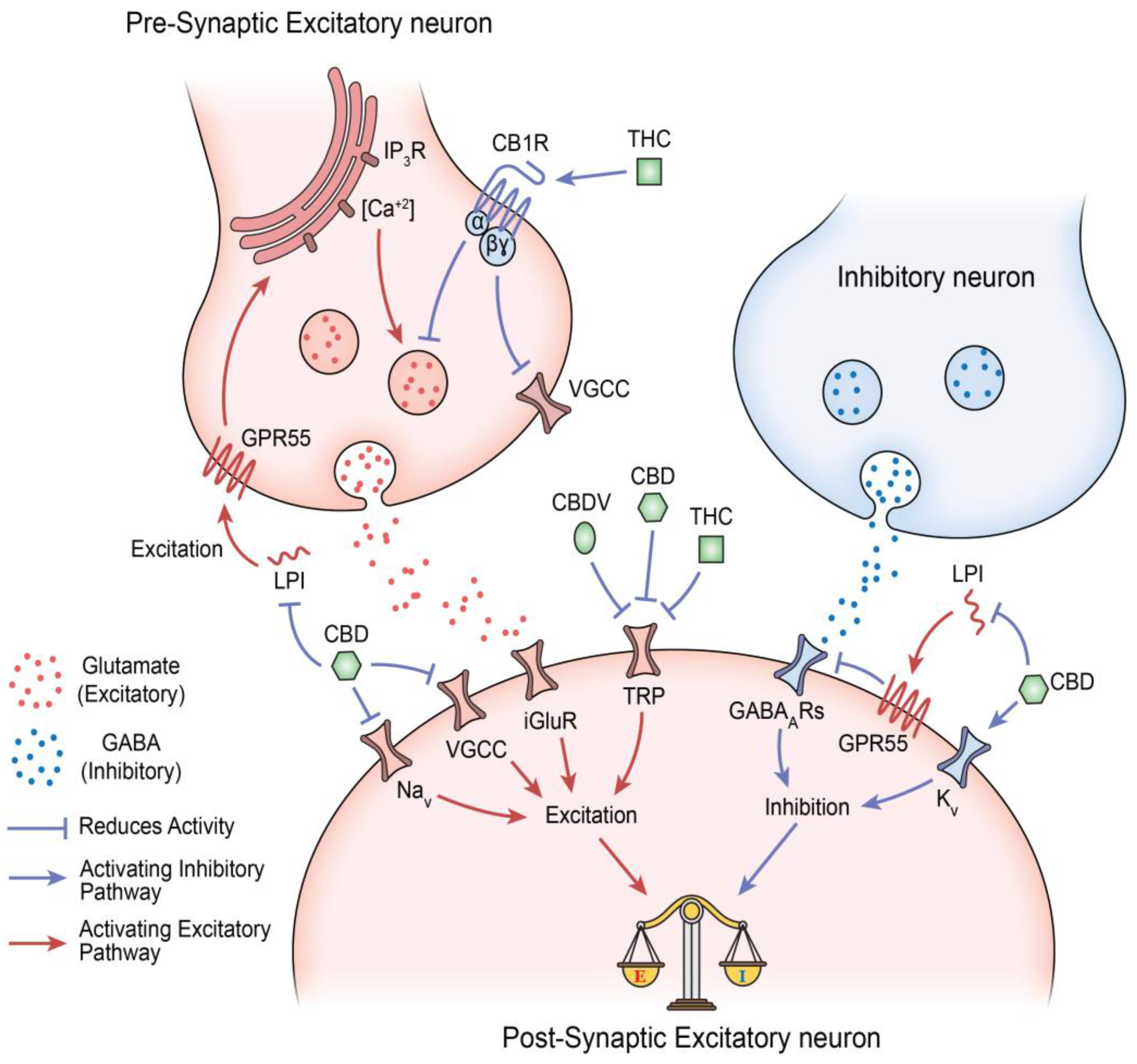
Disclaimer/Publisher’s Note: The statements, opinions and data contained in all publications are solely those of the individual author(s) and contributor(s) and not of MDPI and/or the editor(s). MDPI and/or the editor(s) disclaim responsibility for any injury to people or property resulting from any ideas, methods, instructions or products referred to in the content. |
© 2026 by the authors. Licensee MDPI, Basel, Switzerland. This article is an open access article distributed under the terms and conditions of the Creative Commons Attribution (CC BY) license (http://creativecommons.org/licenses/by/4.0/).
Copyright: This open access article is published under a Creative Commons CC BY 4.0 license, which permit the free download, distribution, and reuse, provided that the author and preprint are cited in any reuse.


