Preprint
Article

This version is not peer-reviewed.

Spherical Mesoporous Silica Synthesis for Catalytic Applications: pH-Regulated Mesostructural Evolution Mediated by Ostwald Ripening

Submitted:

02 April 2026

Posted:

03 April 2026

You are already at the latest version

Abstract
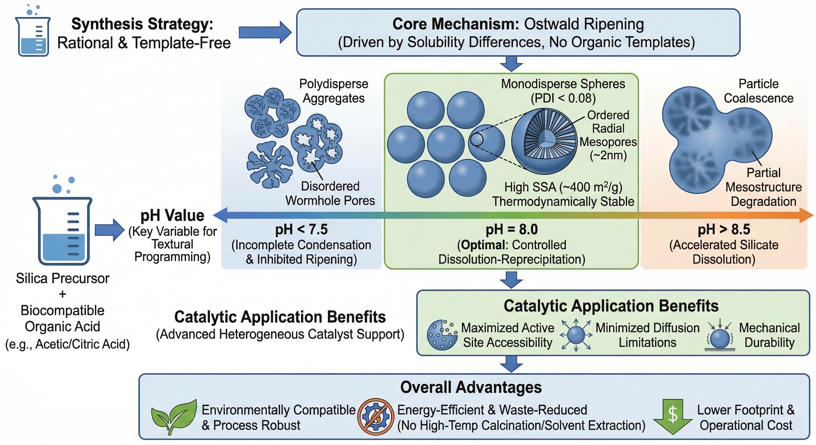
This study introduces a rational, template-free synthetic strategy for the scalable preparation of high-performance monodisperse spherical mesoporous silica particles (MSPs), engineered specifically as advanced heterogeneous catalytic supports. Leveraging Ostwald ripening as the core morphogenetic driver—rather than conventional organic structure-directing agents—the approach achieves both environmental compatibility and process robustness. Precise pH modulation to 8.0 using biocompatible organic acids (e.g., acetic or citric acid) enables controlled silica dissolution–reprecipitation kinetics, yielding MSPs with exceptional sphericity (PDI < 0.08), narrow size distribution, a specific surface area of up to 484 m²/g, uniform pore diameters centered at ~2 nm, and radially aligned, thermodynamically stabilized mesochannels—structural attributes that collectively satisfy stringent design criteria for high-efficiency catalytic carriers, including maximized active-site accessibility, minimized diffusion limitations, and mechanical resilience under reaction conditions. A systematic pH-screening study reveals a distinct structural transition: at pH < 7.5, incomplete condensation and suppressed ripening yield polydisperse aggregates with disordered worm-like porosity; at pH > 8.5, accelerated silicate dissolution induces particle coalescence and partial mesostructural degradation. Critically, pore ordering, channel dimensionality, surface area, and particle morphology are all quantitatively modulated by pH—establishing it as a master variable for hierarchical textural programming. This study compares the methoxychlor (MXC) degradation efficiency of polyhedral Bi2WO6 and MSP/Bi2WO6 under identical irradiation conditions to assess MSP’s catalytic impact. Mechanistic analysis of charge dynamics, interfacial electron transfer, and active species reveals how MSP enhances photocatalytic activity.
Keywords: 
;  ;  ;  ;  ;  

1. Introduction

Mesoporous silica (MS) is widely used in catalysis, adsorption, and biomedicine due to its ordered hexagonal pores, tunable pore size, and high surface area [1,2,3]. MCM-41—a representative M41S material—is typically made using CTAB as a template and TEOS as the silica source under alkaline sol–gel conditions [4,5,6]. But this method has major drawbacks: template removal produces foaming, surfactant-rich wastewater; TEOS and TMOS are expensive and energy-intensive; and synthesis requires pH 9–12, which strongly affects hydrolysis, condensation, and final structure—leading to poor particle uniformity and dispersibility [5,6]. Even optimized CTAB/TEOS systems struggle to control sub-micrometer particle size. Rizzi et al. showed that NaOH-based syntheses need high temperature and large water volumes to limit aggregation—yielding 40–150 nm particles only at the cost of efficiency and scalability. In short, current methods still rely heavily on organic templates, costly precursors, and high-temperature calcination—limiting sustainability and practical scale-up [5].
To advance green manufacturing paradigms, emerging strategies have shifted toward template-minimized or template-free syntheses employing low-cost, abundant sodium silicate (water glass) as the silica source. Inorganic salts—such as ammonium chloride or sodium sulfate—are leveraged to modulate silicate hydration, condensation dynamics, and surface hydrophilicity; alternatively, mild bio-derived ligands or reductants—including fatty acids and plant extracts—have been introduced to guide pore formation without compromising tunability of pore size or humidity-responsive adsorption performance[7,8]. Critically, several of these approaches eliminate the need for high-temperature calcination, thereby preserving structural integrity (e.g., preventing pore sintering or collapse) and retaining a higher density of surface silanol groups—features essential for downstream functionalization (e.g., amination) and broadening application scope in sensing, drug delivery, and heterogeneous catalysis[5,6,7,8].
Concurrently, Ostwald ripening has emerged as a powerful, thermodynamically driven strategy for engineering spherical silica architectures with controlled particle size, hierarchical porosity, and tailored hollow/mesoporous morphologies. By rationally modulating solution pH, ionic strength, or introducing selective salts and reductants, dissolution–reprecipitation equilibria can be directed under mild, near-ambient conditions to yield monodisperse mesoporous or hollow silica spheres with tunable pore diameters[9,10]. Recent mechanistic studies indicate that acid/base switching, cation/anion identity, and their coupling with silicate hydrolysis/condensation rates during ripening govern the emergence and intensity of small-angle X-ray scattering (SAXS) reflections—particularly the (100) peak—which correlate strongly with unimodal versus bimodal particle size distributions[5,6,7,8,9]. Sun et al. developed a NaBH₄-assisted hydrothermal method (designated D1), achieving unprecedented precision in mesopore diameter control across 18–31 nm in spherical mesoporous silica (SMS)—surpassing the previously established ~30-nm threshold for reproducible mesopore uniformity. Complementarily, Bucci et al. integrated experimental characterization with quantitative Ostwald ripening modeling, using benzaldehyde hydrogenation as a model reaction to rigorously evaluate Pt-loaded SMS (D2)[10]. Their work established a direct structure–function relationship: pore confinement governs both metal nanoparticle stabilization and intrinsic catalytic turnover frequency—providing a mechanistic basis for designing sintering-resistant catalysts.
The photocatalytic performance of Bi2WO6 is intrinsically limited by its low specific surface area and sparse distribution of active sites[11]. Integrating monodisperse spherical mesoporous silica—featuring well-defined mesochannels and a high specific surface area—enhances dispersion of the active phase, suppresses agglomeration, facilitates charge-carrier separation and migration, and synergistically improves interfacial redox kinetics and photon utilization efficiency. As a result, the composite exhibits significantly enhanced photocatalytic activity, presenting a rationally grounded strategy for engineering high-performance hybrid photocatalytic materials.
This study reports a surfactant-free, low-waste, and scalable synthesis of spherical mesoporous silica via pH-regulated Ostwald ripening. At pH ≈ 8, the resulting material exhibits high monodispersity, a specific surface area of ∼484 m²·g⁻¹, and a narrow pore size distribution centered at ∼2 nm. The protocol eliminates the need for high-temperature calcination and employs inexpensive, commercially available—or waste-derived—silica precursors (e.g., technical-grade sodium silicate), thereby reducing environmental footprint, simplifying operational requirements, and enabling scalable fabrication of Bi2WO6–mesoporous silica hybrid photocatalysts with enhanced performance.

2. Materials and Methods

2.1. Experimental Materials

All reagents were of analytical grade and used without further purification. The chemicals used in this work included sodium silicate (Na₂SiO₃·9H₂O), nitric acid (HNO), sodium hydroxide (NaOH), potassium chloride (KCl), potassium fluoride (KF), hydrochloric acid (HCl), phenolphthalein indicator, anhydrous ethanol (CHOH), tartaric acid, sodium tungstate dihydrate (NaWO·2HO), bismuth nitrate pentahydrate (Bi(NO)·5HO), glacial acetic acid, mesoporous silica particles (MSP), and deionized water. Sodium silicate, HNO₃, NaOH, KCl, KF, HCl, phenolphthalein, ethanol, and tartaric acid were purchased from Shanghai Lingfeng Chemical Reagent Co., Ltd.

2.2. Synthesis Method

2.2.1. Preparation of Mesoporous Silica by Ostwald Aging Method

A 50 mL aliquot of 0.50 mol/L sodium silicate solution (prepared from NaSiO·9HO and deionized water) was placed in a reaction vessel and monitored with a calibrated pH meter. Under stirring, 0.5 mol/L HCl was added slowly via constant-flow pump until pH 10—triggering turbidity. The mixture was stirred for 1 h. Then, 0.2 mol/L tartaric acid was added at 10 rpm to reach the target pH, followed by another 1 h of stirring. After static aging for 12 h, the product was centrifuged, washed with deionized water until neutral, and dried at 60°C under vacuum.

2.2.2. Preparation of Polyhedral Bi2WO6-Loaded MSP

Briefly, 0.5 mmol Na2WO4·2H2O was dissolved in 10 mL deionized water to form Solution A. Meanwhile, 1.0 mmol Bi(NO3)3·5H2O was dissolved in 25 mL of a mixed solvent composed of H2O, acetic acid, and ethanol (3:1:1, v/v/v) to form Solution B. Then, 0.5 g MSP was added to Solution B and magnetically stirred until uniformly dispersed.
Next, Solution A was slowly introduced dropwise into Solution B using a peristaltic pump. After stirring at room temperature for 30 min, the mixture was transferred into a Teflon-lined stainless-steel autoclave and maintained at 180 °C for 12 h. The obtained solid was collected by filtration, washed several times, dried, and calcined to yield polyhedral MSP@Bi2WO6.

2.2.3. Photocatalytic Degradation Experiments

Photocatalytic degradation of MXC was carried out in a typical batch system. Specifically, 0.50 g of catalyst was dispersed in 100 mL of AOT–MXC buffer solution (pH = 8), where the initial concentrations of MXC and AOT were 40 mg L⁻¹ and 2 g L⁻¹, respectively. Prior to light irradiation, the suspension was magnetically stirred in the dark for 1 h to establish adsorption–desorption equilibrium between MXC and the catalyst surface. Subsequently, the reaction mixture was irradiated under a 500 W halogen tungsten lamp with an illumination intensity of 32.2 × 10³ μW cm⁻² under continuous magnetic stirring. At predetermined time intervals (1, 2, 3, 4, 5, and 6 h), aliquots were withdrawn and immediately cooled to room temperature. The suspension was then centrifuged to remove the catalyst. The supernatant was extracted twice with 15 mL of n-hexane, and the combined organic phases were concentrated using a rotary evaporator under reduced pressure. The obtained residue was redissolved in n-hexane to a final volume of 5 mL, followed by the addition of 1 mL of octacosane solution (30 mg L⁻¹) as an internal standard. The resulting solution was analyzed by gas chromatography–mass spectrometry (GC/MS).

2.2.4. Determination of Adsorption and Degradation Performance

Considering that MXC can be partially adsorbed onto the catalyst surface due to its high specific surface area, the overall removal of MXC was decoupled into adsorption and photocatalytic degradation contributions. The adsorption rate, removal rate, and degradation rate were calculated according to the following equations:
Adsorption rate: (C0-Ca)/C0*100% ; removal rate : (C0-Cd)/C0*100% ; Degradation rate: (Ca-Cd)/C0*100% , where C0 ,Ca and C0 represent the initial concentration, the concentration after adsorption equilibrium in the dark, and the concentration after photocatalytic reaction, respectively.

3. Results

3.1. Materials and Characterization

3.1.1. X-Ray Diffraction Analysis of G-O

Small-angle X-ray diffraction (SAXRD) patterns of samples G-O-6, G-O-7, G-O-8, and G-O-9 are presented in Figure 1. All samples synthesized at pH 6, 8, and 9 exhibit distinct (100) reflections at 2θ = 1.87°, 2.32°, and 2.28°, respectively—confirming the formation of ordered mesoporous frameworks. Sample G-O-6 displays only a weak (100) peak with no higher-order reflections, indicating incipient mesostructural ordering and limited long-range periodicity. In contrast, G-O-9 shows an intensified (100) peak but remains devoid of (110) or (200) reflections, suggesting improved local ordering yet insufficient structural coherence for full hexagonal symmetry. Sample G-O-8—prepared at pH 8—exhibits well-resolved (100), (110), and (200) peaks at 2θ = 2.32°, 3.72°, and 4.62°, respectively, consistent with a partially ordered MCM-41-type structure. This assignment is supported by the use of an inorganic, amorphous silica precursor that undergoes controlled hydrolysis under mildly alkaline conditions. By comparison, G-O-7 (pH 7) yields only a broad hump centered near 2θ = 2.1°, indicative of an amorphous, non-porous silica phase. This arises from suppressed silicate hydrolysis in near-neutral aqueous media: silica solubility is minimal below pH 10, and hydrolysis kinetics are markedly sluggish at pH 7—particularly in the absence of added electrolytes—resulting in negligible condensation and network formation. Notably, colloidal silica sols exhibit maximal stability within pH 5–8 but rapidly aggregate outside this range; thus, the low reactivity observed for G-O-7 reflects both thermodynamic immobility and kinetic inhibition under near-neutral conditions.
The low-angle XRD patterns of both pristine mesoporous silica (MSP) and the loaded sample exhibit characteristic reflections corresponding to an ordered mesostructure. Taking the (100) diffraction peak as an example, the interplanar spacing (d₁₀₀) was calculated based on Bragg’s law (2d sinθ = nλ, n = 1, λ = 0.15406 nm). The calculated d₁₀₀ values are approximately 4.20 nm (2θ = 2.10°) and 4.33 nm (2θ = 2.04°), indicating only negligible variation after loading. The corresponding lattice parameter (a₀), calculated using a₀ = 2d₁₀₀/√3, is in the range of 4.85–5.00 nm, further confirming that the long-range periodic mesostructure of silica is well preserved. In addition, the crystallite size estimated from the Scherrer equation (D = Kλ/(β cosθ)) is approximately 30–45 nm, suggesting the formation of nanoscale crystalline domains of the loaded phase.These results indicate that the introduction of the active component does not significantly alter the mesostructural ordering of the silica framework.

3.1.2. FT-IR Spectroscopy Analysis

FT-IR spectra of all samples were highly consistent; the spectrum of G-O-8 is shown in Figure 1c as a representative example. In the absence of calcination, a broad band centered at 3453 cm⁻¹ is attributed to O–H stretching vibrations of surface silanol groups, confirming their preservation and implying enhanced surface reactivity. The intense band at 1074 cm⁻¹ arises from asymmetric Si–O–Si bridging bond stretching—a hallmark of amorphous silica networks. Additional characteristic silica vibrations are observed at 952 cm⁻¹ (Si–OH bending), 801 cm⁻¹ (Si–O–Si symmetric stretching), and 454 cm⁻¹ (Si–O bending). The band at 1642 cm⁻¹ corresponds to H–O–H bending modes of adsorbed water. Collectively, these features confirm SiO as the dominant phase. A weak but distinct absorption at 1431 cm⁻¹—along with subsidiary peaks at 872 cm⁻¹ and 711 cm⁻¹—is assigned to calcium carbonate (CaCO) impurities. Specifically, the 1431 cm⁻¹ band corresponds to the CO²⁻ asymmetric stretch, 872 cm⁻¹ to the out-of-plane CO²⁻ deformation, and 711 cm⁻¹ to the in-plane O–C–O deformation—spectral signatures diagnostic of calcite-phase CaCO. These impurities originate from incomplete acid washing of the sodium silicate precursor, which contains residual CaCO as a common industrial contaminant. Their presence is corroborated by XRD analysis of the starting material and aligns quantitatively with the low-intensity carbonate reflections observed in the as-synthesized sample.

3.1.3. SEM Analysis

To validate the particle size distributions obtained from dynamic light scattering (DLS), high-resolution scanning electron microscopy (HRSEM) was performed on samples G-O-6, G-O-7, and G-O-8 (Figure 2). As shown in Figure 2A, G-O-8 exhibits uniform, spherical morphology with a narrow size distribution—consistent with efficient Ostwald ripening under mildly basic conditions (pH 8). In contrast, G-O-6 (pH 6) displays irregular particle shapes and significant polydispersity (Figure 2B), indicating suppressed colloidal stability and disrupted growth kinetics under acidic conditions. G-O-7 (pH 7) shows intermediate morphology—less uniform than G-O-8 but more defined than G-O-6—reflecting suboptimal hydrolysis–condensation balance near neutrality. Collectively, these HRSEM images demonstrate that pH is a critical determinant of particle uniformity and structural fidelity in this surfactant-free silica synthesis.

3.1.4. Particle Size Analysis

Particle size distributions were determined using a Malvern Mastersizer 2000 laser diffraction analyzer (measurement range: 2 nm–3 μm). At pH 8, sample G-O-8 exhibited a unimodal distribution centered at 0.83 μm (Z-average = 829.5 nm), with a polydispersity index (PDI) of 0.572—within the instrument’s validated operating range and indicative of moderate colloidal uniformity. In contrast, G-O-6 (pH 6) displayed a bimodal distribution: 37.9% of particles at 0.97 μm and 62.1% at 1.03 μm, yielding a Z-average of 1005 nm (1.05 μm) and a PDI of 0.711—approaching the upper limit of acceptable dispersion. G-O-7 (pH 7) yielded a PDI > 1.0, exceeding the analyzer’s reliability threshold and confirming severe aggregation and morphological heterogeneity.
Size distributions are presented as volume-based histograms overlaid with Gaussian fits. The x-axis denotes particle diameter (μm); the y-axis represents relative volume frequency. A systematic offset exists between laser diffraction and SEM measurements: the former reports hydrodynamic diameters—enlarged by solvation shells and electrostatic repulsion—whereas the latter yields dry, projected diameters. Consequently, laser-derived averages are consistently higher; for G-O-8, the primary mode appears at ~0.8 μm (laser) versus ~0.35 μm (SEM), yet both confirm unimodal, monodisperse behavior under alkaline conditions. Under acidic conditions (G-O-6), both techniques resolve bimodality—laser diffraction at 0.97 μm and 1.03 μm, SEM at ~0.7 μm and ~1.5 μm—indicating heterogeneous nucleation and growth arising from pH gradients induced by tartaric acid addition. These gradients disrupt the uniformity of silicate condensation, which proceeds via parallel acid- and base-catalyzed pathways: protonated silanol species dominate under low pH, while deprotonated silicates drive rapid condensation under alkaline conditions. The reaction can be expressed as follows:
Preprints 206331 i001
The resulting disilicic acid aggregates undergo further hydrolytic condensation to yield trisilicic acid and, ultimately, polysilicic acid oligomers and networks. Uncontrolled reaction kinetics—particularly under acidic conditions—promote rapid, heterogeneous condensation, leading to premature gelation. This behavior aligns quantitatively with the hydrolysis states and gelation onset times documented in Table 2.3.
In contrast, under alkaline conditions, silicate ions and orthosilicate ions participate in oxygen condensation reactions. The corresponding reaction equation is as follows:
Preprints 206331 i002
As the reaction progresses, the initial silicate undergoes polymerization, transitioning from monomers to dimers and subsequently forming higher-order polymers. Under specific conditions, external pressure and heat can be applied to further promote the polymerization of polysilicates, thereby increasing the degree of polymerization and facilitating the continuous growth of particle size, ultimately leading to the formation of larger silica particles. The emergence of a bimodal distribution curve can be attributed to the formation of "secondary particles." Silica gel particles may be formed through two distinct mechanisms: one is the dehydration polymerization of numerous active silicate molecules, which leads to the generation of primary silica gel particles. The other mechanism involves pre-formed silica gel particles serving as cores, onto which new silica molecules aggregate, resulting in continuous particle size growth. This phenomenon is representative of the Ostwald ripening process. The corresponding reaction equation is as follows:
m H 2 Si O 3 + n H 2 Si O 3 n + m Si O 2 + n + m H 2 O  
nSi O 2 + H 2 Si O 3 n + 1 Si O 2 + H 2 O
During the growth of the primary silica gel particle, the majority of newly generated silicate molecules are deposited onto the surface of the primary particle. However, a fraction of these molecules nucleate independently, resulting in the formation of secondary particles. Under neutral conditions, the absence of electrolytes and the lack of nucleating SiO crystallites lead to an irregular and disordered hydrolysis process. Consequently, the morphology of the resulting silica material becomes uncontrolled and non-uniform.

3.1.5. TEM and HRTEM Analysis

As shown in Figure 3, the synthesized G-O-8 sample exhibits a typical spherical mesoporous silica morphology with well-developed internal porous structures. The low-magnification TEM image (Figure 3a) reveals relatively uniform spherical particles with clear boundaries and good dispersion, indicating stable formation under pH 8 conditions. The evident internal contrast suggests a porous rather than dense structure. The internal structure of individual particles (Figure 3b) shows clearly visible stripe-like channels, confirming the formation of an integrated mesoporous framework rather than a disordered amorphous phase. The HRTEM image (Figure 3c) further demonstrates highly ordered, parallel mesoporous channels with uniform pore distribution and well-defined pore walls, indicating a high degree of structural ordering. Such an ordered architecture is beneficial for mass transfer and catalytic applications. At a larger scale (Figure 3d), the particles maintain a generally spherical morphology, although slight aggregation can be observed locally. Importantly, no obvious collapse of the internal pore structure is detected. To further evaluate structural stability after loading, the mesostructural parameters were analyzed by XRD. The calculated d₁₀₀ spacing (~4.20–4.33 nm) and corresponding lattice parameter (a₀≈4.85–5.00 nm) show negligible variation, indicating that the long-range periodic framework is well preserved. In addition, the crystallite size of the loaded phase, estimated by the Scherrer equation (~30–45 nm), corresponds to nanoscale domains rather than pore dimensions. The pore size observed from TEM is consistent with the values obtained from N₂ adsorption–desorption analysis, confirming reliable structural characterization. More importantly, the ordered pore arrangement observed in TEM agrees well with the d-spacing derived from XRD.
Overall, these complementary results demonstrate that the mesoporous silica framework remains structurally intact after modification, with negligible changes in pore size and long-range ordering.

3.1.6. BET Analysis of Mesoporous Silica Products

The nitrogen adsorption isotherms and pore size distributions of samples G-O-6, G-O-7, G-O-8, and G-O-9 are shown in the figure. For G-O-6, the isotherm follows a Type IV pattern according to IUPAC classification. In the low-pressure range (p/p < 0.3), the curve resembles that of microporous materials, indicating single-layer adsorption and the presence of small mesopores. As relative pressure increases to 0.3–0.45, a clear inflection at p/p = 0.39 appears, along with a narrow hysteresis loop. The pore size distribution peaks around 2.3 nm and is broader than that of G-O-8, suggesting coexisting mesoporous and microporous structures. The H3-type hysteresis loop indicates capillary condensation in non-uniform slit-like pores. This is consistent with earlier XRD and particle size distribution results.
Figure 4. Nitrogen adsorption isotherms and pore size distribution of G-O-6(a), G-O-7 (b), G-O-8 (c), G-O-9 (d).
Preprints 206331 g004
For G-O-7, the nitrogen adsorption isotherm follows a Type I pattern according to IUPAC classification. In the low-pressure range (p/p < 0.3), no capillary condensation occurs, and adsorption resembles single-layer behavior. No significant inflection is seen in the mid-pressure range, indicating no abrupt N uptake. Combined with pore size distribution and XRD data, this suggests that under neutral conditions, the silica source undergoes natural hydrolysis, forming mainly irregular microporous structures.
For G-O-8 and G-O-9, the nearly parallel adsorption and desorption curves with distinct hysteresis loops indicate Type IV isotherms. However, G-O-9 shows a narrower H3-type hysteresis loop, typical of capillary condensation in non-uniform slit-like pores. Its adsorption curve inflects at p/p = 0.35, with a pore size distribution centered at 2.5 nm.
For G-O-8, N₂ adsorption increases gradually below p/p = 0.3. At p/p₀ = 0.39, an inflection point appears, indicating capillary condensation in mesoporous channels. Larger pores condense at higher pressures. The adsorption plateau suggests saturation. A hysteresis loop forms when p/p exceeds 0.3, due to incomplete multilayer adsorption-desorption. The loop is gentle and closes at high pressure, indicating an H4-type loop. This reflects uniform pore size and shape, with a BJH pore size of 3.02 nm, consistent with HRTEM results. The isotherm is typical of mesoporous materials.
Specific pore structure parameters are summarized in Table 1.

3.2. Structural Characterization and Photocatalytic Performance of MSP @ BaWO₄

3.2.1. SEM and TEM Analysis of BaWO₄ and BaWO₄@MSP

The morphology and microstructure of the as-prepared BaWO and BaWO@MSP composites were investigated by SEM and TEM, as shown in Figure X. The SEM image of pristine BaWO reveals well-defined polyhedral particles with relatively smooth surfaces and clear edges, indicating good crystallinity and uniform crystal growth. The particle size is mainly in the submicrometer to micrometer range, and a certain degree of aggregation can be observed, which is typical for hydrothermally synthesized tungstate materials.
In contrast, the TEM image of BaWO₄@MSP exhibits a significantly different morphology. The BaWO₄ nanoparticles are uniformly distributed on the surface of mesoporous silica spheres, forming a composite structure with a roughened surface. The MSP support maintains its spherical morphology and porous framework, while numerous small dark dots corresponding to BaWO₄ nanoparticles are clearly visible, indicating successful loading and good dispersion of the active phase.
Importantly, no obvious large-scale aggregation of BaWO₄ is observed after loading, suggesting that the mesoporous silica effectively inhibits particle growth and agglomeration. The intimate interfacial contact between BaWO₄ and MSP is expected to facilitate efficient charge transfer and improve the accessibility of active sites.
Overall, the SEM and TEM results confirm that MSP acts as an effective support for dispersing BaWO₄ nanoparticles, providing a well-defined hierarchical structure that is favorable for enhanced photocatalytic performance.
Figure 5. SEM image of BaWO₄ (a) and TEM image of BaWO₄@MSP (b).
Preprints 206331 g005

3.2.2. BET Surface Area and Pore Volume Analysis

To further evaluate the effect of MSP loading on the textural properties of the catalyst, the specific surface area and pore volume of polyhedral BiWO and BiWO/MSP were compared, as shown in Figure X. The pure polyhedral BiWO exhibited a very low specific surface area of 1.17 m²/g and a pore volume of 0.002010 cm³/g. After loading onto MSP, the specific surface area of Bi₂WO₆/MSP increased to 6.69 m²/g, while the pore volume increased to 0.012756 cm³/g. These results indicate that the introduction of MSP significantly improves the textural properties of the composite catalyst, providing more accessible surface sites and enlarged pore space for adsorption and mass transfer, which is beneficial for the subsequent catalytic degradation performance.
Table 2. Textural properties of Bi₂WO₆ and MSP@Bi₂WO₆.
Sample Specific surface area (m²·g⁻¹) Pore volume (cm³·g⁻¹)
Bi2WO6 1.17 0.002010
MSP@Bi2WO6 6.69 0.012756

3.2.3. Optical Properties and Photocatalytic Performance

The optical absorption properties of polyhedral BiWO and BiWO/MSP were investigated by UV–vis diffuse reflectance spectroscopy (DRS), as shown in Figure 6a. Both samples exhibit strong absorption in the ultraviolet region, while the composite BiWO/MSP shows enhanced absorption intensity and a slight red shift of the absorption edge compared with pure BiWO. This indicates that the introduction of MSP improves the light-harvesting capability of the catalyst, which is beneficial for photocatalytic reactions.
The effect of catalyst dosage on the degradation efficiency is presented in Figure 6b. The degradation efficiency increases with increasing catalyst dosage from 3 to 5 g L⁻¹ and reaches a maximum at approximately 5 g L-1. This enhancement can be attributed to the increased number of active sites. However, further increasing the catalyst dosage leads to a decrease in degradation efficiency, which may be due to light shielding and reduced light penetration in the solution.
The photocatalytic degradation process as a function of reaction time is shown in Figure 6c. The removal efficiency increases steadily with irradiation time, indicating continuous degradation of MXC. Under the optimized conditions (pH = 8, 500 W light source, and catalyst dosage of 5.14 g L⁻¹), the composite catalyst exhibits high photocatalytic activity.
A comparison of the degradation performance between pure BiWO and BiWO/MSP is shown in Figure 6d. The removal efficiency of BiWO/MSP reaches 87.23%, which is significantly higher than that of pure polyhedral BiWO (67.15%), demonstrating the effectiveness of MSP loading in enhancing photocatalytic performance.
The improved photocatalytic activity of BiWO/MSP can be attributed to the synergistic effect between BiWO and MSP. The introduction of MSP enhances the dispersion of BiWO particles, increases the specific surface area and pore volume, and provides more accessible active sites. In addition, the porous structure facilitates mass transfer and improves the interaction between the catalyst and pollutant molecules, leading to superior degradation performance.

4. Discussion

This study demonstrates that pH-regulated Ostwald ripening constitutes an effective, environmentally benign, and template-free strategy for synthesizing monodisperse mesoporous silica spheres with well-defined structural features. Among all samples, the material prepared under near-neutral conditions (pH ≈ 8) exhibits the most favorable microstructure—characterized by uniform spherical morphology, long-range ordered mesochannels, and a narrow particle size distribution. These attributes arise from a finely tuned balance between silica dissolution and reprecipitation, enabling controlled mass redistribution and progressive structural refinement.
At pH <7.5, slow hydrolysis and condensation hinder full framework formation, resulting in weak porosity and poor structural integrity. At pH >8.5, rapid dissolution–reprecipitation causes pore collapse, particle fusion, and loss of mesoscopic order. Thus, Ostwald ripening—and the final structure—depends critically on pH, which controls the balance between growth and dissolution. The sample with a pH of approximately 8 exhibits a 2D hexagonal ordered structure, uniform mesopores and a high specific surface area at 484 m2·g-1. This optimized architecture facilitates rapid mass transport, promoting efficient diffusion of reactants to interior active sites. Consequently, accessible active site density increases, dispersion improves, and nanoparticle aggregation is suppressed—thereby enhancing catalytic efficiency. Moreover, the interconnected mesopore network shortens intraparticle diffusion lengths and mitigates concentration polarization, a key advantage in photocatalytic degradation of sterically hindered organic pollutants. The integration of mesoporous silica particles (MSP) not only extends the visible-light absorption range of Bi2WO6 but also enhances its colloidal stability and dispersion homogeneity. Following MSP loading, Bi2WO6 exhibits markedly improved light-harvesting efficiency, uniform nanoparticle dispersion, and increased density of accessible catalytically active sites. Under optimized reaction conditions, the photocatalytic removal efficiency of metolachlor (MXC) rises from 67.15% to 87.23%, representing a substantial enhancement in overall catalytic performance. In summary, this work establishes a robust structure–performance correlation, demonstrating that precise modulation of Ostwald ripening via pH control enables concurrent optimization of textural properties and catalytic functionality. The scalable, surfactant-free synthesis protocol advances the design of high-performance mesoporous supports for heterogeneous catalysis and environmental remediation.

Author Contributions

X.D: Investigation, Data curation, Writing-original draft, Writing-review and editing, Conceptualization. L.w.: Methodology Data curation, Validation, Writing-review and editing. Y.Y.: Supervision, Writing-review and editing, Funding acquisition. All authors have read and agreed to the published version of the manuscript.

Funding

Please add: “This work was supported by the National Natural Science Foundation of China (20577010, 20971043)”

Data Availability Statement

Data will be made available on request.

Acknowledgments

We extend our heartfelt gratitude to Anbang Dai for his significant contributionsto the conceptualization and validation of this work.

Conflicts of Interest

The authors declare that they have no known competing financial interests orpersonal relationships that could have appeared to influence the work reported in this paper.

References

  1. KRESGE, C T; LEONOWICZ, M E; ROTH, W J; et al. Ordered mesoporous molecular sieves synthesized by a liquid-crystal template mechanism. Nature 1992, 359, 710–2. [Google Scholar] [CrossRef]
  2. ZHAO, D; FENG, J; HUO, Q; et al. Triblock Copolymer Syntheses of Mesoporous Silica with Periodic 50 to 300 Angstrom Pores. 1998, 279, 548–52. [Google Scholar] [CrossRef] [PubMed]
  3. YANAGISAWA, T; SHIMIZU, T; KURODA, K; et al. The Preparation of Alkyltrimethylammonium–Kanemite Complexes and Their Conversion to Microporous Materials. Bulletin of the Chemical Society of Japan 2006, 63, 988–92. [Google Scholar] [CrossRef]
  4. WU, S-H; MOU, C-Y; LIN, H-P. Synthesis of mesoporous silica nanoparticles. Chemical Society Reviews 2013, 42, 3862–75. [Google Scholar] [CrossRef] [PubMed]
  5. VALLET-REGí, M; SCHüTH, F; LOZANO, D; et al. Engineering mesoporous silica nanoparticles for drug delivery: where are we after two decades? Chemical Society Reviews 2022, 51, 5365–451. [Google Scholar] [CrossRef] [PubMed]
  6. XU, B; LI, S; SHI, R; et al. Multifunctional mesoporous silica nanoparticles for biomedical applications. Signal Transduction and Targeted Therapy 2023, 8, 435. [Google Scholar] [CrossRef] [PubMed]
  7. HEIDARNEZHAD, Z; GHORBANI-CHOGHAMARANI, A; TAHERINIA, Z. Surfactant-free synthesis of mesoporous silica materials (Using tetraetylorthosilicate and oleic acid): Preparation, characterization, and catalytic applications. Journal of Molecular Structure 2024, 1306, 137807. [Google Scholar] [CrossRef]
  8. SANGTEANTONG, P; CHAINARONG, K; DONPHAI, W; et al. Green synthesis of surfactant-free mesoporous silica with strong hydrophilicity via metal salt modifications for moisture adsorption. Reaction Chemistry & Engineering 2024, 9, 816–24. [Google Scholar] [CrossRef]
  9. SUN, R; ZHANG, A; SUN, H D; et al. Facile synthesis of monodispersed mesoporous silica nanoparticles with ultra-large mesopores through a NaBH4-assisted hydrothermal process. Journal of Non-Crystalline Solids 2024, 627, 122820. [Google Scholar] [CrossRef]
  10. BUCCI, G; GADELRAB, K; CARTER, W C. Mesoscale Model for Ostwald Ripening of Catalyst Nanoparticles. Journal of The Electrochemical Society 2021, 168, 054515. [Google Scholar] [CrossRef]
  11. ELAOUNI, A; EL OUARDI, M; BAQAIS, A; et al. Bismuth tungstate Bi2WO6: a review on structural, photophysical and photocatalytic properties. RSC Advances 2023, 13, 17476–94. [Google Scholar] [CrossRef] [PubMed]
Figure 1. XRD of product a: G-O-6.b: G-O-8.c: G-O-9 d: G-O-7 and IR spectrum of G-O-8
Preprints 206331 g001
Figure 2. HRSEM of the product and the particle size analysis HRSEM (A:G-O-8 B:G-O-6 ) Particle size analysis (a:G-O-8 b:G-O-6); Particle size distribution and cumulative curve of product (c:G-O-8 d:G-O-6).
Preprints 206331 g002
Figure 3. TEM and HRTEM images of the G-O-8: (a,b,d) TEM images and (c) HRTEM image.
Preprints 206331 g003
Figure 6. Optical properties and photocatalytic performance of Bi₂WO₆ and Bi₂WO₆/MSP: (a) UV–vis diffuse reflectance spectra; (b) effect of catalyst dosage on degradation efficiency; (c) degradation efficiency as a function of irradiation time; (d) comparison of degradation efficiency between Bi₂WO₆ and Bi₂WO₆/MSP.
Preprints 206331 g006
Table 1. Pore structure parameters of samples.
Sample D/nm SBET/(m2·g-1) V/(cm3·g-1)
G-O-6 2.3 352 0.228
G-O-7 <2 201 0.116
G-O-8 3.02 484 0.533
G-O-9 2.51 411 0.284
Disclaimer/Publisher’s Note: The statements, opinions and data contained in all publications are solely those of the individual author(s) and contributor(s) and not of MDPI and/or the editor(s). MDPI and/or the editor(s) disclaim responsibility for any injury to people or property resulting from any ideas, methods, instructions or products referred to in the content.
Copyright: This open access article is published under a Creative Commons CC BY 4.0 license, which permit the free download, distribution, and reuse, provided that the author and preprint are cited in any reuse.
Prerpints.org logo

Preprints.org is a free preprint server supported by MDPI in Basel, Switzerland.

Subscribe

Disclaimer

Terms of Use

Privacy Policy

Privacy Settings

© 2026 MDPI (Basel, Switzerland) unless otherwise stated