Preprint
Article

This version is not peer-reviewed.

Protective Effect of Placental Mesenchymal Stromal Cells in an In Vitro Model of Parkinson’s Disease Using Differentiated Neuroblastoma Cells

Submitted:

30 March 2026

Posted:

31 March 2026

You are already at the latest version

Abstract

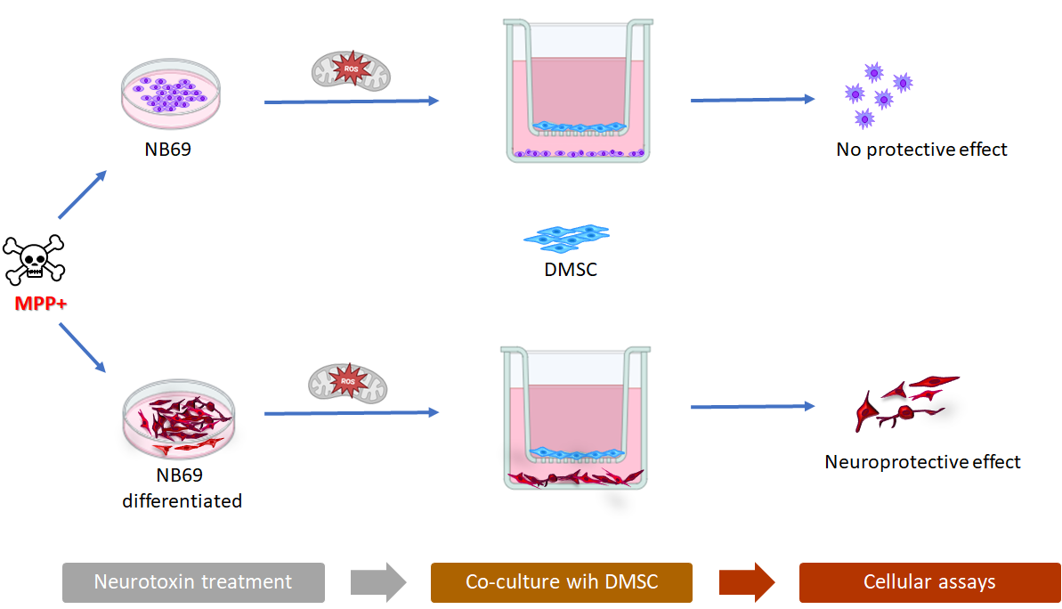
Parkinson’s disease (PD) is the second most prevalent neurodegenerative disorder, characterized by the accumulation of misfolded α-synuclein (α-syn), with the progressive loss of dopaminergic neurons in the substantia nigra. Given the limitations of current therapies, mesenchymal stromal cell (MSC) transplantation has emerged as a promising neuroprotective strategy. This study evaluated the in vitro neuroprotective potential of decidua-derived mesenchymal stem cells (DMSC) using neurotoxin 1-methyl-4-phenylpyridinium (MPP+) neurotoxin-induced damage in a human neuroblastoma cell line (NB69) as a model for PD. NB69 cells were differentiated into a mature dopaminergic phenotype using dibutyryl cyclic adenosine monophosphate (dbcAMP) and subsequently exposed to the MPP+. In proliferative NB69 cells, the effect of DMSC was masked by their inherent anti-tumor activity against the neuroblastoma phenotype. Conversely, in the differentiated NB69 model, DMSC demonstrated a significant protective role against MPP+-induced cytotoxicity. It is interesting that the mechanism by which DMSCs exert a neuroprotective effect against MPP+ damage in differentiated NB69 cells could be through an improvement in mitochondrial function by reducing free radicals. In summary, these findings suggest that DMSC exert a neuroprotective effect in a dopaminergic-like context and highlight the importance of using differentiated cell models to accurately evaluate cell-based therapies for PD in the striatum.

Keywords: 
;  ;  ;  ;  ;  ;  ;  ;  

1. Introduction

Parkinson’s disease (PD) is the second most common neurodegenerative disease and affects 1% of the population over 60 years of age [1]. Between 5 and 10% of PD cases are of genetic origin leading to an early onset of PD. However, the majority of PD cases remain idiopathic and are associated with aging. There are other risk factors, in addition to genetic predisposition, such as environmental toxins, pesticides, heavy metals, traumatic injuries and bacterial or viral infections [2]. All these factors produce an inflammatory process that contribute to the development and progression of parkinsonian symptoms.
PD is characterized by the accumulation of misfolded α-synuclein (α-syn), with the progressive loss of dopamine-secreting neurons of the substantia nigra resulting in motor impairment [3], which manifests mainly as bradykinesia, postural instability, rigidity and resting tremor [4]. Additionally, the PD patients present non-motor symptoms such as mood disorders and cognitive dysfunction [5].
It has been determined the presence of Lewy bodies in several regions of the nervous system, dopaminergic and non-dopaminergic, which could contribute to PD symptoms [6]. Lewy bodies are insoluble cytoplasmic aggregates with fibrillar α-syn [7], and the loss of functional α-syn affects dopamine homeostasis [8]. Furthermore, it has been proposed that α-syn may be involved in the inhibition of mitochondrial complex I and, therefore, in the mitochondrial dysfunction observed in many patients [9], as well as in the systemic brain inflammatory and oxidative stress responses involved in the pathogenesis of PD. [10,11].
One of the most studied toxins is 1-methyl-4-phenyl-1,2,3,6-tetrahydropyridine (MPTP), which induces a set of symptoms very similar to PD and has been widely used as a PD model in experimental animal models as well as in neuroblastoma cell lines [6,12]. MPTP crosses the blood-brain barrier showing high selectivity for the nigrostriatal dopaminergic system because it can enter the cell via the dopamine transporter once it is oxidized by monoamine oxidase B (MAO-B) to 1-methyl-4-phenylpyridinium (MPP+) [13]. The use of this neurotoxin MPP+ constitutes a good model for studying the effect of reactive oxygen species (ROS) generation, and is therefore used to evaluate the antioxidant effects of potential therapeutic agents [14,15]. In the human neuroblastoma cell line SH-SY5Y, MPP+ phosphorylates extracellular signal-regulated kinases (ERK) while decreasing p38 phosphorylation and c-Jun N-terminal kinases (JNK), as neurotoxic mechanisms of MPP+ treatment [16,17]. Inside the cell, MPP+ targets the mitochondria, and at high concentrations, produces a partial inhibition of the mitochondrial complex I activity, resulting in mitochondrial depolarization and increase of oxidative stress [18,19]. These toxic consequences, together with other not fully elucidated effects on energy metabolism, promote the dopaminergic neuronal death described in the parkinsonian syndrome caused by MPTP.
Currently, there is no cure for PD, although there are several therapeutic strategies to treat motor symptoms [20]. However, these treatments focus on treating the motor symptoms of PD with little or no effect on non-motor symptoms or disease progression [21]. Levodopa is the most effective drug for managing the motor symptoms (rigidity, slowness, tremor) of PD [22], although, its long-term use causes side effects such as motor complications in most patients [23]. Therefore, the study and development of new therapies is necessary, and cell-based therapies have emerged as a promising therapeutic approach in the treatment of PD.
Stem cell therapy may be a viable treatment option for PD. This therapy involves replacing and/or repairing damaged dopamine-producing nerve cells through an intracerebral transplant. Some clinical trials have been developed using several types of stem cells, such as embryonic stem cells (ESCs), induced pluripotent stem cells (iPSC) and fetal neural stem cells previously differentiated into dopaminergic neurons [24,25]. These cells are then engrafted in the host tissue, providing an amelioration in PD symptoms [26,27]. However, the expected results have not been achieved, since clinical trials such as the one carried out by Piccini, P., et al. [28] have shown that the efficacy achieved depends on the affected area [29]. Furthermore, some negative effects have been observed, such as the spread of the pathology to the cell graft and an increase in non-motor symptoms, and this has driven the search for alternative strategies.
Some of these strategies are based on the transplantation of dopaminergic neurons derived from other stem cell sources [27], such as mesenchymal stem cells (MSC). MSC, once transplanted into damaged tissue, have been shown to restore dopaminergic connections to the striatum and the areas it supplies, thereby reversing motor deficits [30]. Furthermore, MSC exhibit low immunogenicity and a low capacity to form teratomas, making them promising candidates for cell therapies [31]. Additionally, once transplanted, MSC have the ability to migrate to the site of damage and perform numerous paracrine functions that can result in tissue regeneration and, therefore, symptom improvement [32,33]. Among their most relevant functions are neuroprotection, induction of neurogenesis, immunomodulation, and prevention of protein misfolding [34]. This wide range of functions indicates that MSC have the potential to mitigate various pathological factors in neurodegenerative diseases.
MSC come from a wide variety of tissues, both neonatal and adult, including bone marrow, a tissue already used in the treatment of this pathology [34]. The placenta is an alternative source for obtaining MSC without an invasive isolation procedure and its cells have a series of advantages over MSCs obtained from other source [35,36]. Decidua-derived mesenchymal stromal cells (DMSC) originate from the maternal side of the placenta and have intermediate characteristics between embryonic cells and adult stem cells [36]. Furthermore, DMSC have low risk of viral infection, and as well as other MSC, are hypoimmunogenic and without teratogenic potential, which would justify the security of its use in cell therapy [36]. In vitro and in vivo studies have shown that DMSC have the ability to migrate to damaged tissues, suggesting that DMSC may be therapeutic agents in cell therapy-based treatments, and could also be used as cellular vehicles of therapeutic agents [33,37,38,39,40,41,42,43]. DMSC modulate inflammation by suppressing pro-inflammatory immune responses and promoting anti-inflammatory pathways, largely through regulating immune cells and secreting immunomodulatory factors [37,39].
The first objective of this work was to evaluate whether the human neuroblastoma cell line NB69 exposed to the neurotoxin MPP+, could be a suitable in vitro model of dopaminergic (DA) neurons for PD research and to compare it with differentiated NB69 cells. Having established this, the main objective of this work was to study the potential role of DMSC in the treatment of PD by studying their possible neuroprotective role against MPP+-induced neurotoxicity in the NB69 neuroblastoma cell line.

2. Results

2.1. MPP+ Induces Cell Death by Apoptosis in NB69 Neuroblastoma Cells

The neurotoxin MPP+ is the active neurotoxic metabolite of MPTP and has been shown to induce neurotoxicity in dopaminergic neurons, causing a syndrome clinically and pathologically similar to PD. First, we studied the effect of MPP+ on NB69 neuroblastoma cells. The treatment with the neurotoxin MPP+ showed a clear loss of cell viability in NB69 cells after 48 h of treatment, measured by Alamar blue test. This effect was dose-dependent and increased significantly with increasing dose (Figure 1A). At the lowest studied dose ,0.5 mM, around 20% loss of NB69 viability was observed, which reached 75% at the 5 mM dose. This loss of NB69 viability could be consequence of cytotoxicity, less cell proliferation as well as less cellular metabolic activity. Measuring the activity of cytoplasmic enzymes released into the medium by damaged cells is a biomarker of cellular cytotoxicity. Traditionally, lactate dehydrogenase (LDH) release is associated with necrosis, although it can also occur in late stages of apoptosis or in other forms of lytic cell death. LDH release was only significant at the 5 mM dose, with a 30% increase compared to the control (p < 0.001; Figure 1A). To detect the rate of early apoptosis, cell membrane exposure of phosphatidylserine (PS) was measured by flow cytometry using fluorescently labeled annexin V. Propidium iodide (PI) is a live cell membrane-impermeable fluorescent dye and was used to detect dead cells by flow cytometry. Flow cytometry analysis revealed that cells treated with 5 mM of MPP+ lost viability, showing signs of early (Annexin V+ PI–) and late (Annexin V+ PI+) apoptosis at this dose, and correlating with the positivity in LDH release. Indeed, at this highest dose almost 46% of the cells were apoptotic, with 31.33% showing signs of early apoptosis and 14.59% showing signs of late apoptosis (Figure 1B). NB69 untreated samples (control) showed no signs of apoptosis (Annexin V- PI–, Figure 1B) with 95.49% of viable cells. At the dose of 1 mM MPP+, the cells showed 5.45% and 2.65% early and late apoptosis, respectively, which correlated with negativity in LDH release observed at doses lower than 5 mM.
Since we needed to induce cellular damage in NB69 cells, but not irreversibly, we de-cided to use a dose below 5 mM and select the lower tested dose, 0.5 mM, at which we ob-served a significant loss of viability without signs of cytotoxicity (Figure 1A). When the effect of the 0.5 mM dose was studied over time, a significantly greater negative effect on NB69 viability was found when the exposure time was increased from 48 to 72 h, reaching a 50% decrease in viability compared to the control (Figure 1C). However, no increase in cell death was observed, as measured by LDH release (Figure 1C). According to these results, the experiments in this study were performed with the dose of 0.5 mM and a treatment duration of 72 h.

2.2. Analysis of the Possible Anti-Tumor Effect of DMSCs on NB69 Neuroblastoma Cells

Since the objective was to evaluate the protective effect of DMSC on NB69 cells in this toxicity model, it was first necessary to determine whether the neurotoxin could also affect DMSC. At a dose of 0.5 mM, MPP+ was observed to have a significantly negative effect on DMSC viability, near 20% loss at 72 h, although no LDH release was observed, suggesting that the neurotoxin does not induce DMSC cell death at the dose and time intervals used in this study (Figure 2).
We have previously demonstrated that DMSC have a negative effect on the viability of tumor cells [33]. Therefore, we first studied the effect of DMSC on the growth of NB69 cells, given that they are a neuroblastoma cell line. In these experiments, we observed that DMSC had a negative effect on NB69 viability when co-cultured at a 1:5 ratio (DMSC:NB69) at 72 h (Figure 3A). These results are consistent with the fact that DMSC probably can recognize cellular stress signals from tumoral NB69 cells and respond to them producing loss of cellular viability. Furthermore, we observed that this effect was depended on the ratio between DMSC and NB69, being significant (p < 0.05) in the 1:2 and 1:5 ratios (DMSC:NB69), but not significant in the 1:10 ratio, although an slight decrease in viability was also observed (data not shown). All co-culture experiments from this point forward were performed in a 1:5 ratio of DMSC:NB69.
In this context, the NB69-based in vitro PD model was not suitable for our study investigating the protective role of DMSC. Nevertheless, we performed co-cultures of DMSC with NB69 cells treated with MPP+. As expected, the results showed that the DMSCs did not protect the NB69 cells from MPP+-induced damage (Figure 3B). These results led us to rule out NB69 cells as a model for our study in an undifferentiated state and prompted us to consider a differentiated NB69 state.

2.3. Differentiation of the Human Neuroblastoma Cell Line NB69 into Dopaminergic Neurons by dbcAMP Treatment

To study the effect of DMSC on differentiated NB69, we treated NB69 cells with dbcAMP. Exposure of NB69 cells to a 5-day treatment with dbcAMP has been described to induce changes in this neuroblastoma cell line, such as increase tyrosine hydroxylase (TH) activity and decrease in proliferation, compatible with a shift toward a dopaminergic-like phenotype [44]. Differentiation of human neuroblastoma NB69 cells was performed by adding dbcAMP to the culture medium. As expected, as a consequence of differentiation, NB69 cells acquired significant morphological and biochemical changes (Figure 4). The morphological modifications involved extensive neurite growth, so differentiated NB69 exhibit a more neuron-like morphology compared to NB69 cells (p < 0.05; Figure 4A). It was also observed that the cell body became polarized, the neurites extended and branched (Figure 4B). In addition, the differentiated NB69 cells appeared to extend neurite-like processes towards neighbor cells, which suggested the formation of intercellular connections. The biochemical changes consisted of an increase in the immunofluorescence levels of the TH protein, due both to an increase in the number of TH-positive cells following differentiation by dbcAMP, as well as, a higher level of TH protein expression within the cells (Figure 4C). Furthermore, NB69 cell differentiation was confirmed by a significant decrease of the cell proliferation rate (Figure 4D).

2.4. Effect of Neurotoxin MPP+ on Differentiated NB69 in the Presence or Absence of DMSC

Since we had observed that DMSC have a negative effect on the viability of NB69 cells at 72 h of co-culture (Figure 3A), we first study the effect of the co-culture of DMSC on differentiated NB69 cells in the same 1:5 ratio (DMSC:NB69). DMSC did not exert negative effect on the viability of differentiated NB69 cells (Figure 5A). This reduced susceptibility to DMSC effects is compatible with these cells displaying a more stable and neuron-like phenotype.
The role of DMSC on damaged differentiated NB69 cells treated with MPP+ was studied, revealing that DMSC exerted a significant protective effect against the damage caused by MPP+ (Figure 5B). In contrast to undifferentiated NB69 cells, co-culturing with DMSC restored the viability lost due to MMP+ treatment in differentiated NB69 cells. This result is highly relevant because it shows that DMSC kills the NB69 tumor cells while protecting the differentiated NB69 cells, which are much more similar to TH+ dopaminergic neurons. Since we performed these experiments by co-culturing NB69 cells with DMSC in a Transwell chamber, the results suggest that the regenerative capabilities of DMSC are mediated by paracrine mechanisms, confirming the role of the factors secreted by them in neuronal regeneration.

2.5. Study of the In Vitro Migratory Capacity of DMSC in the Presence of NB69 Cells and Differentiated NB69 Cells

Due to the migratory capacity of DMSC to sites of cellular damage [33], we also analyzed whether MPP+-induced toxicity could affect DMSC migratory capacity in vitro. Parkinson disease models are commonly based on MPTP treatment and it is essential to evaluate whether DMSC could be administered intravenously or whether local transplantation would be necessary in future neurotoxin in vivo studies [33,42]. For that purpose, DMSC were co-cultured with NB69 cells or differentiated NB69 cells previously treated or not with 0.5 mM MPP+ for 72 h. The results showed that NB69 cells act as a chemoattractant for DMSC and induces their migration compared to that induced by cell-free culture medium (DMEM), although difference was not statistically significant (Figure 6A). Interestingly, MPP+ treatment cells did not increase DMSC migration toward NB69 cells (Figure 6A). These results suggest that NB69 neuroblastoma cells secrete chemoattractants that stimulate DMSC migration. This effect likely masks the chemoattractive effect of MMP+ damage. By contrast, differentiated NB69 cells did not induce DMSC migration, whereas MPP+-damaged differentiated NB69 cells significantly increased DMSC migration compared to the control (p < 0.05; Figure 6B). These results demonstrate that factors present in MPP+-damaged differentiated NB69 cells significantly attract DMSC and support the use of the intravenous route in in vivo PD studies. These factors are not present in healthy differentiated NB69 cells.

2.6. Study of the Mechanism by which DMSC Protect NB69 Cells Damaged by the Toxic MPP+

Next, we wanted to investigate some mechanism by which DMSC might protect neurons from damage caused by MPP+ neurotoxin. To assess whether the protective effect of DMSC was related to an improvement of mitochondrial function, we used the MitoSOX-based assay which detect superoxide/ROS production inside mitochondria by microscopy and flow cytometry. When NB69 cells were treated with 0.5 mM MPP for 48 h, a significant accumulation of superoxide levels was observed compared to the control group (Figure 6). This was due to an increase in both the number of positive cells and the fluorescence intensity within the cells (Figure 6A). This was quantitatively confirmed by measuring the mean fluorescence intensity (MFI) in the two groups, control and MPP+ treated NB69 cells (Figure 6B, *p < 0.05). These results confirm that the neurotoxin MPP+ inhibits mitochondrial respiration and that mitochondrial dysfunction produces high levels of reactive oxygen species (ROS). On the other hand, co-culture with DMSC exerted no effect on superoxide level in MPP+-treated NB69 cells (Figure 6B). The absence of an effect of co-culture with DMSC on superoxide production is consistent with the lack of improvement in cell viability observed in NB69 cells treated with MPP+. This suggests that DMSC are unable to effectively modulate oxidative stress in this model.
Figure 7. Representative fluorescence intensity profiles in untreated NB69 cells (control) and MPP+-treated cells. (A) Live NB69 cells were left untreated (control), treated with MPP+ for 48 h (MPP+), or treated with MPP+ and co-cultured with DMSC for 72 h (MPP+ cc DMSC). Cells were then labeled with MitoSOX™ Red indicator and pictures were taken by a ZOE Fluorescent Cell Imager. The images are representative of each condition in which five images were captured at random. (B) Mean fluorescence intensity (MFI) of each positive cell after MitoSOX staining, from five randomly selected representative images. MFI is a quantitative measure used to determine the levels of mitochondrial superoxide, a key ROS. An increase in MFI in MPP+-treated NB69 cells without or with co-culture with DMSC (MPP+ and MPP+ cc DMSC, respectively) indicates increased mitochondrial superoxide production after 48 h of treatment.
Preprints 205788 g007
At the same time, we conducted MitoSox staining assay in the differentiated NB69 cell line with or without MMP+ treatment, and cocultured with DMSC. By the quantification of live images, we observed low baseline levels of fluorescence intensity in the differentiated NB69 cells (D-Control), indicating low levels of ROS. Treatment with 0.5 mM MPP significantly increased superoxide anion levels fivefold compared to the control group (D-MPP+; ****p < 0.0001). However, when co-cultured with DMSC, ROS levels decreased significantly by almost 50% (D-MPP+ cc DMSC; ***p < 0.001), although they remained significantly higher than those in the control group (Figure 8A,B). We observed a similar pattern using flow cytometry (Figure 8C). In differentiated NB69 cells (D-Control), only 23% of the total population was positive for mitochondrial ROS. When differentiated NB69 cells were treated with 0.5 Mm MPP+, the ROS-positive population increased significantly to over 46% (D-MPP+; p < 0.001), but when these damaged cells were co-cultured with DMSC, the percentage of positive cells decreased to 35% (D-MPP+ cc DMSC; p < 0.01), suggesting a reparative role of DMSC on damaged neuronal-like cells. These results suggest that one of the mechanisms by which DMSC exert a neuroprotective effect against MPP+ neurotoxin is by preserving mitochondrial function.

3. Discussion

In vitro experimental models of Parkinson's disease (PD) offer an alternative for understanding neuronal deterioration and thus reducing the use of laboratory animals. For in vitro studies, the use of cell lines is widespread due to their high reproducibility. However, these cells do not reproduce the morphology and physiology of a neuronal cell due to their oncogenic properties. Therefore, differentiation toward a less tumorigenic phenotype is being studied to achieve a better approach to the disease [45]. The development of in vitro models for PD includes cell lines, primary cultures of the substantia nigra, primary cultures of the striatum, iPSCs, and midbrain organoids [46]. Each of these available in vitro models has advantages and disadvantages, but a reliable and optimal in vitro model for understanding all aspects of the etiology of PD is not yet available. New differentiation protocols for both iPSC cells and organoids appear to be in vitro models that possess better molecular and functional characteristics for use in the study of neurodegenerative diseases such as PD, but they are much more complex and difficult to manage. Therefore, the study of new models that can complement this knowledge is necessary. Human neuroblastoma NB69 cells have been used as an interesting model for studying the effects of neurotoxic and neuroprotective drugs on catecholaminergic neurons [44,47,48]. The human neuroblastoma cell line NB69 is rich in catecholamines and has a suitable biochemical and pharmacological profile as an in vitro model of PD (Mena et al., 1989).
Here, we have evaluated whether the human neuroblastoma cell line NB69 can serve as an in vitro model of DA neurons for PD research and have compared it with differentiated NB69 cells. The differentiation of the NB69 neuroblastoma cell line was achieved by adding dbcAMP to the culture medium, as an inducer of neuronal differentiation and neurite growth (neuritogenesis/neurite inducer) [44]. This compound has been previously used and tested as a neural differentiator for a wide variety of neural stem cells and cell lines [49,50]. We observed that NB69 cells treated with dbcAMP showed morphological and biochemical changes. These changes consisted of the development of neuritic processes and the presence of a greater number of TH-positive cells, suggesting that the compound induced the differentiation of NB69 towards a dopaminergic-like phenotype. These changes are consistent with those observed in the analyses by Mena, M.A., et al. 1995 [44], which also include the study of TH enzyme activity and a decrease in cell proliferation, which would confirm the change to a mature dopaminergic neuron.
In experimental PD research, the use of neurotoxic models, both in vitro and in vivo, is common. These models attempt to reproduce certain characteristics of the disease. They are very useful for understanding its causes and origins, as well as for searching for potential treatments, since there is no effective drug capable of halting its progression. The neurotoxin methylphenylpyridinium (MPP+) was used as an in vitro model of PD, given that it is a good and reliable inducer of oxidative stress [16,17]. The effect on the viability of the NB69 neuroblastoma cell line and its differentiated counterparts, and of DMSC was analyzed. As expected, viability was reduced in both phenotypes given that the neurotoxic effect of MPP+ has been extensively studied and confirmed [13]. This toxic effect was greater in the NB69 cells than in differentiated NB69 cells, possibly because differentiation produces a greater number of mitochondria, which could make them less susceptible to MPP+ toxicity [51].
Given the lack of an effective therapy for the treatment of PD, there is currently great expectation about the use of cell therapy in this pathology. MSC appear to be a promising alternative for modifying the course of PD, as they provide neuroprotection, reduce neuroinflammation, and potentially repair damaged dopaminergic pathways [52]. The most commonly used MSC are those derived from the umbilical cord (UC-MSC) or adipose tissue (AD-MSC), and they have been shown to act primarily through paracrine signaling, that is, through the secretion of neurotrophic factors and exosomes, thus promoting neuronal survival and modulating the microenvironment to mitigate oxidative stress [53]. In this study, we used decidua mesenchymal stromal cells (DMSC). To our knowledge, no studies have examined the potential use of this type of stem cell in treating Parkinson's disease. Before examining the impact of DMSC on MPP+-related toxicity in the in vitro PD model presented here, we assessed the influence of MPP+ on DMSC viability. In DMSC, MPP+ also affected viability, though to a much lesser degree than in NB69 cells. The effect in DMSC could be mediated by the organic cation transporter 3 (OCT3), which is expressed in numerous tissues, with particularly high expression in the placenta [54], in both the decidua and the trophoblast [55]. OCT3 in the placenta has been described as playing an important role in the transport of cationic compounds such as MPP+ from the fetal circulation to the placenta [56]. Furthermore, the dopamine receptor DA-1 is present in the decidua of term placentas, possibly due to the role of dopamine in regulating prostaglandin production [57]. MSC obtained from the basal decidua have also been shown to be resistant to toxic environments, suggesting that they could be useful in the treatment of numerous diseases associated with oxidative stress and other types of stress [58].
We investigated the potential therapeutic effects of DMSC in an MPP+-induced neurotoxicity model using dopaminergic NB69 cells and their differentiated counterparts. We had previously observed that DMSC possess a marked antitumor effect both in vitro and in vivo [33,42]. Therefore, the initial step was to analyze the effect of DMSC on NB69 cells in their two phenotypes: undifferentiated and differentiated. The results obtained by co-culturing NB69 cells with DMSC demonstrate that DMSC have a negative effect on the viability of the undifferentiated NB69 phenotype. However, DMSC co-cultured with differentiated NB69 cells did not demonstrate the expected antitumor effect, instead showing a slight increase in viability. In conclusion, the in vitro Parkinson's disease model based on the NB69 tumor cell line is not the most suitable method for studying the effect of DMSC as cell therapy in this disease. The model based on the differentiated NB69 cell line should be used [59].
A study was conducted to examine the therapeutic effect of DMSC on MPP+-induced toxicity in NB69 cells. The results indicated that DMSC had no impact on MPP+-induced damage in NB69 cells and did not offer any protection. These results could be explained by the previously demonstrated anti-tumor properties of DMSC. However, DMSC demonstrated a therapeutic effect in the MPP+ toxicity model in differentiated NB69 cells, as it induced increased viability and significantly protected against cell damage, restoring 100% viability. Some studies have observed that MSC have the capacity to exert a neuroprotective effect against oxidative stress through the secretion of neurotrophic factors (NTFs) including brain-derived neurotrophic factor (BDNF), nerve growth factor (NGF), and glial cell-derived neurotrophic factor (GDNF) [34,60]. Conversely, it has been established that decidua cells exhibit a degree of resistance to oxidative stress when exposed to elevated levels during pregnancy [61]. Given these considerations, it is reasonable to conclude that DMSC will be able to exert their therapeutic effects even in environments with high oxidative stress, such as that generated by MPP+ treatment. This phenomenon could be attributed to a paracrine effect of DMSC, resulting in the release of neurotrophic factors (NTFs).
MPTP (1-methyl-4-phenyl-1,2,3,6-tetrahydropyridine) administration is a widely used in vivo model for PD. It is essential to determine whether DMSC can be administered intravenously or via local transplantation for future in vivo neurotoxin studies. Therefore, in vitro migration assays were used to analyze whether neuronal damage could attract DMSC. The results show that DMSC are significantly attracted to undifferentiated NB69 cells, regardless of whether the NB69 cells have been damaged by MPP+. This may be due to the tumorigenic nature of the NB69 cells, which is consistent with previous research conducted in our laboratory demonstrating DMSC attraction to tumorous breast tissue [33,42]. Interestingly, the results showed that DMSC are only significantly attracted to differentiated NB69 cells when those cells have been damaged by MPP+. These results align with the observation that MSC migrate to areas of tissue damage and suggest that DMSC can be administered intravenously [33].
MPP+ is one of the most widely used neurotoxins in in vitro models of PD because it specifically targets dopaminergic neurons and induces their death by inhibiting complex I of the mitochondrial respiratory chain [15]. This model simulates some of the crucial pathological features of PD, including mitochondrial dysfunction, oxidative stress, and apoptosis [62]. The use of MitoSOX™ Red allows for the direct assessment of mitochondrial superoxide anion (O₂•–) production in live cells. Under basal conditions, most cell types exhibit low to moderate red fluorescence, corresponding to physiological levels of superoxide generated by the respiratory chain [63]. A significant increase in MitoSOX fluorescence is observed under conditions that cause mitochondrial dysfunction or an increase in electron flow, such as the inhibition of complexes I or III (e.g., rotenone or antimycin A) or metabolic overload [64]. The results presented here suggest that DMSC protect mitochondrial function against damage caused by the neurotoxin MPP+ in differentiated NB69 cells. This may be one of the mechanisms by which DMSC exert a regenerative effect in this disease.
Mitochondrial dysfunction contributes significantly to the progression of various diseases, making it a key therapeutic target. Preclinical evidence indicates that MSC can exert therapeutic effects in various pathologies, including neurological diseases, by enhancing mitochondrial function in damaged tissues [65]. Specifically, andrographolide and other antioxidants attenuated MPP+-induced oxidative stress through mitophagy and the clearance of alpha-synuclein via autophagy. This suggests that andrographolide has a neuroprotective effect in PD [66]. This antioxidant further reversed MPP+-induced mitochondrial membrane potential (MMP) depolarization and enhanced antioxidant capacity through the Nrf-2 antioxidant system. It has been suggested that MSC regulate mitochondrial biogenesis, mitophagy, and mitochondrial fission/fusion [67]. Furthermore, mitochondrial transfer from MSC via tunnel nanotubes and microvesicular transport has also been shown to improve mitochondrial function, thereby contributing to the resolution of injury and inflammation. Mitochondria derived from UC-MSC reduce DA neurons cell death caused by MPP+ and other toxins [68]. Furthermore, a murine model of PD has been shown to reduce the expression of proinflammatory cytokines, alleviating oxidative stress and inflammation, and normalizing mitochondrial dysfunction [69]. A previous study demonstrated that mitochondrial transfer from astrocytes derived from iPSC-derived MSC restored rotenone-induced ATP levels in DA neurons [70]. There is increasing evidence that mitochondrial donation by MSC is an active and regulated biological response that restores metabolic homeostasis in damaged tissues [71]. Future studies will explore whether DMSC transfers mitochondria to NB69 cells to protect them from MPP+-induced mitochondrial damage, and how this transfer occurs. One possibility is that it occurs through pathways such as nucleotide transfer tubules (NTTs) or extracellular vesicles (EVs). Another possibility is that this mechanism is due to passive or reactive behavior.

4. Materials and Methods

4.1. Isolation and Culture of Decidua Mesenchymal Stromal Cells (DMSC)

Human placentas were collected from healthy mothers at the Department of Obstetrics and Gynecology during vaginal or cesarean deliveries, following written informed consent approved by the Ethics Committee of Hospital Universitario 12 de Octubre. Placental membranes were dissected and cells were obtained as previously described [36]. Briefly, the tissue was digested twice with trypsin for 30 min, and isolated cells were cultured at 37 °C, 5% CO2 and 95% humidity in Dulbecco’s modified Eagle’s medium (DMEM) supplemented with 2 mM of glutamax, 0.1 mM of sodium pyruvate, 1% nonessential amino acids (NEAA), 1% penicillin/streptomycin, 10% of fetal bovine serum (FBS) and 10 ng/mL of Epidermal Growth Factor (EGF).

4.2. Cell Culture and Differentiation of Neuroblastoma Cell Line NB69

Human neuroblastoma cell line NB69 was kindly provided by Dr. Mª Angeles Mena [72]. NB69 cells were grown in DMEM supplemented with 2 mM of glutamax, 0.1 mM of sodium pyruvate, 1% NEAA, 1% penicillin/streptomycin, and 10% of FBS at 37 °C in a humidified atmosphere of air with 5% CO2.
For cell differentiation, NB69 cells were seeded at an initial concentration of 1x10⁴ cells/cm2 and the differentiation was carried out during 5 days by the addition of 2mM dbcAMP to the culture medium supplemented with 2% FBS, according to the published procedure by Mena, M.A. et al., 1989 [72]. Differentiation stage of NB69 cells was assessed by three approaches, immunofluorescence assay for tyrosine hydroxylase (TH) protein expression, measurement of neurite size, as well as quantification of cell proliferation rate (see below).

4.3. Preparation of MPP+ Toxin and Treatment of NB69 Cells

1-Methyl-4-phenylpyridinium iodide (MPP+) was freshly prepared in phosphate-buffered saline (PBS) just before use. Different doses and incubation times with the MPP+ toxin were tested in this study (see Results section).

4.4. Cell Viability Assay

Cell viability after MPP+ treatment was quantified using the Alamar Blue assay (Invitrogen, Thermo Fisher Scientific, Madrid, Spain), following the manufacturer's instructions. Specifically, 25,000 cells/cm2 were seeded and treated with MPP+ at different times, specified in each figure caption. The culture medium was then replaced with complete DMEM containing 1% Alamar Blue reagent and incubated for 1 h at 37 °C to allow viable cells to convert resazurin (blue) to resorufin (violet). The fluorescence signal was measured at 590 nm using a multimodal plate reader (2300 Enspire, PerkinElmer España S.L., Madrid, Spain).

4.5. Cell Death Assay

MPP+-induced cytotoxicity was determined by measuring lactate dehydrogenase (LDH) release from damaged NB69 cells into the culture medium using the Pierce LDH Cytotoxicity Assay Kit (Thermo Scientific, Madrid, Spain), following the manufacturer's instructions. Briefly, the cell culture medium was collected after treatment, the reaction mixture was added in a 1:1 ratio, and the samples were incubated for 30 min at room temperature, protected from light. The reaction was then stopped, and the absorbance was quantified at 460 nm and 680 nm. The difference between the absorbance value at 680 nm (instrument background signal) and the absorbance value at 490 nm indicates LDH activity. Labeling with annexin V and propidium iodide (PI) was performed on cells in suspension, in darkness, and at room temperature. After 15 min of incubation, the labeled cells were analyzed by flow cytometry (FACScalibur, BD).

4.6. Cell Proliferation Assay

Cell proliferation in NB69 cells was quantified using the Cell Proliferation Colorimetric ELISA (Roche Diagnostics, S.L., Sant Cugat del Vallès, Barcelona, Spain) by measuring the incorporation of 5-bromo-2-deoxyuridine (BrdU) during DNA synthesis in dividing cells. Incorporated BrdU was detected using a colorimetric immunoassay according to the manufacturer's instructions. Briefly, the BrdU labeling solution was added directly to the cell culture and incubated for 24 h. The cells were fixed, and the BrdU-labeled DNA was denatured to make BrdU accessible to the anti-BrdU-POD antibody. The substrate solution was then added, and absorbance was measured at 370 nm and 492 nm using the Enspire 2300 Multimodal Plate Reader (PerkinElmer España S.L., Madrid, Spain).

4.7. Indirect Co-Culture Assay of NB69 and DMSC Cells

To study the effect of DMSC on NB69 cells and differentiated NB69 cells, an indirect co-culture assay was established using Transwell inserts containing a polycarbonate membrane with a pore size of 0.4 μm. NB69 cells were seeded at a concentration of 1x10⁵ cells per 24-well plates. DMSCs were seeded in a 1:2, 1:5 or 1:10 ratio to NB69 cells on the Transwell inserts. Both cell lines were cultured separately for 24 h to allow them to form the cell monolayer. The co-culture was established by placing the DMSC-Transwells onto the NB69 wells. For differentiated NB69 cells, the co-culture was established after differentiation was completed. Before establishing the co-culture with DMSC, the neurotoxin MPP+ was added to the differentiated NB69 at a concentration of 0.5 mM to allow the toxin to take effect for 6 h before proceeding with the co-culture. Viability was assessed using the Alamar Blue assay 72 h after establishing the co-culture with DMSC, as described above.

4.8. DMSC Migration Assay

DMSC were seeded in Transwell inserts with an 8 μm pore size polycarbonate membrane, and NB69 cells were seeded into the plate wells. Cells were seeded in a 1:2, 1:5 or 1:10 ratio (DMSC:NB69). Five different chemoattractant conditions were placed at the bottom of the chambers: DMEM (negative control), NB69 cells, NB69 cells treated with 0.5 mM MPP+, differentiated NB69 cells, and differentiated NB69 cells treated with 0.5 mM MPP+. The cells were co-cultured for 3 h, and DMSC migration was then studied using the CytoSelect Cell Migration Assay kit (Cell Biolabs, Bionova Cientifica, S.L., Madrid, Spain) [33]. Non-migratory cells were removed from the top of the Transwell membrane migratory cells were stained and, once the color was extracted, the absorbance was quantified at 560 nm using the Enspire 2300 plate reader (PerkinElmer España S.L., Madrid, Spain).

4.9. Immunofluorescence

To identify dopamine-secreting neurons, TH immunoreactivity was studied by inmunofluorescnece. Differentiated NB69 cells were fixed in 4% paraformaldehyde for 15 min, permeabilized with 0.3% Triton X-100 for 10 min, and blocked with 1% BSA in PBS for 30 min. Cells were incubated overnight at 4 °C with a rabbit polyclonal anti-TH antibody (1:250) (Merck Millipore, Madrid, Spain) and for 1 h at room temperature with the secondary anti-FITC antibody (1:200) diluted in PBS with 1% BSA. Nuclei were stained with 0.1 ng/ml of 4′,6-diamidino-2-phenylindole (DAPI) for 15 min. Fluorescence was visualized using a Leica DMIL microscope equipped with a DC.3000S SCMEX 3 camera, and images were acquired using ImageFocus v4 software (Euromex Microscopen, The Netherlands). Total cell fluorescence intensity and total background fluorescence were quantified using ImageJ (National Institute of Mental Health, Bethesda, Maryland, USA). Corrected total cell fluorescence (CTCF) was calculated using the formula: CTCF = Integrated density − (Selected cell area × Mean fluorescence of background readings).

4.10. Measurement of Neurite Length

Neurites and somas were delineated and measured from images of NB69 cells and differentiated NB69 cells using ImageJ software. The software automatically calculates the length of the plotted segments and exports the data. The axon and soma length data were analyzed to obtain the axon-soma ratio.

4.11. Detection of Mitochondrial of Reactive Oxygen Species (ROS) Levels

The mitochondrial superoxide anion was measured using the fluorogenic dye MitoSOX. For this purpose, the NB69 medium was removed, and the cells were washed twice with Hank’s Balanced Salt Solution (HBSS). Subsequently, 5 µM of MitoSOX diluted in HBSS was added to each well, and the cells were incubated at 37 °C in a humidified environment for 30 min. The culture was then rinsed twice with HBSS to facilitate inspection with a fluorescence microscope.
Live cells were observed using a fluorescence microscope (ZOE Fluorescent Cell Imager, Bio-Rad) for NB69 cells and a Leica Dmi8 inverted microscope for differentiated cells.
Fluorescence intensity was quantified using ImageJ software. Subsequently, the mean fluorescence intensity (MFI) of each positive cell (i.e., the region of interest [ROI]) was measured. For each condition, five representative images were randomly captured, and the mean of all regions of interest (ROIs) was calculated as data for that particular well.
In one of the studies, after fluorescence microscopy imaging, differentiated NB69 cells were detached from their well and analyzed using a Cytek Aurora spectral flow cytometer (Cytek Biosciences, Fremont, CA, USA) to determine the percentage of Mito-SOX-positive cells. The data were analyzed using Floreada.io software.

4.12. Statistical Analysis

The measured parameters were statistically analyzed using Student’s t-test and are expressed as mean ± SD. Results were considered statistically significant when p < 0.05.

5. Conclusions

This study revealed relevant aspects of the in vitro modeling of Parkinson's disease. The neurotoxic effect of MPP+ is well-known, but its impact on other cell types requires further verification. Second, although the neuroblastoma cell line is commonly used to study PD, our results show that it is not ideal for all studies. In fact, this project shows that the neuroblastoma cell line model is not suitable for studying cell therapy with DMSC due to the DMSC´s antitumor effect. A study using dbcAMP-differentiated NB69 cells showed that DMSC have a significant neuroprotective effect against MPP+-induced cell damage in this more neuronal and dopaminergic cell type. This neuroprotective effect is due, at least in part, to an improvement in mitochondrial function; this mechanism should be studied further. Based on these findings, human DMSC could be used in cell therapy treatments for Parkinson's disease and other neurodegenerative pathologies.

Author Contributions

“Conceptualization, AIF. and PdT.; methodology, ACV, LSC, DA, PdT.; formal analysis, ACV, LSC, DA, PdT., and AIF.; investigation, ACV, and LSC; resources, AIF; data curation, ACV, PdT, AIF.; writing—original draft preparation, ACV, and AIF.; writing—review and editing, ACV, LSC, DA, JAM, MJMJ, JJM, PdT, and AIF; visualization, X.X.; supervision, PdT, and AIF; funding acquisition, AIF and JAM. All authors have read and agreed to the published version of the manuscript.”.

Funding

This research was funded by ASOCIACIÓN PARKINSON MADRID and FUNDACIÓN NEUROCIENCIAS Y ENVEJECIMIENTO (Grant number 2012/0032), FUNDACIÓN FRANCISCO SORIA MELGUIZO, grant number 2013/0116). This research was also funded by the INSTITUTO DE SALUD CARLOS III (ISCIII), Ministry of Economy, Industry, and Competitiveness, cofounded by the European Regional Development Fund, grant numbers PI15/01803, PI18/01278, DTS22-00166. This research was also funded by Grant PID2022-140785OB-I00 funded by MCIN/AEI/ 10.13039/501100011033 and by “European Union NextGenerationEU/PRTR”. Moreover, the project has received two grants for the hiring of Laboratory Technicians from the Comunidad de Madrid in 2016 and 2020 (Youth Employment Operational Program (YEI): Contracts for Research assistants and laboratory technicians of the Community of Madrid and the European Social Fund, PEJ16/MED/TL-1388 and PEJ-2020-TL/BMD-19230), and a grant for the contract of a research assistant (PEJ-2024-AI/SAL-GL-33566).

Institutional Review Board Statement

The study was conducted in accordance with the Declaration of Helsinki, and approved by the Institutional Ethics Committee of HOSPITAL UNIVERSITARIO 12 DE OCTUBRE (Nº CEI: 12/080, date of approval April 24, 2012; Nº CEI: 16/275, date of approval September 27, 2016; Nº CEIm: 18/525, date of approval January 15, 2019; Nº CEIm: 22/568, date of approval January 17, 2023).

Data Availability Statement

The data supporting the conclusions of this article will be made available to interested researchers upon request to the authors.

Acknowledgments

We would like to express our special gratitude to the ASOCIACIÓN PARKINSON MADRID for their financial contribution to some of the experiments, given that their funding is primarily for patients and their families.

Conflicts of Interest

The authors declare no conflicts of interest. The funders had no role in the design of the study; in the collection, analyses, or interpretation of data; in the writing of the manuscript; or in the decision to publish the results.

References

  1. Connolly, B.S.; Lang, A.E. Pharmacological treatment of Parkinson disease: a review. JAMA 2014, 311, 1670–1683. [Google Scholar] [CrossRef]
  2. Wirdefeldt, K.; Adami, H.O.; Cole, P.; Trichopoulos, D.; Mandel, J. Epidemiology and etiology of Parkinson's disease: a review of the evidence. Eur J Epidemiol 2011, 26, S1–58. [Google Scholar] [CrossRef] [PubMed]
  3. Marti, M.J.; Tolosa, E.; Campdelacreu, J. Clinical overview of the synucleinopathies. Mov Disord 2003, 18 Suppl 6, S21–7. [Google Scholar] [CrossRef] [PubMed]
  4. Rcom-H'cheo-Gauthier, A.; Goodwin, J.; Pountney, D.L. Interactions between calcium and alpha-synuclein in neurodegeneration. Biomolecules 2014, 4, 795–811. [Google Scholar] [CrossRef] [PubMed]
  5. Chaudhuri, K.R.; Schapira, A.H. Non-motor symptoms of Parkinson's disease: dopaminergic pathophysiology and treatment. Lancet Neurol 2009, 8, 464–474. [Google Scholar] [CrossRef]
  6. Dexter, D.T.; Jenner, P. Parkinson disease: from pathology to molecular disease mechanisms. Free Radic Biol Med 2013, 62, 132–144. [Google Scholar] [CrossRef]
  7. Mensikova, K.; Matej, R.; Colosimo, C.; Rosales, R.; Tuckova, L.; Ehrmann, J.; Hrabos, D.; Kolarikova, K.; Vodicka, R.; Vrtel, R.; Prochazka, M.; Nevrly, M.; Kaiserova, M.; Kurcova, S.; Otruba, P.; Kanovsky, P. Lewy body disease or diseases with Lewy bodies? NPJ Parkinsons Dis 2022, 8, 3. [Google Scholar] [CrossRef]
  8. Perez, R.G.; Hastings, T.G. Could a loss of alpha-synuclein function put dopaminergic neurons at risk? J Neurochem 2004, 89, 1318–1324. [Google Scholar] [CrossRef]
  9. Franco-Iborra, S.; Vila, M.; Perier, C. The Parkinson Disease Mitochondrial Hypothesis: Where Are We at? Neuroscientist 2016, 22, 266–277. [Google Scholar] [CrossRef]
  10. Stolzenberg, E.; Berry, D.; Yang, D.; Lee, E.Y.; Kroemer, A.; Kaufman, S.; Wong, G.C.L.; Oppenheim, J.J.; Sen, S.; Fishbein, T.; Bax, A.; Harris, B.; Barbut, D.; Zasloff, M.A. A Role for Neuronal Alpha-Synuclein in Gastrointestinal Immunity. J Innate Immun 2017, 9, 456–463. [Google Scholar] [CrossRef]
  11. Pajares, M.; I.R., A.; Manda, G.; Bosca, L.; Cuadrado, A. Inflammation in Parkinson's Disease: Mechanisms and Therapeutic Implications. Cells 2020, 9, 7. [Google Scholar] [CrossRef] [PubMed]
  12. Gao, H.L.; Li, C.; Nabeka, H.; Shimokawa, T.; Saito, S.; Wang, Z.Y.; Cao, Y.M.; Matsuda, S. Attenuation of MPTP/MPP(+) toxicity in vivo and in vitro by an 18-mer peptide derived from prosaposin. Neuroscience 2013, 236, 373–393. [Google Scholar] [CrossRef] [PubMed]
  13. Singer, T.P.; Ramsay, R.R. Mechanism of the neurotoxicity of MPTP. An update. FEBS Lett 1990, 274(1–2), 1–8. [Google Scholar] [PubMed]
  14. Liu, M.; Zuo, S.; Guo, X.; Peng, J.; Xing, Y.; Guo, Y.; Li, C.; Xing, H. The Study of Overexpression of Peroxiredoxin-2 Reduces MPP(+)-Induced Toxicity in the Cell Model of Parkinson's Disease. Neurochem Res 2023, 48, 2129–2137. [Google Scholar] [CrossRef]
  15. Risiglione, P.; Leggio, L.; Cubisino, S.A.M.; Reina, S.; Paterno, G.; Marchetti, B.; Magri, A.; Iraci, N.; Messina, A. High-Resolution Respirometry Reveals MPP(+) Mitochondrial Toxicity Mechanism in a Cellular Model of Parkinson's Disease. Int J Mol Sci 2020, 21, 21. [Google Scholar] [CrossRef]
  16. Dong, G.; Fan, L.; Li, C.; Jiao, Y.; Li, X.; Li, H.; Liang, Y.; Ren, Y.; Wang, L.; Xiao, D.; Xiao, J.; Wu, T.; Li, L. Schisanhenol Inhibits MPTP/MPP(+)-Induced Ferroptosis in Dopaminergic Neurons Via Nrf2/TrxR1/GPX4 Pathway against Parkinson's Disease. Neurochem Res 2025, 51, 26. [Google Scholar] [CrossRef]
  17. Svay, T.; Pariyar, R.; Bastola, T.; Yoon, C.S.; Lee, E.S.; Oh, H. Seo, J., 1beta,6alpha-Dihydroxyeudesm-4(15)-ene Protects Against MPP(+)-Induced Cytotoxicity in SH-SY5Y Cells: An In Vitro Model of Parkinson's Disease. J Med Food 2026, 29, 71–81. [Google Scholar] [CrossRef]
  18. Nakamura, K.; Bindokas, V.P.; Marks, J.D.; Wright, D.A.; Frim, D.M.; Miller, R.J.; Kang, U.J. The selective toxicity of 1-methyl-4-phenylpyridinium to dopaminergic neurons: the role of mitochondrial complex I and reactive oxygen species revisited. Mol Pharmacol 2000, 58, 271–278. [Google Scholar] [CrossRef]
  19. Perfeito, R.; Cunha-Oliveira, T.; Rego, A.C. Reprint of: revisiting oxidative stress and mitochondrial dysfunction in the pathogenesis of Parkinson disease-resemblance to the effect of amphetamine drugs of abuse. Free Radic Biol Med 2013, 62, 186–201. [Google Scholar] [CrossRef]
  20. Kim, T.Y.; Lee, B.D. Current therapeutic strategies in Parkinson's disease: Future perspectives. Mol Cells 2025, 48, 100274. [Google Scholar] [CrossRef]
  21. Seppi, K.; Ray Chaudhuri, K.; Coelho, M.; Fox, S.H.; Katzenschlager, R.; Perez Lloret, S.; Weintraub, D.; Sampaio, C. the collaborators of the Parkinson's Disease Update on Non-Motor Symptoms Study Group on behalf of the Movement Disorders Society Evidence-Based Medicine, C., Update on treatments for nonmotor symptoms of Parkinson's disease-an evidence-based medicine review. Mov Disord 2019, 34, 180–198. [Google Scholar]
  22. Zahoor, I.; Shafi, A.; Haq, E. Pharmacological Treatment of Parkinson's Disease. In Parkinson's Disease: Pathogenesis and Clinical Aspects; Stoker, T.B., Greenland, J.C., Eds.; Brisbane (AU), 2018. [Google Scholar]
  23. Warren Olanow, C.; Kieburtz, K.; Rascol, O.; Poewe, W.; Schapira, A.H.; Emre, M.; Nissinen, H.; Leinonen, M.; Stocchi, F. Stalevo Reduction in Dyskinesia Evaluation in Parkinson's Disease, I., Factors predictive of the development of Levodopa-induced dyskinesia and wearing-off in Parkinson's disease. Mov Disord 2013, 28, 1064–1071. [Google Scholar] [CrossRef]
  24. Liu, Z.; Cheung, H.H. Stem Cell-Based Therapies for Parkinson Disease. Int J Mol Sci 2020, 21, 21. [Google Scholar] [CrossRef]
  25. Zhang, A.L.; Wen, L. Synergetic pathways for Parkinson's disease therapy: The intersection of exercise and stem cell science. World J Stem Cells 2026, 18, 113930. [Google Scholar] [CrossRef] [PubMed]
  26. Jang, S.E.; Qiu, L.; Chan, L.L.; Tan, E.K.; Zeng, L. Current Status of Stem Cell-Derived Therapies for Parkinson's Disease: From Cell Assessment and Imaging Modalities to Clinical Trials. Front Neurosci 2020, 14, 558532. [Google Scholar] [CrossRef] [PubMed]
  27. Lindvall, O. Clinical translation of stem cell transplantation in Parkinson's disease. J Intern Med 2016, 279, 30–40. [Google Scholar] [CrossRef] [PubMed]
  28. Piccini, P.; Pavese, N.; Hagell, P.; Reimer, J.; Bjorklund, A.; Oertel, W.H.; Quinn, N.P.; Brooks, D.J.; Lindvall, O. Factors affecting the clinical outcome after neural transplantation in Parkinson's disease. Brain 2005, 128 (Pt 12), 2977–2986. [Google Scholar] [CrossRef]
  29. Clark, B.J.; Lelos, M.J.; Loring, J.F. Advancing Parkinson's disease treatment: cell replacement therapy with neurons derived from pluripotent stem cells. Stem Cells 2024, 42, 781–790. [Google Scholar] [CrossRef]
  30. Politis, M.; Lindvall, O. Clinical application of stem cell therapy in Parkinson's disease. BMC Med 2012, 10, 1. [Google Scholar] [CrossRef]
  31. Thanaskody, K.; Jusop, A.S.; Tye, G.J.; Wan Kamarul Zaman, W.S.; Dass, S.A.; Nordin, F. MSCs vs. iPSCs: Potential in therapeutic applications. Front Cell Dev Biol 2022, 10, 1005926. [Google Scholar] [CrossRef]
  32. Liu, L.; Eckert, M.A.; Riazifar, H.; Kang, D.K.; Agalliu, D.; Zhao, W. From blood to the brain: can systemically transplanted mesenchymal stem cells cross the blood-brain barrier? Stem Cells Int 2013, 2013, 435093. [Google Scholar] [CrossRef]
  33. Vegh, I.; Grau, M.; Gracia, M.; Grande, J.; de la Torre, P.; Flores, A.I. Decidua mesenchymal stem cells migrated toward mammary tumors in vitro and in vivo affecting tumor growth and tumor development. Cancer Gene Ther 2013, 20, 8–16. [Google Scholar] [CrossRef] [PubMed]
  34. Volkman, R.; Offen, D. Concise Review: Mesenchymal Stem Cells in Neurodegenerative Diseases. Stem Cells 2017, 35, 1867–1880. [Google Scholar] [CrossRef] [PubMed]
  35. Torre, P.; Flores, A.I. Current Status and Future Prospects of Perinatal Stem Cells. Genes (Basel) 2020, 12((1)). [Google Scholar] [CrossRef] [PubMed]
  36. Macias, M.I.; Grande, J.; Moreno, A.; Dominguez, I.; Bornstein, R.; Flores, A.I. Isolation and characterization of true mesenchymal stem cells derived from human term decidua capable of multilineage differentiation into all 3 embryonic layers. Am J Obstet Gynecol 2010, 203, 495 e9–495 e23. [Google Scholar] [CrossRef]
  37. Bravo, B.; Gallego, M.I.; Flores, A.I.; Bornstein, R.; Puente-Bedia, A.; Hernandez, J.; de la Torre, P.; Garcia-Zaragoza, E.; Perez-Tavarez, R.; Grande, J.; Ballester, A.; Ballester, S. Restrained Th17 response and myeloid cell infiltration into the central nervous system by human decidua-derived mesenchymal stem cells during experimental autoimmune encephalomyelitis. Stem Cell Res Ther 2016, 7, 43. [Google Scholar] [CrossRef]
  38. de la Torre, P.; Paris, J.L.; Fernandez-de la Torre, M.; Vallet-Regi, M.; Flores, A.I. Endostatin Genetically Engineered Placental Mesenchymal Stromal Cells Carrying Doxorubicin-Loaded Mesoporous Silica Nanoparticles for Combined Chemo- and Antiangiogenic Therapy. Pharmaceutics 2021, 13((2)). [Google Scholar] [CrossRef]
  39. De La Torre, P.; Perez-Lorenzo, M.J.; Alcazar-Garrido, A.; Collado, J.; Martinez-Lopez, M.; Forcen, L.; Masero-Casasola, A.R.; Garcia, A.; Gutierrez-Velez, M.C.; Medina-Polo, J.; Munoz, E.; Flores, A.I. Perinatal mesenchymal stromal cells of the human decidua restore continence in rats with stress urinary incontinence induced by simulated birth trauma and regulate senescence of fibroblasts from women with stress urinary incontinence. Front Cell Dev Biol 2022, 10, 1033080. [Google Scholar] [CrossRef]
  40. Paris, J.L.; de la Torre, P.; Cabanas, M.V.; Manzano, M.; Flores, A.I.; Vallet-Regi, M. Suicide-gene transfection of tumor-tropic placental stem cells employing ultrasound-responsive nanoparticles. Acta Biomater 2019, 83, 372–378. [Google Scholar] [CrossRef]
  41. Paris, J.L.; de la Torre, P.; Flores, A.I. New Therapeutic Approaches for Allergy: A Review of Cell Therapy and Bio- or Nano-Material-Based Strategies. Pharmaceutics 2021, 13, 12. [Google Scholar] [CrossRef]
  42. Paris, J.L.; de la Torre, P.; Manzano, M.; Cabanas, M.V.; Flores, A.I.; Vallet-Regi, M. Decidua-derived mesenchymal stem cells as carriers of mesoporous silica nanoparticles. In vitro and in vivo evaluation on mammary tumors. Acta Biomater 2016, 33, 275–282. [Google Scholar] [CrossRef] [PubMed]
  43. Paris, J.L.; de la Torre, P.; Victoria Cabanas, M.; Manzano, M.; Grau, M.; Flores, A.I.; Vallet-Regi, M. Vectorization of ultrasound-responsive nanoparticles in placental mesenchymal stem cells for cancer therapy. Nanoscale 2017, 9, 5528–5537. [Google Scholar] [CrossRef] [PubMed]
  44. Mena, M.A.; Casarejos, M.J.; Bonin, A.; Ramos, J.A.; Garcia Yebenes, J. Effects of dibutyryl cyclic AMP and retinoic acid on the differentiation of dopamine neurons: prevention of cell death by dibutyryl cyclic AMP. J Neurochem 1995, 65, 2612–2620. [Google Scholar] [CrossRef] [PubMed]
  45. Lopes, F.M.; Bristot, I.J.; da Motta, L.L.; Parsons, R.B.; Klamt, F. Mimicking Parkinson's Disease in a Dish: Merits and Pitfalls of the Most Commonly used Dopaminergic In Vitro Models. Neuromolecular Med 2017, 19((2–3)), 241–255. [Google Scholar] [CrossRef]
  46. Ferrari, E.; Cardinale, A.; Picconi, B.; Gardoni, F. From cell lines to pluripotent stem cells for modelling Parkinson's Disease. J Neurosci Methods 2020, 340, 108741. [Google Scholar] [CrossRef]
  47. Mena, M.A.; Pardo, B.; Casarejos, M.J.; Fahn, S.; Garcia de Yebenes, J. Neurotoxicity of levodopa on catecholamine-rich neurons. Mov Disord 1992, 7, 23–31. [Google Scholar] [CrossRef]
  48. Rodriguez-Martin, E.; Canals, S.; Casarejos, M.J.; de Bernardo, S.; Handler, A.; Mena, M.A. L-DOPA and glia-conditioned medium have additive effects on tyrosine hydroxylase expression in human catecholamine-rich neuroblastoma NB69 cells. J Neurochem 2001, 78, 535–545. [Google Scholar] [CrossRef]
  49. Harberts, J.; Siegmund, M.; Schnelle, M.; Zhang, T.; Lei, Y.; Yu, L.; Zierold, R.; Blick, R.H. Robust neuronal differentiation of human iPSC-derived neural progenitor cells cultured on densely-spaced spiky silicon nanowire arrays. Sci Rep 2021, 11, 18819. [Google Scholar] [CrossRef]
  50. Kim, H.; Zahir, T.; Tator, C.H.; Shoichet, M.S. Effects of dibutyryl cyclic-AMP on survival and neuronal differentiation of neural stem/progenitor cells transplanted into spinal cord injured rats. PLoS One 2011, 6, e21744. [Google Scholar] [CrossRef]
  51. Khwanraj, K.; Phruksaniyom, C.; Madlah, S.; Dharmasaroja, P. Differential Expression of Tyrosine Hydroxylase Protein and Apoptosis-Related Genes in Differentiated and Undifferentiated SH-SY5Y Neuroblastoma Cells Treated with MPP(.). Neurol Res Int 2015, 2015, 734703. [Google Scholar] [CrossRef]
  52. Tambe, P.; Undale, V.; Sanap, A.; Bhonde, R.; Mante, N. The prospective role of mesenchymal stem cells in Parkinson's disease. Parkinsonism Relat Disord 2024, 127, 107087. [Google Scholar] [CrossRef]
  53. Tew, V.K.; Barathan, M.; Nordin, F.; Law, J.X.; Ng, M.H. Emerging Role of Mesenchymal Stromal Cell and Exosome Therapies in Treating Cognitive Impairment. Pharmaceutics 2025, 17((3)). [Google Scholar] [CrossRef] [PubMed]
  54. Lee, W.K.; Wolff, N.A.; Thevenod, F. Organic cation transporters: physiology, toxicology and special focus on ethidium as a novel substrate. Curr Drug Metab 2009, 10, 617–631. [Google Scholar] [CrossRef]
  55. Uhlen, M.; Fagerberg, L.; Hallstrom, B.M.; Lindskog, C.; Oksvold, P.; Mardinoglu, A.; Sivertsson, A.; Kampf, C.; Sjostedt, E.; Asplund, A.; Olsson, I.; Edlund, K.; Lundberg, E.; Navani, S.; Szigyarto, C.A.; Odeberg, J.; Djureinovic, D.; Takanen, J.O.; Hober, S.; Alm, T.; Edqvist, P.H.; Berling, H.; Tegel, H.; Mulder, J.; Rockberg, J.; Nilsson, P.; Schwenk, J.M.; Hamsten, M.; von Feilitzen, K.; Forsberg, M.; Persson, L.; Johansson, F.; Zwahlen, M.; von Heijne, G.; Nielsen, J.; Ponten, F. Proteomics. Tissue-based map of the human proteome. Science 2015, 347, 1260419. [Google Scholar] [CrossRef] [PubMed]
  56. Sata, R.; Ohtani, H.; Tsujimoto, M.; Murakami, H.; Koyabu, N.; Nakamura, T.; Uchiumi, T.; Kuwano, M.; Nagata, H.; Tsukimori, K.; Nakano, H.; Sawada, Y. Functional analysis of organic cation transporter 3 expressed in human placenta. J Pharmacol Exp Ther 2005, 315, 888–895. [Google Scholar] [CrossRef] [PubMed]
  57. Yanagawa, T.; Kishimoto, Y.; Tada, K.; Arai, F.; Kondo, Y.; Kudo, T. Presence of dopamine DA-1 receptors in human decidua. Placenta 1997, 18((2–3)), 169–172. [Google Scholar] [CrossRef]
  58. Khatlani, T.; Algudiri, D.; Alenzi, R.; Al Subayyil, A.M.; Abomaray, F.M.; Bahattab, E.; AlAskar, A.S.; Kalionis, B.; El-Muzaini, M.F.; Abumaree, M.H. Preconditioning by Hydrogen Peroxide Enhances Multiple Properties of Human Decidua Basalis Mesenchymal Stem/Multipotent Stromal Cells. Stem Cells Int 2018, 2018, 6480793. [Google Scholar] [CrossRef]
  59. Carvajal-Oliveros, A.; Roman-Martinez, C.; Reynaud, E.; Martinez-Martinez, E. The BE (2)-M17 neuroblastoma cell line: revealing its potential as a cellular model for Parkinson's disease. Front Cell Neurosci 2024, 18, 1485414. [Google Scholar] [CrossRef]
  60. Yari, H.; Mikhailova, M.V.; Mardasi, M.; Jafarzadehgharehziaaddin, M.; Shahrokh, S.; Thangavelu, L.; Ahmadi, H.; Shomali, N.; Yaghoubi, Y.; Zamani, M.; Akbari, M.; Alesaeidi, S. Emerging role of mesenchymal stromal cells (MSCs)-derived exosome in neurodegeneration-associated conditions: a groundbreaking cell-free approach. Stem Cell Res Ther 2022, 13, 423. [Google Scholar] [CrossRef]
  61. Abomaray, F.M.; Al Jumah, M.A.; Alsaad, K.O.; Jawdat, D.; Al Khaldi, A.; AlAskar, A.S.; Al Harthy, S.; Al Subayyil, A.M.; Khatlani, T.; Alawad, A.O.; Alkushi, A.; Kalionis, B.; Abumaree, M.H. Phenotypic and Functional Characterization of Mesenchymal Stem/Multipotent Stromal Cells from Decidua Basalis of Human Term Placenta. Stem Cells Int 2016, 2016, 5184601. [Google Scholar] [CrossRef]
  62. Angelova, P.R.; Barilani, M.; Lovejoy, C.; Dossena, M.; Vigano, M.; Seresini, A.; Piga, D.; Gandhi, S.; Pezzoli, G.; Abramov, A.Y.; Lazzari, L. Mitochondrial dysfunction in Parkinsonian mesenchymal stem cells impairs differentiation. Redox Biol 2018, 14, 474–484. [Google Scholar] [CrossRef]
  63. Mukhopadhyay, P.; Rajesh, M.; Yoshihiro, K.; Hasko, G.; Pacher, P. Simple quantitative detection of mitochondrial superoxide production in live cells. Biochem Biophys Res Commun 2007, 358, 203–208. [Google Scholar] [CrossRef]
  64. Dikalov, S.I.; Harrison, D.G. Methods for detection of mitochondrial and cellular reactive oxygen species. Antioxid Redox Signal 2014, 20, 372–382. [Google Scholar] [CrossRef]
  65. Chen, H.; Chen, X.; Zhou, Z.H.; Zheng, J.R.; Lu, Y.; Lin, P.; Lin, Y.F.; Zheng, Y.C.; Xiong, B.; Xu, R.W.; Cui, L.; Zhao, X.Y. Mesenchymal stromal cell-mediated mitochondrial transfer unveils new frontiers in disease therapy. Stem Cell Res Ther 2025, 16, 546. [Google Scholar] [CrossRef] [PubMed]
  66. Prasertsuksri, P.; Kraokaew, P.; Pranweerapaiboon, K.; Sobhon, P.; Chaithirayanon, K. Neuroprotection of Andrographolide against Neurotoxin MPP(+)-Induced Apoptosis in SH-SY5Y Cells via Activating Mitophagy, Autophagy, and Antioxidant Activities. Int J Mol Sci 2023, 24((10)). [Google Scholar] [CrossRef] [PubMed]
  67. Mukkala, A.N.; Jerkic, M.; Khan, Z.; Szaszi, K.; Kapus, A.; Rotstein, O. Therapeutic Effects of Mesenchymal Stromal Cells Require Mitochondrial Transfer and Quality Control. Int J Mol Sci 2023, 24, 21. [Google Scholar] [CrossRef] [PubMed]
  68. Eo, H.; Yu, S.H.; Choi, Y.; Kim, Y.; Kang, Y.C.; Lee, H.; Kim, J.H.; Han, K.; Lee, H.K.; Chang, M.Y.; Oh, M.S.; Kim, C.H. Mitochondrial transplantation exhibits neuroprotective effects and improves behavioral deficits in an animal model of Parkinson's disease. Neurotherapeutics 2024, 21, e00355. [Google Scholar] [CrossRef]
  69. Thakur, P.; Nehru, B. Inhibition of neuroinflammation and mitochondrial dysfunctions by carbenoxolone in the rotenone model of Parkinson's disease. Mol Neurobiol 2015, 51, 209–219. [Google Scholar] [CrossRef]
  70. Cheng, X.Y.; Biswas, S.; Li, J.; Mao, C.J.; Chechneva, O.; Chen, J.; Li, K.; Li, J.; Zhang, J.R.; Liu, C.F.; Deng, W.B. Human iPSCs derived astrocytes rescue rotenone-induced mitochondrial dysfunction and dopaminergic neurodegeneration in vitro by donating functional mitochondria. Transl Neurodegener 2020, 9, 13. [Google Scholar] [CrossRef]
  71. Tran, C.M.; Nguyen, T.T. Mesenchymal stem cells and the rescue of mitochondrial damage in different disease models (Review). World Acad Sci J 2026, 8, 4. [Google Scholar] [CrossRef]
  72. Mena, M.A.; Garcia de Yebenes, J.; Dwork, A.; Fahn, S.; Latov, N.; Herbert, J.; Flaster, E.; Slonim, D. Biochemical properties of monoamine-rich human neuroblastoma cells. Brain Res 1989, 486, 286–296. [Google Scholar] [CrossRef]
Figure 1. Effects of MPP+ on NB69 neuroblastoma cells. (A) Percentage of cell viability and cytotoxicity (LDH release) of the NB69 cell line treated with the neurotoxin MPP+ at doses of 0.5, 1, 2 or 5 mM for 48 h compared to untreated cells (Control) (B) Evaluation by flow cytometry of cell viability and type of cell death (apoptosis versus necrosis) of untreated (control) and treated NB69 cells with doses of 1 or 5 mM of the neurotoxin MPP+ for 48 h. Cells were stained with annexin V, a marker of apoptosis, and with propidium iodide, a marker of necrosis or loss of membrane integrity. (C) Cell viability and LDH concentration (% relative to control) of the NB69 cell line treated with the neurotoxin MPP+ at 0.5 mM for 48 or 72 h. Data are represented as mean ± SD. * p < 0.05, ** p < 0.01, *** p < 0.001, **** p < 0.0001 vs. control.
Preprints 205788 g001
Figure 2. Effect of 0.5mM of the neurotoxin MPP+ on DMSC viability after 72 h of treatment. (A) DMSC cell viability and (B) DMSC cell death measured as LDH release into the culture medium. Values are represented as mean ± SD and calculated as a percentage relative to untreated cells (% control). ****p < 0.0001 vs. control.
Preprints 205788 g002
Figure 3. Effects of DMSCs on NB69 neuroblastoma cells with and without MPP+ treatment. (A) Cell viability (% relative to control) of the NB69 cell line in the presence of DMSC in 1:5 ratios after 72 h of co-culture (cc) (n = 2). (B) Cell viability (% relative to control) of the NB69 cell line treated with 0.5 mM MPP+ and co-cultured without and with DMSC for 72 h. Data are represented as mean ± SD. ****p < 0.0001 vs. control.
Preprints 205788 g003
Figure 4. Differentiation of NB69 neuroblastoma cells into dopaminergic neurons. (A) Bright field and fluorescence microscopy of TH positive cells of the NB69 cell line (top panels) or differentiated with 2 mM dbcAMP for 5 days (bottom panels). (B) Relationship between axon length and soma length of NB69 cells after 7 days in culture with and without the differentiation compound dbcAMP. (C) Fluorescence intensity for TH in NB69 cells and differentiated NB69 cells with 2 mM dbcAMP. (D) Cell proliferation rate of differentiated NB69 relative to control. Data is represented as mean ± SD. *p < 0.05 vs control. Scale bar = 100 μm.
Preprints 205788 g004
Figure 5. Effects of DMSCs on differentiated NB69 neuroblastoma cells with and without MPP+ treatment. (A) Cell viability (% relative to control, black bar) of the differentiated NB69 cell line in the presence of DMSC in 1:5 ratios after 72 h of co-culture (cc) (n = 2). (B) Cell viability (% relative to control) of the NB69 cell line treated with 0.5 mM MPP+ and then co-cultured without and with DMSC for 72 h. Data are represented as mean ± SD. **p < 0.01, ***p < 0.001.
Preprints 205788 g005
Figure 6. Migration of DMSC towards malignant or differentiated NB69 cells, in the absence or presence of the toxic MPP+. Relative migration of DMSC values in the presence of NB69 cells (A) or differentiated NB69 cells (B) without treatment or after treatment with 0.5 mM MPP+ for 72 h. Data are calculated as a percentage respect to control migration without cells (DMEM). Values are represented as mean ± SD. *p < 0.05 vs control.
Preprints 205788 g006
Figure 8. Effect of co-culture with DMSC on the quantification of superoxide radicals in mitochondria using the MitoSox probe in differentiated NB69 cells. NB69 cells were left untreated (D-Control) or treated with 0.5 mM MPP for 48 h without co-culture with DMSC (D-MPP+) or cultured with DMSC (D-MPP+ cc DMSC). (A) Representative images of each condition were taken using a fluorescence microscope (20×). (B) The graph shows the quantification of red fluorescence, representing the mean ± SD, analyzed via t-test. (C) Quantification by flow cytometry of the number of cells positive to MitoSOX staining and calculated with respect to the control (no treatment). ** p < 0.01; *** p < 0.001; **** p < 0.0001.
Preprints 205788 g008
Disclaimer/Publisher’s Note: The statements, opinions and data contained in all publications are solely those of the individual author(s) and contributor(s) and not of MDPI and/or the editor(s). MDPI and/or the editor(s) disclaim responsibility for any injury to people or property resulting from any ideas, methods, instructions or products referred to in the content.
Copyright: This open access article is published under a Creative Commons CC BY 4.0 license, which permit the free download, distribution, and reuse, provided that the author and preprint are cited in any reuse.
Prerpints.org logo

Preprints.org is a free preprint server supported by MDPI in Basel, Switzerland.

Subscribe

Disclaimer

Terms of Use

Privacy Policy

Privacy Settings

© 2026 MDPI (Basel, Switzerland) unless otherwise stated