

Submitted:
27 March 2026
Posted:
30 March 2026
You are already at the latest version
Abstract
Pancreatic ductal adenocarcinoma (PDAC) remains one of the deadliest malignancies, largely due to late-stage diagnosis and lacks effective early detection procedures. Despite advances in the discovery of biomarkers, imaging technologies, and artificial intelligence, clinically scalable frameworks for detection of early PDAC have not yet emerged. This minireview evaluates the current diagnostic approaches for PDAC including serum biomarkers, cross-sectional imaging, invasive diagnostic procedures, and emerging non-invasive strategies. We further synthesize the recent developments in liquid biopsy and multi-omics profiling and AI-assisted diagnostics which enable the detection of molecular and radiographic features in association with PDAC. We argue that the principal barrier to reaching improved PDAC outcomes is not the lack of diagnostic innovation, but that fragmented advancements do not translate into integrated, scalable, multi-modal diagnostic frameworks. Advancing such integrated detection strategies may enable diagnosis at earlier, potentially curable stages and ultimately improve patient outcomes.
Keywords:
diagnostics
; pancreatic cancer
; pancreatic ductal adenocarcinoma
; imaging
; biomarkers
; radiomics
Introduction
Pancreatic Ductal Adenocarcinoma (PDAC), responsible for almost 90–95% of pancreatic cancer cases, remains one of the deadliest forms of cancer despite decades of advances in cancer research [1,2]. Although the overall cancer survival rate has substantially improved to almost 70% in the last few decades, PDAC survival has only increased modestly, to about 13%, reflecting its persistent mortality burden. PDAC is characterized by aggressive biological characteristics such as early metastasis, treatment-resistant tumors, and a longer asymptomatic phase during early disease progression [1]. This is further compounded by epidemiological trends demonstrating a rising global incidence of PDAC and its growing contribution to cancer-related mortality [3].
Current diagnostic strategies have not produced significant improvements in early PDAC detection. Existing screening approaches are largely restricted to high-risk populations due to PDAC having a low population incidence and limited predictive power of known risk factors [4]. The continued reliance on carbohydrate antigen 19-9 (CA19-9) reflects a broader limitation in PDAC diagnostics, where single-analyte approaches fail to capture the biological heterogeneity of the disease [5]. Additionally, studies conflict on the predictive validity of CA19-9 alone, highlighting the constraints of single-analyte methods and the necessity for multi-marker diagnostic strategies [5,6].
While imaging modalities such as computed tomography (CT), magnetic resonance imaging (MRI), and endoscopic ultrasonography (EUS) are essential for clinical diagnosis, their effectiveness is constrained by factors including cost, accessibility, operator dependency, and decreased sensitivity for small or early-stage lesions [7,8,9] This reduced sensitivity for small lesions contrasts with the early molecular signals captured by emerging biomarkers, underscoring the complementary yet insufficient nature of each modality when used in isolation [5,6,7,8,9]. Collectively, these limitations highlight a major diagnostic gap: despite the availability of multiple technologies, no validated, minimally invasive, and scalable approach currently exists to detect PDAC at a feasible and curable stage.
This minireview is guided by the central thesis that the major obstacle to improving PDAC outcomes is not a lack of diagnostic innovation but rather the absence of clinically validated, integrated and scalable early detection models. Hence, this review critically examines existing diagnostic approaches and integrates emerging ones, in support of advances across the molecular systems, imaging, and computation disciplines. It considers both traditional clinical practices and cutting-edge technologies, such as liquid biopsy platforms, multi-omics biomarker identification, and imaging aided by artificial intelligence. In doing so, it evaluates the translational readiness of these approaches from the evolving diagnostic landscape and shows their application within the last several years in the context of clinical implementation.
While these developments have advanced considerably, the early diagnosis of PDAC remains an unresolved challenge. One of the obstacles is the so-called “low prevalence paradox” in which the very low lifetime risk of PDAC (approximately 0.89%) limits the feasibility of population-wide screening and can create false-positive results [1]. These limitations reflect a systems-level failure in PDAC diagnostics, in which biomarker, imaging, and computational approaches remain fragmented rather than integrated into unified, clinically actionable early detection frameworks. This disconnect between technological progress and clinical implementation remains a central translational barrier.
At the same time, diagnostic research has rapidly advanced. Liquid biopsy technologies have enabled detection of circulating DNA, RNA, and extracellular vesicles in peripheral blood, offering a minimally invasive avenue for early detection and disease monitoring. In addition, the use of multi-omics for different kinds of biomarkers, such as genomic, transcriptomic, and proteomics, has enhanced the capabilities to identify biomarkers and better predict their prognosis through biological methods [10]. The identification of subtle radiographic characteristics that may precede traditional clinical assessment of PDAC has been further enhanced by artificial intelligence-assisted imaging. However, these approaches are often developed in isolation, with limited standardization, insufficient prospective validation, and unclear pathways for clinical integration [11,12]. Moreover, socioeconomic disparities, metabolic disturbances before diagnosis, and complete risk stratification models are underrepresented within the diagnostic systems [6,13,14].
Given these scientific and translational challenges, there is a clear need for a focused minireview that not only highlights recent advances but critically evaluates their clinical relevance and implementation potential. This review synthesizes evidence across molecular, imaging, and computational diagnostics to identify ongoing gaps and assess the case for integrated, multi-modal early detection strategies. It clarifies key priorities for the development of clinically applicable early detection frameworks by integrating insights from molecular biology, imaging, and computational methods. Ultimately, improving early diagnosis represents the most promising strategy in reducing PDAC-associated mortality.
Emerging PDAC Biomarker Diagnostics
Poor early detection of PDAC can be attributed to a lack of disease specific biomarkers. Currently, the only FDA approved PDAC serum biomarker is CA19-9, but it has low sensitivity and specificity during early disease stages, it has been linked to other conditions like pancreatitis and other cancers, and its expression is known to be influenced by patient genetics, rendering its main use as a prognostic marker in already diagnosed cases [5,15]. This has led to researchers focusing on identifying novel markers of early-stage PDAC with the hope for earlier intervention.
Biomarker Panel Screening
Recent work by Krusen et al. [5] screened for serum protein markers linked to early disease stages. In a PDAC biomarker panel, increased levels of thrombospondin 2 (THBS2), aminopeptidase N (ANPEP), and polymeric immunoglobin receptor (PIGR), all linked to tumor progression and metastasis, along with CA19-9, were found to improve early detection when compared to healthy controls [5]. Importantly, these results were validated in phase 2 multi-institutional clinical studies, showing a 95% specificity and 86% sensitivity for early stages [5]. Further, a blood panel presents multiple benefits. It is minimally invasive, low-cost, and blood is easily obtained from patients. Additionally, it could be integrated into regular screening for high-risk individuals, or if FDA approved, multiplexed detection tools like lateral flow assays could be developed to increase its diagnostic accessibility and enable large scale population screening.
While protein screening is fast and cost effective, the development of high-throughput next-generation sequencing allows for detection of disease associated RNA and DNA to become an increasingly useful diagnostic modality. For instance, Kane et al. [6] analyzed differentially expressed proteins in combination with micro-RNA found in the pancreatic cyst fluid of low- and high-risk patients. This allowed for development of an 11-marker multi-omics screening panel with high sensitivity and specificity, stating an area under the receiver operating characteristic curve (AUC) of 0.97. Interestingly, their work found that CA19-9 was not significant for categorizing patients as high or low risk, pointing to its lack of specificity despite being the only approved marker [6]. Multi-marker approaches also have increased screening benefits. As patient-to-patient genetic variation may reduce or mute the expression of one marker on its own, causing false negatives, screening for a combination of biomarkers can allow for a more accurate diagnosis. However, combining marker types and increasing number of tested markers can lead to increased cost and diagnosis time due to the additional lab work or required multiplexing technologies.
Liquid Biopsy Approaches
Biopsy analysis from the tumor itself is the most accurate way to obtain a diagnosis, but it is invasive and can put the patient at surgical risk for hard to accesses tumor locations. As such, liquid biopsies have emerged as an alternative approach, utilizing easily obtainable samples to gather diagnostic information. Instead of looking at markers from the body’s disease response, such as proteins, this approach collects DNA and RNA that are shed from the tumor site, or even broken away tumor cells, which can be found in bodily fluids to gather disease information.
Short, freely circulating DNA released from cancer cells have recently gained interest as specific diagnostic and prognostic biomarkers due to the development of next-generation sequencing methods and ease of collection from blood or urine. Termed cell-free DNA (cfDNA), their features such as length, nucleosome footprint, end motif, and copy number alterations can provide information about disease presence and progression [16]. Work by Wu et al. [16] looked to develop a cfDNA detection model for PDAC, achieving an AUC of 0.886, detecting early disease and prognostic outcomes, and they were able to differentiate between malignant and benign pancreatic tumors. Their work highlights the potential of cfDNA in combination with other diagnostics. As imaging for PDAC can be inconclusive of disease or tumor type, cfDNA could be used in combination to aid physicians in diagnostic decision making. As well, Wu et al. Found that cfDNA in combination with CA19-9 further improved diagnostic accuracy [16]. However, cfDNA approaches are just emerging, will take time to validate before clinical use, and as there is a lack of DNA rapid diagnostic options so they currently would not aid in reducing diagnosis timelines.
Circulating tumor cells (CTCs) are an alternative to traditional biopsies as they are cells that have released from the primary tumor into the circulatory system, which can then be sampled for diagnostic information. A recent study from Shishido et al. [17] points to CTCs as potential diagnostic pancreatic cancer markers. The authors suggests that portal vein blood sampling can be used to capture and identify CTCs that have broken away from PDAC for earlier diagnosis, and be used to predict 1-year survival outcomes [17]. However, there are limitations to CTCs that should be considered, such as they are associated with disease progression and metastasis making them a better marker for monitoring over identification of disease. CTCs are also very rare in blood and the bodies normal defenses work to destroy them, and they require techniques like flow cytometry to separate them out for identification, making it non-ideal for rapid diagnostic approaches.
Like cfDNA and CTCs, small extracellular vesicles (sEVs) are potential disease biomarkers found in easy to collect samples like blood. They are small membrane bound vesicles released from cells, containing proteins, lipids, and RNA, and their profile can be linked to normal cell behaviour and disease [18]. Current limitations of sEV in PDAC diagnosis come down to identifying profiles linked to disease states, and technology surrounding their isolation and detection [18]. Greenberg et al. [18] recently published work identifying the gene ATP6V0b, a sEV pancreatic tumor marker, which they isolated using a pH-responsive magnetic bead approach followed by next-generation sequencing. It was reported that ATP6V0b had up to a 0.88 AUC showing that sEVs methods hold promise as a potential PDAC diagnostic tool, and that cost-effective isolation approaches are being developed [18]. One limitation noted is that isolation methods for sEVs can affect their profiles, and while ultracentrifugation is the currently used isolation method, it requires expensive equipment, is time consuming, and produces low yields of sEVs [18]. So as novel isolation methods are developed, standards will need to be implemented to ensure their reliable use in diagnostics.
Biomarker Outlook
Identification of disease specific biomarkers is often the gold standard in diagnosis, but this relies on identifying a specific marker that can be tied to the disease of interest, at a detectable level. The approaches discussed here look to identify PDAC diagnostic markers through improvements in sequencing technology, multiplexing, and data analysis. Faster panel screening for disease related proteins and RNA, with and without the only approved biomarker, CA19-9, shows promise and have the potential to reduce false diagnosese [5,6]. Liquid biopsy approaches, enabled through improvements in marker detection and better understanding of tumor biology, are also promising tools for earlier PDAC diagnosis outcomes.
Comparison of liquid biopsy and panel screening techniques highlights limitations and benefits of each. Screening for multiple protein or RNA markers utilizes an established technology but requires finding the right combination that will have an acceptable specificity and selectivity for PDAC and can increase processing time. Liquid biopsy methods could instead rely on a single marker to confirm disease presence, but as this is a newer technology, work is still required to establish the required markers, like sEVs or cfDNA, as well as rapid, reliable, and cost-effective detection methods. It is more likely that combinations of markers will be utilized to confirm a diagnosis, as they can help prevent false negatives from genetic variation or processing errors, and that may include those from the developing liquid biopsy methods.
Imaging Techniques for PDAC Detection
Computed Tomography
Contrast-enhanced CT remains the first-line imaging modality for suspected PDAC due to its accessibility and ability to assess tumor extent and vascular involvement. Recent studies confirm that CT maintains high overall diagnostic performance for pancreatic cancer. A contemporary imaging review reported that CT achieves approximately 89-97% sensitivity for PDAC detection, reinforcing its role as the primary diagnostic tool in clinical practice [19].
However, CT performance declines in early-stage disease. Tumors smaller than 2 cm and isoattenuating lesions remain difficult to detect on standard imaging, which contributes to delayed diagnosis [20]. Advances in imaging protocols and multiphasic acquisition have improved lesion conspicuity, but sensitivity in small lesions remains lower than that of EUS [19]. This is supported by Costache and Costache, who reported that CT sensitivity decreases to approximately 69% for tumors smaller than 2 cm, highlighting a key limitation in early-stage detection [21].
Emerging computational approaches demonstrate the upper limits of CT performance. A recent large-scale deep learning study using over 1,500 patients reported AUC values of 0.939–0.999, sensitivity of 95.3%, and specificity of 98.7% for detecting small PDAC (<2 cm), with maintained performance in external validation cohorts (AUC 0.918–0.945; sensitivity 91.5%) [22]. While these results exceed routine clinical performance, they highlight that CT contains detectable disease features even before they are visually apparent.
Magnetic Resonance Imaging
MRI provides superior soft tissue contrast and is particularly useful when CT findings are inconclusive. Comparative imaging studies demonstrate that MRI performs similarly to CT in detecting PDAC, with reported sensitivities of 93.5% for MRI versus 96% for CT in some cohorts [23]. MRI offers specific advantages in characterizing small lesions and evaluating the pancreatic ductal system. Advanced techniques such as diffusion-weighted imaging (DWI) further improve diagnostic performance. Recent work has shown that DWI-based MRI can achieve sensitivity and specificity exceeding 95% for PDAC detection, reflecting its ability to detect changes in tissue cellularity [24].
MRI is also particularly valuable for liver metastasis detection and for evaluating patients who cannot undergo contrast-enhanced CT. However, longer acquisition times and limited availability restrict its routine use as a first-line modality. Overall, MRI serves as a critical complementary tool, especially for small lesions, equivocal CT findings, and metastatic assessment.
Endoscopic Ultrasound
EUS is widely regarded as the most sensitive imaging modality for detecting small pancreatic tumors. Recent clinical studies report that EUS-guided fine-needle aspiration or biopsy achieves sensitivity of 85–92% and specificity of 96–98% for diagnosing PDAC [25]. The strength of EUS lies in its ability to detect lesions that are not visible on CT or MRI, particularly tumors smaller than 2 cm. In addition, EUS allows real-time tissue sampling, making it the only imaging modality that provides both visualization and histological confirmation.
Recent advances, including contrast-enhanced EUS and improved biopsy techniques, have further increased diagnostic accuracy. For example, contrast-enhanced EUS combined with biopsy has demonstrated diagnostic accuracy of 93.8%, compared to 91.3% with conventional approaches [25]. Despite these advantages, EUS is limited by operator dependency and inability to assess distant metastases. As a result, it is typically used in conjunction with CT or MRI rather than as a standalone modality.
Functional Imaging
Fluorodeoxyglucose-positron emission tomography (FDG-PET)/CT provides metabolic information that complements anatomical imaging and is primarily used for staging and detection of metastatic disease. Studies report that PET/CT achieves approximately 90% sensitivity for PDAC detection, although it does not significantly outperform CT or MRI for primary tumor identification [19]. The main advantage of PET/CT lies in detecting occult metastases and evaluating systemic disease spread. This can influence treatment decisions, particularly by identifying patients who are not surgical candidates due to previously unrecognized metastatic disease.
More advanced hybrid imaging techniques, such as PET/MRI, have shown improved diagnostic accuracy compared to PET/CT alone. Studies report diagnostic accuracy of up to 96.6% for PET/MRI versus 86.6% for PET/CT, highlighting the benefit of combining metabolic and high-resolution anatomical imaging [23]. However, PET/CT remains limited by false-positive uptake in inflammatory conditions such as pancreatitis, reducing specificity in certain clinical scenarios.
Artificial Intelligence in PDAC Diagnosis
The integration of AI in PDAC diagnostics has emerged as a rapidly advancing area in oncology, with meaningful progress across imaging interpretation, and molecular-biomarker discovery. AI offers the ability to detect patterns in high-dimensional, multimodal data that may go unnoticed by technologists and can address some of the most persistent shortcomings of conventional PDAC diagnostic methods.
AI- Assisted Imaging Interpretation
Contrast-enhanced CT remains as the first-line detection modality recommended for PDAC detection; however, early-stage tumors can be difficult to identify, and inter-reader variability among radiologists can be a challenge [11]. In contrast, AI systems are not subject to inter-reader variability, attentional bias, or perceptual fatigue, and can be trained on datasets larger than what an individual radiologist can encounter.
The PANORAMA study represents the most rigorous evaluation of AI-assisted CT interpretation for PDAC detection [11]. The objectives of this study were to establish average radiologist’s performance, benchmark state-of-the-art AI through open global challenges, and to critically compare both on paired data to generate evidence supporting clinical integration of AI in PDAC detection. Rather than evaluating a single model, the investigators assembled an ensemble of the three highest- performing algorithms. This served to capture the upper bounds of current AI capability. On the sequestered 1,130- patient test cohort, the ensemble achieved an AUC of 0.92, with a sensitivity of 85.7% and a specificity of 83.5%. The study then directly compared the AI system against 68 radiologists from 40 centers across 12 countries who independently reviewed the same scan subset. The pooled radiologist AUC was 0.88 indicating that the AI system demonstrated statistical superiority (p=0.001) while generating 38% fewer false positives. The reduction of false positives is substantial as this triggers an invasive cascade through the diagnostic process, including procedural risk, patient anxiety, and considerable healthcare costs.
Additionally, the PANCANAI model explored retrospective detection of PDAC on scans acquired before pre-diagnostic imaging [26]. Since PDAC does not appear instantaneously and has subtle morphological changes that may be present in imaging months or years before a tumor is formally diagnosed, this aspect of AI integration into imaging is valuable. The PANCANAI model, validated on a large biopsy-confirmed cohort, illustrated that AI could identify PDAC suspicion in more than half of CT scans acquired at least one year prior to histopathologic diagnosis. The study divided scans into two groups: concurrent diagnosis scans acquired within two months of histopathologic diagnosis, and pre-diagnosis scans acquired with a median time of seven months before a formal diagnosis was established. On concurrent diagnosis scans, the PANCANAI model achieved a sensitivity of 91.8%. With the pre-diagnosis group, the model achieved a sensitivity of 68.7. Furthermore, sensitivity on CT scans acquired at least one year prior and at stage I was 53.9% and 82.9%, respectively. These findings suggest that AI systems can detect subclinical imaging features well before conventional diagnostic thresholds are met. As such, these systems could compress the diagnostic timeline and enable earlier clinical interventions in a disease where outcomes are strongly stage dependent.
AI-Driven Molecular Biomarker Discovery
Beyond imaging integration, AI is playing a transformative role in the discovery and validation of molecular biomarkers for PDAC. The complexity of profiling PDAC tissues that yield thousands of differentially expressed features into those that carry genuine, reproducible diagnostic signals amidst biological noise, represents a significant burden on the healthcare system. As such, machine learning (ML) is a viable method to evaluate combinatorial feature interactions across high-dimensional genomic, transcriptomic, and proteomic datasets.
The inadequacy of CA19-9 as a single diagnostic marker has driven ML efforts across multiple molecular domains. For instance, in the proteomic domain, a diagnosis-specific ensemble learning approach was applied to serum samples from patients and identified a proteomic biomarker signature that achieved an AUC of 0.98 and a sensitivity of 0.99 at 90% [27]. Comparing this to the AUC of CA19-9 alone with the AUC of 0.79 and a sensitivity of 0.67, it is evident that the model has outperformed CA19-9 alone.
This improvement reflects a shift in diagnostic strategy as relying on a single analyte can introduce vulnerability in diagnostic approaches as the analyte can be subject to variability and expression constraints. In contrast, ML models can integrate multiple protein signals whose expression patterns carry complementary diagnostic information. As demonstrated by this study, the result is a diagnostic tool that maintains high sensitivity in clinical scenarios where CA19-9 fails most severely; patients with ambiguous early symptoms or whose results would have generated false positive CA19-9 readings. The combination of high specificity and sensitivity minimizes unnecessary downstream procedures such as invasive biopsy procedures.
Collectively, these developments reflect a meaningful shift in the role of AI in PDAC diagnostics: from pattern recognition within a single data modality toward a unifying computational layer capable of integrating heterogeneous clinical, molecular, and imaging inputs into actionable diagnostic decisions. Rather than operating as isolated tools, these AI systems are positioned as integrative frameworks that synthesize complementary information and capture the multidimensional nature of disease progression.
Limitations, Challenges, and Critical Analysis
While progress has been made in PDAC diagnostics, it has been hindered by major biological, technological, and translational barriers that preclude detection and assessment. Thus, despite the current technological improvement, currently available diagnostic strategies are insufficient for early diagnosis and for better health outcomes.
The main hurdle for the screening is the low prevalence of PDAC in the general population. It is known that diagnostic tests with moderate sensitivity to PDAC are unlikely to give a positive outcome in asymptomatic subjects and can result in numerous false-positive tests and unnecessary secondary studies [3]. Consequently, in practice, the population-based evaluation is not possible, and most approaches do not focus on diagnosis but target for prevention of high-risk patients. This is, however, limited because risk stratification is quite poorly constructed and the data may not be accurate enough for the full spectrum of people who were to be involved. This therefore leads to most of these patients are diagnosed at later stages as present screening tools may not work until a disease is well recognized and taken care of at a later stage.
From a biomarker perspective, the most common target, CA19-9, lacks sensitivity and specificity to late-stage clinical progression. Its affinity to the early stage of disease, which is usually elevated in both patients with advanced disease and those with less significant inflammatory risk, is in turn problematic in the screening department. Liquid biopsy for cfDNA, CTCs, and sEVs, is a promising next-generation method for less invasive testing. However, such interventions are subject to lack of diagnostic precision and reproducibility given that circulating concentrations are low in early stages of cancer, which in turn makes comparisons with historical clinical trials for early development of biomarkers in this study with non-clinical populations not reliable. Such weaknesses imply that biomarker-based approaches will not hold standard clinical screening because they are not currently reliable in time and consistency [18].
Existing imaging forms used in PDAC diagnosis, such as CT, MRI, and EUS, are not particularly effective in detecting early-stage tumors or distinguishing between malignant and benign areas [21], and the accuracy of diagnosis is highly dependent on the skills of the operator, in particular in the EUS procedure [8]. Furthermore, difficult access costs, as well as waiting time and a general time-strain to start treatment, make accurate diagnosis difficult from early days. The reality is today, imaging methods can only provide one or more of the results, and less on the other.
AI-supported diagnostics is emerging as a promising tool for imaging processing and biomarker discovery, but there is a shortage in clinical practice, for example with large, high-quality annotated data sets, complex model interpretation, and the difficulties of generalizing them [11,12]. There is also the issue of AI biases, regulatory approval, trust, and accuracy issues in clinical applications. In this regard, AI-based systems, although working well in a controlled study environment, have yet to become truly useful at their clinical implementation even for PDAC.
A critical gap remains to develop new diagnostic technologies in medicine and make them scalable and self-sufficient. There will be new, novel approaches with promising results in a controlled environment, but few of these technologies have had a successful implementation into clinical workflows. This translation bottleneck also is emblematic of underlying systemic issues of an over-evangelized system with expensive systems and insufficient longitudinal measurement. All the things we find in biomarkers, imaging technologies, and AI-based methods, these current diagnostic technologies are quite different and not enough as a stand-alone solution.
As a result of these factors, the challenges with PDAC diagnosis affect the patient at a system level in terms of diagnostic and therapeutic challenges like disease control, diagnosis costs, clinical delivery methods, and clinical management. It will also require better risk stratification models, standardized methodologies, and diagnostic techniques, which will lead to cost improvements, and effective diagnostic tools which are minimally invasive and would support earlier identification with patients and better patient diagnosis and treatment options which could reduce mortality. Without such systemic and cultural impediments, advances in PDAC diagnostic technologies are unlikely to boost the early detection and reduce mortality rate in PDAC.
Conclusions and Future Outlook
PDAC remains one of the most lethal malignancies with its persistently poor prognosis largely driven by the failure to detect the disease at an early, treatable stage. However, the evolving diagnostic landscape spanning molecular biomarker discovery, advanced imaging, and AI assisted diagnostics demonstrate meaningful progress.
Biomarker-based diagnostics have advanced considerably beyond the limitations of CA19-9 as a single analyte strategy. Multi-protein panels incorporating THBS2, ANPEP, and PIGR, alongside multi-omics and liquid biopsy approaches, represent a substantial step. However, low analyte concentrations at early disease stages remain barriers to clinical adoption.
Imaging remains the backbone of PDAC clinical evaluation. Imaging modalities such as CT, MRI, EUS-FNA, and FDG-PET/CT continue to enhance diagnostic performance while emerging radiomic approaches offer an additional layer of quantitative insight from existing scans. Despite this progress, diagnostic accuracy for early-stage lesions remains limited across modalities and practical barriers including cost and accessibility further constrain their use as routine early detection tools.
AI represents the most transformative opportunity in this space. The PANORAMA study demonstrated that an AI ensemble applied to CT scans outperforms pooled radiologist performance, and the PANCANAI model identified imaging signatures in scans acquired up to a year before formal diagnosis. However, AI-based diagnostic systems face significant barriers in clinical implementation due to challenges in model interpretability and limited external validation across diverse populations.
The future of PDAC diagnostics will likely be defined not by a single dominant modality, but by convergence of molecular, imaging, and computational approaches. Prospective, multi-institutional validation of emerging biomarker panels and AI models are needed as most published studies rely on retrospective cohorts. Performance in real-world clinical environments differs compared to controlled settings. Additionally, attention to additive versus redundant contributions of combined modalities should be evaluated to optimize these tools in clinical workflows without additional cost or patient burden. Lastly, improved risk stratification models that incorporate genetic predisposition, metabolic precursors, and socioeconomic determinants will be essential in identifying those that will benefit most from intensified screening. In summary, the field demonstrates scientific foundation, and the remaining challenge lies in effective clinical translation into measurable reductions in PDAC mortality.
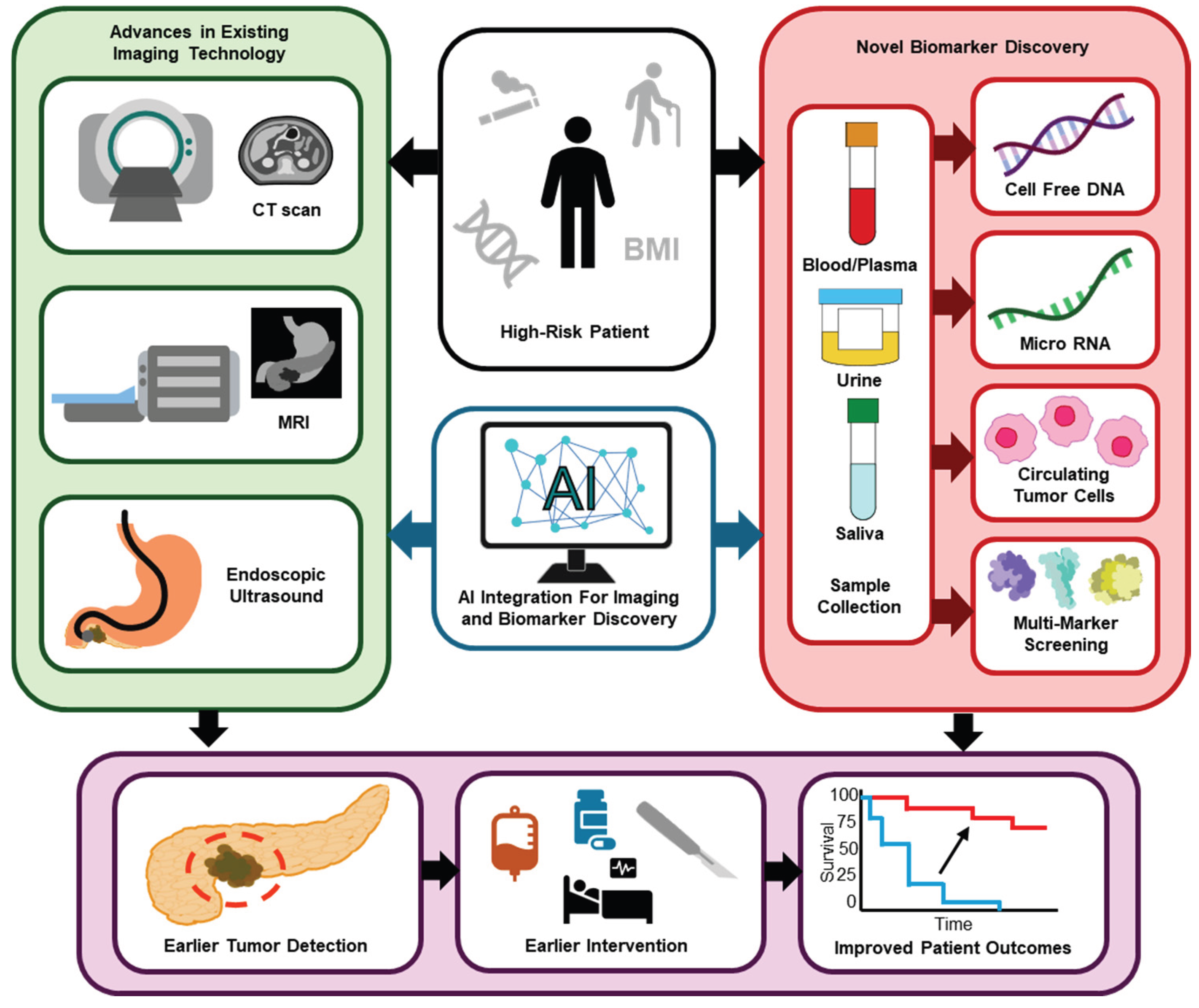
Figure 1.
Current strategies to improve pancreatic ductal adenocarcinoma detection for earlier diagnosis and better patient outcomes. Figure created using InkScape—Draw Freely (2026). Version 1.4.3 (InkScape).
Figure 1.
Current strategies to improve pancreatic ductal adenocarcinoma detection for earlier diagnosis and better patient outcomes. Figure created using InkScape—Draw Freely (2026). Version 1.4.3 (InkScape).
Author Contributions
S.W. drafted the abstract and section 1, introduction. J.R. drafted section 2, biomarkers, and created the TOC figure. B.D. drafted section 3, imaging technologies. N.M. drafted section 4, artificial intelligence applications, and section 6, conclusion/future directions. D.A.V. drafted section 5, the critical analysis of limitations and challenges. R.P. supervised the work and provided feedback throughout the writing process. J.R., B.D., N.M., S.W., and D.V.M. contributed equally to the conceptualization of the review, definition of its scope, manuscript revision, with R.P. assisting in approval of the final version. All authors agree to be accountable for the content of the review.
References
- Stoop, T.F.; et al. Pancreatic cancer. Lancet 2025, 405, 1182–1202. [Google Scholar] [CrossRef] [PubMed]
- Williams, H.L.; et al. Spatially-resolved single-cell assessment of pancreatic cancer expression subtypes reveals co-expressor phenotypes and extensive intra-tumoral heterogeneity. Cancer Res 2023, 83, 441–455. [Google Scholar] [CrossRef]
- Li, T.; Lin, C.; Wang, W. Global, regional, and national burden of pancreatic cancer from 1990 to 2021, its attributable risk factors, and projections to 2050: a systematic analysis of the global burden of disease study 2021. BMC Cancer 2025, 25, 189. [Google Scholar] [CrossRef]
- Partyka, O.; et al. Overview of Pancreatic Cancer Epidemiology in Europe and Recommendations for Screening in High-Risk Populations. Cancers 2023, 15, 3634. [Google Scholar] [CrossRef]
- Krusen, B.M.; et al. Improving a Plasma Biomarker Panel for Early Detection of Pancreatic Ductal Adenocarcinoma with Aminopeptidase N (ANPEP) and Polymeric immunoglobin receptor (PIGR). Clin Cancer Res 2026, 32, 756–769. [Google Scholar] [CrossRef]
- Kane, L.E.; et al. Multi-omic biomarker panel in pancreatic cyst fluid and serum predicts patients at a high risk of pancreatic cancer development. Sci Rep 2025, 15, 129. [Google Scholar] [CrossRef]
- Parker, R.A.; et al. Early features of pancreatic cancer on magnetic resonance imaging (MRI): a case–control study. Abdom Radiol 2024, 49, 1489–1501. [Google Scholar] [CrossRef]
- Krishnan, A. Imaging-pathology correlation in pancreatic cancer: Methodological considerations and future directions. World J Gastrointest Oncol 2025, 17, 103282. [Google Scholar] [CrossRef]
- Peters, M.L.B.; et al. Cost-Effectiveness Analysis of Screening for Pancreatic Cancer Among High-Risk Populations. JCO Oncol Pract 2024, 20, 278–290. [Google Scholar] [CrossRef] [PubMed]
- Wang, Y.; et al. Multi-omic profiling of intraductal papillary neoplasms of the pancreas reveals distinct patterns and potential markers of progression. Cancer Cell 2025, 43, 2156–2170.e6. [Google Scholar] [CrossRef] [PubMed]
- Alves, N.; et al. Artificial intelligence and radiologists in pancreatic cancer detection using standard of care CT scans (PANORAMA): an international, paired, non-inferiority, confirmatory, observational study. Lancet Oncol. 2026, 27, 116–124. [Google Scholar] [CrossRef]
- Chi, H.; et al. Proposing new early detection indicators for pancreatic cancer: combining machine learning and neural networks for serum miRNA-based diagnostic model. Front. Oncol. 2023, 13, 1244578. [Google Scholar] [CrossRef]
- Anthes, L.E.; Hajizadeh, M. Socioeconomic inequalities in pancreatic cancer incidence in Canada: evidence from Cancer Registry data. J Public Health (Berl.) 2022, 30, 801–810. [Google Scholar] [CrossRef]
- Babic, A.; et al. Adipose tissue and skeletal muscle wasting precede clinical diagnosis of pancreatic cancer. Nat Commun 2023, 14, 4317. [Google Scholar] [CrossRef] [PubMed]
- Gu, X.; Minko, T. Targeted Nanoparticle-Based Diagnostic and Treatment Options for Pancreatic Cancer. Cancers 2024, 16, 1589. [Google Scholar] [CrossRef]
- Wu, J.; et al. Cell-free DNA testing for the detection and prognosis prediction of pancreatic cancer. Nat. Commun. 2025, 16, 6645. [Google Scholar] [CrossRef] [PubMed]
- Shishido, S.N.; et al. Circulating tumor cell and oncosome subtypes in portal and peripheral venous circulations may be used for diagnosis and prognostication of pancreatic cancer. npj Precis. Onc. 2025, 9, 397. [Google Scholar] [CrossRef] [PubMed]
- Greenberg, Z.F.; et al. Nanomaterial isolated extracellular vesicles enable high precision identification of tumor biomarkers for pancreatic cancer liquid biopsy. J Nanobiotechnol 2025, 23, 467. [Google Scholar] [CrossRef]
- Bilreiro, C.; Andrade, L.; Santiago, I.; Marques, R.M.; Matos, C. Imaging of pancreatic ductal adenocarcinoma—An update for all stages of patient management. European Journal of Radiology Open 2024, 12, 100553. [Google Scholar] [CrossRef]
- Al-Hawary, M.M.; et al. Pancreatic Ductal Adenocarcinoma Radiology Reporting Template: Consensus Statement of the Society of Abdominal Radiology and the American Pancreatic Association. Gastroenterology 2014, 146, 291–304.e1. [Google Scholar] [CrossRef]
- Costache, M.I.; Costache, C.A. Which is the best imaging method in pancreatic adenocarcinoma diagnosis and staging. Curr. HEALTH Sci. J. 2017, 43, 132–136. [Google Scholar]
- Li, W.; et al. Early and Prediagnostic Detection of Pancreatic Cancer from Computed Tomography. 2026. [Google Scholar] [CrossRef]
- Li, J.; et al. The progress of PET/MRI in clinical management of patients with pancreatic malignant lesions. Front Oncol 2023, 13, 920896. [Google Scholar] [CrossRef]
- Lin, Z.; et al. Early Detection of Pancreatic Cancer: Current Advances and Future Opportunities. Biomedicines 2025, 13. [Google Scholar] [CrossRef] [PubMed]
- Bi, Y.; et al. Diagnosis methods for pancreatic cancer with the technique of deep learning: a review and a meta-analysis. Front Oncol 2025, 15, 1597969. [Google Scholar] [CrossRef] [PubMed]
- Degand, L.; et al. Validation of a Pretrained Artificial Intelligence Model for Pancreatic Cancer Detection on Diagnosis and Prediagnosis Computed Tomography Scans. Invest Radiol 2025, 61, 75–83. [Google Scholar]
- Ney, A.; et al. Identification of a serum proteomic biomarker panel using diagnosis specific ensemble learning and symptoms for early pancreatic cancer detection. PLoS Comput Biol 2024, 20, e1012408. [Google Scholar] [CrossRef] [PubMed]
Disclaimer/Publisher’s Note: The statements, opinions and data contained in all publications are solely those of the individual author(s) and contributor(s) and not of MDPI and/or the editor(s). MDPI and/or the editor(s) disclaim responsibility for any injury to people or property resulting from any ideas, methods, instructions or products referred to in the content. |
© 2026 by the authors. Licensee MDPI, Basel, Switzerland. This article is an open access article distributed under the terms and conditions of the Creative Commons Attribution (CC BY) license (http://creativecommons.org/licenses/by/4.0/).
Copyright: This open access article is published under a Creative Commons CC BY 4.0 license, which permit the free download, distribution, and reuse, provided that the author and preprint are cited in any reuse.


