
Preprint
Review

This version is not peer-reviewed.

Rediscovering the Gut–Mito–Ear Axis: A Systems-Biology Framework for Ototoxic Vulnerability and Microbiome-Targeted Prevention

A peer-reviewed version of this preprint was published in:
Cells 2026, 15(9), 769. https://doi.org/10.3390/cells15090769

Submitted:

27 March 2026

Posted:

27 March 2026

You are already at the latest version

Abstract
Ototoxicity is traditionally viewed as a local cochlear adverse effect of indispensable therapies such as cisplatin and aminoglycosides. However, emerging evidence suggests that cochlear vulnerability is shaped by systemic physiology, including inflammatory tone, vascular barrier integrity, and metabolic state. In this Review, we propose a Gut–Mito–Ear axis in which gut ecosystem function influences circulating mediator modules that converge on two cochlear mediator nodes: blood–labyrinth barrier (BLB) gating and mitochondrial stress tolerance. We synthesize evidence showing that gut perturbation can alter cochlear outcomes in vivo, that at least one microbiota-derived metabolite signal can directly protect hearing in experimental settings, and that BLB dysfunction and inflammatory trafficking are mechanistically relevant to cisplatin- and aminoglycoside-induced injury. We further organize the literature using an evidence-weighted framework that distinguishes direct cochlear causality from mechanistic plausibility and explicitly retains negative studies as boundary-setting evidence. Finally, we outline a translational roadmap in which microbiome-targeted prevention is pursued through mediator-anchored, non-interference-aware strategies and evaluated across linked state variables spanning exposure context, gut function, defined mediator modules, BLB gating, mitochondrial stress tolerance, and auditory phenotype. Framed in this way, the Gut–Mito–Ear axis is presented not as an established mechanism but as an operational, falsifiable systems-biology model that defines minimum and ideal standards for A-tier evidence, interpretable criteria for boundary-setting A− evidence, and testable predictions for causal validation.
Keywords: 
;  ;  ;  ;  ;  ;  ;  

1. Introduction

Hearing loss remains one of the most prevalent sensory impairments worldwide, and treatment-related ototoxicity is a major contributor to irreversible acquired sensorineural hearing loss. Despite decades of mechanistic progress, clinically useful prevention has remained limited, in part because susceptibility varies substantially across individuals and treatment contexts and in part because the cochlea is still too often conceptualized as an isolated end organ [1,2,3,4,5,6,7,8]. A more useful view is that ototoxicity reflects a systems-level failure in which toxic exposure interacts with host physiology to determine whether cochlear stress remains reversible or becomes permanent.
This broader perspective becomes compelling when one considers the metabolic demands of the inner ear. Hair cells, spiral ganglion neurons, and the stria vascularis operate near a bioenergetic edge, requiring tightly controlled mitochondrial output to sustain mechanoelectrical transduction, synaptic signaling, and ionic homeostasis [9,10,11,12,13]. In such a setting, mitochondrial stress responses are not secondary details but a convergent decision node that links diverse upstream insults to irreversible sensory injury [7,10,14].
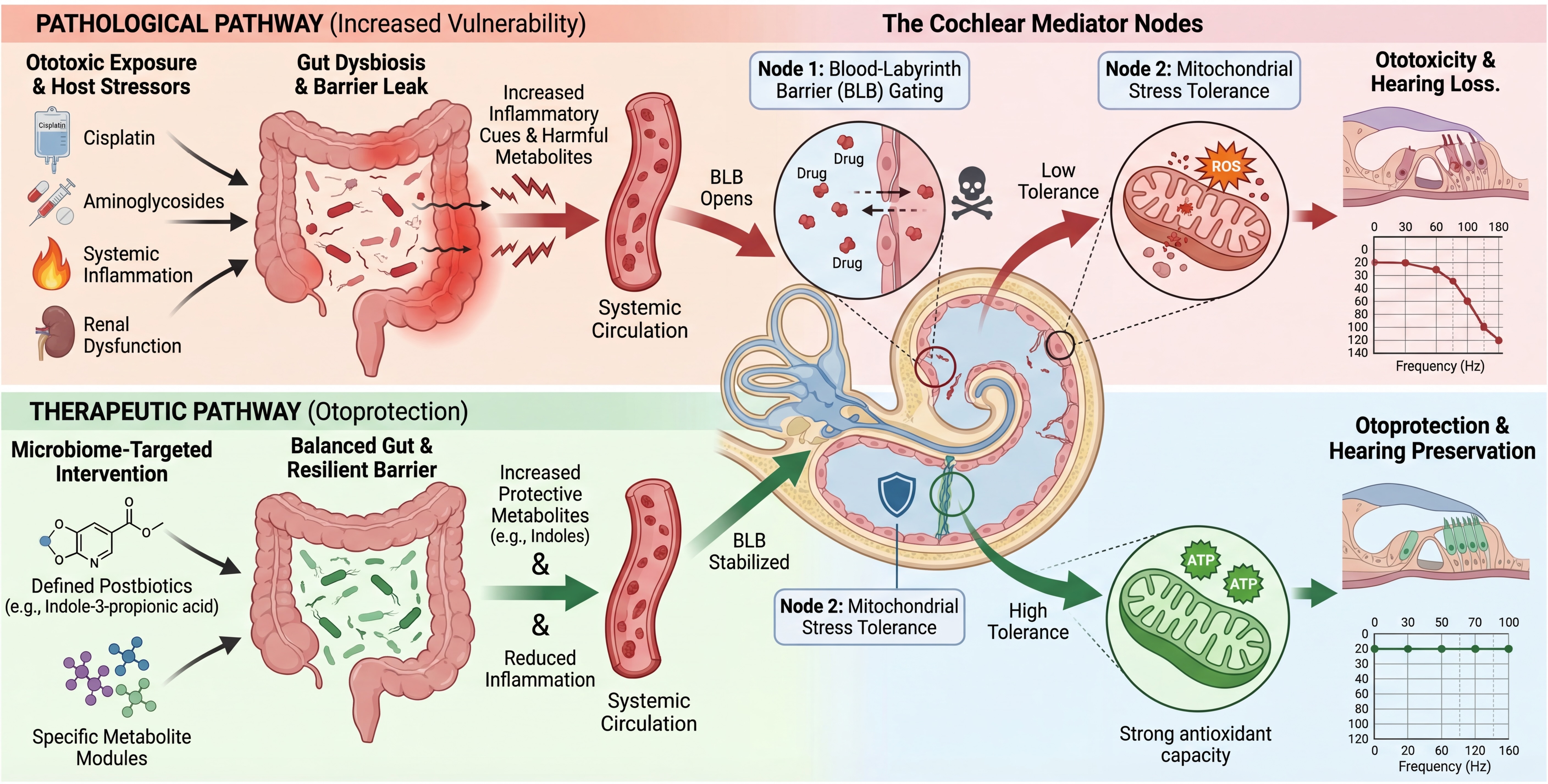
At the same time, cochlear injury is shaped by how toxic signals reach the inner ear. The blood–labyrinth barrier (BLB) and the cochlear immune microenvironment are now recognized as active regulators of exposure and damage amplification rather than passive background structures [15,16,17,18,19,20,21,22,23,24]. Cisplatin can induce BLB hyperpermeability and strial dysfunction, whereas inflammatory states can increase aminoglycoside trafficking into the cochlea and exacerbate injury [16,17]. These features make the cochlea not merely a target of toxins, but a distal sensor of systemic stress states. As summarized in Figure 1, we conceptualize ototoxicity as a systems-level process in which gut-derived inflammatory and metabolic signals converge on BLB gating and cochlear mitochondrial stress responses to shape sensory-cell fate. Rather than treating the cochlea as an isolated toxicologic endpoint, this framework highlights how distal perturbations in gut ecology may shift the threshold between reversible stress and irreversible auditory injury. This systems view provides the conceptual bridge for considering the intestinal microbiome as an upstream modifier of cochlear vulnerability.
Within this framework, the intestinal microbiome emerges as a plausible upstream regulator of ototoxic vulnerability. The gut ecosystem shapes circulating metabolite pools, calibrates immune tone, and influences epithelial and vascular barrier behavior throughout the body. Recent interventional studies now suggest that these systemic processes can reach the auditory system: intestinal inflammation and microbiota modulation have been shown to alter auditory thresholds and cochlear inflammatory signatures, while microbiota-targeted intervention in noise injury reshaped metabolite programs and was associated with hearing protection [25,26,27].
Crucially, the emerging picture is not uniformly positive, and explicit boundary conditions strengthen rather than weaken the framework. In an acute noise-induced hearing loss model, antibiotic-mediated microbiota depletion and germ-free status did not alter susceptibility, indicating that microbiome effects are neither universal nor exposure-agnostic [28]. Likewise, only a limited number of gut-derived metabolites have been linked directly to cochlear outcomes. Among these, indole-3-propionic acid provides one of the clearest causal signals by reducing oxidative stress and apoptosis in a chemical ototoxicity model [27].
Taken together, these observations support a Gut–Mito–Ear axis in which gut ecosystem function alters circulating mediators that converge on two cochlear mediator nodes—BLB-pericyte gating and mitochondrial stress tolerance—to determine sensory cell fate. The purpose of this Review is not to overstate that framework, but to define where direct evidence exists, where mechanistic plausibility is strong, and where decisive experiments are still needed [25,26,27,28,29,30,31,32]. To clarify the terminology used throughout this Review, key definitions and scope are summarized in Box 1. These definitions distinguish compositional from functional dysbiosis, specify how postbiotics are used in the present context, and explain why BLB gating and mitochondrial stress tolerance are treated as central mediator nodes rather than secondary outcomes. This framing is essential before moving from concept to the clinical problem that motivates the Gut–Mito–Ear axis.
Box 1. Definitions and Scope of the Gut–Mito–Ear Axis.
The Gut–Mito–Ear axis refers to a systems-level model in which gut microbial ecology and gut barrier/immune states shape circulating mediators that influence cochlear injury.
Two mediator nodes are central to this framework: blood–labyrinth barrier (BLB) gating, which regulates cochlear exposure, and mitochondrial stress tolerance, which regulates cochlear cell fate.
Ototoxicity is used to describe cochlear injury associated with exposure to platinum-based agents or environmental agents, including functional, structural, barrier-related, and mitochondrial endpoints.
Dysbiosis is considered primarily in functional terms, with emphasis on altered metabolite output and inflammatory signaling rather than taxonomy alone.
Postbiotics are defined pragmatically as microbiota-derived or microbiota-sensitive molecules with measurable biological effects; among these, indole-related metabolites currently have the strongest experimental support.
This Review does not treat the Gut–Mito–Ear axis as established fact, but instead evaluates where evidence is direct, where it remains mechanistically plausible, and what is required for causal validation.

2. Clinical Landscape of Ototoxic Exposures and the Unmet Need for Mechanism-Led, Non-Interfering Otoprotection

Drug-induced ototoxicity is among the most consequential irreversible toxicities encountered in oncology and infectious disease practice. A global meta-analysis estimated that objectively measured hearing loss occurs in a substantial proportion of platinum-exposed patients, and pediatric cohorts have repeatedly shown that these deficits carry long-term developmental, educational, and psychosocial consequences [6,7,8,33,34,35]. In this setting, hearing loss is not a minor side effect but a survivorship issue with lifelong implications.
Cisplatin remains a backbone therapy for many pediatric and adult solid tumors, yet it carries a well-established risk of permanent bilateral sensorineural hearing loss, often beginning at high frequencies and progressing with cumulative exposure [1,2,3,4,5,6,7,8,33,34,36]. Younger age, treatment intensity, and disease context repeatedly emerge as major modifiers of risk [7,34,37,38]. Despite this burden, real-world monitoring remains inconsistent. Baseline audiometry is often omitted, surveillance during therapy is incomplete, and follow-up commonly declines precisely when delayed deficits may become clinically evident [37,39,40].
The current prevention landscape is defined as much by its constraints as by its successes. Sodium thiosulfate represents a genuine milestone because it demonstrated clinically meaningful otoprotection in pediatric patients with localized, non-metastatic solid tumors and ultimately received regulatory approval in this context [8,41,42,43]. At the same time, the sodium thiosulfate experience clarified the central translational rule in this field: any otoprotective adjunct must preserve the efficacy of the primary therapy. Concerns regarding disease setting, timing, and potential non-interference remain central to how the field now evaluates supportive interventions [43,44,45].
Aminoglycosides present a parallel but distinct challenge. They remain essential in severe Gram-negative infection, sepsis, and selected tuberculosis regimens, yet exposure often occurs in patients with fluctuating renal clearance and high systemic inflammatory burden [16,46,47,48,49]. Mechanistically, aminoglycosides enter hair cells through defined routes, accumulate intracellularly, and induce oxidative and mitochondrial injury; clinically, inflammatory priming can increase cochlear trafficking and worsen toxicity [16,46,47,50].
In contrast to cisplatin, the aminoglycoside field has advanced most clearly through pharmacogenetics. Variants in the mitochondrial 12S ribosomal RNA gene (MT-RNR1), such as m.1555A>G, markedly increase susceptibility, and guidance from the Clinical Pharmacogenetics Implementation Consortium (CPIC) now enables genotype-informed prescribing and avoidance when appropriate [48,49,51,52]. This has effectively created a model of precision supportive care in which systemic risk can be stratified even when no approved otoprotective drug is available.
Across both cisplatin and aminoglycoside settings, renal dysfunction, inflammation, and polypharmacy repeatedly shape risk. Supportive-care regimens often include broad-spectrum antibiotics, proton pump inhibitors, antiemetics, corticosteroids, analgesics, and nutritional perturbations, all of which can influence gut microbial function and metabolite output [53,54,55,56,57,58,59]. Chemotherapy-associated gastrointestinal dysfunction adds another layer, linking treatment context to gut functional rewiring [29,53,60]. These same systemic states can also modify BLB behavior and cochlear drug trafficking [16,17]. For this reason, the microbiome is not a peripheral add-on to ototoxicity biology, but a plausible upstream regulator of the inflammatory and metabolic states that determine cochlear exposure and resilience.

3. Cochlear Mitochondrial Vulnerability: Why the Inner Ear Behaves Like a Metabolic Edge Sensor

The cochlea is built for high-flux bioenergetics. Hair cells, spiral ganglion neurons, lateral-wall fibrocytes, and the stria vascularis sustain constant ionic transport and rapid sensory signaling with little regenerative reserve, making mitochondrial integrity central to cochlear survival [9,10,11,12,13,61,62,63,64]. This architecture enables precise hearing but also creates fragility, because persistent respiratory demand inevitably generates mitochondrial reactive oxygen species and magnifies the consequences of even modest failures in quality control.
Both cisplatin and aminoglycosides ultimately converge on mitochondrial dysfunction, but they do so within a broader sensory–vascular–immune unit rather than within hair cells alone. Cisplatin injures hair cells, the stria vascularis, and spiral ganglion neurons through intersecting pathways that include uptake and retention, oxidative stress, inflammatory amplification, and prolonged cochlear residency [1,2,3,4,9,65,66]. Aminoglycosides likewise induce oxidative and mitochondrial injury, yet their toxic effects depend strongly on trafficking and intracellular localization [16,46,47,50].
An increasingly useful way to understand this biology is to treat mitochondrial quality control as a limited stress budget. Cochlear tissues do not survive simply by suppressing reactive oxygen species; they must also preserve turnover, membrane integrity, and metabolic flexibility. Once this reserve is exhausted, cell death programs are engaged and inflammatory signaling becomes self-reinforcing [7,67,68,69,70,71].
This perspective is highly relevant to the Gut–Mito–Ear axis. Gut-derived inflammatory tone or metabolite programs need not directly damage the cochlea. Instead, they may shift the threshold at which cochlear mitochondria move from adaptive recovery to maladaptive collapse. Metabolic signaling pathways, such as nicotinamide adenine dinucleotide (NAD+)/sirtuin signaling and AMP-activated protein kinase (AMPK), support this view by demonstrating that the cochlea can be preconditioned before overt injury [13,64,72,73].
For this reason, cochlear mitochondrial vulnerability serves as one of two obligatory mediator nodes in this Review. The other is BLB-pericyte gating. Only when both are measured can one distinguish lower systemic exposure from preserved barrier integrity or from a true increase in cochlear resilience [16,17,18,19,20,21,22,23,24].

4. Pharmacomicrobiomics of Ototoxic Exposures: How Cisplatin and Aminoglycosides Reshape Gut Ecology

Pharmacomicrobiomics provides the upstream logic for the Gut–Mito–Ear axis. Drugs alter microbial communities and their metabolic outputs, while microbial ecosystems reciprocally influence drug disposition, efficacy, and toxicity through direct metabolism, transporter modulation, and immune regulation [54,74,75,76,77,78,79]. This framework is especially relevant in ototoxicity because the clinical environments in which cisplatin and aminoglycosides are used are rich in co-medications and systemic stressors that intensify drug–microbiome interactions.
Although cisplatin is not an antibiotic, it can still reshape gut ecology and host response. Studies outside the ear have shown that microbiota depletion can protect against cisplatin-induced systemic injury and that microbiota transfer can restore susceptibility, indicating that cisplatin toxicity is already microbiome-sensitive at the organismal level [26,28,49,60]. These studies do not, on their own, prove a gut-to-ear mechanism, but they provide the causal scaffold required for cochlear translation.
Cisplatin-associated gastrointestinal dysfunction provides an additional bridge. Supportive-care regimens and bowel dysmotility can reshape microbial composition and fecal metabolomes, including pathways related to bile-acid and taurine metabolism [29,53,60,76]. This shifts the question away from descriptive dysbiosis and toward functional chemical outputs that could influence BLB gating or mitochondrial stress tolerance.
Aminoglycosides define a complementary pharmacomicrobiomic regime. Because they are antimicrobial by design, they impose strong pressure on gut ecology, and antibiotic-induced dysbiosis is known to reshape host metabolomes and inflammatory states [55,56,57,58,59,78,79]. At the same time, their direct cochleotoxicity makes interpretation more difficult: any experiment using aminoglycosides as microbiome-disrupting tools must separate gut-mediated effects from direct drug injury. This is precisely why BLB-pericyte and mitochondrial mediators are indispensable in experimental design.
Taken together, cisplatin and aminoglycosides define two different but convergent routes into systems ototoxicity. Cisplatin demonstrates that a non-antibiotic drug can still drive dysbiosis, barrier injury, and metabolite remodeling, whereas aminoglycosides demonstrate that antibiotic exposure can directly interact with inflammatory trafficking into the ear [16,19,53,60].

6. Routes from Gut to Ear: BLB Gating, Pericyte Control, and Immune–Vascular Conduits

A systems-level Gut–Mito–Ear axis requires a conduit that converts systemic metabolic and immune states into cochlear exposure, and the BLB is the most plausible structure for this role. It is not a passive wall but a regulated interface composed of endothelial cells, pericytes, basement membrane, and immune-like perivascular elements [15,16,17,18,22,81].
Recent work has clarified two principal levers of BLB regulation: paracellular control through tight junctions and transcellular control through transcytosis. Mfsd2a, a regulator better known for its role in blood–brain barrier biology, also shapes BLB formation and function by influencing both junctional integrity and endothelial transport behavior [15,18]. This means that cochlear exposure can increase even without gross endothelial loss, simply because the barrier changes its function.
Cisplatin provides especially strong support for a barrier-centered model. It induces BLB hyperpermeability, disrupts strial structure, lowers endocochlear potential, and promotes exposure escalation within the cochlea [17,20,24]. Pericytes of the stria vascularis are themselves vulnerable targets of cisplatin injury, with changes in viability, oxidative stress, and signaling pathways consistent with pro-permeability and pro-inflammatory states [21].
Immune and perivascular cells add another layer of control. Human BLB systems show that cytokine-rich inflammatory conditions can compromise barrier integrity, and macrophage-related pathways appear capable of shaping not only tissue damage but also the extent of cochlear exposure itself [16,17,22,23].
The relevance of these observations to gut biology becomes clearest in aminoglycoside settings. Endotoxemia-mediated inflammation increases aminoglycoside trafficking into the cochlea and potentiates ototoxicity [16,19]. Because dysbiosis and gut barrier dysfunction are established drivers of systemic inflammation, the BLB becomes the most plausible route by which gut state influences aminoglycoside injury. This is also why BLB-pericyte gating must be measured alongside mitochondrial readouts in any serious Gut–Mito–Ear experiment [16,17,22].

7. Integrated Evidence-Weighted Network Model of the Gut–Mito–Ear Axis

The literature surrounding the Gut–Mito–Ear axis is heterogeneous by design, spanning direct intervention studies, cochlear mechanistic work, drug–microbiome interaction studies outside the ear, and negative results that define limits of generalization. A conventional narrative can easily flatten these different forms of support and overstate what is currently established. For this reason, the field is better organized as an evidence-weighted network than as an undifferentiated list of plausible links. As shown in Figure 2, the proposed Gut–Mito–Ear axis can be structured across five linked state layers: exposure context, gut functional state, defined mediator-module state, cochlear mediator-node state, and auditory phenotype. Defined mediator-module state refers to tractable circulating modules that can be perturbed and quantitatively tracked experimentally, as summarized in Box 2. This organization distinguishes upstream associative relationships from cochlear-node-level mechanistic support and from direct intervention evidence, while explicitly retaining interpretable negative evidence as a boundary-setting component rather than treating it as a simple null result. Within this framework, the central question is not whether a link is attractive, but whether it has advanced from association to full causal-chain validation under a defined systems-biology standard.
Box 2. Translational Priorities for Microbiome-Targeted Otoprotection.
Translational principle: Any microbiome-targeted otoprotective strategy must preserve the efficacy of the primary therapy, including anticancer activity in cisplatin settings and infection control in aminoglycoside settings.
Preferred entry point: Defined postbiotics or predefined mediator modules are currently the most tractable candidates because they allow dose control, pharmacokinetic monitoring, and pathway-specific testing.
Operational definition:Defined mediator modules” refer to pre-specified, quantifiable families of circulating mediators (metabolites and immune/inflammatory signals) that can be perturbed and tracked in intervention studies; examples include (i) indole-derived module (e.g., indole-3-propionic acid), (ii) short-chain fatty acid module, (iii) bile-acid-related module, (iv) lipid mediator module (e.g., sphingolipid-related), and (v) inflammatory–immune module (e.g., cytokine/endotoxin-related).
Most advanced candidates: The indole-derived module currently provides the strongest proof of principle, whereas other candidate modules remain promising but incompletely validated for drug-related ototoxicity.
Best early studies: Mechanistic bridging studies should test whether candidate interventions alter predefined mediator modules, including inflammatory tone, BLB gating, and mitochondrial stress responses, before large-scale efficacy studies are attempted.
Clinical positioning: Microbiome-targeted strategies should be developed as biomarker-guided adjuncts, not as empirical supplements, and should be prioritized in exposure contexts where systemic inflammation or barrier dysfunction is expected to amplify cochlear vulnerability.
Translational goal: The field should move stepwise from mediator modulation to exposure control and finally to auditory benefit, rather than moving directly from microbiome association to clinical supplementation.
Clinical risk stratification: In host states associated with immunocompromise, marked mucosal barrier injury, severe infection/critical illness, central venous access, or intense broad-spectrum antibiotic exposure, defined postbiotics, ex vivo serum/BLB testing, or delayed recovery-phase intervention should generally take priority over empirical live-biotic administration.
Box 3. Minimum Systems-Biology and Causal-Validation Framework for Gut–Mito–Ear Studies.
State variables:
Exposure state: Studies should define the exposure context, including drug identity, dose, schedule, route, co-medications, diet, renal function, and inflammatory status.
Gut state: Microbiome studies should report functional gut outputs, including metabolomic, inflammatory, and barrier-related measures, rather than taxonomy alone.
Mediator-module state: Claims about gut-to-ear coupling should specify at least one prespecified, quantified mediator module that is measured before and after the intervention.
Cochlear-node state: Two cochlear mediator nodes should be assessed whenever feasible: BLB gating and mitochondrial stress tolerance.
Auditory-phenotype state: Functional and structural cochlear outcomes should be included whenever feasible, ideally combining auditory testing with tissue-level or cochlear exposure readouts.
Minimum readout panel:
BLB gating/pericyte panel: At least one direct or surrogate barrier readout should be included, such as tracer or drug permeability, TEER or an equivalent barrier-resistance measure, junctional integrity, transcytosis-related markers, or pericyte injury signals.
Mitochondrial stress-tolerance panel: At least one cochlear mitochondrial stress readout should be included, such as mitochondrial reactive oxygen species, membrane potential, oxidative phosphorylation or ATP-linked status, mitophagy, or apoptosis-related markers.
Auditory panel: Whenever feasible, studies should include ABR and/or DPOAE together with structural assessment of hair cells, spiral ganglion neurons, or lateral-wall/strial injury.
Exposure-coupling panel: In drug-exposure models, cochlear drug burden or a validated surrogate of cochlear trafficking should be measured whenever feasible.
Operational evidence-tier rules:
A-tier, minimum standard: An A-tier study should include (i) a direct gut-level intervention or a defined-mediator intervention, (ii) a clinically relevant ototoxic exposure or clearly defined susceptibility context, (iii) an auditory phenotype endpoint, preferably ABR and/or DPOAE with or without structural corroboration, and (iv) at least one concordant cochlear-facing mechanistic readout. Ideally, this mechanistic readout is a formal cochlear mediator node (BLB gating or mitochondrial stress tolerance), but in early proof-of-principle studies, a justified proximal surrogate may suffice. Studies lacking an auditory phenotype endpoint should not be classified as A-tier.
A-tier, ideal standard: Ideal A-tier evidence additionally demonstrates matched exposure conditions, direct assessment of both principal cochlear mediator nodes (BLB gating and mitochondrial stress tolerance) or an explicitly justified reason for one-node incompleteness, and at least one temporal-ordering, necessity, blockade, rescue, or reversal experiment within the same framework. No single study in the current literature yet satisfies all the ideal criteria.
A− tier, inclusion rule: A− is reserved for direct negative tests of the axis that include an explicit gut-level or defined-mediator perturbation, a relevant exposure or susceptibility context, an auditory phenotype endpoint, and sufficient confirmation that the perturbation and downstream assessment were interpretable.
A− tier, exclusion rule: Negative studies should not be classified as A− when the perturbation is weak or unverified, the exposure context is mismatched, the auditory phenotype is not assessed, the design is underpowered for the stated claim, or cochlear-node/exposure-coupling readouts are too limited to interpret the null result as a true boundary condition.
B-tier: Strong mechanistic support exists at the BLB, immune-trafficking, or mitochondrial node, but without direct gut-mediated cochlear causality.
C-tier: Evidence is associative, upstream-only, observational, or hypothesis-generating.
Minimum criteria for claiming the full Gut–Mito–Ear causal chain:
A full-chain claim is strongest when the same experimental framework shows:
(1) a prespecified gut intervention or mediator-module shift;
(2) a downstream change in BLB gating/pericyte state under matched exposure;
(3) a concordant change in cochlear mitochondrial stress readouts; and
(4) a corresponding shift in auditory phenotype.
Whenever feasible, at least one necessity, blockade, rescue, or reversal step should be included to distinguish mediation from correlation.
Interpretive standard:
A Gut–Mito–Ear claim should not be considered complete if hearing changes are reported without evidence that the intended mediator module shifted and that at least one cochlear mediator node changed in the predicted direction.
Testable predictions generated by this framework:
Prediction 1: Under matched aminoglycoside exposure, a higher systemic inflammatory mediator-module signal should be associated with greater BLB permeability/drug trafficking and larger ABR threshold shifts.
Prediction 2: In cisplatin settings, interventions that shift a predefined mediator module but fail to alter BLB gating or mitochondrial stress tolerance should not produce a durable otoprotective signal at the auditory level.
Prediction 3: Across transfer or rescue experiments, the magnitude of hearing protection should scale with the intermediate response at the cochlear node; that is, larger normalization of BLB gating and mitochondrial stress should predict larger preservation of auditory function.
To avoid conflating fundamentally different forms of support within a single conceptual model, this Review applies the operational evidence-tier framework summarized in Box 3 throughout the Gut–Mito–Ear axis. A-tier evidence is divided conceptually into a minimum and an ideal standard. Minimum A-tier requires a direct gut-level or defined-mediator intervention under a relevant exposure or susceptibility context, a measurable shift in auditory phenotype, and at least one concordant cochlear-facing mechanistic readout; ideally this readout is a formal cochlear mediator node, whereas in early proof-of-principle studies a justified proximal surrogate may suffice. Ideal A-tier evidence additionally demonstrates matched exposure conditions, direct assessment of both principal cochlear mediator nodes—BLB gating and mitochondrial stress tolerance—and at least one experiment establishing temporal ordering, rescue, blockade, or necessity within the same framework. A− tier is reserved for direct negative tests that remain interpretable because the perturbation, exposure context, and auditory phenotype were adequately defined; it is not assigned to underpowered, exposure-mismatched, or mechanistically uninterpretable null studies. B-tier evidence includes strong mechanistic support at the level of cochlear mediator nodes—such as BLB gating, pericyte injury, immune-dependent trafficking, or mitochondrial stress tolerance—but without direct gut-mediated cochlear causality. C-tier evidence includes associative, upstream-only, or hypothesis-generating observations linking gut or systemic states to cochlear risk without direct validation of the full causal chain. This tiered approach is used not only to classify the literature, but also to distinguish what is already experimentally actionable from what still requires causal validation under the minimum systems-biology standard defined in Box 3.
To make the term “systems-biology” operational in the present Review, and to align the conceptual model in Figure 2 with a testable framework, the Gut–Mito–Ear axis can be represented as five linked state layers: (i) exposure context, (ii) gut functional state, (iii) defined mediator-module state, (iv) cochlear mediator-node state, and (v) auditory phenotype. Exposure context includes drug identity, dose, schedule, route, renal function, inflammatory status, co-medications, and nutritional stress. A gut functional state includes microbial ecology, intestinal barrier function, and functional output, rather than taxonomy alone. Defined mediator-module state refers to tractable circulating modules that can be perturbed and quantitatively tracked experimentally, as summarized in Box 2. Cochlear mediator-node state comprises BLB gating/pericyte integrity and mitochondrial stress tolerance. Auditory phenotype includes functional, structural, and cochlear drug-exposure endpoints. Within this framework, an edge is considered informative only when an upstream perturbation changes a pre-specified downstream state variable under matched exposure conditions, rather than by narrative plausibility alone. The minimum readout panel, interpretive rules, and testable predictions required to support this framework are summarized in Box 3.
Using this framework, the current A-tier literature remains limited but highly informative. It includes studies showing that gut perturbation can alter auditory phenotype in vivo, that microbiota-targeted intervention can modify ototoxic vulnerability in experimental settings, and that at least one defined microbiota-derived mediator can directly protect hearing [25,26,27,28,29]. At the same time, A− evidence is equally important because it prevents conceptual inflation. The negative finding in acute noise-induced hearing loss indicates that Gut–Mito–Ear effects are not universal and are likely to depend on exposure class, timescale, inflammatory state, and host background [28]. In this sense, negative studies do not weaken the framework; they define where the linked state-variable model should and should not be expected to operate.
Much of the remaining literature falls into the B-tier category, and this body of work is essential for understanding how the axis may function mechanistically even when direct gut-mediated cochlear causality has not yet been established. Cisplatin-induced BLB hyperpermeability, strial pericyte injury, cytokine-driven disruption of human BLB models, immune-dependent amplification of cochlear exposure, and mitochondrial-stress signaling together support the idea that systemic signals can modify both cochlear exposure and cochlear stress handling [16,17,18,19,20,21,22,23,24,50,61,62,63,67,68,69,72]. By contrast, C-tier evidence remains largely upstream or associative, including drug-induced dysbiosis, metabolome remodeling, and host–microbiome interaction studies that support biological plausibility but stop short of demonstrating concordant changes across defined mediator modules, cochlear mediator nodes, and auditory phe-notype within the same experimental framework [48,49,53,54,60,74,75,76,77,78,79,82].
The study-level logic of this framework is translated into Table 1, which maps each report by exposure state, experimental context or intervention, gut or mediator-module state, cochlear mediator-node readouts, auditory phenotype, and evidence tier. Read together, Figure 2 and Table 1 distinguish where direct causal-chain testing has already been achieved, where boundary-setting evidence exists, and where apparently promising links remain upstream, partial, or hypothesis-generating.
The practical value of this framework lies in its prioritization. The most informative next studies will not be those that merely add more microbiome association datasets, but those that convert high-value B- and C-tier links into A-tier causal evidence. In cisplatin settings, this means determining whether gut perturbation can transfer susceptibility via changes in BLB gating and mitochondrial stress tolerance. In aminoglycoside settings, it means testing whether gut-driven inflammatory tone alters trafficking across the BLB and shifts mitochondrial injury at matched exposure [16,17,19]. In this sense, the evidence-weighted model is not simply a way of organizing the literature. It is a way of making the field experimentally tractable

8. Translational Opportunities: From Gut–Mito–Ear Biology to Probiotics/Postbiotics and Biomarker-Linked Otoprotection

Any translational program built on the Gut–Mito–Ear axis must begin with a conservative principle: microbiome-targeted otoprotection is credible only if it is both testable and non-disruptive to primary therapy [41,42,43,44,52,86]. The sodium thiosulfate experience made this point unmistakably clear. Even biologically effective otoprotection must satisfy the higher bar of non-interference with anticancer efficacy, and this lesson should shape how microbiome-directed strategies are framed in both preclinical and clinical studies [42,43,44,45].
From this perspective, defined postbiotics and chemically tractable metabolite modules occupy the most attractive translational position. They allow dose control, pharmacokinetic tracking, and pathway-necessity testing at the cochlear node. Indole-3-propionic acid is currently the clearest example because it already provides direct evidence of hearing protection in a chemical ototoxicity model [27]. Bile-acid derivatives such as tauroursodeoxycholic acid can serve as useful downstream comparators for stress-proteostasis rescue [11]. Probiotics and defined consortia remain attractive upstream strategies, but they should be advanced only within a function-first design centered on validated metabolite outputs rather than taxonomic labels [30].
Translation also requires matching intervention windows to exposure biology. In cisplatin settings, the most rational windows likely include pre-conditioning to stabilize BLB-pericyte gating, concurrent administration to reduce exposure escalation during dosing, and recovery-phase support to facilitate resolution of inflammation and organelle stress [9,11,17,65]. In aminoglycoside settings, the most actionable niche may be inflammatory high-risk states, where endotoxemia or severe infection is expected to increase cochlear trafficking [16,19].
What makes this concept clinically plausible rather than merely interesting is the possibility of anchoring interventions to biomarkers. Functional gut outputs—captured as predefined mediator modules (see Box 2)—should be interpreted alongside inflammatory markers and mediator-focused assays of BLB gating and mitochondrial tolerance. Human BLB platforms offer a particularly useful bridge, because they allow patient serum or defined metabolite mixtures to be tested directly for barrier-opening effects before moving to larger translational studies [16,22]. The experimental and translational sequence implied by these considerations is summarized in Figure 3, which links systemic exposure context, gut perturbation, cochlear mediator nodes, and validation platforms within a single stepwise model. Complementing this visual roadmap, Box 2 summarizes the main translational priorities for microbiome-targeted otoprotection, including non-interference with primary therapy, postbiotic-first development, and biomarker-guided study design. Seen in this way, Gut–Mito–Ear translation should proceed stepwise from mediator modulation to exposure modification and finally to auditory benefit, rather than leaping directly from microbiome association to supplementation.
For this reason, the most persuasive translational strategy is not broad empirical supplementation, but stepwise mechanism-led development. Early studies should first demonstrate a shift in the intended mediator module, then show that this shift alters BLB behavior or mitochondrial stress handling, and only then ask whether those changes translate into hearing preservation without compromising anti-tumor or antimicrobial efficacy. In this respect, postbiotic-first translation is likely to be more interpretable than probiotic-first translation, while still leaving room for defined consortia or diet-based approaches once causal metabolite–mediator relationships are better resolved.
This distinction becomes clinically important in host states where safety and interpretability are both reduced. In patients with profound neutropenia, clinically significant mucositis or other gastrointestinal barrier injury, severe infection or critical illness, central venous access, or intense broad-spectrum antibiotic exposure, live-biotic strategies may be harder to interpret and may carry greater translational risk than defined postbiotics or ex vivo validation-first approaches [38,45,87,88]. For this reason, translational studies should stratify participants by host vulnerability and exposure context rather than treating all cisplatin- or aminoglycoside-exposed patients as a single population. A pragmatic patient-group risk–benefit framework for microbiome-targeted otoprotection, including settings in which live-biotic strategies should generally be deferred in favor of postbiotic-first or ex vivo validation-first approaches, is summarized in Table 2 [38,45,87,88].

9. Key Gaps, Pitfalls, and a Roadmap to A-Tier Causality

Despite rapid progress, the Gut–Mito–Ear axis is not yet proven in the contexts where it matters most clinically. The field now includes direct gut-to-ear manipulations, defined metabolite-to-ear signals, barrier- and mitochondrial-mediator studies, and negative evidence that constrain overgeneralization. What is still missing is the experiment that ties these layers together under clinically relevant ototoxic exposure [16,17,25,26,27,28,29].
The principal pitfall is mediation failure. Too many studies stop at association or at phenotype rescue without demonstrating how the effect occurred. In cisplatin models, reduced systemic inflammation or improved renal function could lower cochlear exposure and falsely appear as specific otoprotection [17]. In aminoglycoside settings, altered inflammatory tone can increase cochlear trafficking without necessarily changing intrinsic hair-cell resilience [16,19]. For this reason, a systems-level Gut–Mito–Ear claim should require explicit state-variable measurements across the gut, mediator, cochlear node, and auditory levels within the same experimental framework. Box 3 defines the minimum systems-biology standard used in this Review: the required state variables, the minimum readout panel for BLB gating and mitochondrial stress tolerance, the operational rules for assigning evidence tiers, and the minimum criteria needed to interpret a study as testing the full gut-to-ear causal chain rather than a set of loosely linked associations.
Against this standard, progress toward A-tier causality becomes easier to define. In cisplatin models, the critical question is not simply whether a gut intervention improves hearing, but whether it first shifts a defined mediator module, then alters BLB gating and mitochondrial stress tolerance under matched systemic exposure, and only then improves auditory phenotype [26,27,28,49,60]. In aminoglycoside settings, the most informative designs are those that build on the already established inflammation–trafficking axis and test whether gut intervention reduces cochlear drug entry, preserves mitochondrial stress tolerance, and attenuates hearing loss in parallel [16,19]. By structuring the field around linked state changes rather than descriptive dysbiosis alone, the Gut–Mito–Ear axis becomes not only a conceptual model but also a prediction-generating, falsifiable systems framework.
Within this framework, one practical route to A-tier causality begins with transfer experiments. In cisplatin models, susceptibility should be tested by transferring microbiota or gut functional states between donors and recipients and determining whether auditory phenotype, BLB gating, and mitochondrial stress tolerance shift together under matched exposure conditions [26,27,28,49,60]. Defined-metabolite rescue should then be used to determine whether the phenotype can be reconstructed or reversed, ideally using one A-tier benchmark metabolite (Table 1) as an initial reference point and asking whether additional predefined mediator modules (Box 2) can reproduce or counteract the same phenotype under matched systemic exposure [26,27,28,49,60].
Aminoglycoside models offer a particularly strong opportunity because the inflammation-dependent trafficking axis has already been directly implicated in cochlear drug entry and injury [16,19]. A high-priority roadmap experiment would therefore combine controlled inflammatory priming with gut intervention and test whether BLB gating, cochlear aminoglycoside accumulation, mitochondrial stress responses, and auditory phenotype change in parallel and in the predicted direction [16,19]. If such designs are paired with human BLB bridging assays and receptor- or pathway-level necessity tests at the cochlear node, the field would move substantially closer to genuine A-tier causality.
In practical terms, two experimental directions deserve the highest priority. The first is transfer-based modeling of cisplatin susceptibility with explicit mediator-module measurement and concurrent analysis of BLB and mitochondrial mediation [26,27,28,49,60]. The second is a gut intervention strategy designed to reduce inflammation-driven aminoglycoside trafficking across the BLB while preserving mitochondrial stress tolerance and auditory phenotype [16,19]. These are the experiments most likely to convert the Gut–Mito–Ear axis from a systems-level hypothesis supported by partial evidence into an operational framework with clear translational direction.

10. Conclusions

The Gut–Mito–Ear axis proposed in this Review does not replace established cochlear mechanisms of ototoxicity. Rather, it integrates them into a broader systems framework in which gut ecosystem function, systemic inflammatory tone, BLB gating, and mitochondrial stress handling jointly determine whether cochlear injury remains reversible or becomes permanent [1,2,3,4,5,6,7,8,9,10,11,12,13,14,15,16,17,18,19,20,21,22,23,24,30,31,32,46,47,51,65,90].
Three conclusions can be stated with reasonable confidence. First, gut perturbation can alter cochlear outcomes in vivo under certain conditions, demonstrating that a gut-to-ear route is biologically plausible [25,26,28]. Second, a defined microbiota-derived metabolite can directly protect hearing, providing an important causal link between metabolites and the ear [27]. Third, BLB gating is a mechanistic mediator of ototoxicity, and inflammatory states can amplify inner-ear drug trafficking and injury [16,17,18,19,20,21,22,23,24].
At the same time, the field should resist premature generalization. Negative evidence in acute noise models indicates that microbiome effects are context-dependent, and several candidate mediator modules remain promising but incompletely validated in cisplatin and aminoglycoside settings (see Box 2) [11,28,31,32]. What is defensible now is not the claim that probiotics already prevent ototoxicity, but the argument that microbiome-sensitive metabolite and inflammatory modules provide a tractable, testable route to prevention strategies that complement essential therapies without compromising their primary effect.
If future studies can explicitly connect gut intervention to BLB gating, cochlear mitochondrial readouts, and auditory outcomes under controlled ototoxic exposure, then microbiome-targeted otoprotection will become an experimentally grounded adjunct rather than an aspirational concept. In that sense, the Gut–Mito–Ear axis is less a speculative new theory than a rediscovery of something clinically obvious but mechanistically underappreciated: the inner ear is embedded within whole-body physiology, and its injury threshold can only be fully understood when that physiology is brought back into view.

11. Patents

Not applicable.

Author Contributions

Conceptualization, Y.-S.H. and S.K.A.; methodology, C.D.Y., Y.-S.H. and S.K.A.; investigation, C.D.Y., H.K., J.J.P. and J.H.S.; resources, J.J.P., J.H.S., Y.-S.H. and S.K.A.; data curation, C.D.Y. and H.K.; writing—original draft preparation, C.D.Y.; writing—review and editing, C.D.Y., H.K., J.J.P., J.H.S., S.-J.L., Y.-S.H. and S.K.A.; visualization, H.K. and S.-J.L.; supervision, Y.-S.H. and S.K.A.; project administration, Y.-S.H. and S.K.A.; funding acquisition, Y.-S.H. and S.K.A. All authors have read and agreed to the published version of the manuscript.

Funding

This work has been supported by the Biomedical Research Institute Fund (GNUHBRIF-2024-0001) from Gyeongsang National University Hospital.

Institutional Review Board Statement

Not applicable.

Data Availability Statement

No new data were created or analyzed in this study. Data sharing is not applicable to this article.

Acknowledgments

During the preparation of this work, the authors used ChatGPT 5.2 (OpenAI) to improve the readability and clarity of the manuscript. After using this tool, the authors reviewed and edited the content as needed and take full responsibility for the content of the published article.

Conflicts of Interest

The authors declare no conflicts of interest.

Abbreviations

The following abbreviations are used in this manuscript:
ABR auditory brainstem response
AMPK AMP-activated protein kinase
ARHL age-related hearing loss
BA Bile acid
BLB blood–labyrinth barrier
CPIC Clinical Pharmacogenetics Implementation Consortium
DPOAE distortion product otoacoustic emission
DSS dextran sulfate sodium
ER endoplasmic reticulum
FMT fecal microbiota transplantation
HC hair cell
ICU intensive care unit
IPA indole-3-propionic acid
MT-RNR1 mitochondrial 12S ribosomal RNA gene
mtROS mitochondrial reactive oxygen species
NAD+ nicotinamide adenine dinucleotide
NIHL noise-induced hearing loss
ROS reactive oxygen species
SGN spiral ganglion neuron
SPIOCA superparamagnetic iron oxide nanoparticle assembly
TCP 3,5,6-trichloro-2-pyridinol
TEER transendothelial electrical resistance
TUDCA tauroursodeoxycholic acid

References

  1. Wang, X.; Zhou, Y.; Wang, D.; Wang, Y.; Zhou, Z.; Ma, X.; Liu, X.; Dong, Y. Cisplatin-induced ototoxicity: From signaling network to therapeutic targets. Biomed Pharmacother 2023, 157, 114045. [CrossRef]
  2. Bai, J.; Wang, W.; Fu, Z.; Yang, J.; Guo, Y.; Guan, G. Bridging the gap: Mechanisms and novel translational strategies to prevent cisplatin-induced ototoxicity. Hear Res 2026, 469, 109487. [CrossRef]
  3. Lee, D.S.; Schrader, A.; Zou, J.; Ang, W.H.; Warchol, M.E.; Sheets, L. Direct targeting of mitochondria by cisplatin leads to cytotoxicity in zebrafish lateral-line hair cells. iScience 2024, 27, 110975. [CrossRef]
  4. Sung, C.Y.W.; Hayase, N.; Yuen, P.S.T.; Lee, J.; Fernandez, K.; Hu, X.; Cheng, H.; Star, R.A.; Warchol, M.E.; Cunningham, L.L. Macrophage depletion protects against cisplatin-induced ototoxicity and nephrotoxicity. Sci Adv 2024, 10, eadk9878. [CrossRef]
  5. Sun, Y.; Zou, S.; Xu, X.; Xu, S.; Sun, H.; Tang, M.; Kong, W.; Chen, X.; He, Z. Inhibition of the cGAS-STING Pathway Reduces Cisplatin-Induced Inner Ear Hair Cell Damage. Neurosci Bull 2025, 41, 359-373. [CrossRef]
  6. Dillard, L.K.; Lopez-Perez, L.; Martinez, R.X.; Fullerton, A.M.; Chadha, S.; McMahon, C.M. Global burden of ototoxic hearing loss associated with platinum-based cancer treatment: A systematic review and meta-analysis. Cancer Epidemiol 2022, 79, 102203. [CrossRef]
  7. Moke, D.J.; Luo, C.; Millstein, J.; Knight, K.R.; Rassekh, S.R.; Brooks, B.; Ross, C.J.D.; Wright, M.; Mena, V.; Rushing, T.; et al. Prevalence and risk factors for cisplatin-induced hearing loss in children, adolescents, and young adults: A multi-institutional North American cohort study. Lancet Child Adolesc Health 2021, 5, 274-283. [CrossRef]
  8. Chattaraj, A.; Syed, M.P.; Low, C.A.; Owonikoko, T.K. Cisplatin-Induced Ototoxicity: A Concise Review of the Burden, Prevention, and Interception Strategies. JCO Oncol Pract 2023, 19, 278-283. [CrossRef]
  9. Tang, Q.; Wang, X.; Jin, H.; Mi, Y.; Liu, L.; Dong, M.; Chen, Y.; Zou, Z. Cisplatin-induced ototoxicity: Updates on molecular mechanisms and otoprotective strategies. Eur J Pharm Biopharm 2021, 163, 60-71. [CrossRef]
  10. He, H.; Han, Z.; Cheng, S.; Zhou, Y. Hearing the call of mitochondria: Insight into its role in sensorineural hearing loss. Neurobiol Dis 2025, 213, 107030. [CrossRef]
  11. Abrigo, J.; Olguín, H.; Tacchi, F.; Orozco-Aguilar, J.; Valero-Breton, M.; Soto, J.; Castro-Sepúlveda, M.; Elorza, A.A.; Simon, F.; Cabello-Verrugio, C. Cholic and deoxycholic acids induce mitochondrial dysfunction, impaired biogenesis and autophagic flux in skeletal muscle cells. Biol Res 2023, 56, 30. [CrossRef]
  12. Zangerolamo, L.; Carvalho, M.; Barbosa, H.C.L. The Critical Role of the Bile Acid Receptor TGR5 in Energy Homeostasis: Insights into Physiology and Therapeutic Potential. Int J Mol Sci 2025, 26. [CrossRef]
  13. Ruggiero, A.; Romano, A.; Maurizi, P.; Talloa, D.; Fuccillo, F.; Mastrangelo, S.; Attinà, G. Platinum-Induced Ototoxicity in Pediatric Cancer Patients: A Comprehensive Approach to Monitoring Strategies, Management Interventions, and Future Directions. Children (Basel) 2025, 12. [CrossRef]
  14. Han, H.; Hu, S.; Hu, Y.; Liu, D.; Zhou, J.; Liu, X.; Ma, X.; Dong, Y. Mitophagy in ototoxicity. Front Cell Neurosci 2023, 17, 1140916. [CrossRef]
  15. Xu, X.; Xu, K.; Chen, F.; Yu, D.; Wang, X. Mfsd2a regulates the blood-labyrinth-barrier formation and function through tight junctions and transcytosis. Hear Res 2024, 450, 109048. [CrossRef]
  16. Koo, J.W.; Quintanilla-Dieck, L.; Jiang, M.; Liu, J.; Urdang, Z.D.; Allensworth, J.J.; Cross, C.P.; Li, H.; Steyger, P.S. Endotoxemia-mediated inflammation potentiates aminoglycoside-induced ototoxicity. Sci Transl Med 2015, 7, 298ra118. [CrossRef]
  17. Gu, J.; Tong, L.; Lin, X.; Chen, Y.; Wu, H.; Wang, X.; Yu, D. The disruption and hyperpermeability of blood-labyrinth barrier mediates cisplatin-induced ototoxicity. Toxicol Lett 2022, 354, 56-64. [CrossRef]
  18. Zhang, Y.; Li, L.; Qin, S.; Yuan, J.; Xie, X.; Wang, F.; Hu, S.; Yi, Y.; Chen, M. C-phycocyanin alleviated cisplatin-induced oxidative stress and inflammation via gut microbiota-metabolites axis in mice. Front Nutr 2022, 9, 996614. [CrossRef]
  19. Chen, C.H.; Huang, C.Y.; Lin, H.H.; Wang, M.C.; Chang, C.Y.; Cheng, Y.F. Association of Sodium Thiosulfate With Risk of Ototoxic Effects From Platinum-Based Chemotherapy: A Systematic Review and Meta-analysis. JAMA Netw Open 2021, 4, e2118895. [CrossRef]
  20. Orgel, E.; Villaluna, D.; Krailo, M.D.; Esbenshade, A.; Sung, L.; Freyer, D.R. Sodium thiosulfate for prevention of cisplatin-induced hearing loss: Updated survival from ACCL0431. Lancet Oncol 2022, 23, 570-572. [CrossRef]
  21. Meijer, A.J.M.; Diepstraten, F.A.; van den Heuvel-Eibrink, M.M.; Bleyer, A. Prevention of cisplatin-induced hearing loss in children: Achievements and challenges for evidence-based implementation of sodium Thiosulfate. Front Oncol 2024, 14, 1336714. [CrossRef]
  22. Brock, P.; Meijer, A.; Kogner, P.; Ansari, M.; Capra, M.; Geller, J.; Heuvel-Eibrink, M.V.D.; Knight, K.; Kruger, M.; Lindemulder, S.; et al. Sodium thiosulfate as cisplatin otoprotectant in children: The challenge of when to use it. Pediatr Blood Cancer 2023, 70, e30248. [CrossRef]
  23. Mai, Z.; Han, Y.; Liang, D.; Mai, F.; Zheng, H.; Li, P.; Li, Y.; Ma, C.; Chen, Y.; Li, W.; et al. Gut-derived metabolite 3-methylxanthine enhances cisplatin-induced apoptosis via dopamine receptor D1 in a mouse model of ovarian cancer. mSystems 2024, 9, e0130123. [CrossRef]
  24. Chambers, L.M.; Esakov Rhoades, E.L.; Bharti, R.; Braley, C.; Tewari, S.; Trestan, L.; Alali, Z.; Bayik, D.; Lathia, J.D.; Sangwan, N.; et al. Disruption of the Gut Microbiota Confers Cisplatin Resistance in Epithelial Ovarian Cancer. Cancer Res 2022, 82, 4654-4669. [CrossRef]
  25. Sato, K.; Araya, K.; Wu, C.; Dong, E.; Watson, R.; Maskey, M.; Nguyen, C.D.; Khanipov, K.; Yang, Q.; Wu, Z. The gut microbiota is not linked to acute noise-induced hearing loss in C57BL/6 mice. Sci Rep 2025, 15, 23457. [CrossRef]
  26. Sekulic, M.; Puche, R.; Bodmer, D.; Petkovic, V. Human blood-labyrinth barrier model to study the effects of cytokines and inflammation. Front Mol Neurosci 2023, 16, 1243370. [CrossRef]
  27. Godur, D.A.; Denton, A.J.; Eshraghi, N.; Mittal, J.; Cooper, J.; Moosa, M.; Mittal, R. Modulation of Gut Microbiome as a Therapeutic Modality for Auditory Disorders. Audiol Res 2023, 13, 741-752. [CrossRef]
  28. Yin, Q.; Shi, G.; Zhu, L. Association between gut microbiota and sensorineural hearing loss: A Mendelian randomization study. Front Microbiol 2023, 14, 1230125. [CrossRef]
  29. Kociszewska, D.; Chan, J.; Thorne, P.R.; Vlajkovic, S.M. The Link between Gut Dysbiosis Caused by a High-Fat Diet and Hearing Loss. Int J Mol Sci 2021, 22. [CrossRef]
  30. Meijer, A.J.M.; Li, K.H.; Brooks, B.; Clemens, E.; Ross, C.J.; Rassekh, S.R.; Hoetink, A.E.; van Grotel, M.; van den Heuvel-Eibrink, M.M.; Carleton, B.C. The cumulative incidence of cisplatin-induced hearing loss in young children is higher and develops at an early stage during therapy compared with older children based on 2052 audiological assessments. Cancer 2022, 128, 169-179. [CrossRef]
  31. Santucci, N.M.; Garber, B.; Ivory, R.; Kuhn, M.A.; Stephen, M.; Aizenberg, D. Insight into the current practice of ototoxicity monitoring during cisplatin therapy. J Otolaryngol Head Neck Surg 2021, 50, 19. [CrossRef]
  32. Lee, D.S.; Travis, E.Y.; Wong, S.K.; Munyemana, M.A.; Mueller, L.; Rowling, C.C.; Rich, J.T.; Pipkorn, P.; Puram, S.V.; Jackson, R.S.; et al. Trends in ototoxicity monitoring among cisplatin-treated patients with cancer. J Cancer Surviv 2025, 19, 1750-1760. [CrossRef]
  33. Bovee, S.; Klump, G.M.; Köppl, C.; Pyott, S.J. The Stria Vascularis: Renewed Attention on a Key Player in Age-Related Hearing Loss. Int J Mol Sci 2024, 25. [CrossRef]
  34. Xu, S.; Yang, N. The Role and Research Progress of Mitochondria in Sensorineural Hearing Loss. Mol Neurobiol 2025, 62, 6913-6921. [CrossRef]
  35. Sheth, S.; Mukherjea, D.; Rybak, L.P.; Ramkumar, V. Mechanisms of Cisplatin-Induced Ototoxicity and Otoprotection. Front Cell Neurosci 2017, 11, 338. [CrossRef]
  36. Wu, P.; Barros-Becker, F.; Ogelman, R.; Camci, E.D.; Linbo, T.H.; Simon, J.A.; Rubel, E.W.; Raible, D.W. Multiple mechanisms of aminoglycoside ototoxicity are distinguished by subcellular localization of action. Front Neurol 2024, 15, 1480435. [CrossRef]
  37. Anfuso, C.D.; Cosentino, A.; Agafonova, A.; Zappalà, A.; Giurdanella, G.; Trovato Salinaro, A.; Calabrese, V.; Lupo, G. Pericytes of Stria Vascularis Are Targets of Cisplatin-Induced Ototoxicity: New Insights into the Molecular Mechanisms Involved in Blood-Labyrinth Barrier Breakdown. Int J Mol Sci 2022, 23. [CrossRef]
  38. Blijlevens, N.M.A.; de Mooij, C.E.M. Mucositis and Infection in Hematology Patients. Int J Mol Sci 2023, 24. [CrossRef]
  39. Zhang, Y.; Fu, X.; Li, Y.; Li, W.; Hong, G.; Guo, S.; Xiao, Y.; Liu, Z.; Ding, S.; Bi, X.; et al. Macrophage-mediated immune response aggravates hearing disfunction caused by the disorder of mitochondrial dynamics in cochlear hair cells. Hum Mol Genet 2023, 32, 1137-1151. [CrossRef]
  40. Luo, Y.; Wu, H.; Min, X.; Chen, Y.; Deng, W.; Chen, M.; Yang, C.; Xiong, H. SIRT1 prevents noise-induced hearing loss by enhancing cochlear mitochondrial function. Cell Commun Signal 2025, 23, 160. [CrossRef]
  41. Wen, Y.; Zong, S.; Liu, T.; Du, P.; Li, H.; Xiao, H. Tauroursodeoxycholic acid attenuates cisplatin-induced ototoxicity by inhibiting the accumulation and aggregation of unfolded or misfolded proteins in the endoplasmic reticulum. Toxicology 2021, 453, 152736. [CrossRef]
  42. Nyaku, M.; Richardson, E.; Martinon-Torres, F.; Kuter, B.J. Evaluation of the Safety and Immunogenicity of M-M-RII (Combination Measles-mumps-rubella Vaccine): Clinical Trials of Healthy Children and Adults Published Between 2010 and 2019. Pediatr Infect Dis J 2021, 40, 1046-1054. [CrossRef]
  43. Tan, W.J.T.; Vlajkovic, S.M. Molecular Characteristics of Cisplatin-Induced Ototoxicity and Therapeutic Interventions. Int J Mol Sci 2023, 24. [CrossRef]
  44. Breglio, A.M.; Rusheen, A.E.; Shide, E.D.; Fernandez, K.A.; Spielbauer, K.K.; McLachlin, K.M.; Hall, M.D.; Amable, L.; Cunningham, L.L. Cisplatin is retained in the cochlea indefinitely following chemotherapy. Nat Commun 2017, 8, 1654. [CrossRef]
  45. Tom, F.S.; Tucker, K.J.; McCracken, C.M.; McGregor, J.C.; Gore, S.J. Infectious complications of probiotic use: A matched case-control study. Infect Control Hosp Epidemiol 2022, 43, 1498-1500. [CrossRef]
  46. Fu, X.; Wan, P.; Li, P.; Wang, J.; Guo, S.; Zhang, Y.; An, Y.; Ye, C.; Liu, Z.; Gao, J.; et al. Mechanism and Prevention of Ototoxicity Induced by Aminoglycosides. Front Cell Neurosci 2021, 15, 692762. [CrossRef]
  47. Han, L.; Wang, Z.; Wang, D.; Gao, Z.; Hu, S.; Shi, D.; Shu, Y. Mechanisms and otoprotective strategies of programmed cell death on aminoglycoside-induced ototoxicity. Front Cell Dev Biol 2023, 11, 1305433. [CrossRef]
  48. Gaafar, D.; Baxter, N.; Cranswick, N.; Christodoulou, J.; Gwee, A. Pharmacogenetics of aminoglycoside-related ototoxicity: A systematic review. J Antimicrob Chemother 2024, 79, 1508-1528. [CrossRef]
  49. Rivetti, S.; Romano, A.; Mastrangelo, S.; Attinà, G.; Maurizi, P.; Ruggiero, A. Aminoglycosides-Related Ototoxicity: Mechanisms, Risk Factors, and Prevention in Pediatric Patients. Pharmaceuticals (Basel) 2023, 16. [CrossRef]
  50. Freyer, D.R.; Brock, P.R.; Chang, K.W.; Dupuis, L.L.; Epelman, S.; Knight, K.; Mills, D.; Phillips, R.; Potter, E.; Risby, D.; et al. Prevention of cisplatin-induced ototoxicity in children and adolescents with cancer: A clinical practice guideline. Lancet Child Adolesc Health 2020, 4, 141-150. [CrossRef]
  51. Prayuenyong, P.; Baguley, D.M.; Kros, C.J.; Steyger, P.S. Preferential Cochleotoxicity of Cisplatin. Front Neurosci 2021, 15, 695268. [CrossRef]
  52. Karasawa, T.; Steyger, P.S. An integrated view of cisplatin-induced nephrotoxicity and ototoxicity. Toxicol Lett 2015, 237, 219-227. [CrossRef]
  53. Pisani, A.; Petito, V.; Paciello, F.; Emoli, V.; Masi, L.; Hizam, V.M.; Puca, P.; Montuoro, R.; Chierico, F.D.; Putignani, L.; et al. Intestinal inflammation and microbiota modulation impact cochlear function: Emerging insights in gut-ear axis. Cell Commun Signal 2025, 23, 357. [CrossRef]
  54. McDermott, J.H.; Wolf, J.; Hoshitsuki, K.; Huddart, R.; Caudle, K.E.; Whirl-Carrillo, M.; Steyger, P.S.; Smith, R.J.H.; Cody, N.; Rodriguez-Antona, C.; et al. Clinical Pharmacogenetics Implementation Consortium Guideline for the Use of Aminoglycosides Based on MT-RNR1 Genotype. Clin Pharmacol Ther 2022, 111, 366-372. [CrossRef]
  55. Al Aameri, R.F.H.; Alanisi, E.M.A.; Al Sallami, D.; Alberts, I.; Tischkau, S.; Rybak, L.P.; Ramkumar, V. Role of RGS17 in cisplatin-induced cochlear inflammation and ototoxicity via caspase-3 activation. Front Immunol 2025, 16, 1470625. [CrossRef]
  56. Tang, W.; Zong, S.; Du, P.; Yu, X.; Li, T.; Liu, B.; Zhong, Y.; Lei, W.; Yu, W.; Xiao, H. SIRT3 regulates PFKFB3-mediated glycolysis to attenuate cisplatin-induced ototoxicity both in vivo and in vitro. Arch Toxicol 2025, 99, 2143-2156. [CrossRef]
  57. Gulnaz, A.; Chang, J.-E.; Maeng, H.-J.; Shin, K.-H.; Lee, K.-R.; Chae, Y.-J. A mechanism-based understanding of altered drug pharmacokinetics by gut microbiota. Journal of Pharmaceutical Investigation 2023, 53, 73-92. [CrossRef]
  58. Burke, K.; Li, Y. Impact of Gut Microbiota on Drug Metabolism and Absorption. Current Pharmacology Reports 2025, 11, 49. [CrossRef]
  59. Ting, N.L.; Lau, H.C.; Yu, J. Cancer pharmacomicrobiomics: Targeting microbiota to optimise cancer therapy outcomes. Gut 2022, 71, 1412-1425. [CrossRef]
  60. Chen, S.; Zhou, Y.; Wu, J.; Lin, J.; Hong, Z.; Wang, Y.; Han, Y.; Luo, X. Interleukin 8 exacerbates age-related hearing loss through regulating perivascular-resident macrophage-like melanocytes viability and the permeability of the endothelial cells. Int Immunopharmacol 2025, 146, 113820. [CrossRef]
  61. Tan, W.J.T.; Santos-Sacchi, J.; Tonello, J.; Shanker, A.; Ivanova, A.V. Pharmacological Modulation of Energy and Metabolic Pathways Protects Hearing in the Fus1/Tusc2 Knockout Model of Mitochondrial Dysfunction and Oxidative Stress. Antioxidants (Basel) 2023, 12. [CrossRef]
  62. Neng, L.; Zhang, F.; Kachelmeier, A.; Shi, X. Endothelial cell, pericyte, and perivascular resident macrophage-type melanocyte interactions regulate cochlear intrastrial fluid-blood barrier permeability. J Assoc Res Otolaryngol 2013, 14, 175-185. [CrossRef]
  63. Brock, P.R.; Knight, K.R.; Freyer, D.R.; Campbell, K.C.; Steyger, P.S.; Blakley, B.W.; Rassekh, S.R.; Chang, K.W.; Fligor, B.J.; Rajput, K.; et al. Platinum-induced ototoxicity in children: A consensus review on mechanisms, predisposition, and protection, including a new International Society of Pediatric Oncology Boston ototoxicity scale. J Clin Oncol 2012, 30, 2408-2417. [CrossRef]
  64. Jin, S.; Guan, T.; Wang, S.; Hu, M.; Liu, X.; Huang, S.; Liu, Y. Dioscin Alleviates Cisplatin-Induced Mucositis in Rats by Modulating Gut Microbiota, Enhancing Intestinal Barrier Function and Attenuating TLR4/NF-κB Signaling Cascade. Int J Mol Sci 2022, 23. [CrossRef]
  65. Gong, S.; Feng, Y.; Zeng, Y.; Zhang, H.; Pan, M.; He, F.; Wu, R.; Chen, J.; Lu, J.; Zhang, S.; et al. Gut microbiota accelerates cisplatin-induced acute liver injury associated with robust inflammation and oxidative stress in mice. J Transl Med 2021, 19, 147. [CrossRef]
  66. Yu, M.; Li, J.; Zeng, X. Mechanistic insights into cisplatin-induced ototoxicity: The central role of transient receptor potential channels. Front Mol Biosci 2025, 12, 1694131. [CrossRef]
  67. Nel Van Zyl, K.; Matukane, S.R.; Hamman, B.L.; Whitelaw, A.C.; Newton-Foot, M. Effect of antibiotics on the human microbiome: A systematic review. Int J Antimicrob Agents 2022, 59, 106502. [CrossRef]
  68. Wang, Q.; Irons, E.E.; Zhang, W.; Zhao, F.; Chang, M.H.; Dai, E.; Jeon, J.; Hong, H.; Maeda, R.; Kim, M.; et al. Cisplatin-Induced APE2 Overexpression Disrupts MYH9 Function and Causes Hearing Loss. Cancer Res Commun 2025, 5, 994-1007. [CrossRef]
  69. Brock, P.R.; Maibach, R.; Childs, M.; Rajput, K.; Roebuck, D.; Sullivan, M.J.; Laithier, V.; Ronghe, M.; Dall’Igna, P.; Hiyama, E.; et al. Sodium Thiosulfate for Protection from Cisplatin-Induced Hearing Loss. N Engl J Med 2018, 378, 2376-2385. [CrossRef]
  70. He, M.; Shi, J.; Liu, A.; Xu, Y.J.; Liu, Y. Antibiotic-induced gut microbiota dysbiosis altered host metabolism. Mol Omics 2023, 19, 330-339. [CrossRef]
  71. Mukhopadhya, I.; Louis, P. Gut microbiota-derived short-chain fatty acids and their role in human health and disease. Nat Rev Microbiol 2025, 23, 635-651. [CrossRef]
  72. Zadrożniak, M.; Biskupski, M.; Szymański, M.; Łuszczki, J.J. Experimental and Clinical Approaches to Preventing Aminoglycoside-Induced Ototoxicity: A Scoping Review. Antioxidants (Basel) 2025, 14. [CrossRef]
  73. Gupta, P.; Dutta, S.; Dutta, K.; Bhattacharjee, P.; Hazra, A.; Jash, R. Interconnection between gut microbial metabolites and mitochondrial ROS production: Implications for cellular health. Mol Cell Biochem 2026, 481, 41-65. [CrossRef]
  74. Gu, X.; Zhu, S.; Tian, W.; Li, X.; Cai, Y.; Zheng, C.; Xu, X.; Zhao, C.; Liu, H.; Sun, Y.; et al. Deglycosylated azithromycin alleviates cisplatin-evoked constipation in mice by altering host metabolome and gut microbiota composition. Front Microbiol 2025, 16, 1437662. [CrossRef]
  75. Zhang, Y.; Zhang, J.; Duan, L. The role of microbiota-mitochondria crosstalk in pathogenesis and therapy of intestinal diseases. Pharmacol Res 2022, 186, 106530. [CrossRef]
  76. Zachos, K.A.; Gamboa, J.A.; Dewji, A.S.; Lee, J.; Brijbassi, S.; Andreazza, A.C. The interplay between mitochondria, the gut microbiome and metabolites and their therapeutic potential in primary mitochondrial disease. Front Pharmacol 2024, 15, 1428242. [CrossRef]
  77. Fishbein, S.R.S.; Mahmud, B.; Dantas, G. Antibiotic perturbations to the gut microbiome. Nat Rev Microbiol 2023, 21, 772-788. [CrossRef]
  78. Zhang, J.; Zhang, C.; Zhang, Q.; Yu, L.; Chen, W.; Xue, Y.; Zhai, Q. Meta-analysis of the effects of proton pump inhibitors on the human gut microbiota. BMC Microbiol 2023, 23, 171. [CrossRef]
  79. Garg, K.; Mohajeri, M.H. Potential effects of the most prescribed drugs on the microbiota-gut-brain-axis: A review. Brain Res Bull 2024, 207, 110883. [CrossRef]
  80. Guo, Z.; Wu, Y.; Chen, B.; Kong, M.; Xie, P.; Li, Y.; Liu, D.; Chai, R.; Gu, N. Superparamagnetic iron oxide nanoparticle regulates microbiota-gut-inner ear axis for hearing protection. Natl Sci Rev 2024, 11, nwae100. [CrossRef]
  81. Schwartz, D.J.; Langdon, A.E.; Dantas, G. Understanding the impact of antibiotic perturbation on the human microbiome. Genome Med 2020, 12, 82. [CrossRef]
  82. Noman, A.; Mukherjee, S.; Le, T.N. Manipulating the blood labyrinth barrier with mannitol to prevent cisplatin-induced hearing loss. Hear Res 2022, 426, 108646. [CrossRef]
  83. Mao, S.; Zhang, Z.; Huang, M.; Zhang, Z.; Hong, Y.; Tan, X.; Gui, F.; Cao, Y.; Lian, F.; Chen, R. Protective effects of indole-3-propionic acid against TCP-induced hearing loss in mice by mitigating oxidative stress and promoting neutrophil recruitment. Sci Rep 2025, 15, 9434. [CrossRef]
  84. Li, N.; Zhang, X.; Cui, Y.; Wu, H.; Yu, Y.; Yu, S. Dysregulations of metabolites and gut microbes and their associations in rats with noise induced hearing loss. Front Microbiol 2023, 14, 1229407. [CrossRef]
  85. Yang, T.; Gao, Z.; Huang, H.; Zhang, C.; Tang, Y.; Qu, Q.; Li, H.; Ke, J.; Chen, Z.; Feng, M.; et al. Gut-Metabolome-Proteome Interactions in Age-Related Hearing Loss: Insights from Fecal Microbiota Transplantation and Multi-Omics Analyses. Adv Sci (Weinh) 2026, e14269. [CrossRef]
  86. Ma, Y.; Liu, X.; Wang, J. Small molecules in the big picture of gut microbiome-host cross-talk. EBioMedicine 2022, 81, 104085. [CrossRef]
  87. Kullar, R.; Goldstein, E.J.C.; Johnson, S.; McFarland, L.V. Lactobacillus Bacteremia and Probiotics: A Review. Microorganisms 2023, 11. [CrossRef]
  88. Besselink, M.G.; van Santvoort, H.C.; Buskens, E.; Boermeester, M.A.; van Goor, H.; Timmerman, H.M.; Nieuwenhuijs, V.B.; Bollen, T.L.; van Ramshorst, B.; Witteman, B.J.; et al. Probiotic prophylaxis in predicted severe acute pancreatitis: A randomised, double-blind, placebo-controlled trial. Lancet 2008, 371, 651-659. [CrossRef]
  89. Su, G.L.; Ko, C.W.; Bercik, P.; Falck-Ytter, Y.; Sultan, S.; Weizman, A.V.; Morgan, R.L. AGA Clinical Practice Guidelines on the Role of Probiotics in the Management of Gastrointestinal Disorders. Gastroenterology 2020, 159, 697-705. [CrossRef]
  90. Qiao, X.; Li, W.; Zheng, Z.; Liu, C.; Zhao, L.; He, Y.; Li, H. Inhibition of the HMGB1/RAGE axis protects against cisplatin-induced ototoxicity via suppression of inflammation and oxidative stress. Int J Biol Sci 2024, 20, 784-800. [CrossRef]
Figure 1. Conceptual framework of the proposed Gut–Mito–Ear axis in ototoxicity. Ototoxic exposures, including cisplatin and aminoglycosides, are placed within a systems-level model in which gut ecology, gut barrier function, and systemic inflammatory tone shape cochlear vulnerability beyond the inner ear itself. In the gut, drug exposure, supportive-care medications, inflammation, and nutritional stress may alter microbial composition and functional output, thereby changing circulating mediator modules such as indole-derived metabolites, short-chain fatty acids, bile-acid-related signals, lipid mediators, and inflammatory cues. These systemic signals are proposed to converge on two principal cochlear mediator nodes: blood–labyrinth barrier (BLB) gating, which regulates cochlear exposure and trafficking, and mitochondrial stress tolerance, which regulates whether cochlear cells adapt to stress or undergo degeneration. Through this framework, the cochlea is viewed not as an isolated toxicologic endpoint, but as a distal metabolic and inflammatory sensor whose injury threshold is shaped by whole-body physiology.
Preprints 205360 g001
Figure 2. Evidence-weighted network model of the proposed Gut–Mito–Ear axis. The network organizes the literature across five linked state layers: exposure context, gut functional state, defined mediator-module state, cochlear mediator-node state, and auditory phenotype. Exposure-context nodes include clinically relevant ototoxic exposures and host modifiers that shape systemic susceptibility, including cisplatin, aminoglycosides, noise, and inflammatory/host modifier contexts. Gut functional-state nodes represent dysbiosis/microbial ecology shift, altered intestinal barrier function, and microbiota-sensitive functional output. Defined mediator-module nodes represent tractable circulating modules that can be perturbed and quantitatively tracked, including indole-derived, short-chain fatty acid, bile-acid-related, lipid mediator, and inflammatory–immune modules. Cochlear mediator-node nodes include the two principal cochlear mediator nodes—blood–labyrinth barrier (BLB) gating/pericyte integrity and mitochondrial stress tolerance—together with related immune-trafficking and cochlear drug burden/trafficking processes. Auditory-phenotype nodes include auditory brainstem response (ABR) and/or distortion-product otoacoustic emission (DPOAE) threshold shift, hair-cell/spiral ganglion neuron injury, and lateral-wall/strial injury. Edge labels indicate operational evidence tier: A-tier, direct causal or defined-mediator intervention evidence; A− tier, interpretable direct negative evidence that defines a boundary condition; B-tier, strong mechanistic support at the cochlear-node level without direct gut-mediated cochlear causality; and C-tier, associative, upstream-only, or hypothesis-generating evidence. The figure is intended not only as a conceptual summary, but also as a prioritization framework for identifying which links are already experimentally actionable and which still require full causal-chain validation. Operational definitions of state variables, minimum readouts, and evidence-tier assignment criteria are summarized in Box 3.
Preprints 205360 g002
Figure 3. Experimental and translational roadmap for validating the Gut–Mito–Ear axis. This figure outlines a stepwise strategy for testing and translating the proposed Gut–Mito–Ear framework. (A) Ototoxicity is placed within a systemic exposure context that includes cisplatin or aminoglycoside treatment together with modifiers such as inflammation, renal dysfunction, supportive-care medications, and nutritional stress. (B) These conditions are proposed to reshape gut ecology, barrier function, and microbiota-sensitive metabolite output, thereby altering circulating defined mediator modules (see Box 2). (C) These systemic signals are hypothesized to converge on two principal cochlear mediator nodes: blood–labyrinth barrier (BLB) gating, which regulates cochlear exposure and trafficking, and mitochondrial stress tolerance, which regulates whether cochlear cells adapt to stress or undergo degeneration. (D) The framework can be tested through a staged experimental and translational pipeline incorporating fecal microbiota transplantation, defined metabolite rescue, human BLB models, cochlear explant or hair-cell assays, in vivo auditory outcomes, and a patient-risk triage step that prioritizes postbiotic-first or ex vivo validation-first approaches in neutropenia, mucositis, severe infection, or critical illness before live-biotic translation is attempted.
Preprints 205360 g003
Table 1. Evidence Map Supporting the Gut–Mito–Ear Axis in Ototoxicity.
Evidence Tier Exposure State Experimental Context / Intervention Gut or Mediator-Module State BLB Gating / Pericyte Readout Mitochondrial Stress-
Tolerance Readout
Auditory Phenotype Interpretive Inference Reference
A Intestinal inflammation DSS colitis + donor-dependent FMT Microbiota-dependent inflammatory mediator-state BLB disruption; inflammatory-cell/macrophage influx Increased oxidative stress ABR worsening; cochlear injury partly reversed by FMT Meets minimum A-tier because direct gut-state perturbation shifts auditory phenotype with concordant barrier- and stress-node movement; ideal A-tier criteria remain incomplete [53]
A Noise-induced hearing loss Oral microbiota-targeted intervention (SPIOCA) Dysbiosis remodeling with sphingolipid-linked mediator-module shift No direct BLB gating readout reported Reduced cochlear inflammatory signaling; no direct mitochondrial stress-tolerance readout reported Improved auditory phenotype in NIHL model Meets minimum A-tier as proof-of-principle because direct microbiota-targeted intervention shifts auditory phenotype with cochlear-facing mechanistic change, but formal BLB and mitochondrial-node assignment remains incomplete [80]
A− Acute NIHL Antibiotic depletion / germ-free comparison Microbiota depletion No convincing BLB gating shift detected No convincing mitochondrial stress-tolerance shift detected No change in acute NIHL susceptibility Included as A− because a direct microbiota-depletion test under a defined NIHL context showed no shift in auditory phenotype and no convincing cochlear-node support, thereby providing boundary-setting rather than indeterminate negative evidence [25]
A Chemical ototoxicity (TCP) Defined metabolite supplementation IPA (indole-derived mediator module) No direct BLB gating readout reported Reduced cochlear ROS and apoptosis Preserved auditory phenotype; HC and SGN protection Meets minimum A-tier because a defined microbiota-derived mediator improves auditory phenotype with concordant mitochondrial-stress reduction; BLB gating and ideal full-chain criteria were not tested [83]
A Age-related hearing loss Germ-free / FMT + multi-omics Microbiota-dependent metabolite signaling No direct BLB gating or pericyte readout reported Reduced oxidative stress and senescence-related markers Altered ARHL trajectory Meets minimum A-tier for mediator–auditory coupling with partial mitochondrial support, but formal BLB assessment and ideal full-chain criteria remain incomplete [34]
B Cisplatin ototoxicity Cisplatin exposure No direct gut or mediator-module perturbation; cisplatin-associated vascular injury state BLB hyperpermeability; strial dysfunction No direct mitochondrial stress-tolerance readout emphasized Worsened cochlear injury BLB gating is a mechanistic mediator of cisplatin ototoxicity, but direct gut-mediated cochlear causality is not tested [17]
B Cisplatin ototoxicity Strial pericyte model Pericyte injury state Permeability-related pathway activation; pericyte dysfunction Increased ROS No direct auditory phenotype reported Pericytes function as active cochlear gatekeepers and direct cisplatin targets at the cochlear mediator-node level [37]
B Inflammatory BLB disruption Human BLB co-culture / chip models Cytokine-driven inflammatory mediator-state TEER decrease; junctional loss; permeability increase No direct mitochondrial stress-tolerance readout reported No direct auditory phenotype reported Human-relevant BLB models support systemic inflammation as a modifier of cochlear exposure [26]
B Cisplatin ototoxicity Macrophage depletion Immune/inflammatory mediator-state shift via macrophage depletion Reduced cochlear platinum accumulation; altered exposure-retention pathways No direct mitochondrial stress-tolerance readout reported Reduced ototoxicity Immune cells may shape both cochlear exposure retention and injury amplification without direct gut-level causality [4]
B Aminoglycoside ototoxicity Endotoxemia + aminoglycoside Inflammation-dependent mediator-state shift Increased cochlear drug entry under inflammatory priming No direct mitochondrial stress-tolerance readout reported Exacerbated hearing loss Systemic inflammation can increase aminoglycoside trafficking into the inner ear through BLB-linked exposure gating [16]
B Cisplatin ototoxicity BA-derivative intervention TUDCA (bile-acid-related mediator module) No direct BLB gating readout reported Reduced ER-stress / proteostasis burden; improved cochlear stress handling Attenuated cochlear injury Bile-acid-related signaling is experimentally tractable at the cochlear stress node, but gut-mediated causality remains unproven [41]
C Chronic noise exposure Observational microbiome–metabolome profiling Gut dysbiosis with serum metabolic shifts No direct BLB gating or pericyte mediation tested No direct mitochondrial stress-tolerance mediation tested NIHL-associated auditory phenotype with gut and metabolomic shifts Noise exposure may be associated with gut–metabolome remodeling, but full causal-chain validation is lacking [84]
C Human hearing phenotype Pilot observational cohort Gut taxa / resistome associations No direct BLB gating or pericyte mediation measured No direct mitochondrial stress-tolerance mediation measured Association with hearing status Human gut–hearing associations remain preliminary and upstream of cochlear-node validation [85]
C Cisplatin systemic toxicity Microbiome / metabolome studies outside the ear Drug-induced dysbiosis; metabolite remodeling No direct BLB gating readouts No direct mitochondrial stress-tolerance readouts No direct auditory phenotype measured; systemic toxicity modified by gut state Cisplatin is microbiome-sensitive at the organismal level, but cochlear translation remains to be tested [24,65]
Representative studies are organized according to exposure state, experimental context or intervention, gut or defined mediator-module state, BLB gating/pericyte readouts, mitochondrial stress-tolerance readouts, auditory phenotype, and evidence tier. This format distinguishes studies that test movement across linked state variables from those that provide only partial mechanistic support or upstream association. Evidence tiers were assigned using the operational criteria defined in Box 3. A-tier designations in the present table reflect the minimum A-tier standard unless explicitly stated otherwise; this requires a direct gut-level or defined-mediator intervention, a relevant exposure or susceptibility context, an auditory phenotype endpoint, and at least one concordant cochlear-facing mechanistic readout. Ideal A-tier evidence would additionally include matched exposure conditions, direct assessment of both BLB gating and mitochondrial stress tolerance, and a temporal-ordering, rescue, blockade, or necessity experiment; no study in the present table satisfies all ideal criteria. A− tier was assigned only to direct negative tests that remained interpretable because perturbation, exposure context, and auditory phenotype were adequately defined; underpowered, exposure-mismatched, or mechanistically uninterpretable null studies were not classified as A−. B-tier denotes strong mechanistic support at the cochlear-node level without direct gut-mediated cochlear causality, and C-tier denotes associative, upstream-only, or hypothesis-generating evidence. Read in conjunction with Figure 2, this table identifies where direct causal-chain testing has already been conducted and where apparently promising links remain to be fully validated. Abbreviations: ABR, auditory brainstem response; ARHL, age-related hearing loss; BA, bile acid; BLB, blood–labyrinth barrier; DSS, dextran sulfate sodium; DPOAE, distortion-product otoacoustic emissions; ER, endoplasmic reticulum; FMT, fecal microbiota transplantation; HC, hair cell; IPA, indole-3-propionic acid; NIHL, noise-induced hearing loss; ROS, reactive oxygen species; SGN, spiral ganglion neuron; TCP, 3,5,6-trichloro-2-pyridinol; TEER, transepithelial electrical resistance; TUDCA, tauroursodeoxycholic acid.
Table 2. Pragmatic patient-group risk–benefit considerations for microbiome-targeted otoprotection.
Clinical context Main translational opportunity Main translational concern Preferred early strategy Position of live-biotic strategies
Standard-risk cisplatin setting (no profound neutropenia, no major mucosal injury, clinically stable) Biomarker-guided reduction of ototoxic vulnerability during planned exposure Non-interference with anticancer efficacy; exposure-timing confounding Postbiotic-first; mediator-module tracking; BLB/serum bridging assays May be considered only after mediator-output validation and non-interference safeguards
Cisplatin with profound neutropenia and/or clinically significant mucositis or gastrointestinal barrier injury Possible modulation of inflammatory amplification during recovery Reduced interpretability; mucosal translocation risk; supportive-care confounding Defined postbiotics, ex vivo validation-first approaches, or delayed recovery-phase testing Generally defer during peak neutropenia/mucositis
Aminoglycoside treatment during severe infection, sepsis, or endotoxemic high-risk states Reduction of inflammation-linked BLB trafficking and cochlear drug entry Primary need for infection control; unstable physiology; concurrent antibiotics alter live-biotic viability and interpretation Host-/mediator-targeted adjuncts or postbiotic-first designs under matched antibiotic exposure Avoid empirical probiotic-first use in the acute unstable phase
Critically ill or ICU-level patients with hemodynamic instability, ischemic gut concern, or central venous access Limited early-phase translational opportunity; possible later recovery-phase restoration Rare but consequential invasive probiotic infection; poor interpretability in unstable gut/barrier states No early live-biotic proof-of-concept; consider delayed post-acute evaluation only Generally defer
Recovery or survivorship phase after exposure, once infection is controlled and barrier injury has improved Restoration of mediator-module balance and longer-term resilience support Durability, adherence, and residual confounding Stepwise biomarker-guided postbiotic to consortium/diet escalation May be reconsidered after safety screening and exposure-context stabilization
This table is intended as a pragmatic translational triage framework rather than a formal clinical practice guideline. It prioritizes non-interference with primary therapy and safety in host states associated with mucosal barrier injury, severe infection, critical illness, or immunocompromise, where live-biotic interventions may be less interpretable and, in rare cases, associated with invasive infection [38,45,87,88,89].
Disclaimer/Publisher’s Note: The statements, opinions and data contained in all publications are solely those of the individual author(s) and contributor(s) and not of MDPI and/or the editor(s). MDPI and/or the editor(s) disclaim responsibility for any injury to people or property resulting from any ideas, methods, instructions or products referred to in the content.
Copyright: This open access article is published under a Creative Commons CC BY 4.0 license, which permit the free download, distribution, and reuse, provided that the author and preprint are cited in any reuse.
Prerpints.org logo

Preprints.org is a free preprint server supported by MDPI in Basel, Switzerland.

Subscribe

Disclaimer

Terms of Use

Privacy Policy

Privacy Settings

© 2026 MDPI (Basel, Switzerland) unless otherwise stated