

Submitted:
18 March 2026
Posted:
18 March 2026
You are already at the latest version
Abstract
Objective To biomechanically and clinically compare a novel locking screw–intramedullary pin system with dynamic cross pinning for stabilization of supracondylar femur fractures in dogs. Materials and Methods Thirty-six canine cadaveric femora were randomly allocated into two groups (n = 18 each). Group I was stabilized using dynamic cross pinning, and Group II using the novel locking screw–intramedullary pin system. A standardized transverse supracondylar osteotomy was created. Constructs were subjected to axial compression and three-point bending until failure. Ultimate load to failure and displacement at failure were recorded. Torsional testing was attempted; however, rotational slippage prevented meaningful torque analysis. Clinically, twelve client-owned dogs (< 20 kg) with supracondylar femoral fractures were randomly assigned (n = 6/group). Primary clinical outcome was weight-bearing score at 60 days. Secondary outcomes included pain score, inflammation score, joint range of motion, implant stability, and radiographic callus formation. Results Group II constructs demonstrated significantly higher ultimate load to failure under axial compression and three-point bending compared with Group I (p < 0.05). In the clinical cohort, dogs treated with the locking screw–intramedullary pin system showed significantly improved weight-bearing, earlier restoration of joint motion, fewer implant-related complications, and more consistent radiographic healing at 60 days (p < 0.05). Conclusion The locking screw–intramedullary pin system demonstrated greater resistance to axial and bending loads in vitro and was associated with improved short-term functional outcomes compared with dynamic cross pinning in growing dogs. Further studies with larger sample sizes and validated torsional testing are warranted.
Keywords:
biomechanics
; locking screw
; supracondylar
; femur
; dog
1. Introduction
Fractures of the femur are among the most frequently encountered long-bone injuries in dogs, accounting for approximately 45% of appendicular fractures (38). The majority are diaphyseal (73.21%), followed by supracondylar fractures (19.64%) (40). Supracondylar fractures are particularly challenging due to the flared metaphyseal anatomy of the distal femur, limited bone stock for fixation, and proximity to the stifle joint (35). In immature animals, preservation of the distal femoral physis further complicates implant selection and placement (34). Internal fixation is generally required for femoral fractures, as conservative management rarely provides sufficient stability (5). Several techniques have been described for supracondylar femoral fractures in dogs, including simple intramedullary pinning (3, 37), crossed Kirschner wires (3, 24, 36), and dynamic intramedullary cross pinning (3). However, these methods may be associated with complications such as pin migration, inadequate resistance to bending and rotational forces, loss of reduction in comminuted fractures, and interference with the distal growth plate in juvenile patients. Locking fixation systems, including interlocking nails (ILN), plate–rod constructs, and string-of-pearls (SOP) plates, have demonstrated improved mechanical stability in long-bone fractures (6–8). Despite their biomechanical advantages, these systems may require specialized instrumentation, targeting devices, or greater surgical exposure, and may not always be readily applicable in small-breed or growing dogs with limited distal fragment size. There remains a need for a fixation method that combines the axial and bending support of intramedullary devices with enhanced distal locking stability, while maintaining technical simplicity. The locking screw–intramedullary pin system evaluated in this study was designed to improve distal fragment stabilization by integrating a transverse locking interface between a threaded metaphyseal screw and an intramedullary Steinmann pin. By mechanically linking the distal epiphysis to the intramedullary implant, the construct aims to reduce pin migration and improve resistance to axial and bending loads. Biomechanical evaluation of orthopedic implants represents the foundational step in validating new fixation strategies before clinical application (11, 31). Therefore, the present study was undertaken to develop and evaluate a locking screw–intramedullary pin system through both in vitro biomechanical testing and clinical assessment in dogs with supracondylar femoral fractures. We hypothesized that this construct would demonstrate greater resistance to axial compression and bending compared with dynamic cross pinning and would be associated with improved short-term clinical outcomes.
2. Materials and Methods
A locking screw–intramedullary pin system was fabricated using medical-grade 316L stainless steel. The implant consisted of a 5.6 mm diameter threaded screw incorporating a transverse hole within its widened shaft segment to accommodate a 3 mm Steinmann pin (Figure 1). A 2.5 mm locking screw was inserted perpendicular to the intramedullary pin to prevent axial migration and rotational instability.
For biomechanical evaluation, thirty-six canine femora were harvested from cadavers euthanized for reasons unrelated to this study. The femora were obtained from dogs weighing 18.6 ± 3.2 kg (range 12–24 kg). Specimens from toy or very small breeds (<8 kg) were not included to avoid variability related to reduced cortical thickness and bone diameter. Femora with comparable total length and mid- to distal diaphyseal diameters were selected to reduce geometric variability during biomechanical testing. Specimens were screened radiographically to exclude pre-existing pathology.
A standardized transverse supracondylar osteotomy was created in each femur approximately 2 cm proximal to the condyles using an oscillating saw under irrigation.
Specimens were randomly allocated into two groups (n = 18 each). Group I fractures were stabilized using two 1.5 mm Steinmann pins inserted in a crossed configuration through the medial and lateral epicondyles. Group II fractures were stabilized using the locking screw–intramedullary pin system, involving insertion of the metaphyseal screw into the distal epiphysis, retrograde passage of a 3 mm Steinmann pin through the transverse hole, and perpendicular locking with a 2.5 mm screw (Figure 2).
For mechanical testing, the proximal and distal ends of each femur were embedded in polymethylmethacrylate (PMMA) blocks, leaving the osteotomy site exposed. Approximately 3 cm of bone at each end was embedded to ensure rigid fixation within the testing apparatus. Testing was performed using a universal testing machine (Tinius Olsen H50KS Universal Testing Machine, 50 kN capacity, Tinius Olsen Ltd., UK). at Indian Institute of Technology (IIT) Mandi, Himachal Pradesh, India (Figure 3).
Axial compression testing was conducted at a displacement rate of 1.5 mm/min until failure. Ultimate load at failure (kN) and displacement, with the cranial cortex under tension and the caudal cortex under compression.
Three-point bending testing was performed using a 4 cm support span, with the loading nose centered over the osteotomy site. Load was applied at 2 mm/min until failure, and ultimate bending load and displacement were recorded. at failure (mm) were recorded. Three-point bending was performed in the medio-lateral plane.
Torsional testing was attempted by applying angular rotation; however, reliable torque data could not be obtained due to rotational slippage at the bone–fixture interface. Therefore, torsional strength comparisons were not included in the final analysis.
For the clinical study, twelve client-owned dogs with supracondylar femoral fractures presented to the Referral Veterinary Polyclinic, ICAR–IVRI, Izatnagar were included irrespective of age, breed, or sex. Cases were randomly divided into two groups (n = 6 each): Group I treated with dynamic cross pinning and Group II treated with the locking screw–intramedullary pin system.
Diagnosis was confirmed radiographically. All surgeries were performed under aseptic conditions. Postoperative management included administration of antibiotics, analgesics, and activity restriction.
Clinical outcomes were evaluated using predefined ordinal grading systems adapted from validated veterinary orthopedic scoring methods. Postoperative inflammation was graded on a 0–3 scale (0 = none, 3 = severe). Pain was assessed using the (12) scoring system (1–4 scale). Lameness was evaluated using a modified Vasseur system (20, 22) grading system, with separate standing and walking scores (0–5 each) combined into a composite net weight-bearing score. Radiographic callus formation was graded according to the (16) method (1–4 scale based on callus severity). Implant stability was assessed using a four-point ordinal grading system ranging from excellent alignment (score 4) to implant failure (score 1). Functional recovery was evaluated using a four-grade clinical outcome scale (excellent to poor). Stifle joint function was assessed using objective goniometric measurements of flexion and extension angles at each postoperative time point. The range of motion was calculated and compared with the contralateral limb. Based on overall recovery patterns, joint function was descriptively categorized to aid clinical interpretation. Cases demonstrating persistent limitation in joint motion at Day 60 were classified as delayed recovery, whereas those approaching contralateral limb mobility were categorized as near-baseline recovery.
Statistical Analysis
Data were analyzed using SPSS software (version 22.0). Normality of continuous variables was assessed using the Shapiro–Wilk test. Biomechanical parameters that followed a normal distribution were expressed as mean ± standard error and compared using independent-samples t-tests. Ordinal clinical scoring data were expressed as median and interquartile range and analyzed using non-parametric methods (Mann–Whitney U test). A p-value < 0.05 was considered statistically significant.
3. Results
The biomechanical and clinical performance of the locking screw–intramedullary pin system (Group II) was compared with dynamic cross pinning (Group I). Outcomes were evaluated based on ultimate load to failure under axial compression and bending, as well as clinical and radiographic parameters over a 60-day period.
3.1. Biomechanical Evaluation
Constructs from both groups underwent destructive testing under axial compression and three-point bending. Torsional testing was attempted but did not yield analyzable data.
3.1.1. Axial Compression
Load–displacement curves for both constructs demonstrated a linear pattern up to failure. The mean ultimate load to failure was 2.82 ± 0.23 kN in Group I and 3.77 ± 0.23 kN in Group II. Group II demonstrated a significantly higher ultimate compressive load compared with Group I (p < 0.05) (Table 1).
Displacement at failure was recorded for both groups and is presented in Table 1.
3.1.2. Three-Point Bending
Under three-point bending, both constructs showed progressive load–displacement behavior until failure. The mean ultimate bending load was 1.41 ± 0.19 kN for Group I and 2.18 ± 0.27 kN for Group II. Group II demonstrated significantly higher bending load to failure (p < 0.05).
Displacement at failure was also significantly greater in Group II (12.67 ± 0.44 mm) compared with Group I (10.88 ± 0.82 mm) (p < 0.05), indicating greater deformation tolerance prior to structural failure (Table 1).
3.1.3. Torsion Testing
Torsional loading did not produce reliable torque measurements in either group. During rotational application, slippage occurred at the bone–fixture interface, preventing accurate torque recording. Consequently, torsional strength comparisons were not included in the final analysis (Table 1).
3.2. Clinical Evaluation
The twelve clinical cases ranged in age from 2.3 months to 1.4 years. The mean age was 5.75 ± 0.72 months in Group I and 3.43 ± 0.52 months in Group II, with no statistically significant difference between groups (p > 0.05). The mean body weight of clinical cases was 15.25 ± 1.9 kg in Group I and 11.5 ± 5 kg in Group II with no statistically significant difference between groups (p > 0.05). There were 8 males and 4 females. Falls from height were the most common cause of injury (10/12 cases), followed by automobile accidents. Fracture configurations included transverse and short oblique patterns (Table 2).
3.2.1. Post-Operative Inflammation
Median inflammation scores decreased progressively in both groups over time. By Day 30, Group II demonstrated lower inflammation scores compared with Group I, and this difference was statistically significant (p < 0.05) (Table 2).
3.2.2. Pain Assessment
Pain scores increased transiently during the early postoperative period in both groups and gradually declined thereafter. Between-group differences over the study period were statistically significant (p < 0.05) (Table 2).
3.2.3. Radiographic Callus Formation
Radiographic evaluation demonstrated progressive callus formation and remodeling in both groups. By Day 60, Group II exhibited more consistent remodeling patterns compared with Group I. Differences in radiographic scores between groups were statistically significant (p < 0.05) (Figure 4, Figure 5, Figure 6 and Figure 7; Table 2).
3.2.4. Weight-Bearing Capability
Weight-bearing scores improved progressively in Group II over the 60-day period. By Day 60, Group II achieved near-complete functional limb use, whereas Group I showed comparatively slower improvement. The difference between groups was statistically significant (p < 0.05) (Table 2).
3.2.5. Extension Angle
Mean stifle extension angles were not significantly different between groups at baseline (p > 0.05). Group II demonstrated faster recovery toward preoperative extension values over time. By Day 60, extension angles were significantly improved in Group II compared with Group I (p < 0.05) (Table 2).
3.2.6. Flexion Angle
Flexion angles increased in both groups during the early postoperative phase and improved gradually thereafter. At Day 60, Group II returned closer to baseline values, while Group I maintained higher flexion angles. Differences between groups were statistically significant (p < 0.05) (Table 2).
3.2.7. Functional Recovery
By Day 60, five of six dogs in Group II achieved full functional recovery, whereas three of six dogs in Group I demonstrated satisfactory recovery. The median recovery score was significantly higher in Group II (p < 0.05) (Table 2).
3.2.8. Implant Status and Positioning
Implant migration was observed in three dogs in Group I, including two cases of early failure. In Group II, one case demonstrated minor screw-related positioning error. Overall implant stability was superior in Group II (Table 2).
3.2.9. Stifle Joint Function
Stifle joint function improved over time in both groups. In this study, stifle joint function was assessed as a composite clinical parameter incorporating flexion angle, extension angle, and functional weight-bearing ability. Group II demonstrated faster restoration of overall joint function compared with Group I. By Day 60, the difference in recovery pattern between groups was statistically significant (p < 0.05), with most dogs in Group II achieving near-baseline joint function, whereas delayed functional recovery was more common in Group I.
4. Discussion
Femoral fractures represent a substantial proportion of appendicular skeletal injuries in dogs, with a high incidence in young, active animals (9). Supracondylar fractures pose particular technical challenges due to limited distal bone stock, metaphyseal flaring, and proximity to the stifle joint (35). Although intramedullary pinning techniques remain widely used because of their technical simplicity (29), complications such as pin migration, instability, and soft tissue irritation have been consistently reported (4, 26, 37). The present study evaluated a locking screw–intramedullary pin construct designed to improve distal fragment stabilization while maintaining the mechanical advantages of intramedullary fixation. In vitro testing demonstrated significantly higher ultimate load to failure under axial compression and three-point bending in the locking construct compared with dynamic cross pinning. These findings suggest improved resistance to axial and bending loads, which are clinically relevant forces encountered during weight-bearing and locomotion (10).
Locking fixation systems, including interlocking nails and SOP plate constructs, have previously demonstrated superior mechanical performance compared with non-locking techniques (6–8). The current construct differs from conventional interlocking nail systems in that distal locking is achieved through a transverse screw–pin interface rather than cortical bolts requiring targeting devices. This design may offer practical advantages in small-breed or immature dogs where distal fragment size limits traditional interlocking nail application. Torsional testing in the present study did not yield analyzable torque data due to slippage at the bone–fixture interface. Rotational stability is an important consideration in supracondylar fractures, and the absence of reliable torsional measurements represents a significant limitation of this biomechanical evaluation. Future studies employing improved torsional fixation jigs or cyclic loading protocols are warranted to more comprehensively assess rotational resistance. Clinically, Group II demonstrated improved weight-bearing scores, faster restoration of stifle extension and flexion, and fewer implant-related complications over the 60-day observation period. Implant migration occurred more frequently in the dynamic cross pinning group, consistent with previously reported complications of smooth pin constructs (24). Improved distal fixation in the locking construct may have contributed to enhanced implant stability and functional recovery; however, causal relationships cannot be definitively established given the small sample size. Radiographic callus formation and remodeling progressed in both groups, with more consistent remodeling patterns observed in Group II by Day 60. Intramedullary fixation is known to promote callus formation through relative stability and load sharing (18), whereas rigid compression constructs may demonstrate reduced external callus (15). The remodeling patterns observed in this study align with these mechanobiological principles (11). Several limitations must be acknowledged. First, the clinical sample size was small (n = 6 per group), limiting statistical power and generalizability. Second, cases varied in age and fracture configuration, which may have influenced healing dynamics. Third, torsional biomechanical data could not be analyzed. Finally, all procedures were performed at a single tertiary referral center by experienced surgeons, which may limit extrapolation to general practice settings. Despite these limitations, the locking screw–intramedullary pin construct demonstrated improved resistance to axial and bending loads in vitro and was associated with favorable short-term clinical outcomes compared with dynamic cross pinning in growing dogs. These findings support further investigation through larger, controlled studies incorporating comprehensive torsional and cyclic loading analysis.
5. Conclusions
Within the limitations of this study, the locking screw–intramedullary pin system demonstrated greater resistance to axial compression and bending loads compared with dynamic cross pinning in a cadaveric supracondylar fracture model. Clinically, the construct was associated with improved implant stability and earlier functional recovery over a 60-day period. Larger studies with expanded biomechanical testing are required to confirm these findings and define the role of this system in comparison with established locking fixation techniques.
Author Contributions
MK (Mahesh Kumar) and KS (Kiranjeet Singh) conceptualized and designed the study. MK was primarily responsible for clinical execution, surgical application of the locking screw–intramedullary pin system, and collection of clinical and radiographic data. AG (Aswathy Gopinathan) assisted in perioperative management, postoperative follow-up, and data recording. MA (Manish Arya) contributed to biomechanical interpretation, imaging assessment, and compilation of results. SKY (Sanjay Kumar Yadav) assisted in case selection, clinical supervision, and data verification. PS (Prabha Sharma) contributed to clinical monitoring, postoperative care, and data organization. AK (Akshay Kumar) assisted in surgical procedures and clinical data collection. RM (Renu Motwani) supported radiographic evaluation and documentation. SS (Sruthy S) assisted in data compilation. CSN (Chaithra SN) contributed to statistical analysis and preparation of tables and figures. KS provided overall supervision, critically reviewed the methodology and results, and guided manuscript preparation. MK drafted the initial manuscript. All authors participated in interpretation of the findings, critically revised the manuscript, and approved the final version.
Ethical Approval and Owner Consent
The present study was conducted as a clinical investigation on client-owned dogs presented for fracture management. As all procedures performed were part of standard diagnostic and therapeutic veterinary care, no experimental intervention was undertaken solely for research purposes. Therefore, approval from an Institutional Animal Ethics Committee was not mandatory. Written informed consent was obtained from all owners prior to inclusion of their animals in the study, permitting the use of clinical data, imaging, and outcomes for academic and publication purposes. All procedures were carried out in accordance with accepted veterinary ethical standards and animal welfare guidelines.
Acknowledgments
I sincerely thank Director and Head, Division Surgery, for his valuable guidance and constant support during this work.
Conflicts of Interest
The authors declare that the research was conducted in the absence of any commercial or financial relationships that could be construed as a potential conflict of interest.
Abbreviations
IM Intramedullary; ILN – Interlocking Nail; SOP – String of Pearls; PMMA – Polymethylmethacrylate; SE – Standard Error; kN – Kilonewton; SPSS – Statistical Package for the Social Sciences; IVRI – Indian Veterinary Research Institute; ICAR – Indian Council of Agricultural Research.
References
- Adil, Y. Studies on the comparison of intramedullary cross pinning and intramedullary stack pinning for prevention of axial rotation in femur fractures in dogs. MVSc Thesis, Punjab Agricultural University, Ludhiana, India, 2001. [Google Scholar]
- Aithal, H.P.; Singh, G.R.; Bisht, G.S. Fractures in dogs: A survey of 402 cases. Indian J. Vet. Surg. 1999, 20, 15–21. [Google Scholar]
- Aithal, H.P.; Singh, G.R. Evaluation of fixation devices for resistance of bending and rotation in supracondylar femoral fracture fixation in dogs. Indian J. Anim. Sci. 1998, 68, 1123–1126. [Google Scholar]
- Aydin, U.; Özaydin, I.; Aksoy, Ö.; Ermutlu, C.; Kilic, E.; Yildiz, U.; Tanriverdi, E. Clinical and radiological evaluation of tie-in osteosynthesis with intramedullary threaded pin in diaphyseal humeral, tibial and femoral fractures in dogs. Kafkas Univ. Vet. Fak. Derg. 2022, 28, 637–645. [Google Scholar]
- Beale, B. Orthopedic clinical techniques: Femur fracture repair. Clin. Tech. Small Anim. Pract. 2004, 19, 134–150. [Google Scholar] [CrossRef] [PubMed]
- Bernarde, A.; Diop, A.; Maurel, N.; Viguier, E. In vitro biomechanical comparison between bone plate and interlocking nail. Vet. Comp. Orthop. Traumatol. 2002, 15, 57–66. [Google Scholar]
- Blake, C.A.; Boudrieau, R.J.; Torrance, B.S.; Tacvorian, E.K.; Cabassu, J.B.; Gaudette, G.R.; Kowaleski, M.P. Single cycle to failure in bending of standard and locking plate constructs. Vet. Comp. Orthop. Traumatol. 2011, 24, 408–417. [Google Scholar] [CrossRef]
- Bonin, G.A.; Baker, S.T.; Davis, C.A.; Bergerson, C.M.; Hildebrandt, A.A.; Hulse, D.A.; Kerwin, S.C.; Moreno, M.R.; Saunders, W.B. In vitro biomechanical comparison of LC-DCP/rod and CRIF/rod fixation in a canine femoral gap model. Vet. Surg. 2014, 43, 860–868. [Google Scholar] [CrossRef]
- Braden, T.D.; Eicker, S.W.; Abdinoor, D.; Prieur, W.D. Characteristics of 1000 femur fractures in the dog and cat. Vet. Comp. Orthop. Traumatol. 1995, 8, 203–209. [Google Scholar]
- Carter, D.R.; Vasu, R.; Spengler, D.M.; Dueland, R.T. Stress fields in the unplated and plated canine femur calculated from in vivo strain measurements. J. Biomech. 1981, 14, 63–70. [Google Scholar] [CrossRef]
- Claes, L. Improvement of clinical fracture healing: Lessons from mechanobiological research. J. Biomech. 2021, 115, 110148. [Google Scholar] [CrossRef]
- Cross, A.R.; Budsberg, S.C.; Keefe, T.J. Kinetic gait analysis in a sodium urate-induced synovitis model in dogs. Am. J. Vet. Res. 1997, 58, 626–631. [Google Scholar] [CrossRef]
- Cunningham, J.L. Textbook of Current Orthopaedics, 5th ed.; Mosby: London, UK, 2001; pp. 457–464. [Google Scholar]
- Dueland, R.T.; Johnson, K.A. Interlocking nail fixation of diaphyseal fractures in the dog: A multicenter study. Vet. Surg. 1993, 22, 377–384. [Google Scholar]
- Durall, I.; Diaz, M.C.; Morales, I. Experimental compression of femoral fractures using an interlocking intramedullary pin. Vet. Comp. Orthop. Traumatol. 1993, 6, 93–99. [Google Scholar]
- Dvorak, M.; Nečas, A.; Zatloukal, J. Complications of long bone fracture healing in dogs. Acta Vet. Brno 2000, 69, 107–114. [Google Scholar] [CrossRef]
- Fisher, S.C.; McLaughlin, R.M.; Elder, S.H. Biomechanical comparison of fixation methods for femoral neck fractures in dogs. Vet. Comp. Orthop. Traumatol. 2012, 25, 36–41. [Google Scholar]
- Johnson, A.L.; Smith, C.W.; Schaeffer, D.J. Fragment reconstruction versus bridging plate fixation for comminuted femoral fractures in dogs. J. Am. Vet. Med. Assoc. 1998, 213, 1157–1161. [Google Scholar] [CrossRef] [PubMed]
- Johnson, A.L. Fundamentals of orthopedic surgery and fracture management. In Small Animal Surgery, 4th ed.; Fossum, T.W., Ed.; Elsevier: St. Louis, USA, 2013; pp. 1033–1105. [Google Scholar]
- Kaur, J. Evaluation of interlocking nailing technique for fixation of long bone fractures in small animals. MVSc Thesis, CSK HPKV, Palampur, India, 2017. [Google Scholar]
- Khan, A. Designing and clinical application of cannulated cancellous cap to prevent intramedullary pin migration in dogs. MVSc Thesis, IVRI, Bareilly, India, 2020; pp. 25–46. [Google Scholar]
- Kumar, H. Comparative evaluation of interlocking nailing and end-threaded pinning for fixation of femoral fractures in dogs. Indian J. Anim. Sci. 2020, 90, 90–92. [Google Scholar] [CrossRef]
- Madhu, D.N.; Ahmad, R.A.; Sivanarayanan, T.B.; Kumar, R.; Dubey, P.; Aithal, H.P.; Amarpal; Kinjavdekar, P. Surgical management of supracondylar femur fracture in dogs. Indian J. Can. Pract. 2014, 15, 158–160. [Google Scholar]
- Mahajan, S.K.; Singh, S.S.; Bains, S.S.; Sandhu, H.S.; Singh, N. Management of supracondylar femoral fractures in dogs. Indian J. Vet. Surg. 2007, 28, 120–122. [Google Scholar]
- Millis, D.; Levine, D. Canine Rehabilitation and Physical Therapy, 2nd ed.; Elsevier: St. Louis, USA, 2014. [Google Scholar]
- Palmer, R.H. Decision making in fracture treatment: The fracture patient scoring system. Proc. ACVS Vet. Symp., 1994; pp. 388–390. [Google Scholar]
- Perren, S.M. Evolution of internal fixation of long bone fractures. J. Bone Joint Surg. Br. 2002, 84, 1093–1110. [Google Scholar] [CrossRef]
- Piermattei, D.L.; Flo, G.L. Fractures of the femur and patella. In Handbook of Small Animal Orthopaedics and Fracture Repair, 3rd ed.; WB Saunders: Philadelphia, USA, 1997; pp. 469–475. [Google Scholar]
- Priyanka; Singh, T.; Mohindroo, J.; Verma, P.; Udheiya, R.; Umeshwori, N. Evaluation of intramedullary pinning for tibial fractures in dogs. Pharma Innov. J. 2019, 8, 291–297. [Google Scholar]
- Sarangom, S.B. Development of plate-interlocking nail construct for femoral fractures in dogs. PhD Dissertation, IVRI, Bareilly, India, 2018; p. 165 p. [Google Scholar]
- Schemitsch, E.H.; Bhandari, M.; Boden, S.D.; Bourne, R.B.; Bozic, K.J.; Jacobs, J.J.; Zdero, R. Evidence-based approach to new orthopaedic devices. J. Bone Joint Surg. Am. 2010, 92, 1030–1037. [Google Scholar] [CrossRef]
- Sharir, A.; Barak, M.M.; Shahar, R. Whole bone mechanics and mechanical testing. Vet. J. 2008, 177, 8–17. [Google Scholar] [CrossRef]
- Shiju, S.M.; Ayyappan, S.; Ganesh, T.N.; Suresh Kumar, R. Plate-rod technique for diaphyseal femoral fractures in dogs. Indian J. Vet. Surg. 2010, 31, 41–42. [Google Scholar]
- Simon, M.S.; Ganesh, R.; Ayyappan, S.; Kumar, R.S. Incidence of pectoral limb fractures in dogs. Int. J. Vet. Anim. Sci. Res. 2011, 7, 94–96. [Google Scholar]
- Simpson, D.J.; Lewis, D.D. Fractures of the femur. In Textbook of Small Animal Surgery; Slatter, D.H., Ed.; WB Saunders: Philadelphia, USA, 2003; pp. 2078–2080. [Google Scholar]
- Singh, S. Comparative evaluation of cross-pinning techniques for supracondylar femur fractures in dogs. MVSc Thesis, Punjab Agricultural University, Ludhiana, India, 2000. [Google Scholar]
- Stigen, O. Supracondylar femoral fractures treated with normograde pinning in dogs and cats. J. Small Anim. Pract. 1999, 40, 519–523. [Google Scholar] [CrossRef] [PubMed]
- Strauss, G.; Hertwig, C.H.; Brinker, W.O.; Hohn, R.B.; Prieur, W.D.; Piermattei, D.L.; Flo, G.L.; Denny, H.R.; Schebitz, D.H. Classification of long bone fractures in dogs and cats. Vet. Comp. Orthop. Traumatol. 1990, 3, 41–50. [Google Scholar]
- Sukhiani, H.R.; Holmberg, D.L. Biomechanical comparison of pin fixation techniques for distal femoral physeal fractures in dogs. Vet. Surg. 1997, 26, 398–407. [Google Scholar] [CrossRef] [PubMed]
- Tercanlioglu, H.; Sarierler, M. Femur fractures and treatment options in dogs. Lucr. Stiint. Ser. Med. Vet. 2009, 42, 98–101. [Google Scholar]
Figure 1.
(A) locking screw intramedullary pin system; (B) 1-upper cut, 2-integrated hole, 3-narrow end, 4-wide portion, 5-smaller locking screw.
Figure 1.
(A) locking screw intramedullary pin system; (B) 1-upper cut, 2-integrated hole, 3-narrow end, 4-wide portion, 5-smaller locking screw.
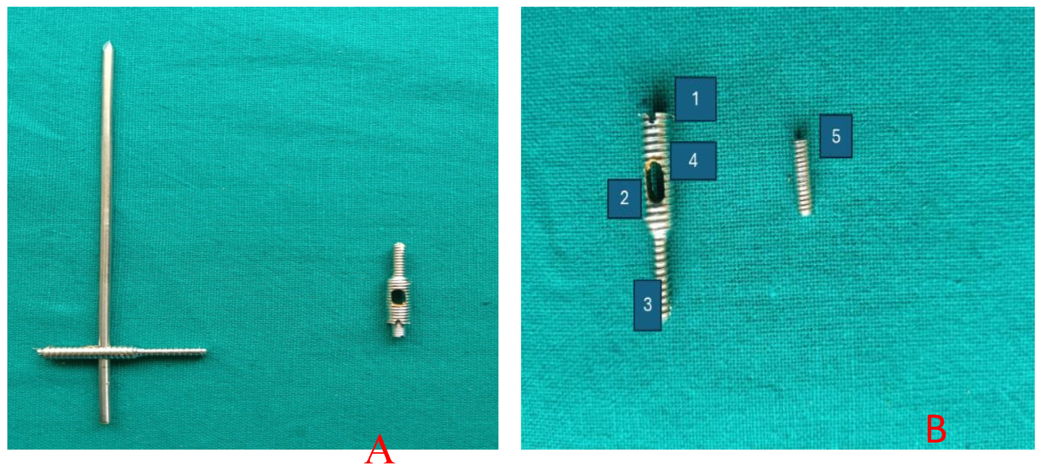
Figure 2.
Constructs prepared for biomechanical testing. (A) Dynamic cross pinning configuration. (B) Locking screw–intramedullary pin construct prior to mechanical testing.
Figure 2.
Constructs prepared for biomechanical testing. (A) Dynamic cross pinning configuration. (B) Locking screw–intramedullary pin construct prior to mechanical testing.
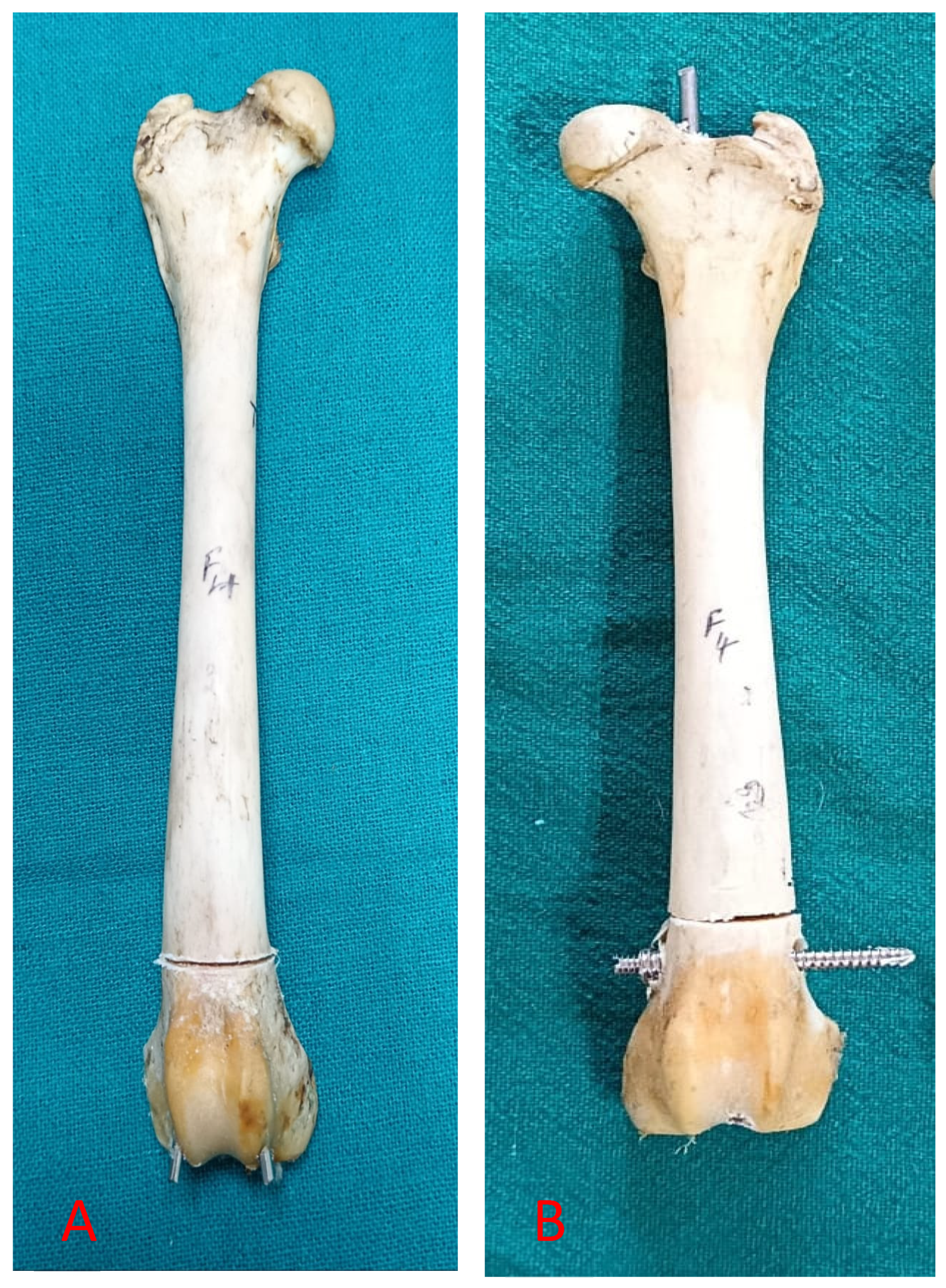
Figure 3.
Universal Testing Machine (UTM) used for biomechanical testing of the prepared constructs.
Figure 3.
Universal Testing Machine (UTM) used for biomechanical testing of the prepared constructs.
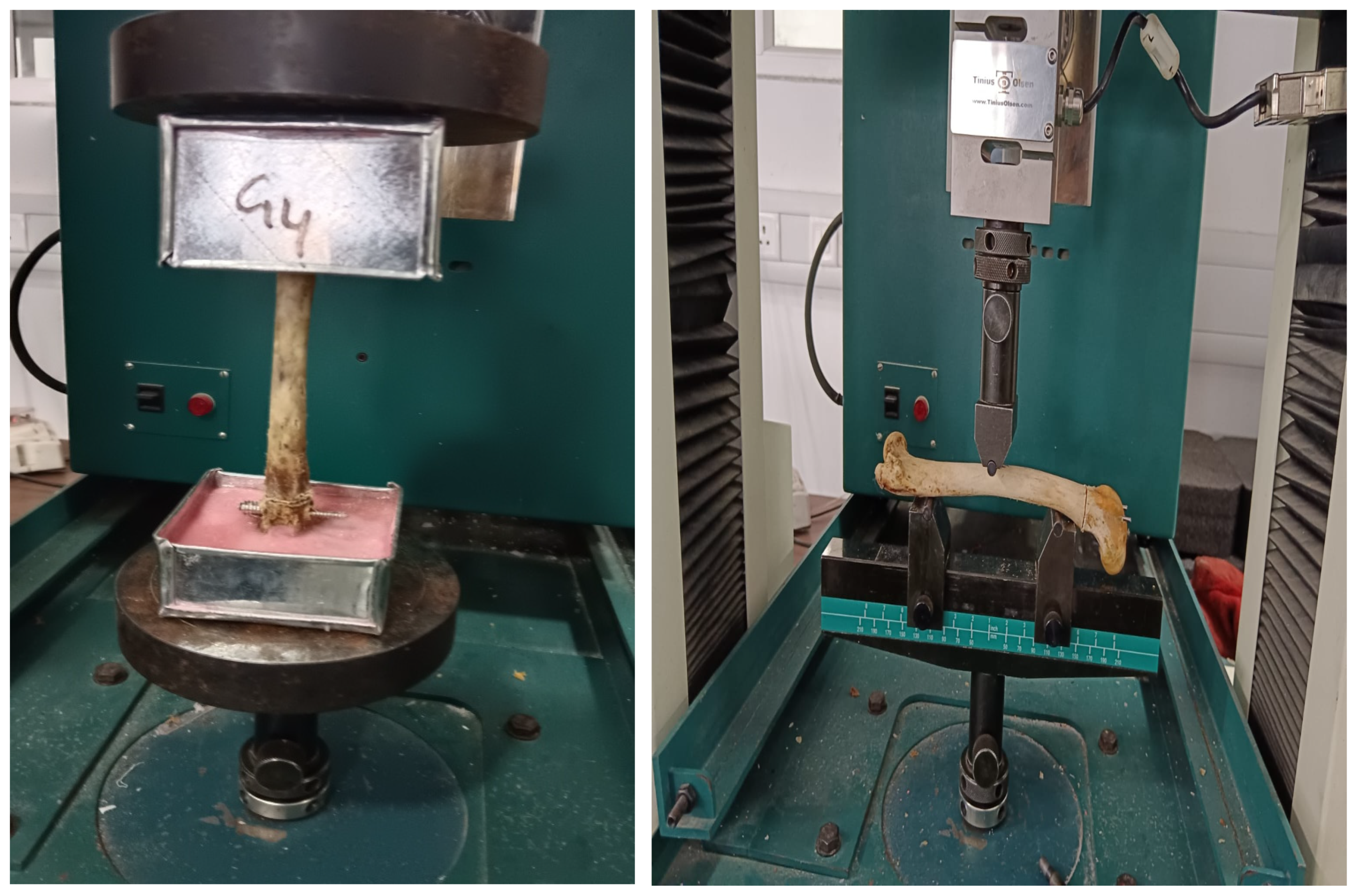
Figure 4.
(a) Group A (case 1) Medio-lateral radiographs showing progression at different time points: 0th day, post-operation, 15th day, 30th day, and 60th day. (b) Group A (case 1) Antero-posterior radiographs showing progression at different time points: 0th day post-operation, 15th day, 30th day, and 60th day.
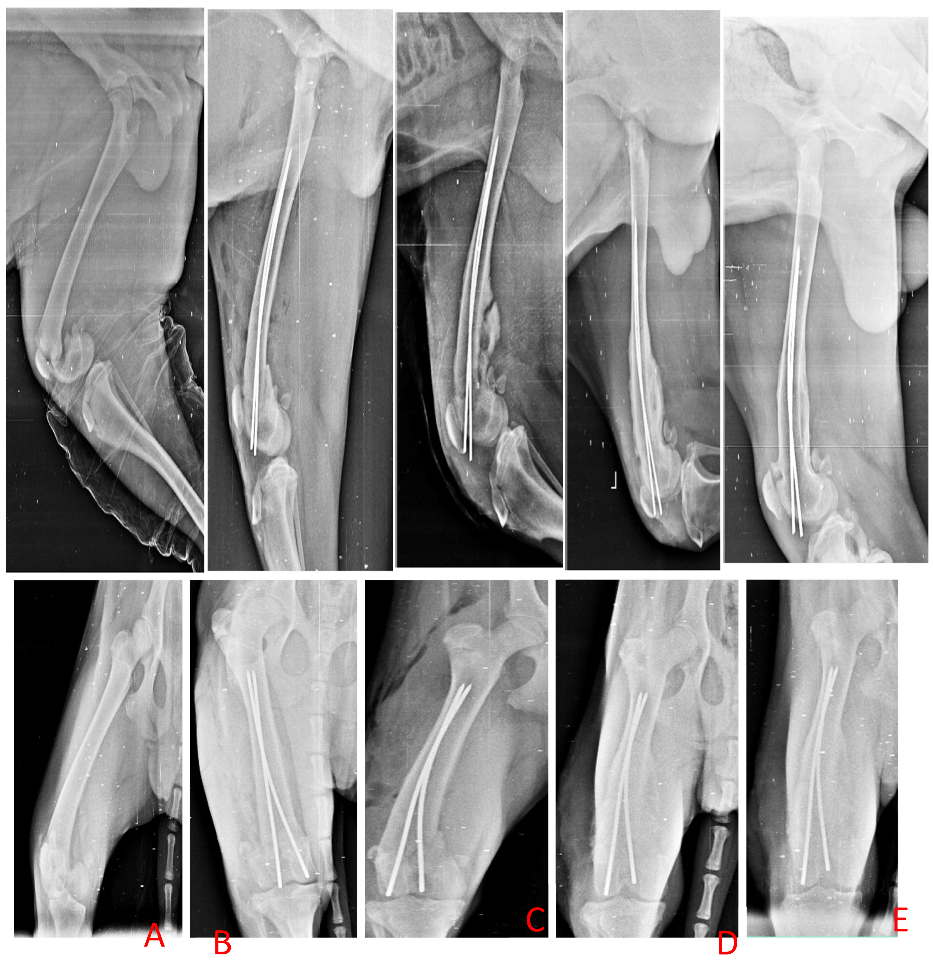
Figure 5.
(a) Group A (case 2) Medio-lateral radiographs showing progression at different time points: 0th day post-operation and 16th day (pin migration). (b) Group A (case 2) Antero-posterior radiographs showing progression at different time points: 0th day post-operation and 16th day (pin migration).
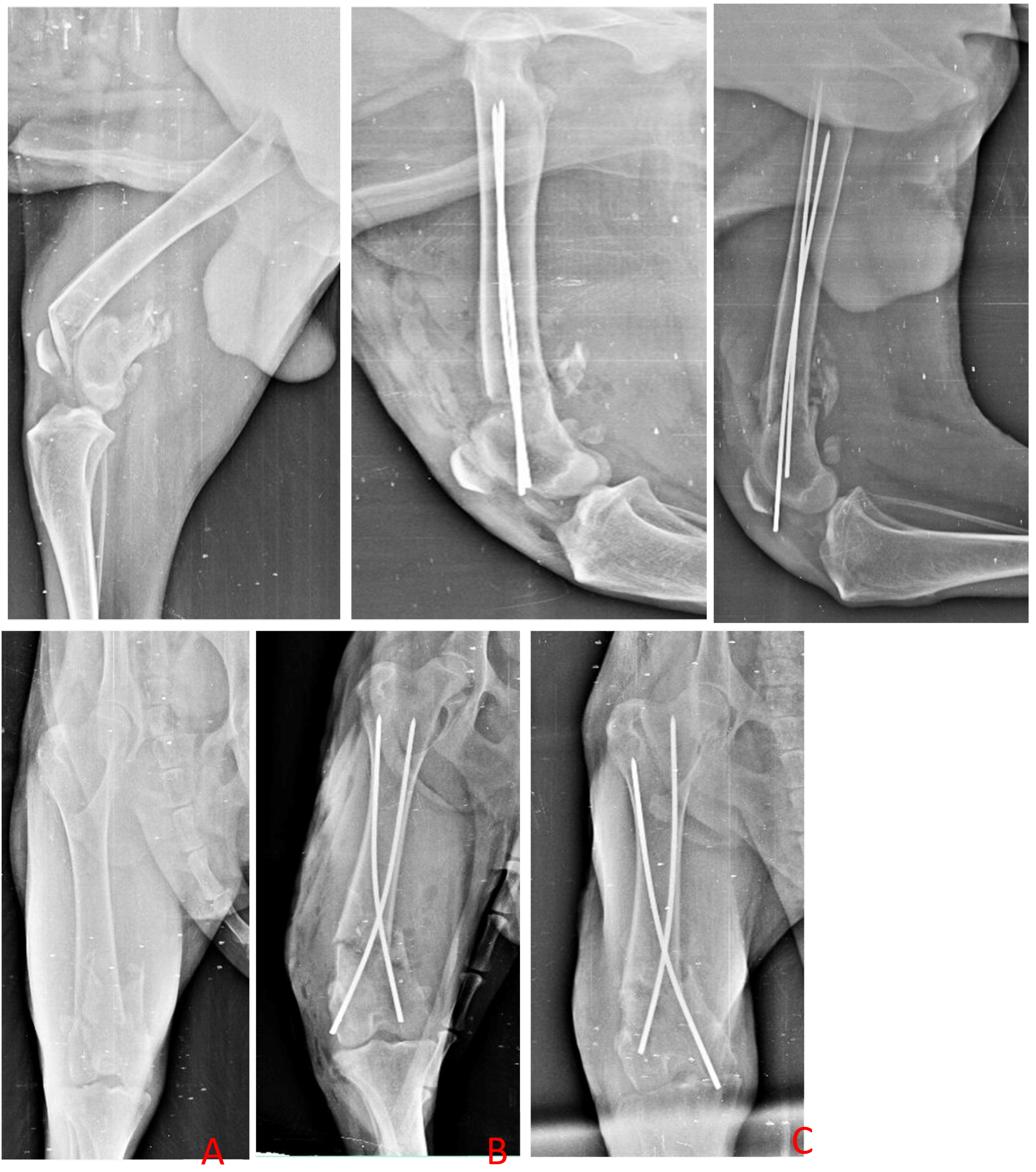
Figure 6.
(a) Group A (case 1) Medio-lateral radiographs showing progression at different time points: 0th day post-operation, 15th day, 30th day, and 60th day. (b) Group A (case 1) Antero-posterior radiographs showing progression at different time points: 0th day post-operation, 15th day, 30th day, and 60th day.
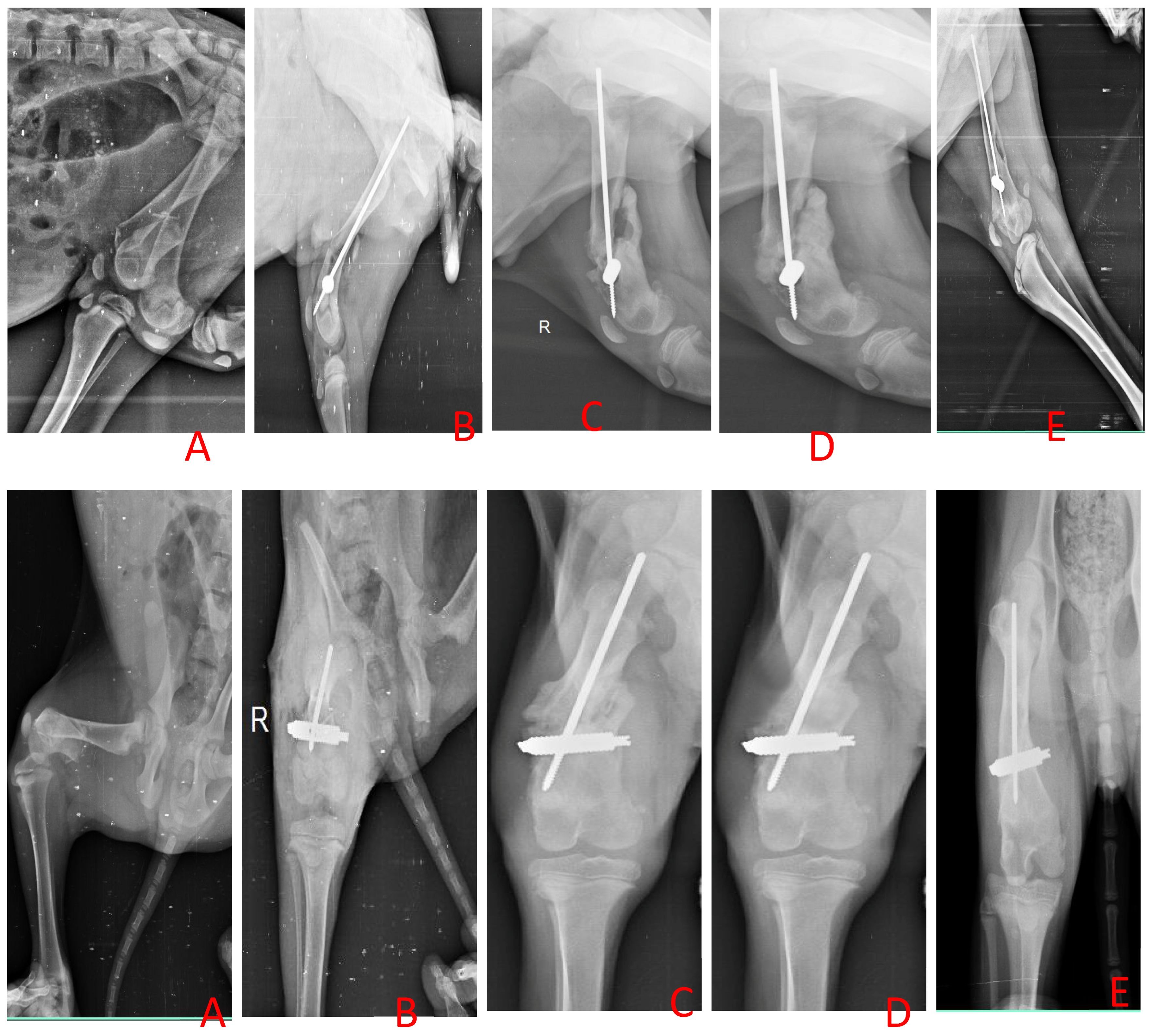
Figure 7.
(a) Group A (case 1) Medio-lateral radiographs showing progression at different time points: 0th day post-operation, 15th day, 30th day, and 60th day. (b) Group A (case 1) Antero-posterior radiographs showing progression at different time points: 0th day post-operation, 15th day, 30th day, and 60th day.
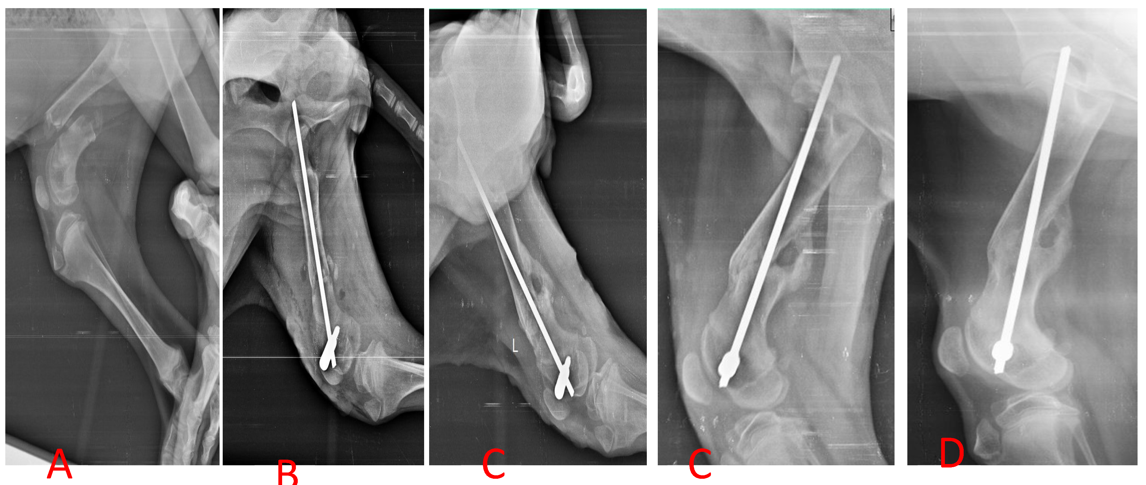
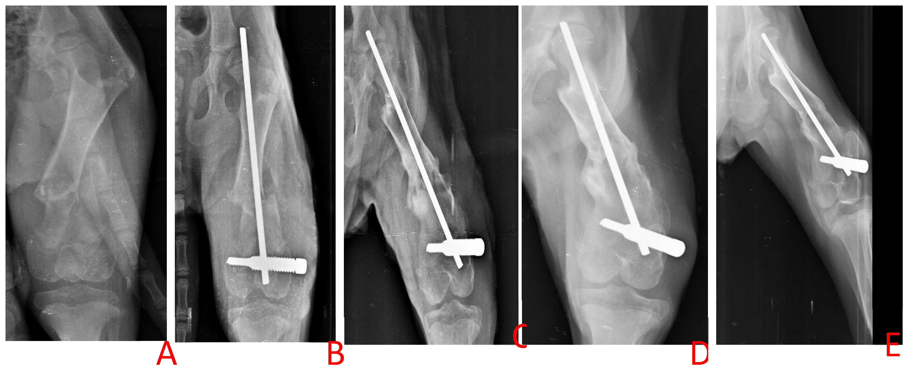

Table 1.
Biomechanical Performance of Constructs (Mean ± SE).
| Test Type | Parameter | Group I | Group II | p-value |
| Axial Compression | Ultimate load to failure (kN) | 2.82 ± 0.23 | 3.77 ± 0.23 | <0.05 |
| Three-Point Bending | Ultimate load to failure (kN) | 1.41 ± 0.19 | 2.18 ± 0.27 | <0.05 |
| Displacement at failure (mm) | 10.88 ± 0.82 | 12.67 ± 0.44 | <0.05 | |
| Axial Torsion | Torque (Nm) | Not measurable | Not measurable | — |
Table 2.
Comparative Clinical Outcomes in Group I and Group II.
| Parameter | Day | Group I (Dynamic Cross Pin) | Group II (Locking Screw–IM Pin) | p-Value |
| Surgery Duration (min) | — | 35 ± 4.8 | 31.17 ± 2.2 | >0.05 |
| Post-operative Inflammation (Median, IQR) | 0 | 2 (1–3) | 1.5 (1–3) | >0.05 |
| 15 | 1.5 (1–3) | 1 (1–3) | <0.05 | |
| 30 | 1 (1–3) | 0 (0–3) | <0.05 | |
| Pain Score (Median, IQR) | 0 | 1 (1–4) | 2 (1–4) | >0.05 |
| 15 | 2 (1–4) | 3 (1–4) | <0.05 | |
| 30 | 3 (1–4) | 4 (1–4) | <0.05 | |
| Radiographic Callus (Median, IQR) | 15 | 2 (1–4) | 1 (1–4) | <0.05 |
| 30 | 3 (1–4) | 2 (1–4) | <0.05 | |
| 60 | 4 (1–4) | 3 (1–4) | <0.05 | |
| Net Weight Bearing (Median, IQR) | 0 | 3.5 (2–6) | 4 (2–4) | >0.05 |
| 15 | 3.5 (2–8) | 6 (4–9) | <0.05 | |
| 30 | 4 (2–6) | 9 (2–9) | <0.05 | |
| 60 | 3.5 (2–7) | 10 (9–10) | <0.05 | |
| Extension Angle (Mean ± SE) | 0 | 152.8 ± 2.44 | 153.8 ± 1.7 | >0.05 |
| 15 | 122.3 ± 2.17 | 137.8 ± 1.85 | <0.05 | |
| 30 | 138 ± 1.85 | 151.8 ± 2.45 | <0.05 | |
| 60 | 151.7 ± 2.32 | 158 ± 2.3 | <0.05 | |
| Flexion Angle (Mean ± SE) | 0 | 33.20 ± 0.54 | 33.48 ± 0.54 | >0.05 |
| 15 | 41.99 ± 0.66 | 41.52 ± 1.22 | <0.05 | |
| 30 | 45.23 ± 1.6 | 40.48 ± 0.6 | <0.05 | |
| 60 | 42.66 ± 1.2 | 32.4 ± 0.45 | <0.05 | |
| Functional Recovery (Median, IQR) | 60 | 1.5 (1–4) | 3.5 (1–4) | <0.05 |
| Implant Failure | — | 3/6 | 1/6 | — |
| Stifle Joint Function | 60 | Delayed recovery | Near-baseline recovery | <0.05 |
Footnotes: IQR = interquartile range; SE = standard error. Inflammation score: 0 = none, 1 = mild, 2 = moderate, 3 = severe inflammation. Pain score: graded on a 1–4 scale (1 = severe pain, 4 = no pain) based on Cross et al. (1997). Radiographic callus: graded using the Dvorak method (1 = abundant callus, 4 = minimal callus). Net weight-bearing score: composite lameness score (0–10) derived from a modified Vasseur grading system. Functional recovery: four-point ordinal scale (4 = excellent recovery, 1 = poor outcome). Stifle joint function: categorized based on flexion–extension recovery patterns. Delayed recovery = persistent functional limitation at Day 60. Near-baseline recovery = restoration of joint function approaching pre-injury mobility.
Disclaimer/Publisher’s Note: The statements, opinions and data contained in all publications are solely those of the individual author(s) and contributor(s) and not of MDPI and/or the editor(s). MDPI and/or the editor(s) disclaim responsibility for any injury to people or property resulting from any ideas, methods, instructions or products referred to in the content. |
© 2026 by the authors. Licensee MDPI, Basel, Switzerland. This article is an open access article distributed under the terms and conditions of the Creative Commons Attribution (CC BY) license (http://creativecommons.org/licenses/by/4.0/).
Copyright: This open access article is published under a Creative Commons CC BY 4.0 license, which permit the free download, distribution, and reuse, provided that the author and preprint are cited in any reuse.