

Submitted:
16 March 2026
Posted:
17 March 2026
You are already at the latest version
Abstract
Down syndrome (DS), caused by trisomy 21, is the most prevalent genetic condition associated with accelerated aging and near-universal development of early-onset Alzheimer’s disease (AD). Beyond gene-dosage imbalance, trisomy 21 induces widespread transcriptional, metabolic, and proteomic remodeling that establishes a chronic state of proteotoxic and oxidative stress from early development. Increasing evidence identifies DS as a disorder of proteostasis network failure, in which sustained translational pressure, redox disequilibrium, and degradation pathway insuffi-ciency progressively erode cellular resilience. In the DS brain, persistent endoplasmic reticulum stress with PERK-dominant signaling, mitochondrial dysfunction characterized by oxidative phosphorylation deficits and exces-sive reactive oxygen species production, and impaired antioxidant responses create a highly vulnerable intracellular environment. Concomitantly, degradation systems become compromised: proteasomal catalytic activity declines, ubiquitin-dependent signaling is remodeled, and chronic mTOR hyperactivation suppresses autophagic and mi-tophagic flux. The coordinated impairment of the ubiquitin–proteasome system and autophagy establish a feed-forward cycle of proteotoxic accumulation and redox amplification. Within this framework, Alzheimer-like neuropathology in DS emerges not solely from amyloid precursor protein triplication but as the late manifestation of decades-long pro-teostasis exhaustion. Therapeutic strategies aimed at restoring global proteostasis and redox balance may therefore represent a more effective systems-level approach to mitigating neurodegeneration in DS.
Keywords:
Down syndrome
; proteostasis
; oxidative stress
; mitochondrial dysfunction
; Alzheimer’s disease
1. Introduction
Down syndrome (DS), caused by full or partial trisomy of chromosome 21 (Chr21), is the most common chromosomal aneuploidy compatible with postnatal survival and the leading genetic cause of intellectual disability, affecting approximately 1 in 800–1,000 live births worldwide [1]. Over recent decades, advances in cardiac surgery, antimicrobial therapies, and endocrine management have markedly increased life expectancy, transforming DS into a lifespan condition. As survival has improved, it has become increasingly evident that DS is not merely a neurodevelopmental disorder, but a systemic and progressive condition characterized by premature aging and early emergence of age-associated comorbidities. Clinically, DS presents with intellectual disability and a broad constellation of multisystem alterations—including congenital heart defects, thyroid dysfunction, immune dysregulation, gastrointestinal anomalies, metabolic disturbances, and increased susceptibility to hematological malignancies. However, this phenotypic heterogeneity reflects a deeper biological principle: trisomy 21 does not simply increase the expression of individual genes but reshapes regulatory networks across the genome [2]. The additional chromosome imposes genome-wide transcriptional, epigenetic, and proteomic remodeling that alters developmental trajectories and systemic homeostasis from embryogenesis onward [3]. At the molecular level, overexpression of more than 200 Chr21 genes perturbs chromatin organization, modifies DNA methylation patterns, alters histone marks, and reshapes non-coding RNA networks [4]. These changes extend beyond proportional transcript amplification, affecting global gene expression programs involved in metabolism, immune signaling, mitochondrial function, and cellular stress responses. Consequently, trisomic cells operate in a state of persistent regulatory disequilibrium. Increased gene dosage elevates global protein synthesis and disrupts stoichiometric relationships within multiprotein complexes, creating a chronic imbalance between protein production, folding, and degradation. This sustained pressure on protein quality control systems establishes a proteotoxic background that intersects with redox imbalance and metabolic dysregulation [5]. The central nervous system (CNS) is particularly vulnerable to these disturbances. Neurodevelopment in DS is characterized by reduced neural progenitor proliferation, altered neuronal differentiation, impaired synaptogenesis, and diminished dendritic arborization, leading to reduced brain volume—especially within the hippocampus and frontal cortex. These structural abnormalities unfold within a cellular environment marked by mitochondrial dysfunction, increased oxidative stress, and chronic low-grade inflammation. Triplication of interferon receptor genes enhances innate immune responsiveness, while overexpression of redox-regulating genes such as SOD1 perturbs antioxidant balance [6]. The convergence of these mechanisms establishes a stress-prone neural milieu that predisposes to premature biological aging. Among the most consequential gene-dosage effects is triplication of the amyloid precursor protein (APP) gene, which confers an almost inevitable risk of early Alzheimer-related neuropathology [7]. Yet Alzheimer’s disease (AD) in DS cannot be attributed solely to amyloid overproduction. Amyloid-β accumulation unfolds within a broader landscape of progressive mitochondrial fragility, impaired proteostasis, chronic oxidative stress, and diminished adaptive stress responses. Thus, AD pathology in DS emerges not as an isolated consequence of APP dosage [8], but as the downstream manifestation of long-standing cellular disequilibrium.
Individuals with DS represent the most prevalent genetically determined form of early-onset Alzheimer’s disease (EOAD), with nearly universal development of AD neuropathology by the fourth decade of life [9]. However, amyloid accumulation in DS occurs within a biological context of systemic and brain-specific accelerated aging. Trisomy 21 establishes a persistently stress-prone cellular state decades before chronological aging would predict, lowering resilience thresholds and amplifying the neurotoxic effects of amyloid-β (Aβ) and tau pathology. Beyond APP triplication, overexpression of Chr21 genes such as SOD1, DYRK1A, and RCAN1 contributes to dysregulation of redox signaling, kinase activity, mitochondrial function, and calcium homeostasis [10]. These alterations promote sustained oxidative stress, metabolic instability, and impaired stress adaptation. Mitochondrial dysfunction emerges as a central vulnerability node, characterized by impaired oxidative phosphorylation, altered mitochondrial dynamics, defective mitophagy, and excessive production of reactive oxygen species (ROS) [11]. Bioenergetic insufficiency not only compromises neuronal function but also weakens energy-dependent proteostasis mechanisms, reinforcing a feed-forward cycle between redox imbalance and proteotoxic stress. Persistent oxidative and metabolic stress promotes premature activation of cellular senescence programs across neurons and glial populations. Senescent cells exhibit activation of p53/p21 and p16^INK4a^ pathways, chronic DNA damage signaling, mitochondrial impairment, and extensive epigenetic remodeling [12]. A hallmark of senescence is progressive loss of proteostasis capacity, including impaired autophagic flux, reduced proteasomal activity, and maladaptive unfolded protein responses [13]. In this context, mitochondrial dysfunction limits ATP availability for protein folding and degradation, while oxidative modifications increase the aggregation propensity of Aβ and tau. The senescence-associated secretory phenotype (SASP) further sustains inflammatory signaling and activates tau kinases, directly linking premature cellular aging to synaptic dysfunction and network instability [14]. Neuropathologically, AD in DS mirrors sporadic AD in its anatomical progression but develops decades earlier. Amyloid deposition begins in early adulthood, followed by widespread tau pathology by the fourth decade, whereas clinical dementia manifests later as compensatory mechanisms progressively fail [15]. Biomarker studies—including cerebrospinal fluid analyses and PET imaging—demonstrate a prolonged preclinical phase during which mitochondrial dysfunction, oxidative stress, and proteostatic insufficiency silently accumulate. This predictable and genetically defined trajectory positions DS as a powerful human model for dissecting the interplay between accelerated aging and neurodegeneration [16].
2. Trisomy 21 and Proteotoxic Stress
Aneuploidy imposes intrinsic stress on cellular systems, and trisomy 21 exemplifies this principle [17]. Increased gene dosage elevates global protein synthesis and disrupts the stoichiometric balance required for correct assembly of multiprotein complexes. Proteins encoded on chromosome 21 frequently require binding partners located on other chromosomes; when produced in excess, they remain unassembled or misfolded, increasing the burden on molecular chaperones and degradation pathways [18]. This imbalance establishes a chronic proteostatic strain in which stress-response pathways remain persistently engaged. Proteotoxic stress in DS is further amplified by mitochondrial dysfunction and redox imbalance. Oxidative modifications destabilize protein structure and promote aggregation, while impaired ATP production compromises the efficiency of energy-dependent clearance systems, including the ubiquitin–proteasome system and autophagy–lysosome pathways. Rather than reflecting isolated pathway failures, proteostasis disruption in DS represents a network-level disturbance in which increased protein synthesis, defective degradation, oxidative stress, altered translational control, and organelle dysfunction converge [19]. Neurons are particularly susceptible to this chronic imbalance due to their high metabolic demand, complex proteome dynamics, and limited regenerative capacity. Over time, sustained proteostatic insufficiency reduces synaptic protein turnover, weakens adaptive stress responses, and lowers the threshold for aggregation-driven toxicity [20]. Aging superimposed on this pre-stressed background further diminishes buffering capacity, accelerating the transition from compensatory stress signaling to irreversible neurodegeneration. Viewed through this integrative lens, DS emerges as a paradigmatic condition in which gene-dosage imbalance drives chronic proteotoxic stress, accelerates biological aging, and creates a permissive environment for Alzheimer-like pathology. The following sections will dissect how endoplasmic reticulum stress, mitochondrial dysfunction, and degradation pathway impairment interact within a unified proteostasis–redox network and will explore therapeutic strategies aimed at restoring systemic resilience rather than targeting single pathogenic proteins.
2.1. Endoplasmic Reticulum Stress and Unfolded Protein Response in Down Syndrome Neuropathology
Trisomy 21 imposes a chronic proteotoxic burden that persistently engages endoplasmic reticulum (ER) stress signaling and reshapes unfolded protein response (UPR) dynamics in DS. Rather than representing a transient adaptive reaction, ER stress in DS appears developmentally established and progressively maladaptive. The ER, a central hub for protein folding, calcium homeostasis, and post-translational modification, is highly sensitive to disturbances in redox balance and proteome load. In trisomic cells, increased protein synthesis, oxidative modifications, and altered calcium handling converge to destabilize ER proteostasis, promoting accumulation of misfolded proteins and sustained activation of UPR pathways [21,22] . Although the UPR is classically defined as a conserved adaptive program that reduces translation and enhances folding capacity [23], its chronic engagement can shift from protective to deleterious, ultimately promoting apoptosis and functional decline [24]. Multiple independent lines of evidence indicate that ER stress and maladaptive UPR activation are embedded features of DS neuropathology. Proteomic and biochemical analyses of human DS brain tissue demonstrate increased activation of ER stress sensors and dysfunction of key ER chaperones [25,26,27]. Accumulation of oxidatively modified proteins within the ER lumen activates the three canonical UPR branches—PERK, ATF6, and IRE1—via dissociation from the master regulator GRP78/BiP [22]. Notably, GRP78 itself is oxidatively modified in DS brain [19], impairing its regulatory capacity and providing a direct mechanistic link between redox imbalance and sustained ER stress signaling [28]. Among UPR branches, the PERK pathway emerges as the dominant and persistently engaged arm in DS. Biochemical studies of frontal cortex from individuals with DS reveal chronic activation of the PERK/eIF2α/ATF4 axis [26]. PERK-mediated phosphorylation of eIF2α reduces global protein synthesis, thereby limiting ER client load [29], while selectively enhancing translation of stress-responsive transcripts such as ATF4 [30]. Under physiological conditions, ATF4 coordinates adaptive outputs, including induction of chaperones, redox-regulatory genes, autophagy components, and amino acid metabolism pathways. Negative feedback through GADD34 allows re-establishment of translational homeostasis [31]. In DS, however, this regulatory balance appears disrupted. Reduced GADD34 levels and persistent eIF2α phosphorylation indicate failure to restore translational equilibrium, leading to sustained activation of pro-apoptotic mediators such as CHOP and BCL-2 family members [32]. Importantly, PERK hyperactivation in DS is closely associated with Alzheimer-like neuropathological features. Both young and older DS brains exhibiting AD pathology show increased pPERK, peIF2α, ATF4, and CHOP levels [26], paralleling observations in sporadic AD, where ER stress markers correlate with Braak stage progression [26,33,34,35,36]. These findings suggest that PERK-dominant UPR signaling is not a secondary epiphenomenon but a central contributor to neurodegenerative vulnerability. A critical pathogenic feature in DS is the uncoupling between PERK activation and antioxidant defense. Under normal conditions, PERK signaling promotes nuclear translocation of Nrf2, enhancing antioxidant gene expression. In DS, despite sustained PERK activation, Nrf2-driven transcription is suppressed [26]. This maladaptive dissociation is linked to overexpression of Bach1, a chromosome 21–encoded transcriptional repressor of Nrf2 targets [37,38], resulting in continued translational repression without adequate redox compensation. Consequently, the DS brain exhibits a state of PERK dominance combined with antioxidant insufficiency, amplifying oxidative damage and reinforcing proteostatic imbalance [39].
Systems-level proteomic analyses further support the centrality of ER stress in DS. Large-scale studies of post-mortem frontal cortex from young and aged individuals with DS reveal early and persistent dysregulation of stress-response and proteostasis pathways that intensify with aging and AD conversion [40,41,42,43]. Importantly, these alterations are detectable before overt neurodegeneration, indicating that UPR dysregulation is embedded within the DS molecular phenotype rather than representing a late-stage response. Energy metabolism, synaptic transmission, and cellular stress-response networks consistently emerge as the most affected pathway clusters, reinforcing the concept that chronic ER stress contributes to progressive neuronal dysfunction [40]. Proteostasis abnormalities in DS are not restricted to the CNS. Proteomic profiling of peripheral blood mononuclear cells (PBMCs) from children with DS demonstrates widespread dysregulation of metabolic, trafficking, and stress-response pathways, including activation of ER stress and UPR components [26,44]. Increased oxidative protein damage and sustained UPR signatures are detectable early in life, supporting the concept of a developmentally established “pre-stressed” proteostatic state. Similarly, lymphoblastoid cell lines derived from children with DS display persistent UPR induction and early endosomal alterations, consistent with systemic proteotoxic burden [26,45]. Broader analyses of the proteostasis network in DS cells confirm coordinated changes in chaperone systems, UPR mediators, and proteasomal machinery, alongside heightened sensitivity to additional proteotoxic challenges [18,41,44,46]. Reduced buffering capacity and lower stress tolerance thresholds suggest that trisomic cells operate closer to proteostasis collapse.
Murine models of DS provide temporal and mechanistic resolution of ER stress dynamics [47]. The Ts65Dn model exhibits early activation of ER stress pathways in the brain, with selective engagement of the PERK branch detectable prior to overt neurodegeneration [48]. Increased phosphorylation of PERK and eIF2α at young ages indicates that ER stress signaling is an upstream event in DS neuropathology. Longitudinal profiling reveals that UPR activation is not static; early hyperactivation may transition into partial signaling exhaustion or maladaptive remodeling with aging, consistent with progressive loss of stress-response efficiency [49,50]. This temporal evolution aligns with the broader concept of premature molecular aging in the DS brain.
Mechanistic validation is provided by studies in the Ts2Cje model, where pharmacological inhibition of PERK signaling restores translational balance, enhances Nrf2 nuclear translocation, and corrects Nrf2/Bach1 imbalance [26]. These interventions demonstrate that PERK hyperactivation is not merely correlative but causally linked to oxidative and proteostatic dysfunction, highlighting its therapeutic tractability. The more recently developed Ts66Yah model, which improves construct validity by eliminating non-Hsa21 orthologous regions [51], confirms early and persistent dysregulation of stress-response and proteostasis pathways across the lifespan [52]. Although not restricted to ER markers, proteomic analyses consistently identify UPR components among the most perturbed molecular signatures, reinforcing the centrality of chronic stress-response engagement in DS neuropathology [40]. Age-dependent cognitive decline in Ts66Yah mice parallels progressive redox imbalance and proteostasis disruption, supporting the notion that ER stress contributes to long-term functional vulnerability.
Beyond the canonical UPR, the integrated stress response (ISR) is also activated in DS. Costa-Mattioli and colleagues demonstrated phosphorylation of eIF2α in Ts65Dn hippocampus, post-mortem DS brain tissue, and DS-derived induced pluripotent stem cells (iPSCs), mediated in part by PKR activation [27]. Importantly, normalization of eIF2α phosphorylation restored proteostatic balance and improved cognitive performance in DS models [27], underscoring the pathological relevance of sustained translational repression.
Collectively, convergent evidence from human brain, peripheral cells, and multiple DS mouse models positions ER stress and UPR dysregulation as central nodes in DS pathobiology. Trisomy 21 establishes chronic proteotoxic and oxidative pressure that persistently activates ER stress sensors, biases UPR signaling toward PERK dominance, uncouples antioxidant defense, and progressively undermines synaptic and neuronal integrity. While acute UPR activation may initially buffer proteome imbalance, sustained engagement—particularly of the PERK–ATF4–CHOP axis—promotes translational repression, pro-apoptotic signaling, and heightened vulnerability to Alzheimer-like neurodegeneration [53]. These findings collectively identify ER stress modulation, restoration of Nrf2 activity, and reinforcement of proteostasis capacity as rational therapeutic strategies to counteract neurodegenerative progression in Down syndrome [25,26,40].
3. Mitochondrial Stress in Down Syndrome
In DS, mitochondria emerge as a central bioenergetic and redox hub through which trisomy-driven proteotoxic pressure is translated into progressive cellular dysfunction. Beyond their canonical role in ATP production, mitochondria regulate calcium buffering, redox signaling, lipid metabolism, and apoptotic pathways—functions that are particularly critical in neurons and glial cells with high metabolic demand and limited regenerative capacity [54]. In trisomy 21, mitochondrial destabilization leads to increased production of reactive oxygen and nitrogen species (ROS/RNS), propagating oxidative modifications of proteins and lipids, including carbonylation, nitration, and peroxidation-derived adducts [55,56]. Persistent oxidative stress damages mitochondrial DNA (mtDNA), respiratory chain components, and membrane integrity [57], further impairing electron transport chain (ETC) efficiency and reducing ATP output. This bioenergetic decline not only compromises neuronal function but also destabilizes energy-dependent proteostasis systems, amplifying protein misfolding and aggregation under oxidizing conditions [58]. Mitochondria therefore act as a mechanistic bridge linking gene-dosage imbalance to accelerated aging and Alzheimer-like neurodegeneration in DS [55,56,58,59,60]. Hallmarks of mitochondrial dysfunction in DS include reduced oxidative phosphorylation (OXPHOS) efficiency, chronic redox imbalance, and disruption of mitochondrial quality control (MQC) programs encompassing biogenesis, dynamics, and mitophagy [28,61]. These alterations are not restricted to the central nervous system; they contribute to systemic phenotypes such as cognitive impairment, congenital cardiac defects, and premature aging traits [1,56,61]. Importantly, mitochondrial dysfunction is detectable early in development, with widespread abnormalities reported in fetal trisomy 21-derived cells [62,63,64]. Structural fragmentation of the mitochondrial network, cristae disorganization, and reduced connectivity indicate chronic imbalance in mitochondrial turnover and distribution [65]. During neurogenesis and synaptic maturation—periods of intense bioenergetic demand—such instability is particularly detrimental, correlating with impaired neuronal proliferation, altered differentiation, reduced dendritic complexity, and increased susceptibility to apoptosis in DS models [28,56,60,66,67,68,69].
3.1. Mitochondrial Unfolded Protein Response (UPRmt) in Down Syndrome
The mitochondrial unfolded protein response (UPRmt) constitutes a critical adaptive program that attempts to buffer mitochondrial proteotoxic stress by coordinating retrograde signaling to the nucleus [70]. Upon accumulation of misfolded proteins within the mitochondrial matrix, UPRmt induces transcriptional programs that enhance expression of mitochondrial chaperones, proteases, antioxidant defenses, and metabolic regulators [70,71,72]. This response aims to restore ETC integrity, stabilize protein folding, and prevent propagation of mitochondrial stress to the broader cellular environment.
In DS, however, UPRmt engagement appears early but functionally incomplete. In the Ts2Cje model, perinatal activation of an ATF5/GRP75-dependent program in frontal cortex is accompanied by reduced expression of other stress mediators, including ATF4, CHOP, and SIRT3 [26,73,74]. This partial activation suggests that mitochondria in DS detect proteotoxic stress yet fail to mount a fully coordinated protective response. Such imbalance may limit stabilization of respiratory chain complexes and compromise mitochondrial proteome maintenance under sustained oxidative conditions [1]. Consequently, incomplete UPRmt engagement may contribute to persistent mitochondrial fragility during neurodevelopmental windows when bioenergetic precision is essential.
When UPRmt buffering proves insufficient, cells rely increasingly on broader MQC mechanisms—including biogenesis, fusion–fission dynamics, and mitophagy—to maintain a functional mitochondrial pool [73]. In DS, evidence indicates that these compensatory systems are chronically stressed and frequently maladaptive, favoring accumulation rather than efficient turnover of dysfunctional organelles.
Impaired OXPHOS is a consistent and defining feature of DS biology. Across human tissues, patient-derived cells, and mouse models, trisomy 21 is associated with ETC inefficiency, particularly involving complex I deficits, reduced bioenergetic reserve, and increased ROS generation [56,60,68,74,75,76,77,78]. Some evidence suggests that trisomic cells downregulate OXPHOS as an adaptive attempt to limit electron leak and oxidative damage [79]. While this strategy may transiently reduce ROS production, it compromises ATP availability and diminishes metabolic flexibility, particularly under fluctuating energy demands.
In the developing and adult brain—where synaptic plasticity and circuit refinement require rapid, localized ATP production—chronic OXPHOS dampening may have profound consequences for neuronal resilience. In Ts2Cje frontal cortex, dysfunction of complexes I and IV emerges early during developmental stages characterized by maximal synaptogenesis [74]. Moreover, lifespan-dependent remodeling of OXPHOS subunits—where some components are upregulated and others downregulated—suggests instability in ETC assembly and turnover under persistent proteostatic pressure [52,74]. Proteomic profiling of human DS frontal cortex with and without AD neuropathology consistently identifies mitochondrial and energy-related pathways among the most altered signatures [40], underscoring the centrality of bioenergetic stress across DS and DSAD trajectories. Emerging evidence also implicates complex IV as a metabolic bottleneck, with upstream metabolic inhibitors converging on cytochrome c oxidase function [80].
ETC complexes are multi-subunit structures requiring coordinated nuclear and mitochondrial gene expression, import, folding, and assembly. Under chronic proteotoxic and oxidative stress, this coordination becomes destabilized. Thus, OXPHOS inefficiency in DS is both a consequence and an amplifier of mitochondrial proteostasis failure, reinforcing a feed-forward loop of ROS production, energetic insufficiency, and progressive proteostatic collapse in the brain.
3.2. Mitochondrial Quality Control (MQC) in Down Syndrome
MQC integrates mitochondrial biogenesis, fusion–fission dynamics, and mitophagy to preserve organelle integrity under stress [81] In DS, converging data indicate that MQC is persistently challenged, particularly in neural tissue where spatially restricted ATP production and tight redox regulation are critical [55,56]. Morphologically, fragmentation of the mitochondrial network and cristae disorganization reflect imbalance in fusion–fission regulation [55,78,82]. Reduced fusion competence (MFN2/OPA1 dysfunction and altered OPA1 processing) combined with enhanced DRP1-mediated fission promotes respiratory inefficiency and ROS amplification [55,74,77,83,84]. Notably, experimental normalization of mitochondrial dynamics improves bioenergetics and supports neurogenic programs in DS models, confirming that dynamics are functionally causal rather than epiphenomenal [75,83,84,85,86,87]. Mitophagy impairment further exacerbates mitochondrial accumulation. Evidence of defective PINK1/Parkin signaling, incomplete autophagic flux, and inefficient removal of damaged mitochondria indicates compromised turnover capacity [74,88,89]. Persistent mTOR hyperactivation—reported early and sustained in DS brain—restrains ULK1-mediated initiation of autophagy and mitophagy [74,88,89,90], thereby linking nutrient-sensing dysregulation to defective mitochondrial clearance. ULK1-dependent phosphorylation of mitophagy receptors such as FUNDC1 [90] provides a mechanistic bridge between mTOR status and cargo recruitment, reinforcing the notion that mTOR dysregulation [59,91,92,93,94] directly impairs mitochondrial quality control. Simultaneously, dysregulation of mitonuclear signaling pathways—including AMPK–PGC-1α–NRF1–TFAM—limits mitochondrial renewal and mtDNA maintenance under sustained stress [55,74,87]. Trisomy-linked gene dosage further compounds MQC dysfunction. Overexpression of ETS2 promotes mitochondrial apoptotic signaling in DS neuronal models [95], while intersectin-1 (ITSN1) influences mitochondrial death pathways via trafficking modules [96]. Repression of PGC-1α activity through DYRK1A/RCAN1–calcineurin/NFAT signaling [69,97,98] and NRIP1 overexpression [99] constrains mitochondrial biogenesis. Additionally, HSA21-encoded microRNAs, such as miR-155-5p targeting TFAM [100] and let-7c-5p potentially targeting ANT1/SLC25A4 [101], further destabilize mitochondrial maintenance. Collectively, these trisomy-driven inputs create an imbalance wherein mitochondrial damage accumulates faster than MQC systems can compensate, accelerating redox stress and proteostatic vulnerability.
3.3. Insulin Resistance as a Proteostasis–Redox Switch in the DS Brain
In the CNS, insulin signaling functions as a metabolic rheostat that integrates nutrient sensing with mitochondrial competence, redox control, and proteostasis [102,103,104,105]. Through the IRS–PI3K–AKT cascade, insulin promotes metabolic flexibility, restrains excessive ROS generation, and modulates mTOR-dependent translation and autophagy pathways [102,103,104,105]. In DS, this insulin–mTOR axis is disrupted early and prominently in the brain [40,59,60,68,69,89,91]. Aberrant activation of PI3K/AKT/mTOR signaling has been documented in post-mortem DS tissue [88,89,91], reinforcing mTOR hyperactivation as a core trisomy-linked stress signature. Sustained mTOR activity suppresses ULK1-dependent autophagy initiation, impairs mitophagy, and favors persistence of ROS-generating mitochondria [88,89,91]. Brain insulin resistance (BIR) likely contributes upstream to this maladaptive state [59,60,68], linking metabolic signaling failure to mitochondrial dysfunction and oxidative damage. Neuronal-derived extracellular vesicles from young individuals with DS reveal coordinated insulin pathway disruption and aberrant mTOR activity [59], suggesting that this imbalance is developmentally established. O-GlcNAcylation adds an additional nutrient-sensitive regulatory layer. Reduced glucose uptake and insulin resistance decrease hexosamine biosynthetic pathway flux and UDP-GlcNAc availability, lowering protein O-GlcNAcylation and altering the balance between phosphorylation and adaptive stress signaling. In DS models, O-GlcNAc dysregulation coexists with OXPHOS defects and oxidative stress [106]. Taken together, these findings position insulin resistance not merely as a metabolic comorbidity but as a proteostasis–redox switch that shapes mitochondrial resilience and neurodegenerative vulnerability across the DS lifespan.
4. Ubiquitin–Proteasome System Dysfunction in Down Syndrome Brain
The ubiquitin–proteasome system (UPS) is the principal intracellular machinery responsible for selective protein degradation and dynamic regulation of protein turnover. Beyond its degradative function, ubiquitin-dependent signaling orchestrates stress responses, apoptosis, transcriptional regulation, and metabolic adaptation, thereby functioning as a central regulator of proteostasis [107]. Protein targeting to the proteasome requires coordinated ubiquitin conjugation via E1, E2, and E3 enzymes, leading to polyubiquitin chain formation—most commonly through Lys48 linkages—recognized by the 26S proteasome, a multicatalytic complex endowed with chymotrypsin-like, trypsin-like, and caspase-like activities [108]. Oxidative modification of proteins can facilitate proteasomal recognition through partial unfolding, functionally linking redox balance to proteolytic efficiency [109]. In DS, converging evidence indicates progressive disruption of this finely tuned system. A landmark study by our research group provided the first comprehensive characterization of the polyubiquitinylation profile in DS frontal cortex before and after Alzheimer neuropathology. Isolation of polyubiquitinated proteins from postmortem samples revealed a profound age-dependent remodeling of the ubiquitome [110]. Proteins displaying aberrant ubiquitin tagging were predominantly involved in protein quality control and energy metabolism, and oxidative modifications were closely associated with altered ubiquitin conjugation patterns [110,111] . These findings demonstrate that ubiquitin signaling dysregulation represents an early molecular alteration accompanying DS brain aging rather than a late consequence of amyloid deposition [110]. Functional analyses further revealed significant reductions in proteasomal catalytic activities—including chymotrypsin-like, trypsin-like, and caspase-like activities in DS frontal cortex, supporting impaired substrate clearance during early disease stages [110]. Similar reductions in proteasomal activity and increased ubiquitin-positive aggregates have been reported in DS fibroblasts and in the cerebellum of Ts65Dn mice [19,112] , reinforcing the concept of systemic UPS vulnerability. Mechanistically, oxidative post-translational damage represents a major contributor to UPS dysfunction in DS. Redox proteomics analyses identified ubiquitin carboxyl-terminal hydrolase L1 (UCH-L1), a neuron-enriched deubiquitinating enzyme essential for ubiquitin recycling, as an early target of oxidative modification in DS brain [19]. Subsequent studies demonstrated aberrant polyubiquitinylation of UCH-L1, suggesting structural instability and functional impairment [113]. Given its role in maintaining free ubiquitin pools, oxidative inactivation of UCH-L1 may further compromise proteasomal efficiency. Similar carbonylation-associated loss of UCH-L1 activity has been described in AD brain, reinforcing the pathogenic relevance of this mechanism [114]. Accumulation of polyubiquitinated proteins in DS cortex indicates that ubiquitinated substrates are not efficiently degraded and may remain stalled within the system. Increased oxidation of SOD1, a feature observed in DS brain, has been linked to inhibition of proteasome activity, further supporting direct interference of chronic redox imbalance with proteolytic capacity [111,114]. In addition to oxidative mechanisms, gene dosage effects inherent to trisomy 21 may contribute to UPS perturbation. Chromosome 21 encodes multiple UPS regulators, including deubiquitinating enzymes such as USP25 and USP16, as well as proteins involved in ubiquitin conjugation and proteasome assembly [115]. Although the direct functional consequences of their triplication remain incompletely defined, altered expression of UPS-related genes may destabilize ubiquitin homeostasis in DS brain. Furthermore, triplication of BACH1 perturbs the nuclear BACH1/NRF2 balance, impairing antioxidant responses and amplifying oxidative stress [37,38,116]. Given the sensitivity of proteasome function to redox status, BACH1 gene dosage represents an important indirect contributor to UPS vulnerability. Altogether, enzymatic impairment, oxidative damage, and gene dosage imbalance converge to compromise ubiquitin-dependent proteolysis in DS. As proteasomal degradation becomes progressively insufficient, cells increasingly rely on complementary degradative pathways particularly the autophagy lysosomal system to maintain proteostasis.
5. Autophagy in Down Syndrome
Autophagy is a highly conserved degradation pathway responsible for the clearance of long-lived proteins, aggregates, and damaged organelles, including mitochondria and ER [117] . Through autophagosome formation and lysosomal fusion, autophagy ensures cytoplasmic quality control and metabolic adaptation under stress [118]. Its activity is tightly regulated by nutrient-sensitive signaling pathways, most prominently the mTOR axis [119]. In DS, accumulating evidence indicates profound and early autophagy dysregulation. Post-mortem DS frontal cortex exhibits hyperactivation of the PI3K/AKT/mTOR pathway associated with reduced autophagosome formation and altered expression of autophagy-related proteins [89]. Notably, mTORC1 hyperactivation is detectable during prenatal and early postnatal development in the DS hippocampus and persists across the lifespan [88], indicating chronic suppression of autophagic induction. These alterations correlate with increased Aβ accumulation and tau phosphorylation [120,121]. Experimental models reproduce these findings. Ts1Cje and Ts65Dn mice show reduced autophagic flux, altered LC3 processing, and dysregulation of Atg proteins, together with accumulation of pathogenic protein species [91,122,123] . Similarly, DS-derived fibroblasts display defective macroautophagy and impaired PINK1/PARKIN-dependent mitophagy [89]. Pharmacological inhibition of mTOR restores autophagic flux and partially rescues mitochondrial defects [93,124,125]. Additional trisomy-linked mechanisms further constrain autophagic efficiency. APP triplication and accumulation of its C99 fragment impair lysosomal acidification through interference with the V0-ATPase proton pump [126]. Oxidative modifications of autophagy-related and lysosomal proteins—including V0-ATPase and cathepsin D—have been documented in DS brain and are associated with impaired LC3-II formation [19]. Chronic inflammatory signaling also contributes to autophagy modulation [6]. Collectively, autophagy impairment in DS is early, persistent, and multifactorial, contributing to accumulation of misfolded proteins and dysfunctional organelles.
6. Interplay Between UPS and Autophagy in Down Syndrome
Maintenance of proteostasis depends on coordinated activity of UPS and autophagy within an integrated degradation network. In DS brain, dysfunction of these systems appears synergistic rather than compensatory. Reduced proteasomal activity and oxidative modifications of proteasome components [111] coexist with chronic mTOR hyperactivation and lysosomal impairment suppressing autophagic flux [89,91] . Proteomic analyses further reveal widespread alterations in proteins involved in degradation and redox regulation [40]. Ubiquitin signaling constitutes a central convergence point. When proteasomal degradation is insufficient, ubiquitinated substrates are redirected toward selective autophagy via adaptor proteins such as p62/SQSTM1 [127]. However, in DS—where autophagy is simultaneously compromised—this compensatory mechanism becomes ineffective, leading to accumulation of ubiquitinated aggregates [128]. Redox imbalance further amplifies this collapse. Oxidative stress directly damages proteasome subunits and autophagy machinery [129], while defective mitophagy sustains mitochondrial ROS production [40]. Thus, oxidative stress acts both upstream and downstream of proteostasis dysfunction, reinforcing a feed-forward cycle of degradation failure. Within this integrated framework, coordinated impairment of UPS and autophagy emerges as a central driver of proteostasis exhaustion and accelerated Alzheimer-like neurodegeneration in Down syndrome, providing a strong mechanistic rationale for therapeutic strategies aimed at restoring global degradative capacity (Figure 1).
7. Therapeutic Implications
The central involvement of degradation systems in DS proteostasis imbalance identifies multiple therapeutic entry points. Rather than targeting individual pathogenic proteins, increasing evidence supports strategies aimed at restoring global proteostasis capacity and redox resilience through modulation of interconnected stress-response and clearance pathways. Several studies demonstrated the ability of different compounds to target components of the proteostasis network in both preclinical and clinical studies (Table1).
7.1. Autophagy Modulation
Autophagy has emerged as a pivotal node linking mitochondrial dysfunction, proteotoxic stress, and neurodegeneration in DS. Several pharmacological interventions targeting the Akt/mTOR axis have demonstrated the ability to restore impaired autophagic flux and improve protein clearance [89,130] [131]. In DS models, inhibition of mTOR signaling consistently reactivates autophagy and ameliorates downstream neuropathology. Aminooxyacetic acid (AOAA), a cystathionine β-synthase inhibitor, corrects CBS/H₂S pathway overactivity in Dp(17)3Yey/+ mice, improving cognition and synaptic ATP production while attenuating ER stress, gliosis, and autophagic alterations [132]. Rapamycin represents the most extensively studied autophagy modulator in DS. Intranasal rapamycin administration in Ts65Dn mice normalizes hippocampal and cortical mTOR signaling, restores autophagic flux (increased LC3-II and autophagy-related proteins), improves insulin signaling, reduces APP processing and tau hyperphosphorylation, and rescues hippocampal-dependent memory [93,125]. Prenatal rapamycin treatment in Ts1Cje mice corrects synaptic plasticity abnormalities and spine morphology alterations, highlighting developmental sensitivity of mTOR-dependent mechanisms [130]. Similarly, short-term intraperitoneal rapamycin restores spatial long-term memory in Ts1Cje mice, indirectly implicating autophagy induction in cognitive rescue [133]. Second-generation mTOR inhibitors such as AZD8055 restore macroautophagy and PINK1/PARKIN-dependent mitophagy in DS fibroblasts, rescuing mitochondrial clearance [89,130] . Metformin likewise improves mitophagy and lysosomal function in trisomic cell [78,89], reinforcing the therapeutic tractability of nutrient-sensing pathways. Additional interventions converge on mitochondrial-autophagy crosstalk. Polydatin reactivates mitochondrial bioenergetics and mitophagy while preventing premature senescence via modulation of microRNA-155 in T21 fibroblasts [85]. Intranasal Thiamet G restores O-GlcNAcylation, enhances autophagy induction, and rescues mitochondrial function in DS models [106]. The KYCCSRK peptide, derived from Biliverdin Reductase-A, restores brain insulin signaling, reduces oxidative damage, and limits amyloidogenic processing, indirectly reinforcing proteostasis capacity [134]. Collectively, these findings indicate that carefully titrated activation of autophagy and mitophagy represents a promising strategy to counteract degradation insufficiency in DS.
7.2. ISR and UPR Modulation
Chronic activation of the integrated stress response (ISR), characterized by persistent eIF2α phosphorylation, contributes to translational repression and impaired synaptic plasticity in DS [27]. Pharmacological inhibition of PKR using small-molecule inhibitors (PKRi) or early postnatal fluoxetine administration restores hippocampal neurogenesis and long-term memory in Ts65Dn mice [135,136,137]. ISRIB, a potent eIF2B activator, rescues de novo protein synthesis and improves memory performance in DS models [27], directly demonstrating the pathological relevance of sustained ISR activation. Targeting the PERK arm of the UPR has also shown therapeutic promise. Intranasal administration of the PERK inhibitor GSK2606414 in Ts2Cje mice reduces chronic PERK signaling, restores translational balance, and reactivates Nrf2-dependent antioxidant responses by correcting the Nrf2/Bach1 imbalance [26]. Beyond ER stress, early mitochondrial stress responses (UPRmt) have emerged as additional targets. Altered ATF5/GRP75 signaling in Ts2Cje frontal cortex contributes to early oxidative distress [138], identifying mitochondrial proteostasis pathways as potential intervention nodes.
7.3. Ubiquitin–Proteasome System (UPS) Targeting.
Given early proteasomal impairment in DS brain, modulation of the UPS represents an additional therapeutic frontier. Trisomy 21 includes genes encoding deubiquitinases such as USP16 and USP25. Inhibition of USP16 rescues stem cell proliferation defects in DS models [139,140,141], while USP25 overexpression has been implicated in impaired neurogenesis and cognitive deficits, nominating it as a potential pharmaceutical target [142]. Importantly, crosstalk between UPS and autophagy suggests that enhancing autophagic clearance may relieve proteasomal burden. Intranasal rapamycin reduces accumulation of Lys63-linked polyubiquitinated proteins in Ts65Dn mice without affecting Lys48 linkages, indicating selective rebalancing of degradation pathways [143]. These findings support a systems-level approach in which coordinated restoration of UPS and autophagy may prove more effective than isolated pathway targeting.
7.4. Antioxidant and Redox-Modulating Strategies
Given the central role of oxidative stress in destabilizing proteostasis, antioxidant-based approaches have long been explored in DS. Preclinical studies demonstrated beneficial effects of α-tocopherol supplementation in Ts65Dn mice, including reduced lipid peroxidation, attenuation of cholinergic degeneration, and improved spatial memory [144,145,146]. However, large randomized clinical trials in adults with DS and AD failed to show significant cognitive benefit despite multi-year antioxidant administration [147]. Coenzyme Q10 improved oxidative DNA damage markers in children with DS, although long-term efficacy remained inconsistent [148,149,150]. Among polyphenols, epigallocatechin gallate (EGCG) has received the most attention. In Ts65Dn mice, EGCG restores neurogenesis, rebalances excitatory/inhibitory transmission, and improves learning [144,151,152,153,154,155]. Clinical trials in young adults demonstrated modest improvements in memory and executive function after 12 months of treatment [137,152,153,154,155]. Beyond DYRK1A inhibition, EGCG also modulates epigenetic regulators such as DNMT1 and ADAR1 and influences homocysteine metabolism and MMP/TIMP balance, potentially affecting amyloid dynamics [156]. Other flavonoids, including apigenin and 7,8-dihydroxyflavone (7,8-DHF), improve neurodevelopmental outcomes and mitochondrial bioenergetics in DS models [157,158,159,160] . Melatonin reduces oxidative damage and improves spatial learning in Ts65Dn mice [161,162,163]. Metformin modulates AMPK/NF-κB signaling and mitigates oxidative stress [164], while lithium restores REST levels and reduces oxidative damage in DS-derived neurons [165]. CAPE and its analogue VP961 restore the BACH1/NRF2 axis, promoting ARE-driven cytoprotective gene expression [37]. GLP-1 receptor agonists have also shown neuroprotective and antioxidant effects. The GLP-1 cleavage product improves hippocampal LTP and cognition in Ts65Dn mice [166], and other GLP-1RAs activate the Nrf2/HO-1 axis in neurodegenerative models [167], providing a rationale for further exploration in DS.
Taken together, therapeutic evidence in DS increasingly supports a shift from single-target interventions toward strategies aimed at restoring integrated proteostasis networks. Modulation of mTOR signaling, ISR/UPR pathways, mitochondrial quality control, UPS components, and redox balance converges on a shared objective: re-establishing cellular buffering capacity against chronic proteotoxic and oxidative stress (Figure 2). Because proteostasis failure in DS is developmentally established and progressively amplified across the lifespan, timing of intervention may be critical. Early modulation of stress-response and degradative pathways could potentially delay or attenuate the trajectory toward Alzheimer-like neurodegeneration.
Concluding Remarks
DS should no longer be conceptualized solely as a gene-dosage disorder, but rather as a systems-level perturbation of proteostasis networks established from early development. Trisomy 21 imposes chronic translational pressure, redox disequilibrium, and degradation pathway insufficiency that progressively erode cellular resilience across the lifespan. Sustained ER stress with PERK-dominant signaling, incomplete mitochondrial stress adaptation, impaired ubiquitin–proteasome activity, and suppressed autophagic flux converge into a coordinated failure of proteostasis. Rather than isolated defects, these alterations form an interconnected vulnerability network in which mitochondrial dysfunction, oxidative stress, and degradation insufficiency reinforce one another in a feed-forward manner. Within this framework, insulin resistance and chronic mTOR hyperactivation function as metabolic switches that lock trisomic cells into maladaptive stress signaling states, limiting adaptive clearance responses and amplifying proteotoxic burden. Alzheimer-like neuropathology in DS thus emerges not simply as a consequence of APP triplication, but as the late manifestation of decades-long proteostasis exhaustion. This integrated perspective reframes DS as a human model of premature aging, providing unique insight into the mechanistic interface between redox imbalance, mitochondrial fragility, and neurodegeneration. Therapeutic strategies aimed at restoring global proteostasis capacity rather than targeting individual aggregates may offer broader and more durable benefits, particularly if implemented during early developmental or preclinical windows.
Future research should prioritize longitudinal systems-level approaches to identify biomarkers of proteostasis resilience and define optimal timing for intervention. In doing so, DS may serve not only as a target for precision therapeutic development, but also as a translational framework for understanding the broader biology of age-related neurodegeneration.
Data Availability Statement
No new data were created or analyzed in this study.
Conflicts of Interest
The authors declare that no conflict of interest exists.
Abbreviations
The following abbreviations are used in this manuscript:
AD – Alzheimer's disease
AMPK – AMP-activated protein kinase
ANT1 – Adenine nucleotide translocator 1
AOAA – Aminooxyacetic acid
APP – Amyloid precursor protein
ARE – Antioxidant response element
ATF4 – Activating transcription factor 4
ATF5 – Activating transcription factor 5
ATF6 – Activating transcription factor 6 (sensore UPR)
ATP – Adenosine triphosphate
Aβ (A-beta) – Amyloid-β / Amiloide-beta
BACH1 – BTB and CNC homology 1
BCL-2 – B-cell lymphoma 2
BIR – Brain insulin resistance
C99 – C-terminal 99-amino acid APP fragment
CAPE – Caffeic acid phenethyl ester
CBS – Cystathionine β-synthase
CHOP – C/EBP homologous protein
Chr21 – Chromosome 21
CNS – Central nervous system
CoQ10 – Coenzyme Q10
DNMT1 – DNA methyltransferase 1
DRP1 – Dynamin-related protein 1
DS – Down syndrome
DSAD – Down syndrome with Alzheimer's disease neuropathology
DYRK1A – Dual-specificity tyrosine-regulated kinase 1A
EGCG – Epigallocatechin gallate
eIF2α (eIF2-alpha) – Eukaryotic initiation factor 2 alpha
eIF2B – Eukaryotic initiation factor 2B
EOAD – Early-onset Alzheimer's disease
ER – Endoplasmic reticulum
ETC – Electron transport chain
ETS2 – ETS proto-oncogene 2
FUNDC1 – FUN14 domain containing 1
GADD34 – Growth arrest and DNA damage-inducible protein 34
GLP-1 – Glucagon-like peptide-1
GLP-1RA – GLP-1 receptor agonist
GRP75 – 75 kDa glucose-regulated protein
GRP78/BiP – 78 kDa glucose-regulated protein / Binding immunoglobulin protein
H₂S – Hydrogen sulfide
HO-1 – Heme oxygenase 1
Hsa21 – Homo sapiens chromosome 21
HSP70 – Heat shock protein 70
iPSC – Induced pluripotent stem cell
IRE1 – Inositol-requiring enzyme 1
IRS – Insulin receptor substrate
ISR – Integrated stress response
LC3 – Microtubule-associated protein 1 light chain 3
LTP – Long-term potentiation
MFN2 – Mitofusin 2
miR-155-5p – microRNA-155-5p
MMP – Matrix metalloproteinase
MQC – Mitochondrial quality control
mtDNA – Mitochondrial DNA
mTOR – mammalian target of rapamycin
mTORC1 – mTOR complex 1
NF-κB (NF-kappa-B) – Nuclear factor kappa-light-chain-enhancer of activated B cells
NRF1 – Nuclear respiratory factor 1
NRIP1 – Nuclear receptor interacting protein 1
Nrf2 – Nuclear factor erythroid 2-related factor 2
OPA1 – Optic atrophy 1
OXPHOS – Oxidative phosphorylation
p16INK4a – Cyclin-dependent kinase inhibitor 2A
p21 – Cyclin-dependent kinase inhibitor 1
p53 – Tumor protein p53
p62/SQSTM1 – Sequestosome 1
PBMC – Peripheral blood mononuclear cell
PERK – Protein kinase R-like ER kinase
PET – Positron emission tomography
PGC-1α (PGC-1-alpha) – Peroxisome proliferator-activated receptor gamma coactivator 1-alpha
PI3K – Phosphoinositide 3-kinase
PINK1 – PTEN-induced kinase 1
PKR – Protein kinase R (EIF2AK2)
PKRi – PKR inhibitor
RCAN1 – Regulator of calcineurin 1
REST – RE1-silencing transcription factor
RNS – Reactive nitrogen species
ROS – Reactive oxygen species
SASP – Senescence-associated secretory phenotype
SIRT3 – Sirtuin 3
SLC25A4 – Solute carrier family 25 member 4 (ANT1)
SOD1 – Superoxide dismutase 1 (Cu/Zn-SOD)
TFAM – Mitochondrial transcription factor A
TIMP – Tissue inhibitor of metalloproteinases
UCH-L1 – Ubiquitin carboxyl-terminal hydrolase L1
UDP-GlcNAc – Uridine diphosphate N-acetylglucosamine
ULK1 – Unc-51-like kinase 1
UPR – Unfolded protein response
UPRmt – Mitochondrial unfolded protein response
UPS – Ubiquitin–proteasome system
USP16 – Ubiquitin-specific peptidase 16
USP25 – Ubiquitin-specific peptidase 25
V0-ATPase – Vacuolar-type H⁺-ATPase
References
- Antonarakis, S.E.; Skotko, B.G.; Rafii, M.S.; Strydom, A.; Pape, S.E.; Bianchi, D.W.; Sherman, S.L.; Reeves, R.H. Down syndrome. Nat Rev Dis Primers 2020, 6, 9. [CrossRef]
- Chou, C.Y.; Liu, L.Y.; Chen, C.Y.; Tsai, C.H.; Hwa, H.L.; Chang, L.Y.; Lin, Y.S.; Hsieh, F.J. Gene expression variation increase in trisomy 21 tissues. Mamm Genome 2008, 19, 398-405. [CrossRef]
- Zhegalova, I.V.; Vasiluev, P.A.; Flyamer, I.M.; Shtompel, A.S.; Glazyrina, E.; Shilova, N.; Minzhenkova, M.; Markova, Z.; Petrova, N.V.; Dashinimaev, E.B., et al. Trisomies Reorganize Human 3D Genome. Int J Mol Sci 2023, 24. [CrossRef]
- Reichard, J.; Zimmer-Bensch, G. The Epigenome in Neurodevelopmental Disorders. Front Neurosci 2021, 15, 776809. [CrossRef]
- Ippolito, M.R.; Zerbib, J.; Eliezer, Y.; Reuveni, E.; Vigano, S.; De Feudis, G.; Shulman, E.D.; Savir Kadmon, A.; Slutsky, R.; Chang, T., et al. Increased RNA and Protein Degradation Is Required for Counteracting Transcriptional Burden and Proteotoxic Stress in Human Aneuploid Cells. Cancer Discov 2024, 14, 2532-2553. [CrossRef]
- Waugh, K.A.; Minter, R.; Baxter, J.; Chi, C.; Galbraith, M.D.; Tuttle, K.D.; Eduthan, N.P.; Kinning, K.T.; Andrysik, Z.; Araya, P., et al. Triplication of the interferon receptor locus contributes to hallmarks of Down syndrome in a mouse model. Nat Genet 2023, 55, 1034-1047. [CrossRef]
- Tcw, J.; Goate, A.M. Genetics of beta-Amyloid Precursor Protein in Alzheimer's Disease. Cold Spring Harb Perspect Med 2017, 7. [CrossRef]
- Wiseman, F.K.; Pulford, L.J.; Barkus, C.; Liao, F.; Portelius, E.; Webb, R.; Chavez-Gutierrez, L.; Cleverley, K.; Noy, S.; Sheppard, O., et al. Trisomy of human chromosome 21 enhances amyloid-beta deposition independently of an extra copy of APP. Brain 2018, 141, 2457-2474. [CrossRef]
- Fortea, J.; Zaman, S.H.; Hartley, S.; Rafii, M.S.; Head, E.; Carmona-Iragui, M. Alzheimer's disease associated with Down syndrome: a genetic form of dementia. Lancet Neurol 2021, 20, 930-942. [CrossRef]
- Gomez, W.; Morales, R.; Maracaja-Coutinho, V.; Parra, V.; Nassif, M. Down syndrome and Alzheimer's disease: common molecular traits beyond the amyloid precursor protein. Aging (Albany NY) 2020, 12, 1011-1033. [CrossRef]
- Zong, Y.; Li, H.; Liao, P.; Chen, L.; Pan, Y.; Zheng, Y.; Zhang, C.; Liu, D.; Zheng, M.; Gao, J. Mitochondrial dysfunction: mechanisms and advances in therapy. Signal Transduct Target Ther 2024, 9, 124. [CrossRef]
- Shreeya, T.; Ansari, M.S.; Kumar, P.; Saifi, M.; Shati, A.A.; Alfaifi, M.Y.; Elbehairi, S.E.I. Senescence: A DNA damage response and its role in aging and Neurodegenerative Diseases. Front Aging 2023, 4, 1292053. [CrossRef]
- Labbadia, J.; Morimoto, R.I. The biology of proteostasis in aging and disease. Annu Rev Biochem 2015, 84, 435-464. [CrossRef]
- Zhu, J.; Wu, C.; Yang, L. Cellular senescence in Alzheimer's disease: from physiology to pathology. Transl Neurodegener 2024, 13, 55. [CrossRef]
- Sparks, L.D.; Kryscio, R.J.; Hunsaker, J.C., 3rd. Early age-related progression of AD-like neuropathology in Down's syndrome. Am J Neurodegener Dis 2013, 2, 121-128.
- Qu, H.Q.; Liu, Y.; Connolly, J.J.; Mentch, F.D.; Kao, C.; Hakonarson, H. Risk of Alzheimer's disease in Down syndrome: Insights gained by multi-omics. Alzheimers Dement 2025, 21, e14604. [CrossRef]
- Zhu, J.; Tsai, H.J.; Gordon, M.R.; Li, R. Cellular Stress Associated with Aneuploidy. Dev Cell 2018, 44, 420-431. [CrossRef]
- Aivazidis, S.; Coughlan, C.M.; Rauniyar, A.K.; Jiang, H.; Liggett, L.A.; Maclean, K.N.; Roede, J.R. The burden of trisomy 21 disrupts the proteostasis network in Down syndrome. PLoS One 2017, 12, e0176307. [CrossRef]
- Di Domenico, F.; Coccia, R.; Cocciolo, A.; Murphy, M.P.; Cenini, G.; Head, E.; Butterfield, D.A.; Giorgi, A.; Schinina, M.E.; Mancuso, C., et al. Impairment of proteostasis network in Down syndrome prior to the development of Alzheimer's disease neuropathology: redox proteomics analysis of human brain. Biochim Biophys Acta 2013, 1832, 1249-1259. [CrossRef]
- Hohn, A.; Tramutola, A.; Cascella, R. Proteostasis Failure in Neurodegenerative Diseases: Focus on Oxidative Stress. Oxid Med Cell Longev 2020, 2020, 5497046. [CrossRef]
- Di Domenico, F.; Lanzillotta, C. The disturbance of protein synthesis/degradation homeostasis is a common trait of age-related neurodegenerative disorders. Adv Protein Chem Struct Biol 2022, 132, 49-87. [CrossRef]
- Nagar, P.; Sharma, P.; Dhapola, R.; Kumari, S.; Medhi, B.; HariKrishnaReddy, D. Endoplasmic reticulum stress in Alzheimer's disease: Molecular mechanisms and therapeutic prospects. Life Sci 2023, 330, 121983. [CrossRef]
- Sidhom, E.; O'Brien, J.T.; Butcher, A.J.; Smith, H.L.; Mallucci, G.R.; Underwood, B.R. Targeting the Unfolded Protein Response as a Disease-Modifying Pathway in Dementia. Int J Mol Sci 2022, 23. [CrossRef]
- Bravo, R.; Parra, V.; Gatica, D.; Rodriguez, A.E.; Torrealba, N.; Paredes, F.; Wang, Z.V.; Zorzano, A.; Hill, J.A.; Jaimovich, E., et al. Endoplasmic reticulum and the unfolded protein response: dynamics and metabolic integration. Int Rev Cell Mol Biol 2013, 301, 215-290. [CrossRef]
- Lanzillotta, C.; Di Domenico, F. Stress Responses in Down Syndrome Neurodegeneration: State of the Art and Therapeutic Molecules. Biomolecules 2021, 11. [CrossRef]
- Lanzillotta, C.; Zuliani, I.; Tramutola, A.; Barone, E.; Blarzino, C.; Folgiero, V.; Caforio, M.; Valentini, D.; Villani, A.; Locatelli, F., et al. Chronic PERK induction promotes Alzheimer-like neuropathology in Down syndrome: Insights for therapeutic intervention. Prog Neurobiol 2021, 196, 101892. [CrossRef]
- Zhu, P.J.; Khatiwada, S.; Cui, Y.; Reineke, L.C.; Dooling, S.W.; Kim, J.J.; Li, W.; Walter, P.; Costa-Mattioli, M. Activation of the ISR mediates the behavioral and neurophysiological abnormalities in Down syndrome. Science 2019, 366, 843-849. [CrossRef]
- Perluigi, M.; Di Domenico, F.; Butterfield, D.A. Oxidative damage in neurodegeneration: roles in the pathogenesis and progression of Alzheimer disease. Physiol Rev 2024, 104, 103-197. [CrossRef]
- Hetz, C.; Saxena, S. ER stress and the unfolded protein response in neurodegeneration. Nat Rev Neurol 2017, 13, 477-491. [CrossRef]
- Costa-Mattioli, M.; Walter, P. The integrated stress response: From mechanism to disease. Science 2020, 368. [CrossRef]
- Ma, Y.; Hendershot, L.M. Delineation of a negative feedback regulatory loop that controls protein translation during endoplasmic reticulum stress. J Biol Chem 2003, 278, 34864-34873. [CrossRef]
- Fawcett, T.W.; Martindale, J.L.; Guyton, K.Z.; Hai, T.; Holbrook, N.J. Complexes containing activating transcription factor (ATF)/cAMP-responsive-element-binding protein (CREB) interact with the CCAAT/enhancer-binding protein (C/EBP)-ATF composite site to regulate Gadd153 expression during the stress response. Biochem J 1999, 339 ( Pt 1), 135-141.
- Hoozemans, J.J.; van Haastert, E.S.; Nijholt, D.A.; Rozemuller, A.J.; Eikelenboom, P.; Scheper, W. The unfolded protein response is activated in pretangle neurons in Alzheimer's disease hippocampus. Am J Pathol 2009, 174, 1241-1251. [CrossRef]
- Hoozemans, J.J.; Veerhuis, R.; Van Haastert, E.S.; Rozemuller, J.M.; Baas, F.; Eikelenboom, P.; Scheper, W. The unfolded protein response is activated in Alzheimer's disease. Acta Neuropathol 2005, 110, 165-172. [CrossRef]
- Scheper, W.; Nijholt, D.A.; Hoozemans, J.J. The unfolded protein response and proteostasis in Alzheimer disease: preferential activation of autophagy by endoplasmic reticulum stress. Autophagy 2011, 7, 910-911. [CrossRef]
- Ma, T.; Trinh, M.A.; Wexler, A.J.; Bourbon, C.; Gatti, E.; Pierre, P.; Cavener, D.R.; Klann, E. Suppression of eIF2alpha kinases alleviates Alzheimer's disease-related plasticity and memory deficits. Nat Neurosci 2013, 16, 1299-1305. [CrossRef]
- Pagnotta, S.; Tramutola, A.; Barone, E.; Di Domenico, F.; Pittala, V.; Salerno, L.; Folgiero, V.; Caforio, M.; Locatelli, F.; Petrini, S., et al. CAPE and its synthetic derivative VP961 restore BACH1/NRF2 axis in Down Syndrome. Free Radic Biol Med 2022, 183, 1-13. [CrossRef]
- Di Domenico, F.; Pupo, G.; Mancuso, C.; Barone, E.; Paolini, F.; Arena, A.; Blarzino, C.; Schmitt, F.A.; Head, E.; Butterfield, D.A., et al. Bach1 overexpression in Down syndrome correlates with the alteration of the HO-1/BVR-a system: insights for transition to Alzheimer's disease. J Alzheimers Dis 2015, 44, 1107-1120. [CrossRef]
- Buttari, B.; Tramutola, A.; Rojo, A.I.; Chondrogianni, N.; Saha, S.; Berry, A.; Giona, L.; Miranda, J.P.; Profumo, E.; Davinelli, S., et al. Proteostasis Decline and Redox Imbalance in Age-Related Diseases: The Therapeutic Potential of NRF2. Biomolecules 2025, 15. [CrossRef]
- Di Domenico, F.; Greco, V.; Tramutola, A.; Rataj-Baniowska, M.; Barone, E.; Lanzillotta, C.; Pieroni, L.; Butterfield, D.A.; Herault, Y.; Pagnotta, S., et al. Proteome Signature of Alzheimer-Like Phenotypes in Frontal Cortices From Young and Old Individuals With Down Syndrome. Mol Neurobiol 2025, 63, 126. [CrossRef]
- Rastogi, M.; Bartolucci, M.; Nanni, M.; Aloisio, M.; Vozzi, D.; Petretto, A.; Contestabile, A.; Cancedda, L. Integrative multi-omic analysis reveals conserved cell-projection deficits in human Down syndrome brains. Neuron 2024, 112, 2503-2523 e2510. [CrossRef]
- Farrell, C.; Buhidma, Y.; Mumford, P.; Heywood, W.E.; Hallqvist, J.; Flores-Aguilar, L.; Andrews, E.J.; Rahimzadah, N.; Taso, O.S.; Doran, E., et al. Apolipoprotein E abundance is elevated in the brains of individuals with Down syndrome-Alzheimer's disease. Acta Neuropathol 2025, 149, 49. [CrossRef]
- Marta-Ariza, M.; Leitner, D.F.; Kanshin, E.; Suazo, J.; Giusti Pedrosa, A.; Thierry, M.; Lee, E.B.; Devinsky, O.; Drummond, E.; Fortea, J., et al. Comparison of the amyloid plaque proteome in Down syndrome, early-onset Alzheimer's disease, and late-onset Alzheimer's disease. Acta Neuropathol 2025, 149, 9. [CrossRef]
- Lanzillotta, C.; Greco, V.; Valentini, D.; Villani, A.; Folgiero, V.; Caforio, M.; Locatelli, F.; Pagnotta, S.; Barone, E.; Urbani, A., et al. Proteomics Study of Peripheral Blood Mononuclear Cells in Down Syndrome Children. Antioxidants (Basel) 2020, 9. [CrossRef]
- Botte, A.; Laine, J.; Xicota, L.; Heiligenstein, X.; Fontaine, G.; Kasri, A.; Rivals, I.; Goh, P.; Faklaris, O.; Cossec, J.C., et al. Ultrastructural and dynamic studies of the endosomal compartment in Down syndrome. Acta Neuropathol Commun 2020, 8, 89. [CrossRef]
- Liu, Y.; Borel, C.; Li, L.; Muller, T.; Williams, E.G.; Germain, P.L.; Buljan, M.; Sajic, T.; Boersema, P.J.; Shao, W., et al. Systematic proteome and proteostasis profiling in human Trisomy 21 fibroblast cells. Nat Commun 2017, 8, 1212. [CrossRef]
- Granholm, A.C. Vertebrate and Invertebrate Animal Models for the Study of Down Syndrome. Int J Mol Sci 2025, 26. [CrossRef]
- Lanzillotta, C.; Tramutola, A.; Meier, S.; Schmitt, F.; Barone, E.; Perluigi, M.; Di Domenico, F.; Abisambra, J.F. Early and Selective Activation and Subsequent Alterations to the Unfolded Protein Response in Down Syndrome Mouse Models. J Alzheimers Dis 2018, 62, 347-359. [CrossRef]
- Peng, L.; Baradar, A.A.; Aguado, J.; Wolvetang, E. Cellular senescence and premature aging in Down Syndrome. Mech Ageing Dev 2023, 212, 111824. [CrossRef]
- Roth, G.M.; Sun, B.; Greensite, F.S.; Lott, I.T.; Dietrich, R.B. Premature aging in persons with Down syndrome: MR findings. AJNR Am J Neuroradiol 1996, 17, 1283-1289.
- Duchon, A.; Del Mar Muniz Moreno, M.; Chevalier, C.; Nalesso, V.; Andre, P.; Fructuoso-Castellar, M.; Mondino, M.; Po, C.; Noblet, V.; Birling, M.C., et al. Ts66Yah, a mouse model of Down syndrome with improved construct and face validity. Dis Model Mech 2022, 15. [CrossRef]
- Lanzillotta, C.; Baniowska, M.R.; Prestia, F.; Sette, C.; Nalesso, V.; Perluigi, M.; Barone, E.; Duchon, A.; Tramutola, A.; Herault, Y., et al. Shaping down syndrome brain cognitive and molecular changes due to aging using adult animals from the Ts66Yah murine model. Neurobiol Dis 2024, 196, 106523. [CrossRef]
- Ludwig, M.P.; Galbraith, M.D.; Eduthan, N.P.; Hill, A.A.; Clay, M.R.; Tellez, C.M.; Wilky, B.A.; Elias, A.; Espinosa, J.M.; Sullivan, K.D. Proteasome Inhibition Sensitizes Liposarcoma to MDM2 Inhibition with Nutlin-3 by Activating the ATF4/CHOP Stress Response Pathway. Cancer Res 2023, 83, 2543-2556. [CrossRef]
- Song, J.; Herrmann, J.M.; Becker, T. Quality control of the mitochondrial proteome. Nat Rev Mol Cell Biol 2021, 22, 54-70. [CrossRef]
- Zuo, X. Mitochondrial Imbalance in Down Syndrome: A Driver of Accelerated Brain Aging? Aging Dis 2025, 16, 2674-2694. [CrossRef]
- Valenti, D.; Vacca, R.A. Brain Mitochondrial Bioenergetics in Genetic Neurodevelopmental Disorders: Focus on Down, Rett and Fragile X Syndromes. Int J Mol Sci 2023, 24. [CrossRef]
- Palozza, P.; Barone, E.; Mancuso, C.; Picci, N. The protective role of carotenoids against 7-keto-cholesterol formation in solution. Mol Cell Biochem 2008, 309, 61-68. [CrossRef]
- Barone, E.; Head, E.; Butterfield, D.A.; Perluigi, M. HNE-modified proteins in Down syndrome: Involvement in development of Alzheimer disease neuropathology. Free Radic Biol Med 2017, 111, 262-269. [CrossRef]
- Perluigi, M.; Picca, A.; Montanari, E.; Calvani, R.; Marini, F.; Matassa, R.; Tramutola, A.; Villani, A.; Familiari, G.; Domenico, F.D., et al. Aberrant crosstalk between insulin signaling and mTOR in young Down syndrome individuals revealed by neuronal-derived extracellular vesicles. Alzheimers Dement 2022, 18, 1498-1510. [CrossRef]
- Lanzillotta, C.; Tramutola, A.; Di Giacomo, G.; Marini, F.; Butterfield, D.A.; Di Domenico, F.; Perluigi, M.; Barone, E. Insulin resistance, oxidative stress and mitochondrial defects in Ts65dn mice brain: A harmful synergistic path in down syndrome. Free Radic Biol Med 2021, 165, 152-170. [CrossRef]
- Mollo, N.; Cicatiello, R.; Aurilia, M.; Scognamiglio, R.; Genesio, R.; Charalambous, M.; Paladino, S.; Conti, A.; Nitsch, L.; Izzo, A. Targeting Mitochondrial Network Architecture in Down Syndrome and Aging. Int J Mol Sci 2020, 21. [CrossRef]
- Valenti, D.; de Bari, L.; De Filippis, B.; Henrion-Caude, A.; Vacca, R.A. Mitochondrial dysfunction as a central actor in intellectual disability-related diseases: an overview of Down syndrome, autism, Fragile X and Rett syndrome. Neurosci Biobehav Rev 2014, 46 Pt 2, 202-217. [CrossRef]
- Valenti, D.; Braidy, N.; De Rasmo, D.; Signorile, A.; Rossi, L.; Atanasov, A.G.; Volpicella, M.; Henrion-Caude, A.; Nabavi, S.M.; Vacca, R.A. Mitochondria as pharmacological targets in Down syndrome. Free Radic Biol Med 2018, 114, 69-83. [CrossRef]
- Ganguly, B.B.; Kadam, N.N. Therapeutics for mitochondrial dysfunction-linked diseases in Down syndrome. Mitochondrion 2023, 68, 25-43. [CrossRef]
- Zamponi, E.; Helguera, P.R. The Shape of Mitochondrial Dysfunction in Down Syndrome. Dev Neurobiol 2019, 79, 613-621. [CrossRef]
- Stagni, F.; Giacomini, A.; Emili, M.; Guidi, S.; Bartesaghi, R. Neurogenesis impairment: An early developmental defect in Down syndrome. Free Radic Biol Med 2018, 114, 15-32. [CrossRef]
- Uguagliati, B.; Stagni, F.; Emili, M.; Giacomini, A.; Russo, C.; Guidi, S.; Bartesaghi, R. Early Appearance of Dendritic Alterations in Neocortical Pyramidal Neurons of the Ts65Dn Model of Down Syndrome. Dev Neurosci 2022, 44, 23-38. [CrossRef]
- Tramutola, A.; Lanzillotta, C.; Di Domenico, F.; Head, E.; Butterfield, D.A.; Perluigi, M.; Barone, E. Brain insulin resistance triggers early onset Alzheimer disease in Down syndrome. Neurobiol Dis 2020, 137, 104772. [CrossRef]
- Dierssen, M.; Fructuoso, M.; Martinez de Lagran, M.; Perluigi, M.; Barone, E. Down Syndrome Is a Metabolic Disease: Altered Insulin Signaling Mediates Peripheral and Brain Dysfunctions. Front Neurosci 2020, 14, 670. [CrossRef]
- Shpilka, T.; Haynes, C.M. The mitochondrial UPR: mechanisms, physiological functions and implications in ageing. Nat Rev Mol Cell Biol 2018, 19, 109-120. [CrossRef]
- Martinus, R.D.; Garth, G.P.; Webster, T.L.; Cartwright, P.; Naylor, D.J.; Hoj, P.B.; Hoogenraad, N.J. Selective induction of mitochondrial chaperones in response to loss of the mitochondrial genome. Eur J Biochem 1996, 240, 98-103. [CrossRef]
- Zhao, Q.; Wang, J.; Levichkin, I.V.; Stasinopoulos, S.; Ryan, M.T.; Hoogenraad, N.J. A mitochondrial specific stress response in mammalian cells. EMBO J 2002, 21, 4411-4419. [CrossRef]
- Eckl, E.M.; Ziegemann, O.; Krumwiede, L.; Fessler, E.; Jae, L.T. Sensing, signaling and surviving mitochondrial stress. Cell Mol Life Sci 2021, 78, 5925-5951. [CrossRef]
- Lanzillotta, S.; Esteve, D.; Lanzillotta, C.; Tramutola, A.; Lloret, A.; Forte, E.; Pesce, V.; Picca, A.; Di Domenico, F.; Perluigi, M., et al. Altered mitochondrial unfolded protein response and protein quality control promote oxidative distress in down syndrome brain. Free Radic Biol Med 2025, 227, 80-93. [CrossRef]
- Scala, I.; Valenti, D.; Scotto D'Aniello, V.; Marino, M.; Riccio, M.P.; Bravaccio, C.; Vacca, R.A.; Strisciuglio, P. Epigallocatechin-3-Gallate Plus Omega-3 Restores the Mitochondrial Complex I and F(0)F(1)-ATP Synthase Activities in PBMCs of Young Children with Down Syndrome: A Pilot Study of Safety and Efficacy. Antioxidants (Basel) 2021, 10. [CrossRef]
- Vacca, R.A.; Valenti, D. Green tea EGCG plus fish oil omega-3 dietary supplements rescue mitochondrial dysfunctions and are safe in a Down's syndrome child. Clin Nutr 2015, 34, 783-784. [CrossRef]
- Valenti, D.; Rossi, L.; Marzulli, D.; Bellomo, F.; De Rasmo, D.; Signorile, A.; Vacca, R.A. Inhibition of Drp1-mediated mitochondrial fission improves mitochondrial dynamics and bioenergetics stimulating neurogenesis in hippocampal progenitor cells from a Down syndrome mouse model. Biochim Biophys Acta Mol Basis Dis 2017, 1863, 3117-3127. [CrossRef]
- Izzo, A.; Nitti, M.; Mollo, N.; Paladino, S.; Procaccini, C.; Faicchia, D.; Cali, G.; Genesio, R.; Bonfiglio, F.; Cicatiello, R., et al. Metformin restores the mitochondrial network and reverses mitochondrial dysfunction in Down syndrome cells. Hum Mol Genet 2017, 26, 1056-1069. [CrossRef]
- Helguera, P.; Seiglie, J.; Rodriguez, J.; Hanna, M.; Helguera, G.; Busciglio, J. Adaptive downregulation of mitochondrial function in down syndrome. Cell Metab 2013, 17, 132-140. [CrossRef]
- Petrosino, M.; Zuhra, K.; Kieronska-Rudek, A.; Janickova, L.; Bremer, O.; Khalaf, M.; Logue, B.A.; Szabo, C. Cyanide overproduction impairs cellular bioenergetics in Down syndrome. Neurotherapeutics 2025, 22, e00719. [CrossRef]
- Ye, L.; Fu, X.; Li, Q. Mitochondrial Quality Control in Health and Disease. MedComm (2020) 2025, 6, e70319. [CrossRef]
- Lanzillotta, S.; Rolfi, L.R.; Zulli, B.; Barone, E. Metabolic breakdown: Linking insulin resistance and mitochondrial dysfunction to neurodegeneration in Alzheimer's disease. Neural Regen Res 2026, 21, 2227-2237. [CrossRef]
- Mollo, N.; Nitti, M.; Zerillo, L.; Faicchia, D.; Micillo, T.; Accarino, R.; Secondo, A.; Petrozziello, T.; Cali, G.; Cicatiello, R., et al. Pioglitazone Improves Mitochondrial Organization and Bioenergetics in Down Syndrome Cells. Front Genet 2019, 10, 606. [CrossRef]
- Piccoli, C.; Izzo, A.; Scrima, R.; Bonfiglio, F.; Manco, R.; Negri, R.; Quarato, G.; Cela, O.; Ripoli, M.; Prisco, M., et al. Chronic pro-oxidative state and mitochondrial dysfunctions are more pronounced in fibroblasts from Down syndrome foeti with congenital heart defects. Hum Mol Genet 2013, 22, 1218-1232. [CrossRef]
- Valenti, D.; Abbrescia, D.I.; Marzano, F.; Ravagnan, G.; Tullo, A.; Vacca, R.A. Polydatin reactivates mitochondrial bioenergetics and mitophagy while preventing premature senescence by modulating microRNA-155 and its direct targets in human fibroblasts with trisomy 21. Free Radic Biol Med 2025, 235, 200-212. [CrossRef]
- Valenti, D.; Stagni, F.; Emili, M.; Guidi, S.; Bartesaghi, R.; Vacca, R.A. Impaired Brain Mitochondrial Bioenergetics in the Ts65Dn Mouse Model of Down Syndrome Is Restored by Neonatal Treatment with the Polyphenol 7,8-Dihydroxyflavone. Antioxidants (Basel) 2021, 11. [CrossRef]
- Lanzillotta, C.; Prestia, F.; Greco, V.; Iavarone, F.; Cordella, F.; Sette, C.; Forte, E.; Tramutola, A.; Lanzillotta, S.; Cassano, T., et al. Enhancing protein O-GlcNAcylation in down syndrome mice mitigates memory dysfunctions through the rescue of mitochondrial bioenergetics, stress responses and pathological markers. Redox Biol 2025, 85, 103769. [CrossRef]
- Iyer, A.M.; van Scheppingen, J.; Milenkovic, I.; Anink, J.J.; Adle-Biassette, H.; Kovacs, G.G.; Aronica, E. mTOR Hyperactivation in down syndrome hippocampus appears early during development. J Neuropathol Exp Neurol 2014, 73, 671-683. [CrossRef]
- Bordi, M.; Darji, S.; Sato, Y.; Mellen, M.; Berg, M.J.; Kumar, A.; Jiang, Y.; Nixon, R.A. mTOR hyperactivation in Down Syndrome underlies deficits in autophagy induction, autophagosome formation, and mitophagy. Cell Death Dis 2019, 10, 563. [CrossRef]
- Wu, W.; Tian, W.; Hu, Z.; Chen, G.; Huang, L.; Li, W.; Zhang, X.; Xue, P.; Zhou, C.; Liu, L., et al. ULK1 translocates to mitochondria and phosphorylates FUNDC1 to regulate mitophagy. EMBO Rep 2014, 15, 566-575. [CrossRef]
- Perluigi, M.; Pupo, G.; Tramutola, A.; Cini, C.; Coccia, R.; Barone, E.; Head, E.; Butterfield, D.A.; Di Domenico, F. Neuropathological role of PI3K/Akt/mTOR axis in Down syndrome brain. Biochim Biophys Acta 2014, 1842, 1144-1153. [CrossRef]
- Perluigi, M.; Di Domenico, F.; Barone, E.; Butterfield, D.A. mTOR in Alzheimer disease and its earlier stages: Links to oxidative damage in the progression of this dementing disorder. Free Radic Biol Med 2021, 169, 382-396. [CrossRef]
- Di Domenico, F.; Tramutola, A.; Barone, E.; Lanzillotta, C.; Defever, O.; Arena, A.; Zuliani, I.; Foppoli, C.; Iavarone, F.; Vincenzoni, F., et al. Restoration of aberrant mTOR signaling by intranasal rapamycin reduces oxidative damage: Focus on HNE-modified proteins in a mouse model of down syndrome. Redox Biol 2019, 23, 101162. [CrossRef]
- Tramutola, A.; Lanzillotta, C.; Arena, A.; Barone, E.; Perluigi, M.; Di Domenico, F. Increased Mammalian Target of Rapamycin Signaling Contributes to the Accumulation of Protein Oxidative Damage in a Mouse Model of Down's Syndrome. Neurodegener Dis 2016, 16, 62-68. [CrossRef]
- Helguera, P.; Pelsman, A.; Pigino, G.; Wolvetang, E.; Head, E.; Busciglio, J. ets-2 promotes the activation of a mitochondrial death pathway in Down's syndrome neurons. J Neurosci 2005, 25, 2295-2303. [CrossRef]
- Predescu, S.A.; Predescu, D.N.; Knezevic, I.; Klein, I.K.; Malik, A.B. Intersectin-1s regulates the mitochondrial apoptotic pathway in endothelial cells. J Biol Chem 2007, 282, 17166-17178. [CrossRef]
- Arron, J.R.; Winslow, M.M.; Polleri, A.; Chang, C.P.; Wu, H.; Gao, X.; Neilson, J.R.; Chen, L.; Heit, J.J.; Kim, S.K., et al. NFAT dysregulation by increased dosage of DSCR1 and DYRK1A on chromosome 21. Nature 2006, 441, 595-600. [CrossRef]
- Handschin, C.; Rhee, J.; Lin, J.; Tarr, P.T.; Spiegelman, B.M. An autoregulatory loop controls peroxisome proliferator-activated receptor gamma coactivator 1alpha expression in muscle. Proc Natl Acad Sci U S A 2003, 100, 7111-7116. [CrossRef]
- Izzo, A.; Manco, R.; Bonfiglio, F.; Cali, G.; De Cristofaro, T.; Patergnani, S.; Cicatiello, R.; Scrima, R.; Zannini, M.; Pinton, P., et al. NRIP1/RIP140 siRNA-mediated attenuation counteracts mitochondrial dysfunction in Down syndrome. Hum Mol Genet 2014, 23, 4406-4419. [CrossRef]
- Quinones-Lombrana, A.; Blanco, J.G. Chromosome 21-derived hsa-miR-155-5p regulates mitochondrial biogenesis by targeting Mitochondrial Transcription Factor A (TFAM). Biochim Biophys Acta 2015, 1852, 1420-1427. [CrossRef]
- Izzo, A.; Manco, R.; de Cristofaro, T.; Bonfiglio, F.; Cicatiello, R.; Mollo, N.; De Martino, M.; Genesio, R.; Zannini, M.; Conti, A., et al. Overexpression of Chromosome 21 miRNAs May Affect Mitochondrial Function in the Hearts of Down Syndrome Fetuses. Int J Genomics 2017, 2017, 8737649. [CrossRef]
- Neth, B.J.; Craft, S. Insulin Resistance and Alzheimer's Disease: Bioenergetic Linkages. Front Aging Neurosci 2017, 9, 345. [CrossRef]
- Arnold, S.E.; Arvanitakis, Z.; Macauley-Rambach, S.L.; Koenig, A.M.; Wang, H.Y.; Ahima, R.S.; Craft, S.; Gandy, S.; Buettner, C.; Stoeckel, L.E., et al. Brain insulin resistance in type 2 diabetes and Alzheimer disease: concepts and conundrums. Nat Rev Neurol 2018, 14, 168-181. [CrossRef]
- Tramutola, A.; Di Domenico, F.; Perluigi, M.; Barone, E. Biliverdin reductase-A is a key modulator in insulin signaling and metabolism. Trends Endocrinol Metab 2025, 10.1016/j.tem.2025.08.007. [CrossRef]
- Chen, W.; Cai, W.; Hoover, B.; Kahn, C.R. Insulin action in the brain: cell types, circuits, and diseases. Trends Neurosci 2022, 45, 384-400. [CrossRef]
- Zuliani, I.; Lanzillotta, C.; Tramutola, A.; Francioso, A.; Pagnotta, S.; Barone, E.; Perluigi, M.; Di Domenico, F. The Dysregulation of OGT/OGA Cycle Mediates Tau and APP Neuropathology in Down Syndrome. Neurotherapeutics 2021, 18, 340-363. [CrossRef]
- Kwon, Y.T.; Ciechanover, A. The Ubiquitin Code in the Ubiquitin-Proteasome System and Autophagy. Trends Biochem Sci 2017, 42, 873-886. [CrossRef]
- Komander, D.; Rape, M. The ubiquitin code. Annu Rev Biochem 2012, 81, 203-229. [CrossRef]
- Grimm, S.; Hohn, A.; Grune, T. Oxidative protein damage and the proteasome. Amino Acids 2012, 42, 23-38. [CrossRef]
- Tramutola, A.; Perluigi, M. Polyubiquitin Profile in Down Syndrome and Alzheimer's Disease Brain. Methods Mol Biol 2021, 2261, 79-91. [CrossRef]
- Di Domenico, F.; Pupo, G.; Tramutola, A.; Giorgi, A.; Schinina, M.E.; Coccia, R.; Head, E.; Butterfield, D.A.; Perluigi, M. Redox proteomics analysis of HNE-modified proteins in Down syndrome brain: clues for understanding the development of Alzheimer disease. Free Radic Biol Med 2014, 71, 270-280. [CrossRef]
- Necchi, D.; Lomoio, S.; Scherini, E. Dysfunction of the ubiquitin-proteasome system in the cerebellum of aging Ts65Dn mice. Exp Neurol 2011, 232, 114-118. [CrossRef]
- Tramutola, A.; Di Domenico, F.; Barone, E.; Arena, A.; Giorgi, A.; di Francesco, L.; Schinina, M.E.; Coccia, R.; Head, E.; Butterfield, D.A., et al. Polyubiquitinylation Profile in Down Syndrome Brain Before and After the Development of Alzheimer Neuropathology. Antioxid Redox Signal 2017, 26, 280-298. [CrossRef]
- Mi, Z.; Graham, S.H. Role of UCHL1 in the pathogenesis of neurodegenerative diseases and brain injury. Ageing Res Rev 2023, 86, 101856. [CrossRef]
- Valero, R.; Marfany, G.; Gonzalez-Angulo, O.; Gonzalez-Gonzalez, G.; Puelles, L.; Gonzalez-Duarte, R. USP25, a novel gene encoding a deubiquitinating enzyme, is located in the gene-poor region 21q11.2. Genomics 1999, 62, 395-405. [CrossRef]
- Dhakshinamoorthy, S.; Jain, A.K.; Bloom, D.A.; Jaiswal, A.K. Bach1 competes with Nrf2 leading to negative regulation of the antioxidant response element (ARE)-mediated NAD(P)H:quinone oxidoreductase 1 gene expression and induction in response to antioxidants. J Biol Chem 2005, 280, 16891-16900. [CrossRef]
- Mizushima, N.; Levine, B. Autophagy in Human Diseases. N Engl J Med 2020, 383, 1564-1576. [CrossRef]
- Yim, W.W.; Mizushima, N. Lysosome biology in autophagy. Cell Discov 2020, 6, 6. [CrossRef]
- Kim, Y.C.; Guan, K.L. mTOR: a pharmacologic target for autophagy regulation. J Clin Invest 2015, 125, 25-32. [CrossRef]
- Colacurcio, D.J.; Pensalfini, A.; Jiang, Y.; Nixon, R.A. Dysfunction of autophagy and endosomal-lysosomal pathways: Roles in pathogenesis of Down syndrome and Alzheimer's Disease. Free Radic Biol Med 2018, 114, 40-51. [CrossRef]
- Tramutola, A.; Triplett, J.C.; Di Domenico, F.; Niedowicz, D.M.; Murphy, M.P.; Coccia, R.; Perluigi, M.; Butterfield, D.A. Alteration of mTOR signaling occurs early in the progression of Alzheimer disease (AD): analysis of brain from subjects with pre-clinical AD, amnestic mild cognitive impairment and late-stage AD. J Neurochem 2015, 133, 739-749. [CrossRef]
- Troca-Marin, J.A.; Alves-Sampaio, A.; Montesinos, M.L. An increase in basal BDNF provokes hyperactivation of the Akt-mammalian target of rapamycin pathway and deregulation of local dendritic translation in a mouse model of Down's syndrome. J Neurosci 2011, 31, 9445-9455. [CrossRef]
- Sheppard, O.; Plattner, F.; Rubin, A.; Slender, A.; Linehan, J.M.; Brandner, S.; Tybulewicz, V.L.; Fisher, E.M.; Wiseman, F.K. Altered regulation of tau phosphorylation in a mouse model of down syndrome aging. Neurobiol Aging 2012, 33, 828 e831-844. [CrossRef]
- Urbano-Gamez, J.D.; Casanas, J.J.; Benito, I.; Montesinos, M.L. Prenatal treatment with rapamycin restores enhanced hippocampal mGluR-LTD and mushroom spine size in a Down's syndrome mouse model. Mol Brain 2021, 14, 84. [CrossRef]
- Tramutola, A.; Lanzillotta, C.; Barone, E.; Arena, A.; Zuliani, I.; Mosca, L.; Blarzino, C.; Butterfield, D.A.; Perluigi, M.; Di Domenico, F. Intranasal rapamycin ameliorates Alzheimer-like cognitive decline in a mouse model of Down syndrome. Transl Neurodegener 2018, 7, 28. [CrossRef]
- Im, E.; Jiang, Y.; Stavrides, P.H.; Darji, S.; Erdjument-Bromage, H.; Neubert, T.A.; Choi, J.Y.; Wegiel, J.; Lee, J.H.; Nixon, R.A. Lysosomal dysfunction in Down syndrome and Alzheimer mouse models is caused by v-ATPase inhibition by Tyr(682)-phosphorylated APP betaCTF. Sci Adv 2023, 9, eadg1925. [CrossRef]
- Kumar, A.V.; Mills, J.; Lapierre, L.R. Selective Autophagy Receptor p62/SQSTM1, a Pivotal Player in Stress and Aging. Front Cell Dev Biol 2022, 10, 793328. [CrossRef]
- Aivazidis, S.; Jain, A.; Rauniyar, A.K.; Anderson, C.C.; Marentette, J.O.; Orlicky, D.J.; Fritz, K.S.; Harris, P.S.; Siegel, D.; Maclean, K.N., et al. SNARE proteins rescue impaired autophagic flux in Down syndrome. PLoS One 2019, 14, e0223254. [CrossRef]
- Puente-Bedia, A.; Berciano, M.T.; Martinez-Cue, C.; Lafarga, M.; Rueda, N. Oxidative-Stress-Associated Proteostasis Disturbances and Increased DNA Damage in the Hippocampal Granule Cells of the Ts65Dn Model of Down Syndrome. Antioxidants (Basel) 2022, 11. [CrossRef]
- Troca-Marin, J.A.; Casanas, J.J.; Benito, I.; Montesinos, M.L. The Akt-mTOR pathway in Down's syndrome: the potential use of rapamycin/rapalogs for treating cognitive deficits. CNS Neurol Disord Drug Targets 2014, 13, 34-40. [CrossRef]
- Alldred, M.J.; Chao, H.M.; Lee, S.H.; Beilin, J.; Powers, B.E.; Petkova, E.; Strupp, B.J.; Ginsberg, S.D. Long-term effects of maternal choline supplementation on CA1 pyramidal neuron gene expression in the Ts65Dn mouse model of Down syndrome and Alzheimer's disease. FASEB J 2019, 33, 9871-9884. [CrossRef]
- Panagaki, T.; Janickova, L.; Petrovic, D.; Zuhra, K.; Ditroi, T.; Juranyi, E.P.; Bremer, O.; Ascencao, K.; Philipp, T.M.; Nagy, P., et al. Neurobehavioral dysfunction in a mouse model of Down syndrome: upregulation of cystathionine beta-synthase, H(2)S overproduction, altered protein persulfidation, synaptic dysfunction, endoplasmic reticulum stress, and autophagy. Geroscience 2024, 46, 4275-4314. [CrossRef]
- Andrade-Talavera, Y.; Benito, I.; Casanas, J.J.; Rodriguez-Moreno, A.; Montesinos, M.L. Rapamycin restores BDNF-LTP and the persistence of long-term memory in a model of Down's syndrome. Neurobiol Dis 2015, 82, 516-525. [CrossRef]
- Tramutola, A.; Lanzillotta, S.; Aceto, G.; Pagnotta, S.; Ruffolo, G.; Cifelli, P.; Marini, F.; Ripoli, C.; Palma, E.; Grassi, C., et al. Intranasal Administration of KYCCSRK Peptide Rescues Brain Insulin Signaling Activation and Reduces Alzheimer's Disease-like Neuropathology in a Mouse Model for Down Syndrome. Antioxidants (Basel) 2023, 12. [CrossRef]
- Bianchi, P.; Ciani, E.; Guidi, S.; Trazzi, S.; Felice, D.; Grossi, G.; Fernandez, M.; Giuliani, A.; Calza, L.; Bartesaghi, R. Early pharmacotherapy restores neurogenesis and cognitive performance in the Ts65Dn mouse model for Down syndrome. J Neurosci 2010, 30, 8769-8779. [CrossRef]
- Du, R.H.; Tan, J.; Sun, X.Y.; Lu, M.; Ding, J.H.; Hu, G. Fluoxetine Inhibits NLRP3 Inflammasome Activation: Implication in Depression. Int J Neuropsychopharmacol 2016, 19. [CrossRef]
- Jammi, N.V.; Whitby, L.R.; Beal, P.A. Small molecule inhibitors of the RNA-dependent protein kinase. Biochem Biophys Res Commun 2003, 308, 50-57. [CrossRef]
- Lanzillotta, S.; Esteve, D.; Lanzillotta, C.; Tramutola, A.; Lloret, A.; Forte, E.; Pesce, V.; Picca, A.; Di Domenico, F.; Perluigi, M., et al. Corrigendum to "Altered mitochondrial unfolded protein response and protein quality control promote oxidative distress in down syndrome brain" [Free Radic. Biol. Med. 227 (2025) 80-93]. Free Radic Biol Med 2025, 234, 283. [CrossRef]
- Adorno, M.; Sikandar, S.; Mitra, S.S.; Kuo, A.; Nicolis Di Robilant, B.; Haro-Acosta, V.; Ouadah, Y.; Quarta, M.; Rodriguez, J.; Qian, D., et al. Usp16 contributes to somatic stem-cell defects in Down's syndrome. Nature 2013, 501, 380-384. [CrossRef]
- Reinitz, F.; Chen, E.Y.; Nicolis di Robilant, B.; Chuluun, B.; Antony, J.; Jones, R.C.; Gubbi, N.; Lee, K.; Ho, W.H.D.; Kolluru, S.S., et al. Inhibiting USP16 rescues stem cell aging and memory in an Alzheimer's model. Elife 2022, 11. [CrossRef]
- Xu, J.C.; Dawson, V.L.; Dawson, T.M. Usp16: key controller of stem cells in Down syndrome. EMBO J 2013, 32, 2788-2789. [CrossRef]
- Cai, F.; Song, B.; Yang, Y.; Liao, H.; Li, R.; Wang, Z.; Cao, R.; Chen, H.; Wang, J.; Wu, Y., et al. USP25 contributes to defective neurogenesis and cognitive impairments. FASEB J 2023, 37, e22971. [CrossRef]
- !!! INVALID CITATION !!! {Di Domenico, 2019 #2}.
- Lockrow, J.; Prakasam, A.; Huang, P.; Bimonte-Nelson, H.; Sambamurti, K.; Granholm, A.C. Cholinergic degeneration and memory loss delayed by vitamin E in a Down syndrome mouse model. Exp Neurol 2009, 216, 278-289. [CrossRef]
- Lott, I.T.; Doran, E.; Nguyen, V.Q.; Tournay, A.; Head, E.; Gillen, D.L. Down syndrome and dementia: a randomized, controlled trial of antioxidant supplementation. Am J Med Genet A 2011, 155A, 1939-1948. [CrossRef]
- Shichiri, M.; Yoshida, Y.; Ishida, N.; Hagihara, Y.; Iwahashi, H.; Tamai, H.; Niki, E. alpha-Tocopherol suppresses lipid peroxidation and behavioral and cognitive impairments in the Ts65Dn mouse model of Down syndrome. Free Radic Biol Med 2011, 50, 1801-1811. [CrossRef]
- Mancuso, C.; Siciliano, R.; Barone, E. Curcumin and Alzheimer disease: this marriage is not to be performed. J Biol Chem 2011, 286, le3; author reply le4. [CrossRef]
- Larsen, E.L.; Padella, L.; Bergholdt, H.K.M.; Henriksen, T.; Santoro, L.; Gabrielli, O.; Poulsen, H.E.; Littarru, G.P.; Orlando, P.; Tiano, L. The effect of long-term treatment with coenzyme Q10 on nucleic acid modifications by oxidation in children with Down syndrome. Neurobiol Aging 2018, 67, 159-161. [CrossRef]
- Tiano, L.; Padella, L.; Santoro, L.; Carnevali, P.; Principi, F.; Bruge, F.; Gabrielli, O.; Littarru, G.P. Prolonged coenzyme Q10 treatment in Down syndrome patients: effect on DNA oxidation. Neurobiol Aging 2012, 33, 626 e621-628. [CrossRef]
- Galano, A.; Reiter, R.J. Melatonin and its metabolites vs oxidative stress: From individual actions to collective protection. J Pineal Res 2018, 65, e12514. [CrossRef]
- Valenti, D.; De Rasmo, D.; Signorile, A.; Rossi, L.; de Bari, L.; Scala, I.; Granese, B.; Papa, S.; Vacca, R.A. Epigallocatechin-3-gallate prevents oxidative phosphorylation deficit and promotes mitochondrial biogenesis in human cells from subjects with Down's syndrome. Biochim Biophys Acta 2013, 1832, 542-552. [CrossRef]
- Catuara-Solarz, S.; Espinosa-Carrasco, J.; Erb, I.; Langohr, K.; Notredame, C.; Gonzalez, J.R.; Dierssen, M. Principal Component Analysis of the Effects of Environmental Enrichment and (-)-epigallocatechin-3-gallate on Age-Associated Learning Deficits in a Mouse Model of Down Syndrome. Front Behav Neurosci 2015, 9, 330. [CrossRef]
- De la Torre, R.; De Sola, S.; Pons, M.; Duchon, A.; de Lagran, M.M.; Farre, M.; Fito, M.; Benejam, B.; Langohr, K.; Rodriguez, J., et al. Epigallocatechin-3-gallate, a DYRK1A inhibitor, rescues cognitive deficits in Down syndrome mouse models and in humans. Mol Nutr Food Res 2014, 58, 278-288. [CrossRef]
- Souchet, B.; Duchon, A.; Gu, Y.; Dairou, J.; Chevalier, C.; Daubigney, F.; Nalesso, V.; Creau, N.; Yu, Y.; Janel, N., et al. Prenatal treatment with EGCG enriched green tea extract rescues GAD67 related developmental and cognitive defects in Down syndrome mouse models. Sci Rep 2019, 9, 3914. [CrossRef]
- Souchet, B.; Guedj, F.; Penke-Verdier, Z.; Daubigney, F.; Duchon, A.; Herault, Y.; Bizot, J.C.; Janel, N.; Creau, N.; Delatour, B., et al. Pharmacological correction of excitation/inhibition imbalance in Down syndrome mouse models. Front Behav Neurosci 2015, 9, 267. [CrossRef]
- Tyagi, S.C.; Smolenkova, I.; Zheng, Y.; Singh, M. Epigenetic Control of Hyperuricemia and Gout by Gene Writer DNMT1 and RNA Editor ADAR1: Mechanism of Gout and Amyloid Dissolution in Down Syndrome. Biochem Genet 2026, 64, 386-400. [CrossRef]
- Giacomini, A.; Stagni, F.; Emili, M.; Uguagliati, B.; Rimondini, R.; Bartesaghi, R.; Guidi, S. Timing of Treatment with the Flavonoid 7,8-DHF Critically Impacts on Its Effects on Learning and Memory in the Ts65Dn Mouse. Antioxidants (Basel) 2019, 8. [CrossRef]
- Guedj, F.; Siegel, A.E.; Pennings, J.L.A.; Alsebaa, F.; Massingham, L.J.; Tantravahi, U.; Bianchi, D.W. Apigenin as a Candidate Prenatal Treatment for Trisomy 21: Effects in Human Amniocytes and the Ts1Cje Mouse Model. Am J Hum Genet 2020, 107, 911-931. [CrossRef]
- Amir, M.; Shafi, S.; Parveen, S.; Reshi, A.A.; Ahmad, A. Network Pharmacology Identifies Intersection Genes of Apigenin and Naringenin in Down Syndrome as Potential Therapeutic Targets. Pharmaceuticals (Basel) 2024, 17. [CrossRef]
- Javadi, B.; Sobhani, Z. Role of apigenin in targeting metabolic syndrome: A systematic review. Iran J Basic Med Sci 2024, 27, 524-534. [CrossRef]
- Corrales, A.; Martinez, P.; Garcia, S.; Vidal, V.; Garcia, E.; Florez, J.; Sanchez-Barcelo, E.J.; Martinez-Cue, C.; Rueda, N. Long-term oral administration of melatonin improves spatial learning and memory and protects against cholinergic degeneration in middle-aged Ts65Dn mice, a model of Down syndrome. J Pineal Res 2013, 54, 346-358. [CrossRef]
- Corrales, A.; Vidal, R.; Garcia, S.; Vidal, V.; Martinez, P.; Garcia, E.; Florez, J.; Sanchez-Barcelo, E.J.; Martinez-Cue, C.; Rueda, N. Chronic melatonin treatment rescues electrophysiological and neuromorphological deficits in a mouse model of Down syndrome. J Pineal Res 2014, 56, 51-61. [CrossRef]
- Parisotto, E.B.; Vidal, V.; Garcia-Cerro, S.; Lantigua, S.; Wilhelm Filho, D.; Sanchez-Barcelo, E.J.; Martinez-Cue, C.; Rueda, N. Chronic Melatonin Administration Reduced Oxidative Damage and Cellular Senescence in the Hippocampus of a Mouse Model of Down Syndrome. Neurochem Res 2016, 41, 2904-2913. [CrossRef]
- Buczynska, A.; Malinowski, P.; Zbikowski, A.; Kretowski, A.J.; Zbucka-Kretowska, M. Metformin modulates oxidative stress via activation of AMPK/NF-kappaB signaling in Trisomy 21 fibroblasts: an in vitro study. Front Mol Biosci 2025, 12, 1577044. [CrossRef]
- Lam, X.J.; Maniam, S.; Ling, K.H.; Cheah, P.S. Lithium restores nuclear REST and Mitigates oxidative stress in down syndrome iPSC-Derived neurons. Neuroscience 2025, 567, 86-95. [CrossRef]
- Day, S.M.; Yang, W.; Wang, X.; Stern, J.E.; Zhou, X.; Macauley, S.L.; Ma, T. Glucagon-Like Peptide-1 Cleavage Product Improves Cognitive Function in a Mouse Model of Down Syndrome. eNeuro 2019, 6. [CrossRef]
- Ghosh, P.; Fontanella, R.A.; Scisciola, L.; Pesapane, A.; Taktaz, F.; Franzese, M.; Puocci, A.; Ceriello, A.; Prattichizzo, F.; Rizzo, M.R., et al. Targeting redox imbalance in neurodegeneration: characterizing the role of GLP-1 receptor agonists. Theranostics 2023, 13, 4872-4884. [CrossRef]
- Sano, M.; Aisen, P.S.; Andrews, H.F.; Tsai, W.Y.; Lai, F.; Dalton, A.J.; International Down, S.; Alzheimer's Disease, C. Vitamin E in aging persons with Down syndrome: A randomized, placebo-controlled clinical trial. Neurology 2016, 86, 2071-2076. [CrossRef]
- Mustafa Nachvak, S.; Reza Neyestani, T.; Ali Mahboob, S.; Sabour, S.; Ali Keshawarz, S.; Speakman, J.R. alpha-Tocopherol supplementation reduces biomarkers of oxidative stress in children with Down syndrome: a randomized controlled trial. Eur J Clin Nutr 2014, 68, 1119-1123. [CrossRef]
- Parisotto, E.B.; Garlet, T.R.; Cavalli, V.L.; Zamoner, A.; da Rosa, J.S.; Bastos, J.; Micke, G.A.; Frode, T.S.; Pedrosa, R.C.; Wilhelm Filho, D. Antioxidant intervention attenuates oxidative stress in children and teenagers with Down syndrome. Res Dev Disabil 2014, 35, 1228-1236. [CrossRef]
- Stagni, F.; Giacomini, A.; Emili, M.; Trazzi, S.; Guidi, S.; Sassi, M.; Ciani, E.; Rimondini, R.; Bartesaghi, R. Short- and long-term effects of neonatal pharmacotherapy with epigallocatechin-3-gallate on hippocampal development in the Ts65Dn mouse model of Down syndrome. Neuroscience 2016, 333, 277-301. [CrossRef]
- de la Torre, R.; de Sola, S.; Hernandez, G.; Farre, M.; Pujol, J.; Rodriguez, J.; Espadaler, J.M.; Langohr, K.; Cuenca-Royo, A.; Principe, A., et al. Safety and efficacy of cognitive training plus epigallocatechin-3-gallate in young adults with Down's syndrome (TESDAD): a double-blind, randomised, placebo-controlled, phase 2 trial. Lancet Neurol 2016, 15, 801-810. [CrossRef]
Figure 1.
Schematic representation of proteostasis network collapse in Down syndrome. Trisomy 21–driven gene dosage imbalance perturbs multiple proteostasis modules, including endoplasmic reticulum (ER) stress and integrated stress responses (ISR/UPRmt), with increased PERK–eIF2α–ATF4 signaling, reduced global protein translation. Concomitantly, degradation pathways are impaired, as indicated by altered UCHL1 levels, defective proteolytic enzyme activity, and dysregulated autophagy (LC3 II/I and Atg proteins), leading to inefficient clearance of damaged proteins. Mitochondrial dysfunction, characterized by reduced respiratory chain complex I–IV activity, decreased ATP production, increased reactive oxygen species (ROS), and mitochondrial DNA instability, further exacerbates cellular stress. Insulin resistance, with increased IR phosphorylation, impaired IRS–PI3K–Akt signaling, and chronic mTOR activation, contributes to chronic translational pressure and impaired proteostasis maintenance. Together, these alterations converge in proteotoxic stress and progressive accumulation of misfolded/aggregated proteins, which in turn sustain “feed forward” loops that promote synaptic dysfunction, cellular senescence, redox disequilibrium, accelerated proteostasis aging, and Alzheimer like neuropathology in Down syndrome.
Figure 1.
Schematic representation of proteostasis network collapse in Down syndrome. Trisomy 21–driven gene dosage imbalance perturbs multiple proteostasis modules, including endoplasmic reticulum (ER) stress and integrated stress responses (ISR/UPRmt), with increased PERK–eIF2α–ATF4 signaling, reduced global protein translation. Concomitantly, degradation pathways are impaired, as indicated by altered UCHL1 levels, defective proteolytic enzyme activity, and dysregulated autophagy (LC3 II/I and Atg proteins), leading to inefficient clearance of damaged proteins. Mitochondrial dysfunction, characterized by reduced respiratory chain complex I–IV activity, decreased ATP production, increased reactive oxygen species (ROS), and mitochondrial DNA instability, further exacerbates cellular stress. Insulin resistance, with increased IR phosphorylation, impaired IRS–PI3K–Akt signaling, and chronic mTOR activation, contributes to chronic translational pressure and impaired proteostasis maintenance. Together, these alterations converge in proteotoxic stress and progressive accumulation of misfolded/aggregated proteins, which in turn sustain “feed forward” loops that promote synaptic dysfunction, cellular senescence, redox disequilibrium, accelerated proteostasis aging, and Alzheimer like neuropathology in Down syndrome.
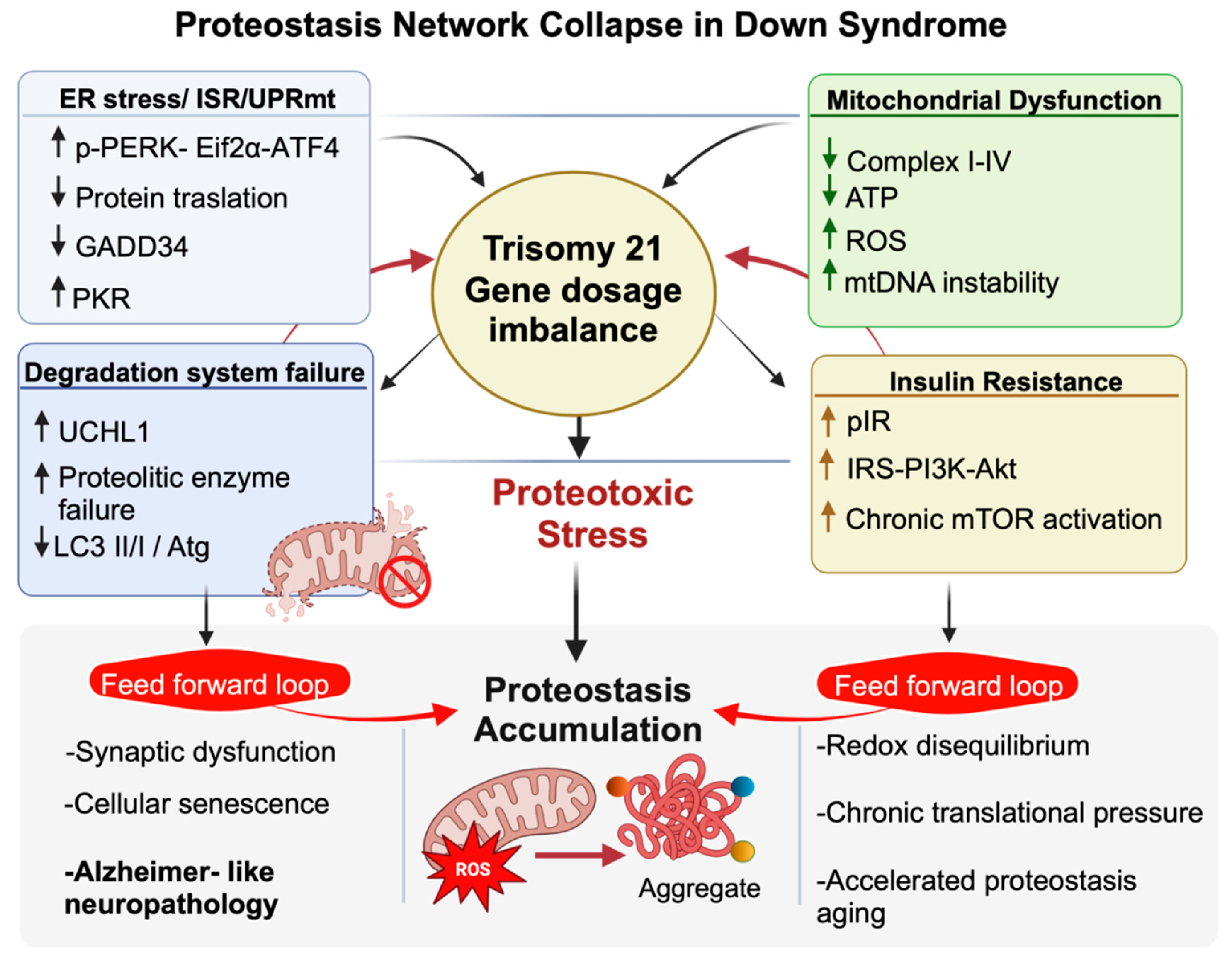
Figure 2.
Major components of the proteostasis network altered in Down syndrome and their pharmacological modulators. In the autophagy panel (right), inhibition of mTOR by rapamycin, AOAA, and Thiamet G restores autophagic flux, promoting autophagosome formation and maturation into autolysosomes. Metformin and polydatin enhance mitophagy, facilitating the selective removal of damaged mitochondria. In the ER-stress and ISR/UPR panel (upper left), activation of PERK and PKR leads to eIF2α phosphorylation and suppression of global protein translation; GSK2606414, PKRi, and ISRIB are shown as pharmacological modulators that attenuate stress signaling and help rebalance protein synthesis. The central proteasome panel highlights the role of the deubiquitinase USP16 in proteasomal dysfunction and the accumulation of ubiquitinated substrates, emphasizing the ubiquitin–proteasome system as a critical hub of proteostasis control. In the mitochondrial UPR and redox panel (lower left), mitochondrial stress and excessive reactive oxygen species (ROS) production are counteracted by α-tocopherol, CoQ10, EGCG, apigenin, 7,8-dihydroxyflavone, and CAPE/VP961, which support mitochondrial proteostasis and antioxidant responses. Together, these pathways illustrate a network-based therapeutic strategy aimed at restoring autophagy and mitophagy, ISR/UPR signaling, ubiquitin–proteasome function, and redox homeostasis to improve cellular proteostasis in Down syndrome.
Figure 2.
Major components of the proteostasis network altered in Down syndrome and their pharmacological modulators. In the autophagy panel (right), inhibition of mTOR by rapamycin, AOAA, and Thiamet G restores autophagic flux, promoting autophagosome formation and maturation into autolysosomes. Metformin and polydatin enhance mitophagy, facilitating the selective removal of damaged mitochondria. In the ER-stress and ISR/UPR panel (upper left), activation of PERK and PKR leads to eIF2α phosphorylation and suppression of global protein translation; GSK2606414, PKRi, and ISRIB are shown as pharmacological modulators that attenuate stress signaling and help rebalance protein synthesis. The central proteasome panel highlights the role of the deubiquitinase USP16 in proteasomal dysfunction and the accumulation of ubiquitinated substrates, emphasizing the ubiquitin–proteasome system as a critical hub of proteostasis control. In the mitochondrial UPR and redox panel (lower left), mitochondrial stress and excessive reactive oxygen species (ROS) production are counteracted by α-tocopherol, CoQ10, EGCG, apigenin, 7,8-dihydroxyflavone, and CAPE/VP961, which support mitochondrial proteostasis and antioxidant responses. Together, these pathways illustrate a network-based therapeutic strategy aimed at restoring autophagy and mitophagy, ISR/UPR signaling, ubiquitin–proteasome function, and redox homeostasis to improve cellular proteostasis in Down syndrome.
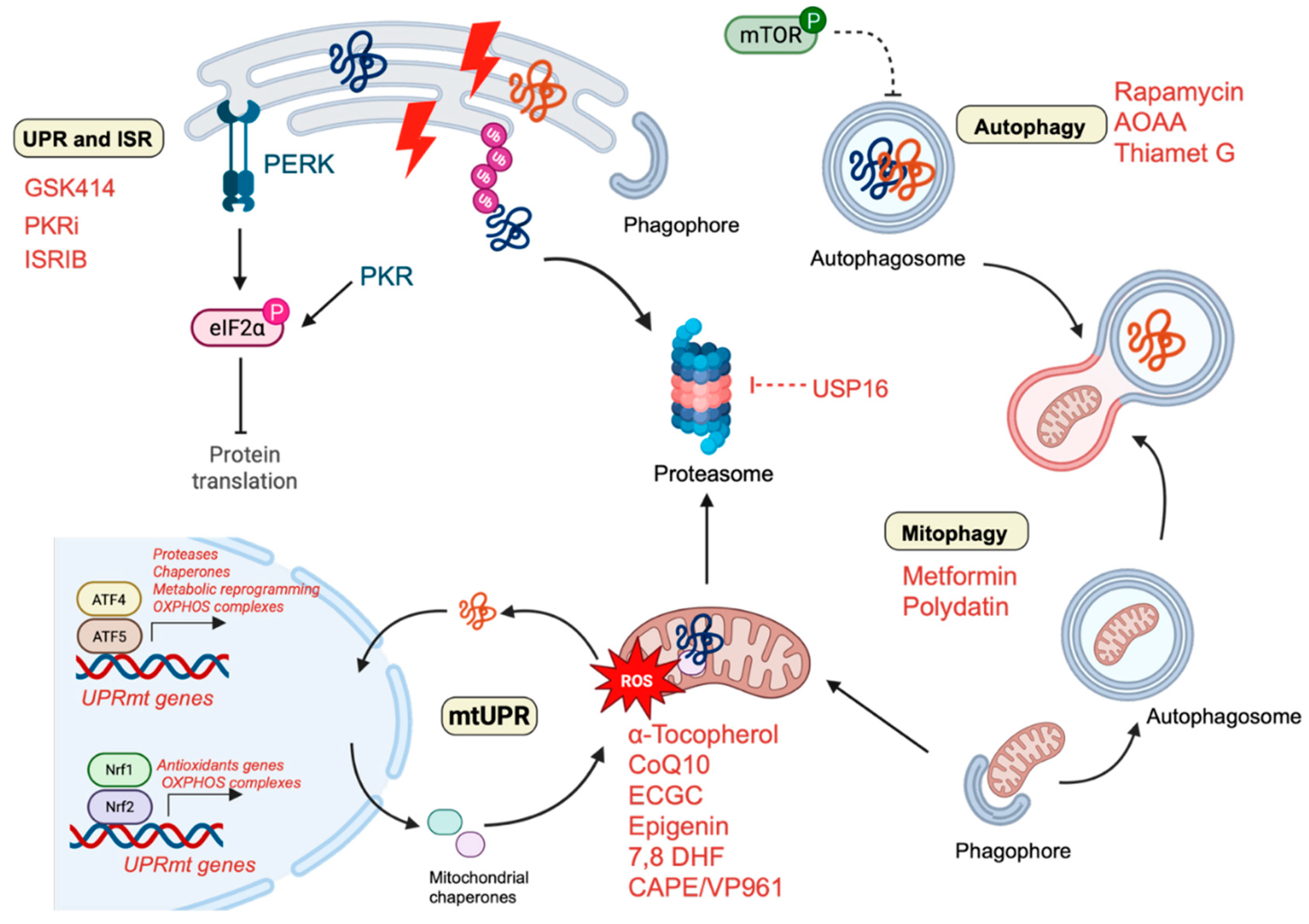

Table 1.
List of all the compounds used in DS neuropathology in clinical and pre-clinical studies.
| Compound | Target | study type | Dosage | Length of the treatment | Administration route | Model | Ref. | Outcomes |
| Unfolded Protein Response and Integrated stress Response inhibitors | ||||||||
| GSK2606414 | PERK | Preclinical study | 0.1 μg/μl | 5 days (1 x day) | intranasal treatment | Ts2Cje | [26] | Restored protein synthesis; reduced OS |
| ISRIB | eiF2a | Preclinical study | 2.5 mg/kg | 7 days (once every 2 days) | i.p. injection | Ts65dn | [27] | Restored protein synthesis; improved long-term memory |
| PKRi | PKR | Preclinical study | 0.1 mg/kg | 6 days (1 x day) | i.p. injection | Ts65dn | [135,136,137] | Rescued long-term memory and synaptic plasticity |
| Fluoxetine | PKR (indirect) | Preclinical study | Not specified | Early postnatal | Systemic | Ts65dn | [135,136,137] | Rescued long-term memory; neurogenesis |
| UPS modulators | ||||||||
| USP16 | USP16 | Preclinical study | Not specified | Not specified | In vitro | Ts65Dn DS stem cells | [143] | Rescued proliferation defects |
| Rapamycin | mTOR | Preclinical study | 1ug | 90 days (1x day,3x week) | Intranasal | Ts65Dn | [93,125] | Reduced Lys63-linked polyubiquitinated proteins |
| Autophagy modulators | ||||||||
| AOAA | CBS/H₂S pathway | Preclinical study | 1 mg/kg/day | 14 days, daily administration | intraperitoneally | Dp(17)3Yey/+ | [132] | Improved cognition; restored autophagy |
| Rapamycin | mTOR | Preclinical study | 1ug | 90 days (1x day,3x week) | Intranasal | Ts65Dn | [93,125] | Reduced APP/tau pathology; rescued hippocampal tasks |
| Preclinical study | 1mg/kg | 3 consecutive days during gestation | i.p. injection to pregnant dams | Ts1Cje | [130] | Corrected synaptic plasticity; | ||
| Preclinical study | 10 mg/kg | 5 days (1 x day) | i.p. injection | Ts1Cje | [133] | Restored spatial long-term memory | ||
| AZD8055 | mTORC1/2 | Preclinical study | 0,1 uM | 2, 4 and 8h | In vitro | Human fibroblasts | [89] | Restored autophagy and mitophagy |
| Metformin | AMPK/mTOR | Preclinical study | 0,5mM | 72h | In vitro | Human T21 fibroblasts | [78,89] | Restored mitophagy and lysosomal clearance |
| Polydatin | Mitophagy; miR-155 | Preclinical study | 10uM | 24-72h | In vitro | Human T21 fibroblasts | [85] | Mitochondrial bioenergetics and mitophagy |
| KYCCSRK peptide | BVR-A | Preclinical study | 0,5mM | 2 weeks | Intranasal | Ts2Cje | [134] | Restored insulin signaling and mitochondrial function |
| Thiamet G | O-GlcNAcylation | Preclinical study | 25 ug | 5 days (2×day) | Intranasal | Ts2Cje | [106] | Boosted autophagy induction |
| Antioxidants | ||||||||
| α-Tocopherol | ROS | Preclinical study | 50 mg/Kg | 5 months | diet supplementation | Ts65dn | [144] | Reduced OS; improved spatial working memory |
| Preclinical study | 0.1% w/w for Kg of diet | Pregnancy and pups | maternal supplementation | Ts65dn | [146] | Improved cognition; reduced lipid peroxidation | ||
| Randomized, double-blind, placebo-controlled trial |
900 IU+ ascorbic acid (200 mg) + α-lipoic acid (600 mg) |
2 years (daily) | oral | DS and AD individuals | [145] | No cognition improvement | ||
| Randomized, placebo-controlled clinical trial | 1000 IU | over 3 years (twice daily) | oral | DS over 50 years | [168] | No cognitive improvement | ||
| randomized controlled trial | 266 mg + α-lipoic acid (100 mg/day) | 4-months (daily) | oral | DS children | [169] | Reduced OS at DNA level | ||
| Clinical study | 400 mg + Vitamin C (500 mg/day) |
over 6 months (daily) | oral | DS children and teenagers |
[170] | Reduced blood levels of lipid peroxidation | ||
| CoQ10 | Mitochondrial ETC | Clinical study | 4 mg/kg/day | 6 or 20 months (daily) |
Oral | Children DS | [149] | Inhibited DNA oxidative damage; inconsistent long-term effects |
| Clinical study | 4 mg/kg/day | 4-year (daily) |
Oral | Children DS | [148] | No reduced OS level level at RNA or DNA level |
||
| Apigenin | NF-κB; antioxidant | Preclinical study | 2 μM (in vitro); 200-250 mg/kg/day (in vivo) | Prenatal+postnatal | Oral/systemic | T21 amniocytes; Ts1Cje | [157,158] | Reduced OS; improved spatial memory (sex-specific) |
| 7,8-DHF | TrkB (BDNF mimetic) | Preclinical study | 5 mg/kg | Postnatal treatment: for 12 days, Adult treatment for 40 days. | Subcutaneous administration | Ts65Dn | [157] | Restored mitochondrial bioenergetics; increased |
| Melatonin | ROS scavenger | Preclinical study | 10 mg/kg/die | 5 months | Oral | Ts65Dn | [161,162,163] | Improved spatial learning; reduced lipid peroxidation |
| 10 mg/kg/die | 6 months | Oral | Ts65Dn | [161,162,163] |
reduced OS and hippocampal senescence | |||
| Metformin | AMPK/NF-κB | Preclinical study | 10,30,50 uM | 48h | Systemic | Human T21 fibroblasts | [164,165] | Mitigated oxidative damage |
| Lithium | REST | Preclinical study | 10mM | 24h | In vitro | iPSC-derived neurons | [165] | Restored REST levels; reduced OS |
| CAPE | BACH1/NRF2 | Preclinical study | 10 μM | 6h | In vitro | human DS lymphoblastoid (LCLs) | [37] | Promoted NRF2 activation |
| VP961 | BACH1/NRF2 | Preclinical study | 5 μM | 6h | In vitro | human DS lymphoblastoid (LCLs) | [37] | Promoted NRF2 activation |
| GLP-1 (cleavage product) | GLP-1R; mitochondrial ROS | Preclinical study | 500ng/g | 2-3 weeks | Ip injection | 9 mo Ts65Dn | [166] | Decreased mitochondrial OS |
| EGCG | DYRK1A; ROS | Preclinical study | 20 μM | 72 h (changed every 24 hours) | cells treatment | Human DS cell cultures | [151] | Reduced OS and mitochondrial energy deficit |
| Preclinical study | 2–3 mg/day | 1 month | water supplementation | Ts65Dn/TgDyrk1A | [153] | Improved cognition | ||
| Preclinical study | 225 mg/kg/day | 4 weeks | water supplementation | Ts65Dn | [155] | Restored excitatory/inhibitory (E/I) imbalance (GABA modulation) | ||
| Preclinical study | 25 mg/Kg/day | P3 to P15 | subcutaneous injection | Ts65Dn | [171] | Restored neurogenesis at P15; no cognitive improvement at P45 | ||
| Preclinical study | 30 mg/kg/ day | 30 days | water supplementation | Ts65Dn | [152] | Rescued CA1 dendritic spine density, improved cognition | ||
| Preclinical study | 50 mg/kg | T1 (21 days) T2 (mating until 90 days) T3(P60-P90) |
diet supplementation | Dp(16)1Yey | [154] | Rescued GAD67; restored VGAT1/VGLUT1 balance; improved novel object recognition memory | ||
| phase I randomized controlled clinical trials |
9 mg/kg/day | 6 and 12 months | diet supplementation | Young adults with DS | [153] [172] |
Reduced plasma homocysteine; rescued cognitive performances | ||
Disclaimer/Publisher’s Note: The statements, opinions and data contained in all publications are solely those of the individual author(s) and contributor(s) and not of MDPI and/or the editor(s). MDPI and/or the editor(s) disclaim responsibility for any injury to people or property resulting from any ideas, methods, instructions or products referred to in the content. |
© 2026 by the authors. Licensee MDPI, Basel, Switzerland. This article is an open access article distributed under the terms and conditions of the Creative Commons Attribution (CC BY) license (http://creativecommons.org/licenses/by/4.0/).
Copyright: This open access article is published under a Creative Commons CC BY 4.0 license, which permit the free download, distribution, and reuse, provided that the author and preprint are cited in any reuse.


