Submitted:
14 March 2026
Posted:
16 March 2026
You are already at the latest version
Abstract
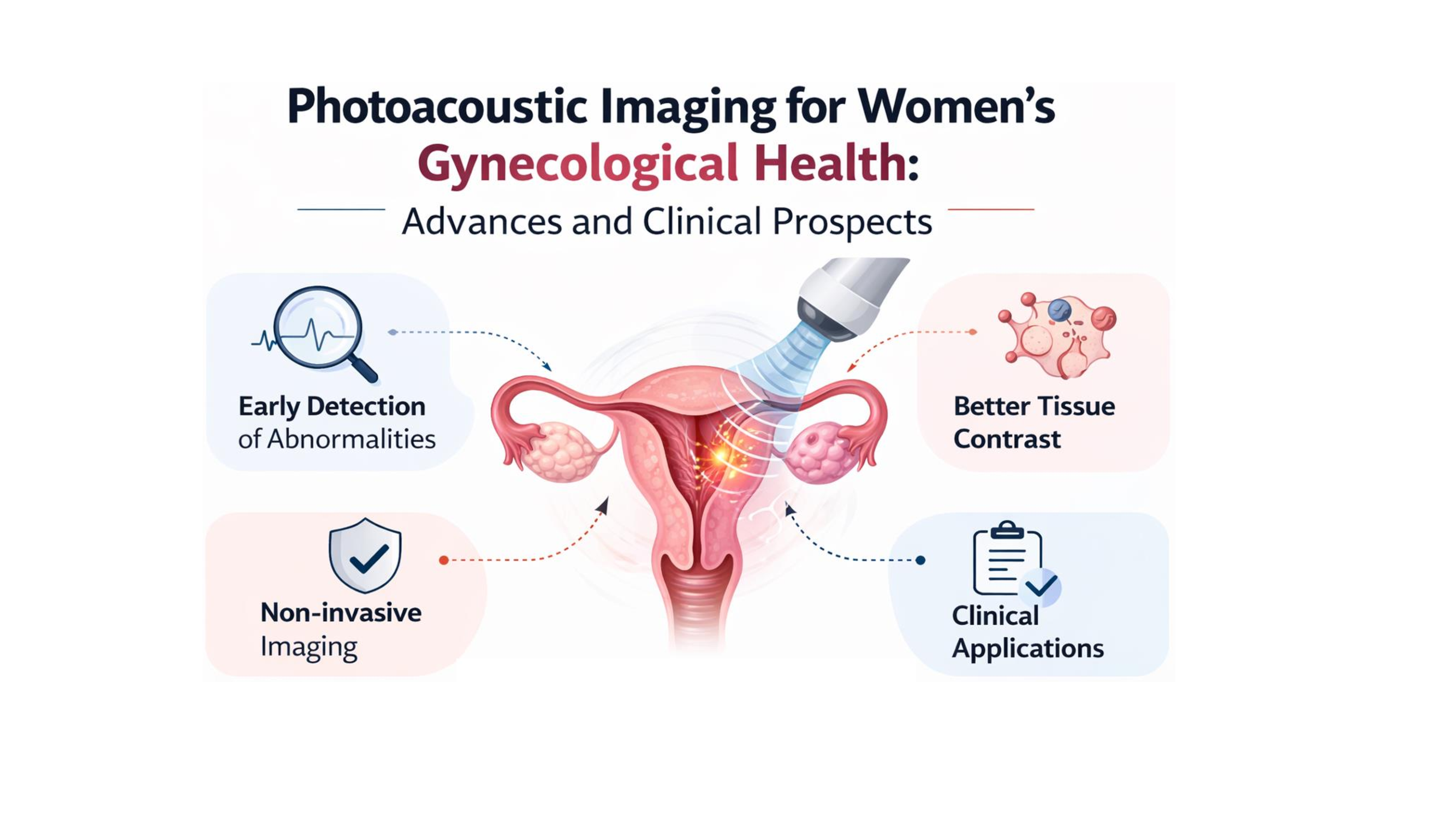
Photoacoustic imaging (PAI) is an emerging hybrid biomedical imaging modality that combines the high molecular contrast of optical excitation with the deep tissue penetration of ultrasound detection. This review presents recent advances in PAI-based techniques for the detection and characterization of gynecological and gynecological diseases in women, with particular focus on endometriosis and uterine-related disorders. We summarize the application of PAI across preclinical and translational studies, highlighting progress in photoacoustic microscopy, spectroscopic photoacoustic imaging, and endoscopic and probe-based implementations for noninvasive, high-resolution tissue evaluation. The role of functional and contrast-enhanced PAI approaches is discussed, emphasizing their ability to enhance diagnostic sensitivity, enable longitudinal monitoring, and provide detailed information on vascular, biochemical, and structural tissue characteristics. Furthermore, the expanding applications of PAI in assessing uterine, cervical, and ovarian pathologies, including tumor detection and tissue remodeling, are reviewed. Finally, current challenges, limitations, and future directions toward clinical translation are addressed. Collectively, this review underscores the potential of photoacoustic imaging as a powerful, noninvasive platform for early diagnosis, disease monitoring, and improved management of women’s health conditions.
Keywords:
photoacoustic imaging
; contrast agents
; women’s health
; gynecological disorders
; endometriosis
; uterine disease
; biomedical imaging
; non-invasive diagnostics
Copyright: This open access article is published under a Creative Commons CC BY 4.0 license, which permit the free download, distribution, and reuse, provided that the author and preprint are cited in any reuse.


