Preprint
Review

This version is not peer-reviewed.

Perspective Approaches to “Trojan Horse” Strategy Development for Combating Bacterial Pathogens

A peer-reviewed version of this preprint was published in:
Pharmaceuticals 2026, 19(5), 701. https://doi.org/10.3390/ph19050701

Submitted:

12 March 2026

Posted:

13 March 2026

You are already at the latest version

Abstract

Background/Objectives: The escalating crisis of antibiotic resistance and the inherent limitations of conventional antibiotics necessitate the development of innovative therapeutic strategies. Targeted drug delivery (TDD) offers a powerful approach to enhance efficacy, minimize systemic toxicity, and circumvent bacterial resistance. This systematic review aims to evaluate the potential of unique bacterial transport systems (BTSs) and surface specific receptors as platforms for TDD via the "Trojan Horse" strategy (THS). Methods: A comprehensive literature review was conducted, focusing on studies that investigated the specificity and mechanisms of BTSs responsible for the uptake of essential metabolites. This includes an analysis of transport systems for siderophores, bacteria-specific sugars, cell wall components, D-amino acids, and vitamins. We assessed preclinical and clinical examples of drug conjugates utilizing these pathways, as well as emerging platforms such as bacteriophage-derived proteins, antibody-antibiotic conjugates, and bacterial extracellular vesicles (EVs). Results: BTSs demonstrate high specificity for their cognate substrates, providing effective molecular gateways for drug conjugate import. The siderophore-cephalosporin conjugate cefiderocol represents a clinically validated example, having received FDA approval. Preclinical studies further reveal that conjugates utilizing sugars (e.g., maltose, trehalose) and vitamins (e.g., B12) can significantly enhance antibiotic uptake and activity against both Gram-positive and Gram-negative pathogens, including drug-resistant strains. Emerging platforms like bacteriophage endolysins and engineered EVs show promise for overcoming biological barriers such as bacterial outer membranes and intracellular host niches. Conclusions: The THS leveraging BTSs represents a clinically viable and promising avenue for next-generation antibacterial therapies. While significant progress has been made, including regulatory approval of cefiderocol, further research is critically needed to identify novel BTSs, optimize drug-linker chemistry, improve the pharmacokinetics and biosafety of conjugates, and translate these innovative platforms into effective treatments for drug-resistant infections.

Keywords: 
;  ;  ;  

1. Introduction

The escalating crisis of antibiotic resistance and the intrinsic shortcomings of traditional antimicrobials pose an existential threat to modern medicine [1]. The core of the problem isn’t just the misuse of antibiotics, but also their ineffectiveness against already resistant strains. Furthermore, even with effective antibiotics, their broad-spectrum action leads to negative consequences: disruption of the microbiome, the development of secondary infections, and the emergence of side effects [2].
Traditional approaches to drug delivery, often relying on systemic administration, are hampered by significant challenges. These include systemic toxicity, poor bioavailability, and off-target effects on healthy tissues, ultimately limiting therapeutic efficacy and increasing the risk of adverse events. These issues are particularly acute in the treatment of infectious diseases, where achieving sufficiently high antibiotic concentrations at the site of infection is crucial for overcoming pathogen resistance and preventing its further development [3]. Inadequate drug concentrations in the targeted area allow for the survival of a subpopulation of pathogens. This incomplete exposure not only fails to resolve the infection but also facilitates the spread of the surviving organisms and, critically, promotes the selection and propagation of resistant strains due to the sustained, low-level exposure toа the drug [4]. Therefore, there is increasing interest in utilizing biological systems for targeted drug delivery (TDD), with bacteria being recognized as a particularly promising approach, especially for fighting infectious diseases [5]. This approach maximizes drug efficacy, minimizes systemic side effects, and reduces the likelihood of resistance development by avoiding unnecessary exposure of non-target microbes [6,7]. Targeted delivery improves the concentration of antibiotics at infection sites, overcoming barriers like bacterial biofilms and intracellular localization that often render traditional drugs ineffective [8,9].
To date, several approaches have been proposed for the implementation of TDD [3]. These include the use of prodrugs [10,11,12], various stimuli-responsive nanocarriers [13,14,15], and pH-responsive drug delivery systems [13].
At the same time, bacteria possess several unique characteristics that produce the basis per se for realization of TDD. In particular, bacteria possess remarkable transport systems, representing complex molecular mechanisms developed to efficiently move substances across their cellular envelopes. These bacterial transport systems (BTSs) are critical for maintaining cellular homeostasis, enabling the uptake of vital nutrients (including a number of unique for bacteria) from the environment and the excretion of metabolites. The existence of such transport systems creates the prerequisites for the development of conjugates containing a naturally transported substance and an antibiotic compound, which must be transported into the bacterial cell, ultimately leading to a negative effect.
Furthermore, the bacterial cell is surrounded by unique structures (cell wall, capsule), which creates an opportunity for specific binding of various molecules and supramolecular structures with high affinity for these cellular components, conjugated with antibiotic agents. These two approaches form the basis for the “Trojan Horse” Strategy (THS) as one of the promising modalities for implementing TDD. The present review is dedicated to a detailed examination of this strategy, along with specific examples of its application and future development for antibacterial therapy. This review explores the potential of BTSs as sophisticated tools for targeted drug delivery. We focus on BTSs specific for key metabolites - including sugars, peptidoglycan fragments, iron-siderophore complexes, and D-amino acids - examining their mechanisms, structural basis, and regulation. We discuss strategies for creating conjugates of these ligands with therapeutic agents and highlight preclinical examples demonstrating improved bioavailability, reduced toxicity, and enhanced efficacy. In parallel, we examine bacteriophage-derived proteins and surface receptors-based structures as an emerging platform for targeted delivery, leveraging their inherent specificity for bacterial recognition and their capacity to deliver diverse therapeutic payloads, including antibiotics and CRISPR-Cas systems.
By analyzing the advantages and limitations of both BTS- and phage-based approaches, this review aims to illuminate their potential for developing novel, more effective, and safer antibacterial therapies.

2. The “Trojan Horse” Strategy in Drug Delivery

The THS operates on a fundamental principle: exploiting the target pathogen’s own metabolic and uptake machinery or specific recognizable structures to deliver a therapeutic agent. This “horse” is a therapeutic agent (the drug or antibiotic or photosensitizer) that is chemically attached (conjugated) to a carrier molecule. This carrier molecule is not chosen randomly; it is specifically designed or selected to resemble a molecule that the target pathogenic bacteria actively and preferentially seek out for its survival and growth or are able for specific binding.
Critically, this approach bypasses many of the natural defenses that bacteria employ against external threats, including antibiotics. These defenses can include impermeable cell walls/membranes, efflux pumps, enzymatic degradation [16]. By being recognized as a “friendly” and necessary molecule, the “Trojan Horse” bypasses these passive and active exclusion mechanisms [10,17].
Thus, the THS offers promise for overcoming bacterial resistance linked to reduced drug permeability and efflux pumps, enabling targeted delivery of antibiotics to pathogenic bacteria. This approach holds the potential for enhanced antimicrobial activity and reduced side effects.
In many publications, the term THS is used in connection with the use of siderophores conjugated with various substances as carriers (see below), historically representing the first examples of this strategy. However, in our view, the THS can be expanded to include other naturally transported compounds such as sugars, amino acids, peptides, etc., as discussed later in this review. Furthermore, we also include within the THS compounds or structures that specifically bind to the bacterial cell surface - such as antibodies or phage structures - as carriers (in this case, cell-surface receptors function as a bait for antibiotic-loaded carriers, thus representing a component of the THS). As a result, this strategy minimizes exposure of host cells to the drug, potentially reducing side effects. The high intracellular concentration of the released drug can lead to more potent antimicrobial activity, even against less susceptible bacteria [10,18,19]. Furthermore, this enables the transport into the pathogen cell of substances (antibiotics) that do not penetrate it for various reasons, yet have intracellular targets within them.
The main principles of the THS include the following: (a) the carrier should be a "native" substance for the pathogen and/or not be toxic to the cell per se. This should significantly reduce the development of resistance to the conjugate as a whole. Particularly advantageous is the use of a carrier (e.g., a substrate) that is essential for bacterial survival, as the latter should also diminish the emergence of resistance. (b) Ideally, the carrier or cell-binding compounds/structures must be unique to the pathogen and not transported by or bound to host cells, which provides the basis for specificity.

2.1. Mechanism of “Trojan Horse” Strategy Action

Pathogenic bacteria have evolved sophisticated systems to acquire vital resources from their environment. These essential molecules often include: 1) nutrients (such as iron, sugars, or amino acids) which are critical for bacterial metabolism and replication [20]; 2) growth factors (specific vitamins or cofactors) that are indispensable for bacterial processes [21]; 3) siderophores - small, high-affinity iron-chelating molecules secreted by many bacteria to scavenge scarce iron from their surroundings [22]; 4) other specific ligands - molecules that bind to unique bacterial surface receptors, which might be involved in nutrient uptake, adhesion, or even entry into host cells (in the case of intracellular pathogens) [23].
The bacterial cell, driven by its inherent need for the essential molecule that the carrier mimics, actively recognizes and binds to the “Trojan Horse” complex. This binding typically occurs via specific surface receptors or transport proteins that are dedicated to importing the natural nutrient or molecule.
Once the “Trojan Horse” complex has successfully traversed the bacterial cell envelope and entered the cytoplasm or periplasm, a second crucial step occurs: the release of the therapeutic agent. The release mechanism is often triggered by the intracellular environment of the bacterium. This can include: pH changes [13] (the internal pH of a bacterium might differ from the external environment, cleaving the bond between the carrier and the drug); enzymatic activity (intracellular bacterial enzymes can be designed to specifically break down the linker molecule holding the drug to the carrier); redox potential (differences in oxidation-reduction potential within the cell can also be used to trigger drug release) [14]. The result is that the therapeutic agent - either in its released active form or as an integral part of the conjugate - is localized precisely inside the target bacterium, where its effective concentration can be significantly higher than if it had been administered conventionally.
When supramolecular structures such as antibodies or phage-derived particles are employed as carriers, the release or delivery of the active agent follows mechanisms distinct from those of low-molecular-weight conjugates. If the supramolecular carrier (e.g., an antibody-antibiotic conjugate, AAC, or a phage-based nanocarrier) is designed to be internalized by host cells (e.g., macrophages) or by the bacteria themselves, the active agent is typically released upon degradation of the carrier within the phagolysosome or endosome. In AACs, for instance, the linker connecting the antibiotic to the antibody is cleaved by proteolytic enzymes (such as cathepsins) inside the host cell, liberating the free, active antibiotic to target intracellular pathogens [10,24,25].
In many applications, the primary goal is not internalization into the bacterium, but rather the high-precision delivery of a toxic payload directly to the bacterial cell surface. In this scenario, the active agent acts while still conjugated to the supramolecular carrier. Carrier-bound enzymes (e.g., endolysins) can directly degrade the peptidoglycan from the outside [26,27].
Photosensitizers or photothermal agents conjugated to antibodies or phages generate lethal reactive oxygen species or heat upon light activation, destroying the bacterial membrane without needing to be released from the carrier [28,29,30].
Carrier-bound antimicrobial peptides can disrupt the bacterial membrane integrity from the surface [31,32].
Some supramolecular carriers, such as hydrogels containing functionalized cellulose [33,34] or extracellular vesicles [35,36], can act as localized drug depots. The active agent is released extracellularly in the immediate vicinity of the bacteria. This release can be passive, through gradual diffusion from the carrier matrix [33,37] or triggered by specific signals in the infection microenvironment, such as bacterial enzymes (e.g., β-lactamases, proteases) [38,39], pH changes [13], or hypoxia [15], leading to carrier disassembly and subsequent drug release.
Thus, in the context of supramolecular carriers, the active agent can be deployed either on-site (acting directly from the carrier) or in-site (released intra- or extracellularly) to exert its bactericidal effect.
By specifically targeting bacterial uptake systems, this strategy minimizes exposure of host cells to the drug, potentially reducing side effects. The high intracellular concentration of the released drug can lead to more potent antimicrobial activity, even against less susceptible bacteria [10,18,19].
In essence, the Trojan Horse strategy is a clever form of biological subterfuge, tricking the bacteria into admitting their own demise by disguising a potent weapon as something they desperately need.

2.2. Types of “Trojan Horses”

Classification of “Trojan Horses” by delivery mechanism allows a better understanding of their mode of action and potential for clinical applications. Below are the main types of such systems grouped according to their mechanism and mode of entry into the bacterial cell (Figure 1).

2.2.1. Metabolite-Based Mimics of Natural "Messengers" and Transport Mechanisms

These “Trojan Horses” mimic molecules or processes naturally used by bacteria to acquire essential resources or transport substances. They imitate key nutrients, such as siderophores, amino acids, and sugars, which bacteria actively transport. The bacterial world is remarkable not only for its diversity but also for its extraordinary ability to adapt to extreme environments. A key element of this adaptation is an efficient system for exchanging metabolites with the surrounding environment, mediated by complex transport systems. While ubiquitous transport mechanisms handle the influx of essential nutrients, certain metabolites are selectively transported by bacteria through dedicated, exclusive systems [23]. These specialized systems not only reflect the unique metabolic requirements of specific bacterial species or communities but also often determine their interactions, survival, and pathogenic potential [23]. Bacteria are equipped with a diverse and intricate array of transport systems that mediate the uptake of necessary nutrients and the removal of toxic compounds. Notably, some of these systems are unique to specific bacterial species or groups, rendering them promising platforms for TDD via THS [23,40].
Key characteristics of BTSs that make them attractive for THS include high specificity and efficient transport of substances across the bacterial cell wall and membrane. BTSs are usually targeted to specific metabolites or signalling molecules, which ensures selective transport into bacterial target cells [41].
Unique BTSs may be classified according to the type of molecules they transport and their mechanism of action.

2.2.1.1. Siderophores

Bacteria utilize siderophores to capture iron. Iron is a critically important element for most living organisms, including bacteria, where it participates in essential processes such as respiration, DNA synthesis, and antioxidant defense. However, in aerobic environments, iron often exists in poorly soluble Fe(III) forms, making it difficult for bacteria to access. In response, bacteria have evolved complex strategies to acquire iron based on the secretion of siderophores.
Siderophores are low-molecular-weight compounds (Mw 200–2000 Da) secreted by bacteria, fungi, and plants, and are highly specific chelators of trivalent iron salts. They exhibit marked selectivity and strong binding capability for iron (Kaff > 1030) [19]. These compounds bind to iron, followed by transport into the cell, thus fulfilling the iron requirements of metabolism under conditions of low environmental concentration of iron. Based on their chemical structure, siderophores (over 500 compounds) are divided into several classes according to the presence of five iron-binding motifs in their structure: catecholate, hydroxamate, phenolate, carboxylate, and α-hydroxy carboxylate [42]. Their structure and synthesis by producer cells are well studied (for a review, see, for example, [43]).
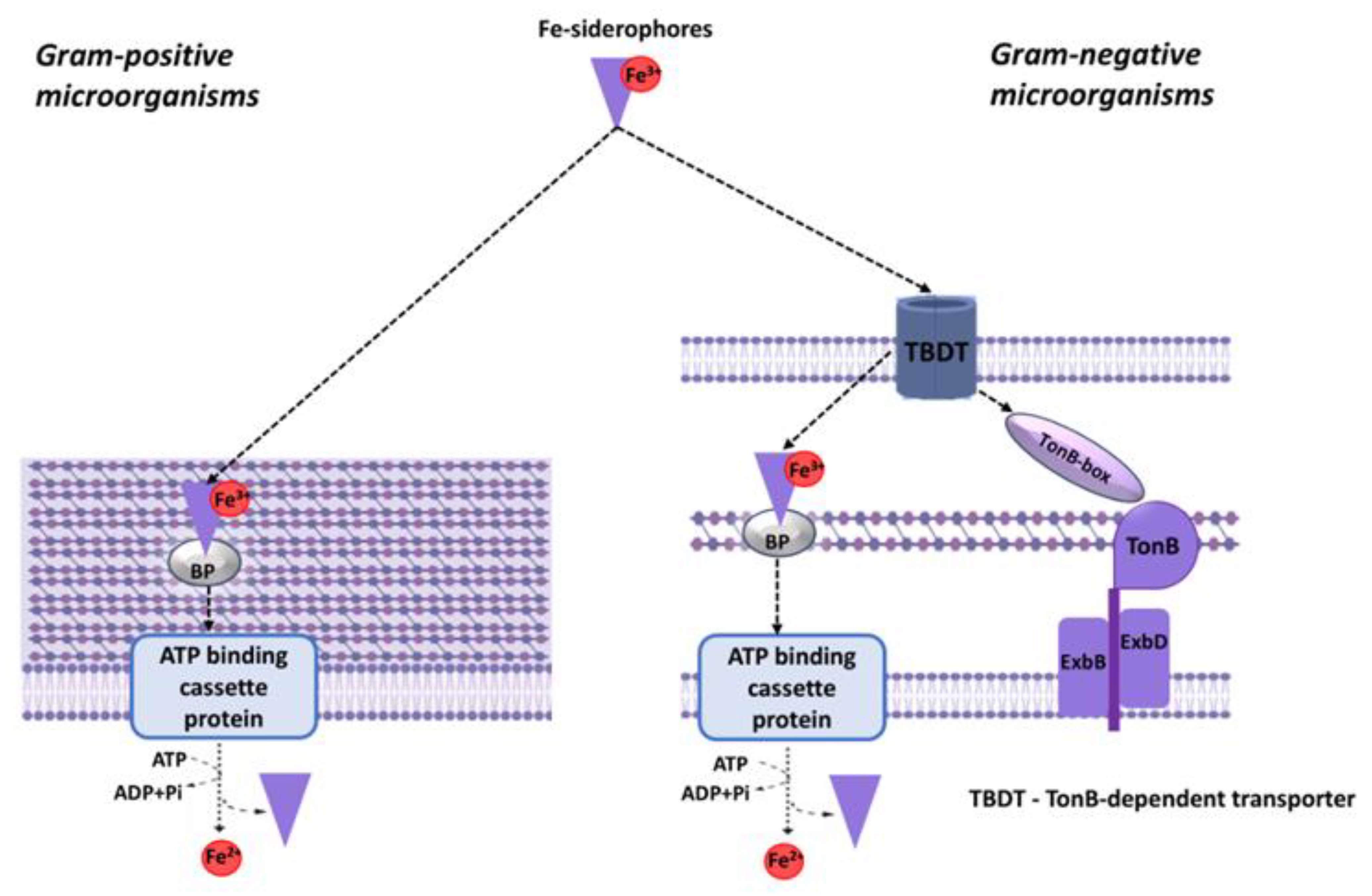
Siderophores are synthesized through two principal mechanisms: one involves the nonribosomal peptide synthetase (NRPS) pathway, and the other is the nonribosomal independent synthesis (NIS) pathway [43]. The transport of siderophores complexed with iron into the bacterial cell differs between Gram-positive and Gram-negative bacteria. In Gram-negative bacteria, the siderophore-iron complex interacts with a receptor on the cell surface, localized in the outer membrane. Active transport of the iron–siderophore complex across the outer membrane is an energy-dependent process [44,45,46] (Figure 2).
The TonB complex, composed of three cytoplasmic membrane proteins (TonB, ExbB, and ExbD), powers the active transport of iron-siderophore complexes. It uses power of the proton motive force across the cytoplasmic membrane for complex translocation through the outer membrane, enabling its entry into the cytosol [47,48,49]. This energy transfer relies on direct contact between TonB’s C-terminal domain and the OMR’s N-terminal domains. After reaching the periplasm, the iron-siderophore complex binds to its cognate periplasmic binding protein (BP). The resulting iron-siderophore-BP complex is then translocated across the cytoplasmic membrane by an ATP-binding cassette (ABC) transporter into the bacterial cytoplasm. Located in the inner membrane, ABC transporters harness ATP hydrolysis to pump substrates from the periplasm into the cytoplasm against concentration gradients [48,50,51]. For Gram-positive bacteria, the mechanism of siderophore-iron transport differs significantly, although the first step also involves binding to a specific receptor on the cell surface. Similarities, by contrast, include the presence of BP receptors and ABC transporters. The complex is then transported with the ABC transporter into the cytoplasm of this Gram-positive bacterium [45,52].
The most advanced direction in the use of bacteria-specific transported compounds in the form of conjugates (THS) today is the siderophore-based approach [53].
In an interesting twist, certain bacteria have evolved to exploit this siderophore-mediated iron acquisition for antibiotic delivery, conjugating toxic compounds to siderophores. These naturally occurring siderophore-drug conjugates, also known as sideromycins [54], represent a promising class of antibiotics. Examples of these sideromycins include the danomycins [55], salmycins [56], albomycins [57,58], ferrimycins [59,60,61], and microcins [62].
To date, a significant number of different siderophore and antibiotic compound conjugates have been synthesized and tested (for review see [18,19,42,63,64,65,66,67,68,69]). A [42] review lists over 50 such conjugates, including those with beta-lactam antibiotics, fluoroquinolones, cephalosporins, and others, linked to various siderophores via both cleavable and non-cleavable linkers. Most of these demonstrate enhanced efficacy against targeted Gram-positive and Gram-negative bacteria compared to their unconjugated antibiotic counterparts. In some cases, the effectiveness of these conjugates against resistant strains has been demonstrated, particularly against Pseudomonas aeruginosa, which exhibits high resistance to a wide range of antibiotics [19]. However, it is important to note that resistance can also develop to siderophore-antibiotic conjugates, as their transport depends on the presence of specific transporters (e.g., TonB). Mutations in these transporters can lead to a loss of activity, resulting in the development of resistance [49,70].
For effective antibacterial action, conjugates transported into the cell, in cases where the bacterial target is located in the cytoplasm (with rare exceptions), must undergo intracellular chemical modification to release the inhibitor. This is precisely why the structure of the linker, which connects the siderophore molecule and the antibiotic, is important for the overall activity of the conjugate [54]. Amino acids [71], succinic acid [72], alkyl chains [73], polyethylene glycol (PEG) chains [74], triazoles [75], acetal groups [76], disulfide bonds [77], thiol-maleimides [78], citric acid [79], and esters [80] have been used as linkers. Among these linkers, some are cleavable within cells and some are non-cleavable. Cleavage occurs through hydrolysis, enzymatic cleavage, and reduction [18,42,66]. However, such linkers can be labile and may be hydrolyzed too early during intracellular transport, leading to a loss of activity, or within the host. An interesting approach involved using a cephalosporin-based linker as a less labile linker, which is selectively cleaved by bacterial β-lactamase [81]. At the same time, conjugates with non-cleavable linkers are effective in cases of periplasmic target localization in Gram-negative bacteria [42].
Siderophores can also mediate the transport of metals that are toxic to bacterial cells and can compete with iron. For instance, a significant efficacy of a linker-free preparation based on a natural sideromycin complexed with colloidal bismuth citrate has been demonstrated against P. aeruginosa and Burkholderia cepacia in both in vitro and in vivo experiments [82].
Although the majority of studies on the application of siderophore conjugates as antibacterial agents have been conducted in vitro, their efficacy has also been demonstrated in vivo using laboratory models of infection. Several pharmaceutical companies (Pfizer, GSK, Takeda, Synphar, and others) have developed conjugates for clinical trials: MC-1, a siderophore-conjugated monocarbam shown activity against MDR P. aeruginosa and ESBL-producing members of the Enterobacteriaceae from Pfizer; BAL30072, a siderophore monosul factam, from Basilea; and S-649266, a catechol–cephalosporin antibiotic, from Shionogi Pharmaceuticals [68]. Unfortunately, almost all of these were halted at the pre-clinical stage or Phase I clinical trial for various reasons (e.g., instability in the body, side effects). However, a recently developed synthetic catecholate – siderophore cephalosporin conjugate (Cefiderocol) [67] by Shionogi & Company Ltd., demonstrating activity against a range of carbapenem-resistant Gram-negative pathogens, has received approval from the US Food and Drug Administration (FDA) [42].
Siderophores are utilized not only for transporting antibacterial compounds but also, as reported, for delivering photoactivators to enhance the photodynamic inactivation of P. aeruginosa [83] and as conjugated vaccine components targeting uropathogenic E. coli [84].

2.2.1.2. Unique Bacterial Sugars and Their Dedicated Transport Systems

Sugar metabolism is fundamental to bacterial life, providing essential building blocks and energy for growth and survival [85]. While many sugars are universally utilized across life forms, bacteria possess unique metabolic pathways and utilize distinct sugar molecules not commonly found or processed in mammals [86,87]. These bacteria-specific sugars, and the dedicated transport systems responsible for their uptake, represent promising targets for the development of THS.
A key feature of bacterial sugar uptake is the phosphotransferase system (PTS), a unique transport and phosphorylation pathway that is not found in eukaryotic cells [88]. While the PTS is particularly prevalent in Gram-positive bacteria, it is also present in many Gram-negative species [89]. Through the PTS, bacteria can efficiently transport and phosphorylate a range of sugars, enabling their rapid utilization, however, some of these sugars are unable to directly enter eukaryotic cells. The PTS employs active transport, utilizing a coordinated network of both cytoplasmic and membrane-bound enzymes. Central to its function is the transfer of a phosphate group from phosphoenolpyruvate (PEP) to the sugar substrate, which is simultaneously transported across the bacterial membrane [90]. The PTS functions through a highly coordinated multi-step process, relying on a series of conserved proteins that act as a phosphorylation cascade [91]. A defining characteristic of the PTS is its unique ability to couple sugar transport with phosphorylation in a single, energy-efficient step. Unlike other transport systems that rely on ATP hydrolysis or electrochemical gradients to drive sugar uptake, the PTS utilizes PEP, a high-energy intermediate in glycolysis, as the phosphoryl donor. This direct link between sugar transport and phosphorylation has several key advantages for bacteria: enhanced energy efficiency, as the system essentially “piggybacks” onto the glycolytic pathway, avoiding the need to expend additional ATP for sugar phosphorylation; rapid metabolic integration, as the direct phosphorylation of the sugar during transport allows for immediate entry into central metabolic pathways, such as glycolysis, bypassing the need for an additional kinase reaction; metabolic regulation, due to the phosphorylated form of the sugar acting as a better substrate for glycolytic enzymes; the ability to overcome kinetic barriers, because phosphorylation lowers the intracellular concentration of the unphosphorylated sugar, maintaining a favorable concentration gradient; and finally, enhanced competitiveness, as the combination of energy efficiency and rapid metabolic integration provides a significant competitive advantage for bacteria in mixed-substrate environments. Thus, the tight coupling of sugar transport and phosphorylation in the PTS represents a highly evolved adaptation that optimizes energy utilization and enhances metabolic efficiency for bacteria, especially in fluctuating or nutrient-poor conditions [88,92]. While the PTS transports a diverse array of sugars, some general characteristics and commonalities can be observed among PTS-dependent substrates. At a fundamental level, PTS sugars are typically small, readily metabolized carbohydrates or carbohydrate derivatives that serve as important carbon and energy sources for bacteria [92,93]. Structurally, many PTS sugars are monosaccharides or simple disaccharides, readily utilized in glycolysis or other central metabolic pathways after phosphorylation. While the PTS transports a diverse array of sugars across bacterial membranes, it’s crucial to recognize that many of these sugars are also utilized by eukaryotes, including humans. While the direct transport of sugars via the phosphotransferase system (PTS) is exclusively bacterial, finding examples of PTS-transported sugars that are entirely absent from human biology is challenging. Most common sugars utilized by bacteria are also metabolized to some extent in humans, albeit through different mechanisms. However, certain bacterial-specific sugar modifications or less common sugars may be transported via the PTS, with extremely limited or no direct utilization in humans. The bacteria-specific sugars, suitable for realization of THS are discussed below (Table 1).

2.2.1.2.1. Rhamnose

Rhamnose is a 6-deoxy sugar (methylpentose) that plays an important role in the structural organization of the bacterial cell and its interaction with the environment. Rhamnose, a deoxyhexose sugar, holds a special place in the biological world due to its peculiar transport properties. While readily utilized by numerous bacterial species, it remains largely inaccessible to mammalian cells [134,135], highlighting a fundamental difference in cellular machinery and metabolic pathways between prokaryotic and eukaryotic organisms.
Rhamnose exists in two anomeric forms: L-rhamnose and D-rhamnose. Of these, L-rhamnose is more commonly encountered in nature and plays a significant role as a component of various plant polysaccharides such as pectin and gums. Additionally, L-rhamnose is a crucial constituent of bacterial cell walls and capsular polysaccharides, where it contributes not only to structural integrity but also to immunological properties by influencing host-pathogen interactions [135,136]. Rhamnose is a critical component of the cell wall of many pathogenic bacteria, including Mycobacterium tuberculosis, where it is part of the α-L-Rha-(1→3)-α-D-GlcNAc linker between peptidoglycan and arabinogalactan, which is essential for bacterial viability [137]. In E. coli, the same disaccharide is present in the O-antigen of lipopolysaccharide, which is required for virulence [137].
Chemically, rhamnose distinguishes itself from more common sugars like glucose through specific structural features: it lacks a hydroxyl group at the carbon atom in position 6 and instead contains a methyl group at carbon position 2. This subtle difference classifies rhamnose as a 6-deoxyhexose sugar, which influences its recognition and transport by cellular transporters specialized for deoxy sugars. The precise stereochemistry and unique groups affect how rhamnose interacts with biological molecules, guiding the specificity of bacterial transport systems for rhamnose and underpinning its absence in mammalian metabolic pathways [135,138].
For many bacteria, rhamnose is a valuable nutrient. Its transport into the bacterial cell is mediated by specific membrane bound ABC transporters [139]. These systems are highly evolved and possess a remarkable selectivity for rhamnose, ensuring its efficient uptake. In stark contrast to bacteria, mammalian cells generally lack the specific transport systems required for rhamnose uptake [134,135]. This exclusion is not due to an inability to metabolize rhamnose if it were present, but rather to the absence of the dedicated transporters [135].
The mechanisms of rhamnose transport into the bacterial cell differ among various microorganism species. In Escherichia coli and other Enterobacteriaceae, the transport of L-rhamnose occurs via a proton-coupled symport system, which is induced when bacteria grow on rhamnose and creates an alkaline pH shift upon adding the sugar to energy-depleted cells [140]. This transport is respiration-dependent, optimal at pH 7, and inhibited by protonophores and ionophores, confirming its energy-dependent nature [140].
In Rhizobium leguminosarum, rhamnose transport is mediated by an ATP-binding cassette (ABC) transport system encoded by a specialized genetic locus that also contains genes for the catabolism of this sugar [141]. This locus includes genes encoding a putative dehydrogenase, isomerase, and sugar kinase, which are necessary for the subsequent metabolism of rhamnose. The regulation of these genes is controlled by a DeoR-type repressor (RhaR), which is encoded in the same transcript as the ABC transporter, with expression being induced by the presence of rhamnose in the medium [141]. Alternative rhamnose transport pathways have been found in Arthrobacter pyridinolis: a phosphoenolpyruvate-dependent phosphotransferase system and malate- or succinate-dependent transport, which stimulates rhamnose uptake in membrane vesicles [142].
Currently, there are several experimental studies where rhamnose or its derivatives have been used to implement the THS.
Thus, a study showed that replacing the native sugar D-desosamine in the macrolide antibiotic YC-17 with L-rhamnose led to improved antibacterial activity against susceptible and resistant strains of Enterococcus faecium and Staphylococcus aureus compared to the parent compound [94]. In another study new esters based on methyl -L-rhamnopyranoside and fatty acids (stearic, hexanoic, decanoic, benzoic) were synthesized. The key idea was to use the natural sugar (rhamnose) as a platform for the covalent attachment of lipophilic “warheads” (fatty acids). The result is an amphiphilic molecule that is “masked” as a harmless sugar ester but possesses potent antimicrobial activity [32]. Esterification of rhamnose with fatty acids sharply increases its lipophilicity. This allows the molecule to better penetrate the lipid membranes of microorganisms. Thus, a new non-azole antifungal agent was created that uses the sugar to deliver a lethal lipophilic payload to the target within the pathogen’s cell [32].
A novel approach to combat P. aeruginosa biofilms has been described using a recombinant protein that specifically binds rhamnose. The researchers created an effective anti-biofilm agent operating on a principle very close to the “Trojan horse” concept [96]. The authors used the recombinant protein rHPLOE - a modified version of the lectin from the Taiwanese horseshoe crab (Tachypleus tridentatus). The uniqueness of this protein is its high specificity for binding L-rhamnose and rhamnose-containing molecules. Given that rhamnose is a component of key structures in the P. aeruginosa biofilm (lipopolysaccharides, di-rhamnolipid, Psl polysaccharide), the authors hypothesized that this protein could act as a “Trojan horse” to locate and disrupt the bacteria’s protective structure. rHPLOE demonstrated the ability to strongly bind to the acellular matrix of the P. aeruginosa PA14 biofilm. This binding occurred exclusively through the recognition of rhamnose residues: it was effectively blocked by L-rhamnose and synthetic rhamnosides, but not by other sugars. The binding of the protein led to a significant reduction in the amount of di-rhamnolipid within the biofilm. Di-rhamnolipid is not merely a biofilm component but an important signaling molecule (quorum regulator) necessary for the formation of its three-dimensional structure and the maintenance of channels. rHPLOE not only inhibited the formation of a new biofilm but also caused the dispersion (disruption) of an already mature biofilm. This, in turn, enhanced the efficacy of traditional antibiotics, which could now better penetrate the bacterial cells. This study brilliantly illustrates an alternative THS, where rhamnose does not deliver the cargo, but rather the rhamnose-binding protein delivers itself, using bacterial rhamnose as the target (“anchor”) [96].
In addition to rhamnose, in the context of drug delivery, it is also necessary to focus on rhamnolipids.
Rhamnolipids, produced by P. aeruginosa (and other Gram-negative bacteria), are glycolipids consisting of one or two molecules of rhamnose linked to hydroxy-fatty acids, and they are involved in bacterial motility, biofilm formation, and the uptake of hydrophobic substrates [143]. Research indicates that mono-rhamnolipids are well-recognized by bacteria and can be taken up by cells and converted into di-rhamnolipids, opening possibilities for the delivery of therapeutic agents conjugated with rhamnolipids [143]. Recent developments include the creation of rhamnose-liposomes functionalized with rhamnolipids and loaded with cinnamaldehyde, which demonstrated enhanced antibacterial activity against Salmonella due to improved binding to the bacterial membrane [95].
By chemically modifying rhamnolipids (by attaching amino acids and, potentially, other cargo), the authors create compounds that likely retain the ability to interact with bacterial membranes or target cells. Rhamnose itself serves as that “familiar” element that facilitates contact [31]. The modified rhamnolipids themselves (especially the arginine derivatives) exhibit enhanced antibacterial activity. In this paradigm, the cationic moiety with the amino acid is the “weapon,” and the rhamnolipid scaffold is the “vehicle” that delivers this weapon to the bacterial membrane. The mechanism of action involves the electrostatic interaction of the cationic “head” with the negatively charged bacterial membrane, leading to its disruption. Rhamnose here acts as part of the “Trojan horse,” ensuring biocompatibility and biodegradability [31].
Summarizing, rhamnose, owing to its unique biochemical role and transport specificity, is an ideal candidate for the development of “Trojan horse” antimicrobial agents. Confirmed strategies range from direct conjugation with antibiotics for their import into the cell to the creation of complex nanoscale platforms (rhamnosomes [95]) and highly specific proteins (lectins [96]) targeting pathogen rhamnose structures. These approaches not only enhance the effectiveness of antimicrobial action but also open pathways for overcoming drug resistance by targeting biofilms and exploiting mechanisms to which bacteria develop resistance more slowly.

2.2.1.2.2. Cellobiose

Cellobiose, a disaccharide linked by β-1,4-glycosidic bonds, serves as a natural substrate specific for bacterial transport systems, enabling targeted delivery of antimicrobial agents via Trojan horse strategies.
Pathogenic bacteria like Escherichia coli, P. aeruginosa, Salmonella enterica, Klebsiella pneumoniae, Clostridium perfringens, and Clostridioides difficile utilize ABC transporters for cellobiose uptake [144,145]. These systems include periplasmic binding proteins, transmembrane channels, and ATPases [146], allowing active transport into the cytoplasm where β-glucosidases hydrolyze cellobiose to glucose [144,147].
Gram-negative species require porins for outer membrane passage before ABC-mediated cytoplasmic entry [148]. Regulation involves induction by cellobiose and catabolite repression by glucose, ensuring selective expression under nutrient stress relevant to infections [144,145,149].
Conjugates link antibiotics or toxins to cellobiose, hijacking ABC transporters for intracellular delivery. The main value of cellobiose lies in its unique β-1,4-linkage. This bond is an ideal target for bacterial enzymes (β-glucosidases), which is utilized in the design of prodrugs activated by the microflora.
Upon hydrolysis by bacterial β-glucosidase, the active drug releases in the cytoplasm, enhancing efficacy against resistant strains. β-glucosidase (cellobiase) is an enzyme that normally cleaves cellobiose into two molecules of glucose within the cytoplasm. In the case of conjugates, this enzyme hydrolyzes the bond between the sugar and the active substance (antibiotic or toxin), thereby activating the drug inside the cell. As an example, cellobiose–nitrofurantoin/linezolid conjugates hijack the ABC (CebE/MsiK) system in E. coli and are hydrolyzed by BglA/BglB; the MIC drops 32-fold, which is confirmed in ΔbglA mutants [144].
Flocculosin represents an innovative two-phase Trojan horse platform in which cellobiose acts as both a transporter and a membrane-active agent. Chemically, flocculosin is a glycolipid composed of cellobiose molecules covalently linked to a lipid core. In the first phase, the individual cellobiose motifs protruding from the glycolipid structure serve as bait. They are recognized and hijack ABC transporters (CebE/MsiK) in pathogenic bacteria such as E. coli and C. difficile, ensuring the intracellular delivery of the entire flocculosin molecule. Once inside, it is hydrolyzed by bacterial β-glucosidases (BglA), releasing the active component and resulting in an 8- to 32-fold reduction in the MIC. In the second phase, which occurs on the outer membrane of Gram-negative bacteria, the lipid moieties of flocculosin self-assemble into multivalent clusters. These clusters, displaying multiple β-1,4-linked glucose residues, aggregate LPS via multivalent hydrogen bonds (involving O3' and O6' hydroxyl groups), leading to flocculation and destabilization of the outer membrane, pore formation, and ATP efflux. This dual strike - targeting the cytoplasm from within and the outer membrane from without - provides Gram-negative specificity and a low potential for resistance development [97].
Materials have been developed in which cellulose (a biocompatible, non-toxic, and renewable polymer contained cellobiose as a structural repeating unit (disaccharide fragment)) is functionalized with various antimicrobial compounds: quaternary ammonium salts, phosphonium salts, and sulfur-containing extractants (2-mercaptobenzothiazole, thiourea). Cellulose itself (control sample) does not exhibit antimicrobial activity, making it an ideal “vehicle”. Upon contact of the functionalized cellulose with microorganisms, due to the specific interaction of cellobiose molecules, the immobilized active compounds occur in close proximity to the cells. The Cel-TDTMABr material (with tetradecyl-trimethylammonium bromide) showed the best antimicrobial activity. Even at a minimal degree of functionalization (ratio of 1:0.012), it provided 100% inhibition of growth for S. aureus, Escherichia coli, and Candida albicans [98].
A nanocomposite was developed based on cellulose nanocrystals isolated from the marine algae Ulva lactuca and selenium nanoparticles synthesized using L-arginine and ascorbic acid as reducing agents [100]. The nanocellulose acts as a physical carrier and stabilizer that provides a high specific surface area and adhesion to bacterial cells; it retains the toxic cargo (selenium nanoparticles) in a nanoscale, bioavailable form; and due to its negative charge (–30 mV), it facilitates electrostatic interaction with the bacterial wall. Selenium, in turn, acts as the ‘warhead’: its nanoparticles penetrate the cell, disrupt membranes, and generate oxidative stress. The composite showed the best antimicrobial activity among all tested samples, surpassing the individual components. For example, the zone of growth inhibition for Candida albicans reached 18 mm, and the MIC for this fungus was only 15.6 μg/mL. The composite also effectively inhibited E. coli, P. aeruginosa, S. aureus, and B. subtilis, whereas Aspergillus fumigatus proved resistant [100].
It was developed a system for the targeted and prolonged delivery of an antibiotic using cellulose fibers modified with β-cyclodextrin (β-CD) [33]. The cyclodextrin was covalently grafted onto the fibers using citric acid as a crosslinking agent, and then the antibiotic ciprofloxacin hydrochloride (CipHCl) was included into the β-CD cavity as a model drug. The grafting yield of β-CD reached 9.7 % under optimal conditions (15 min, 160 °C, pH 3.4). The antibacterial activity of paper containing such fibers was significantly higher and more prolonged than pure antibiotic. For example, the zone of inhibition against E. coli lasted up to 15 days (compared to 3 days for the control), and against S. aureus up to 11 days (compared to 4 days). With a modified fiber content of 50 % or higher, the growth of both bacteria was completely suppressed (100 % inhibition). In this case the antibiotic is released in a controlled manner in close proximity to the bacterial cells, providing a long-lasting antimicrobial effect [33].
Application of nanocrystalline cellulose (NCC) as inert and safe carrier for TDD, including for combating bacteria through various chemical, physical, and enzymatic modifications of NCC has been recently reviewed by Babaei-Ghazvini et al. [99]. Within the antibacterial strategy, nanocrystalline cellulose acts as the “Trojan horse” itself - an inert, safe, biocompatible, and environmentally friendly carrier (“horse”). Its unique properties, such as enormous specific surface area, an abundance of surface hydroxyl groups, and nanoscale structure, make it an ideal platform for “loading” with antibacterial “payloads”: traditional antibiotics (ampicillin, tetracycline), antiseptics (triclosan), and natural antimicrobial compounds (curcumin). This approach allows overcoming problems such as the low solubility of hydrophobic antibiotics and their rapid degradation. Thanks to the possibility of fine-tuning the surface, it can carry a diverse range of antibacterial cargoes, ensure their targeted delivery, and controlled release directly to the infection site, opening new prospects in the fight against bacterial diseases, including resistant forms [99].
A nanocomposite material based on regenerated cellulose nanofibers, covalently conjugated with the photosensitizer protoporphyrin IX (PPIX) and subsequently chelated with a zinc ion (Zn), was developed [34]. From the perspective of the "Trojan Horse" concept, this material implements a strategy where the cellulose nanofiber acts as the "carrier" , and the photosensitizer acts as the "warhead". Unlike molecular "Trojan horses" that utilize active transport across bacterial membranes, here delivery is ensured by physical contact between the nanostructured surface and the bacterial cell. The high surface area of the nanofibers and their hydrophilicity promote effective bacterial adhesion to the material. Upon irradiation with visible light (λ≥420 nm), the photosensitizer generates reactive oxygen species (primarily singlet oxygen), which diffuse over short distances and cause oxidative membrane damage, leading to cell death. The material achieved complete inactivation (99.999+%, a 5-log reduction) of both Gram-positive (S. aureus) and Gram-negative (E. coli) bacteria in just 20 minutes of irradiation. Zinc chelation further enhanced efficacy by increasing the singlet oxygen quantum yield. Thus, this material implements a topological version of the "Trojan Horse": the bacterium, by adhering to the nanofibrous surface, itself ensures close contact with the photosensitizer, which, upon light activation, destroys it from the outside without requiring internalization. This is an example of a physicochemical version of the strategy, where the "Trojan" element is the nanostructured carrier that concentrates the toxic cargo in close proximity to the target [34].
In addition to the use of cellobiose and its polymers for the delivery of toxic compounds to bacterial cells, a whole-cell biosensor for detecting cellulase activity, based on the recognition of cellobiose - a product of cellulose hydrolysis, was developed [150]. The biosensor, named CBGESS (cellobiose-detectable genetic enzyme screening system), utilizes the transcriptional regulator CelR from the bacterium Thermobifida fusca and the gfp reporter gene. In the absence of cellobiose, CelR is bound to the promoter and blocks GFP transcription. When cellobiose appears in the environment (as a result of cellulase action), it enters the cell, binds to CelR, releases the promoter, and triggers the synthesis of green fluorescent protein. From the perspective of the "Trojan Horse" concept, this implements a signal-reporter strategy, rather than the delivery of a toxic substance. However, the principle of "recognition and activation" fully aligns with the "Trojan Horse" logic: cellobiose acts as a "password" or "key" that "unlocks" the genetic construct inside the sensor cell. The E. coli cell carrying the biosensor itself serves as a "sentry": it "senses" the presence of cellobiose (a product of cellulose breakdown) and, in response, begins to fluoresce. The more cellobiose produced during cellulose hydrolysis, the brighter the fluorescence. In the context of discovering new cellulases, CBGESS acts as an analytical "Trojan Horse": it "tricks" the bacterium into producing a fluorescent signal in response to the presence of the target product of an enzymatic reaction [150].
The above analysis convincingly demonstrates that cellobiose and cellulose form a unified hierarchical platform for implementing the THS against bacteria, operating on two complementary levels. Cellobiose provides active enzyme-mediated transport at the molecular level, delivering cargo directly into the cytoplasm. At the same time, cellulose provides passive physicochemical transport at the nanoscale, implementing a topological version of the "Trojan Horse” where the "Trojan" element is the nanostructured carrier that concentrates the toxic cargo in close proximity to the target.

2.2.1.2.3. Maltose

Maltose is transported in bacterial cell with ABC transporters - complex protein assemblies that bind and transport maltose into the bacterial cell with high affinity and specificity [151]. In the periplasmic space (in Gram-negative bacteria) or on the membrane surface (in Gram-positive bacteria), there is the MalE protein, also known as the maltose-binding protein (MBP). It possesses exceptionally high affinity for maltose [151]. This protein acts as a sensitive sensor: upon capturing maltose, it changes its spatial conformation (‘closes’), and then directs itself to the membrane complex [152]. Interestingly, in some bacteria, this protein is adapted to extreme conditions, such as low pH, which emphasizes its evolutionary flexibility [151]. Upon recognizing the closed MBP-maltose complex, the MalF and MalG proteins, which form the channel in the inner membrane, open up to allow substrate passage [152]. Energy for this process is provided by ATP hydrolysis. Two copies of the MalK protein, located on the cytoplasmic side of the membrane, cleave ATP, causing the channel proteins to open and ‘push’ the maltose into the cell [152]. This system is so efficient that it can function even under extreme conditions, such as high temperatures [151,153]. Moreover, in some bacteria, this transporter can recognize not only maltose but also other sugars, such as trehalose, sucrose, or palatinose [153]. In Gram-positive bacteria (for example, certain species of Bacillus or Clostridium), maltose transport can occur via a specific PTS [154].
Human cells lack ABC transport systems specific for maltose. The primary mechanism for maltose assimilation in the human body is hydrolysis in the intestinal lumen by the enzyme maltase (alpha-glucosidase), which is located on the surface of enterocytes (the cells lining the intestine) [155]. The glucose produced as a result of hydrolysis is then transported into the cells via glucose transporters (GLUTs) [156].
One of the most promising strategies is the use of maltose—a natural disaccharide—as a bait or ‘Trojan horse’ for the targeted delivery of drugs into the bacterial cell. An antibiotic molecule or another active substance is chemically attached to the maltose molecule or a longer maltodextrin chain. Critically important here is the selection of the correct linker—a chemical bond that must be stable enough for the conjugate to reach its target but easily cleaved by enzymes inside the bacterium to release the drug. The attachment of ciprofloxacin to maltose at a specific position (C-3) led to the creation of a conjugate that inhibited E. coli growth 5 to 25 times more effectively than the free antibiotic. Scientists used maltopentaose (an oligosaccharide consisting of seven glucose units) to coat liposomes—microscopic vesicles loaded with the antibiotic rifampicin. The maltose ‘cap’ on the liposome surface served as a targeting beacon, causing the liposomes to bind to bacteria 3 times more effectively, and the antibiotic itself showed a 3 times lower minimum inhibitory concentration [101].
Not just the disaccharide maltose, but a longer chain—maltotriose (three glucose molecules)—was chosen as the bait. A fluorescent tag (perylene) was attached to it to enable tracking of the molecule’s path. Two conjugates were created: Cpd-1 (based on maltotriose) and Cpd-2 (based on the longer maltodextrose) [102]. In this study, the researchers provided the first detailed proof that the maltotriose-based conjugate (Cpd-1) is capable of crossing both membranes of the Gram-negative bacterium E. coli, utilizing the bacterium’s specialized transport systems—the LamB channel in the outer membrane and the maltose ABC transporter in the inner membrane. Using a combination of methods (single-channel electrophysiology, spectrofluorimetry, and single-cell microscopy), the authors demonstrated that the short maltotriose penetrates significantly more effectively than the long maltohexaose (Cpd-2). A key discovery was that Cpd-1 does not merely passively enter the cell; upon reaching the cytoplasm, it activates the maltose operon—the bacterium’s genetic program—forcing it to produce more transport proteins (LamB and MalE) and thus enhancing its own entry pathway. It was also established that this conjugate is not effluxed by the main efflux pumps (such as AcrAB-TolC) [102]. Thus, maltotriose confirmed its status as the ideal ‘Trojan horse,’ capable not only of delivering cargo to the cytoplasm but also of auto-inducing its own accumulation, paving the way for the development of highly effective antibacterial agents.
In study of Zhang et al. (2019), maltose is used not to deliver a drug inside the bacterium, but as an economical and safe ‘Trojan signal’ to initiate the production of bactericidal weapons [103]. The scientists engineered a strain of Bacillus subtilis carrying the gene for the antimicrobial peptide T9W (effective against the dangerous pathogen P. aeruginosa), which is controlled by the Pglv promoter, activated by maltose. When maltose was added to the growth medium, it penetrated the B. subtilis cells and switched on the promoter, forcing the producer bacterium to synthesize and secrete the target T9W peptide. Thus, the cheap and non-toxic sugar (maltose) acted as a “Trojan horse”, deceiving the gene expression system and triggering the effective production of a highly active antimicrobial agent, which paves the way for the scalable and safe synthesis of new drugs against resistant infections [103].
The researchers created a complex prodrug molecule, MMCC, in which the five amine groups of colistin (responsible for its toxicity) are reversibly masked by a self-immolative linker, and two "baits" are attached to the construct: mannose (for targeting macrophages, where intracellular bacteria hide) and maltose (for targeting bacteria directly) [104]. Here, maltose plays the role of a bacterial "navigator": it is recognized by specific maltose transporters on the bacterial cell surface, ensuring the selective accumulation of the prodrug precisely at the bacteria. When MMCC enters cells (first into the macrophage and then is released near or inside the bacterium), the high concentration of reducing agents (GSH) cleaves the disulfide bonds in the linker, triggering self-immolation and the release of active colistin directly at the target. Experiments showed that the maltose conjugate was taken up by E. coli bacteria significantly more efficiently than the conjugate without it, and in an intracellular infection model, MMCC demonstrated superior antibacterial efficacy against resistant Klebsiella pneumoniae while exhibiting significantly reduced nephro- and neurotoxicity compared to free colistin. Thus, maltose in the MMCC composition acts as a high-precision "beacon," ensuring the delivery of the toxic payload directly to the target bacteria, allowing a deadly antibiotic to be used safely and effectively [104].
In the study by Wang et al. (2018), the authors developed a trimethoprim prodrug (TM-TMP) in which the antibiotic is conjugated to thiolmaltose (a stabilized analog of maltose) via a self-immolative disulfide linker [105]. The conjugate exemplifies the "Trojan Horse" strategy by utilizing a thiomaltose moiety to hijack the bacterium's own maltodextrin transporters (such as LamB and the maltose ABC transporter), thereby ensuring selective and efficient uptake of the drug specifically into bacterial cells rather than mammalian cells. This targeted delivery mechanism results in a more than 250-fold increase in the aqueous solubility of the antibiotic and dramatically reduces its lipophilicity, improving its pharmacokinetic profile. Furthermore, the thiomaltose-based prodrug demonstrates remarkable stability against hydrolysis by host enzymes like maltase, with less than 1% cleavage over three hours compared to the complete breakdown of natural maltose, ensuring a prolonged half-life in serum. Upon reaching the bacterial cytoplasm, the disulfide linker is cleaved by intracellular thiols (e.g., glutathione), triggering self-immolation and releasing the active antibiotic precisely at the site of action. This approach not only maintains potent antibacterial efficacy—achieving sterile urine in 7 out of 9 infected mice comparable to the parent drug - but also offers significant advantages in biocompatibility, reduced systemic toxicity, and the ability to overcome permeability-based resistance mechanisms by actively transporting the otherwise poorly permeable antibiotic across the bacterial cell envelope [105].
In this study [106], maltodextrin (MD) is used not as a classic "Trojan horse" for delivering a drug into bacteria, but rather as a "Trojan alarm clock" — an agent that awakens dormant bacteria and renders them vulnerable to antibiotics. The authors discovered that oligosaccharides, especially maltodextrin, can be taken up by dormant S. aureus, increasing their metabolic activity (ATP and aconitase levels) and enhancing rifampicin uptake by 58%. To deliver maltodextrin specifically to intracellular bacteria hiding within macrophages, a nanosized prodrug formulation (MDNP) responsive to reactive oxygen species (ROS) was developed. After phagocytosis by macrophages, MDNP accumulates in lysosomes, where it is degraded upon exposure to ROS, releasing free maltodextrin. This leads to the awakening of dormant intracellular S. aureus and restoration of their sensitivity to rifampicin, as confirmed in a reservoir transfer model and a systemic infection model in mice. Thus, maltose here acts as a "Trojan key" that "wakes up" the sleeping enemy inside its cellular refuge, allowing the antibiotic to eliminate it and opening a new strategy for combating chronic and recurrent infections [106].
In recent study, maltose acts as a "Trojan horse," delivering the thiosemicarbazone pharmacophore — a molecule with proven antibacterial activity — directly to bacteria [107]. The authors synthesized a series of conjugates in which thiosemicarbazones of various substituted benzaldehydes are covalently linked to β-maltose (compounds 6a-u). Here, the maltose moiety serves a key "bait" function: it is presumed to be recognized by specific maltose transporters on the bacterial cell surface (e.g., LamB/MalE in Gram-negative bacteria), ensuring selective accumulation of the conjugate in bacteria rather than in human cells. The obtained compounds demonstrated a broad spectrum of antibacterial activity against both Gram-positive (including MRSA) and Gram-negative pathogens, with minimum inhibitory concentrations (MICs) ranging from 0.78 to 400 μg/mL. The most active derivatives (6b, 6c, 6f, 6i, 6n, 6q, 6r) showed MICs of 0.78 μg/mL against several strains, comparable to ciprofloxacin and vancomycin. Structure-activity relationship analysis revealed that electron-withdrawing substituents (Cl, Br, NO₂) provide the highest activity, while electron-donating groups (-OH, -OCH₃) reduce it. The mechanism of action involves inhibition of key bacterial enzymes — DNA gyrase (with compound 6f being particularly active) and topoisomerase IV (with compound 6b being particularly active). Importantly, all highly active compounds showed low cytotoxicity against normal human fibroblasts (WI-38 cell line). Thus, maltose in these conjugates serves not merely as a passive carrier, but as an active navigator, ensuring targeted delivery of the thiosemicarbazone inhibitor to bacterial targets, paving the way for the development of new selective antibacterial agents with improved bioavailability and reduced toxicity [107].
A maltose-derivatized fluorescent “turn-on” probe (Mal-Cz) selectively detects E. coli and S. epidermidis in the presence of P. aeruginosa and M. smegmatis, exploiting the specific maltose ABC transporters (LamB/MalE) found in the target bacteria, without interference [157,158]. Mal-Cz (maltose-carbazole) is activated following transport and subsequent intracellular hydrolysis, generating fluorescence exclusively in maltose-associated pathogens. P. aeruginosa and M. smegmatis, lacking LamB orthologs, are excluded, preventing false positives [159].
Thus, maltose represents a versatile molecular tool that can function as a "bait" (for antibiotic delivery), a "key" (for triggering the production of antimicrobial agents), an "alarm clock" (for reactivating persisters), and a "beacon" (for diagnostics). This opens up broad prospects for the development of a new generation of antibacterial drugs capable of effectively combating chronic, recurrent, and drug-resistant infections.

2.2.1.2.4. Galactose

Bacteria utilize different strategies for monosaccharide galactose import to survive in conditions of fierce competition and nutrient scarcity [146]. In Gram-negative bacteria, such as E. coli, the ABC transporter (Mgl system) plays a key role. It consists of three components: a periplasmic galactose-binding protein (GBP) with high affinity binds galactose; two hydrophobic proteins (transmembrane domains) form the channel; and two ATPases hydrolyze ATP, providing the energy for active transport against the gradient [146]. This mechanism allows for the accumulation of galactose inside the cell in its chemically unchanged form. Furthermore, a PTS system has been discovered in some bacteria, such as the Gram-positive Geobacillus stearothermophilus [160]. In this case, galactose is phosphorylated to galactose-6-phosphate directly during its transfer across the membrane [160]. In E. coli, galactose can be transported by the lactose permease, LacY. It is important to note that it transports not only galactose but also other β-galactosides [161]. LacY belongs to a completely different class of transport systems - it is a secondary active transporter (symporter), whereas ABC transporters are primary active transporters. LacY is a galactoside: Н⁺
symporter that is a member of the Major Facilitator Superfamily (MFS). It utilizes the energy of the proton gradient (electrochemical gradient of hydrogen ions) to transport the substrate against its concentration gradient [162]. Although its primary function is lactose transport, it is also capable of transporting other galactosides. The specificity of LacY is directed towards the galactopyranosyl moiety of the molecule [163].
In the human body, the primary task is the efficient absorption of galactose from food in the small intestine. This process is two-stepped and tightly regulated [164]. The first step - entry into the enterocyte (from the intestinal lumen) - is achieved predominantly via the sodium-glucose cotransporter 1 (SGLT1), located on the apical membrane. SGLT1 is a secondary active transporter that utilizes the energy of the sodium ion gradient (high concentration outside the cell, low inside, maintained by the Na⁺/К⁺-ATPase) to move galactose into the cell against its concentration gradient. The second step - exit into the blood (from the enterocyte) - occurs via the GLUT2 transporter on the basolateral membrane. GLUT2 is a facilitative carrier protein that allows galactose to passively leave the cell into the bloodstream following its concentration gradient [164].
The success of the THS using galactose is based not on the absolute metabolic uniqueness of the sugar itself, but on the specificity of its molecular recognition on the pathogen's surface. In the examples reviewed, galactose acts not so much as a nutritional substrate for import via ABC transporters, but as a high-affinity ligand for bacterial lectins (such as LecA in P. aeruginosa) or adhesins (K88 fimbriae in E. coli). These proteins, which are critically important for virulence factors and biofilm formation, are evolutionarily "tuned" to specifically bind to galactose residues. Thus, even if a portion of the conjugate is non-specifically taken up by mammalian cells, the high local concentration of galactose "baits," combined with the multivalency of the construct (e.g., in galactoclusters or nanoparticles), ensures preferential binding and delivery of the therapeutic cargo specifically to the bacterial target. Moreover, as demonstrated in studies with microspheres [109] or hybrid GalTEBB molecules [110], it is this specific binding to the bacterial surface, rather than active import into the prokaryotic cell, that constitutes the key mechanism of action.
There are interesting examples of using galactose in a ‘Trojan horse’ strategy against bacteria. The most developed direction involves the creation of galactose clusters (galactoclusters) conjugated with siderophore groups [108]. P. aeruginosa bacteria produce a specific virulence factor - the lectin LecA, which binds to D-galactose. The galactose moiety ensures binding to the LecA lectin on the bacterial surface. The siderophore moiety (catechol groups) ensures active transport via the iron uptake system. The attachment of siderophore groups to galactoclusters enhances their ability to inhibit P. aeruginosa compared to using galactose ligands alone [108].
Microspheres containing the antibiotic gentamicin were developed, whose matrix consists of albumin modified with lactose. Lactose - a disaccharide composed of “glucose-galactose”—was used, and in this specific construct, it is the galactose residues that “face outward”, serving as the targeting element [109]. Enterotoxigenic E. coli K88 possess special adhesins (K88 fimbriae) that naturally help them attach to host intestinal cells by recognizing specific sugars on those cells. The authors exploited this feature: galactose “baits” on the microspheres bind to the K88 fimbriae. Thus, the microsphere carrying the antibiotic is delivered directly to the bacterium. The in vitro system demonstrated its effectiveness: the microspheres successfully bound to the bacteria, and the released gentamicin inhibited their growth [109].
Two novel luminescent glycoconjugates, GalTEBB-1 and GalTEBB-2, were synthesized and studied, representing a striking example of the THS in the fight against bacterial infections. The molecules of these compounds are constructed on an "all-in-one" principle: three galactose rings serve as a "bait" for lectins on the bacterial surface (ensuring targeted delivery), while two fluorescent BODIPY fragments are mounted on an oligophenylene ethynylene (OPE) scaffold. These BODIPY fragments act simultaneously as a "signal beacon" for imaging and, as demonstrated in the study, function as potent photosensitizers. Upon light activation, they generate reactive oxygen species (ROS), which are responsible for the observed bactericidal effect. Tests confirmed the success of this strategy: both conjugates exhibited significant light-induced bactericidal activity against dangerous pathogens - Gram-negative P. aeruginosa and Gram-positive S. aureus, with the minimum bactericidal concentration (MBC) for P. aeruginosa being 125 μg/mL. Thus, in this system, galactose acts as a classic "Trojan horse," delivering a powerful theranostic payload—combining imaging capabilities with photodynamic therapy - directly to target bacteria, making GalTEBB-1 and GalTEBB-2 promising candidates for the simultaneous diagnosis and treatment of infections [110].
While the previous examples focused on hijacking bacterial import systems (transporters) for nutrient uptake, an alternative THS exploits bacterial surface structures involved in host colonization. Many pathogenic bacteria display lectins on their surface - proteins specifically designed to bind to sugar residues on host cells, facilitating adhesion and biofilm formation. By targeting these lectins, we can block the infection process itself or deliver a therapeutic payload directly to the bacterial surface. Illustrating this concept, a tetravalent neoglycoconjugate based on D-galactose was developed and synthesized [111]. The structure of this compound is specifically designed to combat bacterial infections by “deceiving” pathogens: four galactose “baits” on a flexible platform are intended to bind with high specificity to lectins on the bacterial surface (such as LecA in P. aeruginosa). At the same time, the hydrophobic “anchor” of the molecule allows it to be incorporated into liposomes, creating a targeted delivery system. Although the authors primarily focus on the anti-adhesive effect (blocking bacterial attachment), the developed structure is ideally suited for the “Trojan Horse” concept: the galactose residues serve for precise target recognition, and the liposomes can be filled with a toxic “payload” (antibiotics or photosensitizers), which will be delivered directly to the bacteria, minimizing the impact on healthy cells of the body [111].
The review by Liu et al. (2024) describes modern methods for the enzymatic biosynthesis of various D-galactose derivatives, and although it does not directly describe the application of these compounds as “Trojan horses”, it lays the fundamental groundwork for creating such strategies [165]. The authors focus on the development of efficient and cost-effective enzymatic cascades for producing galactosyl conjugates (such as lactulose, epilactose, lactobionic acid, the core disaccharides of human milk oligosaccharides) and valuable products derived from galactose itself (including D-tagatose, D-galactitol, and isofloridoside). The review systematizes knowledge on how to obtain pure and functional galactose ligands [165], which, as part of conjugates or nanoparticles, can serve for the targeted delivery of antibacterial payloads, realizing the 'Trojan Horse' principle for combating pathogens.
Based on the presented data, it can be concluded that galactose represents a highly effective and versatile tool for implementing the THS against bacteria. Its effectiveness is due to the presence of specialized transport and binding systems (ABC transporters, PTS, LacY, lectins) in pathogens, which can be "deceived" using galactose conjugates. Modern research successfully demonstrates a wide range of such applications: from targeted antibiotic delivery (microspheres, nanoparticles) and the creation of theranostic systems for simultaneous diagnosis and treatment (GalTEBB conjugates with photosensitizers) to the development of platforms for flexible liposomal drug delivery. Moreover, progress in the field of enzymatic synthesis enables the production of complex galactose structures on an industrial scale, making this strategy economically accessible.

2.2.1.2.5. Raffinose

Raffinose is a non-reducing trisaccharide composed of residues of three simple sugars: D-galactose, D-glucose, and D-fructose. Bacteria transport raffinose into the cell using specialized ABC transporters [166]. This process is highly specific, energy-dependent, and strictly regulated based on the availability of other nutrient sources. Due to the presence of a raffinose-binding protein, the transport system is specifically tuned to this trisaccharide. This allows bacteria to selectively absorb raffinose even in the presence of other substances. The raffinose transport process does not occur constantly. A key regulatory mechanism is catabolite repression: if a more preferred and easily assimilated carbon source, such as glucose, is present in the environment, the genes responsible for raffinose transport and metabolism will be suppressed. The bacterium will first consume glucose, and only after its depletion will it begin to synthesize the necessary proteins for raffinose assimilation [149]. Research on E. coli identified the outer membrane protein RafY (a porin), which facilitates raffinose diffusion across the outer membrane, significantly increasing transport efficiency at low substrate concentrations [148].
Human cells do not possess known specific transporters for raffinose. Raffinose is not absorbed in the human small intestine due to the absence of alpha-galactosidase, the enzyme required for its breakdown. Raffinose reaches the large intestine, where it is fermented by bacteria, producing gases (hydrogen, carbon dioxide, methane), which can lead to bloating and discomfort in some individuals [167].
In the study by Zhou et al. (2026), an original variation of the THS was implemented: raffinose is not used for direct antibiotic delivery but as a prebiotic that selectively stimulates the growth of the probiotic bacterium Limosilactobacillus reuteri HLRE05, transforming it into an efficient producer of the antimicrobial agent reuterin directly within fermented milk [112]. The addition of raffinose solved a key problem - the inherent toxicity of reuterin to the starter microflora itself. It increased the yield of the antimicrobial compound while simultaneously mitigating its negative impact on the fermented milk product's texture, water-holding capacity, and structure. Consequently, the resulting system provided robust protection against contamination and spoilage by S. aureus (including the inhibition of biofilms and virulence) throughout the entire storage period [112]. This demonstrates a novel approach where raffinose acts not as a passive courier, but as an active component that cultivates a "squad of saboteurs" and enhances the efficiency of their "weapon" directly within the food matrix [112].
The use of raffinose for delivering cargo (labels and photosensitizers) is a real, actively developing scientific direction, albeit presently not yet explored. Successes achieved with maltose and other monosaccharides provide a strong foundation for creating similar conjugates based on raffinose. The main advantage of raffinose over them is its high specificity for bacterial transporters, which opens the door to creating highly selective diagnostic and therapeutic agents.

2.2.1.2.6. Lactose

Lactose (milk sugar) is a disaccharide composed of two monosaccharides: β-D-galactose and α-D-glucose, linked by a β(1→4) glycosidic bond. Unlike in humans, where lactose is broken down in the small intestine by the enzyme lactase, after which the absorbed glucose and galactose are transported by the blood, bacteria use a specialized system of its transport - lactose-proton (H⁺) symport [168]. The key role here is played by the LacY protein, a lactose permease that acts as the actual transmembrane transporter [169]. The gene encoding the LacY protein is part of the lac operon. If glucose (a more favorable energy source) is present in the environment, the operon is blocked, and the LacY protein is not synthesized. If glucose is absent but lactose is present, it is converted into allolactose, which removes the block from the operon. The cell begins to mass-produce LacY (for transport) and the enzyme β-galactosidase (LacZ) [170]. Only after LacY has brought lactose inside does LacZ come into play, cleaving lactose into glucose and galactose for further use in glycolysis.
The article by Liu et al. (2025) represents a direct embodiment of the THS for delivering antibiotics to bacteria using lactose [114]. The researchers developed, for the first time, an electrospraying method to create spherical amorphous microparticles based on lactose, encapsulating the antibiotic ciprofloxacin inside. Here, lactose acts as a biocompatible and biodegradable carrier that masks and delivers the antibiotic. The key success of the strategy was confirmed experimentally: the particles retained full antibacterial activity against S. aureus and P. aeruginosa, demonstrating concentration-dependent inhibition of bacterial growth. Notably, "empty" lactose particles (without the antibiotic) at high concentrations even stimulated the growth of P. aeruginosa, confirming their role as an attractive nutrient medium ("bait") for bacteria. Thus, the lactose matrix successfully performs a dual function: it attracts bacteria and ensures the rapid delivery of a lethal dose of the antibiotic directly to the infection site [114].
The researchers developed nanoparticles based on the PLGA (poly(lactic-co-glycolic acid)) polymer, loaded with the anti-tuberculosis drug N-acetylcysteine (NAC), and coated them with a layer of lactose [37]. In this system, lactose performs a dual function, although the deception is aimed at the host rather than the bacteria. Firstly, it acts as a cryoprotectant and dispersant, ensuring the formation of dry powder particles with an optimal aerodynamic size (1–5 μm) for deep lung delivery - the primary habitat of M. tuberculosis. This allows the nanoparticles to physically overcome the first barrier to the infection site. Secondly, as a biocompatible sugar, lactose masks the nanoparticles, allowing them to evade pulmonary defense mechanisms (such as mucociliary clearance and phagocytosis by alveolar macrophages) long enough to reach the lower respiratory tract. Once in the lungs, the lactose coating dissolves, releasing the nanoparticles and providing a prolonged release of NAC. This strategy resulted in a four-fold increase in antimycobacterial activity compared to the free drug, demonstrating that lactose-mediated protection and targeted delivery to the site of infection can dramatically enhance therapeutic efficacy [37].
Similar to the study cited above, a strategy used to deliver anti-tuberculosis drugs directly into the "enemy's lair" - the alveolar macrophages where mycobacteria reside [115]. The researchers developed microparticles based on lactose and leucine using spray drying, encapsulating various nanocarriers loaded with anti-tuberculosis drugs (nanocontainers with benzothiazinone and levofloxacin). In this system, lactose acts as a key structural component of the “Trojan horse”, forming porous microparticles of an optimal aerodynamic size for deep lung delivery, masking the nanocarriers and ensuring their effective deposition in the lower respiratory tract. After the lactose matrix dissolves in the lungs, the drug-loaded nanoparticles are released. These nanoparticles are then taken up by macrophages - the very cells that harbor the pathogen - thus delivering a lethal cargo directly inside the host cells where M. tuberculosis hides. Here, the "bait" function is fulfilled by the entire microparticle, which is designed to be deposited in the lungs and subsequently phagocytosed by macrophages, rather than by lactose directly interacting with the bacteria [115].
The researchers developed a hypoxia-responsive nanocarrier based on calix[4]arene modified with lactose (LacAC4A) and loaded it with the antibiotic ciprofloxacin (Cip) [15]. Here, lactose acts as the "bait": it ensures active targeting of the LecA lectin on the surface of P. aeruginosa, leading to the binding and internalization of the nanoparticles by the bacteria. Additionally, lactose inhibits the formation of biofilms—a key factor in resistance. Once inside the bacteria under hypoxic conditions (characteristic of infected diabetic ulcers), the azo groups of the carrier are reduced by bacterial azoreductases, causing the nanoparticle to disassemble and release ciprofloxacin directly inside the target cell. This system provided a significant enhancement in antibacterial efficacy against MDR P. aeruginosa compared to the free antibiotic, reduced inflammation, and accelerated the healing of infected diabetic wounds in vivo [15].
Two novel lactose esters with unsaturated fatty acids - palmitoleate (URB1076) and nervonate (URB1077) - were synthesized [113]. Although the authors do not explicitly frame their work as implementing the THS, the compounds effectively embody it: lactose acts as a hydrophilic "Trojan horse" (or "bait"), ensuring biocompatibility and likely facilitating recognition by bacterial cells, while the lipophilic fatty acids serve as the antimicrobial "payload" that disrupts membrane integrity. As a result, both esters exhibited pronounced antimicrobial activity against eight pathogenic microorganisms, including E. coli, S. aureus, and P. aeruginosa, with MIC values ranging from 64 to 128 μg/mL - significantly more effective than a traditional paraben mixture (MIC >1024 μg/mL). Furthermore, in time-kill experiments, the conjugates suppressed the growth of foodborne pathogens, confirming the potential of lactose as a non-toxic sugar carrier for delivering lipophilic antimicrobial fragments directly to bacterial cells [113].
Thus, lactose represents a highly effective and multifunctional tool in the THS for delivering antibacterial drugs, performing three key roles: firstly, it serves as a "bait" molecule for active targeting of pathogenic bacteria (such as P. aeruginosa and E. coli), exploiting their natural receptors and transport systems for recognizing galactose; secondly, lactose forms a biocompatible protective matrix or coating that masks the antibacterial cargo (antibiotics, nanoparticles), ensuring its stability and optimal size for delivery to the infection site (e.g., the deep lung regions); thirdly, as part of modern "smart" nanosystems, lactose can perform additional functions, such as inhibiting biofilm formation and enabling controlled drug release in response to specific signals from the bacterial microenvironment (e.g., hypoxia). Collectively, this leads to a significant enhancement in antibacterial efficacy, reduced inflammation, and accelerated healing of infected tissues [15].

2.2.1.2.7. Trehalose

Trehalose is transported by many Gram-positive and Gram-negative bacteria, typically utilizing either PTS systems or ABC transporters for uptake. In mycobacteria, trehalose is a structural metabolite involved in the synthesis of outer membrane components, whereas in other bacteria, trehalose is predominantly a nutritional substrate. Since trehalose is absent in mammals, the lack of host production provides a fundamental basis for the selective uptake of trehalose-linked compounds by bacterial cells inside a microorganism in according with THS. This principle was suggested and experimentally verified using macrophages infected with M. tuberculosis (Mtb) [171].
However, it was also found that exogenous trehalose rapidly induces hepatic autophagy due to transport into hepatocytes using a transporter system similar to the bacterial Tret1 [172]. The degree to which this mechanism impacts the in vivo specificity of trehalose-linked antibacterial compounds is yet to be determined.
The ability of trehalose conjugates to access the mycobacterial periplasm is a significant advantage. This opens the door for designing small molecules that can specifically target functional domains of transmembrane proteins and inhibit their activity. This design approach is not exclusive to mycobacteria and holds potential for application across various other pathogens [173].
The potential for transporting low molecular weight compounds conjugated to trehalose could potentially be exploited to enhance their transport. Structural modifications of trehalose, including the creation of conjugates with various compounds, have been achieved either purely through chemical methods or by utilizing natural enzymatic systems involved in trehalose transformations [116].
It was discovered that structurally modified trehalose analogues inhibit growth and biofilm formation in Mycobacterium smegmatis in the micromolar/millimolar range. The essentiality of the trehalose-specific ABC transporter for this antimicrobial and anti-biofilm activity proves that it is the analogues transported intracellularly that cause the inhibition of cell replication, evidently by inhibiting trehalose metabolism [117].
Among the first trehalose conjugates developed were fluorescent compounds used for the detection of mycobacteria [171]. Subsequent development in this area occurred because the method enables observation in the pathogen’s natural environment, thereby providing innovative prospects for the targeted identification and management of mycobacterial pathogens within intricate biological frameworks [174,175,176]. Specifically, DMN-Tre (a conjugate pairing the fluorogenic dye 4-N,N-Dimethylamino-1,8-naphthalimide with trehalose) selectively targets the mycobacterial cell wall. This selectivity establishes DMN-Tre as a valuable probe for investigating M. tuberculosis physiology, applicable both in laboratory culture and within host cells [177,178]. The use of DMN-Tre enabled the rapid detection of Mtb in macrophages and patient sputum samples [176,179,180].
Development efforts resulted in a trehalose probe incorporating a 3-hydroxychromone (3HC-3) dye. This construct exhibits superior signal strength, boasting a 10-fold increase in fluorescence intensity relative to the DMN-Tre conjugate [179]. To achieve specific labeling of single, viable Bacille Calmette-Guérin (BCG) cells residing within macrophages, a fluorescent probe, cephalosphorinase-dependent green trehalose (CDG-Tre), was engineered. This probe exhibited excellent selectivity for mycobacteria, distinguishing them effectively from other species within the Corynebacterineae suborder [176]. The utility of trehalose derivatized carbazole (Tre-Cz) for selectively identifying mycobacteria was demonstrated. The presence of trehalose within this conjugate was key to its ability to detect mycobacteria across complex matrices, including mixed cultures and patient sputum [181].
The potential for transporting low molecular weight compounds conjugated to trehalose could potentially be utilized to enhance the delivery of established antibiotics into bacterial cells (especially those with impaired intracellular uptake). However, this concept has only recently been realized in recent work published in 2025. Trehalose conjugates of polyketide synthase 13 (Pks13) inhibitors demonstrated that the attachment of trehalose served to significantly enhance either the antimycobacterial potency or improve selectivity (by reducing toxicity) of the Pks13 inhibitors against M. smegmatis and M. tuberculosis [118].
Simultaneously, there are several publications reporting the use of trehalose conjugated with photosensitizers to sensitize bacterial cells for photodynamic inactivation. For example, a trehalose conjugate with BODIPY acting as the photosensitizer (PS) was found to have a minimum inhibitory concentration (MIC) for its photoactivity against M. smegmatis and M. abscessus in the range of 0.5–33 µM [119]. With the trehalose-porphyrin conjugate, only a single concentration (10mM) was tested, yet this concentration successfully yielded a noticeable inhibition of M. smegmatis [120]. In our own experiments, we assessed the antibacterial activity of trehalose conjugates with tricarbocyanine (TCC2Tre) following photodynamic inactivation (PDI) against mycobacteria. We determined that 20 µM of TCC2Tre was sufficient to achieve 99.9% photoinactivation of both M. smegmatis and M. tuberculosis. Furthermore, a 40 µM concentration of TCC2Tre produced a significant killing effect on M. tuberculosis. Crucially, the trehalose-free photosensitizer exhibited substantially lower activity compared to its trehalose-conjugated counterpart when subjected to photoactivation [121].
All the aforementioned examples of using trehalose conjugates for bacterial detection or eradication have thus far been conducted exclusively in vitro or ex vivo, and not within the host organism. Future in vivo studies must take into account that while mammals do not synthesize trehalose, they are capable of breaking down dietary trehalose. Although trehalase is found across broad taxa, in mammals the enzyme is known to be expressed only in the brush border of the intestine and the kidneys [182]. To overcome the degradation of trehalose and its conjugates within the animal body, one potential strategy is the application of non-digestible trehalose analogs [116].

2.2.1.2.8. Mannose

Mannose is a simple sugar (monosaccharide) from the hexose group, which is an isomer of glucose. In bacteria such as E. coli and streptococci, mannose transport occurs via the PTS system [183].
Macrophages lack a mannose transport system for carbon source utilization. Instead, their surfaces express receptors that recognize mannose as a component of the envelopes of bacteria, fungi, and other pathogens representing a key element of innate immunity. The macrophage mannose receptor (MR, CD206) is a transmembrane protein weighing about 175 kDa, belonging to the C-type lectin receptor family [184].
Since mannose receptors are abundantly expressed on alveolar macrophages (lungs) and Kupffer cells (liver), they are used for targeted drug delivery [185]. For example, nanoparticles or proteins coated with mannose are selectively absorbed by macrophages, which allows for the treatment of intracellular infections like tuberculosis [122], minimizing side effects for the rest of the body.
Mannose plays a dual role: it either targets nanocarriers to specific receptors on macrophages - the cells inside which pathogens like M. tuberculosis often hide - ensuring receptor-mediated endocytosis and a high intracellular drug concentration; or it directly binds to bacterial adhesins (e.g., FimH in E. coli), blocking bacterial attachment to host cells and disrupting biofilms [124].
In particular, nanoparticles based on the PLGA-PEG polymer, "stuffed" with the anti-tuberculosis drug rifapentine (RPT) and coated on the outside with mannosamine molecules (a mannose derivative) were developed. Employing a THS, these nanoparticles masquerade as a "friendly" target: mannosamine specifically binds to mannose receptors on the surface of macrophages - the very cells inside which the M. tuberculosis hides. The macrophage, "deceived" by the recognizable sugar, engulfs the nanoparticle through receptor-mediated endocytosis. Once inside the host cell, the nanoparticle releases rifapentine directly into the bacteria's "hideout," which allowed for a 4-fold reduction in the effective drug dose compared to the free form and ensured sustained drug release for up to 60 hours, enhancing the intracellular killing of M. tuberculosis [123].
The authors created nanoparticles (MP-MENP) with two key features: they are coated with mannose to target monocytes/macrophages and possess photothermal properties. As part of a THS, these nanoparticles "hitchhiked" on the body's natural immune cells. After intravenous injection, mannose ensured the uptake of the nanoparticles by circulating monocytes. Following chemotactic signals, these cells naturally migrated to the infection site (caused by multidrug-resistant S. aureus, MRSA), delivering the "stuffed" nanoparticles directly into the bacteria's "lair," including those hiding inside macrophages. Furthermore, mannose also helped target macrophages already infiltrated into the wound. After accumulating at the infection site, the nanoparticles generated heat upon laser irradiation, destroying bacteria both outside and inside cells and triggering an immune response, leading to complete eradication of the infection and visualization of the lesion [125]. Mannose-modified lipid nanoparticles have been used for targeted delivery of rifampicin in the treatment of tuberculosis and other infections. This allows for increased treatment efficacy and reduced side effects [126].
It was developed an optical sensor based on complex double emulsions that uses mannose as a key element to detect Salmonella enterica bacteria. The surface of the emulsion droplets is functionalized with boronic acid, to which mannan (a mannose polysaccharide) is attached [186]. The THS is implemented through a competitive binding mechanism: Salmonella bacteria possess the fimbrial protein FimH, which has a high affinity for mannose. When bacteria are added to the medium, they "snatch" the mannan "bait" from the droplet surface, binding to it more strongly than to the boronate "anchor." This removal of mannan alters the morphology of the liquid droplet, which in turn changes the angle of refraction and the direction of fluorescent emission, allowing for the detection of the pathogen's presence. Thus, mannose acts as a decoy: it "tricks" the bacteria into interacting with the sensor instead of attacking host cells, enabling the detection of the pathogen (down to 100 cells/mL) significantly faster (in just one hour) than traditional methods [186].
Thus, mannose is not merely a marker for delivery, but a fully-fledged tactical agent. It enables either the masking of a therapeutic cargo to penetrate sanctuary cells, the neutralization of bacteria by diverting them to itself, or the visualization of their presence. Thanks to its biocompatibility and ability for chemical modification, mannose paves the way for the creation of new classes of antibacterial drugs and diagnostic tools capable of overcoming multidrug resistance and reducing systemic toxicity, making it an extremely promising tool for personalized antimicrobial therapy.

2.2.1.2.9. Sugar Acids and Their Phosphorylated Forms, Glucans

Sugar acids, such as gluconate and glucuronate, are imported into bacterial cells via specific, active transport systems (e.g., the Gnt family of H⁺ symporters for gluconate. Once inside the cytoplasm, gluconate is phosphorylated by a kinase (GntK) to form gluconate-6-phosphate [187,188,189]. This intracellular metabolite then enters central carbon metabolism, primarily via the Entner-Doudoroff (ED) pathway to generate pyruvate and glyceraldehyde-3-phosphate, or alternatively, it can be catabolized through the pentose phosphate pathway. Both fuel ED pathway for energy (producing NADPH, pyruvate) often under stress/dormancy [190].
In bacterial metabolism, particularly within the ED pathway and gluconate utilization systems, gluconate-6-phosphate serves as a pivotal intermediate metabolite [189]. It forms following the active transport of free (non-phosphorylated) gluconate across the cell membrane and its subsequent phosphorylation inside the cytoplasm [187,188,189]. This process is highly conserved in many bacteria, including E. coli [191], Pseudomonas species [192], and mycobacteria [193].
Two systems contribute to the transport and phosphorylation of gluconate in Escherichia coli. The GntI system, the primary one, contains gntT, gntU, and gntK, which encode high- and low-affinity gluconate transporters and a thermostable gluconokinase, respectively. Expression of GntI is negatively controlled by the product of the gntR gene [187,188,194,195]. The GntII auxiliary system contains gntW and gntV, which are thought to encode a second high-affinity gluconate transporter and a thermosensitive gluconokinase, respectively [188] and performs redundant transport and phosphorylation functions relative to GntI [194]. Gluconate-6-phosphate enters mainly through the ED pathway, producing 3-phosphoglyceraldehyde and pyruvate, which are further metabolized in the Krebs cycle to generate energy and precursors for cell growth [195,196]. Gluconate-6-phosphate can also be catabolized via the pentose phosphate pathway (PPP) [191,197].
In M. tuberculosis, the gene gntK (Rv3116c) is upregulated during dormancy. In a multiple-stress model (involving hypoxia, NO, low pH, and starvation) over 18 days, basic aerobic glucose/gluconate metabolism is repressed (down-regulation of energy pathways). However, gntK (Rv3116c) is part of the adaptive gene cluster and is upregulated to facilitate alternative catabolism, evidenced by an increase in the glyoxylate shunt (glyoxylate shunt ↑) and a 14-fold upregulation of the regulator iclR (iclR ↑14x) [190].
In contrast, eukaryotes (mammals) do not utilize gluconate as a primary substrate: they do not transport it, and gluconate-6-phosphate is formed minimally from glucose-6-phosphate via the pentose phosphate pathway. Mammals lack dedicated gluconate transporters and gluconokinases (homologs of GntP/K), and the ED pathway is absent [198].
The prospect of using gluconate as a vector for the delivery of antibiotics or photosensitizers into bacteria is theoretically sound, but not yet experimentally realized. The existence of specialized active transport systems for gluconate in bacteria is the key argument in favor of the ‘Trojan horse’ concept. As discussed above, bacteria such as E. coli, P. aeruginosa, and mycobacteria possess high-affinity, regulated permeases (e.g., GntT, GntU) that actively import gluconate into the cell for use as a source of carbon and energy. These proteins are ideal “gates”: they are located on the membrane, substrate-specific (for gluconate), and function as pumps, drawing the substance in against the concentration gradient.
In contrast to gluconate, glucuronic acid is already being used as a working vector for delivering antibiotics into bacteria. A key study of 2024 demonstrated the creation of glycosylated mesoporous silica nanoparticles (GLY-MSN), where glucuronic acid functions as a targeting molecule, ensuring enhanced uptake of the nanoparticles by bacterial cells and a significant potentiation of the antibacterial effect [127].
D-glucuronic acid is a component of bacterial polysaccharides, utilized by many bacterial species, and most importantly, serves as a substrate for specific bacterial enzymes—β-glucuronidases. Many bacteria, including E. coli, Salmonella, and Clostridium, produce β-glucuronidases—enzymes that hydrolyze the glucuronide bond. This capability allows for the design of prodrugs that are activated only inside or in the immediate vicinity of the bacterial cell.
The article by Yu et al. (2023) presents a brilliant example of using glucuronic acid as a vector for delivering a fluorescent tag directly into bacteria [128]. In this work, the ERNathG probe (4-hydroxy-1,8-naphthalimide as the fluorophore) contains glucuronic acid as its recognition site. Here, glucuronic acid acts as a key that fits only one lock - the bacterial β-glucuronidase (GUS) enzyme. After the probe enters the bacterial cell (likely via diffusion or non-specific uptake), it encounters GUS, which hydrolyzes the bond between glucuronic acid and the fluorophore. The glucuronic acid is cleaved off, and the released fluorophore (ERNathOH) begins to fluoresce brightly in the yellow channel, allowing for the visualization of the bacteria. Without cleavage (in the absence of GUS), the probe remains non-fluorescent, ensuring a low background signal. The probe was tested on 11 bacterial strains. It yielded a significant signal only in those possessing GUS (E. coli, S. warneri). S. aureus (GUS-negative) showed no staining. This proves that the tag delivery occurs exclusively in those bacteria possessing the enzyme capable of ‘opening the lock’ (cleaving the glucuronic acid). The probe can detect E. coli at concentrations as low as 1 CFU/mL (colony-forming unit per milliliter). A remarkable sensitivity achieved through fluorescence-based detection methods such as flow cytometry or fluorescence microscopy, demonstrating how effectively the signal accumulates after tag delivery. The authors mixed E. coli (GUS+) and S. aureus (GUS-) and showed that the probe stained only the E. coli rods, ignoring the staphylococcal cocci. This provides direct evidence of targeted tag delivery only to the intended bacteria within a complex environment. ERNathG functions effectively at physiological pH [128].
Glucuronic acid is one of the key building blocks of many complex glucans. Glucans are an extensive group of polysaccharides constructed from numerous interconnected sugar molecules. The transport of glycans (complex polysaccharides) into bacteria is a multi-step and highly specialized process that fundamentally differs from the import of simple sugars, such as gluconate or glucuronic acid. Specialized proteins (substrate-binding proteins) capture oligosaccharides formed outside the cell. Subsequently, an ATP-dependent ABC transporter imports them across the cytoplasmic membrane [199]. However, the transport of glucans into bacteria occurs predominantly through PTS for short soluble oligo-/polyglycosides (maltodextrins, β-glucans) but ABC transporters are used for long-chain molecules, involving phosphorylation for accumulation inside the cell [146].
Researchers labeled two different glycans - mannan (derived from yeast) and rhamnogalacturonan-II (of plant origin) - and fed them to Bacteroides thetaiotaomicron bacteria. Those bacterial cells possessing active pathways for the degradation and transport of these glycans became fluorescent, which allowed for a direct correlation between the bacteria’s genotype and its actual metabolic function [200].
In the article by Jaecklein et al. (2025), Glucan Lipid Particles (GLPs) are utilized for the encapsulation and delivery of anti-tuberculosis drugs (clofazimine, isoniazid, linezolid) directly to the lungs. In this system, the glycan on the surface of the particles serves for recognition by macrophages, but the construct itself acts as a platform for delivering drugs to the site where bacteria accumulate [129]. This example demonstrates how glycans can function as part of complex drug delivery systems.
However, to the best of our knowledge, the use of glycans as carriers for delivering antibiotics and other therapeutic agents into bacterial cells within the framework of the THS has not yet been described, although such an approach is theoretically feasible.

2.2.1.2.10. β- Glycosides (Specific to Bacteria)

Glycosides are compounds in which a sugar portion (the glycone) is linked to a non-proteinaceous portion (the aglycone) via a glycosidic bond. β -glycosides, in particular, are characterized by the glycosidic bond being formed through the β-anomer of the carbohydrate [201]. β-Glucosides are sugars primarily of plant origin that exhibit diverse biological and pharmacological activities. The most common β-glucosides are salicin (a salicyl alcohol glucoside), arbutin (a hydroquinone glucoside), and cellobiose (β(1→6)-glucose dimer) [202,203,204].
Bacteria possess specialized systems for utilizing β-glycosides with PTS as a key element of glycoside transport [205]. These systems are particularly attractive for drug delivery and THS because they are fundamentally different from mammalian carbohydrate metabolism. Unlike mammalian β-glucosidases that act on non-phosphorylated glycosides, bacterial systems often require the substrate to be phosphorylated before hydrolysis [147].
Two important stages in the utilization of transported arylglucosides, arbutin, and salicin by bacteria are their hydrolysis by enzymes inside of cells belonging to the class of phosphoglucosidases [206].
Most mammals, including humans, have lost or often possess polymorphic non-functional variants of genes encoding enzymes for hydrolyzing the β-glycosidic bond (such as GBA3), therefore they are unable to efficiently utilize cellobiose and other β-glycosides [207].
As an example of usage of glycosides for THS, ten glycoside derivatives of the antibacterial compound, inhibitor of L-alanine racemase L-R-aminoethylphosphonic acid (L-AEP), were synthesized, which are selectively taken up by bacterial cells through carbohydrate uptake mechanisms [130]. After uptake, the prodrugs are hydrolyzed by bacterial glycosidases, releasing active L-AEP. β-Glycosides, containing L-AEP linked to glucose or galactose via a carbamate bond, demonstrated growth inhibition of a broad spectrum of Gram-negative bacteria (MIC <0.75 mg/mL), with inhibition correlating to the hydrolysis of the corresponding chromogenic glycosides [130].

2.2.1.2.11. Non-Metabolizable Sugar Analogs

The use of non-metabolizable sugar analogs - which can be transported into bacteria but cannot be metabolized by either bacteria or humans - opens up broad possibilities for drug delivery and diagnostic labeling within the THS. Indeed, a non-metabolizable sugar analog conjugated with a drug or label penetrates the bacterial cell via its specific transporters. Inside the bacterium, the analog accumulates because it cannot be cleaved, thus releasing the payload. Human cells either do not absorb such an analog or rapidly efflux it. Rare sugars are also of interest for implementing the THS, as they cannot be transported into mammalian cells.
The following substances could be of interest:
2-Deoxyglucose (2-DG) and α-Methylglucoside are classic non-metabolizable glucose analogues. They are transported into bacteria but cannot be fully broken down [208]. In microbiology, they have long been used to study transport mechanisms and the regulation of metabolism (catabolite repression) [208].
Methyl-α-D-galactopyranoside is another example of a commercially available non-metabolizable sugar analogue (galactose) [209]. It is positioned by manufacturers as a tool for studying sugar transport and enzyme specificity (galactosidases) in bacteria. The similar compound used for directly measuring accumulation (uptake against a concentration gradient without metabolism) was TMG (Methyl-β-D-thiogalactopyranoside), a thio-analog of galactoside that is not metabolized by the cell [210].
D-Allulose is an example of a rare sugar that is metabolized only by a limited number of bacteria (via the enzyme AlsE), but not by humans [211]. This demonstrates an even more nuanced approach: one can target not all bacteria indiscriminately, but specific species possessing the unique enzyme for that sugar [211].
D-Lyxose — an example of a sugar that E. coli does not normally assimilate but can transport via the D-xylose system. Mutant strains can acquire the ability to metabolize it through "side" activities of other enzymes [212].
In addition to these, the arsenal of non-metabolizable analogues (2-deoxyglucose, methyl-galactoside) and rare sugars (like D-allulose) allows for flexible tuning of THS by using appropriate conjugates for antibacterial application or for bacterial visualization.
In this regard, Hajjo et al. (2023) describe a method of combinatorial fluorescent labeling of live anaerobic bacteria, which can be viewed as an implementation of this approach using non-metabolizable sugar analogs [131]. The authors employ azide-modified sugars (AMS), such as GalNAz, ManNAz, and GlcNAz, synthetic analogs of natural sugars (N-acetylgalactosamine, N-acetylmannosamine, and N-acetylglucosamine) captured by bacteria through their own metabolic pathways and incorporated into the polysaccharides of the cell wall (e.g., into the capsule of Bacteroides fragilis) without disrupting cell viability. Since these modified sugars contain a bioorthogonal azide group, they serve as "handles" for a subsequent click chemistry reaction. In the second step, fluorescent labels (cyclooctynes) are added to the medium, which covalently bind to the azide groups directly on the bacterial surface. Thus, bacteria are "tricked" into incorporating "anchors" for fluorophores into their own envelope, allowing for real-time visualization even under anaerobic conditions (e.g., in the gut) where traditional GFP does not function. Although the primary goal of this work is labeling for microbiota studies (rather than drug delivery), the principle fully aligns with the "Trojan horse" idea: a non-metabolizable AMS can be used to transport a payload (a fluorescent label or photosensitizer) into the bacterial cell or its wall [131].
The article by Reintjes et al. (2023) describes a method for creating and applying fluorescently labeled polysaccharides (FLAPS) to visualize carbohydrate uptake by bacteria, which directly illustrates the "Trojan horse" principle [132]. The authors chemically conjugate fluoresceinamine (FLA) to the hydroxyl groups of various polysaccharides (laminarin, chondroitin sulfate, mannan, etc.), producing stable fluorescent probes. Bacteria recognize them as substrates and actively import them through specialized transport systems (such as SusCD-like complexes in Bacteroidetes) into the periplasmic space - a mechanism known as "selfish uptake". The polysaccharides are not immediately metabolized; instead, they accumulate in the periplasm, where the fluorescent label allows the fate of the carbohydrate to be tracked without rapid degradation. After incubating pure cultures or complex microbial communities with FLAPS, researchers use epifluorescence microscopy and flow cytometry to visualize and quantify which specific bacteria have taken up the labeled sugar. The method enables the study of metabolic heterogeneity, foraging strategies (selfish vs. distributive uptake), and, in combination with FACS sorting, even the identification of active community members and their metabolic pathways. Thus, FLAPS act as a classic "Trojan horse": the non-metabolizable (in the context of tracking) sugar carrier delivers a fluorescent "payload" into the bacterium, making the processes of carbohydrate uptake and utilization visible in real time [132].
The article by Sorlin et al. (2023) describes the development and application of fluorine-18-labeled disaccharides, derived from the clinical tracer [¹⁸F]FDG via chemoenzymatic synthesis, for visualizing live bacteria using PET imaging [133]. This serves as a prime example of the THS. The authors used maltose phosphorylase and other phosphorylases to dimerize [¹⁸F]FDG, producing a series of [¹⁸F]-labeled disaccharides, including [¹⁸F]FDM (an α-1,4-linked maltose) and [¹⁸F]FSK (an α-1,3-linked sakebiose). These non-metabolizable sugar analogs act as "Trojan horses": bacteria recognize them through their transport systems (e.g., the maltodextrin transporter) and actively import them, but due to the modification (fluorine at the C2 position), they cannot be fully utilized and accumulate inside the cells, enabling infection visualization. The study demonstrates that [¹⁸F]FDM and [¹⁸F]FSK are efficiently taken up by clinically significant pathogens, including S. aureus (including MRSA) and Acinetobacter baumannii, showing high specificity for live bacteria in mouse myositis and rat vertebral discitis-osteomyelitis models. The tracers are stable in human serum and can be readily synthesized from the widely available [¹⁸F]FDG, paving the way for their clinical application in diagnosing bacterial infections and distinguishing them from sterile inflammation [133].

2.2.1.3. Components of Bacterial Cell Walls

Peptidoglycan (murein) (PG) is the primary structural component of the bacterial cell wall and is entirely absent in human cells. This makes the transport of its components (e.g., muramic acid, D-amino acids) potentially more efficient in bacteria. Most bacteria actively recycle their peptidoglycan [213]. During cell growth and division, peptidoglycan fragments are released into the environment and subsequently transported back into the cytoplasm for reuse [214]. Bacteria utilize specialized transporter proteins, such as AmpG, OppBCDF-MppA, and YejBEF-YepA, for the import of PG fragments [213]. There are transporters for specific components, such as MurP for the transport of N-acetylmuramic acid (MurNAc) [215]. Some bacteria, such as the oral pathogen Tannerella forsythia, have lost the genes for MurNAc synthesis and are entirely dependent on importing it from the outside (e.g., from PG fragments of other bacteria) [216], making this transport pathway a vulnerable target.
N-acetylglucosamine (GlcNAc) and Glucosamine (GlcN)
N-acetylglucosamine (GlcNAc) is a structural component of bacterial cell wall peptidoglycans, fungal and parasitic cell wall chitin, and animal cell extracellular matrix glycosaminoglycans [217,218].
In the bacterial cell wall a matrix of alternating GlcNAc and N-acetylmuramic acid (MurNAc) molecules linked by β (1→4)-glycosidic bonds forms glycan chains cross-linked by peptide bridges [219].
Free GlcNAc is released by glycosidases and, for example, is taken up by bacterial cells via their phosphotransferase system (PTS), while eukaryotes possess specific GlcNAc transporters [218,220]. Most cells cannot synthesize unphosphorylated GlcNAc; therefore, GlcNAc is synthesized de novo either as GlcNAc-6-P (in eukaryotes) or as GlcNAc-1-P (in prokaryotes) inside the cell [218,220]. Consequently, the free form of GlcNAc is never expected inside the cell unless it originates from an external source. Accordingly, based on whether GlcNAc is phosphorylated or unphosphorylated, cells distinguish between endogenous and exogenous GlcNAc [218,220]. Bacteria differ from eukaryotic cells in that they do not synthesize GlcNAc-6-P. Instead, they directly convert glucosamine-6-P to GlcNAc-1-P. These differences are due to the sequence of reactions: glucosamine-6-P is isomerized to glucosamine-1-P and then acetylated to GlcNAc-1-P. This pathway is unique to prokaryotes and is important for the synthesis of peptidoglycan and arabinogalactan in the cell wall [218,220].
E. coli uses the PTS system for GlcNAc transport. The specific transporter is called NagE, which phosphorylates GlcNAc during transport, converting it into GlcNAc-6-phosphate inside the cell [221]. Other bacteria utilize ABC transporters. For instance, in actinomycetes (Streptomyces olivaceoviridis), the ABC transporter has been identified, which specifically imports the GlcNAc [222].
GlcNAc is a component of Group A Carbohydrate (GAC). GAC, conjugated with an appropriate carrier protein, has been used to develop a vaccine against Group A Streptococcus infection. Native GAC consists of a polyrhamnose (polyRha) backbone with N-acetylglucosamine (GlcNAc) attached to every second rhamnose residue, forming a trisaccharide repeating motif [223,224]. The study showed that the GAC conjugate elicited higher levels of anti-GAC IgG and a stronger ability to bind to Group A Streptococcus strains than polyRha, in both mice and rabbits [224].
Glucosamine (GlcN), which differs from GlcNAc by the absence of an acetyl group, can also be transported and utilized by bacteria. For a long time, GlcN transporters remained poorly understood. A breakthrough occurred in 2016 with a study that characterized the Avi_5305 protein from the bacterium Agrobacterium vitis [225]. This is a substrate-binding protein (SBP) of an ABC transporter that specifically binds D-glucosamine and D-galactosamine. It became the first representative of the large Pfam13407 protein family for which such specificity was demonstrated. The crystal structure of the Avi_5305-glucosamine complex revealed the molecular basis of recognition: the interaction of the sugar’s amino group with the amino acid Tyr168 (a so-called cation-π interaction) plays a key role, enabling the distinction between glucosamine and glucose [225].
Thus, glucosamine and N-acetylglucosamine can be transported into both eukaryotic and bacterial cells, and experimental studies have demonstrated the effectiveness of using these compounds as “Trojan horses”.
Glucosamine has been utilized for the synthesis of conjugates with several fluoroquinolone antibiotics (ciprofloxacin, norfloxacin, and moxifloxacin) by forming a bond with the amide group at position 3 of the antibiotic. These conjugates demonstrated enhanced activity against both Gram-positive and Gram-negative bacteria (E. coli, methicillin-resistant S. aureus, Listeria monocytogenes), as well as certain fungi (C. albicans, S. chartarum, and P. chrysogenum). Furthermore, some conjugates exhibited activity against a fluoroquinolone-resistant E. coli clinical isolate [226,227].
It is important to note that, despite the fact that eukaryotic cells are also capable of transporting glucosamine and N-acetylglucosamine, the synthesized conjugates displayed cytotoxic effects 100 times less than the free fluoroquinolons [226,227]. The authors propose that the observed effects of these novel conjugates are attributed to their specific transport into bacteria and fungi via sugar-dependent phosphotransferase systems, which are absent in animal cells [226,227]. It can be assumed that eukaryotic transport systems are less efficient than bacterial systems for the transport of conjugated compounds.
Similar conjugates of fluoroquinolones with an acetylated glucosamine molecule, it turned out, had a higher degree of biodegradation in the environment than the parent antibiotics [228].
Copolymers of glucosamine with lactide were synthesized to create particles for sustained release of rifampicin. Glucosamine on the surface of these particles is hypothesized to aid their binding to the target bacterial cell. The study was conducted on Mycobacterium smegmatis, a model for tuberculosis research, indicating the potential of this approach for developing anti-tuberculosis drug delivery systems [229].
N-acetylmuramic acid (MurNAc) and muropeptides
MurNAc is a unique component of peptidoglycan and is absent in human cells.
For the import of MurNAc (either as a monosaccharide or as part of fragments), bacteria utilize two main types of transport systems, and the choice of system depends on the bacterial species and the form of the substrate. PTS is the primary mechanism for MurNAc import in many Firmicutes and Proteobacteria, including model organisms such as E. coli and S. aureus. The transporter is the specialized PTS-permease MurP [230]. Transport is inextricably linked with phosphorylation. MurP (containing EIIB and EIIC domains) transports the substrate across the membrane and simultaneously phosphorylates it, using phosphate from phosphoenolpyruvate (PEP), which is transferred via a cascade of common PTS proteins (Enzyme I, HPr) and the specific EIIA (Crr protein in E. coli) [231]. In E. coli, MurP transports MurNAc, converting it into MurNAc-6-phosphate inside the cell. Interestingly, E. coli’s MurP can also import anhydro-MurNAc (anhMurNAc), but it does not phosphorylate it due to the presence of the 1,6-anhydro ring [231]. In S. aureus, MurP performs a dual function. It transports the disaccharide MurNAc-GlcNAc, which is the main degradation product of its own autolysin Atl. During transport, only MurNAc is phosphorylated, forming MurNAc-6-phosphate-GlcNAc [230].
In some bacteria, particularly alpha-proteobacteria, the ampG gene, which encodes the classical muropeptide transporter in E. coli, is absent. Instead, they utilize ABC transporters [232]. The YejBEF-YepA system, identified in Agrobacterium tumefaciens. It consists of two membrane proteins (YejB and YejE), an ATPase (YejF) that provides energy, and SBP in the periplasm. In A. tumefaciens, the YepA protein, which is unique to alpha-proteobacteria, was found to be specific for muropeptides. This system imports larger fragments—muropeptides—which include not only the MurNAc-GlcNAc disaccharide but also oligopeptides attached to MurNAc, as well as cross-linked dimers of these fragments [232].
It is important to mention AmpG, as it is the primary pathway for the import of peptidoglycan degradation products in many Gram-negative bacteria, including E. coli and P. aeruginosa. AmpG is a Major Facilitator Superfamily (MFS) protein, not an ABC transporter. It transports anhydro-muropeptides, such as GlcNAc-anhydro-MurNAc and GlcNAc-anhydro-MurNAc-peptides, from the periplasm into the cytoplasm. These fragments are formed during the degradation of peptidoglycan by lysozyme and lytic transglycosylases [232].
Since MurNAc is absolutely unique to bacteria, and its transporters (MurP, AmpG, YejBEF-YepA) are absent in humans, any conjugates created based on its structure will possess high specificity for bacteria, minimizing toxicity to mammalian cells.
A GlcNAc-1,6-anhydroMurNAc-fluorophore conjugate was synthesized. This molecule mimics the natural substrate of the AmpG transporter—GlcNAc-anhydroMurNAc-peptides, which are formed during peptidoglycan degradation and imported into the cytoplasm for recycling. Using live E. coli spheroplasts, the authors demonstrated that the conjugate is efficiently transported into the cells via AmpG [233]. Thus, the AmpG transporter is capable of moving not only natural molecules across the membrane but also artificial constructs with bulky ‘cargo’ (in this case, a fluorophore)
Liang et al. (2017) used UDP-MurNAc derivatives as probes for labeling bacterial peptidoglycan biosynthesis pathways, enabling visualization and study of cell wall dynamics without mammalian interference [234].
  Using muropeptides for a "Trojan horse" antibiotic strategy presents a significant paradox: the very mechanism that makes them effective vectors for bacterial uptake (their recognition by bacterial importers) also triggers potent immune surveillance in the host, which can limit their application [235]. The primary limitation is that muropeptides are not "stealth" molecules. They are potent pathogen-associated molecular patterns (PAMPs) that are constantly monitored by the host's innate immune system [235]. Recognition leads to rapid inflammatory response, potentially causing off-target effects or conjugate clearance before reaching bacteria. Muropeptide-antibiotic conjugate could trigger inflammation at the infection site, exacerbating tissue damage. Widespread distribution increases risk of conjugate interaction with host cells, leading to unintended systemic effects. However, vast array of muropeptide structures exist due to bacterial species variations and modifications (O-acetylation, N-deacetylation) [235] and not all muropeptides are pro-inflammatory. Recent research using advanced computational tools (like PGN_MS2) has identified muropeptides from beneficial bacteria (e.g., Bifidobacterium) that possess anti-inflammatory activity, suppressing LPS-induced inflammation [235]. If such muropeptides retain the ability to be imported by bacteria, they could serve as "silent" or even "protective" vectors. Medicinal chemistry could be employed to modify the muropeptide structure. The goal would be to retain the molecular features necessary for recognition by bacterial importers while disrupting the epitopes that bind to host NOD receptors. If systemic administration is too risky, the Trojan horse conjugate could be delivered directly to the site of infection (e.g., topical application for skin infections, or inhaled formulations for lung infections). This could achieve high local concentrations at the bacterial target while minimizing systemic exposure and subsequent immune activation.

2.2.1.3.3. Arabinose and Arabinooligosaccharides

Arabinose is a natural monosaccharide from the aldopentose group, existing in two stereoisomeric forms: D-arabinose and the more common L-arabinose. For bacteria, arabinose is not only a potential carbon source but also an important structural component, especially for mycobacteria, where it participates in the synthesis of arabinogalactan and lipoarabinomannan - key elements of the cell wall that influence its permeability and antibiotic resistance [236].
The transport of arabinose into the bacterial cell is carried out by specialized systems that can vary between species. In bacteria such as E. coli, transport is mediated by the products of the araE, araF, araG, and araH genes, which are part of the arabinose operon and ensure the uptake of the sugar from the environment
Tetracyclic conjugates linking biologically active 1,4-naphthoquinones with thio-derivatives of various sugars, including L-arabinose, were synthesized. The conjugates featuring the L-arabinose fragment and a hydroxyl group at a specific position on the naphthoquinone core displayed high antimicrobial activity. In liquid media, these juglone-arabinose tetracycles were most active against Gram-positive bacteria (S. aureus, Bacillus cereus), exhibiting a MIC of 6.25 µM. Their activity was comparable to that of the antibiotics vancomycin and gentamicin [237].
Arabinose, particularly in its D-form and with the correct linkage type (C2-linker), can serve as a highly specific ‘beacon’ for delivering various cargoes (such as diagnostic dyes or biofilm-disrupting agents) directly to target bacteria. A library of multivalent probes based on D-arabinofuranose (Araf)—a sugar not found in the human body but present in the cell wall of certain bacteria—was created. It turned out that the method of binding to the bacterium was critically important. Probes in which arabinose was attached to polymeric microparticles via the C2 position showed the best and most selective binding to S. aureus, unlike attachment via other positions (C1 or C5) [238]. These conjugates not only bound to the bacteria but were also capable of disrupting S. aureus biofilm formation [238], which confirms their specific interaction with the bacterial cell surface.
Arabinooligosaccharides are short sugar chains composed of xylose (the main chain) with arabinose side branches. In scientific literature, they are most commonly referred to as arabinoxylan-oligosaccharides (AXOS). Their value for our task lies in the fact that, for some bacteria, highly specialized transport systems exist, naturally evolved specifically for the uptake of these oligosaccharides intact [239]. ABC transporters play a key role. These are complex protein assemblies that span the bacterial membrane. They have three main components, but the most important for our purpose is the SBP, which is located outside the cell and acts as a ‘bait’ or ‘trap’ [239]. This protein performs a key function in bacterial carbohydrate metabolism, however, its structural localization directly depends on the type of bacterial cell: in Gram-negative bacteria, it is located in the periplasmic space, where it functions as the first component of an ABC transport system, capturing oligosaccharides that penetrate through outer membrane pores [240]. In contrast, in Gram-positive bacteria, which lack an outer membrane, this protein is anchored to the outer side of the cytoplasmic membrane or linked to it by a lipid anchor, allowing it to directly contact the external environment. The protein BlAXBP, associated with an ABC transporter in the Gram-positive bacterium Bifidobacterium animalis, captures arabinoxylan-oligosaccharides [241]. BlAXBP possesses exceptionally broad substrate specificity. It can bind oligosaccharides with various arabinose derivatives and even recognize them in two opposite orientations. This is direct proof that the transporter is ‘tolerant’ to modifications of the substrate’s structure, meaning it can, in principle, be used for delivering conjugates where a beneficial molecule is attached to the oligosaccharide. The protein’s binding pocket is spacious enough to accommodate various modifications [241].
It turns out that genes encoding AXOS transporters are often found in the same cluster as genes encoding intracellular enzymes (glycoside hydrolases). After transport, AXOS are hydrolyzed by enzymes such as arabinosidases, releasing arabinose [239]. The arabinose is then metabolized through various bacterial pathways. Eukaryotic cells lack dedicated transport systems for AXOS, underscoring the potential of these molecules for the THS. However, this approach has yet to be explored/implemented.
Studies in humans and animal models (rats, pigs) unequivocally show that AXOS, whose main component is arabinooligosaccharides, are not digested in the small intestine and reach the large intestine intact, where they are fermented by gut bacteria. This process leads to the production of short-chain fatty acids, such as acetate, propionate, and butyrate [242,243].
Arabinogalactan
Arabinogalactan (AG) belongs to the main group of carbohydrates known as hemicelluloses, which are polysaccharides found in the seeds, leaves, roots, and fruits of all families of higher plants [244]. AG is an important structural component of the mycobacterial cell wall as the main structure of the mycobacterial cell wall is the mycolyl-arabinogalactan-peptidoglycan (mAGP) complex [245,246,247,248]. AG serves as a crucial link between peptidoglycan and the mycolic acid layer. This ensures the integrity of the cell envelope and its hydrophobic properties, which hinder the penetration of antibiotics and contribute to the survival of mycobacteria within macrophages [247,248].
In the context of AG, at least two fundamentally different transport mechanisms can be identified, which may be related to its utilization or integration into the bacterial cell wall.
Transport of the Galactan Backbone via ABC Transporter Wzm-Wzt. This is the most well-characterized system in mycobacteria. This ABC transport system is responsible for the export of a polysaccharide precursor that will subsequently be incorporated into the cell wall. It is critically important for the biosynthesis of the mAGP complex and, consequently, for the integrity and survival of mycobacteria [249,250].
In Maribacter sp., growth on AG induces three TonB-dependent SusC/D-type transporters [251]. These are classic systems for the capture and import of large polysaccharides in bacteria living in resource-competitive environments (e.g., in the gut or marine environments). Along with the transporters, numerous glycoside hydrolases located in the periplasm are induced, which cleave the imported oligosaccharides [251].
Arabinogalactan acts as a stabilizing matrix for nanoparticles and, possibly, facilitates their delivery to the bacterial cell. The study by Shurygina et al. (2011) is dedicated to analyzing the mechanism of the bactericidal action of a nanocomposite consisting of silver nanoparticles in a zero-valent state (Ag(0)), stabilized by sulfated arabinogalactan [252]. The key role of arabinogalactan in this system is that, at the first stage of interaction with the bacterial cell (E. coli), it is precisely due to the high adhesion of sulfated arabinogalactan to the microbial cell that the transport and concentration of antimicrobial nanosilver directly onto the target bacterium occurs. Arabinogalactan itself does not possess bactericidal action, but acts as an effective carrier matrix, ensuring the delivery of nanoparticles to the bacterial surface. After this delivery, a complex process of "co-evolution" begins: silver ions are released from the nanoparticles, reach the membrane and bind to it, triggering redox reactions that lead to the formation of new, smaller silver nanoparticles (including triangular shapes) directly on the bacterial membranes, which further enhances the antimicrobial effect. Thus, arabinogalactan performs the function of a targeted delivery system, transporting a toxic cargo to the bacterial cell [252].
The study by Tantsyrev and colleagues (2025) focuses on the development and characterization of water-soluble nanocomposites based on the AG and elemental iodine, obtained using "green" mechanochemical methods [253]. The key problem the authors address is that iodine, while being one of the most powerful and broad-spectrum antiseptics (active against Gram-positive and Gram-negative bacteria, their resistant forms, spores, fungi, protozoa, and viruses), possesses high toxicity, chemical instability, and poor water solubility, which severely limits its medical application. In this system, arabinogalactan, extracted from Siberian larch wood, acts as an ideal carrier platform, as it is non-toxic, excellently water-soluble, and, most importantly, exhibits membranotropic properties due to the presence of galactopyranose residues that have an affinity for cell membrane receptors. Using solid-phase mechanoactivation (a simple mechanical grinding process without solvents), the authors obtained stable nanocomposites (AG-I-NPs) in which iodine nanoparticles are uniformly distributed and stabilized within the arabinogalactan matrix. The antimicrobial activity of the obtained nanocomposite (with an iodine content of 13.97%) was tested against a broad spectrum of pathogens, including E. coli, P. aeruginosa, Klebsiella pneumoniae (including an extended-spectrum β-lactamase producer), S. aureus, Enterococcus faecalis, and the fungus Candida albicans. The results showed a pronounced antimicrobial effect [253]. Thus, the role of arabinogalactan in this work is multifunctional: it acts as a stabilizing matrix, preventing the aggregation and inactivation of iodine; ensures its high-water solubility; and, thanks to its membranotropic properties, potentially facilitates the targeted delivery and concentration of the antimicrobial agent onto or inside the microbial cell, which can significantly enhance the effectiveness of iodine and minimize its toxicity.

2.2.1.4. D-Amino Acids

While L-amino acids are the primary building blocks of proteins, D-amino acids, their mirror image isomers, are less common in living nature. However, D-amino acids play a vital and multifaceted role in the bacterial world, functioning as regulators of biofilm formation, key components of the cell wall (peptidoglycan), and even neurotransmitters. Their critical role in bacterial life includes cell wall synthesis, regulation of physiological processes, and intercellular communication [254].
Bacteria synthesize over 10 types of D-amino acids, with D-alanine and D-glutamate being the most commonly used for cross-linking in peptidoglycan [254]. S. aureus utilizes D-amino acids, particularly D-alanine and D-glutamate, for peptidoglycan biosynthesis. It can not only synthesize but also export D-amino acids to maintain cell wall integrity and stability [255]. E. coli transports D-alanine and D-glutamate for the construction and modification of peptidoglycans [256]. Although Gram-negative bacteria have less peptidoglycan in their cell walls than Gram-positive bacteria, D-amino acids are still essential for its synthesis.
Some bacteria are capable of utilizing them from the environment, often via specific transport systems. For instance, D-alanine is an essential component of peptidoglycan, and many bacteria possess specialized transporters for its import [257]. Furthermore, D-amino acids can act as regulatory molecules, influencing the structure and stability of biofilms. Certain bacteria release D-amino acids to prevent biofilm formation by other microorganisms, thereby competing for resources and space [254].
D-amino acid transport is often mediated by di- or tripeptide transporters (Dpp/Tpp) [258]. The primary and most universal pathway for the entry of D-amino acids into bacterial cells is their “masking” within short peptides. Broad-spectrum peptide transporters play a leading role here, primarily Dpp (dipeptide permease) and Opp (oligopeptide permease). These transporters, belonging to the ATP-binding cassette (ABC transporter) family, are notable for their insensitivity to the stereochemistry of individual amino acid residues. Their binding site recognizes the peptide bond and the overall backbone structure of the molecule, disregarding chirality. Thanks to this “blindness” to chirality, the Dpp system efficiently takes up dipeptides containing D-alanine, D-glutamate, or other D-forms (e.g., D-Ala-D-Ala or D-Ala-Gly) from the external environment. After transport into the cell, these peptides are cleaved by intracellular peptidases, releasing free D-amino acids for metabolic use [257]. This mechanism is crucial for bacterial nutrition (allowing the utilization of cell wall fragments from other microorganisms) and for recycling their own peptidoglycan during cell wall remodeling.
Beyond the peptide pathway, specialized transporters for the import of free D-amino acids also exist. The most well-studied of these is the CycA system (also known as DagA), prevalent in E. coli and other bacteria [259]. This is a secondary transporter that functions as an H+-symporter and, despite its preference for L-forms (L-alanine, L-serine, glycine), is also capable of transporting D-alanine and its toxic structural analog—the antibiotic D-cycloserine.
D-amino acids are increasingly recognized as critical, yet distinct, players in bacterial physiology and host-microbe interactions. While rare in mammalian metabolic pathways compared to L-amino acids, significant amounts of D-amino acids (e.g., D-serine, D-alanine) exist in mammals, mainly produced by gut microbiota and acting as neurotransmitters or regulators in endocrine system [257]. Despite the relative selectivity of D -amino acids, their application in THS within infected cells and animal tissues has shown promise.
Thus, a novel method for specific bacterial labeling enabling simultaneous diagnosis and therapy (theranostics) of infections caused by drug-resistant bacteria has been developed. A metabolic probe combining D-alanine and an aggregation-induced emission (AIE) photosensitizer, TPACN-D-Ala, has been created. This probe is used for in vivo labeling of bacterial cell walls, allowing for simultaneous visualization and destruction of bacteria under light irradiation. TPACN-D-Ala enables the detection and treatment of biofilms and intracellular bacteria (e.g. S. aureus (including MRSA)) with multidrug resistance, without the use of traditional antibiotics. The method is based on the specific incorporation of a modified D-alanine molecule into the bacterial peptidoglycan but not in the structures of host cells [260].
D-amino acid derivatives are used as metabolic labels for specific and live visualization of peptidoglycan synthesis in bacteria. D-amino acid derivatives (such as D-alanine derivatives) modified with fluorescent tags or other chemical groups were utilized for metabolic incorporation into peptidoglycan. The following bacteria and strains were used in the experiments: Chlamydia, Bacillus subtilis 168, E. coli K12, S. aureus, Streptomyces venezuelae, and Lactococcus lactis. These bacteria represent models of Gram-positive and Gram-negative microorganisms, demonstrating the versatility of the D-amino acid metabolic labeling method for visualizing peptidoglycan synthesis in diverse bacterial species [261].
The work by Fura et al. (2015) demonstrated the concept of using D-amino acids as a “Trojan horse” for immune labeling of bacteria and their killing. The researchers synthesized unnatural D-amino acid derivatives to which an antigenic molecule - dinitrophenyl (DNP) - was attached. The key mechanism involved promiscuous bacterial transpeptidases, responsible for incorporating natural D-amino acids into the peptidoglycan cell wall, “mistakenly” recognizing and covalently attaching this D-DNP-carboxamide conjugate to the peptidoglycan. As a result, the bacterial surface became effectively “marked” with DNP molecules. This led to opsonization: anti-DNP antibodies (either exogenously added or present in blood serum) bound to the labeled bacteria, thereby marking them for efficient destruction by the immune system [262]. This experiment serves as direct proof of the concept, where a D-amino acid acts as a “Trojan horse,” delivering a foreign “cargo” (DNP) to the bacterial surface, with the delivery being carried out metabolically, through the bacterium’s own enzymes.
The study by Kuru et al. (2019) provided a deeper understanding of the mechanism by which fluorescent D-amino acids are incorporated into peptidoglycan [263]. A key discovery was that fluorescent D-amino acids are incorporated not via the cytoplasmic pathway, as previously assumed, but directly in the periplasm by D,D-transpeptidases and, if present, L,D-transpeptidases. This is significant because it means that the target “cargo” (in this case, a fluorophore) is delivered directly to the site of cell wall assembly, bypassing the cytoplasm. Furthermore, an unexpected observation was made: the well-known antituberculosis drug D-cycloserine, in addition to its primary inhibitory action, is itself incorporated into peptide bridges by these same transpeptidases, indicating its ability to mimic natural substrates [263].
2.2.1.5. Vitamins
Bacteria, like all living organisms, require vitamins. However, many pathogenic bacteria are unable to synthesize them independently and are forced to acquire pre-formed vitamins (or their precursors) from the surrounding environment—from the tissues and cells of the host organism. For this purpose, they possess highly specific transport proteins on their cell surface that actively ‘draw’ these vital micronutrients inward.
Vitamin conjugates represent a promising direction in the development of modern antimicrobial therapy. They exploit natural vitamin transport pathways or vitamin photochemistry to improve antimicrobial delivery, selectivity, and potency, especially against resistant pathogens. The most popular candidates are B-group vitamins. For instance, many bacteria (including S. aureus and M. tuberculosis) are auxotrophic for vitamin B1 (thiamine), B2 (riboflavin), B7 (biotin), B12, and B9 (folate). They cannot survive without external uptake of these. Transport systems for these vitamins operate with high intensity, making them ideal entry points.
A number of studies have shown promising prospects for applying this strategy (Table 2).
For instance, integrating a vitamin into the chemical structure of an antibiotic [264,265,266,267,284] increases its capacity to enter bacterial cells, leading to enhanced therapeutic effectiveness against infections. This becomes especially critical given the growing problem of microbial resistance towards traditional antibiotics.
The article [264] demonstrates the use of vitamin B12 as a potential candidate for targeted delivery into Gram-negative bacteria. Almost all Gram-negative bacteria have a TonB-dependent transporter on their outer membrane [285], that has high affinity and transports the vitamin against its concentration gradient which makes it a good option for THS.
B12 - ampicillin conjugates [264] showed >500 fold activity gain against Gram negative bacteria versus ampicillin alone. B12–colistin [265,266,267] use the host or bacterial B12 uptake machinery to enhance cellular/intestinal entry and overcome permeability barriers, especially in Gram-negative bacteria and oral delivery settings. B12–colistin systems generally maintain antimicrobial potency comparable to free colistin while decreasing nephrotoxicity 20–60% and improving oral bioavailability estimates to 50–100% in vitro models [265,266,267].
The use of conjugates of vitamin B12 with antisense peptide nucleic acids (PNA) or antisense oligonucleotides has been demonstrated to significantly enhance nucleic acid penetration into E. coli and S. typhimurium, leading to the suppression of protein synthesis [268,269].
Vitamin B12 (cyanocobalamin) is used as a key element of the THS to create the first oral formulation of the antibiotic colistin (CT) [267]. The researchers modified a conjugate of colistin with hyaluronic acid by attaching vitamin B12 to it. Here, the vitamin acts as the "Trojan Horse": its own active transport system in the small intestine (ileum) is exploited to force the uptake and transport of the entire polymer-antibiotic construct across the intestinal wall into the bloodstream. This solved the main problem of colistin—its zero bioavailability when administered orally. As a result, the conjugates not only retained their antimicrobial activity and reduced toxicity but also achieved high intestinal permeability, making their absorption in the gastrointestinal tract possible by deceiving the vitamin transporters of both bacteria and intestinal cells [267]. The researchers constructed a system based on chondroitin sulfate (CHS) and a chitosan conjugate with vitamin B12 (CSB12), where the vitamin serves as the "bait" molecule. Due to the presence of its own active transport system for vitamin B12 in the ileum (via specific transcobalamin proteins), the CHS-CT-CSB12 complexes are effectively captured and transported across the intestinal barrier. The "payload" of this Trojan horse is colistin, which is released in the intestine while retaining its antimicrobial activity (MIC 1 μg/mL against P. aeruginosa) [266]. The use of the vitamin makes it possible to overcome the problem of the antibiotic's low bioavailability and reduce its systemic toxicity through targeted delivery via vitamin transporters.
The study by (Mills et. al. 2023 vitamin B2 (riboflavin) acts as a "Trojan Horse" in two capacities simultaneously: as a photosensitizer for photodynamic therapy and as a photolabile protecting group for the controlled release of an antibiotic [284]. The researchers created a VanB2 conjugate by attaching riboflavin to vancomycin, a ligand that specifically binds to Gram-positive bacterial cells. Upon exposure to blue light, riboflavin generates reactive oxygen species, providing rapid photodynamic killing of bacteria (including MRSA and VRE) directly at the binding site. Simultaneously, an unexpected photoinduced bond cleavage reaction occurs, causing the riboflavin to detach and release vancomycin in its unmodified form. Thus, the vitamin serves as a "trigger" that, upon a light signal, activates both photodynamic damage and the release of a second antibacterial agent [284].
In study Xu et al. (2022) the authors conclude that vitamin C may be a promising antimicrobial agent against CR-HvKP and other bacterial pathogens, given its safety for mammalian cells and its low tendency to form resistant strains [270]. Vitamin C (ascorbic acid) acts as a multifunctional "Trojan Horse" against carbapenem-resistant hypervirulent Klebsiella pneumoniae (CR-hvKP) strains. At high doses, vitamin C penetrates bacterial cells and triggers the Fenton reaction inside them (due to the high iron ion content in hvKP), generating powerful oxidative stress (reactive oxygen species) that destroys bacterial structures - DNA, lipids, and proteins. Furthermore, at sub-MIC concentrations, vitamin C acts as an efflux pump inhibitor, disrupting the transport of exopolysaccharides (EPS) and capsular polysaccharides (CPS) to the cell surface, thereby suppressing biofilm formation and reducing virulence. Vitamin C also suppresses the expression of genes responsible for capsule synthesis (rmpA, rmpA2) and fimbriae. Thus, vitamin C exploits the metabolic characteristics of particular bacteria (high iron ion intracellular content) to deliver a lethal oxidative charge inside the cell, while simultaneously depriving them of their defense mechanisms [270].
Vitamin-conjugated metallic nanoparticles enhance antimicrobial effects via improved uptake, redox balance, and dose sparing while acting as drug carriers. The "Trojan Horse" concept is implemented through the conjugation of vitamins with metallic nanoparticles (VC-MNPs), where the vitamins act as the "bait," ensuring targeted delivery of the therapeutic "weapon"—the metallic core. Vitamins (such as folic acid, B12, biotin, riboflavin, C, and D) perform a dual function: they serve as ligands for receptor-mediated uptake of the nanoparticles by target cells (e.g., through folate or transcobalamin receptors overexpressed on cancer cells or bacteria), and they also enhance the biocompatibility and stability of the nanoparticles. The metallic core (Au, Ag, Fe₃O₄, etc.) is the "payload," which delivers a lethal cargo: generating reactive oxygen species, releasing toxic ions, or enabling photodynamic/photothermal therapy. Thus, VC-MNPs deceive cells by penetrating through "legitimate" vitamin transport pathways, after which they activate cytotoxic mechanisms, effectively combating infections and cancer, overcoming drug resistance, and minimizing side effects [286].
Thus, the use of vitamins in the THS opens up promising opportunities for overcoming the key challenges of modern antimicrobial therapy. B-group vitamins (B12, B2), vitamin C, and vitamin-conjugated nanoparticles perform a dual function: they serve as highly specific ligands for active transport through bacterial and intestinal receptors, ensuring targeted delivery of a lethal payload (antibiotics, photosensitizers, or metallic cores) directly into the target cell. In addition to their role as "bait," vitamins can act as independent combat agents - generating reactive oxygen species (riboflavin), triggering oxidative stress via the Fenton reaction (vitamin C), or inhibiting efflux pumps and the synthesis of virulence factors. This multifunctional approach not only enhances antimicrobial efficacy but also reduces toxicity to the host organism, restores antibiotic sensitivity in resistant strains, and overcomes protective barriers such as biofilms.
Surface receptor-based “Trojan” horses
Targeting specific surface proteins or receptors on bacterial cells forms the basis of the THS. In this approach, a carrier molecule mimics a harmless or even beneficial ligand for the bacterial cell, enabling it to bind with high specificity to a particular surface receptor. This interaction serves a dual purpose: either the targeted delivery of a lethal payload (such as an antibiotic or toxin) into the cell, or the triggering of signaling cascades that ultimately lead to pathogen death. The use of surface receptors as targets offers significant potential for overcoming antimicrobial resistance, as such precise, receptor-mediated mechanisms are more difficult for bacteria to circumvent through non-specific resistance pathways. We examined four main classes of compounds that act as such vectors: antimicrobial peptides, which possess membrane-active properties; lectins, which are specific to carbohydrate determinants on the bacterial surface; antibodies, which provide the highest binding specificity; and bacteriophage receptor-binding proteins, which are evolutionarily "tuned" to recognize specific host receptors.

2.2.2.1. Antimicrobial and Other Peptides

Peptides, both natural and synthetic, are of considerable interest as potential vehicles for the targeted delivery of active compounds into bacterial cells. Within this category, the most widely cited are antimicrobial peptides (AMPs), which themselves exhibit intrinsic antimicrobial activity. AMPs have been isolated from numerous bacterial, fungal, plant, and animal species. To date, over 3,940 AMPs—including 3,146 natural, 314 synthetic, and 190 predicted variants - have been annotated in the Antimicrobial Peptide Database (APD3; https://aps.unmc.edu/). Typically composed of 10 to 50 amino acids, antimicrobial peptides are usually cationic and amphiphilic, with diverse secondary structures. They constitute a crucial part of the innate immune response against bacteria, viruses, and other pathogens [287,288]. The antibacterial effect of AMPs is associated either with their direct action on the cell membrane (disrupting its structure) or with the inhibition of various intracellular processes (e.g., protein biosynthesis) following their translocation into the cell. AMPs do not readily induce resistance due to the diversity of their targets. However, for this very reason, the selectivity of AMPs between bacterial and eukaryotic cells is not absolute. The cationic property of AMPs mainly contributes to cell selectivity [289], because the surface of bacterial membranes is more negatively charged than that of mammalian cells [290]. The cell membranes of bacteria are rich in acidic phospholipids, such as phosphatidylglycerol and cardiolipin [290]. Nevertheless, the specificity of AMPs also depends on many of their other structural features [291], which necessitates their chemical modification -for example, the introduction of D-amino acids, fluorinated amino acids, and other unusual amino acids into peptides—to improve their selective action against pathogens [290].
The use of various AMPs as carriers and the development of hybrid AMP-antibiotic compounds have led to the synthesis of conjugates such as those with chloramphenicol [292], neomycin B [293], kanamycin [294], gentamicin [295], ciprofloxacin [296], levofloxacin [297], and vancomycin [298]. Some of them have shown better therapeutic potential relative to the free form of antibiotics (for a review, see [3]). Nevertheless, since the antibiotic carriers (AMPs) are themselves antimicrobial compounds, these conjugates cannot be classified as true Trojan horses. Indeed, although AMPs have a lower chance of inducing resistance due to their non-specific modes of action and diverse targets [299,300], the possibility that resistant strains may eventually emerge should not be completely discounted [301].
In this context, the use of peptides that are non-toxic to bacteria but capable of penetrating the bacterial cell - known as cell-penetrating peptides (CPPs) - appears promising for reducing the development of resistance. The uptake mechanisms of CPPs can be receptor-mediated; however, most CPPs derived from the AMP family act through non-receptor-mediated pathways, interacting directly with the membrane [302]. CPPs represent the most attractive option for the delivery of non-cell-permeable antibiotics [295]. Conjugation of antibiotics to CPPs can enhance their cellular uptake, resulting in improved antibacterial potential [303].
A notable example of aminoglycoside-CPP hybrids was reported by Schmidt et al., in which tobramycin was linked at its 5''-hydroxy function via a succinate spacer to the N-terminus of the designed CPP Pen. The resulting conjugate, named pentobra, retained the high antibacterial activity of tobramycin. However, due to its efficient spontaneous membrane permeation, pentobra demonstrated 10⁶-fold and 10⁴-fold greater efficacy in killing S. aureus and E. coli persisters, respectively, at a concentration of 25 µM compared to tobramycin alone, while remaining non-toxic to eukaryotic cells [304].
Purkayastha et al. synthesized a number of fluoroquinolones conjugated with the cell-penetrating β-peptide β³-h-octa-arginine. Some of the obtained conjugates demonstrated activity close to that of the free antibiotics [305].
A series of chloramphenicol (CAM) amides with polyamines (PAs) were synthesized. The conjugates were internalized into E. coli cells via the spermidine-preferential uptake system and demonstrated activity against S. aureus and E. coli [292].
Close to CPP, some aminoamides could be considered as carriers for THS. As examples, the synthesis of conjugates based on cyclohexylalanine and arginine with nalidixic acid (NA) can be cited. In comparison to conventional NA, which has a low level of potency against S. aureus, these conjugates exhibited significantly improved antibacterial activity [303].
When compared to unmodified neomycin B, the prepared lysine-neomycin conjugates exhibited a 4- to 8-fold enhanced activity against the Gram-negative bacterium P. aeruginosa, and up to a 12-fold enhancement was observed relative to the unligated reference peptides [293].
The use of peptides as Trojan horses holds great promise due to the structural variability of peptides. Modern genetic engineering techniques, combined with AI-driven algorithms, now enable the design of artificial peptides with high penetrating capacity that are non-toxic to bacterial cells while exhibiting minimized proteolytic degradation and reduced toxic effects on mammalian cells. This opens up an important area for the development of hybrid molecules to combat antibiotic resistance.

2.2.2.2. Lectins

Lectins are non-immunoglobulin proteins that possess the ability to specifically and reversibly bind to carbohydrates (glycoconjugates). Lectins possess the ability to recognize and specifically and reversibly bind to the carbohydrate moieties of glycoconjugates on cell surfaces, including bacterial ones, without altering their covalent structure [306,307]. Lectins participate in many biological processes, such as intercellular communication, host-pathogen interaction, and they also play a role in the immune response [308,309].
The ability of lectins to interact with bacterial glycans has been exploited in the development of lectin-functionalized nanoparticles (or lectin-conjugated nanoparticles, or lectin-decorated nanoparticles) [310]. Fluorescent markers/probes for visualizing and detecting sugar chains are produced based on lectins combined with nanoparticles [310].
Particular lectins can also be used for treating infections through targeted drug delivery, owing to their ability to specifically bind to carbohydrate structures on microbial surfaces. Meiers and colleagues, in several studies, presented novel antibiotic prodrugs targeting P. aeruginosa lectins, whose chronic infections are characterized by biofilm formation, a major virulence factor [311,312].
The extracellular lectins LecA [313] and LecB [314] - key structural components of P. aeruginosa biofilms—were utilized as targets for the targeted delivery of fluoroquinolones, conjugated with lectin probes via cleavable peptide linkers to create prodrugs with favorable absorption, distribution, metabolism, and elimination (ADME) properties and reduced in vitro toxicity [312]. This mechanism of action was developed to increase the concentration of the antibiotic at the site of infection with the aim of reducing biofilm resistance to antimicrobial agents and minimizing the systemic side effects of toxic fluoroquinolones.
The researchers engineered a nanoscale transport complex based on apoferritin - a protein capable of opening and closing like a cage [315]. First, apoferritin was functionalized with WGA (wheat germ agglutinin), a lectin with high specificity for carbohydrates on bacterial cell surfaces. Then, the antibiotic ampicillin was loaded inside this protein capsule using a pH shift (2.5 → 7.4). The resulting complex (apoferritin-WGA-ampicillin) was tested on Gram-positive Bacillus subtilis bacteria in a standard disk diffusion assay, the bactericidal activity of the complex was 10 times higher than that of the free antibiotic. Its effectiveness was also confirmed in whole blood, where the complex completely suppressed bacterial growth at an ampicillin dose that was ineffective in its free form [315].
It was noted that uropathogenic E. coli (UPEC) penetrates bladder cells due to the FimH lectin, which binds to mannose on the urothelium. Hidden inside these cells, the bacteria become unreachable by antibiotics, leading to chronic and recurrent infections. The authors propose turning this mechanism against the infection: using plant lectins (e.g., WGA and LCA) as "targeting sights" to deliver drugs precisely to the same compartments where the bacteria hide [316]. Through experiments on urothelial cells, they found that the WGA lectin (specific to GlcNAc) has the highest capacity to bind to and penetrate these cells. The most crucial finding was that lectins with different carbohydrate specificities (WGA and LCA) ultimately accumulated in the same intracellular compartments typically occupied by invading UPEC. This means that by "loading" antibiotic onto particles decorated with WGA lectin, one could create a smart delivery system that follows the bacterial invasion pathway, penetrates their hideouts, and eradicates the pathogen where conventional drugs are powerless [316].
Lectins (concanavalin A, ConA) was used not as a passive target, but as an active "hunting tool" on the surface of microscopic engines, realizing a dynamic version of the THS [317]. The researchers created self-propelled micro-rockets just 8 μm long, coated with a gold layer onto which ConA lectin was immobilized via a self-assembled monolayer. Due to ConA's ability to specifically bind to polysaccharides on the cell wall of Gram-negative bacteria, these micro-rockets, moving through a sample at speeds of up to 80 μm/s, actively "hunt" and capture E. coli cells on the fly, while ignoring other microorganisms (e.g., S. cerevisiae). After delivering the cargo to the desired location, the lectin-bacterial complex can be disrupted by passing the micro-rockets through an acidic glycine buffer (pH 2.5), allowing the bacteria to be released and the engines to be potentially reused. The culmination of the THS was the demonstration of dual functionality: a micro-rocket carrying lectin simultaneously captured an E. coli and a magnetic poly(lactic-co-glycolic acid) (PLGA) polymer particle mimicking a drug carrier [317]. A particle composed of PLGA serves as a synthetic carrier for a therapeutic agent; the lectin ConA is covalently immobilized on the particle surface. The delivery of this container directly to the bacterial cell is achieved through the specific binding of the lectin ConA to polysaccharides on the E. coli cell wall, suggesting that in future applications, the release of an antibiotic from such a targeted carrier would ensure pathogen eradication [317].
Thus, in the THS lectins act as high-precision navigators capable of delivering therapeutic payloads directly to pathogens, including their intracellular reservoirs, which is unattainable for conventional antibiotics. The presented works demonstrate the evolution of this approach: from targeting bacterial lectins LecA and LecB with prodrugs to combat P. aeruginosa biofilms, to the creation of nanotransport systems. Lectins combined with nanotechnologies enable the creation of intelligent delivery systems capable of outsmarting pathogens by fighting fire with fire.

2.2.2.3. Antibodies

One of the key approaches in the Trojan horse strategy is the use of antibody-antibiotic conjugates (AACs). The idea is to link a powerful, but potentially toxic or poorly cell-penetrating antibiotic to a monoclonal antibody that targets a specific bacterial antigen [10,24]. The antibody in the conjugate recognizes and binds to a unique antigen on the surface of the bacterial cell. This could be a cell wall component (e.g., teichoic acids in S. aureus) or other surface structures [24]. After binding, the pathogen carrying antibody-antibiotic complex is taken up by a host immune cell (e.g., a macrophage) through the process of phagocytosis. This allows the antibiotic to be "smuggled" inside the cells, where surviving bacteria often "hide," evading both the immune system and standard doses of antibiotics. Inside the cell, within the phagolysosome, specific enzymes (e.g., cathepsins) cleave the linker connecting the antibody and the antibiotic. The antibiotic is released in high local concentrations and eliminates the intracellular bacteria [10,24]. This approach offers several advantages. It allows for the "revival" of antibiotics previously abandoned due to systemic toxicity, as the conjugate delivers them in a targeted manner. Also, by increasing the concentration of the drug directly at the site of infection, the risk of systemic side effects is reduced. Furthermore, the conjugate may act even against bacteria hidden inside cells, which are often the cause of chronic and recurrent infections [10,24].
To date, the greatest successes in this field have been achieved in the fight against S. aureus (including MRSA). Preclinical studies have shown that antibiotic conjugates targeting S. aureus teichoic acids effectively eliminate intracellular bacterial reservoirs, a feat unattainable with standard vancomycin [318]. The drug DSTA4637A (Genentech) is an antibody against S. aureus conjugated with a rifamycin-class antibiotic and has already undergone preclinical trials in rats and primates, demonstrating complex but predictable pharmacokinetics [319].
In this review by Yu et al., the authors comprehensively examine the current state and future prospects of AACs as a promising platform for combating bacterial antibiotic resistance [320]. The authors systematically analyze three key components of such conjugates: antibodies that provide highly specific targeting of bacterial antigens (surface proteins, lipopolysaccharides, teichoic acids); antimicrobial payloads (traditional antibiotics, antimicrobial peptides, nanoparticles); and linkers, which can be cleavable (sensitive to the infection microenvironment conditions) or non-cleavable. The article also summarizes the main site-specific conjugation strategies (based on cysteine, lysine, noncanonical amino acids, and biocatalysis) and discusses current applications of AACs - from highly sensitive pathogen detection to targeted infection therapy, including the elimination of intracellular S. aureus and biofilms. Despite the significant potential of AACs in reducing side effects and improving therapeutic efficacy, the authors emphasize the existing challenges on the path to clinical implementation, such as biodegradability, control of in vivo behavior, the impact of conjugation on component activity, and the importance of optimizing the drug-to-antibody ratio, all of which require further research [320].
In research by Zhou et al., the authors present the results of a preclinical study investigating the pharmacokinetics and pharmacodynamics of an innovative conjugate, DSTA4637A (THIOMAB™-antibiotic conjugate, TAC), designed to combat infections caused by S. aureus [25]. TAC consists of a monoclonal antibody that specifically binds to teichoic acids of the S. aureus cell wall and a potent rifamycin-class antibiotic, dmDNA31, which exerts its bactericidal effect by inhibiting bacterial RNA polymerase. In TAC the antibiotic was attached to antibodies via a protease-cleavable linker. In mouse experiments, it was established that in the absence of infection, the pharmacokinetics of TAC correspond to the behavior of a typical monoclonal antibody (biphasic profile, long half-life), and conjugation with the antibiotic does not significantly affect antibody clearance. In a mouse model of systemic S. aureus infection, administration of TAC at doses of 25 and 50 mg/kg demonstrated a potent and prolonged antibacterial effect, leading to a significant (up to 7.6 log10) reduction in bacterial load in the heart, kidneys, and bones on days 7 and 14 after a single dose. Importantly, the presence of infection and target binding did not significantly impact the pharmacokinetics of TAC within the studied efficacious dose range, confirming the stability of the linker and the potential of this platform for treating difficult-to-manage staphylococcal infections, including intracellular pathogen reservoirs [25].
The researchers developed an AAC based on the monoclonal antibody 26F8, which specifically binds to the lipopolysaccharide O-antigen on the surface of P. aeruginosa, and the antibiotic G2637 (an arylamycin analog), which has moderate activity in its free form [321]. The antibiotic was attached to the antibody via a cathepsin-cleavable cBuCit linker, achieving a drug-to-antibody ratio of 6. In vitro experiments showed that after macrophage phagocytosis of AAC-bacteria complexes, intracellular cleavage of the linker occurs, releasing free G2637 antibiotic. The concentration of the released antibiotic inside the cells reached approximately ~16 μM, significantly exceeding its minimum inhibitory concentration (MIC ~2 μM). Consequently, the AAC enabled effective intracellular killing of P. aeruginosa within macrophages, with the molar concentration of AAC-associated antibiotic being roughly two orders of magnitude lower than the concentration of free antibiotic required to achieve a similar effect against extracellular bacteria. This work demonstrates that the AAC strategy can potentiate the action of moderately active antibiotics by locally concentrating them within phagocytes, opening new possibilities for treating difficult-to-manage infections [321].
In the work by Iwase et al. (2023), the Photoimmuno-Antimicrobial Strategy (PIAS) is described in detail - an innovative approach that directly implements the "Trojan horse" concept for combating bacteria (using MRSA as an example), as well as fungi and viruses [28]. The technology utilizes a conjugate of a monoclonal antibody with a photosensitizing dye - a phthalocyanine derivative (IRDye 700DX). The antibody acts as a "guidance system," specifically delivering the dye to the surface of the target bacterium. Upon irradiation with near-infrared light, the dye instantly changes its molecular structure, releasing an axial ligand. This photochemical change, combined with a partial contribution from singlet oxygen, induces conformational changes in the entire conjugate, leading to the physical disruption of the bacterial cell wall from the outside due to mechanical stress and local compromise of membrane integrity. Thus, the antibody does not merely mark the pathogen but serves as a key element of the "Trojan horse," ensuring the delivery of the photosensitizer precisely to the target, where it is activated by light, allowing for the selective elimination of even antibiotic-resistant strains without affecting surrounding cells or normal microbiota [28].
In the article by Mussini et al. (2022), the authors present the development of a versatile supramolecular complex for targeted antimicrobial photodynamic inactivation, which implements the THS by using antibodies as the targeting module [29]. The tetrameric protein streptavidin serves as the primary platform, covalently labeled with multiple molecules of the photosensitizer eosin (EITC). Immunoglobulin G (IgG) from human serum, which naturally binds to protein A on the surface of S. aureus, is attached to this complex via biotinylation. Thus, the antibody acts as a "guidance system," delivering the entire cargo (streptavidin with multiple eosin molecules) directly to the cell wall of the target bacterium. Using fluorescence correlation spectroscopy and STED microscopy, the authors confirmed the specific binding of the complex to S. aureus and its absence on non-target bacteria (E. coli). Upon irradiation with green light, eosin generates singlet oxygen, leading to effective bacterial photoinactivation: an 8-log reduction in colony-forming units was achieved at a complex concentration of 0.5 μM and a light dose of 50 J/cm². Crucially, without pre-incubation with IgG, the photoinactivation efficiency was significantly lower (only a 4-log reduction), confirming the key role of the antibody as a "Trojan horse" that ensures selective delivery of the photosensitizer and, consequently, enhances its bactericidal action [29].
In the article by Bardhan et al. (2013), the authors present a first method using M13 bacteriophage as a multifunctional targeting vector for optical imaging of bacterial infections in vivo, directly implementing the "Trojan horse" concept with the involvement of antibodies [322]. The key innovation lies in covalently attaching approximately 150 molecules of the fluorescent dye Alexa Fluor 750 to the surface of the M13 phage, which naturally targets the F-pili of certain E. coli strains, creating a signal-amplifying carrier (M13-Dye). To target other pathogens lacking F-pili, the authors developed a simple one-step modification method: a biotin acceptor peptide was genetically engineered onto the p3 coat protein of the M13 phage. After biotinylation, a streptavidin-linked antibody against S. aureus is attached to the phage. Thus, the antibody acts as a "guidance system," delivering the enormous cargo (a phage with hundreds of dye molecules) precisely to the cell wall of the target bacterium. In mouse experiments, the anti-S. aureus-M13-Dye conjugate provided selective accumulation at the site of S. aureus infection, with a 3.7-fold increase in fluorescence signal compared to the control, confirmed by bacterial bioluminescence and histological tissue staining [322].
The key advantage of all the technologies reviewed is the ability of antibodies to provide highly specific delivery of therapeutic or diagnostic payloads directly to pathogens, allowing for local concentration of the active agent at the infection site. Research confirms that AACs can overcome intracellular bacterial reservoirs, potentiate the action of moderately active antibiotics, and ensure selective elimination of even resistant strains such as MRSA and P. aeruginosa. However, despite promising preclinical results and confirmed efficacy for some AACs, there is a need for further research to optimize the drug-to-antibody ratio, study conjugate behavior in vivo, and overcome limitations related to tissue penetration and antibody specificity.

Bacteriophage-Based “Trojan” Horses

The concept of using bacteriophages as "Trojan horses" represents one of the most compelling strategies in modern antibacterial research. This approach repurposes bacteriophages - viruses that specifically infect bacteria - as vehicles to deliver therapeutic cargo into bacterial cells. Unlike natural lytic phages that kill their host through replication, engineered “Trojan” phages are designed to act as precision delivery systems. Their exceptional specificity allows them to function as "smart" therapeutic agents that can analyze the cellular environment and deliver a predefined payload, such as lethal genes or CRISPR systems, only when conditions are met [323,324]. By harnessing the natural infectivity of phages while programming them to carry exogenous cargo, researchers have transformed these bacterial viruses into versatile molecular tools for combating pathogens, including those that are intracellular or antibiotic-resistant [325].

2.2.3.1. Engineering Phages

In the context of using phages as "Trojan horses," the review by Shim (2023) describes how bacteriophages can be reprogrammed into highly specific vectors for delivering lethal cargo into the bacterial cell, thereby overcoming the limitations of traditional chemotherapeutic drugs. As prominent examples of this approach, application of engineered phages for the targeted delivery of CRISPR-Cas systems can be used [324], which act as programmable antimicrobial agents that selectively destroy pathogens carrying antibiotic resistance genes or virulence factors. Although the use of living, replicating phages entails risks (e.g., horizontal gene transfer), the "Trojan horse" concept itself is also implemented in safer forms—through the application of phage vectors (phagemids) for nucleic acid delivery or the use of phage structures as containers for the direct injection of antibacterial molecules [324].
The concept of "phage syringes" was developed - non-replicating phage structures that serve exclusively as containers and delivery systems [326]. By constructing a modular T7 phage genome containing only the genes for structural proteins and synthesizing it de novo, the researchers used a cell-free expression system for capsid self-assembly in the presence of penicillin G. The antibiotic was spontaneously encapsulated inside the forming phage particles. The resulting "syringes," loaded with penicillin, retained the ability to specifically infect E. coli and inject their contents into the cytoplasm, thereby overcoming the β-lactamase resistance mechanism and effectively eradicating penicillin-resistant bacterial strains [326] (Figure 3).
It was developed a system consisting of three components: (1) the phage as a high-capacity carrier (with thousands of coat protein copies), (2) an antibiotic molecule (chloramphenicol) converted into a prodrug by attachment via a labile ester bond, and (3) a targeting module on the phage surface for recognition of S. aureus [327]. The study successfully tested two targeting strategies: the use of S. aureus-specific peptides displayed on the phage, and the use of the ZZ domain (binding immunoglobulins) for antibody-mediated targeting. The key result was that phages loaded with chloramphenicol inhibited bacterial growth as effectively as a 10-20 times higher concentration of the free antibiotic, demonstrating a local concentration effect. This approach enables the "revival" of toxic or non-selective antibiotics by converting them into a safe prodrug that is activated only in the immediate vicinity of the pathogen, paving the way for the development of fundamentally new antibacterial strategies [327]. The significant innovation was the development of a fundamentally new conjugation chemistry that solved the problem of limited phage cargo capacity due to the hydrophobicity of antibiotics [328]. The researchers proposed using aminoglycoside antibiotics (specifically, neomycin) as soluble branched linkers that attach to the carboxyl groups of the phage coat proteins and carry multiple chloramphenicol molecules. This made it possible to increase the payload to 40,000 antibiotic molecules per phage while maintaining its integrity. Using the ZZ domain on the phage surface to bind bacteria-specific antibodies, the authors achieved complete growth inhibition of pathogens such as S. aureus, Streptococcus pyogenes, and E. coli. The key result was an approximately 20,000-fold increase in antibiotic efficacy compared to free chloramphenicol, demonstrating the enormous potential of phage nanoparticles as a platform for the "revival" of toxic but potent antibacterial drugs [328] (Figure 4).
  • The review by Yacoby and Benhar (2008) summarizes advances in the genetic and chemical engineering of filamentous bacteriophages (M13, fd, f1) for the creation of therapeutic nanoparticles [329]. Three key areas of their application are described: in vivo imaging, gene delivery to mammalian cells, and targeted drug delivery. All approaches are based on the use of phage display to present targeting molecules (peptides, antibodies) on the phage surface. For imaging, phages are conjugated with fluorophores, radioisotopes, or MRI contrast agents (creating "magnetophages"). For gene therapy, the ability of phages to internalize into target cells is used to deliver transgenes, with efficiency enhanced by hybrid "phage-AAV" vectors (a hybrid between a recombinant adeno-associated virus genome (rAAV) and a filamentous bacteriophage capsid). In the field of drug delivery, "Trojan horse phages," were created where the phage acts as a high-capacity carrier (up to 40,000 molecules), displaying antibiotics (e.g., chloramphenicol) on its surface via soluble linkers and ensuring their release directly at the pathogen [329].
  • The extensive review by Karimi et al. (2016) focuses on the use of bacteriophages and phage-inspired nanocarriers for the targeted delivery of therapeutic cargo [330]. The authors examine in detail the fundamental properties of various phage types (filamentous M13, icosahedral MS2, phages T4, T7, and lambda). Key attention is given to phage display technology as a tool for creating targeting molecules (peptides, antibodies) that ensure specific binding to target cells (cancerous or bacterial). The article describes three main areas of application: drug delivery (e.g., conjugation of antibiotics or chemotherapeutic agents to the phage surface), gene therapy (using phages as vectors for delivering transgenes to mammalian cells), and the creation of hybrid nanomaterials (combining phages with inorganic particles, polymers, or eukaryotic viruses to enhance properties) [330].
  • The article by Vaks and Benhar (2011) represents an important study evaluating the behavior of engineered "Trojan horse phages" in a living organism (in vivo) before moving on to therapeutic efficacy experiments [331]. The authors tested how modified filamentous phages carrying the antibiotic chloramphenicol (conjugated via a soluble neomycin linker) interact with the mouse body. Key results showed that the drug conjugation process radically changes the phage's properties: it completely loses its ability to infect bacteria (which is an advantage from a biosafety perspective) and, more importantly, becomes significantly less toxic and immunogenic compared to unmodified phages. Mice receiving high doses of drug-loaded phages showed no signs of toxicity, and the level of anti-phage antibodies in them was 4-10 times lower than in mice injected with ordinary phages. Furthermore, conjugation with the antibiotic via an aminoglycoside linker increased the circulation time of the phages in the blood. Thus, the study demonstrates that chemical modification of the phage for drug delivery not only creates an effective "Trojan horse" but paradoxically improves its safety profile, reducing immunogenicity and toxicity [331].
  • The body of research summarized here demonstrates the evolution and validation of the phage as a "Trojan horse" concept, progressing from in vitro proof-of-concept through significant technological refinements to critically important in vivo safety assessment. These advances transform the bacteriophage from a natural predator into a programmable, safe, and highly effective nanomedicine platform capable of "reviving" otherwise toxic antibiotics for targeted and efficient antibacterial therapy.

2.2.3.2. Phage-Derived Endolysins

Endolysins are synthesized in bacterial cells infected with a phage during the late stages of the phage replication cycle. Bacteriophage endolysins are capable of destroying the peptidoglycan of Gram-positive bacteria; however, the penetration of endolysins to the peptidoglycan of Gram-negative bacteria is prevented by the outer membrane [26,332,333]. Holins and spanins, encoded by the lysin operon genes, facilitate the movement of lysin to the peptidoglycan [333]. Holin accumulates in the cytoplasmic membrane of bacteria and, at a precisely programmed time, forms pores to allow endolysins to cross this membrane and reach the peptidoglycan [26]. In the case of Gram-negative bacteria, an additional protein, spanin, is required to subsequently disrupt the outer membrane, releasing the newly formed phage particles [334].
Endolysins are divided into six main categories, namely: N-acetyl-β-D-glucosaminidase, N-acetyl-β-D-muramidase (lysozyme), lytic transglycosylase, N-acetylmuramoyl-L-alanine amidase, L-alanoyl-D-glutamate endopeptidase, and D-alanyl-glycyl endopeptidase [333]. Endolysins from phages of Gram-positive bacteria typically consist of two domains, with an enzymatic catalytic domain at the N-terminus and a cell wall-binding domain at the C-terminus, separated by a short linker [333]. Endolysins from phages of Gram-negative bacteria typically have a globular shape and are represented as a single enzymatic catalytic domain [333]. Endolysins are of interest as potential antimicrobial agents because their lytic activity can also be manifested upon exogenous administration of the lysin. When externally applied to bacterial cells as recombinant proteins, they can cause rapid cell lysis and death, which is a desirable property for an antimicrobial agent. Upon exogenous application of endolysins, lysis occurs without the use of holins or other partner enzymes [332].
The initial interest in endolysins was driven by promising alternative to traditional antibiotics [26]. However, as research has deepened, it has become evident that the unique properties of these enzymes—high specificity for the bacterial cell wall and the presence of domains responsible for binding to the target cell surface—open up broader possibilities for their application. In particular, a critical challenge in using endolysins against Gram-negative bacteria remains the need to overcome the outer membrane, which acts as an insurmountable barrier for most native enzymes. This limitation has spurred the development of a new direction: the use of endolysins not only as a lethal payload but also as a platform for the targeted delivery of various molecules into the bacterial cell. This strategy, which can be seen as a molecular-level evolution of the "Trojan horse" approach, involves creating hybrid constructs where the endolysin acts either as a "carrier" for therapeutic or sensing molecules, or is itself the cargo delivered to the site of action using additional transport systems [26].
Chimeric lysins (chimeolysins) represent a modified form of natural phage endolysins, often created by shuffling their catalytic N-terminal domains and cell-wall binding domains. This rearrangement can lead to enhanced activity, can make the lysin more soluble, or can expand its host range [332].
A chimeric endolysin, ClyS, active against S. aureus, including MRSA was constructed [335]. Although the THS is not directly implemented in the article, it contains key prerequisites for its development. First, the authors demonstrate the modularity of endolysins: replacing the C-terminal domain with a domain from another phage (phiNM3) resulted in an enzyme with improved properties while retaining species specificity. Second, it is shown that the isolated cell-wall targeting domain itself is capable of specifically and firmly attaching to the surface of staphylococci [335]. This ability of the binding domain to act as a "targeting vector" is a fundamental basis for the THS, where such a domain could potentially be used to deliver not only a lethal catalytic domain, but also other therapeutic payloads or labels to the bacterial cell.
A chimeric lysin, ClyF, was developed by domain shuffling (combining the catalytic domain from the Ply187 lysin and the cell-wall binding domain from PlySs2), demonstrating potent antibacterial activity against both planktonic and biofilm forms of MRSA [336]. The combination of domains from different phages not only expanded the activity spectrum and enhanced the enzyme's efficacy in complex biological environments (such as milk and serum) but also overcame the limitations of natural lysins by improving their stability [336]. This illustrates how endolysin engineering, based on the swapping or rearrangement of their modules, embodies the logic of the "Trojan horse" at the molecular level: one domain ensures targeted delivery to the bacterium (binding), while the other performs the "killing" function - a feature particularly valuable for combating persistent biofilms.
The native endolysin lysep3 from an E. coli phage was unable to cross the outer membrane and lyse cells from the outside [27]. The authors solved this problem by creating a hybrid protein, lysep3-D8, by attaching the binding domain D8 from a Bacillus amyloliquefaciens phage endolysin to lysep3. In this construct, the D8 domain acts as the "Trojan horse": it has an affinity for the lipopolysaccharides (LPS) of the outer membrane and, by binding to them, disrupts its integrity. This paves the way for the main "payload" - the catalytic domain of lysep3 - to reach its target, peptidoglycan, leading to bacterial cell lysis and death. As a result, the hybrid protein acquired the ability to kill not only a wide range of E. coli strains, but also other Gram-negative bacteria (P. aeruginosa, Acinetobacter baumannii), which the original components could not do individually [27].
To overcome the limitations of endolysin action against Gram-negative bacteria, an innovative technology was developed that combines the enzymatic activity of endolysins with receptor-binding proteins (RBPs) [337]. RBPs are components of bacteriophages and are involved in host-phage interactions [338]. RBPs, located at the distal end of the baseplate or on the tip of the tail fibers, interact with surface receptors of bacterial cells [339].
The problem of the permeability of the outer membrane of Gram-negative bacteria was also overcome - by constructing a hybrid protein: the RBP Pb5 from phage T5, which acts as a "guide" and specifically binds to the outer membrane protein FhuA on E. coli cells, was combined with the enzymatic domain of an endolysin (muralytic activity) [337]. This hybrid, named Innolysin, acts as a Trojan horse: Pb5 delivers the endolysin precisely to the target bacterial surface, where it presumably destabilizes the outer membrane, gains access to the peptidoglycan, and destroys the cell. Innolysin Ec21, selected from 228 variants by high-throughput screening, demonstrated significant bactericidal activity (up to a 3.31 log reduction in cell counts) against clinically relevant E. coli strains resistant to third-generation cephalosporins, confirming the potential of this approach for delivering a lethal "cargo" to previously inaccessible pathogens [337].
The article by Heselpoth et al. (2019) represents a new class of hybrid molecules, named "lysocins," by combining a bacteriocin (pyocin S2) and an endolysin (GN4) [340]. In this construct, pyocin S2 acts as the "Trojan horse": its domains I-III are responsible for highly specific binding to the FpvAI receptor on the surface of P. aeruginosa and, most importantly, for the active transport of the payload across the outer membrane using the bacterium's Ton-dependent import system. The delivered "payload" is the GN4 endolysin, which, once inside the periplasmic space, degrades the peptidoglycan and kills the cell from within [340]. This approach solved the main problem of using endolysins against Gram-negative bacteria - overcoming the outer membrane—and demonstrated high efficacy in serum, the ability to disrupt biofilms, and the capacity to protect mice from lethal infection, confirming the immense therapeutic potential of this THS.
The cell-binding domain (CBD) of the CTP1L endolysin from phage ΦCTP1, active against Clostridium tyrobutyricum, was used as a highly specific "targeting vector" [341]. By attaching this domain to green fluorescent protein (GFP), they created a chimeric protein, GFP-CBD, which acts as a "Trojan horse": it specifically binds to the cell wall of target bacteria (various Clostridium species causing cheese spoilage), delivering a fluorescent label to them. This strategy enabled the visualization of not only vegetative cells but also clostridial spores, and for the first time, allowed the detection of C. tyrobutyricum directly within the matrix of cheese exhibiting the late blowing defect [341]. Thus, the endolysin, or more precisely its binding domain, served not as a lethal payload but as a navigation system for delivering a reporter molecule, demonstrating the powerful diagnostic potential of this "Trojan horse" concept.
Endolysins were used for delivering fluorescent labels for the detection and differentiation of bacteria. In this work, the authors isolated the CBDs from 12 different Listeria phage endolysins and fused them to fluorescent proteins of various colors (BFP, CFP, YFP, RedStar) [342]. Due to the fact that different CBDs have varying specificity for Listeria serovars, the authors were able to simultaneously identify and differentiate different Listeria strains in a mixed culture in a single experiment (multiplex assay) based on their emitted color. Furthermore, they demonstrated the practical application of this concept by isolating Listeria from contaminated food products using magnetic particles coated with a broad-specificity CBD and then differentiating them using two colored CBD probes [342]. Thus, endolysins, or more precisely their binding domains, served as ideal "Trojan horses" for the highly targeted delivery of diagnostic labels.
The cell-binding domain (CBD3) of a bacteriophage endolysin was used to deliver the photosensitizer IRDye 700DX inside staphylococcal cells and biofilms [30]. The authors showed that the CBD3-700DX conjugate specifically binds to the cell wall of S. aureus (including MRSA) and S. epidermidis. Upon activation with red light, it generates reactive oxygen species (including H₂O₂), effectively killing high concentrations of planktonic bacteria and disrupting biofilms. A key finding was the ability of CBD3 to act as a "Trojan horse" for eukaryotic cells: the conjugate penetrated HeLa cells (similar to endolysins), retained the ability to bind intracellular bacteria, and, after photoactivation, selectively killed intracellular S. aureus without damaging uninfected host cells [30]. Thus, the endolysin here acts not merely as a lytic enzyme, but as a highly specific vector (carrier) that "tricks" both bacteria and mammalian cells, delivering a phototoxic payload directly into hidden reservoirs of infection.
Due to their modular structure, particularly the presence of highly specific CBDs, bacteriophage endolysins have become a versatile platform for implementing the THS against bacteria. This strategy involves using the CBD as a targeting vector for the directed delivery of various payloads to the bacterial cell: from lethal catalytic domains (in chimeric lysins and lysocins) and membrane-disrupting peptides to fluorescent labels (for diagnostics) and photosensitizers (for targeted photodynamic therapy). Key achievements have been overcoming the outer membrane of Gram-negative bacteria (by fusion with membrane-active domains or by exploiting bacterial importers, as in lysocins and Innolysins), as well as the ability to eliminate intracellular pathogens. This advances endolysins beyond simple antibiotics, transforming them into highly precise theranostic agents for combating multidrug resistance.

Bacterial Enzymes for Drug Activation (Prodrugs)

Certain bacterial enzymes can convert inactive prodrugs into their active cytotoxic forms specifically within the bacterial cell. Bacterial enzymes that activate prodrugs represent a promising target for the development of novel antibiotics within the ‘Trojan Horse’ strategy. This approach allows for the creation of highly selective drugs that are activated only inside the bacterial cell, which reduces toxicity to the human body and helps overcome resistance mechanisms [11]. The active antibiotic is chemically modified (‘masked’), turning it into an inactive prodrug. This masking allows it to penetrate biological barriers unimpeded and reach the site of infection without harming the host cells. Specific bacterial transport systems can be utilized for the delivery of the prodrug directly into the bacterial cell. For example, conjugating the antibiotic with a siderophore (an iron-scavenging molecule) allows the bacterium to be ‘tricked’: it recognizes the siderophore as a necessary nutrient and actively imports the entire complex inside itself. Inside the bacterium, a specific bacterial enzyme removes the ‘mask,’ releasing the active antibiotic, which then destroys the cell from within. The key point here is that the enzyme activating the prodrug must be unique to bacteria and absent in humans to prevent premature activation and systemic toxicity [11].
The review by Jubeh et al. (2020) systematizes modern strategies for creating antibacterial prodrugs that utilize pathogen enzymes as "Trojan horses" for targeted drug delivery [12]. The key approach involves masking the active toxin with a protective group that is selectively removed by bacterial enzymes, thereby minimizing effects on the host organism and healthy microbiota. The most developed application involves β-lactamases: prodrugs based on a cephalosporin scaffold (e.g., conjugates with ciprofloxacin or NO donors) are activated by these enzymes, releasing the antibiotic directly within resistant bacteria. Also, under active investigation are nitroreductases (activation of nitrofurans, ADC111), glycosidases (triclosan prodrugs), and siderophore systems (enterobactin-antibiotic conjugates), which exploit bacterial iron uptake mechanisms to target the cell. The review emphasizes that this approach not only enhances efficacy but also helps overcome resistance by "repurposing" the very mechanisms of bacterial resistance [12].
Researchers from the Children’s Hospital of Philadelphia (CHOP) focused on identifying enzymes in S. aureus, including the methicillin-resistant strain MRSA. They identified two bacterial enzymes - GloB and FrmB - that possess high substrate specificity, distinct from the specificity of human enzymes [11]. Using the structure of these enzymes, scientists plan to develop prodrugs that will be selectively activated only in the presence of S. aureus, paving the way for the creation of highly targeted drugs against this infection. Glyoxalase II (GloB) in Staphylococci (e.g., S. schleiferi, S. pseudintermedius, S. aureus) activates pivaloyloxymethyl (POM) ester prodrugs like POM-ERJ, a phosphonate inhibitor of isoprenoid biosynthesis enzyme DXR. GloB, a metallo-β-lactamase superfamily member, cleaves the ester bond, enabling cell-impermeable antibiotics to penetrate via lipophilicity and activate intracellularly; mutants lacking GloB show 10-50-fold resistance [343].
Nitroreductase (NTR) in M. tuberculosis activates nitroaryl prodrugs, such as 2-nitrothiazole ester-modified moxifloxacin [344]. The prodrug enters persister cells (drug-tolerant non-replicators), where NTR reduces the nitro group to release active moxifloxacin, killing dormant subpopulations that prolong TB therapy [344].
Mycobacterial nitroreductases, including Ddn, NfnB, DprE1, and others, act as "Trojan horses" for the activation of anti-tuberculosis prodrugs [345]. These enzymes are essential for normal bacterial physiological processes such as respiration, cell wall synthesis, and protection against oxidative stress, but they also reduce the nitro groups of drugs (e.g., pretomanid and delamanid), converting them into toxic metabolites. This selective reduction occurs predominantly in mycobacteria due to the specificity of the enzymes and cofactors, such as F420, ensuring targeted drug delivery and minimizing its activation in other organisms [345].
Novel prodrugs based on piperazine diazeniumdiolates protected with a nitroaromatic group were synthesized [346]. These compounds themselves do not release nitric oxide (NO). However, in the presence of the nitroreductase NfsB from E. coli, enzymatic reduction of the nitro group occurs, triggering a cascade of reactions that lead to the release of NO—a potent antimicrobial agent. Experiments confirmed that NO release occurs exclusively with the participation of the enzyme, while control groups without nitroreductase showed no activity. Antibacterial tests demonstrated that the activated prodrug reduces E. coli viability by up to 94%, proving the effectiveness of this approach: the bacterium itself activates the toxic compound through its own enzyme, thereby minimizing effects on the host organism and helping to combat resistance [346].
А prodrug was developed that utilizes β-lactamase - an enzyme responsible for bacterial antibiotic resistance - as a "Trojan horse" for the targeted delivery of ciprofloxacin [38]. The molecule consists of a cephalosporin scaffold linked to ciprofloxacin via an ester bond. In its intact form, the prodrug is inactive because modification of the carboxyl group of ciprofloxacin abolishes its ability to inhibit DNA gyrase. However, upon penetration into β-lactamase-producing bacteria, the enzyme hydrolyzes the β-lactam ring, leading to the release of active ciprofloxacin. Experiments showed that the prodrug selectively kills clinical isolates of E. coli expressing various β-lactamases (CTX-M, NDM, KPC) with efficacy comparable to free ciprofloxacin, while having no effect on bacteria lacking this enzyme [38]. This approach not only exploits the resistance mechanism for drug activation but also helps preserve the microbiota, minimize side effects, and reduce the risk of secondary infections.
The study [347] investigated the possibility of using mycobacterial esterases as a "Trojan horse" for the activation of ester prodrugs, comparing their hydrolysis with that in human plasma and rat liver homogenate. Using a series of benzoate esters with various alcohol moieties, it was shown that all compounds are hydrolyzed by the mycobacterial homogenate, releasing active benzoic acid, with the hydrolysis rate depending on the structure of the alkoxy substituent. A key finding is that mycobacterial esterases are less sensitive to steric hindrance compared to mammalian enzymes, allowing the use of bulky groups (e.g., tert-butyl) to create prodrugs resistant to hydrolysis in plasma and liver, yet readily activated directly within bacterial cells. The work confirms that the esterase mechanism can be effectively utilized for targeted delivery of anti-tuberculosis drugs, enhancing their selectivity and reducing systemic toxicity [347].
Gram-negative bacteria present a particular challenge due to the presence of an additional outer membrane. A group of scientists has developed a platform for creating prodrugs activated by proteases located in the bacterial periplasmic space [39]. The conjugate consists of three parts: a siderophore (for active transport across the outer membrane), a peptide linker cleavable by bacterial proteases, and the antibiotic itself. This strategy enabled the conversion of daptomycin (an antibiotic effective only against Gram-positive bacteria) into a drug active against Gram-negative Acinetobacter species. Similar results were obtained for antibiotics belonging to the oxazolidinone and macrolide classes [39].
Thus, the use of bacterial enzymes as "Trojan horses" represents a highly effective and promising strategy for the development of next-generation antibacterial drugs. The key advantage of this approach lies in its selectivity: the prodrug is activated exclusively within the pathogen by its own enzymes (such as β-lactamases, nitroreductases, esterases, and specific proteases), which minimizes toxic effects on the host organism and preserves the healthy microbiota. Further development of this strategy - from identifying novel unique bacterial enzymes (such as GloB and FrmB in S. aureus) to the fine chemical tuning of prodrug structures - opens the way for creating highly targeted drugs capable of eradicating even persisted forms of bacteria. This not only enhances therapeutic efficacy but also slows the development of drug resistance.
Extracellular vesicles
Extracellular vesicles (EVs) are small membrane-bound structures (30–1000 nm) secreted by bacterial, eukaryotic, and plant cells, containing lipids, proteins, nucleic acids, and metabolites. Bacterial EVs (BEVs) play a key role in intercellular communication, pathogenesis, and the transfer of antibiotic resistance factors [348]. In recent years, EVs have attracted attention as promising drug carriers and a basis for vaccines. Vesicles derived from bacterial membranes or artificially created to mimic their structure can be loaded with antibiotics and feature a modified surface for binding to target bacteria, followed by fusion and cargo delivery. Bacterial EVs are formed either by membrane budding or cell autolysis; in Gram-negative bacteria, these are OMVs (outer membrane vesicles), while in Gram-positive bacteria, smaller vesicles are released through pores in the cell wall. They transport molecules for interspecies interactions, including adhesion, invasion, and modulation of the host immune response [349]. BEVs adhere to the surface of other bacteria, inhibiting peptidoglycan remodeling, which leads to defects in cell division and reduced pathogenicity. They invade via endocytosis or membrane fusion, transferring DNA for horizontal gene transfer of resistance or inducing autolysis (for example, P. aeruginosa EVs fuse with the membrane of Gram-positive bacteria). This promotes interspecies competition and adaptation [350]. In antibacterial strategies, BEVs are loaded with antibiotics for delivery inside target bacteria, utilizing natural uptake mechanisms as a “Trojan horse” to overcome barriers. For instance, EVs can capture antibiotics (such as levofloxacin, ciprofloxacin) and transport them, enhancing the bactericidal effect and bypassing resistance; they also serve as carriers for vaccines or adjuvants. This approach is promising against biofilms and MDR strains, where EVs destroy growth by 99.99% [348,351].
Interestingly, host-derived EVs can naturally function as antimicrobial delivery systems across different kingdoms of life (reviewed in [352]). This represents a biological parallel to engineered approaches, where the host itself uses EVs as "Trojan horses" to deliver lethal cargo to invading pathogens. The review highlights that human neutrophils, upon encountering bacteria like S. aureus, produce antibacterial EVs loaded with potent proteins such as cathepsin G and azurocidin, which can directly target and kill the pathogens. Similarly, plant hosts deploy EVs containing small RNAs that are taken up by attacking fungi (e.g., Botrytis cinerea), where they silence essential virulence genes - a natural example of using EVs as a "Trojan horse" for cross-kingdom interference [352].
It was also found that streptomycetes use EVs as a natural THS to deliver antimicrobial compounds to competitors, which directly relates to the discussed topic. Many streptomycetes strains package various specialized metabolites (actinorhodin, candidicin, anthracyclines, actinomycin) into vesicles, which differ in chemical structure and mechanism of action [353]. The key finding is that these vesicles fuse with the membranes of target cells (including pathogenic bacteria S. aureus, K. pneumoniae and fungi C. albicans, C. neoformans), delivering the cargo directly into the cytoplasm rather than relying on diffusion of dissolved molecules. The authors prove that the antimicrobial activity of the vesicles is due precisely to the packaged metabolites (knocking out their synthesis genes renders the vesicles inactive), and the membrane fusion process itself ensures efficient delivery of the "cargo" inside the cell [353].
The concept of using bacterial membrane vesicles (MVs) as a "Trojan horse" to kill other bacteria was experimentally implemented [354]. The authors discovered that vesicles naturally released by P. aeruginosa (n-MVs) contain potent peptidoglycan hydrolases (autolysins), including a key 26 kDa enzyme. These vesicles are capable of fusing with the outer membrane of Gram-negative bacteria (such as E. coli) or firmly attaching to the cell wall of Gram-positive bacteria (e.g., S. aureus), releasing the autolysins directly into the periplasmic space or onto the surface of the target cell. This led to localized hydrolysis of peptidoglycan and bacterial death. A key improvement to the strategy was the use of gentamicin-induced vesicles (g-MVs). During their formation under antibiotic exposure, these vesicles encapsulated small amounts of gentamicin in their lumen along with the autolysins. Thus, g-MVs acted as a "Trojan horse," delivering a "combined payload" to target cells: the autolysins disrupted the cell wall, while the antibiotic entered the cytoplasm and inhibited protein synthesis. This provided a powerful synergistic effect: g-MVs killed bacteria 2.5 times more effectively than an equivalent concentration of free antibiotic. Particularly important was that g-MVs overcame the resistance mechanism in P. aeruginosa 8803 strain with impaired permeability to aminoglycosides, delivering gentamicin inside the cell while bypassing its protective barriers [354]. This study laid the foundation for the development of a fundamentally new class of antibiotics based on vesicles, capable of combating hard-to-treat pathogens.
EVs derived from Gram-positive S. aureus are used for targeted delivery of the antibiotic ceftiofur (CEF) inside host cells to combat intracellular bacterial infections [35]. The researchers isolated the vesicles, loaded them with the antibiotic (CEVs), and applied them to treat infections caused by the same pathogen. Due to the natural affinity of the vesicles for target cells and their ability to fuse with membranes, the construct efficiently penetrated infected epithelial cells, delivering the antibiotic directly to the compartments where bacteria hide. This resulted in 12-fold more effective killing of intracellular bacteria compared to the free antibiotic. In a mouse model of infected skin wounds, a hydrogel with CEVs (CEVH) accelerated healing, reduced bacterial load, and modulated inflammation [35].
EVs derived from the Gram-positive bacterium Streptococcus parauberis (SpEVs) are used to deliver the antimicrobial peptide LL37 to bacteria of the same species [36]. Using a co-incubation method, the authors loaded LL37 into the vesicles (SpEVs-LL37), which preserved the structural integrity of the carrier and even enhanced its stability. SpEVs, due to their natural affinity for parent bacterial cells, efficiently delivered the peptide inside S. parauberis. As a result, the intracellular delivery of LL37 caused a significant increase in membrane permeability and reactive oxygen species generation, leading to enhanced antibacterial activity (MIC reduced from 10 to 5 μg/mL) compared to the free peptide. Furthermore, encapsulation reduced the cytotoxicity of LL37 to mammalian cells [36].
Contemporary research convincingly demonstrates that EVs represent a versatile and highly effective platform for implementing the THS in the fight against bacterial infections. Thus, the use of extracellular vesicles as natural nanocarriers allows for overcoming biological barriers (cell walls, eukaryotic cell membranes), enabling the synergistic action of combined payloads (enzymes + antibiotics), and effectively combating intracellular pathogens, biofilms, and antibiotic-resistant strains, thereby realizing an ancient evolutionary principle of covertly delivering a lethal cargo to target cells.

3. Conclusions

The escalating threat of antibiotic resistance, coupled with the inherent limitations of conventional broad-spectrum therapeutics, necessitates a paradigm shift in how we approach antibacterial therapy. This review has explored the transformative potential of TDD as a strategic alternative, with a particular focus on leveraging unique BTSs and other biological structures/mechanisms for realization of THS to outmaneuver pathogens.
Bacterial transport systems represent a highly promising platform for THS development due to their specificity, efficiency, and essential role in bacterial survival. By exploiting the natural uptake pathways for siderophores, unique sugars (such as rhamnose, trehalose, and maltose), cell wall components (like peptidoglycan fragments and D-amino acids), and vitamins, it is possible to deliver therapeutic payloads directly into the bacterial cytoplasm or periplasm. This approach offers several key advantages. It enables the "revival" of antibiotics that are poorly permeable or toxic, significantly increasing their local concentration at the target site and potentiating their antimicrobial activity, as demonstrated by the FDA-approved siderophore-cephalosporin conjugate, cefiderocol. By confining drug release to the bacterial cell, it minimizes systemic exposure and off-target effects on the host microbiome and tissues, a critical advantage over traditional antibiotics. The Trojan horse strategy can bypass common resistance mechanisms, such as reduced membrane permeability and efflux pumps, by actively importing the drug-conjugate through essential, high-affinity transporters. To date, a substantial number of conjugates of antibiotic compounds and reporter molecules with naturally transported substrates have been designed and synthesized. Many of these conjugates have demonstrated markedly enhanced efficacy as antibacterial and diagnostic agents compared with the corresponding unconjugated compounds. However, most of these studies have been conducted in vitro, and only a limited number have been evaluated in vivo in animal models. Although several candidates are currently undergoing clinical trials, only one has so far been approved for practical clinical use.
Beyond metabolite-based mimics, the review highlights the remarkable versatility of other biological platforms. Bacteriophages and their derived proteins (endolysins, receptor-binding proteins) offer unparalleled specificity for bacterial recognition and can be engineered to deliver lethal CRISPR systems, antibiotics, or photosensitizers. Similarly, antibodies, antimicrobial peptides, and lectins provide high-precision targeting of surface receptors, enabling both direct killing and the delivery of conjugated therapeutics to intra- and extracellular pathogens. Furthermore, the use of extracellular vesicles as natural nanocarriers and bacterial enzymes for prodrug activation represents an evolutionarily refined and highly selective approach to drug delivery.
Despite the significant progress and promising preclinical results, several challenges remain. The development of resistance to the carrier molecule itself (e.g., via mutations in TonB or specific transporters), the potential immunogenicity of biological vectors, the stability of drug-linker conjugates in vivo, and the complex regulatory landscape for these novel therapeutics require further investigation. Future research must focus on expanding the arsenal of unique bacterial targets to cover a broader spectrum of pathogens; designing cleavable linkers that are stable in systemic circulation but efficiently processed by bacterial enzymes or the intracellular environment; engineering ligands and carriers to reduce immunogenicity and enhance their pharmacokinetic profiles; conducting rigorous preclinical and clinical studies to translate these promising concepts into safe and effective treatments for drug-resistant infections.
In conclusion, the Trojan horse strategy, by exploiting the fundamental biology of bacteria, offers a powerful and versatile framework for the next generation of antimicrobials. By turning a pathogen's own machinery against it, this approach holds the key to overcoming resistance, reducing toxicity, and ultimately, transforming the way we combat infectious diseases.

Author Contributions

Conceptualization, M.S. and A.K.; methodology, G.R.; writing—original draft preparation, M.S., A.S., G.D and N.K.; writing—review and editing, M.S., A.S., G.D and N.K.; visualization, G.D and N.K.; supervision, M.S.; project administration, M.S.; funding acquisition, M.S. All authors have read and agreed to the published version of the manuscript.

Funding

This research was funded by Russian Science Foundation, grant number 24-45-10015, https://rscf.ru/en/project/25-45-10015/

Data Availability Statement

Data sharing not applicable. No new data were created or analyzed in this study.

Conflicts of Interest

The authors declare no conflicts of interest

Abbreviations

The following abbreviations are used in this manuscript:
[¹⁸F]FDM
[¹⁸F]FSK
2-DG
AAC
ABC
ADME
AG
AG-I-NPs
AIE
AMPs
AMS
APD
ATP
AXOS
BCG
BEV
BODIPY
BP
BTS
CAM
CBD
CBGESS
CDG-Tre
CEF
Cel-TDTMABr
CEV
CEVH
CFU
CHOP
CHS
Cip
CipHCl
ConA
CPP
CPS
CR-HvKP
CT
DMN-Tre
DNP
Dpp
ED pathway
EITC
EPS
ERNathG
EV
FACS
FDA
FLA
FLAPS
GAC
GalTEBB
GBP
GFP
GlcN
GlcNAc
GLPs
GLUT
GLY-MSN
GntK
GSH
GUS
LacAC4A
L-AEP
LCA
LPS
mAGP
Mal-Cz
MBC
MBP
MD
MDNP
MDR
MFS
MIC
MMCC
MP-MENP
MR
MRSA
MurNAc
MV
NA
NAC
NCC
NIS
NO
NRPS
NTR
OMV
Opp
PA
PDI
PEG
PEP
PG
PIAS
PLGA
PNA
POM
PPIX
PPP
PS
PTS
rAAV
RBP
ROS
RPT
SBP
SGLT1
SpEV
TAC
TB
TCC2Tre
TDD
THS
TMG
TM-TMP
Tre-Cz
UPEC
VC-MNPs
WGA
β-CD
 
 
α-1,4-linked maltose
α-1,3-linked sakebiose
2-Deoxyglucose
antibody-antibiotic conjugates
ATP-binding cassette
absorption, distribution, metabolism, and elimination
arabinogalactan
arabinogalactan-iodine nanoparticles
aggregation-induced emission
antimicrobial peptides
azide-modified sugars
antimicrobial peptide database
adenosine triphosphate
arabinoxylan-oligosaccharides
Bacille Calmette-Guérin
bacterial extracellular vesicles
boron dipyrromethene/4,4-difluoro-4-bora-3a,4a-diaza-s-indacene
binding protein
bacterial transport system
chloramphenicol
cell binding domain
cellobiose-detectable genetic enzyme screening system
cephalosphorinase-dependent green trehalose
ceftiofur
cellobiose tetradecyl-trimethylammonium bromide
 
extracellular vesicles with ceftiofur
extracellular vesicles with ceftiofur in hydrogel
colony-forming unit
Children’s Hospital of Philadelphia
chondroitin sulfate
ciprofloxacin
ciprofloxacin hydrochloride
concanavalin A
cell-penetrating peptides
capsular polysaccharides
carbapenem-resistant hypervirulent Klebsiella pneumoniae
colistin
4-N,N-Dimethylamino-1,8-naphthalimide trehalose conjugate
dinitrophenyl
dipeptide permease
Entner-Doudoroff pathway
 
eosin 5-isothiocyanate
exopolysaccharides
β-d-glucuronic acid 4-hydroxy-1,8-naphthalimide p-toluene sulfonyl
extracellular vesicles
fluorescently activated cell sorting
US Food and Drug Administration
fluoresceinamine
fluorescently labeled polysaccharides
group A carbohydrate
galactose -BODIPY conjugate
galactose-binding protein
green fluorescent protein
glucosamine
N-acetylglucosamine
glucan lipid particles
glucose transporter
glycosylated mesoporous silica nanoparticles
gluconate kinase
glutathione
bacterial β-glucuronidase
lactose-modified azo-calix[4]arene
L-R-aminoethylphosphonic acid
Lens culinaris agglutinin
lipopolysaccharide
mycolyl-arabinogalactan-peptidoglycan
maltose-carbazole
minimum bactericidal concentration
maltose-binding protein
maltodextrin
maltodextrin nanoparticles
multidrug resistant
Major Facilitator Superfamily
minimum inhibitory concentration
mannose-maltose-colistin conjugate
mannose-functionalized manganese-eumelanin coordination nanoparticles
mannose receptor
methicillin-resistant Staphylococcus aureus
N-acetylmuramic acid
membrane vesicles
nalidixic acid
N-acetylcysteine
nanocrystalline cellulose
nonribosomal independent synthesis
nitric oxide
nonribosomal peptide synthetase
nitroreductase
outer membrane vesicles
oligopeptide permease
polyamine
photodynamic inactivation
polyethylene glycol
phosphoenolpyruvate
peptidoglycan
Photoimmuno-Antimicrobial Strategy
poly(lactic-co-glycolic acid)
peptide nucleic acid
pivaloyloxymethyl
protoporphyrin IX
pentose phosphate pathway
photosensitizer
phosphotransferase system
recombinant adeno-associated virus
receptor-binding proteins
reactive oxygen species
rifapentine
substrate-binding protein
sodium-glucose cotransporter 1
Streptococcus parauberis extracellular vesicles
THIOMAB™-antibiotic conjugate
tuberculosis
tricarbocyanine trehalose conjugate
Targeted drug delivery
“Trojan Horse” Strategy
Methyl-β-D-thiogalactopyranoside
thiomaltose trimethoprim conjugate
trehalose – carbazole
uropathogenic E. coli
vitamin conjugate with metallic nanoparticles
wheat germ agglutinin
β-cyclodextrinMultidisciplinary Digital Publishing Institute

References

  1. Chinemerem Nwobodo, D.; Ugwu, M.C.; Oliseloke Anie, C.; Al-Ouqaili, M.T.S.; Chinedu Ikem, J.; Victor Chigozie, U.; Saki, M. Antibiotic Resistance: The Challenges and Some Emerging Strategies for Tackling a Global Menace. J Clin Lab Anal 2022, 36, e24655. [CrossRef]
  2. Konstantinidis, T.; Tsigalou, C.; Karvelas, A.; Stavropoulou, E.; Voidarou, C.; Bezirtzoglou, E. Effects of Antibiotics upon the Gut Microbiome: A Review of the Literature. Biomedicines 2020, 8, 502. [CrossRef]
  3. Nazli, A.; He, D.L.; Liao, D.; Khan, M.Z.I.; Huang, C.; He, Y. Strategies and Progresses for Enhancing Targeted Antibiotic Delivery. Adv Drug Deliv Rev 2022, 189, 114502.
  4. Martinez, M.N.; Papich, M.G.; Drusano, G.L. Dosing Regimen Matters: The Importance of Early Intervention and Rapid Attainment of the Pharmacokinetic/Pharmacodynamic Target. Antimicrob Agents Chemother 2012, 56, 2795–2805. [CrossRef]
  5. Aparicio-Blanco, J.; Vishwakarma, N.; Lehr, C.M.; Prestidge, C.A.; Thomas, N.; Roberts, R.J.; Thorn, C.R.; Melero, A. Antibiotic Resistance and Tolerance: What Can Drug Delivery Do against This Global Threat? Drug Deliv Transl Res 2024, 14, 1725–1734. [CrossRef]
  6. Nwankwo, E.I.; Emeihe, E.V.; Ajegbile, M.D.; Olaboye, J.A.; Maha, C.C. Innovative Drug Delivery Methods for Combating Antimicrobial Resistance. Int Med Sci Res J 2024, 4, 834–858. [CrossRef]
  7. Domling, A. The Role of Targeted Drug Delivery in Medicine, Antibiotics and Microorganisms. Drug Des Open access 2023, 12, 248. [CrossRef]
  8. Hussain, S.; Joo, J.; Kang, J.; Kim, B.; Braun, G.B.; She, Z.G.; Kim, D.; Mann, A.P.; Mölder, T.; Teesalu, T.; et al. Antibiotic-Loaded Nanoparticles Targeted to the Site of Infection Enhance Antibacterial Efficacy. Nat Biomed Eng 2018, 2, 95–103. [CrossRef]
  9. Feng, W.; Chitto, M.; Moriarty, T.F.; Li, G.; Wang, X. Targeted Drug Delivery Systems for Eliminating Intracellular Bacteria. Macromol Biosci 2023, 23, 2200311.
  10. Li, X.; Dong, S.; Pan, Q.; Liu, N.; Zhang, Y. Antibiotic Conjugates: Using Molecular Trojan Horses to Overcome Drug Resistance. Biomed Pharmacother 2025, 186, 118007. [CrossRef]
  11. Miller, J.J.; Shah, I.T.; Hatten, J.; Barekatain, Y.; Mueller, E.A.; Moustafa, A.M.; Edwards, R.L.; Dowd, C.S.; Planet, P.J.; Muller, F.L.; et al. Structure-Guided Microbial Targeting of Antistaphylococcal Prodrugs. Elife 2021, 10, e66657. [CrossRef]
  12. Jubeh, B.; Breijyeh, Z.; Karaman, R. Antibacterial Prodrugs to Overcome Bacterial Resistance. Molecules 2020, 25, 1543.
  13. Li, D.; Tang, G.; Yao, H.; Zhu, Y.; Shi, C.; Fu, Q.; Yang, F.; Wang, X. Formulation of PH-Responsive PEGylated Nanoparticles with High Drug Loading Capacity and Programmable Drug Release for Enhanced Antibacterial Activity. Bioact Mater 2022, 16, 47–56. [CrossRef]
  14. Zhang, W.; Hu, E.; Wang, Y.; Miao, S.; Xu, B.; Chen, D. Emerging Antibacterial Strategies with Application of Targeting Drug Delivery System and Combined Treatment. Int J Nanomedicine 2021, 26, 4005.
  15. Li, J.; Hu, Y.; Hu, B.; Wang, W.; Xu, H.; Hu, X.; Ding, F.; Li, H.; Wang, K.; Zhang, X.; et al. Lactose Azocalixarene Drug Delivery System for the Treatment of Multidrug-Resistant Pseudomonas Aeruginosa Infected Diabetic Ulcer. Nat Commun 2022, 13, 6279. [CrossRef]
  16. Shleeva, M.O.; Demina, G.R.; Savitsky, A.P. A Systematic Overview of Strategies for Photosensitizer and Light Delivery in Antibacterial Photodynamic Therapy for Lung Infections. Adv Drug Deliv Rev 2024, 215, 115472. [CrossRef]
  17. Pham, T.N.; Loupias, P.; Dassonville-Klimpt, A.; Sonnet, P. Drug Delivery Systems Designed to Overcome Antimicrobial Resistance. Med Res Rev 2019, 39, 2343–2396. [CrossRef]
  18. Schalk, I.J. A Trojan-Horse Strategy Including a Bacterial Suicide Action for the Efficient Use of a Specific Gram-Positive Antibiotic on Gram-Negative Bacteria. J Med Chem 2018, 61, 3842–3844. [CrossRef]
  19. Górska, A.; Sloderbach, A.; Marszałł, M.P. Siderophore-Drug Complexes: Potential Medicinal Applications of the “Trojan Horse” Strategy. Trends Pharmacol Sci 2014, 35, 442–449. [CrossRef]
  20. Barber, M.F.; Fitzgerald, J.R. Mechanisms of Host Adaptation by Bacterial Pathogens. FEMS Microbiol Rev 2024, 48, fuae019. [CrossRef]
  21. Izquierdo Lafuente, B.; Verboom, T.; Coenraads, S.; Ummels, R.; Bitter, W.; Speer, A. Vitamin B 12 Uptake across the Mycobacterial Outer Membrane Is Influenced by Membrane Permeability in Mycobacterium Marinum. Microbiol Spectr 2024, 12, e03168-23. [CrossRef]
  22. Khasheii, B.; Mahmoodi, P.; Mohammadzadeh, A. Siderophores : Importance in Bacterial Pathogenesis and Applications in Medicine and Industry. Microbiol Res J 2021, 250, 126790.
  23. Davies, J.S.; Currie, M.J.; Wright, J.D.; Newton-Vesty, M.C.; North, R.A.; Mace, P.D.; Allison, J.R.; Dobson, R.C.J. Selective Nutrient Transport in Bacteria: Multicomponent Transporter Systems Reign Supreme. Front Mol Biosci 2021, 8, 699222. [CrossRef]
  24. Darbandi, A.; Abdi, M.; Dashtbin, S.; Yaghoubi, S.; Sholeh, M.; Kouhsari, E.; Darbandi, T.; Ghanavati, R.; Taheri, B. Antibody – Antibiotic Conjugates : A Comprehensive Review on Their Therapeutic Potentials Against Bacterial Infections. J Clin Lab Anal 2024, 38, e25071. [CrossRef]
  25. Zhou, C.; Lehar, S.; Gutierrez, J.; Rosenberger, C.M.; Ljumanovic, N.; Dinoso, J.; Koppada, N.; Hong, K.; Baruch, A.; Carrasco-Triguero, M.; et al. Pharmacokinetics and Pharmacodynamics of DSTA4637A: A Novel THIOMABTM- Antibiotic Conjugate against Staphylococcus Aureus in Mice. MAbs 2016, 8, 1612–1619. [CrossRef]
  26. Wojciechowska, M. Endolysins and Membrane-Active Peptides: Innovative Engineering Strategies against Gram-Negative Bacteria. Front Microbiol 2025, 16, 1603380. [CrossRef]
  27. Wang, S.; Gu, J.; Lv, M.; Guo, Z.; Yan, G.; Yu, L.; Du, C.; Feng, X.; Han, W.; Sun, C.; et al. The Antibacterial Activity of E . Coli Bacteriophage Lysin Lysep3 Is Enhanced by Fusing the Bacillus Amyloliquefaciens Bacteriophage Endolysin Binding Domain D8 to the C-Terminal Region. J Microbiol 2017, 55, 403–408. [CrossRef]
  28. Iwase, T.; Ito, K.; Nishimura, T.; Miyakawa, K.; Ryo, A.; Kobayashi, H.; Mitsunaga, M. Photoimmunotechnology as a Powerful Biological Tool for Molecular-Based Elimination of Target Cells and Microbes , Including Bacteria , Fungi and Viruses. Nat Protoc 2023, 18, 3390–3412. [CrossRef]
  29. Mussini, A.; Uriati, E.; Hally, C.; Nonell, S.; Bianchini, P.; Diaspro, A.; Pongolini, S.; Delcanale, P.; Abbruzzetti, S.; Viappiani, C. Versatile Supramolecular Complex for Targeted Antimicrobial Photodynamic Inactivation. Bioconjugate Chem. 2022, 33, 666–676. [CrossRef]
  30. Bispo, M.; Santos, S.B.; Melo, L.D.R.; Azeredo, J.; van Dijl, J.M. Targeted Antimicrobial Photodynamic Therapy of Biofilm- Embedded and Intracellular Staphylococci with a Phage Endolysin’s Cell Binding Domain. Microbiol Spectr 2022, 10, e01466-21.
  31. Ramos da Silva, A.; Manresa, M.Á.; Pinazo, A.; García, M.T.; Pérez, L. Rhamnolipids Functionalized with Basic Amino Acids: Synthesis, Aggregation Behavior, Antibacterial Activity and Biodegradation Studies. Colloids Surfaces B Biointerfaces 2019, 181, 234–243. [CrossRef]
  32. Sanaullah, A.F.M.; Devi, P.; Hossain, T.; Sultan, S. Bin; Badhon, M.M.U.; Hossain, M.E.; Uddin, J.; Patwary, M.A.M.; Kazi, M.; Matin, M.M. Rhamnopyranoside-Based Fatty Acid Esters as Antimicrobials: Synthesis, Spectral Characterization, PASS, Antimicrobial, and Molecular Docking Studies. Molecules 2023, 28, 986. [CrossRef]
  33. Dong, C.; Ye, Y.; Qian, L.; Zhao, G.; He, B.; Xiao, H. Antibacterial Modification of Cellulose Fibers by Grafting b -Cyclodextrin and Inclusion with Ciprofloxacin. Cellulose 2014, 21, 1921–1932. [CrossRef]
  34. Dong, J.; Ghiladi, R.; Wang, Q.; Cai, Y.; Wei, Q. Protoporphyrin-IX Conjugated Cellulose Nanofibers That Exhibit High Antibacterial Photodynamic Inactivation Efficacy. Nanotechnology 2018, 29, 265601.
  35. Yuan, P.; Zhou, S.; Li, Q.; Li, L.; Qu, S. Extracellular Vesicles Enhance the Efficacy of Ceftiofur against Intracellular Bacterial Infections. Synth Syst Biotechnol 2025, 10, 1200–1207. [CrossRef]
  36. Jayathilaka, E.H.T.T.; Dias, M.K.H.M.; Nikapitiya, C.; De Zoysa, M. Extracellular Vesicles from Streptococcus Parauberis Facilitate the Efficient Delivery of LL37 with Enhanced Antibacterial Activity. Fish Shellfish Immunol 2026, 168, 110989. [CrossRef]
  37. Chaudhary, K.R.; Viegas, C.; Pirela, P.; Atalaia, M.; Ruivinho, B.; Arora, S.; Singh, A.; Brandão, P.; Singh, C.; Fonte, P. Inhalable N - Acetylcysteine - Loaded Lactose - Coated PLGA Nanoparticles for Tuberculosis Treatment. Pharm Res 2025, 42, 1153–1165. [CrossRef]
  38. Evans, L.E.; Krishna, A.; Ma, Y.; Webb, T.E.; Marshall, D.C.; Tooke, C.L.; Spencer, J.; Clarke, T.B.; Armstrong, A.; Edwards, A.M. Exploitation of Antibiotic Resistance as a Novel Drug Target: Development of a β-Lactamase-Activated Antibacterial Prodrug. J Med Chem 2019, 62, 4411–4425. [CrossRef]
  39. Boyce, J.H.; Dang, B.; Ary, B.; Edmondson, Q.; Craik, C.S.; DeGrado, W.F.; Seiple, I.B. Platform to Discover Protease-Activated Antibiotics and Application to Siderophore–Antibiotic Conjugates. J Am Chem Soc 2020, 142, 21310–21321. [CrossRef]
  40. Lorca, G.L.; Barabote, R.D.; Zlotopolski, V.; Tran, C.; Winnen, B.; Hvorup, R.N.; Stonestrom, A.J.; Nguyen, E.; Huang, L.W.; Kim, D.S.; et al. Transport Capabilities of Eleven Gram-Positive Bacteria: Comparative Genomic Analyses. Biochim Biophys Acta - Biomembr 2007, 1768, 1342–1366. [CrossRef]
  41. Peter, M.F.; Ruland, J.A.; Kim, Y.; Hendricks, P.; Schneberger, N.; Siebrasse, J.P.; Thomas, G.H.; Kubitscheck, U.; Hagelueken, G. Conformational Coupling of the Sialic Acid TRAP Transporter HiSiaQM with Its Substrate Binding Protein HiSiaP. Nat Commun 2024, 15, 217. [CrossRef]
  42. Rayner, B.; Verderosa, A.D.; Ferro, V.; Blaskovich, M.A.T. Siderophore Conjugates to Combat Antibiotic-Resistant Bacteria. RSC Med Chem 2023, 14, 800–822. [CrossRef]
  43. Xie, B.; Wei, X.; Wan, C.; Zhao, W.; Song, R.; Xin, S.; Song, K. Exploring the Biological Pathways of Siderophores and Their Multidisciplinary Applications: A Comprehensive Review. Molecules 2024, 29, 2318. [CrossRef]
  44. Andrews, S.C.; Robinson, A.K.; Rodríguez-Quiñones, F. Bacterial Iron Homeostasis. FEMS Microbiol Rev 2003, 27, 215–237. [CrossRef]
  45. Krewulak, K.D.; Vogel, H.J. Structural Biology of Bacterial Iron Uptake. Biochim Biophys Acta - Biomembr 2008, 1778, 1781–1804. [CrossRef]
  46. Schauer, K.; Rodionov, D.A.; de Reuse, H. New Substrates for TonB-Dependent Transport: Do We Only See the “Tip of the Iceberg”? Trends Biochem Sci 2008, 33, 330–338. [CrossRef]
  47. Miethke, M.; Marahiel, M.A. Siderophore-Based Iron Acquisition and Pathogen Control. Microbiol Mol Biol Rev 2007, 71, 413–451. [CrossRef]
  48. Ballouche, M.; Cornelis, P.; Baysse, C. Iron Metabolism: A Promising Target for Antibacterial Strategies. Recent Pat Antiinfect Drug Discov 2009, 4, 190–205. [CrossRef]
  49. Ferguson, A.D.; Coulton, J.W.; Diederichs, K.; Welte, W.; Braun, V.; Fiedler, H. Crystal Structure of the Antibiotic Albomycin in Complex with the Outer Membrane Transporter FhuA. Protein Sci 2000, 9, 956–963. [CrossRef]
  50. Schalk, I.J.; Mislin, G.L.A. Bacterial Iron Uptake Pathways: Gates for the Import of Bactericide Compounds. J Med Chem 2017, 60, 4573–4576. [CrossRef]
  51. Lubelski, J.; Konings, W.N.; Driessen, A.J.M. Distribution and Physiology of ABC-Type Transporters Contributing to Multidrug Resistance in Bacteria. Microbiol Mol Biol Rev 2007, 71, 463. [CrossRef]
  52. Wencewicz, T.A.; Long, T.E.; Mo, U.; Miller, M.J. Trihydroxamate Siderophore − Fluoroquinolone Conjugates Are Selective Sideromycin Antibiotics That Target Staphylococcus Aureus. Bioconjug Chem 2013, 24, 473–486.
  53. Huang, Y.; Jiang, Y.; Wang, H.; Wang, J.; Shin, M.C.; Byun, Y.; He, H.; Liang, Y.; Yang, V.C. Curb Challenges of the “Trojan Horse” Approach: Smart Strategies in Achieving Effective yet Safe Cell-Penetrating Peptide-Based Drug Delivery. Adv Drug Deliv Rev 2013, 65, 1299–1315. [CrossRef]
  54. Wencewicz, T.A.; Mollmann, U.; Long, T.E.; Miller, M.J. Is Drug Release Necessary for Antimicrobial Activity of Siderophore-Drug Conjugates ? Syntheses and Biological Studies of the Naturally Occurring Salmycin ‘“ Trojan Horse ”’ Antibiotics and Synthetic Desferridanoxamine-Antibiotic Conjugates. Biometals 2009, 22, 633–648. [CrossRef]
  55. Huber, P.; Leuenberger, H.; Keller-Schierlein, W. 26. Stoffwechselprodukte von Mikroorganisrnen. Helv Chim Acta 1986, 69, 236–245.
  56. Vertesy, L.; Aretz, W.; Fehlhaber, H.-W.; Kogler, H. 3. Salmycin A-D, Antibiotika Aus Streptomyces Violaceus, DSM 8286, Mit Siderophor-Aminoglycosid-Struktur Von. Helv Chim Acta 1995, 78, 46–60.
  57. Benz, G.; Schröder, T.; Kurz, J.; Wünsche, C.; Karl, W.; Steffen, G.; Pfitzner, J.; Schmidt, D. Konstitution Der Deferriform Der Albomycine Δ1, Δ2 Und ε. Angew Chem. 1982, 94, 552–553.
  58. Gause, G. Recent Studies on Albomycin, a New Antibiotic. BMJ 1955, 12, 1177–1179.
  59. Bickel, H.; Mertens, P.; Prelog, V.; Seibl, J.; Walser, A. Constitution of Ferrimycin A1. Antimicrob Agents Chemother (Bethesda) 1965, 5, 1965.
  60. Bickel, H.; Gaumann, E.; Nussberger, G.; Reusser, P.; Vischer, E.; Voser, W.; Wettstein, A.; Zahner, H. Stoffwechselprodukte von Actinomyceten. 25 Mitteilung: Über Die Isolierung Und Charakterisierung Der Ferrimycine A1 Und A2, Neuer Antibiotika Der Sideromycin-Gruppe. Helv Chim Acta 1960, 43, 2105–2118.
  61. Sackmann, W.; Preusser, P.; Neipp, L.; Kradolfer, F.; Gross, F. Ferrimycin A, a New Iron Containing Antibiotic. Antibiot Chemother 1962, 12, 34–45.
  62. Thomas, X.; Destoumieux-garzo, D.; Peduzzi, J.; Afonso, C.; Blond, A.; Birlirakis, N.; Goulard, C.; Dubost, L.; Thai, R.; Tabet, J.; et al. Siderophore Peptide , a New Type of Post-Translationally Modified Antibacterial Peptide with Potent Activity. J Biol Chem 2004, 279, 28233–28242. [CrossRef]
  63. Miller, M.J. Microbial Iron Chelators as Drug Delivery Agents: The Rational Design and Synthesis of Siderophore-Drug Conjugates. Acc Chem Res 1993, 26, 241–249.
  64. Mollmann, U.; Heinisch, L.; Bauernfeind, A.; Kohler, T.; Ankel-Fuchs, D. Siderophores as Drug Delivery Agents: Application of the ‘“ Trojan Horse ”’ Strategy. Biometals 2009, 22, 615–624. [CrossRef]
  65. Roosenberg II, J.M.; Lin, Y.-M.; Lu, Y.; Miller, M.J. Studies and Syntheses of Siderophores, Microbial Chelators, and Analogs as Potential Drug Delivery Agents. Curr Med Chem 2000, 7, 159–197.
  66. Negash, K.H.; Norris, J.K.S.; Hodgkinson, J.T. Siderophore – Antibiotic Conjugate Design: New Drugs for Bad Bugs? Molecules 2019, 24, 3314.
  67. Tonziello, G.; Caraffa, E.; Pinchera, B.; Granata, G.; Petrosillo, N. Present and Future of Siderophore-Based Therapeutic and Diagnostic Approaches in Infectious Diseases. Infect Dis Rep 2019, 11, 8208. [CrossRef]
  68. Tillotson, G.S. Trojan Horse Antibiotics — A Novel Way to Circumvent Gram-Negative Bacterial Resistance? Infect Dis Res Treat 2016, 9, 45–52. [CrossRef]
  69. Ribeiro, M.; Simões, M. Advances in the Antimicrobial and Therapeutic Potential of Siderophores. Environ Chem Lett 2019, 17, 1485–1494. [CrossRef]
  70. Fardeau, S.; Mullié, C.; Dassonville-Klimpt, A.; Audic, N.; Sasaki, A.; Sonnet, P. Bacterial Iron Uptake: A Promising Solution against Multidrug Resistant Bacteria. In Science against microbial pathogens: communicating current research and technological advances; Méndez-Vilas, A., Ed.; ©FORMATEX, 2011; pp. 695–705.
  71. Dolence, E.K.; Lin, C.-E.; Miller, M.J.; Payne, S.M. Synthesis and Siderophore Activity of Albomycin-like Peptides Derived from N5-Acetyl-N5- Hydroxy-L-Ornithine. J Med Chem 1991, 34, 956–968.
  72. Milner, S.J.; Seve, A.; Snelling, A.M.; Thomas, G.H.; Kerr, K.G.; Routledge, A.; Duhme-Klair, A.-K. Staphyloferrin A as Siderophore-Component in Fluoroquinolone-Based Trojan Horse Antibiotics. Org Biomol Chem 2013, 11, 3461–3468. [CrossRef]
  73. Ito, A.; Nishikawa, T.; Matsumoto, S.; Yoshizawa, H.; Sato, T.; Nakamura, R.; Tsuji, M.; Yamano, Y. Siderophore Cephalosporin Cefiderocol Utilizes Ferric Iron Transporter Systems for Antibacterial Activity against Pseudomonas Aeruginosa. Antimicrob Agents Chemother 2016, 60, 7396–7401. [CrossRef]
  74. Zheng, T.; Nolan, E.M. Enterobactin-Mediated Delivery of β-Lactam Antibiotics Enhances Antibacterial Activity Against Pathogenic Escherichia Coli. J Am Chem Soc 2014, 136, 9677–9691.
  75. Paulen, A.; Gasser, V.; Hoegy, F.; Perraud, Q.; Pesset, B.; Schalk, I.J.; Mislin, G.L.A. Synthesis and Antibiotic Activity of Oxazolidinone-Catechol Conjugates against Pseudomonas Aeruginosa. Org Biomol Chem 2015, 13, 11567–11579. [CrossRef]
  76. Rivault, F.; Liebert, C.; Burger, A.; Hoegy, F.; Abdallah, M.A.; Schalk, I.J.; Mislin, G.L.A. Synthesis of Pyochelin – Norfloxacin Conjugates. Bioorg Med Chem Lett 2007, 17, 640–644. [CrossRef]
  77. Neumann, W.; Nolan, E.M. Evaluation of a Reducible Disulfide Linker for Siderophore - Mediated Delivery of Antibiotics. J Biol Inorg Chem 2018, 23, 1025–1036. [CrossRef]
  78. Juárez-Hernández, R.E.; Miller, P.A.; Miller, M.J. Syntheses of Siderophore − Drug Conjugates Using a Convergent Thiol − Maleimide System. ACS Med Chem Lett 2012, 3, 799–803.
  79. Milner, S.J.; Snelling, A.M.; Kerr, K.G.; Abd-El-Aziz, A.; Thomas, G.H.; Hubbard, R.E.; Routledge, A.; Duhme-Klair, A.-K. Probing Linker Design in Citric Acid-Ciprofloxacin Conjugates. Bioorg Med Chem 2014, 22, 4499–4505. [CrossRef]
  80. Zheng, T.; Nolan, E.M. Evaluation of (Acyloxy)Alkyl Ester Linkers for Antibiotic Release from Siderophore-Antibiotic Conjugates. Bioorg Med Chem Lett 2015, 25, 4987–4991. [CrossRef]
  81. Lamont, I.L.; Beare, P.A.; Ochsner, U.; Vasil, A.I.; Vasil, M.L. Siderophore-Mediated Signaling Regulates Virulence Factor Production in Pseudomonas Aeruginosa. Proc Natl Acad Sci U S A 2002, 99, 7072–7077.
  82. Wang, C.; Xia, Y.; Wang, R.; Li, J.; Chan, C.-L.; Kao, R.Y.; Toy, P.H.; Ho, P.-L.; Li, H.; Sun, H. Metallo-Sideromycin as a Dual Functional Complex for Combating Antimicrobial Resistance. Nat Commun 2023, 14, 5311. [CrossRef]
  83. Shisaka, Y.; Iwai, Y.; Yamada, S.; Uehara, H.; Tosha, T.; Sugimoto, H.; Shiro, Y.; Stan, J.K.; Ogawa, K.; Watanabe, Y.; et al. Hijacking the Heme Acquisition System of Pseudomonas Aeruginosa for the Delivery of Phthalocyanine as an Antimicrobial. ACS Chem Biol 2019, 14, 1637–1642. [CrossRef]
  84. Mike, L.A.; Smith, S.N.; Sumner, C.A.; Eaton, K.A.; Mobley, H.L.T. Siderophore Vaccine Conjugates Protect against Uropathogenic Escherichia Coli Urinary Tract Infection. Proc Natl Acad Sci U S A 2016, 113, 13468–13473. [CrossRef]
  85. Kawada-Matsuo, M.; Oogai, Y.; Komatsuzawa, H. Sugar Allocation to Metabolic Pathways Is Tightly Regulated and Affects the Virulence of Streptococcus Mutans. Genes (Basel) 2017, 8, 11. [CrossRef]
  86. Bräsen, C.; Esser, D.; Rauch, B.; Siebers, B. Carbohydrate Metabolism in Archaea: Current Insights into Unusual Enzymes and Pathways and Their Regulation. Microbiol Mol Biol Rev 2014, 78, 89–175. [CrossRef]
  87. Herget, S.; Toukach, P. V; Ranzinger, R.; Hull, W.E.; Knirel, Y.A.; Lieth, C. Von Der Statistical Analysis of the Bacterial Carbohydrate Structure Data Base ( BCSDB ): Characteristics and Diversity of Bacterial Carbohydrates in Comparison with Mammalian Glycans. BMC Struct Biol 2008, 8, 35. [CrossRef]
  88. Stülke, J.; Schmalisch, M.H. The Bacterial Phosphotransferase System: A Perfect Link of Sugar Transport and Signal Transduction. In Topics in Current Genetics; 2004; pp. 179–205.
  89. Saier, M.H. The Bacterial Phosphotransferase System: New Frontiers 50 Years after Its Discovery. J Mol Microbiol Biotechnol 2015, 25, 73–78. [CrossRef]
  90. Saier, M.H.; Simoni, R.D.; Roseman, S. Sugar Transport. Properties of Mutant Bacteria Defective in Proteins of the Phosphoenolpyruvate: Sugar Phosphotransferase System. J Biol Chem 1976, 251, 6584–6597. [CrossRef]
  91. Kremling, A.; Pflüger-Grau, K.; Chavarría, M.; Puchalka, J.; dos Santos, V.M.; Lorenzo, V. de Modeling and Analysis of Flux Distributions in the Two Branches of the Phosphotransferase System in Pseudomonas Putida. BMC Syst Biol 2012, 6, 149. [CrossRef]
  92. Deutscher, J.; Aké, F.M.D.; Derkaoui, M.; Zébré, A.C.; Cao, T.N.; Bouraoui, H.; Kentache, T.; Mokhtari, A.; Milohanic, E.; Joyet, P. The Bacterial Phosphoenolpyruvate:Carbohydrate Phosphotransferase System: Regulation by Protein Phosphorylation and Phosphorylation-Dependent Protein-Protein Interactions. Microbiol Mol Biol Rev 2014, 78, 231–256. [CrossRef]
  93. Dills, S.S.; Apperson, A.; Schmidt, M.R.; Saier, M.H. Carbohydrate Transport in Bacteria. Microbiol Rev 1980, 44, 385–418. [CrossRef]
  94. Shinde, P.B.; Han, A.R.; Cho, J.; Lee, S.R.; Ban, Y.H.; Yoo, Y.J.; Kim, E.J.; Kim, E.; Song, M.C.; Park, J.W.; et al. Combinatorial Biosynthesis and Antibacterial Evaluation of Glycosylated Derivatives of 12-Membered Macrolide Antibiotic YC-17. J Biotechnol 2013, 168, 142–148. [CrossRef]
  95. Zafeer, N.; Mushtaq, S.; Shabbir, S.; Noor, T.; Imran, M. Rhamnolipids Functionalized Intrinsically Active Liposomes Loaded with Cinnamaldehyde: Potent Antimicrobial and Antibiofilm Activity against Salmonella Typhimurium and Salmonella Enteritidis. Food Sci Biotechnol 2025, 34, 1063–1078. [CrossRef]
  96. Fu, T.K.; Ng, S.K.; Chen, Y.E.; Lee, Y.C.; Demeter, F.; Herczeg, M.; Borbás, A.; Chiu, C.H.; Lan, C.Y.; Chen, C.L.; et al. Rhamnose Binding Protein as an Anti-Bacterial Agent—Targeting Biofilm of Pseudomonas Aeruginosa. Mar Drugs 2019, 17, 355. [CrossRef]
  97. Mimee, B.; Pelletier, R.; Bélanger, R.R. In Vitro Antibacterial Activity and Antifungal Mode of Action of Flocculosin, a Membrane-Active Cellobiose Lipid. J Appl Microbiol 2009, 107, 989–996. [CrossRef]
  98. Nemeş, N.S.; Ardean, C.; Davidescu, C.M.; Negrea, A.; Ciopec, M.; Duţeanu, N.; Negrea, P.; Paul, C.; Duda-Seiman, D.; Muntean, D. Antimicrobial Activity of Cellulose Based Materials. Polymers (Basel) 2022, 14, 735. [CrossRef]
  99. Babaei-Ghazvini, A.; Patel, R.; Vafakish, B.; Yazdi, A.F.A.; Acharya, B. Nanocellulose in Targeted Drug Delivery: A Review of Modifications and Synergistic Applications. Int J Biol Macromol 2024, 278, 135200. [CrossRef]
  100. El-sheekh, M.M.; Yousuf, W.E.; Mohamed, T.M.; Kenawy, E. Synergistic Antimicrobial Action of Nanocellulose, Nanoselenium , and Nanocomposite against Pathogenic Microorganisms. Int J Biol Macromol 2024, 268, 131737. [CrossRef]
  101. Atay, H.Y. Antibacterial Activity of Chitosan-Based Systems. In Functional Chitosan; Springer Nature Singapore Pte Ltd. 2019, 2019; pp. 457–489 ISBN 9789811502637.
  102. Dumont, E.; Vergalli, J.; Pajovic, J.; Bhamidimarri, S.P.; Morante, K.; Wang, J.; Dmitrijs, L.; Suna, E.; Stavenger, R.A.; Winterhalter, M.; et al. Mechanistic Aspects of Maltotriose-Conjugate Translocation to the Gram-Negative Bacteria Cytoplasm. Life Sci Alliance 2018, 2, e201800242. [CrossRef]
  103. Zhang, L.; Li, G.; Zhan, N.; Sun, T.; Cheng, B.; Li, Y.; Shan, A. Expression of a Pseudomonas Aeruginosa -Targeted Antimicrobial Peptide T9W in Bacillus Subtilis Using a Maltose-Inducible Vector. Process Biochem 2019, 81, 22–27. [CrossRef]
  104. Liu, G.; Lu, D.; Zhu, S.; Hao, M.; Wang, X.; Zhang, Y. A New Self-Immolative Colistin Prodrug with Dual Targeting Functionalities and Reduced Toxicity for the Treatment of Intracellular Bacterial Infections. J Biomed Mater Res 2022, 110, 1590–1598. [CrossRef]
  105. Xiaojian, W.; Borges, C.A.; Ning, X.; Rafi, M.; Zhang, J.; Park, B.; Takemiya, K.; Sterzo, C. Lo; Taylor, W.R.; Riley, L.; et al. A Trimethoprim Conjugate of Thiomaltose Has Enhanced Antibacterial Efficacy In Vivo Xiaojian. Bioconjug Chem. 2018, 29, 1729–1735. [CrossRef]
  106. Yang, X.; Tang, X.; Yi, S.; Guo, T.; Liao, Y.; Wang, Y.; Zhang, X. Maltodextrin-Derived Nanoparticles Resensitize Intracellular Dormant Staphylococcus Aureus to Rifampicin. Carbohydr Polym 2025, 348, 122843. [CrossRef]
  107. Van, H.T.K.; Thanh, N.D. Maltose-Conjugated Thiosemicarbazones from Substituted Benzaldehydes: Synthesis and Inhibitory Activity against Gram-(+) and Gram-( − ) Bacteria. ACS Med Chem Lett 2026. [CrossRef]
  108. Madaoui, A.M.; Vidal, O.; Meyer, A.; Noël, M.; Lacroix, J.; Vasseur, J.; Marra, A.; Morvan, F. Modified Galacto- or Fuco-Clusters Exploiting the Siderophore Pathway to Inhibit the LecA- or LecB-Associated Virulence of Pseudomonas Aeruginosa. ChemBioChem 2020, 21, 3433–3448. [CrossRef]
  109. Sarabia-sainz, A.; Sarabia-Sainz, H.M.; Montfort, G.R.-С.; Mata-Haro, V.; Guzman-Partida, A.M.; Guzman, R.; Garcia-Soto, M.; Vazquez-Moreno, L. K88 Fimbrial Adhesin Targeting of Microspheres Containing Gentamicin Made with Albumin Glycated with Lactose. Int J Mol Sci 2015, 16, 22425–22437. [CrossRef]
  110. Gangemi, C.M.A.; Monforte, M.; Arrigo, A.; Bonaccorsi, P.M.; Conoci, S.; Iaconis, A.; Puntoriero, F.; Franco, D.; Barattucci, A. Synthesis of Bodipy-Tagged Galactoconjugates and Evaluation of Their Antibacterial Properties. Molecules 2024, 29, 2299.
  111. Magasumova, A.I.; Shuina, E.D.; Sebyakin, Y.L.; Budanova, U.A.; Shchelik, I.S. Tetravalent Neoglycoconjugate Based on the D-Galactose for Medicine Purposes. Fine Chem Technol 2015, 10, 22–26.
  112. Zhou, M.; Hu, Y.; He, S.; Zhao, K.; Hu, J.; Wei, H.; Zhang, Z. Raffinose and Glycerol Co-Modulation of Reuterin Production in Fermented Milk : Antimicrobial Efficacy against Staphylococcus Aureus and Quality Improvement. Int J Food Microbiol 2026, 445, 111496. [CrossRef]
  113. Lucarini, S.; Fagioli, L.; Campana, R.; Cole, H.; Duranti, A.; Baffone, W.; Vllasaliu, D.; Casettari, L. Unsaturated Fatty Acids Lactose Esters: Cytotoxicity, Permeability Enhancement and Antimicrobial Activity. Eur J Pharm Biopharm 2016, 107, 88–96. [CrossRef]
  114. Liu, S.; Gaisford, S.; Williams, G.R. Ciprofloxacin-Loaded Electrosprayed Lactose Particles. Int J Pharm 2025, 680, 125748. [CrossRef]
  115. Thiyagarajan, D.; Huck, B.; Nothdurft, B.; Koch, M.; Rudolph, D.; Rutschmann, M.; Feldmann, C.; Hozsa, C.; Furch, M.; Besecke, K.F.W.; et al. Spray - Dried Lactose - Leucine Microparticles for Pulmonary Delivery of Antimycobacterial Nanopharmaceuticals. Drug Deliv Transl Res 2021, 11, 1766–1778. [CrossRef]
  116. O’Neill, M.K.; Piligian, B.F.; Olson, C.D.; Woodruff, P.J.; Swarts, B.M. Tailoring Trehalose for Biomedical and Biotechnological Applications. Pure Appl Chem 2017, 89, 1223–1249. [CrossRef]
  117. Wolber, J.M.; Urbanek, B.L.; Meints, L.M.; Piligian, B.F.; Irene, C.; Zochowski, K.M.; Woodruff, P.J.; Swarts, B.M. The Trehalose-Specific Transporter LpqY-SugABC Is Required for Antimicrobial and Anti-Biofilm Activity of Trehalose Analogues in Mycobacterium Smegmatis. Carbohydr Res 2017, 450, 60–66. [CrossRef]
  118. Kumbalathara, U.S.S.; Halicki, P.C.B.; Kalera, K.; Swarts, B.M.; Rohde, K.H.; Sucheck, S.J. Synthesis and Evaluation of Trehalose-Pks13 Inhibitor Conjugates Targeting Mycobacteria. Carbohydr Res 2025, 553, 109506. [CrossRef]
  119. Dutta, A.K.; Choudhary, E.; Wang, X.; Záhorszka, M.; Forbak, M.; Lohner, P.; Jessen, H.J.; Agarwal, N.; Kordulákova, J.; Jessen-Trefzer, C. Trehalose Conjugation Enhances Toxicity of Photosensitizers against Mycobacteria. ACS Cent Sci 2019, 5, 644–650. [CrossRef]
  120. Dixon, C.F.; Nottingham, A.N.; Lozano, A.F.; Sizemore, J.A.; Russell, L.A.; Valiton, C.; Newell, K.L.; Babin, D.; Bridges, W.T.; Parris, M.R.; et al. Synthesis and Evaluation of Porphyrin Glycoconjugates Varying in Linker Length: Preliminary Effects on the Photodynamic Inactivation OfMycobacterium Smegmatis. RSC Adv 2021, 11, 7037–7042. [CrossRef]
  121. Kozobkova, N. V.; Samtsov, M.P.; Lugovski, A.P.; Bel’ko, N. V.; Tarasov, D.S.; Kaprelyants, A.S.; Savitsky, A.P.; Shleeva, M.O. Photoinactivation of Mycobacterium Tuberculosis and Mycobacterium Smegmatis by Near-Infrared Radiation Using a Trehalose-Conjugated Heptamethine Cyanine. Int J Mol Sci 2024, 25, 8505. [CrossRef]
  122. Vieira, A.C.C.; Chaves, L.L.; Pinheiro, M.; Lima, S.A.C.; Ferreira, D.; Sarmento, B.; Reis, S.; Chaves, L.L.; Pinheiro, M.; Costa, S.A. Mannosylated Solid Lipid Nanoparticles for the Selective Delivery of Rifampicin to Macrophages. Artif Cells, Nanomedicine, Biotechnol 2018, 46, 653–663. [CrossRef]
  123. Luan, H.; Peng, C.; Yasin, P.; Shang, Q.; Xiang, W.; Song, X. Mannosamine-Engineered Nanoparticles for Precision Rifapentine Delivery to Macrophages : Advancing Targeted Therapy Against Mycobacterium Tuberculosis. Drug Des Devel Ther 2025, 19, 2081–2102.
  124. Sajeev, D.; Shenoy, P.A.; Nayak, U.Y. Mannose-Functionalized Nano-Therapeutics : A Targeted Strategy for Combating Bacterial Infections. J Drug Deliv Sci Technol 2026, 116, 107924. [CrossRef]
  125. Pang, X.; Xu, H.; Geng, Q.; Han, Y.; Zhang, H.; Liu, H.; Zhang, X.; Miao, M. Nanotheranostic Trojan Horse for Visualization and Photo-Immunotherapy of Multidrug- Resistant Bacterial Infection. J Nanobiotechnology 2023, 21, 492.
  126. Chae, J.; Kang, S.H.; Kim, J.; Choi, Y.; Kang, S.H.; Choi, J. Targeted and Efficient Delivery of Rifampicin to Macrophages Involved in Non-Tuberculous Mycobacterial Infection via Mannosylated Solid Lipid Nanoparticles. Nanoscale Adv 2023, 5, 4536–4545. [CrossRef]
  127. Mukhopadhyay, S.; Narayan, R.; Gadag, S.; Shenoy, P.A.; Garg, S.; Ashwini, T.; Nayak, U.Y. Development of Levofloxacin Glycosylated Mesoporous Silica Nanoparticles for Urinary Tract Infections. J Appl Pharm Sci 2024, 14, 174–179. [CrossRef]
  128. Yu, Z.; Zhang, J.; Chen, J.; Zhao, L.; Yu, D.; Liu, L.; Dong, S. A New Fluorescent Probe Tool: ERNathG. Anal Chem 2023, 95, 4261–4265. [CrossRef]
  129. Jaecklein, E.; Papavinasasundaram, K.; Ostroff, G.R.; Sassetti, C.; Soto, E.R. Targeted Delivery of Antitubercular Drugs Using Glucan Lipid Particles. Microbiol Spectr 2025, 13, e0274424.
  130. Bovill, R.; Evans, P.G.; Howse, G.L.; Osborn, H.M.I. Synthesis and Biological Analysis of Novel Glycoside Derivatives of L-AEP, as Targeted Antibacterial Agents. Bioorg Med Chem Lett 2016, 26, 3774–3779. [CrossRef]
  131. Hajjo, H.; Bhardwaj, N.; Gefen, T.; Geva-zatorsky, N. Combinatorial Fluorescent Labeling of Live Anaerobic Bacteria via the Incorporation of Azide-Modified Sugars into Newly Synthesized Macromolecules. Nat Protoc 2023, 18, 3767–3789. [CrossRef]
  132. Reintjes, G.; Klassen, L.; Abbott, D.W. Visualization of Carbohydrate Uptake Using Fluorescent Polysaccharides. In Carbohydrate-Protein Interactions: Methods and Protocols, Methods in Molecular Biology; 2023; Vol. 2657, pp. 241–249 ISBN 9781071631515.
  133. Sorlin, A.M.; Marina, L.; Rabbitt, S.J.; Alanizi, A.A.; Shuere, R.; Bobba, K.N.; Blecha, J.; Sakhamuri, S.; Evans, M.J.; Bayles, K.W.; et al. Chemoenzymatic Syntheses of Fluorine-18-Labeled Disaccharides from [ 18 F] FDG Yield Potent Sensors of Living Bacteria. J Am Chem Soc 2023, 2023, 145, 17632−17642. [CrossRef]
  134. Adibekian, A.; Stallforth, P.; Hecht, M.-L.; Werz, D.B.; Gagneux, P.; Seeberger, P.H. Comparative Bioinformatics Analysis of the Mammalian and Bacterial Glycomes. Chem Sci 2011, 2, 337–344. [CrossRef]
  135. Mistou, M.-Y.; Sutcliffe, I.C.; Sorge, N.M. Van Bacterial Glycobiology: Rhamnose-Containing Cell Wall Polysaccharides in Gram-Positive Bacteria. FEMS Microbiol Rev 2016, 40, 464–479. [CrossRef]
  136. Li, S.; Chen, F.; Li, Y.; Wang, L.; Li, H.; Gu, G.; Li, E. Rhamnose-Containing Compounds: Biosynthesis and Applications. Molecules 2022, 27, 5315.
  137. Harnagel, A.P.; Sheshova, M.; Zheng, M.; Zheng, M.; Skorupinska-Tudek, K.; Swiezewska, E.; Lupoli, T.J. Preference of Bacterial Rhamnosyltransferases for 6-Deoxysugars Reveals a Strategy To Deplete O-Antigens. J Am Chem Soc 2023, 145, 15639–15646. [CrossRef]
  138. Rodionova, I.A.; Li, X.; Thiel, V.; Stolyar, S.; Stanton, K.; Fredrickson, J.K.; Bryant, D.A.; Osterman, A.L.; Best, A.A.; Rodionov, D.A. Comparative Genomics and Functional Analysis of Rhamnose Catabolic Pathways and Regulons in Bacteria. Front Microbiol 2013, 4, 407. [CrossRef]
  139. Rivers, D.; Oresnik, I.J. Carbohydrate Kinase ( RhaK ) -Dependent ABC Transport of Rhamnose in Rhizobium Leguminosarum Demonstrates Genetic Separation of Kinase and Transport Activities. J Bacteriol 2013, 195, 3424–3432. [CrossRef]
  140. Muiry, J.A.R.; Gunn, T.C.; McDonald, T.P.; Bradley, S.A.; Tate, C.G.; Henderson, P.J.F. Proton-Linked L-Rhamnose Transport, and Its Comparison with L-Fucose Transport in Enterobacteriaceae. Biochem J 1993, 290, 833–842. [CrossRef]
  141. Richardson, J.S.; Hynes, M.F.; Oresnik, I.J. A Genetic Locus Necessary for Rhamnose Uptake and Catabolism in Rhizobium Leguminosarum Bv. Trifolii. J Bacteriol 2004, 186, 8433–8442. [CrossRef]
  142. Levinson, S.L.; Krulwich, T.A. Alternate Pathways of L-Rhamnose Transport in Arthrobacter Pyridinolis. Arch Biochem Biophys 1974, 160, 445–450.
  143. Wittgens, A.; Kovacic, F.; Müller, M.M.; Gerlitzki, M.; Santiago-Schübel, B.; Hofmann, D.; Tiso, T.; Blank, L.M.; Henkel, M.; Hausmann, R.; et al. Novel Insights into Biosynthesis and Uptake of Rhamnolipids and Their Precursors. Appl Microbiol Biotechnol 2017, 101, 2865–2878. [CrossRef]
  144. Jourdan, S.; Francis, I.M.; Kim, M.J.; Salazar, J.J.C.; Planckaert, S.; Frère, J.; Matagne, A.; Kerff, F.; Devreese, B.; Loria, R.; et al. The CebE/MsiK Transporter Is a Doorway to the Cello- Induction of Streptomyces Scabies Pathogenicity. Sci Rep 2016, 6, 27144 |. [CrossRef]
  145. Neelakanta, G.; Sankar, T.S.; Schnetz, K. Characterization of a b -Glucoside Operon (Bgc) Prevalent in Septicemic and Uropathogenic Escherichia Coli Strains. Appl Environ Microbiol 2009, 75, 2284–2293. [CrossRef]
  146. Jeckelmann, J.; Erni, B. Transporters of Glucose and Other Carbohydrates in Bacteria. Pflugers Arch. 2020, 472, 1129–1153. [CrossRef]
  147. Schaefler, S.; Malamy, A. Taxonomic Investigations on Expressed and Cryptic Phospho . f-Glucosidases in Enterobacteriaceae. J Bacteriol 1969, 99, 422–433.
  148. Ulmke, C.; Lengeler, J.W.; Schmid, K. Identification of a New Porin , RafY , Encoded by Raffinose Plasmid PRSD2 of Escherichia Coli. J Bacteriol 1997, 179, 5783–5788.
  149. Bringhurst, R.M.; Gage, D.J. Control of Inducer Accumulation Plays a Key Role in Succinate-Mediated Catabolite Repression in Sinorhizobium Meliloti. J Bacteriol 2002, 184, 5385–5392. [CrossRef]
  150. Kwon, K.K.; Yeom, S.-J.; Lee, D.-H.; Jeong, K.J.; Lee, S.-G. Development of a Novel Cellulase Biosensor That Detects Crystalline Cellulose Hydrolysis Using a Transcriptional Regulator. Biochem Biophys Res Commun 2018, 495, 1328–1334. [CrossRef]
  151. Hulsmann, A.; Lurz, R.; Scheffel, F.; Schneider, E. Maltose and Maltodextrin Transport in the Thermoacidophilic Gram- Positive Bacterium Alicyclobacillus Acidocaldarius Is Mediated by a High-Affinity Transport System That Includes a Maltose Binding Protein Tolerant to Low PH. J Bacteriol 2000, 182, 6292–6301.
  152. Ehrmann, M.; Ehrle, R.; Hofmann, E.; Boos, W.; Schlo¨sser, A. The ABC Maltose Transporter. Mol Microbiol 1998, 29, 685–694.
  153. Silva, Z.; Sampaio, M.-M.; Henne, A.; Bohm, A.; Gutzat, R.; Boos, W.; da Costa, M.S.; Santos, H. The High-Affinity Maltose / Trehalose ABC Transporter in the Extremely Thermophilic Bacterium Thermus Thermophilus HB27 Also Recognizes Sucrose and Palatinose. J Bacteriol 2005, 187, 1210–1218. [CrossRef]
  154. Vastermark, A.; Saier Jr., M.H.S. Time to Stop Holding the Elevator: A New Piece of the Transport Protein Mechanism Puzzle. Structure 2016, 24, 845–846. [CrossRef]
  155. Rose, D.R.; Chaudet, M.M.; Jones, K. Structural Studies of the Intestinal a -Glucosidases, Maltase-Glucoamylase and Sucrase-Isomaltase. JPGN 2018, 66, S11–S13. [CrossRef]
  156. Thorens, B. Glucose Transporters in the Regulation of Intestinal, Renal, and Liver Glucose Fluxes. Am J Physiol 2018, 270, G541-53.
  157. Ning, X.; Lee, S.; Wang, Z.; Kim, D.; Stubblefield, B.; Gilbert, E.; Murthy, N. Maltodextrin-Based Imaging Probes Detect Bacteria in Vivo with High Sensitivity and Specificity. Nat Mater 2019, 10, 602–607. [CrossRef]
  158. Zlitni, A.; Gowrishankar, G.; Steinberg, I.; Haywood, T.; Gambhir, S.S. Maltotriose-Based Probes for Fluorescence and Photoacoustic Imaging of Bacterial Infections. Nat Commun 2020, 11, 1250. [CrossRef]
  159. Liyanage, S.H.; Yan, M. Maltose-Derivatized Fluorescence Turn-On Imaging Probe for Bacteria Detection. ACS Infect Dis 2023, 9, 2560–2571. [CrossRef]
  160. Shulami, S.; Zehavi, A.; Belakhov, V.; Salama, R.; Lansky, S.; Baasov, T.; Shoham, G.; Shoham, Y. Cross-Utilization of b -Galactosides and Cellobiose in Geobacillus Stearothermophilus. J Biol Chem 2020, 295, 10766–10780. [CrossRef]
  161. Serdiuk, T.; Madej, M.G.; Sugihara, J.; Kawamura, S.; Mari, S.A.; Kaback, H.R. Substrate-Induced Changes in the Structural Properties of LacY. PNAS 2014, 111, E1571-80. [CrossRef]
  162. Guan, L.; Mirza, O.; Verner, G.; Iwata, S.; Kaback, H.R. Structural Determination of Wild-Type Lactose Permease. PNAS 2007, 104, 15294–15298. [CrossRef]
  163. Kumar, H.; Finer-moore, J.S.; Kaback, H.R.; Stroud, R.M. Structure of LacY with an α -Substituted Galactoside : Connecting the Binding Site to the Protonation Site. PNAS 2015, 112, 9004–9009. [CrossRef]
  164. Levin, J. Digestion and from Molecules Absorption of Carbohydrates and Membranes. Am J Clin Nutr 2018, 59, 690S-698S. [CrossRef]
  165. Liu, J.; Feng, X.; Liang, L.; Sun, L.; Meng, D. Enzymatic Biosynthesis of D-Galactose Derivatives: Advances and Perspectives. Int J Biol Macromol 2024, 267, 131518. [CrossRef]
  166. Davidson, A.L.; Dassa, E.; Orelle, C.; Chen, J. Structure , Function , and Evolution of Bacterial ATP-Binding Cassette Systems. Microbiol Mol Biol Rev 2008, 72, 317–364. [CrossRef]
  167. Elango, D.; Rajendran, K.; Laan, L. Van Der; Sebastiar, S.; Raigne, J.; Thaiparambil, N.A.; Haddad, N. El; Raja, B.; Doria, E. Raffinose Family Oligosaccharides : Friend or Foe for Human and Plant Health ? Front Plant Sci 2022, 13, 829118. [CrossRef]
  168. Page, M.G.P. The Role of Protons in the Mechanism of Galactoside Transport via the Lactose Permease of Escherichia Coli. Biochim Biopt~vsica Acta 1987, 897, 112–126.
  169. Abramson, J.; Smirnova, I.; Kasho, V.; Verner, G.; Kaback, H.R.; Iwata, S. Structure and Mechanism of the Lactose Permease of Escherichia Coli. Science (80- ) 2003, 301. [CrossRef]
  170. Wong, P.; Gladney, S.; Keasling, J.D. Mathematical Model of the Lac Operon: Inducer Exclusion , Catabolite Repression , and Diauxic Growth on Glucose and Lactose. Biotechnol. Prog. 1997, 13, 132–143.
  171. Backus, K.M.; Boshoff, H. l; Barry, C.S.; Boutureira, O.; Patel, M.K.; D’Hooge, F.; Lee, S.S.; Via, L.E.; Tahlan, K.; Barry III, C.E.; et al. Uptake of Unnatural Trehalose Analogs as a Reporter for Mycobacterium Tuberculosis. Nat Chem Biol 2011, 7, 228–235. [CrossRef]
  172. Mayer, A.L.; Higgins, C.B.; Heitmeier, M.R.; Kraft, T.E.; Qian, X.; Crowley, J.R.; Krzysztof, L.H.; Beatty, W.L.; Kevin, E.Y.; Hruz, P.W.; et al. SLC2A8 ( GLUT8 ) Is a Mammalian Trehalose Transporter Required for Trehalose-Induced Autophagy. Sci Rep 2016, 6, 38586. [CrossRef]
  173. Rapacka-Zdo´nczyk, A.; Wo´zniak, A.; Michalska, K.; Piera´nski, M.; Ogonowska, P.; Grinholc, M.; Nakonieczna, J. Factors Determining the Susceptibility of Bacteria to Antibacterial Photodynamic Inactivation. Front Med 2021, 8, 642609. [CrossRef]
  174. Biegas, K.J.; Swarts, B.M. Chemical Probes for Tagging Mycobacterial Lipids. Curr Opin Chem Biol 2021, 65, 57–65. [CrossRef]
  175. Banahene, N.; Gepford, D.M.; Biegas, K.J.; Swanson, D.H.; Hsu, Y.; Murphy, B.A.; Taylor, Z.E.; Lepori, I.; Siegrist, M.S.; Obregón-henao, A.; et al. Far-Red Molecular Rotor Fluorogenic Trehalose Probe for Live Mycobacteria Detection and Drug-Susceptibility Testing. Angew Chem Int Ed Engl 2023, 62, e202213563. [CrossRef]
  176. Dai, T.; Xie, J.; Zhu, Q.; Kamariza, M.; Jiang, K.; Bertozzi, C.R.; Rao, J. A Fluorogenic Trehalose Probe for Tracking Phagocytosed Mycobacterium Tuberculosis. J Am Chem Soc 2020, 142, 15259–15264. [CrossRef]
  177. Sahile, H.A.; Rens, C.; Shapira, T.; Andersen, R.J.; Av-Gay, Y. DMN-Tre Labeling for Detection and High-Content Screening of Compounds against Intracellular Mycobacteria. ACS Omega 2020, 5, 3661–3669. [CrossRef]
  178. Dinkele, R.; Gessner, S.; Mckerry, A.; Leonard, B.; Seldon, R.; Koch, A.S.; Morrow, C.; Gqada, M.; Kamariza, M.; Bertozzi, C.R.; et al. Capture and Visualization of Live Mycobacterium Tuberculosis Bacilli from Tuberculosis Patient Bioaerosols. PLoS Pathog 2021, 17, e1009262. [CrossRef]
  179. Kamariza, M.; Keyser, S.G.L.; Utz, A.; Knapp, B.D.; Ealand, C.; Ahn, G.; Cambier, C.J.; Chen, T.; Kana, B.; Huang, K.C.; et al. Toward Point-of-Care Detection of Mycobacterium Tuberculosis: A Brighter Solvatochromic Probe Detects Mycobacteria within Minutes. JACS Au 2021, 1, 1368–1379. [CrossRef]
  180. Kamariza, M.; Shieh, P.; Ealand, C.S.; Peters, J.S.; Chu, B.; Rodriguez-rivera, F.P.; Sait, M.R.B.; Treuren, W. V; Martinson, N.; Kalscheuer, R.; et al. Rapid Detection of Mycobacterium Tuberculosis in Sputum with a Solvatochromic Trehalose Probe. Sci Transl Med 2018, 10, eaam6310.
  181. Liyanage, S.H.; Raviranga, N.G.H.; Ryan, J.G.; Shell, S.S.; Ramström, O.; Kalscheuer, R.; Yan, M. Azide-Masked Fluorescence Turn-On Probe for Imaging Mycobacteria. JACS Au 2023, 3, 1017–1028. [CrossRef]
  182. Arguelles, J.-C. Why Can’t Vertebrates Synthesize Trehalose ? J Mol Evol 2014, 79, 111–116. [CrossRef]
  183. Bourassa, S.; Gauthier, L.; Giguere, R.; Vadeboncoeur, C. A III TM " Protein Is Involved in the Transport of Glucose , Mannose and Fructose by Oral Streptococci. Oral Microbiol Immunol 1990, 5, 288–297.
  184. Zande, H.J.P. Van Der; Nitsche, D.; Schlautmann, L.; Guigas, B.; Burgdorf, S. The Mannose Receptor : From Endocytic Receptor and Biomarker to Regulator of (Meta)Inflammation. Front Immunol 2021, 12, 765034. [CrossRef]
  185. Kogelberg, H.; Tolner, B.; Sharma, S.K.; Lowdell, M.W.; Qureshi, U.; Robson, M.; Hillyer, T.; Pedley, R.B.; Vervecken, W.; Contreras, R.; et al. Clearance Mechanism of a Mannosylated Antibody – Enzyme Fusion Protein Used in Experimental Cancer Therapy. Glycobiology 2006, 17, 36–45. [CrossRef]
  186. Zeininger, L.; Nagelberg, S.; Harvey, K.S.; Savagatrup, S.; Herbert, M.B.; Yoshinaga, K.; Capobianco, J.A.; Kolle, M.; Swager, T.M. Rapid Detection of Salmonella Enterica via Directional Emission from Carbohydrate-Functionalized Dynamic Double Emulsions. ACS Cent Sci 2019, 5, 789–795. [CrossRef]
  187. Tong, S.; Porco, A.; Isturiz, T.; Conway, T. Cloning and Molecular Genetic Characterization of the Escherichia Coli GntR , GntK , and GntU Genes of GntI , the Main System for Gluconate Metabolism. J Bacteriol 1996, 178, 3260–3269.
  188. Porco, A.; Peekhaus, N.; Bausch, C.; Tong, S.; Isturiz, T.; Conway, T. Molecular Genetic Characterization of the Escherichia Coli GntT Gene of GntI , the Main System for Gluconate Metabolism. J Bacteriol 1997, 179, 1584–1590.
  189. Conway, T. The Entner-Doudoroff Pathway: History , Physiology and Molecular Biology. FEMS Microbiol Rev 1992, 103, 1–28. [CrossRef]
  190. Deb, C.; Lee, C.; Dubey, V.S.; Daniel, J.; Abomoelak, B.; Tatiana, D.; Pawar, S.; Rogers, L.; Kolattukudy, P.E. A Novel In Vitro Multiple-Stress Dormancy Model for Mycobacterium Tuberculosis Generates a Lipid-Loaded , Drug-Tolerant , Dormant Pathogen. PLoS One 2009, 4, e6077. [CrossRef]
  191. Klemm, P.; Tong, S.; Nielsen, H. The GntP Gene of Escherichia Coli Involved in Gluconate Uptake. J Bacteriol 1996, 178, 61–67.
  192. Whiting, B.P.H.; Midgley, M.; Dawes, E.A. The Regulation of Transport of Glucose , Gluconate and 2-Oxogluconate and of Glucose Catabolism in Pseudomonas Aeruginosa. Biochem J 1976, 154, 659–668. [CrossRef]
  193. Ji, L.; Xie, J. GntR Family Regulators of the Pathogen of Fish Tuberculosis Mycobacterium Marinum. Biochem Biophys Res Commun 2011, 410, 780–785. [CrossRef]
  194. Gómez, K.M.; Rodríguez, A.; Rodriguez, Y.; Ramírez, A.H.; Istúriz, T. The Subsidiary GntII System for Gluconate Metabolism in Escherichia Coli: Alternative Induction of the Gnt V Gene. Biol Res 2011, 44, 269–275.
  195. Qu, M.; Li, L.; Zan, X.; Cui, F.; Sun, L. Molecular Characterization of a Transcriptional Regulator GntR for Gluconate Metabolism in Industrial 2-Ketogluconate Producer Pseudomonas Plecoglossicida JUIM01. Microorganisms 2025, 13, 1395.
  196. Kopp, D.; Sunna, A. Alternative Carbohydrate Pathways – Enzymes , Functions and Engineering. Crit Rev Biotechnol 2020, 40, 895–912. [CrossRef]
  197. Eisenberg, R.C.; Dobrogosz, W.J. Gluconate Metabolism in Escherichia Colil. J Bacteriol 1967, 93, 941–949.
  198. Corkins, M.E.; Wilson, S.; Cocuron, J.; Alonso, A.P.; Bird, X.A.J. The Gluconate Shunt Is an Alternative Route for Directing Glucose into the Pentose Phosphate Pathway in Fission Yeast. J Biol Chem 2017, 292, 13823–13832. [CrossRef]
  199. Tjo, H.; Jiang, V.; Joseph, J.A.; Conway, J.M. Maltodextrin Transport in the Extremely Thermophilic, Lignocellulose Degrading Bacterium Anaerocellum Bescii (f. Caldicellulosiruptor Bescii). J Bacteriol 2025, 207, e0040124.
  200. Hehemann, J.; Reintjes, G.; Amann, R.; Abbott, D.W.; Klassen, L.; Smith, A.D.; Ndeh, D.; Abbott, D.W. Single Cell Fluorescence Imaging of Glycan Uptake by Intestinal Bacteria. ISME J 2019, 13, 1883–1889. [CrossRef]
  201. York, W.S.; Hawkins, R. Preparation of Oligomeric β -Glycosides from Cellulose and Hemicellulosic Polysaccharides via the Glycosyl Transferase Activity of a Trichoderma Reesei Cellulase. Glycobiology 2000, 10, 193–201.
  202. Harwani, D. Regulation of Gene Expression: Cryptic b -Glucoside (Bgl) Operon of Escherichia Coli as a Paradigm. Brazilian J Microbiol 2014, 45, 1139–1144.
  203. Johnson, J.B.; Mani, J.S.; Broszczak, D.; Valeris, P.; Naiker, M. Hitting the Sweet Spot: A Systematic Review of the Bioactivity and Health Benefits of Phenolic Glycosides from Medicinally Used Plants. Phyther Res 2021, 35, 3484–3508. [CrossRef]
  204. Zhang, M.; Liu, C.; Xi, D.; Bi, H.; Cui, Z.; Zhuang, Y.; Yin, H.; Liu, T. Metabolic Engineering of Escherichia Coli for High-Level Production of Salicin. ACS Omega 2022, 7, 33147–33155. [CrossRef]
  205. Shafeeq, S.; Kuipers, O.P.; Kloosterman, T.G. Cellobiose-Mediated Gene Expression in Streptococcus Pneumoniae : A Repressor Function of the Novel GntR- Type Regulator BguR. PLoS One 2013, 8, e57586. [CrossRef]
  206. Desai, S.K.; Nandimath, K.; Mahadevan, S. Diverse Pathways for Salicin Utilization in Shigella Sonnei and Escherichia Coli Carrying an Impaired Bgl Operon. Arch Microbiol 2010, 192, 821–833. [CrossRef]
  207. Marques, M.L.; Serrano, C.; Cardoso, A.R.; Salazar, R. GBA3: A Polymorphic Pseudogene in Humans That Experienced Repeated Gene Loss during Mammalian Evolution. Sci Rep 2020, 10, 11565. [CrossRef]
  208. Thompson, J.; Turner, K.W.; Thomas, T.D. Catabolite Inhibition and Sequential Metabolism of Sugars by Streptococcus Lactis. J Bacteriol 1978, 133, 1163–1174. [CrossRef]
  209. Sahin-To´th, M.; Gunawan, P.; Lawrence, M.C.; Toyokuni, T.; Kaback, H.R. Binding of Hydrophobic D -Galactopyranosides to the Lactose Permease of Escherichia Coli. Biochemistry 2002, 41, 13039–13045.
  210. Varela, M.F.; Wilson, T.H.; Shepherd, S.; Dehne, T.A.; Rector, A.C. Membrane Biology Mutants of the Lactose Carrier of Escherichia Coli Which Show Altered Sugar Recognition Plus a Severe Defect in Sugar Accumulation. J Membr Biol 2000, 174, 199–205.
  211. Ndjite, G.M.; Jiang, A.K.; Ravel, C.T.; Grant, M.R.; Jiang, X.; Hall, B. Gut Microbial Utilization of the Alternative Sweetener, D-Allulose, via AlsE. Commun Biol 2025, 8, 970. [CrossRef]
  212. Stevens, F.J.; Wu, T.T. Growth on D Lyxose of a Mutant Strain of Escherichia Coli K12 Using a Novel Isomerase and Enzymes Related to D Xylose Metabolism. J Gen Microbiol 1976, 97, 257–265. [CrossRef]
  213. Gilmore, M.C.; Cava, F. Bacterial Peptidoglycan Recycling. Trends Microbiol 2025, 33, 340–353. [CrossRef]
  214. Richter, P.; Merz, A.; Biboy, J.; Paczia, N.; Glatter, T.; Vollmer, W. Peptidoglycan Recycling Is Critical for Cell Division , Cell Wall Integrity and β-Lactam Resistance in Caulobacter Crescentus. bioRxiv Prepr 2025. [CrossRef]
  215. Ruscitto, A.; Hottmann, I.; Stafford, G.P.; Schäffer, C.; Mayer, C. Identification of a Novel N -Acetylmuramic Acid Transporter in Tannerella Forsythia. J Bacteriol 2016, 198, 3119–3125. [CrossRef]
  216. Hottmann, I.; Mayer, V.M.T.; Tomek, M.B.; Friedrich, V.; Calvert, M.B.; Titz, A.; Schäffer, C.; Mayer, C. N -Acetylmuramic Acid ( MurNAc ) Auxotrophy of the Oral Pathogen Tannerella Forsythia: Characterization of a MurNAc Kinase and Analysis of Its Role in Cell Wall Metabolism. Frointiers Microbiol 2018, 9, 19. [CrossRef]
  217. Naseem, S.; Konopka, J.B. N-Acetylglucosamine Regulates Virulence Properties in Microbial Pathogens. PLoS Pathog 2015, 11, e1004947. [CrossRef]
  218. Ansari, S.; Kumar, V.; Bhatt, D.N.; Irfan, M. N-Acetylglucosamine Sensing and Metabolic Engineering for Attenuating Human and Plant Pathogens. Bioengineering 2022, 9, 64. [CrossRef]
  219. Wang, M.; Chan, E.W.C.C.; Yang, C.; Chen, K.; So, P.; Chen, S. N-Acetyl-D-Glucosamine Acts as Adjuvant That Re-Sensitizes Starvation-Induced Antibiotic-Tolerant Population of E. Coli to b-Lactam. ISCIENCE 2020, 23, 101740. [CrossRef]
  220. Konopka, J.B. N-Acetylglucosamine Functions in Cell Signaling. Scientifica (Cairo) 2012, 2012, 1–15.
  221. Plumbridge, J.; Vimr, E. Convergent Pathways for Utilization of the Amino Sugars N -Acetylglucosamine , N -Acetylmannosamine , and N -Acetylneuraminic Acid by Escherichia Coli. J Bacteriol 1999, 181, 47–54.
  222. Saito, A.; Schrempf, H. Mutational Analysis of the Binding Affinity and Transport Activity for N -Acetylglucosamine of the Novel ABC Transporter Ngc in the Chitin-Degrader Streptomyces Olivaceoviridis. Mol Gen Genomics 2004, 271, 545–553. [CrossRef]
  223. Burns, K.; Dorfmueller, H.C.; Wren, B.W.; Mawas, F.; Shaw, H.A. Progress towards a Glycoconjugate Vaccine against Group A Streptococcus. NPJ Vaccines 2023, 8, 48. [CrossRef]
  224. Pitirollo, O.; Di, R.; Henriques, P.; Gasperini, G.; Mancini, F.; Carducci, M.; Massai, L.; Rossi, O.; Geert, A.; Cod, J.D.C.; et al. Elucidating the Role of N -Acetylglucosamine in Group A Carbohydrate for the Development of an Effective Glycoconjugate Vaccine against Group A Streptococcus. Carbohydr Polym 2023, 311, 120736.
  225. Yadava, U.; Vetting, M.W.; Obaidi, A.; Carter, M.S.; Gerlt, J.A.; Almo, S.C. Structure of an ABC Transporter Solute-Binding Protein Specific for the Amino Sugars Glucosamine and Galactosamine Research Communications. Acta Cryst 2016, F72, 467–472. [CrossRef]
  226. Mohammed, A.A.M.; Suaifan, G.A.R.Y.; Shehadeh, M.B.; Okechukwu, P.N. Design , Synthesis , and Biological Evaluation of 1 , 8-Naphthyridine Glucosamine Conjugates as Antimicrobial Agents. Drug Dev Res 2019, 1–8. [CrossRef]
  227. Mohammed, A.A.M.; Suaifan, G.A.R.Y.; Shehadeh, M.B.; Okechukwu, P.N. Design , Synthesis and Antimicrobial Evaluation of Novel Glycosylated- Fl Uoroquinolones Derivatives. Eur J Med Chem 2020, 202, 112513. [CrossRef]
  228. Lorenz, S.; Suaifan, G.; Kümmerer, K. Designing Benign Molecules: The Influence of O-Acetylated Glucosamine-Substituents on the Environmental Biodegradability of Fluoroquinolones. Chemosphere 2022, 309, 136724. [CrossRef]
  229. Ragusa, J.; Gonzalez, D.; Li, S.; Noriega, S.; Skotak, M.; Larsen, G. Glucosamine / L-Lactide Copolymers as Potential Carriers for the Development of a Sustained Rifampicin Release System Using Mycobacterium Smegmatis as a Tuberculosis Model. Heliyon 2019, e01539. [CrossRef]
  230. Kluj, R.M.; Ebner, P.; Adamek, M.; Ziemert, N.; Mayer, C.; Borisova, M. Recovery of the Peptidoglycan Turnover Product Released by the Autolysin Atl in Staphylococcus Aureus Involves the Phosphotransferase System Transporter MurP and the Novel 6-Phospho- N -Acetylmuramidase MupG. Front Microbiol 2018, 9, 2725. [CrossRef]
  231. Dahl, U.; Jaeger, T.; Tra, B.; Sattler, J.M.; Mayer, C. Identification of a Phosphotransferase System of Escherichia Coli Required for Growth on N -Acetylmuramic Acid. J Bacteriol 2004, 186, 2385–2392. [CrossRef]
  232. Gilmore, M.C.; Cava, F. Peptidoglycan Recycling Mediated by an ABC Transporter in the Plant Pathogen Agrobacterium Tumefaciens. Nat Commun 2022, 13, 7927. [CrossRef]
  233. Perley-Robertson, G.E.; Yadav, A.K.; Winogrodzki, J.L.; Stubbs, K.A.; Mark, B.L.; Vocadlo, D.J. A Fluorescent Transport Assay Enables Studying AmpG Permeases Involved in Peptidoglycan Recycling and Antibiotic Resistance . A Fluorescent Transport Assay Enables Studying AmpG Permeases Involved in Peptidoglycan Recycling and Antibiotic Resistance. ACS Chem Biol 2016, 11, 626–635. [CrossRef]
  234. Liang, H.; Demeester, K.E.; Hou, C.; Parent, M.A.; Caplan, J.L.; Grimes, C.L. Metabolic Labelling of the Carbohydrate Core in Bacterial Peptidoglycan and Its Applications. Nat Commun 2017, 8, 15015. [CrossRef]
  235. Orsini Delgado, M.L.; Gamelas Magalhaes, J.; Morra, R.; Cultrone, A. Muropeptides and Muropeptide Transporters Impact on Host Immune Response. Gut Microbes 2024, 16, 2418412. [CrossRef]
  236. Zhou, L.; He, Z.G.; Li, W. AraR, an L Arabinose Responding Transcription Factor, Negatively Regulates Resistance of Mycobacterium Smegmatis to Isoniazid. Biochem 2019, 84, 540–552.
  237. Sabutski, Y.E.; Menchinskaya, E.S.; Shevchenko, L.S.; Chingizova, E.A.; Chingizov, A.R.; Popov, R.S.; Denisenko, V.A.; Mikhailov, V. V; Aminin, D.L.; Polonik, S.G. Synthesis and Evaluation of Antimicrobial and Cytotoxic Activity of Oxathiine-Fused Quinone-Thioglucoside Conjugates of Substituted. Molecules 2020, 25, 3577.
  238. Walke, G.; Santi, C.; Haydon, C.; Joshi, P.; Takebayashi, Y.; Rama, S.; Dorh, J.; Hotha, S.; Spencer, J.; Galan, M.C. C2-Linked Arabinose-Functionalized Polystyrene Microbeads Selectively Target Staphylococcus Aureus. JACS Au 2024, 4, 4537–4543. [CrossRef]
  239. Saito, Y.; Shigehisa, A.; Watanabe, Y.; Tsukuda, N.; Moriyama-Ohara, K.; Hara, T.; Matsumoto, S.; Tsuji, H.; Matsukia, T. Multiple Transporters and Glycoside Hydrolases Are Involved in Arabinoxylan-Derived Oligosaccharide Utilization in Bifidobacterium Pseudocatenulatum. Appl Environ Microbiol 2020, 86, e01782-20.
  240. Gilliland, G.L.; A, Q.F. Structure of the L-Arabinose-Binding Protein from Escherichia Coli at 2.4 Å Resolution. J Mol Biol 1981, 146, 341–362.
  241. Ejby, M.; Fredslund, F.; Vujicic-zagar, A.; Svensson, B.; Slotboom, D.J.; Hachem, M.A. Structural Basis for Arabinoxylo-Oligosaccharide Capture by the Probiotic Bifidobacterium Animalis Subsp . Lactis Bl-04. Mol Micro 2013, 90, 1100–1112. [CrossRef]
  242. Cloetens, L.; Preter, V. De; Swennen, K.; Broekaert, W.F.; Courtin, C.M.; Delcour, J.A.; Rutgeerts, P.; Verbeke, K. Dose-Response Effect of Arabinoxylooligosaccharides on Gastrointestinal Motility and on Colonic Bacterial Metabolism in Healthy Volunteers Lieselotte. J Am Coll Nutr 2008, 27, 512–518. [CrossRef]
  243. Grootaert, C.; Jan, A.; Christophe, M.; Broekaert, W.F.; Verstraete, W.; Wiele, T. Van De Microbial Metabolism and Prebiotic Potency of Arabinoxylan Oligosaccharides in the Human Intestine. Trends Food Sci Technol 2007, 18, 64–71. [CrossRef]
  244. Dion, C.; Chappuis, E.; Ripoll, C. Does Larch Arabinogalactan Enhance Immune Function ? A Review of Mechanistic and Clinical Trials. Nutr Metab (Lond) 2016, 13, 28. [CrossRef]
  245. Jankute, M.; Grover, S.; Rana, A.K.; Besra, G.S. Arabinogalactan and Lipoarabinomannan Biosynthesis: Structure, Biogenesis and Their Potential as Drug Targets Monika. Futur Microbiol 2012, 7, 129–147.
  246. Jankute, M.; Cox, J.A.G.; Harrison, J.; Besra, G.S. Assembly of the Mycobacterial Cell Wall. Annu Rev Microbiol 2015, 69, 405–423. [CrossRef]
  247. Alderwick, L.J.; Harrison, J.; Lloyd, G.S.; Birch, H.L. The Mycobacterial Cell Wall — Peptidoglycan. Cold Spring Harb Perspect Med 2015, 5, a021113.
  248. Wu, X.; Wu, Y.; Zheng, R.; Tang, F.; Qin, L.; Lai, D.; Zhang, L.; Chen, L.; Yan, B.; Yang, H.; et al. Sensing of Mycobacterial Arabinogalactan by Galectin- 9 Exacerbates Mycobacterial Infection. EMBO Rep 2021, 22, e51678 |. [CrossRef]
  249. Savková, K.; Huszár, S.; Baráth, P.; Pakanová, Z.; Kozmon, S.; Vancová, M.; Tesarová, M.; Blaško, J.; Kalinák, M.; Singh, V.; et al. An ABC Transporter Wzm – Wzt Catalyzes Translocation of Lipid-Linked Galactan across the Plasma Membrane in Mycobacteria. PNAS 2021, 118, e2023663118. [CrossRef]
  250. Dianišková, P.; Korduláková, J.; Škovierová, H.; Kaur, D.; Brennan, P.J.; Mikušová, K. Investigation of ABC Transporter from Mycobacterial Arabinogalactan Biosynthetic Cluster. Gen Physiol Biophys 2011, 30, 239–250. [CrossRef]
  251. Kalenborn, S.; Zühlke, D.; Riedel, K.; Amann, R.I.; Harder, J. Proteomic Insight into Arabinogalactan Utilization by Particle-Associated Maribacter Sp . MAR _ 2009 _ 72. FEMS Microbiol Ecol 2024, 100, fiae045. [CrossRef]
  252. Shurygina, I.A.; Sukhov, B.G.; Fadeeva, T. V; Umanets, V.A.; Shurygin, M.G.; Ganenko, T. V; Kostyro, Y.A.; Grigoriev, E.G.; Trofimov, B.A. Bactericidal Action of Ag ( 0 ) -Antithrombotic Sulfated Arabinogalactan Nanocomposite : Coevolution of Initial Nanocomposite and Living Microbial Cell to a Novel Nonliving Nanocomposite. Nanomedicine Nanotechnology, Biol Med 2011, 7, 827–833. [CrossRef]
  253. Tantsyrev, A.; Zvereva, M.; Fadeeva, T.; Nevezhina, A.; Shurygina, I.; Titova, Y.; Ivanov, A. Arabinogalactan-Associated Mechanochemical Synthesis and Antimicrobial Activity of Iodine-Containing Nanocomposites : The Use of Green Technology for Antimicrobic Agents Design. Int J Biol Macromol 2025, 318, 144971. [CrossRef]
  254. Cava, F.; Lam, H.; De Pedro, M.A.; Waldor, M.K. Emerging Knowledge of Regulatory Roles of D-Amino Acids in Bacteria. Cell Mol Life Sci 2011, 68, 817–831. [CrossRef]
  255. Suzuki, Y.; Kawada-Matsuo, M.; Ton That Thuan, V.; Nguyen-Tra Le, M.; Sakaguchi, T.; Komatsuzawa, H. D-Alanine Synthesis and Exogenous Alanine Affect the Antimicrobial Susceptibility of Staphylococcus Aureus Yujin. Antimicrob Agents Chemother 2025, 69, e01936-24.
  256. Caparros, M.; Pisabarro, A.G.; De Pedro, M.A. Effect of D-Amino Acids on Structure and Synthesis of Peptidoglycan in Escherichia Coli. J Bacteriol 1992, 174, 5549–5559. [CrossRef]
  257. Aliashkevich, A.; Alvarez, L.; Cava, F. New Insights Into the Mechanisms and Biological Roles of D -Amino Acids in Complex Eco-Systems. Front Microbiol 2018, 9, 683. [CrossRef]
  258. Clark, V.L.; Young, F.E. Active Transport of D-Alanine and Related Amino Acids by Whole Cells of Bacillus Subtilis. J Bacteriol 1974, 120, 1085–1092.
  259. Hook, C.; Eremina, N.; Zaytsev, P.; Varlamova, D.; Stoynova, N. The Escherichia Coli Amino Acid Uptake Protein CycA: Regulation of Its Synthesis and Practical Application in L -Isoleucine Production. Microorganisms 2022, 10, 647.
  260. Mao, D.; Hu, F.; Kenry; Qi, G.; Ji, S.; Wu, W.; Kong, D.; Liu, B. One-Step: In Vivo Metabolic Labeling as a Theranostic Approach for Overcoming Drug-Resistant Bacterial Infections. Mater Horizons 2020, 7, 1138–1143. [CrossRef]
  261. Hsu, Y.P.; Booher, G.; Egan, A.; Vollmer, W.; Vannieuwenhze, M.S. D -Amino Acid Derivatives as in Situ Probes for Visualizing Bacterial Peptidoglycan Biosynthesis. Acc Chem Res 2019, 52, 2713–2722. [CrossRef]
  262. Fura, J.M.; Pires, M.M. Carboxamide-Based Recruitment of Dinitrophenol Antibodies to Bacterial Surfaces via Peptidoglycan Remodeling. PeptideScience 2015, 104, 351–359. [CrossRef]
  263. Kuru, E.; Radkov, A.; Meng, X.; Egan, A.; Alvarez, L.; Dowson, A.; Booher, G.; Breukink, E.; Roper, D.I.; Cava, F.; et al. Mechanisms of Incorporation for D - Amino Acid Probes That Target Peptidoglycan Biosynthesis. ACS Chem Biol 2019, 14, 2745–2756. [CrossRef]
  264. Zhao, S.; Wang, Z.; Wen, X.; Li, S.; Wei, G.; Guo, J.; He, Y. Synthesis of Vitamin B 12 -Antibiotic Conjugates with Greatly Improved Activity against Gram-Negative Bacteria. Org Lett 2020, 22, 6632–6636. [CrossRef]
  265. Stepanova, M.; Levit, M.; Egorova, T.; Nashchekina, Y.; Sall, T.; Demyanova, E.; Guryanov, I.; Korzhikova-Vlakh, E. Poly ( 2-Deoxy-2-Methacrylamido-D-Glucose ) -Based Complex Conjugates of Colistin , Deferoxamine and Vitamin B12 : Synthesis and Biological Evaluation. Pharmaceutics 2024, 16, 1080.
  266. Dubashynskaya, N. V; Borovskoy, A.Y.; Bokatyi, A.N.; Sall, T.S.; Egorova, T.S.; Demyanova, E. V; Murashko, E.A.; Skorik, Y.A. Chondroitin Sulfate / Cyanocobalamin – Chitosan Polyelectrolyte Complexes for Improved Oral Delivery of Colistin. Polysaccharides 2025, 6, 21. [CrossRef]
  267. Dubashynskaya, N. V; Bokatyi, A.N.; Sall, T.S.; Egorova, T.S.; Nashchekina, Y.A.; Dubrovskii, Y.A.; Murashko, E.A.; Vlasova, E.N.; Demyanova, E. V; Skorik, Y.A. Cyanocobalamin-Modified Colistin – Hyaluronan Conjugates : Synthesis and Bioactivity. Int J Mol Sci 2023, 24, 11550. [CrossRef]
  268. Równicki, M.; Wojciechowska, M.; Wierzba, A.J.; Czarnecki, J.; Bartosik, D.; Gryko, D.; Trylska, J. Vitamin B12 as a Carrier of Peptide Nucleic Acid ( PNA ) into Bacterial Cells. Sci Rep 2017, 7, 7644. [CrossRef]
  269. Giedyk, M.; Jackowska, A.; Ro´wnicki, M.; Kolanowska, M.; Trylska, J.; Gryko, D. Vitamin B12 Transports Modified RNA into E. Coli and S. Typhimurium Cells. Chem. Commun. 2019, 55, 763. [CrossRef]
  270. Xu, C.; Dong, N.; Chen, K.; Yang, X.; Zeng, P.; Hou, C.; Chan, E.W.C.; Tao, X.; Chen, S. Bactericidal, Anti-Biofilm, and Anti-Virulence Activity of Vitamin C against Carbapenem-Resistant Hypervirulent Klebsiella Pneumoniae. ISCIENCE 2022, 25, 103894. [CrossRef]
  271. ZZain, N.M.; Stapley, A.G.F.; Shama, G. Green Synthesis of Silver and Copper Nanoparticles Using Ascorbic Acid and Chitosan for Antimicrobial Applications. Carbohydr Polym 2014, 112, 195–202. [CrossRef]
  272. Deeb, N.M. El; Khattab, S.M.; Youssef, M.A.A.; Badr, A.M.A. Green Synthesis of Novel Stable Biogenic Gold Nanoparticles for Breast Cancer Therapeutics via the Induction of Extrinsic and Intrinsic Pathways. Sci Rep 2022, 12, 11518. [CrossRef]
  273. Haque, S.; Siddiqui, S.; Mashraqi, A.; Mandal, R.K.; Wahid, M.; Ahmad, F.; Fatima, B. Green Synthesis and Biomedical Potential of L-Ascorbic Acid-Stabilized Copper Sulfide Nanoparticles as Antibacterial and Antioxidant Agents. Drug Dev Ind Pharm 2025, 51, 577–586. [CrossRef]
  274. Aashima; Pandey, S.K.; Singh, S.; Mehta, S.K. Biocompatible Gadolinium Oxide Nanoparticles as Efficient Agent against Pathogenic Bacteria. J Colloid Interface Sci 2018, 529, 496–504. [CrossRef]
  275. Bužková, A.; Hochvaldová, L.; Večeřová, R.; Malina, T.; Petr, M.; Kašlík, J.; Kvítek, L.; Kolář, M.; Panáček, A.; Prucek, R. Selenium Nanoparticles: Influence of Reducing Agents on Particle Stability and Antibacterial Activity at Biogenic Concentrations. Nanoscale, 2025, 17, 8170–8182. [CrossRef]
  276. Moaness, M.; Mabrouk, M.; Ahmed, M.M.; Das, D.B.; Beherei, H.H. Novel Zinc-Silver Nanocages for Drug Delivery and Wound Healing: Preparation , Characterization and Antimicrobial Activities. Int J Pharm 2022, 616, 121559. [CrossRef]
  277. Boopathi, T.S.; Rajiv, A.; Patel, T.S.G.M.; Lakshay, B.; Salmen, S.H.; Aljawdah, H.M.; Arulselvan, P.; Suriyaprakash, J.; Thangavelu, I. Efficient One - Pot Green Synthesis of Carboxymethyl Cellulose/Folic Acid Embedded Ultrafine CeO2 Nanocomposite and Its Superior Multi - Drug Resistant Antibacterial Activity and Anticancer Activity. Bioprocess Biosyst Eng 2025, 48, 121–131. [CrossRef]
  278. Song, P.; Wu, Y.; Fan, M.; Chen, X.; Dong, M.; Qiao, W.; Dong, N.; Wang, Q. Biomaterials Advances Folic Acid Modified Silver Nanoparticles Promote Endothelialization and Inhibit Calcification of Decellularized Heart Valves by Immunomodulation with Anti-Bacteria Property. Biomater Adv 2025, 166, 214069. [CrossRef]
  279. Safarpour, R.; Pooresmaeil, M.; Namazi, H. Folic Acid Functionalized Ag@MOF(Ag) Decorated Carboxymethyl Starch Nanoparticles as a New Doxorubicin Delivery System with Inherent Antibacterial Activity. Int J Biol Macromol 2024, 282, 137096. [CrossRef]
  280. Gu, Y.; Huang, Y.; Qiu, Z.; Xu, Z.; Li, D.; Chen, L.; Jiang, J.; Gao, L. Vitamin B2 Functionalized Iron Oxide Nanozymes for Mouth Ulcer Healing. Sci China Life Sci 2020, 63, 68–79.
  281. Aiello, M.B.R.; Ghilini, F.; Porcel, J.E.M.; Giovanetti, L.; Schilardi, P.L.; Mártire, D.O. Riboflavin-Mediated Photooxidation of Gold Nanoparticles and Its Effect on the Inactivation of Bacteria ́. Langmuir 2020, 36, 8272–8281. [CrossRef]
  282. Zhou, J.; Zhang, L.; Wei, Y.; Wu, Q.; Mao, K.; Wang, X.; Cai, J.; Li, X.; Jiang, Y. Photothermal Iron-Based Riboflavin Microneedles for the Treatment of Bacterial Keratitis via Ion Therapy and Immunomodulation. Adv Heal Mater 2024, 13, e2304448. [CrossRef]
  283. Tudose, M.; Culita, D.C.; Ionita, P.; Chifiriuc, M.C. Silver Nanoparticles Embedded into Silica Functionalized with Vitamins as Biological Active Materials. Ceram Int 2015, 41, 4460–4467. [CrossRef]
  284. Mills, B.; Kiang, A.; Mohanan, S.M.P.C.; Bradley, M.; Klausen, M. Riboflavin-Vancomycin Conjugate Enables Simultaneous Antibiotic Photo-Release and Photodynamic Killing against Resistant Gram- Positive Pathogens. JACS Au 2023, 3, 3014−3023. [CrossRef]
  285. Noinaj, N.; Guillier, M.; Barnard, T.J.; Buchanan, S.K. TonB-Dependent Transporters: Regulation , Structure , and Function. Annu Rev Microbiol 2010 2010, 64, 43–60. [CrossRef]
  286. Genamo, M.; Geremew, A.; Peace, E.; Carson, L. Vitamin — Conjugated Metallic Nanoparticles : Applications for Antimicrobial and Anti-Cancer Drug Delivery. Molecules 2025, 30, 4248. [CrossRef]
  287. Klahn, P.; Br¨onstrup, M. Bifunctional Antimicrobial Conjugates and Hybrid Antimicrobials. Nat Prod Rep 2017, 34, 832. [CrossRef]
  288. Moravej, H.; Moravej, Z.; Yazdanparast, M.; Heiat, M.; Mirhosseini, A.; Moghaddam, M.M.; Mirnejad, R. Antimicrobial Peptides: Features, Action, and Their Resistance Mechanisms in Bacteria. Microb Drug Resist 2018, 24, 747–767. [CrossRef]
  289. Matsuzaki, K. Why and How Are Peptide - Lipid Interactions Utilized for Self-Defense? Magainins and Tachyplesins as Archetypes. Biochim Biophys Acta 1999, 1462, 1–10.
  290. Matsuzaki, K. Control of Cell Selectivity of Antimicrobial Peptides. Biochim Biophys Acta - Biomembr 2009, 1788, 1687–1692. [CrossRef]
  291. Bobone, S.; Stella, L. Selectivity of Antimicrobial Peptides: A Complex Interplay of Multiple Equilibria. Adv Exp Med Biol 2019, 1117, 175–214. [CrossRef]
  292. Magoulas, G.E.; Kostopoulou, O.N.; Garnelis, T.; Athanassopoulos, C.M.; Kournoutou, G.G.; Leotsinidis, M.; Dinos, G.P.; Papaioannou, D.; Kalpaxis, D.L. Synthesis and Antimicrobial Activity of Chloramphenicol – Polyamine Conjugates. Bioorg Med Chem 2015, 23, 3163–3174. [CrossRef]
  293. Bera, S.; Zhanel, G.G.; Schweizer, F. Synthesis and Antibacterial Activity of Amphiphilic Lysine-Ligated Neomycin B Conjugates. Carbohydr Res 2011, 346, 560–568. [CrossRef]
  294. Brezden, A.; Mohamed, M.F.; Harwood, J.S.; Kuriakose, J.; Seleem, M.N.; Chmielewski, J. Dual Targeting of Intracellular Pathogenic Bacteria with a Cleavable Conjugate of Kanamycin and an Antibacterial Cell-Penetrating Peptide. J Am Chem Soc 2016, 138, 10945−10949. [CrossRef]
  295. Gomarasca, M.; Martins, T.F.C.; Greune, L.; Hardwidge, P.R.; Schmidt, M.A.; Rüter, C. Bacterial-Derived Cell-Penetrating Peptides Deliver Gentamicin to Kill Intracellular Pathogens. Antimicrob. Agents Chemother. 2017, 61, 2545–2565. [CrossRef]
  296. Ptaszyńska, N.; Gucwa, K.; Olkiewicz, K.; Łȩgowska, A.; Okońska, J.; Ruczyński, J.; Gitlin-Domagalska, A.; Dȩbowski, D.; Milewski, S.; Rolka, K. Antibiotic-Based Conjugates Containing Antimicrobial HLopt2 Peptide: Design, Synthesis, Antimicrobial and Cytotoxic Activities ́. ACS Chem Biol 2019, 14, 2233−2242. [CrossRef]
  297. Berry, L.; Domalaon, R.; Brizuela, M.; Zhanel, G.G.; Schweizer, F. Polybasic Peptide–Levofloxacin Conjugates Potentiate Fluoroquinolones and Other Classes of Antibiotics against Multidrug-Resistant Gram- Negative Bacteria. Medchemcomm 2019, 10, 517–527. [CrossRef]
  298. Jelinkova, P.; Splichal, Z.; Jimenez, A.M.; Mazumdar, A.; Sur, V.P.; Milosavljevic, V.; Kopel, P.; Buchtelova, H.; Guran, R.; Zitka, O.; et al. Novel Vancomycin – Peptide Conjugate as Potent Antibacterial Agent against Vancomycin-Resistant Staphylococcus Aureus. Infect Drug Resist 2018, 11, 1807–1817.
  299. Mwangi, J.; Hao, X.; Lai, R.; Zhang, Z. Antimicrobial Peptides : New Hope in the War against Multidrug Resistance. Zool Res 2019, 40, 488–505. [CrossRef]
  300. Mahlapuu, M.; Håkansson, J.; Ringstad, L.; Björn, C. Antimicrobial Peptides : An Emerging Category of Therapeutic Agents. Front Cell Infect Microbiol 2016, 6, 194. [CrossRef]
  301. Andersson, D.I.; Hughes, D.; Kubicek-Sutherland, J.Z. Mechanisms and Consequences of Bacterial Resistance to Antimicrobial Peptides. Drug Resist Updat 2016, 26, 43–57. [CrossRef]
  302. Reissmann, S. Cell Penetration: Scope and Limitations by the Application of Cell-Penetrating Peptides. J Pept Sci 2014, 20, 760–784. [CrossRef]
  303. Ahmed, M.; Kelley, S.O. Enhancing the Potency of Nalidixic Acid toward a Bacterial DNA Gyrase with Conjugated Peptides. ACS Chem Biol 2017, 12, 2563–2569. [CrossRef]
  304. Schmidt, N.W.; Deshayes, S.; Hawker, S.; Blacker, A.; Kasko, A.M.; Wong, G.C.L. Engineering Persister-Specific Antibiotics with Synergistic Antimicrobial Functions. ACS Nano 2014, 8, 8786–8793.
  305. Purkayastha, N.; Capone, S.; Beck, A.K.; Seebach, D. Antibacterial Activity of Enrofloxacin and Ciprofloxacin Derivatives of B-Octaarginine. Chem Biodivers 2015, 12, 179–193.
  306. Sharon, N. Lectins: Past , Present and Future. Biochem Soc Trans 2008, 36, 1457–1460. [CrossRef]
  307. Pinto-junior, V.R. Chimerolectins: Classification, Structural Architecture, and Functional Perspectives. Protein Sci 2025, 34, e70261. [CrossRef]
  308. Santos, J.V. de O.; Porto, A.L.F.; Cavalcanti, I.M.F. Potential Application of Combined Therapy with Lectins as a Therapeutic Strategy for the Treatment of Bacterial Infections. Antibiotics 2021, 10, 520.
  309. Cho, S.; Park, J.; Kim, C. Systemic Lectin-Glycan Interaction of Pathogenic Enteric Bacteria in the Gastrointestinal Tract. Int J Mol Sci 2022, 23, 1451.
  310. Devi, R.V.; Basil-rose, M.R. Lectins as Ligands for Directing Nanostructured Systems. Curr Drug Deliv 2018, 15, 448–452. [CrossRef]
  311. Meiers, J.; Zahorska, E.; Ro, T.; Hauck, D.; Wagner, S.; Titz, A. Directing Drugs to Bugs: Antibiotic-Carbohydrate Conjugates Targeting Bio Fi Lm-Associated Lectins of Pseudomonas Aeruginosa. J Med Chem 2020, 63, 11707–11724. [CrossRef]
  312. Meiers, J.; Rox, K.; Titz, A. Lectin-Targeted Prodrugs Activated by Pseudomonas Aeruginosa for Self-Destructive Antibiotic Release. J Med Chem 2022, 65, 13988−14014. [CrossRef]
  313. Diggle, S.P.; Stacey, R.E.; Dodd, C.; Cámara, M.; Williams, P.; Winzer, K. The Galactophilic Lectin, LecA , Contributes to Biofilm Development in Pseudomonas Aeruginosa. Environ Microbiol 2006, 8, 1095–1104. [CrossRef]
  314. Tielker, D.; Hacker, S.; Loris, R.; Strathmann, M.; Wingender, J.; Wilhelm, S.; Rosenau, F.; Jaeger, K.-E.J. Pseudomonas Aeruginosa Lectin LecB Is Located in the Outer Membrane and Is Involved in Biofilm Formation. Microbiology 2005, 151, 1313–1323. [CrossRef]
  315. Gyurjyan, Q.G.; Poghosyan, G.G.; Gasparyan, V.K. Apoferritin-Functionalized with Wheat Germ Agglutinin and Loaded with Antibiotic for Targeting Bacteria. Iran Biomed J 2025, 29, 228–235. [CrossRef]
  316. Pichl, C.M.; Dunkl, B.; Brauner, B.; Gabor, F.; Wirth, M.; Neutsch, L. Biomimickry of UPEC Cytoinvasion: A Novel Concept for Improved Drug Delivery in UTI. Pathogens 2016, 5, 16. [CrossRef]
  317. Campuzano, S.; Orozco, J.; Kagan, D.; Guix, M.; Gao, W.; Claussen, J.C.; Merkoçi, A.; Wang, J. Bacterial Isolation by Lectin-Modified Microengines. Nano Lett 2012, 12, 396–401. [CrossRef]
  318. Lehar, S.M.; Pillow, T.; Xu, M.; Staben, L.; Kajihara, K.K.; Vandlen, R.; Depalatis, L.; Raab, H.; Hazenbos, W.L.; Morisaki, J.H.; et al. Novel Antibody–Antibiotic Conjugate Eliminates Intracellular S. Aureus. Nature 2015, 527, 323–328. [CrossRef]
  319. Wang-Lin, S.; Zhou, C.; Kamath, A. V; Hong, K.; Koppada, N.; Saad, O.M.; Carrasco-Trigueroc, Montserrat Khojastehd, C.; Deng, R. Minimal Physiologically-Based Pharmacokinetic Modeling of DSTA4637A , A Novel THIOMAB TM Antibody Antibiotic Conjugate against Staphylococcus Aureus , in a Mouse Model. MAbs 2018, 10, 1131–1143. [CrossRef]
  320. Yu, L.; Shang, Z.; Jin, Q.; Chan, S.Y.; Hong, W.; Li, N.; Li, P. Antibody-Antimicrobial Conjugates for Combating Antibiotic Resistance. Adv Heal Mater 2023, 12, e2202207. [CrossRef]
  321. Kajihara, K.K.; Pantua, H.; Hernandez-Barry, H.; Hazen, M.; Deshmukh, K.; Chiang, N.; Ohri, R.; Castellanos, E.R.; Martin, L.; Matsumoto, M.L.; et al. Potent Killing of Pseudomonas Aeruginosa by an Antibody-Antibiotic Conjugate. MBio 2021, 12, e00202-21. [CrossRef]
  322. Bardhan, N.M.; Ghosh, D.; Belcher, A.M. M13 Virus Based Detection of Bacterial Infections in Living Hosts. J Biophotonics 2014, 7, 617–623. [CrossRef]
  323. Altamirano, F.L.G.; Barr, J.J. Phage Therapy in the Postantibiotic Era. Clin Microbiol Rev 2019, 32, e00066-18. [CrossRef]
  324. Shim, H. Three Innovations of Next-Generation Antibiotics: Evolvability, Specificity, and Non-Immunogenicity. Antibiotics 2023, 12, 204. [CrossRef]
  325. Johnston, N. Viral Trojan Horse for Combating Tuberculosis. Drug Discov Today 2002, 7, 333–335. [CrossRef]
  326. Shim, H. Self-Assembling T7 Phage Syringes with Modular Genomes for Targeted Delivery of Penicillin against β-Lactam-Resistant Escherichia Coli. BMC Biotechnol 2025, 25, 63. [CrossRef]
  327. Yacoby, I.; Shamis, M.; Bar, H.; Shabat, D.; Benhar, I. Targeting Antibacterial Agents by Using Drug-Carrying Filamentous Bacteriophages. Antimicrob Agents Chemother 2006, 50, 2087–2097. [CrossRef]
  328. Yacoby, I.; Bar, H.; Benhar, I. Targeted Drug-Carrying Bacteriophages as Antibacterial Nanomedicines. Antimicrob Agents Chemother 2007, 51, 2156–2163. [CrossRef]
  329. Yacoby, I.; Benhar, I. Targeted Filamentous Bacteriophages as Therapeutic Agents. Expert Opin Drug Deliv 2008, 5, 321–330.
  330. Karimi, M.; Mirshekari, H.; Moosavibasri, M.; Bahrami, S.; Moghoofei, M.; Hamblin, M.R.; PII: Bacteriophages and Phage-Inspired Nanocarriers for Targeted Delivery of Therapeutic Cargos. Adv Drug Deliv Rev 2016, 106, 45–62. [CrossRef]
  331. Vaks, L.; Benhar, I. In Vivo Characteristics of Targeted Drug-Carrying Filamentous Bacteriophage Nanomedicines. J Nanobiotechnology 2011, 9, 58.
  332. Murray, E.; Draper, L.A.; Ross, R.P.; Hill, C. The Advantages and Challenges of Using Endolysins in a Clinical Setting. Viruses 2021, 13, 680.
  333. Dicks, L.M.T.; Vermeulen, W. Bacteriophage–Host Interactions and the Therapeutic Potential of Bacteriophages. Viruses 2024, 16, 478. [CrossRef]
  334. Singh, A.; Padmesh, S.; Dwivedi, M.; Kostova, I. How Good Are Bacteriophages as an Alternative Therapy to Mitigate Biofilms of Nosocomial Infections. Infect Drug Resist 2022, 15, 503–532.
  335. Daniel, A.; Euler, C.; Collin, M.; Chahales, P.; Gorelick, K.J.; Fischetti, V.A. Synergism between a Novel Chimeric Lysin and Oxacillin Protects against Infection by Methicillin-Resistant Staphylococcus Aureus. Antimicrob. Agents Chemother. 2010, 54, 1603–1612. [CrossRef]
  336. Yang, H.; Zhang, H.; Wang, J.; Yu, J.; Wei, H. A Novel Chimeric Lysin with Robust Antibacterial Activity against Planktonic and Biofilm Methicillin- Resistant Staphylococcus Aureus. Sci Rep 2017, 7, 40182. [CrossRef]
  337. Zampara, A.; Sørensen, M.C.H.; Grimon, D.; Antenucci, F.; Vitt, A.R.; Bortolaia, V.; Briers, Y.; Brøndsted, L. Exploiting Phage Receptor Binding Proteins to Enable Endolysins to Kill Gram - Negative Bacteria. Sci Rep 2020, 10, 12087. [CrossRef]
  338. Abril, A.G.; Carrera, M.; Notario, V.; S, Á.; Villa, G. The Use of Bacteriophages in Biotechnology and Recent Insights into Proteomics. Antibiotics 2022, 11, 653.
  339. Durr, H.A.; Leipzig, N.D. Advancements in Bacteriophage Therapies and Delivery for Bacterial Infection. Mater Adv 2023, 4, 1249–1257. [CrossRef]
  340. Heselpoth, R.D.; Euler, C.W.; Schuch, R.; Fischetti, V.A. Lysocins: Bioengineered Antimicrobials That Deliver Lysins across the Outer Membrane of Gram-Negative Bacteria. Antimicrob Agents Chemother 2019, 63, e00342-19.
  341. Gomez-Torres, N.; Dunne, M.; Garde, S.; Meijers, R.; Narbad, A.; Avila, M.; Mayer, M.J. Development of a Specific Fluorescent Phage Endolysin for in Situ Detection of Clostridium Species Associated with Cheese Spoilage. Microb Biotechnol 2018, 11, 332–345. [CrossRef]
  342. Schmelcher, M.; Shabarova, T.; Eugster, M.R.; Eichenseher, F.; Tchang, V.S.; Banz, M.; Loessner, M.J. Rapid Multiplex Detection and Differentiation of Listeria Cells by Use of Fluorescent Phage Endolysin Cell Wall Binding Domains. Appl Environ Microbiol 2010, 76, 5745–5756. [CrossRef]
  343. Mikati, M.O.; Miller, J.J.; Osbourn, D.M.; Barekatain, Y.; Ghebremichael, N.; Shah, I.T.; Burnham, C.A.D.; Heidel, K.M.; Yan, V.C.; Muller, F.L.; et al. Antimicrobial Prodrug Activation by the Staphylococcal Glyoxalase GloB. ACS Infect Dis 2020, 6, 3064–3075. [CrossRef]
  344. Kumar, T.A.; Birua, S.; Mallojjala, S.C.; Mukherjee, P.; Singh, S.; Kaul, G.; Ramachandran, A.; Akhir, A.; Chopra, S.; Gadgil, C.J.; et al. A Mycobacteria-Specific Prodrug to Overcome Phenotypic AMR in Mycobacterium Tuberculosis. J Med Chem 2025, 68, 24935–24952. [CrossRef]
  345. Eke, I.E.; Abramovitch, R.B. Functions of Nitroreductases in Mycobacterial Physiology and Drug Susceptibility. J Bacteriol 2025, 207, e0032624. [CrossRef]
  346. Hibbard, H.A.J.; Reynolds, M.M. Synthesis of Novel Nitroreductase Enzyme-Activated Nitric Oxide Prodrugs to Site-Specifically Kill Bacteria. Bioorg Chem 2019, 93, 103318. [CrossRef]
  347. Valente, E.; Testa, B.; Constantino, L. Activation of Benzoate Model Prodrugs by Mycobacteria. Comparison with Mammalian Plasma and Liver Hydrolysis. Eur J Pharm Sci 2021, 162, 105831. [CrossRef]
  348. Yang, J.; Jia, F.; Qiao, Y.; Hai, Z.; Zhou, X. Microbial Pathogenesis Correlation between Bacterial Extracellular Vesicles and Antibiotics: A Potentially Antibacterial Strategy. Microb Pathog 2023, 181, 106167. [CrossRef]
  349. Kawagishia, Y.; Murase, K.; Grebenshchikova, A.; Iibushi, J.; Ma, C.; Kimeua, T.M.; Minowa-Nozawa, A.; Nozawa, T.; Nakagawa, I. Bacterial Extracellular Vesicles Target Different Bacterial Species, Impairing Cell Division and Diminishing Their Pathogenicity. Proc Natl Acad Sci 2025, 122, e2416652122. [CrossRef]
  350. Zeng, B.; Li, Y.; Xia, J.; Xiao, Y.; Khan, N.; Jiang, B.; Liang, Y.; Duan, L. Micro Trojan Horses: Engineering Extracellular Vesicles Crossing Biological Barriers for Drug Delivery. Bioeng Transl Med 2024, 9, e10623. [CrossRef]
  351. Jiang, B.; Lai, Y.; Xiao, W.; Zhong, T.; Liu, F.; Gong, J.; Huang, J. Microbial Extracellular Vesicles Contribute to Antimicrobial Resistance. PLoS Pathog 2024, 20, e1012143. [CrossRef]
  352. Brakhage, A.A.; Zimmermann, A.K.; Rivieccio, F.; Visser, C.; Blango, M.G. Host-Derived Extracellular Vesicles for Antimicrobial Defense. MicroLife 2021, 2, uqab003. [CrossRef]
  353. Meyer, K.J.; Nodwell, J.R. Streptomyces Extracellular Vesicles Are a Broad and Permissive Antimicrobial Packaging and Delivery System. J Bacteriol 2024, 206, e0032523.
  354. Kadurugamuwa, J.L.; Beveridge, T.J. Bacteriolytic Effect of Membrane Vesicles from Pseudomonas Aeruginosa on Other Bacteria Including Pathogens: Conceptually New Antibiotics. J Bacteriol 1996, 178, 2767–2774. [CrossRef]
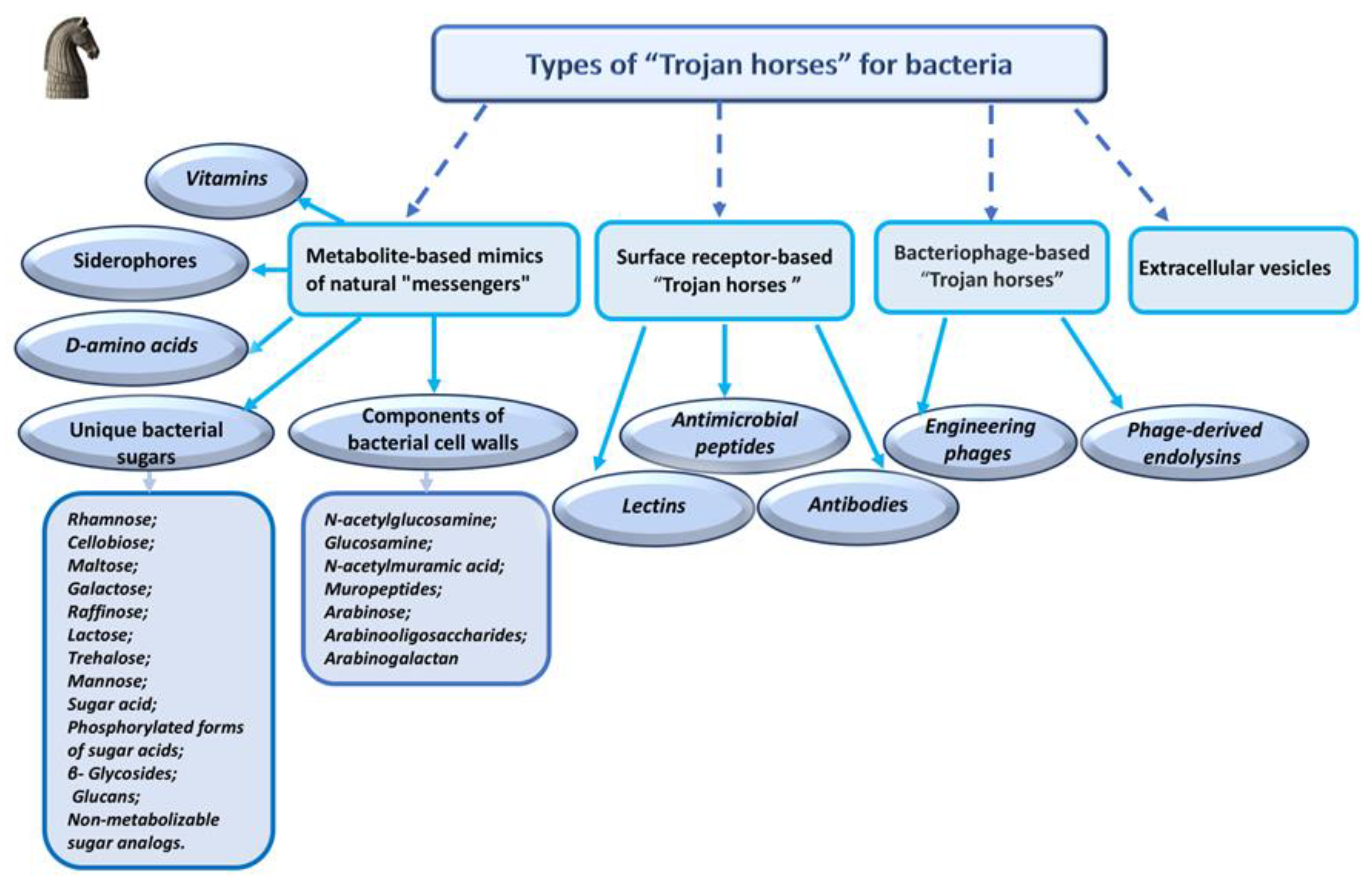
Figure 1. Classification of bacterial “Trojan horse” delivery systems based on their mechanisms of action.
Preprints 202840 g001
Figure 2. Schematic of siderophore-mediated iron uptake in Gram-positive and Gram-negative bacteria.
Preprints 202840 g002
Figure 3. Schematic illustration of “phage syringes” engineered through modular T7 phage genome design and de novo gene synthesis followed by cell-free protein expression, delivering penicillin G (red dots) to target penicillin-resistant Escherichia coli.
Preprints 202840 g003
Figure 4. A schematic representation of the filamentous M13 bacteriophage as a drug delivery vehicle targeting pathogenic bacteria. Target specificity is achieved using antibodies recognized the bacteria. The linkage between the antibodies and the phages is mediated by an IgG binding ZZ domain located on the minor coat protein of the phage. The therapeutic agent employed is chloramphenicol conjugated to neomycin via an ester bond that is cleavable by esterase.
Preprints 202840 g004
Table 1. Sugar conjugates as "Trojan Horses".
Sugar
 
Conjugate "Trojan Horse" strategy
targets
Reference
Rhamnose l-rhamnosyl-10-deoxymethynolide Erythromycin-sensitive and erythromycin-resistant strains of Enterococcus faecium and Staphylococcus aureus [94]
Rhamnolipids functionalized intrinsically active liposomes loaded with cinnamaldehyde Salmonella typhimurium
Salmonella enteritidis
[95]
Rhamnolipid amino acid-arginine derivatives
(RLmix-Arg and monoRLArg)
Gram-positive bacteria, including methicillin-resistant Staphylococcus aureus MRSA [31]
Rhamnose binding protein P. aeruginosa PA14 biofilms [96]
Rhamnopyranoside—based conjugates Bacillus subtilis
Staphylococcus aureus
Escherichia coli
Pseudomonas aeruginosa
[32]
Cellobiose Flocculosin (a membrane-active cellobiose lipid)
 
Gram positive bacteria, including
Staphylococcus aureus MRSA,
Enterococcus faecium VRE
[97]
Functionalized cellulose conjugates
Cel-DDTMABr
Cel-TDTMABr
Cel-HDTMACl
Cel-DDTPPB
Cel-HDTBPBr
Cel-MBT
Cel-THIO
Staphylococcus aureus
Escherichia coli
[98]
Various nanocellulose conjugates
 
Staphylococcus aureus
Escherichia coli
[99]
Nanocomposite of nanocellulose and nanoselenium Bacillus subtilis
Staphylococcus aureus
Escherichia coli
Pseudomonas aeruginosa
Candida albicans
Aspergillus fumigatus
[100]
Antibacterial-modified cellulose fiber E. coli
S. aureus
[33]
PpIX-CNF (protoporphyrin-IX conjugated cellulose nanofibers) Staphylococcus aureus ATCC-6538
Escherichia coli ATCC-8099
[34]
Maltose Chitosan derivatives E. coli
Bacillus subtilis
S. aureus
Listeria monocytogenes
Salmonella enteritidis
S. typhimurium
K. pneumoniae
P. aeruginosa.
[101]
Cpd-1 (maltotriose–perylene conjugate) and Cpd-2 (maltohexaose–perylene conjugate) E. coli [102]
Antimicrobial peptide T9W production in presence of maltose P. aeruginosa, including clinically isolated antibiotic-resistant strains [103]
MMCC (mannose-maltose-colistin conjugate) E. coli [104]
TM-TMP (trimethoprim conjugate of maltodextrin) E. coli [105]
MDNP (ROS-responsive maltodextrine nanoparticles) dormant Staphylococcus aureus [106]
β-maltosyl thiosemicarbazones of substituted benzaldehydes Clostridium difficile
Bacillus subtilis
Staphylococcus epidermidis
Streptococcus pneumoniae
Staphylococcus aureus
MRSA: Methicillin-resistant Staphylococcus aureus
[107]
Galactose Glycoclusters connected to pseudosiderophores Pseudomonas aeruginosa [108]
 
Glycated albumin with lactose (BSA-glucose-β (4-1) galactose) E. coli K88 [109]
The luminescent galactoconjugates
GalTEBB-1
GalTEBB-2.
P. aeruginosa:
S. aureus
 
[110]
Tetravalent neoglycoсonjugate based
on the D-galactose
Pathogenic bacteria with adhesion mediated by galactose recognition
 
[111]
 
Raffinose stimulation of reuterin production by Limosilactobacillus reuteri Staphylococcus aureus biofilms
Staphylococcus aureus CMCC 26003
Escherichia coli O157:H7 NCTC12900
Salmonella typhimurium ATCC 13311
Listeria monocytogenes CMCC 54007
[112]
Lactose Lactose palmitoleate and lactose nervonate Escherichia coli O157:H7 ATCC 35150
Listeria monocytogenes
ATCC 7644
Salmonella enteritidis ATCC 13076
Enterococcus fecalis ATCC 29212,
Pseudomonas aeruginosa ATCC 9027
Staphylococcus aureus ATCC 43387
Yersinia enterocolitica ATCC 27729
[113]
Ciprofloxacin-loaded lactose particles Staphylococcus aureus
Pseudomonas aeruginosa
[114]
Lactose-coated NAC-loaded Poly(lactic-co-glycolic acid) (PLGA) nanoparticles (NAC-PLGA NPs) Mycobacterium tuberculosis H37Rv. [36]
Microparticles (MPs) containing various nanocarriers (NPs) with antimycobacterial substances for pulmonary delivery. Mycobacterium sp. [115]
Cip@LacAC4A lactose-modified azocalix[4]arene (LacAC4A) and ciprofloxacine multidrug-resistant Pseudomonas aeruginosa [15]
Trehalose TreAz analogs M. smegmatis
M. tuberculosis
[116]
Trehalose analogues M. smegmatis [117]
Trehalose conjugates of polyketide synthase 13 (Pks13) M. smegmatis
M. tuberculosis
[118]
Trehalose – BODIPY conjugate M. smegmatis
M. abscessus
[119]
Trehalose-porphyrin conjugate M. smegmatis [120]
TCC2Tre (Trehalose – tricarbocyanine conjugate) M. smegmatis
M. tuberculosis
[121]
Mannose Nanoparticles or proteins coated with mannose M. tuberculosis [122]
RPT-MAN-PLGA-PEG M. tuberculosis [123]
Various mannose conjugates E. coli
Klebsiella pneumoniae
Mycobacterium tuberculosis.
Helicobacter pylori
Pseudomonas aeruginosa
Salmonella enterica
Vibrio cholerae
Yersinia pestis
Porphyromonas gingivalis
[124]
MP-MENP S. aureus, MRSA [125]
Mannose-modified lipid nanoparticles M. tuberculosis [126]
Sugar acids and their phosphorylated forms, glucans Levofloxacin glycosylated mesoporous silica nanoparticles (GLY-MSN) E. coli [127]
ERNathG probe (4-hydroxy-1,8-naphthalimide as the tag) S. warneri
E. coli (DH5α, ATCC 25922)
S. aureus (MRSA, ATCC 6538, CMCC(B) 26003)
B. subtilis (CICC 10088, ATCC 6633)
E. faecalis ATCC 29212
E. aerogenes ATCC 13048
P. aeruginosa ATCC 9027
[128]
Glucan Lipid Particles (GLPs) conjugates with various drugs M. tuberculosis [129]
β- Glycosides (specific to bacteria) Glycoside derivatives of l-R-aminoethylphosphonic acid (l-AEP) E.aerogenes 13048
C sakazakii 29544
K.pneumoniae 13882
S.enterica serovar Typhimurium 14028
S.enterica serovar Virchow 5742
Gram negative bacteria
[130]
Non-metabolizable sugar analogs Azide-modified sugars (AMS) B. fragilis
Anaerobic bacteria
 
[131]
FLAPS Imaging of
Vibrio cholerae
Bacteroides thetaiotaomicron
[132]
Fluorine-18-Labeled Disaccharides Imaging of
Staphylococcus aureus (including MRSA) Acinetobacter baumannii
[133]
Table 2. Vitamin conjugates and complexes as "Trojan Horses".
Vitamin Active compound/ conjugate component Activity assay Antibacterial efficiency Reference
В12 Ampicillin E. coli,
S. typhimurium
500 and 60 times higher than ampicillin and 8 times higher than ciprofloxacin  
[264]
В12 Chloramphenicol succinate E. coli The same as chloramphenicol, but the compound is less toxic.  
[264]
Suc-B12 PMAG- (DFOA, CT)
PMAG – poly (2- 2-deoxy-2-methacrylamido-D-glucose)
DFOA- deferoxamine (siderophore)
CT- colistin (antibiotic)
Pseudomonas aeruginosa PMAG-(DFOA+Fe,CT) conjugates can be considered as promising targeted systems for intravenous delivery, while PMAG-(DFOA+Fe,CT-B12) conjugates are suitable for oral delivery.  
 
[265]
B12 CHS-CT-CSB12
(CHS- chondroitin sulfate
CT- Colistin
CSB12 cyanocobalamin–chitosan conjugate)
P. aeruginosa The effect is not pronounced, but nevertheless the conjugate with vitamin has increased bioavailability.  
[266]
B12 HA–CT–B12
HA– hyaluronic acid
CT–colistin
P. aeruginosa  
[267]
B12 Antisense peptide nucleic acid (PNA) E. coli
S. typhimurium
[268]
B12 Antisence oligonucleotides E. coli
S. typhimurium
Complete destruction of S. aureus was achieved in 2 minutes of illumination, and E. faecalis in 5 minutes under certain conditions (illumination of 455 nm, 30 MW/cm2).  
[269]
Ascorbic acid none carbapenem-resistant hypervirulent Klebsiella pneumoniae (CR-hvKP) for strain KP1088, MIC is 8 mg/ml; 1
 
for the HvKP3 strain, the MIC is 16 mg/ml.
 
[270]
Complexes with metallic nanoparticles
Ascorbic acid Ag (Ag/Cu) Bacillus subtilis and
E. coli
Strongest bactericidal effect (MIC ~0.05–0.08 mg/L).  
[271]
Ascorbic acid Au
 
E. coli, S. aureus, S. enterica, S. mutans, Candida spp. Most effective: >88% kill of E. coli and ~83% of S. aureus under light (via ROS generation),  
[272]
Ascorbic acid Cu2S S. aureus,
E. coli,
K. pneumoniae
Broad-spectrum bactericidal activity. MIC: ~2 mg/mL (E. coli) and 10 µg/mL (other strains)  
[273]
Ascorbic acid Gd2O3 Potent bactericidal effects against multiple pathogens.  
[274]
Ascorbic acid Se S. aureus Strong activity against S. aureus; stabilized Se–VitC NPs retained activity 2–6 months.  
[275]
Ascorbic acid Zn/Ag MOF Strong activity against Gram+ and Gram− bacteria common in wound infections.  
[276]
Folic acid CeO2 Potent against MRSA Inhibit ~95.6% of MRSA growth  
[277]
Folic acid Ag Antibacteria property  
[278]
Folic acid Ag/MOF (nanocapsule) Single folate-targeted nanocapsule can deliver chemotherapeutics while preventing infection & oxidative damage.  
[279]
Riboflavin Fe3O4 S. aureus,
E. coli
Kills >90% of S. aureus and ~88% of E. coli at 0.5 mg/mL.  
[280]
Riboflavin Au Photodynamic antimicrobial therapy (S. aureus, P. aeruginosa) Vitamin B2 + AuNP create synergistic ROS + Au+ antibacterial effect.  
[281]
Riboflavin Fe/MOF Treatment of bacterial keratitis (S. aureus,
P. aeruginosa)
Rapid infection clearance with minimal collateral damage.  
[282]
Biotin, D-Pantothenic acid & Nicotinic acid Ag NPs Effective at low concentrations (15.62–62.5 μg/mL) against planktonic cells & iofilms.  
[283]
 
Disclaimer/Publisher’s Note: The statements, opinions and data contained in all publications are solely those of the individual author(s) and contributor(s) and not of MDPI and/or the editor(s). MDPI and/or the editor(s) disclaim responsibility for any injury to people or property resulting from any ideas, methods, instructions or products referred to in the content.
Copyright: This open access article is published under a Creative Commons CC BY 4.0 license, which permit the free download, distribution, and reuse, provided that the author and preprint are cited in any reuse.
Prerpints.org logo

Preprints.org is a free preprint server supported by MDPI in Basel, Switzerland.

Subscribe

Disclaimer

Terms of Use

Privacy Policy

Privacy Settings

© 2026 MDPI (Basel, Switzerland) unless otherwise stated