Submitted:
04 March 2026
Posted:
06 March 2026
You are already at the latest version
Abstract
This study evaluates the antimicrobial properties of nanocomposite materials based on polyvinyl alcohol (PVA) reinforced with cellulose nanofibrils (CNFs) and/or supplement-ed with biobased additives derived from blueberry pruning wastes, with the objective of developing biodegradable food packaging systems with antimicrobial properties. The nanocomposites were prepared using a solvent-casting processing approach, and their thermal, physicochemical, and antimicrobial properties were assessed. All the nanocomposites exhibited thermal stability up to 200 °C, confirming their suitabil-ity for conventional food packaging processing conditions. Antimicrobial activity tests re-vealed inhibitory effects against both Gram-positive and Gram-negative bacteria. Bleached PVA/CNFs films showed complete growth inhibition (100%) against E. coli and S. aureus. In contrast, unbleached PVA/CNFs and PVA/CNFsB supplemented with blueber-ry-derived additives exhibited selective inhibition against E. coli, highlighting the influ-ence of nanofibril composition and additive incorporation on antimicrobial performance. Zeta potential measurements revealed values of –35.3 mV for the CNFs, confirming their negatively charged surface, which may contribute to interactions with bacterial mem-branes. Additionally, scanning electron microscopy (SEM) showed that the incorporation of CNFs generates nanostructured surfaces with exposed fibrillar domains, where bacteri-al cells become adhered and immobilized. These topographical features suggest that the antimicrobial behavior of the nanocomposites is associated with direct bacteria–surface interactions, supporting a contact-active antimicrobial behavior associated with the CNFs.
Keywords:
nanocomposite
; PVA/CNFs
; antimicrobial properties
; packaging food
1. Introduction
Today, food packaging materials are one of the main challenges in the food industry. There is a growing consumer demand for high-quality fresh food packaged in biobased and preservative-free biodegradable materials [1]. Food packaging systems with antimicrobial properties represent an emerging technology that can enhance both food shelf life and safety. Therefore, the production of packaging with antimicrobial characteristics based on biodegradable materials is an area of growing interest [2,3,4]. Biobased packaging can improve food quality and increase shelf life, functioning as a barrier system against oxygen, environmental odors, humidity, and microorganisms [5,6].
The polymers commonly used for food packaging are those derived from petroleum: mainly polypropylene, polyethylene and polystyrene. Their low cost of obtaining, its availability, its good mechanical, and barrier properties make them the most competitive alternative. However, in view of both the global problem of the accumulation of non-biodegradable plastics and the need to develop materials for the packaging of fresh foods with antimicrobial characteristics, it is essential to advance in processes that are more respectful of the environment and to develop new packaging materials [7].
In this search, biodegradable polymers have been widely studied as substitutes for petroleum-derived plastics. Biopolymers are used in many applications: in the biomedical area (implants), biomimetic materials, material coatings, insulating materials, as additives in paints, some food packaging, additives in adhesives, as thickeners in lubricants and plasticizers [5,8,9]. The main limitation of biodegradable polymers for fresh food packaging lies in their low processing efficiency and suboptimal mechanical or barrier properties (e.g., oxygen/moisture permeability and thermal resistance) [7,8]. To overcome these limitations presented by biopolymers, various materials have been studied to be used as reinforcement or fillers to obtain a bio-nanocomposite and thus improve the properties of the biopolymer [10,11,12,13,14,15,16]. Bio-nanocomposites are composite materials made by mixing a biodegradable polymer (natural or synthetic) with a nanoscale reinforcement material, which typically does not exceed 5% by weight of the mixture. These bio-nanocomposites exhibit notable improvements in mechanical and barrier performance, driven by the synergistic effects of their high surface area at the nanoscale.
Among the biodegradable polymers used for food packaging, polyvinyl alcohol (PVA) stands out. PVA is a low-cost, non-toxic, water-soluble synthetic polymer [17] that is obtained commercially from the hydrolysis of polyvinyl acetate. Despite its synthetic character, this polymer is biodegradable and shows high tensile strength, adequate barrier properties, and resistance to acid and alkaline media [18]. The safety of PVA has been extensively evaluated in various studies by entities in the food and health area (JECFA, FAO, WHO) [19]. This biopolymer, thanks to its hydroxyl groups (OH-), forms hydrogen bonds with the hydrophilic surfaces of biopolymers or biomaterials to produce bio-based compounds [17]. Due to this capacity, PVA has been combined with other reinforcements or fillers to improve its performance and barrier properties, for example with nanoparticles of silver [20], zinc and titanium oxide [21], etc. Heavy metal nanoparticles such as silver, copper and zinc possess effective antimicrobial activities against a wide variety of bacteria [22]. Their high surface area, thermal stability, and low volatility have made metallic nanoparticles attractive candidates for incorporation into active packaging systems [22,23]. However, these elements are inorganic in nature and therefore have limited biocompatibility and biodegradability. On the other hand, the reinforcement of PVA with nanocelluloses has been studied, obtaining a significant improvement in the mechanical and barrier properties of PVA [24,25,26,27,28].
In the last decade, the use of cellulose nanofibrils (CNFs) as a reinforcement or filler agent has been investigated, mainly in biodegradable polymers. CNFs are obtained from cellulose, the most abundant biopolymer in nature. CNFs are commonly obtained by chemical or enzymatic pretreatment, followed by mechanical treatment to achieve a nanometric size of their fibrils. CNFs have been used as a reinforcing agent (due to their large aspect ratio and high specific surface area) in various polymeric matrices to mainly improve their mechanical and barrier properties [29,30]. CNFs can be obtained from any source of lignocellulosic biomass (mainly wood) including agro-industrial waste [31,32,33,34,35], allowing the comprehensive use of natural resources, favoring the circular economy [7].
The use of CNFs in food packaging with antimicrobial properties is a little explored area to date. The available studies focus on the elaboration of CNFs films with antimicrobial properties, mainly through the physical adsorption of metallic nanoparticles [36,37,38] which are gradually released into the matrix, leading to the inhibition of microbial growth. Other studies are based on the incorporation of protein grafts [4,39] or polycationic compounds [40] on the surface of CNFs. Although metals are effective in inhibiting a wide range of microorganisms, they are toxic and their continuous use has generated resistance in microorganisms [41,42]. Polycations are poorly biocompatible, while enzymes suffer from chemical instability. In the reviewed literature, the most studied antimicrobial agents in food packaging applications were Ag nanoparticles [43], ethyl lauryl arginate (LAE) [44], TiO2 [45], Nisin [46], Citric acid and chitosan [47], Gallic acid grafted chitosan [48], Dehydroabietylamine [3], Porphyrin [49], Lysozyme and nisin [39], and Aminosilanes [40].
Most studies on obtaining CNFs have focused on bleached chemical pulps, however, CNFs from unbleached kraft pulps have also been reported to have good barrier properties [7,32,35]. CNFs from unbleached pulps contains residual lignin, which allows the production of pulps with higher yields and lower production costs [50]. It has been investigated that the presence of residual lignin in CNFs has several advantages, such as improved compatibility [51,52] and higher hydrophobicity [53].
Espinosa et al., (2019) obtained CNFs with and without residual lignin from wheat straw. These nanocelluloses were used as a reinforcing agent in PVA films to improve their barrier properties, specifically their antioxidant characteristics. The results showed that this capacity increased from 10 to 50% using unbleached nanocellulose, especially in small amounts of reinforcement of CNFs (1-3% wt.) [7].
Patiño et al. (2022) prepared cellulose nanocrystals nanocomposites incorporated with an antimicrobial agent, ethyl lauroyl arginate (LAE). These LAE-CNC nanocomposites films demonstrated antimicrobial activity against the Gram (+) and (-) bacteria., L. monocytogenes and S. typhimurium., even with just a 1% addition of LAE [44].
Within biopolymers, lignin has been extensively studied mainly due to its antioxidant and antimicrobial properties. The main function of lignin is to provide rigidity to the plant to strengthen the structure of cell walls [54]. Among its various functions in plants, its behavior as an antimicrobial agent stands out [54]. Lignin’s antimicrobial activity is attributed to its phenolic structure, particularly the α, β-unsaturated side chains and γ-methyl substitutions, which enhance the potency of its phenolic fragments [55]. Many investigations have suggested that lignin’s could be applied to stabilize foods due to their antioxidant, antifungal and antimicrobial properties [56,57,58,59,60,61,62]. Its biocidal activity makes it an alternative and more attractive compound than metallic nanoparticles due to its reduced environmental impact.
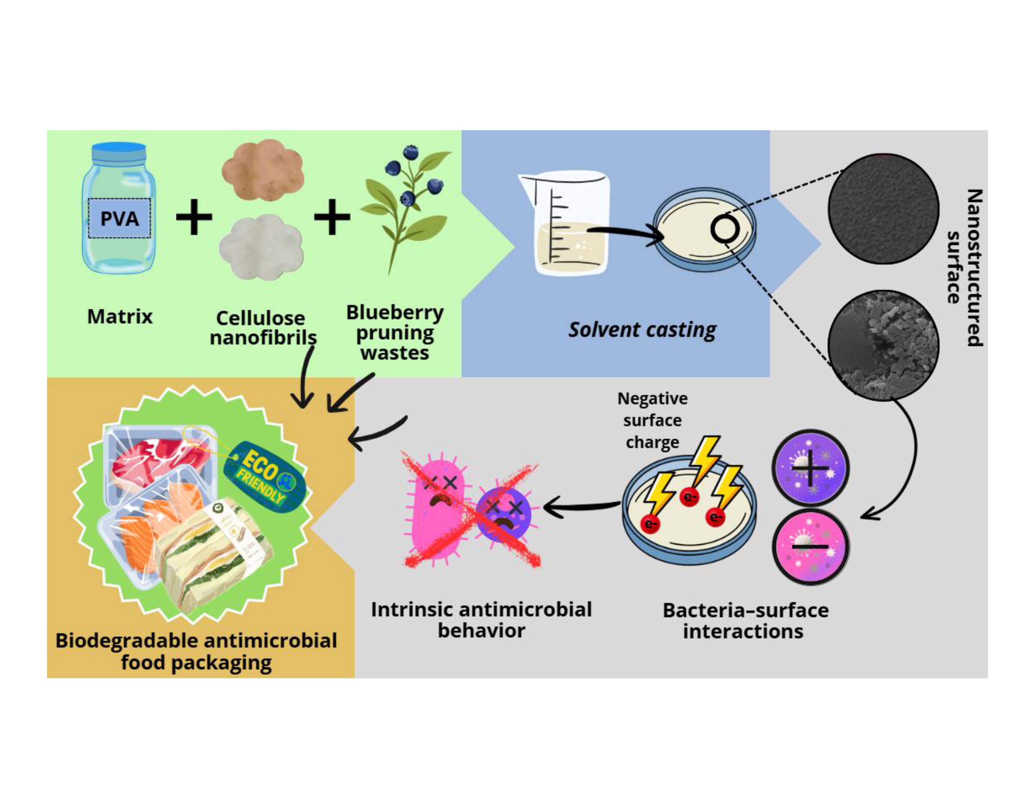
Considering the above, the use of unbleached cellulose nanofibrils (L-CNFs) as reinforcing agents in PVA-based bio-nanocomposites is proposed. The presence of residual lignin in L-CNFs is expected to contribute to inherent antimicrobial properties, enabling the development of active, biodegradable food packaging materials. Figure 1 presents the conceptual framework of the proposed system.
2. Materials and Methods
Raw materials and Chemicals
The unbleached nanocellulose hydrogel (with lignin) was supplied by Bioforest (Arauco cellulose). The blueberry pruning and corn husks waste were provided by the agricultural company FISTUR (Yumbel, Biobío region, Chile). The Polyvinyl alcohol, PVA (M.W.: 146,000–186,000; and degree of hydrolsysis +99%), (2,2,6,6-tetramethylpiperidinyl-1-oxyl) TEMPO, NaBr, NaClO, ethanol, which were purchased from Merck/Sigma Aldrich, Santiago de Chile.
2.1. Removable Extraction from Blueberry Pruning and Corn Husks (Modified TAPPI T204 cm-97)
For its determination, 4.0±0.1 g of sample (dry weight) at equilibrium moisture content, prepared according to TAPPI standard 257 cm-85, were used. The raw material was placed in a pre-weighed filter paper cartridge. On top of the cartridge with the sample inside, another cartridge was placed as a lid, also pre-weighed, to prevent sample losses during extraction. The assembly was placed in a Soxhlet, and 150 mL of analytical-grade acetone was added, approximately up to one and a half siphons. The Soxhlet was connected to a flask with a condenser and heated to reflux under a hood, bringing the mixture to a boil. After 6 hours, during which no fewer than 24 extractions must occur, and once cooled, the Soxhlet was disassembled, and the solvent in the Erlenmeyer flask was partially evaporated at room temperature under a hood. The cartridge with the sample was collected for subsequent analyses, dried in an oven at 105 ºC for 24 hours. Finally, it was weighed, and the extracted mass (mf) was obtained [31].
2.2. CNFs Obtention
The method described by Zuluaga et al. (2009), with some modifications, was applied for obtaining cellulose pulp from corn husks [63].
TEMPO-mediated oxidation of corn husk-cellullose pulp and nanocellulose hydrogels was carried out according to the protocol described by Saíto et al., 2006 with some modifications. A total of 10 g of 3 wt% sample was suspended in 300 mL of deionized water containing 0.050 g TEMPO and 0.50 g NaBr. TEMPO-mediated oxidation began with the addition of 30 mL of a commercial NaClO solution (10% wt.) at room temperature with gentle stirring. The pH was maintained at 10.5 by adding 0.5 M NaOH. The reaction was considered complete once the pH remained stable at 10.5. The oxidized product was filtered, washed with deionized water, and stored at 4 °C [67]. Each experiment was conducted in triplicate. The oxidized fibers were dispersed in deionized water with a pulp concentration of 1 wt.% and then homogenized with a high-pressure homogenizer (NS1001L PANDA 2K-GEA) 8 times at a pressure drop of 800 bar to produce nanocellulose hydrogels [64]. Films (20 g/m2) were prepared in plastic petri dishes, based on dispersions of 0.25% CNFs. The drying temperature was 24-26 °C [31].
2.3. Preparation of PVA/CNFs Nanocomposites
A 5 wt% PVA solution was prepared by dissolving the polymer in distilled water under stirring at 90 °C for 4 h. After cooling to room temperature, a 2 wt% CNFs suspension (bleached, unbleached, or with blueberry extractives) was added and stirred for an additional 4 h. The final films were cast in Petri dishes and dried under ambient conditions (20–25 °C, 40–60% RH) for approximately 7 days until complete water evaporation. Each experiment was performed in triplicate. The final CNFs content in the nanocomposites was approximately 28 wt% relative to dry PVA [7]. Each experiment was conducted in triplicate. Table 1 provides the nomenclature of the nanocomposite samples manufactured for this study.
2.4. Fourier Transform Infrared Spectroscopy (FTIR)
The IR measurements were performed with an Agilent Tensor 27 instrument (Malaysia) in Fourier transform mode (FTIR). The Agilent MicroLab PC software (version IQ/OQ, 21 CFR Part 11 compliant) was used for data acquisition. A total of 40 scans were collected across a spectral range of 400 to 4000 cm−1 [64,66].
2.5. Thermo-Gravimetric Analysis (TGA)
Thermal stability and decomposition profiles were determined using a NETZSCH TG 209 F3 Tarsus® thermal analyzer (MB & Cia, Selb, Germany). Approximately 5 mg of each sample was placed in an alumina crucible and heated from room temperature (~25 °C) up to 600 °C under an inert nitrogen atmosphere, at a constant flow rate of 50 mL/min. The heating rate used was 10 °C/min [64,66].
2.6. Zeta Potential
Zeta potential measurements were performed using a Malvern Zetasizer Ultra (Malvern Panalytical, UK). A total of 1 mL of the sample was dispersed in deionized water and used as the measurement medium. All measurements were conducted at room temperature with a fixed scattering angle of 90°.
Prior to measurement, the zeta potential cell was carefully filled with the sample, ensuring the absence of air bubbles. External electrodes were cleaned thoroughly to avoid contamination, and the refractive indices of both the solvent (water) and the sample were correctly set in the instrument software. The temperature was maintained at 25 °C throughout the analysis. Each measurement was performed in triplicate to ensure reproducibility.
The Zetasizer software provided the average zeta potential value (in mV), the standard deviation, and the corresponding distribution graph for each set of measurements.
2.7. PVA/CNFs Antimicrobial Activity Nanocomposites
The antimicrobial activity of PVA/CNFs nanocomposites was analyzed by modifying the Japanese Industrial Standard (JIS Z 2801:2000) to test antibacterial activity on plastic surfaces. The JIS test is based on a comparison of bacteria counts (S. aureus, and E. coli) in saline solution on reference (PVA) and sample materials (PVA + CNFs) after a defined incubation temperature and time (37 ◦C, 24 h) [68,69]. The material that shows a calculated log10-reduction ≥ 2.0 log10 units after 24 h is considered as an effective antimicrobial agent [68]. Each experiment was performed in triplicate for each nanocomposite.
Figure 2.
Scheme of the antimicrobial activity JIS test for PVA/CNFs nanocomposites.

2.8. Scanning Electron Microscopy (SEM) with Energy Dispersive X-Ray Spectroscopy (EDS)
After the bacterial contact period with S. aureus, the films were removed and cut into 0.5 × 0.5 cm fragments. The samples were transferred to 50 mL Falcon tubes containing 1 mL of 4% paraformaldehyde (PFA) as a fixative agent and incubated for 4 h at room temperature to preserve bacterial morphology and bacteria–surface interactions. Subsequently, the samples were washed three times with phosphate-buffered saline (PBS, pH 7.2) and subjected to gradual dehydration using a graded ethanol series (30, 50, 70, 90, and 100%), with 10 min incubations at each concentration; the absolute ethanol step was repeated. After dehydration, the samples were dried by critical point drying [65].
The dried film fragments were mounted on SEM stubs and coated with a thin gold layer using a sputter coater (SPI-MODULE, USA) to ensure surface conductivity. Morphological analysis was evaluated using a scanning electron microscope (JEOL JSM-6380LV, Japan) equipped with an energy-dispersive X-ray spectroscopy (EDS) detector, with image acquisition performed via a computer-connected system.
3. Results
3.1. Infrared Spectroscopy Analysis (FTIR)
Figure 3 shows the infrared spectra of CNFs films and its nanocomposites. The infrared spectra of CNFs films and PVA/CNFs nanocomposites showed typical bond vibrations of these materials. The broad peak around 3300 cm–1 in all nanocomposite films indicates the presence of hydroxyl groups corresponding to both PVA and CNFs. The C–H stretching vibrations of PVA, and CNFs was identified at the peak of 2900 cm–1 in all samples, while the peak around 1700 cm–1 in the PVA/CNFs and PVA/L-CNFs samples is attributed to the stretching vibration of the C=O bond resulting from TEMPO-mediated oxidation of CNFs. The peak around 1580 cm–1 in all films is due to the bending vibration of the C-H bond. The peak around 1100 cm–1 in all samples is attributed to the stretching vibration of the C-O bond in secondary alcoholic groups or to CH2 bending vibrations [7].
3.2. PVA/CNFs Nanocomposites Thermo-Gravimetric Analysis (TGA)
In Figure 4, L-CNFs and CNFsB samples display a single major degradation step, with degradation starting at approximately 330–350 °C. The slightly improved thermal resistance of CNFB may be attributed to the presence of extractable phenolic compounds from blueberry pruning waste, which may contribute to enhanced thermal shielding during decomposition.
The nanocomposite PVA/CNFs show a two-step degradation pattern. The first mass loss event, between 250–300 °C, is associated with the thermal degradation of the PVA matrix, while the second, occurring above 350 °C, corresponds to the decomposition of the nanocellulose component. TEMPO-oxidized samples showed a slightly lower degradation onset, likely due to the introduction of carboxylic groups. Overall, CNFs reinforcement led to a minor enhancement in thermal stability compared to pure PVA films.[7]. Sirviö et al., 2015, reported that pure PVA films begin to degrade at 262 °C. Therefore, due to the reinforcement of CNFs, the thermal stability of the PVA/CNFs nanocomposite was slightly increased.
3.3. PVA/CNFs Nanocomposites Zeta Potential (mV) Analysis
In Table 2, it can be observed that CNFs exhibit a strong negatively charged surface, attributed to the presence of hydroxyl and/or carboxylate groups [70], particularly when subjected to TEMPO-mediated oxidation pretreatment. This negative charge of CNFs increases the overall charge of the PVA/CNFs nanocomposite when reinforced with CNFs, considering that the PVA matrix is weakly charged and accounts for approximately 98% wt. of the nanocomposite by mass. Consequently, the charges induced by the small fraction of CNFs in the nanocomposite (2% wt.) can interact with bacterial membranes, particularly those of Gram-positive and Gram-negative bacteria, which differ in their cell membrane structure, contributing to destabilization.
3.4. Antimicrobial Activity of PVA/CNFs Nanocomposites
As shown in Figure 5A all the nanocomposites studied demonstrated an inhibitory effect on Gram-negative bacteria (E. coli). Those showing a calculated log10-reduction ≥ 2.0 log10 units compared with PVA after 24 h of incubation are marked with a red asterisk in Figure 5. This means they have a validated antimicrobial activity, according to the JIS 2801 test. The bleached PVA/CNFs tested showed a 100% inhibition of E. coli and S. aureus growth (Figure 5). This is the same effect observed when using a nanocomposite of CNFs alone. PVA/CNFs-T showed a 100% inhibition of E. coli only. The unbleached CNFs (CNF-L), PVA/CNFs-L and PVA/CNFs with blueberries additives tested showed significant antibacterial activity for E. coli only.
Although CNFs in general do not exhibit antimicrobial properties as strong as those of specific antimicrobial compounds, the nanocomposites with CNFs tested have demonstrated measurable activity, which can be attributed to their topo-chemical and supramolecular characteristics. One key factor is their negatively charged surface, resulting from naturally occurring hydroxyl groups or carboxyl groups introduced during pretreatment. This surface charge can interact with bacterial membranes, leading to membrane disruption. In addition, type II cellulose, which has lower crystallinity (approximately 60%), imparts greater flexibility to the nanofibrils. This flexibility may enhance their ability to interact with bacterial cell walls, contributing to their antimicrobial activity.
3.5. Surface Topography and Bacteria–Material Interactions in PVA/CNFs Nanocomposites
The surface morphology of the PVA/CNFs nanocomposites was examined by scanning electron microscopy (SEM) to elucidate the role of nanoscale topographical features on the antibacterial behaviour of the materials. As shown in Figure 6A, the nanocomposite surface exhibits a heterogeneous and rough morphology, characterized by an interconnected network of cellulose nanofibrils partially exposed at the surface of the PVA matrix. This fibrillar architecture, typical of TEMPO-oxidized and highly fibrillated CNFs, has been widely reported to generate high surface area and pronounced nanoscale roughness [71,74]. After incubation with S. aureus (Figure 6B), bacterial cells are observed adhered and immobilized on the PVA/CNFs surface. The bacteria appear preferentially located in regions with higher nanofibrillar density, indicating that surface topography plays a dominant role in governing bacteria–material interactions. Similar SEM observations have been reported for carboxylated and TEMPO-oxidized CNFs systems, where bacteria are physically entrapped within nanofibrillar networks rather than repelled from the surface [71,73].
The immobilization of S. aureus observed in Figure 6B is consistent with a contact-active antibacterial mechanism, previously described for nanocellulose-based materials. In these systems, the antibacterial effect arises from prolonged physical contact between the bacterial cell wall and the nanostructured surface, leading to restricted motility, impaired cell division, and inhibited biofilm formation [73]. SEM images reported by Knutsen et al. (2021) showed bacterial cells surrounded and confined by a dense CNFs network, a phenomenon that closely resembles the bacterial immobilization observed in the present PVA/CNFs nanocomposites.
For Gram-positive bacteria such as S. aureus, the thick peptidoglycan layer provides mechanical rigidity, which explains the absence of severe cell collapse or lysis in the SEM images. However, this structural robustness also promotes strong physical anchoring to rough and fibrillar surfaces, enhancing immobilization effects. Previous studies have demonstrated that nanocellulose surfaces with high aspect ratio fibrils induce mechanical stress at localized contact points, leading to membrane fatigue and loss of bacterial viability over time, even in the absence of chemical biocides [71].
The topographical contribution to antibacterial activity is further reinforced by studies on oxygenated and highly carboxylated CNFs, where SEM analyses revealed that increased fibrillation degree and surface charge result in denser nanofibrillar networks capable of surrounding bacterial cells and limiting nutrient and oxygen diffusion [73,74]. Although oxygenation was not applied in the present system, the morphological similarities observed by SEM suggest that the PVA/CNFs nanocomposites operate through a comparable physical confinement mechanism.
Importantly, the bacterial adhesion observed in Figure 6B does not indicate a loss of antimicrobial functionality. On the contrary, it supports a non-leaching, contact-based antibacterial mode of action, which is particularly advantageous for food packaging applications. Contact-active antimicrobial surfaces prevent bacterial proliferation directly at the material interface while minimizing the risk of antimicrobial migration into food matrices [4].
Overall, the SEM results confirm that the antibacterial performance of PVA/CNFs nanocomposites is strongly governed by their nanoscale surface topography. The combination of nanofibrillar architecture, surface roughness, and physical confinement of bacterial cells aligns well with previously reported antibacterial mechanisms of TEMPO-oxidized, carboxylated, and oxygenated CNFs [71,72,73,74]. These findings highlight the potential of PVA/CNFs nanocomposites as contact-active antimicrobial materials for sustainable food packaging applications.
4. Discussion
Antibacterial metal nanoparticles or antibacterial peptides grafted onto cellulosic materials tend to aggregate upon release, reducing their capture efficiency and shortening their effective lifespan. In contrast, contact-active antibacterial surfaces eliminate the release step, thereby offering greater durability. However, their main limitation lies in their passive mechanism of action, as they require direct contact with bacteria to be effective. Despite this, such materials are ideal candidates for applications where this contact naturally occurs, such as antibacterial filters, water purification systems, food packaging, wound dressings, and antimicrobial coatings.
Conclusions
PVA/CNFs nanocomposites were successfully developed as bio-based antimicrobial materials, and their antibacterial performance was shown to be strongly governed by nanoscale surface topography rather than by the release of antimicrobial agents. The incorporation of cellulose nanofibrils into the PVA matrix generated heterogeneous and rough nanostructured surfaces with exposed nanofibrillar networks, which played a decisive role in bacteria–material interactions.
SEM analysis provided direct evidence that Staphylococcus aureus cells become adhered and immobilized on the surface of the PVA/CNFs nanocomposites. This immobilization is attributed to a contact-active antibacterial mechanism, where nanoscale roughness and fibrillar architecture restrict bacterial mobility and proliferation at the interface. These findings are consistent with previously reported antibacterial behaviors of TEMPO-oxidized, carboxylated, and oxygenated cellulose nanofibrils, in which physical confinement within nanofibrillar networks is a dominant mode of action.
Importantly, the observed bacterial adhesion does not indicate a loss of antimicrobial efficacy but rather confirms a non-leaching and physically driven antibacterial mechanism. This characteristic is particularly advantageous for food packaging applications, as it minimizes the risk of antimicrobial migration into food products while ensuring localized and sustained inhibition of bacterial growth upon contact.
Overall, this study demonstrates that controlling nanoscale surface topography is a key strategy for designing effective antimicrobial polymer nanocomposites based on cellulose nanofibrils. The results highlight the potential of PVA/CNFs nanocomposites as sustainable, contact-active antimicrobial materials for advanced food packaging systems, contributing to improved food safety and extended shelf life.
Supplementary Materials
The following supporting information can be downloaded at the website of this paper posted on Preprints.org, Figure S1: CNFs hydrogel; Figure S2: PVA/CNFs hydrogel; Figure S3: PVA/CNFs casting process; Table S1: S. aureus Colony count using JIS test; Figure S4: Colony count using JIS test; Table S2: E. Coli Colony count using JIS test; Figure S5: Zeta Potential of PVA hydrogel sample (5% wt.); Figure S6: Zeta Potential of CNFs hydrogel sample (2% wt.); Figure S7: Zeta Potential of PVA/CNFs hydrogel nanocomposite sample.
Author Contributions
Conceptualization, F.V and CP.Q.; methodology, F.V, D.P and V.R.; software, E.E and R.C.; validation, R.C., G.P and L.A.; formal analysis; F.V, CP.Q, R.C and C.F.; investigation, F.V and CP.Q; resources, F.V and CP.Q.; data curation, M.P.; writing—original draft preparation, F.V, CP.Q.; writing—review and editing, F.V and R.C.; visualization, V.R; supervision, F.V and R.C.; project administration, F.V and E.E.; funding acquisition, F.V and CP.Q. All authors have read and agreed to the published version of the manuscript.
Funding
This research was funded by the ANID/IDEA-FONDEF ID25I10525, ANID Subvención a la Instalación en la Academia 2021 SA77210108, and ANID/FONDECYT DE INICIACIÖN 11251136.
Institutional Review Board Statement
The study protocol involving pathogenic bacteria was approved by the Bioseguridad Committee of UCSC (protocol code 61/2025 and date of approval 04/09/2025).
Data Availability Statement
The original contributions presented in this study are included in the article. Further inquiries can be directed at the corresponding author.
Acknowledgments
The provision of facilities and technical support by Laboratorio de Microscopía VRID-UdeC (SEM-EDS analysis) is acknowledged.
Conflicts of Interest
The authors declare no conflicts of interest.
References
- Nanomaterials from Renewable Resources for Emerging Applications, 1st ed.; Ahankari, S.S., Mohanty, A.K., Misra, M., Eds.; CRC Press, 2023. [Google Scholar] [CrossRef]
- Ghadiri Alamdari, N.; Salmasi, S.; Almasi, H. Tomato Seed Mucilage as a New Source of Biodegradable Film-Forming Material: Effect of Glycerol and Cellulose Nanofibers on the Characteristics of Resultant Films. Food Bioprocess Technol 2021, 14, 2380–2400. [Google Scholar] [CrossRef]
- Plotniece, A. Selected strategies to fight pathogenic bacteria. Journal of Enzyme Inhibition and Medicinal. Chemistry 2023, 38(1). [Google Scholar] [CrossRef]
- Maresca, Diamante; Mauriello, Gianluigi. Development of Antimicrobial Cellulose Nanofiber-Based Films Activated with Nisin for Food Packaging Applications. Foods 2022, 11(no. 19), 3051. [Google Scholar] [CrossRef] [PubMed]
- Mada, T.; Abera, A.; Duraisamy, R. Review on Bio-Nanocomposite Polymers and its Application on Food Packaging Processes. Journal of Earth and Environmental Waste Management 2023, 1(1), 1–8. [Google Scholar]
- Wu, Yingji. Advanced nanocellulose-based gas barrier materials: Present status and prospects. Chemosphere, Volume 286, Part 3 2022, 2022, 131891. [Google Scholar] [CrossRef] [PubMed]
- Espinosa, E. PVA/(ligno)nanocellulose biocomposite films. Effect of residual lignin content on structural, mechanical, barrier and antioxidant properties. International Journal of Biological Macromolecules 2019, 141, 197–206. [Google Scholar] [CrossRef] [PubMed]
- Dirpan. A Review on Biopolymer-Based Biodegradable Film for Food Packaging: Trends over the Last Decade and Future Research. Polymers 2023, 15(no. 13), 2781. [Google Scholar] [CrossRef]
- Bose. A Comprehensive Review on Significance and Advancements of Antimicrobial Agents in Biodegradable Food Packaging. Antibiotics 2023, 12(no. 6), 968. [Google Scholar] [CrossRef]
- Prilepskii, Artur. Conductive bacterial cellulose: From drug delivery to flexible electronics. Carbohydrate Polymers,Volume 2023, Volume 313, 120850. [Google Scholar] [CrossRef]
- Chakraborty, G.; et al. Applicability of Fe-CNC/GR/PLA composite as potential sensor for biomolecules. J Mater Sci: Mater Electron 2020, 31, 5984–5999. [Google Scholar] [CrossRef]
- Haghighi, Hossein. Characterization of bio-nanocomposite films based on gelatin/polyvinyl alcohol blend reinforced with bacterial cellulose nanowhiskers for food packaging applications. Food Hydrocolloids,Volume 2021, Volume 113(2021), 106454. [Google Scholar] [CrossRef]
- Hashemzehi, M. A comprehensive review of nanocellulose modification and applications in papermaking and packaging: Challenges, technical solutions, and perspectives. BioResources 2022, 17(2), 3718–3780. [Google Scholar] [CrossRef]
- Nicoleta, Ene. The Production of Biodegradable Polymers-medium-chain-length Polyhydroxyalkanoates(mcl-PHA) in Pseudomonas putida for Biomedical EngineeringApplications. Current Pharmaceutical Biotechnology; Volume 2022, Volume 23(Issue 8, Year), e100821195486. [Google Scholar] [CrossRef]
- Tarrés, Quim. Research on the Strengthening Advantages on Using Cellulose Nanofibers as Polyvinyl Alcohol Reinforcement. Polymers 2020, 12(no. 4), 974. [Google Scholar] [CrossRef]
- Nechita, Petronela; Năstac, Silviu Marian. Overview on Foam Forming Cellulose Materials for Cushioning Packaging Applications. Polymers 2022, 14(no. 10), 1963. [Google Scholar] [CrossRef]
- Oun, Ahmed A. Recent advances in polyvinyl alcohol based composite films and their applications in food packaging. Food Packaging and Shelf Life, Volume 2022, Volume 34, 100991. [Google Scholar] [CrossRef]
- Kouser, S.; et al. In vitro evaluation of modified halloysite nanotubes with sodium alginate-reinforced PVA/PVP nanocomposite films for tissue engineering applications. Appl Nanosci 2022, 12, 3529–3545. [Google Scholar] [CrossRef]
- Zhang, Wanli. Advances in sustainable food packaging applications of chitosan/polyvinyl alcohol blend films. Food Chemistry,Volume 2024, Volume 443, 138506. [Google Scholar] [CrossRef] [PubMed]
- Oreibi, I. Polymer nanocomposites comprising PVA matrix and Ag–BaTiO3 nanofillers: a comparative study of structural, dielectric and optical characteristics for optics and quantum nanoelectronic applications. Opt Quant Electron 2024, 56, 119. [Google Scholar] [CrossRef]
- Deng, Hao. A review of starch/polyvinyl alcohol (PVA) blend film: A potential replacement for traditional plastic-based food packaging film. International Journal of Biological Macromolecules 2024, Volume 273, 132926. [Google Scholar] [CrossRef] [PubMed]
- Ahari, Hamed. Employing Nanosilver, Nanocopper, and Nanoclays in Food Packaging Production: A Systematic Review Coatings. 2021, 11(no. 5), 509. [Google Scholar] [CrossRef]
- Brandelli, Adriano; Lopes, Nathalie Almeida; Pinilla, Cristian Mauricio Barreto. Nanostructured Antimicrobials for Quality and Safety Improvement in Dairy Products Foods. 2023, 12(no. 13), 2549. [Google Scholar] [CrossRef]
- Wang, Mengxia. Effect of Length of Cellulose Nanofibers on Mechanical Reinforcement of Polyvinyl Alcohol. Polymers 2022, 14(no. 1), 128. [Google Scholar] [CrossRef]
- Listyarini, A.; Fauzia, V.; Imawan, C. Mechanical and barrier properties of Tapioca/PVA composite films reinforced with pineapple leaf nanocellulose. Journal of Physics: Conference Series 2020, 1428, 012002. [Google Scholar] [CrossRef]
- Syafrina, D. Improving Physical Properties of Polyvinyl Alcohol Film through The Addition of Nanocellulose Prepared from Palm Oil Solid Waste. IOP Conference Series: Materials Science and Engineering 2020, 845, 012027. [Google Scholar] [CrossRef]
- Lee, H. Chemical and physical reinforcement behavior of dialdehyde nanocellulose in PVA composite film: A comparison of nanofiber and nanocrystal. Carbohydrate Polymers 2020, 232, 115771. [Google Scholar] [CrossRef] [PubMed]
- Aprilia, S. Composites polyvinyl alcohol filled with nanocellulose from oil palm waste by formic acid hydrolysis. MATEC Web Conf. 2019, 268, p. 04012. [Google Scholar] [CrossRef]
- Liu, Ming. Chapter 10 - Production of microfibrillated cellulose fibers and their application in polymeric composites. In Micro and Nano Technologies, Nanotechnology in Paper and Wood Engineering; Bhat, Rajeev, Kumar, Ashok, Nguyen, Tuan Anh, Sharma, Swati, Eds.; Elsevier, 2022; pp. Pages 197–229. ISBN 9780323858359. [Google Scholar] [CrossRef]
- Tie, Luna. Nanocellulose fine-tuned poly(acrylic acid) hydrogel for enhanced diclofenac removal. International Journal of Biological Macromolecules 2022, Volume 213, Pages 1029–1036. [Google Scholar] [CrossRef] [PubMed]
- Valdebenito, F. On the nanofibrillation of corn husks and oat hulls fibres. Industrial Crops and Products 2017, 95, 528–534. [Google Scholar] [CrossRef]
- Albornoz-Palma, et al. Effect of lignin and hemicellulose on the properties of lignocellulose nanofibril suspensions. Cellulose 2020, 27, 10631–10647. [Google Scholar] [CrossRef]
- Guimarães, Barbara Maria Ribeiro. Bio-based films/nanopapers of banana tree pseudostem: From lignocellulosic wastes to added-value micro/nanomaterials, 13 May, PREPRINT (Version 1) available at Research Square. 2021. [Google Scholar] [CrossRef]
- Padhi, S.; Singh, A.; Routray, W. Nanocellulose from agro-waste: a comprehensive review of extraction methods and applications. Rev Environ Sci Biotechnol 2023, 22, 1–27. [Google Scholar] [CrossRef]
- Sánchez-Gutiérrez. Production of Cellulose Nanofibers from Olive Tree Harvest—A Residue with Wide Applications. Agronomy 2020, 10(no. 5), 696. [Google Scholar] [CrossRef]
- Zhang, Xiuqiang. Preparation and Characterization of Cellulose Nanofiber/Zinc Oxide Composite Films. Journal of Biobased Materials and Bioenergy 2020, 14, (2)203–208(6). [Google Scholar] [CrossRef]
- García-Castrillo, Marta. Aqueous Cellulose Nanocrystal-Colloidal Au Inks for 2D Printed Photothermia. ACS Sustainable Chemistry & Engineering 2024, 2024 12(4), 1468–1479. [Google Scholar] [CrossRef]
- Polylactic Acid-Based Nanocellulose and Cellulose Composites, 1st ed.; Parameswaranpillai, J., Siengchin, S., Salim, N.V., George, J.J., Poulose, A., Eds.; CRC Press, 2022. [Google Scholar] [CrossRef]
- Ojagh, S.M.A.; et al. Hairy bacterial nanocellulose (2023): preparation and bioconjugation with an antibacterial agent. Cellulose 30, 10905–10922. [CrossRef]
- Zhang, Shuai. Preparation of amino cellulose nanofiber via ε-poly-L-lysine grafting with enhanced mechanical, anti-microbial and food preservation performance. Industrial Crops and Products 2023, 194, 116288. [Google Scholar] [CrossRef]
- Risks, S.S.C.o.E.a.N.I.H. Nanosilver: safety, health and environmental effects and role in antimicrobial resistance. 2014. [Google Scholar]
- Panáček, A. Bacterial resistance to silver nanoparticles and how to overcome it. Nature Nanotechnology 2018, 13(1), 65–71. [Google Scholar] [CrossRef]
- Seydibeyoğlu, et al. Review on Hybrid Reinforced Polymer Matrix Composites with Nanocellulose, Nanomaterials, and Other Fibers. Polymers 2023, 15(no. 4), 984. [Google Scholar] [CrossRef]
- Vidal, Cristian Patiño. Development of an antibacterial coaxial bionanocomposite based on electrospun core/shell fibers loaded with ethyl lauroyl arginate and cellulose nanocrystals for active food packaging. Food Packaging and Shelf Life 2022, 31, 100802. [Google Scholar] [CrossRef]
- El-Gendy, A. TEMPO-Oxidized Cellulose Nanofibers/Polylactic acid/TiO2 as Antibacterial Bionanocomposite for Active Packaging. Egyptian Journal of Chemistry 2017, 60(6), 1007–1014. [Google Scholar] [CrossRef]
- Meira, S.M.M. A novel active packaging material based on starch-halloysite nanocomposites incorporating antimicrobial peptides. Food Hydrocolloids 2017, 63, 561–570. [Google Scholar] [CrossRef]
- Uranga, J. Citric acid-incorporated fish gelatin/chitosan composite films. Food Hydrocolloids 2019, 86, 95–103. [Google Scholar] [CrossRef]
- Liu, J. Effect of gallic acid grafted chitosan film packaging on the postharvest quality of white button mushroom (Agaricus bisporus). Postharvest Biology and Technology 2019, 147, 39–47. [Google Scholar] [CrossRef]
- Alvarado, D.R. A facile strategy for photoactive nanocellulose-based antimicrobial materials. Green Chemistry 2019, 21(12), 3424–3435. [Google Scholar] [CrossRef]
- Fu, H.; Gao, W.; Wang, B.; et al. Effect of lignin content on the microstructural characteristics of lignocellulose nanofibrils. Cellulose 2020, 27, 1327–1340. [Google Scholar] [CrossRef]
- Oliaei, Erfan; Lindström, Tom; Berglund, Lars A. Sustainable Development of Hot-Pressed All-Lignocellulose Composites—Comparing Wood Fibers and Nanofibers. Polymers 2021, 13(no. 16), 2747. [Google Scholar] [CrossRef] [PubMed]
- Albornoz-Palma, Gregory. Effect of lignin on the morphological, rheological, and dielectric characteristics of lignocellulose nanofibrils from Pinus radiata. Industrial Crops and Products 2023, 204, Part B, 117323. [Google Scholar] [CrossRef]
- Liu, Kun. Lignin-containing cellulose nanomaterials: preparation and applications. Green Chem. 2021, 23(24), 9723–9746. [Google Scholar] [CrossRef]
- Behr, Marc; El Jaziri, Mondher; Baucher, Marie. Chapter Three - Glycobiology of the plant secondary cell wall dynamics. In Advances in Botanical Research; Sibout, Richard, Ed.; Academic Press, 2022; Volume 104, pp. Pages 97–131. ISSN 0065-2296, ISBN 9780323912204. [Google Scholar] [CrossRef]
- Priyadarshi, Ruchir. Lignin as a sustainable and functional material for active food packaging applications: A review. Journal of Cleaner Production 2024, 469, 143151. [Google Scholar] [CrossRef]
- Yang, Weijun. Poly(lactic acid)/lignin films with enhanced toughness and anti-oxidation performance for active food packaging. International Journal of Biological Macromolecules 2020, 144, 102–110. [Google Scholar] [CrossRef]
- Tavares, Débora; Woiciechowski, Adenise Lorenci. Lignin from Residual Sawdust of Eucalyptus spp.—Isolation, Characterization, and Evaluation of the Antioxidant Properties. Biomass 2022, 2(no. 3), 195–208. [Google Scholar] [CrossRef]
- Antonino, Leonardo D. Lignin-Based Polyurethanes from the Blocked Isocyanate Approach: Synthesis and Characterization. ACS Omega 2023, 2023 8(30), 27621–27633. [Google Scholar] [CrossRef] [PubMed]
- Ndaba, Busiswa. Influence of extraction methods on antimicrobial activities of lignin-based materials: A review. Sustainable Chemistry and Pharmacy 2020, 18, 100342. [Google Scholar] [CrossRef]
- Nakason, Kamonwat. Antimicrobial and antioxidant activities of lignin by-product from sugarcane leaf conversion to levulinic acid and hydrochar. Sustainable Materials and Technologies 2024, 40, e00973. [Google Scholar] [CrossRef]
- Ruwoldt, Jost. Functional surfaces, films, and coatings with lignin – a critical review 12529 – 12553. RSC Advances 2023, 13((18)). [Google Scholar] [CrossRef]
- Basbasan; Angel, Jr. Lignin Nanoparticles for Enhancing Physicochemical and Antimicrobial Properties of Polybutylene Succinate/Thymol Composite Film for Active Packaging Polymers. 2023, 15(no. 4), 989. [Google Scholar] [CrossRef]
- Zuluaga, R; Putaux, R; Cruz, J; Vélez, J; Mondragon, I; Gañán, P. Cellulose microfibrils from banana rachis: Effect of alkaline treatments on structural and morphological features. Carbohydrate Polymers 2009, 76(1), 51–59. [Google Scholar] [CrossRef]
- Valdebenito, F.; Albornoz, C.; Rivera, V.; Elgueta, E.; Nisar, M.; Lira, S.; Valerio, O.; Narváez, A.; Quezada, C.; Muñoz, R.; Azócar, L.; Sandoval, F. Stable Reusability of Nanocellulose Aerogels with Amino Group Modification in Adsorption/Desorption Cycles for CO2 Capture. Materials 2025, 18(2), 243. [Google Scholar] [CrossRef]
- Chao, Y.; Zhang, T. Optimization of fixation methods for observation of bacterial cell morphology and surface ultrastructures by atomic force microscopy. Appl Microbiol Biotechnol 2011, 92, 381–392. [Google Scholar] [CrossRef]
- Valdebenito, F. Agricultural-Waste Cellulose Nanofibrils to develop a CO2 adsorbent material. In Programa de doctorado en ciencias de recursos naturales; Universidad de la Frontera, 2017. [Google Scholar]
- Saito, T. Homogeneous Suspensions of Individualized Microfibrils from TEMPO Catalyzed Oxidation of Native Cellulose. Biomacromolecules 2006, 7(6), 1687–1691. [Google Scholar] [CrossRef] [PubMed]
- Dohlen, S. Effect of different packaging materials containing poly-[2-(tert-butylamino) methylstyrene] on the growth of spoilage and pathogenic bacteria on fresh meat. International Journal of Food Microbiology 2017, 257, 91–100. [Google Scholar] [CrossRef]
- Hüwe, C. Potential of antimicrobial treatment of linear low-density polyethylene with poly((tert-butyl-amino)-methyl-styrene) to reduce biofilm formation in the food industry. Biofouling 2018, 34(4), 378–387. [Google Scholar] [CrossRef] [PubMed]
- Safari, S.; Sheikhi, A.; van de Ven, T.G. Electroacoustic characterization of conventional and electrosterically stabilized nanocrystalline celluloses. J. Colloid Interface Sci. 2014, 432(1), 151–157. [Google Scholar] [CrossRef] [PubMed]
- Powell, L.C.; Khan, S.; Chinga-Carrasco, G.; Wright, C.J.; Hill, K.E.; Thomas, D.W. An investigation of Pseudomonas aeruginosa biofilm growth on novel nanocellulose fibre dressings. Carbohydrate Polymers 2016, 137, 191–197. [Google Scholar] [CrossRef]
- Chinga-Carrasco, G.; Pasquier, E.; Solberg, A.; Leirset, I.; Stevanic, J.S. Structure and surface chemistry of nanocellulose films relevant for antibacterial applications. Carbohydrate Polymers 2017, 157, 1955–1962. [Google Scholar]
- Knutsen, M.F.; Agrenius, K.; Ugland, H.; Petronis, S.; Haglerod, C.; Håkansson, J.; Chinga-Carrasco, G. Oxygenated nanocellulose: A material platform for antibacterial wound dressing devices. ACS Applied Bio Materials 2021, 4, 7554–7562. [Google Scholar] [CrossRef] [PubMed]
- Chinga-Carrasco, G.; Pasquier, E.; Solberg, A.; Leirset, I.; Stevanic, J.S. Oxygenated nanocellulose for wound healing applications: Increase of washing efficiency after chemical pre-treatment and stability of homogenized gels over 10 months. Carbohydrate Polymers 2023, 314, 120923. [Google Scholar] [CrossRef] [PubMed]
Figure 1.
Conceptual diagram of proposed work to develop a green-packaging food with antimicrobial properties.
Figure 1.
Conceptual diagram of proposed work to develop a green-packaging food with antimicrobial properties.

Figure 3.
FTIR spectra of PVA/CNFs nanocomposite films.

Figure 4.
TGA curves of PVA/CNFs nanocomposite films.

Figure 5.
Antibacterial activity against E. coli (A) and S. aureus (B) of the PVA/CNFs tested. Those showing a calculated log10-reduction ≥ 2.0 log10 units compared with PVA after 24 h of incubation are marked with a red asterisk.
Figure 5.
Antibacterial activity against E. coli (A) and S. aureus (B) of the PVA/CNFs tested. Those showing a calculated log10-reduction ≥ 2.0 log10 units compared with PVA after 24 h of incubation are marked with a red asterisk.

Figure 6.
SEM images of PVA/CNFs nanocomposites alone (A) or after incubation with S. aureus (B).


Table 1.
Nanocomposite nomenclature.
| Nomenclature | Description |
| CNFs | Krat CNFs film |
| CNFs-L | Lignin-CNFs film |
| CNFs-B | CNFs film additivity with blueberry pruning extractable. |
| PVA | Polyvinyl alcohol film |
| PVA/CNFs | PVA/CNFs nanocomposite |
| PVA/CNFs-L | PVA/lignin-CNFs nanocomposite |
| PVA/CNFs-T | PVA/TEMPO-CNFs nanocomposite |
| PVA/CNFs-LT | PVA/lignin-CNFs-TEMPO nanocomposite |
| PVA/CNFs-B | PVA/CNFs additivity with blueberry pruning extractable nanocomposite. |
Table 2.
Zeta potential (mV) of PVA, CNFs and PVA/CNFs samples.
| Sample | Zeta potential (mV) |
| PVA | -1.307 |
| CNFs | -35.29 |
| PVA/CNFs | -3.766 |
Disclaimer/Publisher’s Note: The statements, opinions and data contained in all publications are solely those of the individual author(s) and contributor(s) and not of MDPI and/or the editor(s). MDPI and/or the editor(s) disclaim responsibility for any injury to people or property resulting from any ideas, methods, instructions or products referred to in the content. |
© 2026 by the authors. Licensee MDPI, Basel, Switzerland. This article is an open access article distributed under the terms and conditions of the Creative Commons Attribution (CC BY) license (http://creativecommons.org/licenses/by/4.0/).
Copyright: This open access article is published under a Creative Commons CC BY 4.0 license, which permit the free download, distribution, and reuse, provided that the author and preprint are cited in any reuse.


