Submitted:
15 March 2026
Posted:
16 March 2026
Read the latest preprint version here
Abstract
Elucidating the pathophysiological mechanisms of mental disorders remains a critical challenge in psychiatric research. Recent studies have highlighted the potential involvement of cytoskeletal and molecular motor abnormalities in the development of mental disorders such as schizophrenia and autism spectrum disorder (ASD). This review synthesizes the latest findings on the relationship between cytoskeletal and molecular motor abnormalities and mental disorders. The cytoskeleton, composed of microtubules, actin filaments, and intermediate filaments, along with molecular motors such as kinesins, dyneins, and myosins, plays crucial roles in neurodevelopment, synapse formation, and neurotransmission. In schizophrenia, decreased expression of the microtubule-associated protein MAP2 and abnormalities in the DISC1 gene have been reported, potentially leading to dendritic morphological abnormalities and neurodevelopmental disorders. Additionally, abnormalities in molecular motors such as KIF17 and KIF1A have been implicated in disturbances of synaptic plasticity. In ASD, Myosin Id has been identified as a risk gene, and its localization in dendritic spines has recently been elucidated. Furthermore, abnormalities in actin-related proteins such as SHANK3 and CYFIP1 have been shown to cause synaptic dysfunction. These findings suggest that mental disorders arise from complex pathologies involving multiple cytoskeletal and molecular motor-related protein abnormalities. Future research should focus on elucidating the functions of individual proteins and adopting a comprehensive approach that includes glial cells. Advances in this field may deepen our understanding of the pathophysiological mechanisms of mental disorders and potentially lead to the development of novel therapeutic strategies.
Keywords:
cytoskeleton
; molecular motors
; schizophrenia
; autism spectrum disorder
; microglia
1. Introduction
Neurons possess a highly organized cytoskeletal system that enables them to maintain their unique morphology and specialized functions. This cytoskeletal network, composed primarily of microtubules, actin filaments, and intermediate filaments, plays essential roles in numerous cellular processes, including neuronal development, establishment of polarity, axonal and dendritic outgrowth, and synapse formation [1]. In addition, molecular motors—particularly members of the kinesin superfamily (KIFs)—travel along these cytoskeletal tracks to transport various intracellular cargos, thereby contributing critically to the maintenance of neuronal function [2,3].
Recent research has revealed that abnormalities in cytoskeletal structures and molecular motor proteins are deeply implicated in the pathophysiology of several psychiatric disorders, most notably schizophrenia and autism spectrum disorder (ASD) [4,5]. In this review, we summarize the fundamental functions of the cytoskeleton and molecular motors, and we discuss how disruptions in these systems contribute to psychiatric disease mechanisms, with a particular focus on schizophrenia and ASD.
Psychiatric disorders such as schizophrenia and autism spectrum disorder (ASD) impose a substantial societal burden, yet their molecular–cellular underpinnings remain incompletely understood. While synaptic and neurodevelopmental frameworks have been extensively discussed, the cytoskeleton and intracellular transport machinery—which directly govern neuronal morphogenesis, cargo trafficking, and synaptic organization—are often treated as background cell biology rather than as central disease mechanisms. This review aims to integrate evidence linking cytoskeletal dynamics and molecular motor function to schizophrenia and ASD across multiple scales, from genetic and protein-level alterations to cellular phenotypes (e.g., dendritic architecture, receptor trafficking, spine morphology) and circuit-relevant consequences. We further highlight emerging findings on glial cytoskeletal remodeling, particularly in microglia, to frame neuron–glia interactions and transport dysregulation as a unifying axis for future mechanistic and therapeutic studies. We prioritized studies providing (i) genetic evidence (e.g., GWAS, rare variants, copy-number variants), (ii) human brain evidence (e.g., postmortem expression/proteomics), and/or (iii) mechanistic support from cellular and animal models that links a specific cytoskeletal/motor alteration to neuronal or glial phenotypes relevant to disease. Reference lists of key articles were also screened to identify additional relevant publications. As a narrative review, we did not apply formal systematic-review reporting guidelines; instead, we focused on mechanistically informative evidence and recent advances, which are summarized in Table 1. We also incorporated recent studies where available and cited foundational work when it remains the primary mechanistic reference for specific cytoskeletal or motor pathways.

Table 1.
Cytoskeletal and molecular motor alterations implicated in schizophrenia and ASD, including the
type of evidence, reported alterations, molecular functions, and proposed cellular consequences.
Table 1.
Cytoskeletal and molecular motor alterations implicated in schizophrenia and ASD, including the
type of evidence, reported alterations, molecular functions, and proposed cellular consequences.
 |
Representative cytoskeletal and molecular motor–related molecules associated with schizophrenia and autism spectrum disorder (ASD) are summarized together with the principal lines of evidence, reported alterations, molecular functions, intracellular cargo or pathway relevance, and predicted cellular consequences. The table includes abnormalities involving microtubule-associated proteins, actin regulators, glia-related intermediate filament markers, and motor proteins, and highlights how these alterations may affect intracellular trafficking, dendritic and axonal development, synaptic organization, neuronal migration, and neural circuit maturation. By integrating genetic, postmortem, and functional evidence, this summary provides an overview of the mechanistic links between cytoskeletal/motor dysfunction and vulnerability to psychiatric disease. Abbreviations: ASD, autism spectrum disorder; CNV, copy-number variation; NAA, N-acetylaspartate; NMDA, N-methyl-D-aspartate.
Schizophrenia and autism spectrum disorder (ASD) both involve social-communication difficulties, but differ in developmental trajectory and symptom profile. ASD typically presents in early childhood with persistent impairments and restricted/repetitive behaviors, whereas schizophrenia usually emerges in late adolescence/early adulthood, often after relatively typical premorbid development, and is characterized by psychosis and fluctuating cognitive symptoms. Sensory processing alterations occur in both disorders, yet tend to be stable stimulus-specific hyper-/hyposensitivity in ASD and state-dependent perceptual distortions in schizophrenia. Despite these differences, both conditions share neurodevelopmental and synaptic vulnerabilities that motivate comparison at the cellular level.
Both disorders share convergent neurobiological themes, including altered excitatory–inhibitory balance and perturbations in dopaminergic and glutamatergic signaling, as well as structural and functional changes in cortical–subcortical networks. Polygenic risk is enriched in genes involved in neurodevelopment and synaptic function, and increasing evidence implicates immune dysregulation and neuroinflammation (e.g., maternal immune activation and microglial activation) as modulators of circuit maturation. These shared features provide a framework for examining how cytoskeletal regulation and intracellular transport shape synapse development, receptor trafficking, and network-level function.
It is important to note that both schizophrenia and ASD are highly heterogeneous disorders with diverse genetic architectures and environmental influences. Cytoskeletal and molecular motor dysfunction likely represents only one of several biological pathways contributing to disease pathophysiology. Nevertheless, because intracellular transport systems regulate neuronal morphology, connectivity, and synaptic function, disturbances in these systems may represent a convergent mechanism linking diverse genetic risk factors to circuit-level abnormalities.
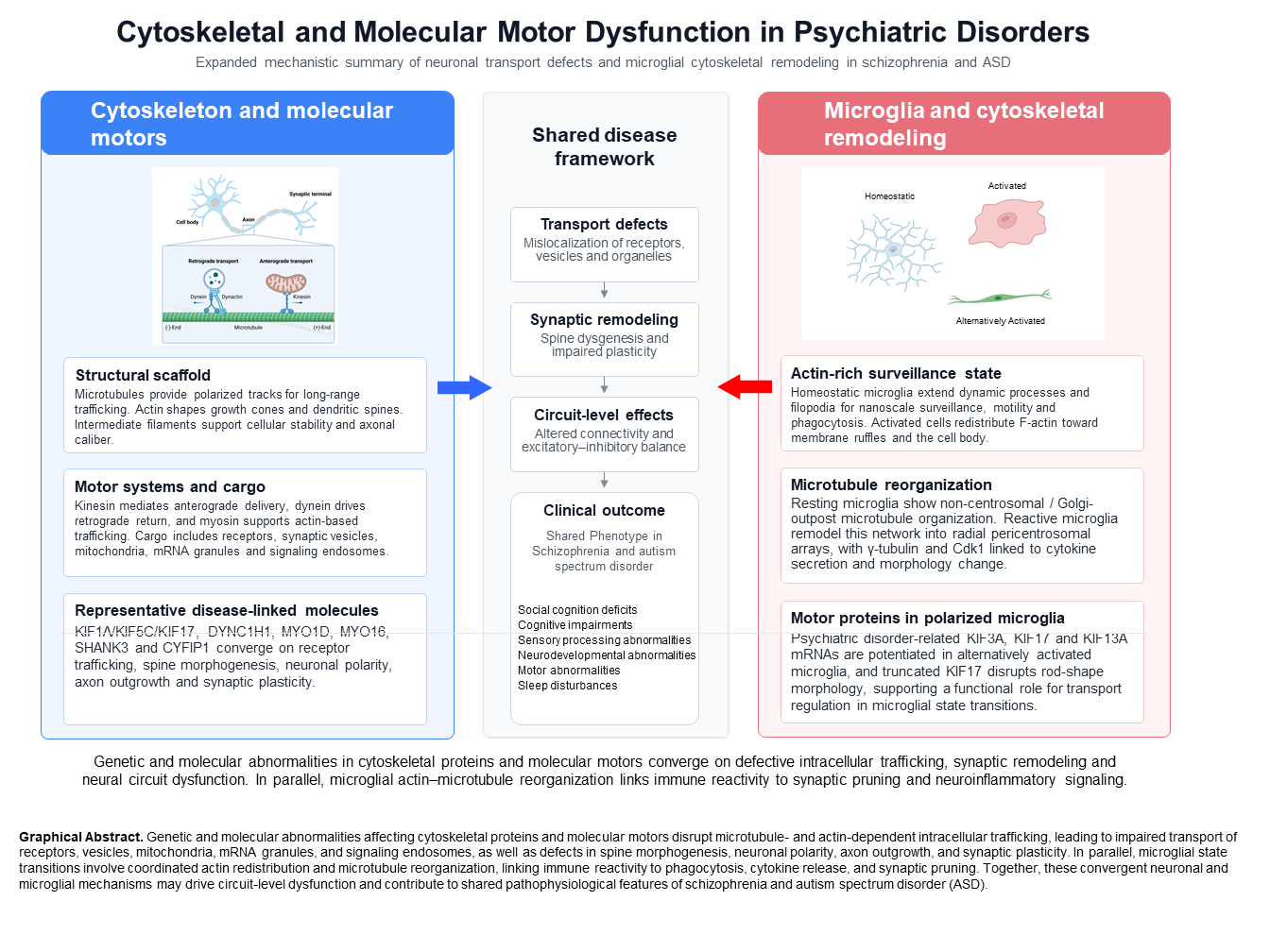
The neuronal cytoskeleton provides both structural support and dynamic scaffolding for morphogenesis and plasticity. Microtubules support axonal/dendritic growth and serve as tracks for long-range transport; actin filaments shape growth cones; dendritic spines mediate activity-dependent structural remodeling; and intermediate filaments contribute to cellular stability and axonal caliber. Perturbations in these systems can therefore impact neuronal polarity, arborization, and synaptic architecture—cellular phenotypes repeatedly implicated in schizophrenia and ASD.
Molecular motors convert ATP hydrolysis into directed movement along cytoskeletal filaments. Kinesins primarily drive anterograde transport on microtubules, dynein mediates retrograde transport, and myosins regulate actin-based trafficking and spine dynamics. By controlling the spatiotemporal delivery of receptors, vesicles, organelles, and mRNAs, these motors directly influence synaptogenesis, synaptic plasticity, and circuit maturation, providing mechanistic links between genetic/molecular perturbations and disease-relevant cellular phenotypes.
Although schizophrenia and autism spectrum disorder (ASD) differ in clinical manifestations, both disorders are increasingly conceptualized as neurodevelopmental conditions involving disruptions in neuronal circuit formation. Cytoskeletal organization and intracellular transport systems are essential for neuronal migration, axon specification, dendritic arborization, and synaptic connectivity. Therefore, abnormalities in cytoskeletal and molecular motor proteins may represent a shared cellular mechanism contributing to circuit-level dysfunction in both disorders.
Molecular motors are protein complexes that utilize the energy derived from ATP hydrolysis to transport cargos along cytoskeletal filaments. In neurons, three major classes of molecular motors—kinesins, dyneins, and myosins—play indispensable roles in maintaining cellular function and supporting neurodevelopment.
Increasing genetic and molecular evidence indicates that specific cytoskeletal and molecular motor proteins are directly linked to psychiatric disorders. For example, alterations in kinesin family members (such as KIF1A, KIF5C, and KIF17), dynein components (DYNC1H1), and actin-associated motors (MYO16 and MYO1D) have been reported in individuals with schizophrenia or ASD. These proteins regulate intracellular trafficking of synaptic vesicles, neurotransmitter receptors, and signaling complexes, suggesting that disruption of intracellular transport may contribute to synaptic dysfunction and abnormal circuit development.
The kinesin superfamily proteins (KIFs) mediate predominantly anterograde transport toward the plus-end of microtubules, delivering cargos from the soma to distal axons and dendrites [20]. Comprehensive studies by Hirokawa and colleagues have identified 45 KIF genes in humans and mice, each responsible for transporting distinct sets of cargos [11]. Key functions of neuronal KIFs include the anterograde transport of synaptic vesicles, neurotransmitter receptors, mitochondria, and mRNA-containing granules, as well as the delivery of neurotrophic factors [21,22,23]. Among them, KIF1A, KIF5, and KIF17 have been shown to be particularly important for neuronal function [10].
Beyond KIF1A and KIF17, the kinesin-1 family (KIF5A/B/C) is a major driver of long-range anterograde transport in neurons, supporting the delivery of diverse cargos—including mitochondria, membranous organelles, and receptor-containing vesicles—to distal axons and dendrites. Disruption of KIF5-mediated transport is therefore expected to affect both neuronal energetics and the spatiotemporal availability of synaptic components, two prerequisites for normal circuit development. In the context of ASD, mutations in KIF5C have been reported and are thought to impair neuronal polarity and axonal outgrowth, processes that depend on correctly oriented microtubule arrays and a continuous supply of structural and signaling cargos. Mechanistically, compromised KIF5C function can lead to delayed or misdirected axon specification, reduced axon elongation, and altered cargo distribution between axons and dendrites, ultimately increasing the risk of miswired connectivity. Importantly, this transport-based framework provides a direct link between a molecular motor defect and higher-order phenotypes, because even modest reductions in processive anterograde transport can accumulate over long axons and during sensitive developmental windows, resulting in persistent network-level alterations. Additional KIF5-family and kinesin candidates are summarized in Table 1.
Dynein, in contrast, mediates retrograde transport toward the minus-end of microtubules, moving cargos from axonal terminals back toward the cell body [24]. Its major roles include the retrograde trafficking of synaptic vesicles, neurotransmitter receptors, and various organelles; the transport of neurotrophic factor signaling endosomes; and the redistribution of cellular components during axon regeneration [25,26,27,28].
Myosins function primarily along actin filaments [29]. Numerous myosin family members are expressed in neurons, where they regulate the transport of diverse cargos and modulate actin architecture to influence synaptic activity. Myosins contribute to dendritic spine morphogenesis, local mobility of synaptic vesicles, growth cone motility, and the regulation of synaptic plasticity [30,31,32,33]. Myosin II, V, and VI, in particular, have been shown to play essential roles in neuronal physiology.
Together, these molecular motor systems coordinate the highly dynamic intracellular transport processes required for neuronal development, synaptic function, and circuit maintenance. Dysfunction in any of these motor proteins can profoundly disrupt neuronal connectivity and is increasingly recognized as a key contributor to neurodevelopmental and psychiatric disorders.
2. Cytoskeletal and Molecular Motor Abnormalities in Schizophrenia
Table 1 summarizes representative cytoskeletal and molecular motor genes implicated in schizophrenia and ASD, together with reported variants, molecular functions, and proposed cellular consequences.
Schizophrenia is a psychiatric disorder characterized by hallucinations, delusions, cognitive impairments, and disturbances in emotion and behavior. Accumulating evidence indicates that abnormalities in cytoskeletal components and molecular motor proteins are present in the brains of individuals with schizophrenia and are closely linked to disease pathophysiology.
Among microtubule-related abnormalities, reduced expression of the microtubule-associated protein MAP2 has been reported in the prefrontal cortex and hippocampus of patients with schizophrenia [34,35]. Because MAP2 plays a critical role in the formation and maintenance of dendrites, decreased expression is thought to contribute to dendritic morphological abnormalities and impaired dendritic function. In addition, DISC1 (Disrupted in Schizophrenia 1), a susceptibility gene for schizophrenia, regulates microtubule stabilization and intracellular transport. Dysfunction of DISC1 is believed to impair neurodevelopment and synaptic plasticity [36,37].
Abnormalities in actin-related proteins have also been identified in schizophrenia. Postmortem studies have reported altered expression of components of the WAVE complex—such as CYFIP1, NCKAP1, and WASF1 [38]—which regulates actin polymerization and contributes to dendritic spine formation. Disruption of this complex is expected to lead to synaptic dysfunction [39]. Increased expression of calponin-3, an actin-binding protein, has also been observed in the prefrontal cortex of patients [40]. Elevated levels of calponin-3 may contribute to aberrant stabilization of the actin cytoskeleton [41].
Regarding intermediate filaments, increased expression of GFAP (glial fibrillary acidic protein) has been detected in the prefrontal cortex and hippocampus of individuals with schizophrenia [42]. As a major intermediate filament protein in astrocytes, elevated GFAP expression likely reflects neuroinflammatory or neuroprotective responses [43].
Genetic studies have suggested that variants affecting the KIF1A pathway may influence synaptic vesicle transport mechanisms relevant to psychiatric disorders. Because KIF1A mediates the anterograde transport of synaptic vesicle precursors, its dysfunction may reduce synaptic transmission efficiency and alter neuronal connectivity.
Dynein-related abnormalities include rare variants in the DYNC1H1 (Dynein Cytoplasmic 1 Heavy Chain 1) gene identified in individuals with schizophrenia [49]. Because DYNC1H1 is a major component of the dynein complex, its dysfunction is expected to impair retrograde axonal transport [50]. Variants in MYO16 (Myosin XVI), a myosin family protein involved in neuronal migration and dendrite formation, have also been reported in schizophrenia [51]. Disruption of MYO16 function may impair neural circuit formation.
Collectively, these cytoskeletal and molecular motor abnormalities contribute to a range of pathological processes observed in schizophrenia, including impaired synapse formation and plasticity, abnormal receptor trafficking, disrupted neurodevelopment, dysfunctional neural circuits, and cognitive deficits. These findings underscore the crucial role of intracellular transport and cytoskeletal regulation in the neurobiology of schizophrenia.
3. Cytoskeletal and Molecular Motor Abnormalities in Autism Spectrum Disorder
Autism spectrum disorder (ASD) is a neurodevelopmental condition characterized by impairments in social communication and interaction, together with restricted, repetitive patterns of behavior and interests. In recent years, multiple abnormalities in cytoskeletal components and molecular motor proteins have been reported in the brains of individuals with ASD.
Myosin ID (MYO1D) has been identified as a risk gene for ASD [52]. We previously showed that EGFP-tagged Myosin Id, when expressed in cultured neurons, accumulates in dendritic spines [53]. Furthermore, this localization critically depends on the TH1 (tail homology 1) domain: deletion of the TH1 domain disrupts MYO1D targeting to dendrites and markedly reduces its accumulation in dendritic spines. These findings strongly suggest that MYO1D interacts with actin filaments within dendritic spines and contributes to the regulation of excitatory synaptic transmission, and that disruption of this function may underlie aspects of ASD pathophysiology. The TH1 domain of MYO1D has also been reported to interact with the C-terminus of aspartoacylase, an N-acetylaspartate (NAA)–acylating enzyme [54]. NAA is present at high concentrations in the mammalian brain, and reduced NAA levels have been observed in children with ASD compared with typically developing controls [55]. These observations further support a functional link between MYO1D and ASD.
Myosin IXb (MYO9B) is known to regulate RhoA activity and to control dendritic morphology in cortical neurons [56]. Variants in MYO9B have been suggested to influence ASD risk [5]. In addition, cohort studies have reported an association between Myosin XVI and ASD [57]. Myosin XVI contributes to neuronal migration and dendritic development, and its dysfunction is thought to lead to defects in neural circuit formation.
Mutations in SHANK3 (SH3 and multiple ankyrin repeat domains 3) are recognized as a major genetic cause of ASD [58]. SHANK3 plays a key role in organizing the actin cytoskeleton in the postsynaptic density; its disruption leads to defective dendritic spine formation and synaptic dysfunction [59]. Duplications and deletions of CYFIP1 (Cytoplasmic FMR1-interacting protein 1) have also been identified as ASD risk factors [60]. CYFIP1 is a component of the WAVE complex and regulates actin polymerization; its dysregulation is believed to cause dendritic spine abnormalities [61].
Rare variants in TUBG1 (γ-tubulin) have been reported in individuals with ASD [62]. γ-Tubulin functions as a nucleation factor for microtubule assembly, and its disruption is expected to affect neuronal polarity and migration [63]. Mutations in ASPM (abnormal spindle-like microcephaly-associated protein) have likewise been implicated as risk factors for ASD [64]. ASPM plays an important role in the division and differentiation of neural stem cells, and its dysfunction is thought to lead to abnormal cortical development [65].
Rare variants in KIF1A have been identified in patients with ASD [66]. KIF1A is essential for the anterograde transport of synaptic vesicle precursors, and its dysfunction is predicted to reduce the efficiency of synaptic transmission [67]. Mutations in KIF5C have also been associated with ASD. KIF5C contributes to neuronal polarity and axonal elongation, and its disruption likely impairs neural circuit formation [68].
Taken together, these cytoskeletal and molecular motor abnormalities appear to contribute to ASD pathophysiology through several interrelated mechanisms. First, disturbances in actin-regulating proteins such as SHANK3 and CYFIP1 result in defective dendritic spine formation and synaptic dysfunction, thereby disrupting the normal development and function of neural circuits. Second, abnormalities in kinesins, such as KIF5C, and dynein-related proteins, such as DYNC1H1, are expected to impair axonal transport and neuronal migration, leading to structural and functional abnormalities in neural networks. Third, defects in proteins such as MYO9B that regulate neuronal migration and axon guidance may interfere with the precise positioning of neurons and the accurate extension of axons, thereby contributing to macroscopic brain abnormalities. Fourth, dysfunction of ASPM is predicted to alter the division and differentiation of neural stem cells and to disrupt the proper formation of cortical lamination. Fifth, impaired synaptic vesicle transport due to abnormalities in KIF1A and KIF5C may reduce the efficiency of neurotransmission and compromise information-processing capacity. The convergence of genetic, molecular, and cellular findings suggests that cytoskeletal and molecular motor abnormalities may represent a mechanistic bridge between diverse ASD risk genes and the synaptic and connectivity phenotypes frequently observed in this disorder.
These findings together suggest that ASD is not caused by a single molecular defect but rather arises from a complex interplay among multiple abnormalities in cytoskeletal and molecular motor–related proteins, which collectively perturb neurodevelopment, synaptic function, and network-level information processing.
4. Microglial Cytoskeleton, Molecular Motors, and Psychiatric Disorders
Recent studies suggest that abnormalities in cytoskeletal organization and molecular motor function in glial cells, as well as in neurons, may contribute to the pathophysiology of psychiatric disorders. In particular, cytoskeletal remodeling during microglia activation—the resident immune cells of the central nervous system—has emerged as a critical factor in disease mechanisms.
Emerging evidence indicates that microglial activation can influence synaptic pruning, neuroinflammatory signaling, and circuit maturation, processes increasingly implicated in both schizophrenia and ASD. Therefore, cytoskeletal remodeling in microglia may represent an additional mechanistic layer linking intracellular structural dynamics to the pathology of psychiatric disease.
Microglia become activated in response to brain inflammation or injury and undergo marked morphological changes. The studies by Rosito et al. (2023) and Adrian et al. (2023) provided a detailed description of the dramatic reorganization of the microtubule cytoskeleton that accompanies microglial activation [69,70]. In the resting state, microglia exhibit an acentrosomal microtubule array that is distributed throughout the cell. Upon activation, however, microtubules are reorganized into a radial array emanating from the centrosome. This reorganization is associated with a morphological transition from a ramified shape to a more rounded, amoeboid (activated) morphology.
In activated microglia, microtubule polymerization is enhanced, and microtubule stability is increased. These changes are regulated by activation of the microtubule-associated protein MAP4 and downregulation of Stathmin 1 (STMN1), a key microtubule-destabilizing protein. During activation, the centrosome also matures and accumulates microtubule-nucleating factors such as γ-tubulin, thereby establishing the centrosome as the principal microtubule-organizing center. At the same time, Golgi-derived, acentrosomal microtubule nucleation is reduced, a process controlled in part by decreased expression of Golgi-associated proteins such as AKAP9.
Cyclin-dependent kinase 1 (Cdk1) has been identified as an upstream regulator of this cytoskeletal remodeling during microglial activation. Activation of Cdk1 promotes microtubule stabilization through phosphorylation-dependent regulation of MAP4, enhances microtubule polymerization via phosphorylation and subsequent degradation of STMN1, and drives centrosome maturation and its function as a microtubule-organizing center. Collectively, these changes are thought to facilitate efficient secretion of proinflammatory cytokines by activated microglia.
Microglial activation and the accompanying cytoskeletal remodeling are implicated in the pathophysiology of a range of neuropsychiatric and neurodegenerative disorders. In Alzheimer’s disease, microglial activation in response to amyloid-β and pathological tau accumulation contributes to disease progression, and cytoskeletal remodeling may regulate phagocytosis of these abnormal proteins and the release of inflammatory mediators. In postmortem brains from individuals with schizophrenia and ASD, microglial activation has also been reported, raising the possibility that aberrant cytoskeletal reorganization in microglia influences synaptic pruning and the development and maintenance of neural circuits, thereby contributing to symptom expression.
In psychiatric disorders such as schizophrenia and ASD, dysregulated synaptic pruning and altered neuroimmune signaling have been proposed as contributors to abnormal circuit development. Because microglial morphology and function depend strongly on cytoskeletal remodeling and molecular motor–dependent trafficking, alterations in these systems may indirectly influence neuronal connectivity through microglia-mediated mechanisms.
Targeting microglial cytoskeletal remodeling is a promising strategy for developing novel treatments for psychiatric disorders. Adrian et al. (2023) demonstrated that pharmacological inhibition of Cdk1 suppresses microglial activation and reduces the secretion of inflammatory cytokines, suggesting potential therapeutic applications in conditions characterized by excessive neuroinflammation. In addition, drugs that modulate microtubule polymerization and stability may help regulate microglial activation and attenuate pathological neuroinflammation. Manipulating the balance between centrosomal and Golgi-derived microtubule organization could provide additional means to control microglial morphology and function, thereby slowing or preventing disease progression.
5. Conclusions and Future Directions
Accumulating evidence indicates that abnormalities in cytoskeletal organization and molecular motor function—particularly within the kinesin (KIF) and myosin families—are deeply involved in the pathophysiology of psychiatric disorders such as schizophrenia and ASD. These abnormalities affect multiple aspects of brain function, including neurodevelopment, synapse formation, neurotransmission, and neuronal plasticity, and are thought to contribute to the biological basis of the characteristic symptoms and cognitive impairments observed in these conditions.
Recent findings on the localization and function of Myosin Id in dendritic spines provide important clues for understanding the disease mechanisms of ASD. In our laboratory, we plan to further dissect the roles of Myosin Id and to clarify in greater detail how its dysfunction contributes to ASD onset and progression. In parallel, elucidating the mechanisms by which KIFs transport neurotransmitter receptors and synaptic vesicles has provided new perspectives on the pathogenesis of psychiatric disorders [71]. A more precise understanding of the functions of individual KIFs and their links to specific psychiatric phenotypes is expected to facilitate the development of novel, mechanism-based therapeutic strategies.
Recent large-scale human genetics studies published in the 2020s further reinforce the relevance of synaptic and neurodevelopmental pathways that intersect with cytoskeletal regulation and intracellular transport. For schizophrenia, a large two-stage GWAS reported associations at 287 genomic loci and highlighted convergence on synaptic organization and neuronal differentiation [72]. For ASD, large-scale exome sequencing has identified more than 100 risk genes and emphasized both developmental and functional mechanisms in excitatory and inhibitory neuronal populations [73]. In parallel, recent work has expanded the microglia-focused literature in psychiatric and neurodevelopmental disorders, including evidence linking microglial activation to interneuron metabolic disruptions relevant to schizophrenia [74], reviews integrating microglia–neuron interactions in schizophrenia [75], and functional genomics studies in human iPSC-derived microglia implicating ASD risk genes in endocytosis and synaptic pruning [76].
In addition, growing evidence suggests that abnormalities in the cytoskeleton and molecular motors of glial cells—particularly microglia—also play a crucial role in psychiatric disease. Cytoskeletal remodeling accompanying microglial activation is closely associated with a range of functional changes, including the production and release of inflammatory mediators and alterations in phagocytic capacity [77]. These insights are highly informative for identifying new therapeutic targets and deepening our understanding of disease mechanisms. Going forward, more comprehensive research approaches that explicitly consider neuron–glia interactions will be essential.
Key future directions include:
- Clarifying the relationships between glia-specific cytoskeletal and molecular motor gene variants and psychiatric disorders;
- Developing novel therapeutic strategies that directly target glial cytoskeletal and molecular motor pathways;
- Elucidating the roles of cytoskeletal and motor systems in neuron–glia interactions; and
- Defining how morphological changes in glial cells relate to functional alterations in the context of psychiatric illnesses.
Through these lines of investigation, we anticipate further advances in our understanding of the pathophysiological mechanisms underlying psychiatric disorders, which in turn should contribute to the development of new therapeutic and preventive strategies. Approaches that specifically target microglial cytoskeletal and molecular motor dynamics may exert their effects through mechanisms distinct from conventional neurotransmitter-based treatments and thus hold promise as alternatives for patients with treatment-resistant conditions. Research on cytoskeletal and molecular motor systems lies at the interface of neuroscience and psychiatry and is expected to become increasingly important in the coming years. Ultimately, progress in this field may help improve the quality of life of individuals suffering from psychiatric disorders.
Acknowledgments
The work conducted in our laboratory has been supported by Grants-in-Aid for Scientific Research (KAKENHI; 16H06276, 19K08065, 22K07611, 19H05201), as well as by the Advanced Pharmaceutical Research Promotion Foundation, the Naito Memorial Foundation for the Promotion of Science, the Takeda Science Foundation, the Kawano Pediatric Medical Research Foundation, the Daido Life Welfare Foundation, the Life Science Foundation of Japan, the Nakatomi Health Science Foundation, the Pharmacological Research Foundation, the Pharmaceutical Research Encouragement Foundation, the Mishima Kaiun Memorial Foundation, the Collaborative Research Program of the National Institute for Basic Biology, and the Open Facility Initiative of the University of Tsukuba. We would also like to express our sincere gratitude to all members of the Laboratory of Anatomy and Neuroscience for their valuable advice and support during the preparation of this review.
Institutional Review Board Statement
Not applicable.
Informed Consent Statement
Not applicable.
Data Availability Statement
No new data were created or analyzed in this study.
Acknowledgments
The work conducted in our laboratory has been supported by Grants-in-Aid for Scientific Research (KAKENHI; 16H06276, 19K08065, 22K07611, 19H05201), as well as by the Advanced Pharmaceutical Research Promotion Foundation, the Naito Memorial Foundation for the Promotion of Science, the Takeda Science Foundation, the Kawano Pediatric Medical Research Foundation, the Daido Life Welfare Foundation, the Life Science Foundation of Japan, the Nakatomi Health Science Foundation, the Pharmacological Research Foundation, the Pharmaceutical Research Encouragement Foundation, the Mishima Kaiun Memorial Foundation, the Collaborative Research Program of the National Institute for Basic Biology, and the Open Facility Initiative of the University of Tsukuba. We would also like to express our sincere gratitude to all members of the Laboratory of Anatomy and Neuroscience for their valuable advice and support during the preparation of this review.
Conflicts of Interest
The authors declare no conflicts of interest.
References
- Fletcher, DA; Mullins, RD. Cell mechanics and the cytoskeleton. Nature 2010, 463(7280), 485–92. [Google Scholar] [CrossRef]
- Hirokawa, N; Noda, Y; Tanaka, Y; Niwa, S. Kinesin superfamily motor proteins and intracellular transport. Nat Rev Mol Cell Biol. 2009, 10(10), 682–96. [Google Scholar] [CrossRef]
- Franker, MAM; Hoogenraad, CC. Microtubule-based transport - basic mechanisms, traffic rules and role in neurological pathogenesis. J Cell Sci. 2013, 126 Pt 11, 2319–29. [Google Scholar] [CrossRef] [PubMed]
- Marchisella, F. Abnormalities of the microtubule system and impaired neurite outgrowth in psychiatric disorders. In Neural Plast; 2016. [Google Scholar]
- De Rubeis, S; He, X; Goldberg, AP; Poultney, CS; Samocha, K; et al.; The DDD Study Synaptic, transcriptional and chromatin genes disrupted in autism. Nature 2014, 515(7526), 209–15. [Google Scholar] [CrossRef]
- Moghaddam, B; Javitt, D. From revolution to evolution: the glutamate hypothesis of schizophrenia and its implication for treatment. Neuropsychopharmacology 2012, 37(1), 4–15. [Google Scholar] [CrossRef] [PubMed]
- Padmanabhan, A; Lynch, CJ; Schaer, M; Menon, V. The default mode network in autism. Biol Psychiatry Cogn Neurosci Neuroimaging 2017, 2(6), 476–86. [Google Scholar] [CrossRef] [PubMed]
- Estes, ML; McAllister, AK. Maternal immune activation: Implications for neuropsychiatric disorders. Science 2016, 353(6301), 772–7. [Google Scholar] [CrossRef]
- Müller, N. Neuroinflammation in schizophrenia and autism spectrum disorders: Recent advances. In Prog Neuropsychopharmacol Biol Psychiatry; 2018. [Google Scholar]
- Hirokawa, N; Takemura, R. Molecular motors and mechanisms of directional transport in neurons. Nat Rev Neurosci. 2005, 6(3), 201–14. [Google Scholar] [CrossRef]
- Miki, H; Setou, M; Kaneshiro, K; Hirokawa, N. All kinesin superfamily protein, KIF, genes in mouse and human. Proc Natl Acad Sci U S A 2001, 98(13), 7004–11. [Google Scholar] [CrossRef]
- Conde, C; Cáceres, A. Microtubule assembly, organization and dynamics in axons and dendrites. Nat Rev Neurosci 2009, 10(5), 319–32. [Google Scholar] [CrossRef]
- Takei, Y; Teng, J; Harada, A; Hirokawa, N. Defects in axonal elongation and neuronal migration in mice with disrupted tau and map1b genes. J Cell Biol. 2000, 150(5), 989–1000. [Google Scholar] [CrossRef]
- Kapitein, LC; Hoogenraad, CC. Building the neuronal microtubule cytoskeleton. Neuron 2015, 87(3), 492–506. [Google Scholar] [CrossRef] [PubMed]
- Dent, EW; Gupton, SL; Gertler, FB. The growth cone cytoskeleton in axon outgrowth and guidance. Cold Spring Harb Perspect Biol. 2011, 3(3), a001800–a001800. [Google Scholar] [CrossRef]
- Cingolani, LA; Goda, Y. Actin in action: the interplay between the actin cytoskeleton and synaptic efficacy. Nat Rev Neurosci 2008, 9(5), 344–56. [Google Scholar] [CrossRef] [PubMed]
- Okamoto, K-I; Nagai, T; Miyawaki, A; Hayashi, Y. Rapid and persistent modulation of actin dynamics regulates postsynaptic reorganization underlying bidirectional plasticity. Nat Neurosci. 2004, 7(10), 1104–12. [Google Scholar] [CrossRef] [PubMed]
- Perrot, R; Eyer, J. Neuronal intermediate filaments and neurodegenerative disorders. Brain Res Bull. 2009, 80(4–5), 282–95. [Google Scholar] [CrossRef]
- Lendahl, U; Zimmerman, LB; McKay, RD. CNS stem cells express a new class of intermediate filament protein. Cell 1990, 60(4), 585–95. [Google Scholar] [CrossRef]
- Hirokawa, N; Niwa, S; Tanaka, Y. Molecular motors in neurons: transport mechanisms and roles in brain function, development, and disease. Neuron 2010, 68(4), 610–38. [Google Scholar] [CrossRef]
- Mandal, A; Drerup, CM. Axonal transport and mitochondrial function in neurons. Front Cell Neurosci 2019, 13, 373. [Google Scholar] [CrossRef]
- Kanai, Y; Dohmae, N; Hirokawa, N. Kinesin transports RNA: isolation and characterization of an RNA-transporting granule. Neuron 2004, 43(4), 513–25. [Google Scholar] [CrossRef]
- Cosker, KE; Segal, RA. Neuronal signaling through endocytosis. Cold Spring Harb Perspect Biol. 2014, 6(2), a020669–a020669. [Google Scholar] [CrossRef]
- Roberts, AJ; Kon, T; Knight, PJ; Sutoh, K; Burgess, SA. Functions and mechanics of dynein motor proteins. Nat Rev Mol Cell Biol. 2013, 14(11), 713–26. [Google Scholar] [CrossRef]
- Caviston, JP; Holzbaur, ELF. Microtubule motors at the intersection of trafficking and transport. Trends Cell Biol. 2006, 16(10), 530–7. [Google Scholar] [CrossRef]
- Vallee, RB; Tsai, J-W. The cellular roles of the lissencephaly gene LIS1, and what they tell us about brain development. Genes Dev. 2006, 20(11), 1384–93. [Google Scholar] [CrossRef]
- Heerssen, HM; Pazyra, MF; Segal, RA. Dynein motors transport activated Trks to promote survival of target-dependent neurons. Nat Neurosci. 2004, 7(6), 596–604. [Google Scholar] [CrossRef]
- Tojima, T; Itofusa, R; Kamiguchi, H. Steering neuronal growth cones by shifting the imbalance between exocytosis and endocytosis. J Neurosci. 2014, 34(21), 7165–78. [Google Scholar] [CrossRef]
- Hartman, MA; Spudich, JA. The myosin superfamily at a glance. J Cell Sci. 2012, 125 Pt 7, 1627–32. [Google Scholar] [CrossRef] [PubMed]
- Rex, CS; Gavin, CF; Rubio, MD; Kramar, EA; Chen, LY; Jia, Y; et al. Myosin IIb regulates actin dynamics during synaptic plasticity and memory formation. Neuron 2010, 67(4), 603–17. [Google Scholar] [CrossRef] [PubMed]
- Kneussel, M; Wagner, W. Myosin motors at neuronal synapses: drivers of membrane transport and actin dynamics. Nat Rev Neurosci. 2013, 14(4), 233–47. [Google Scholar] [CrossRef]
- Wang, Z; Edwards, JG; Riley, N; Provance, DW, Jr.; Karcher, R; Li, X-D; et al. Myosin Vb mobilizes recycling endosomes and AMPA receptors for postsynaptic plasticity. Cell 2008, 135(3), 535–48. [Google Scholar] [CrossRef] [PubMed]
- Huber, KM; Gallagher, SM; Warren, ST; Bear, MF. Altered synaptic plasticity in a mouse model of fragile X mental retardation. Proc Natl Acad Sci U S A 2002, 99(11), 7746–50. [Google Scholar] [CrossRef]
- Jones, LB; Johnson, N; Byne, W. Alterations in MAP2 immunocytochemistry in areas 9 and 32 of schizophrenic prefrontal cortex. Psychiatry Res Neuroimaging 2002, 114(3), 137–48. [Google Scholar] [CrossRef] [PubMed]
- DeGiosio, R; Kelly, RM; DeDionisio, AM; Newman, JT; Fish, KN; Sampson, AR; et al. MAP2 immunoreactivity deficit is conserved across the cerebral cortex within individuals with schizophrenia. NPJ Schizophr 2019, 5(1), 13. [Google Scholar] [CrossRef] [PubMed]
- Brandon, NJ; Sawa, A. Linking neurodevelopmental and synaptic theories of mental illness through DISC1. Nat Rev Neurosci. 2011, 12(12), 707–22. [Google Scholar] [CrossRef]
- Lipska, BK; Peters, T; Hyde, TM; Halim, N; Horowitz, C; Mitkus, S; et al. Expression of DISC1 binding partners is reduced in schizophrenia and associated with DISC1 SNPs. Hum Mol Genet. 2006, 15(8), 1245–58. [Google Scholar] [CrossRef]
- Kähler, AK; Djurovic, S; Rimol, LM; Brown, AA; Athanasiu, L; Jönsson, EG; et al. Candidate gene analysis of the human natural killer-1 carbohydrate pathway and perineuronal nets in schizophrenia: B3GAT2 is associated with disease risk and cortical surface area. Biol Psychiatry 2011, 69(1), 90–6. [Google Scholar] [CrossRef]
- Soderling, SH; Guire, ES; Kaech, S; White, J; Zhang, F; Schutz, K; et al. A WAVE-1 and WRP signaling complex regulates spine density, synaptic plasticity, and memory. J Neurosci. 2007, 27(2), 355–65. [Google Scholar] [CrossRef]
- Föcking, M; Dicker, P; English, JA; Schubert, KO; Dunn, MJ; Cotter, DR. Common proteomic changes in the hippocampus in schizophrenia and bipolar disorder and particular evidence for involvement of cornu ammonis regions 2 and 3. Arch Gen Psychiatry 2011, 68(5), 477–88. [Google Scholar] [CrossRef] [PubMed]
- Rami, G; Caillard, O; Medina, I; Pellegrino, C; Fattoum, A; Ben-Ari, Y; et al. Change in the shape and density of dendritic spines caused by overexpression of acidic calponin in cultured hippocampal neurons. Hippocampus 2006, 16(2), 183–97. [Google Scholar] [CrossRef]
- Toro, CT; Hallak, JEC; Dunham, JS; Deakin, JFW. Glial fibrillary acidic protein and glutamine synthetase in subregions of prefrontal cortex in schizophrenia and mood disorder. Neurosci Lett. 2006, 404(3), 276–81. [Google Scholar] [CrossRef]
- Sofroniew, MV; Vinters, HV. Astrocytes: biology and pathology. Acta Neuropathol 2010, 119(1), 7–35. [Google Scholar] [CrossRef]
- Ratta-Apha, W; Mouri, K; Boku, S; Ishiguro, H; Okazaki, S; Otsuka, I; et al. A decrease in protein level and a missense polymorphism of KIF17 are associated with schizophrenia. Psychiatry Res. 2015, 230(2), 424–9. [Google Scholar] [CrossRef]
- Yin, X; Feng, X; Takei, Y; Hirokawa, N. Regulation of NMDA receptor transport: a KIF17-cargo binding/releasing underlies synaptic plasticity and memory in vivo. J Neurosci. 2012, 32(16), 5486–99. [Google Scholar] [CrossRef]
- Rivero, O; Sich, S; Popp, S; Schmitt, A; Franke, B; Lesch, K-P. Impact of the ADHD-susceptibility gene CDH13 on development and function of brain networks. Eur Neuropsychopharmacol 2013, 23(6), 492–507. [Google Scholar]
- Okada, Y; Yamazaki, H; Sekine-Aizawa, Y; Hirokawa, N. The neuron-specific kinesin superfamily protein KIF1A is a unique monomeric motor for anterograde axonal transport of synaptic vesicle precursors. Cell. 1995, 81(5), 769–80. [Google Scholar] [CrossRef] [PubMed]
- Alsabban, AH; Morikawa, M; Tanaka, Y; Takei, Y; Hirokawa, N. Kinesin Kif3b mutation reduces NMDAR subunit NR2A trafficking and causes schizophrenia-like phenotypes in mice. EMBO J 2020, 39(1), e101090. [Google Scholar] [CrossRef] [PubMed]
- Vissers, LELM; de Ligt, J; Gilissen, C; Janssen, I; Steehouwer, M; de Vries, P; et al. A de novo paradigm for mental retardation. Nat Genet. 2010, 42(12), 1109–12. [Google Scholar] [CrossRef]
- Schiavo, G; Greensmith, L; Hafezparast, M; Fisher, EMC. Cytoplasmic dynein heavy chain: the servant of many masters. Trends Neurosci. 2013, 36(11), 641–51. [Google Scholar] [CrossRef]
- Yue, W; Yu, X; Zhang, D. Progress in genome-wide association studies of schizophrenia in Han Chinese populations. NPJ Schizophr 2017, 3(1), 24. [Google Scholar] [CrossRef] [PubMed]
- Stone, JL; Merriman, B; Cantor, RM; Geschwind, DH; Nelson, SF. High density SNP association study of a major autism linkage region on chromosome 17. Hum Mol Genet. 2007, 16(6), 704–15. [Google Scholar] [CrossRef]
- Koshida, R; Tome, S; Takei, Y. Myosin Id localizes in dendritic spines through the tail homology 1 domain. Exp Cell Res. 2018, 367(1), 65–72. [Google Scholar] [CrossRef]
- Benesh, AE; Fleming, JT; Chiang, C; Carter, BD; Tyska, MJ. Expression and localization of myosin-1d in the developing nervous system. Brain Res. 2012, 1440, 9–22. [Google Scholar] [CrossRef] [PubMed]
- Moffett, JR; Ross, B; Arun, P; Madhavarao, CN; Namboodiri, AMA. N-Acetylaspartate in the CNS: from neurodiagnostics to neurobiology. Prog Neurobiol 2007, 81(2), 89–131. [Google Scholar] [CrossRef]
- Long, H; Zhu, X; Yang, P; Gao, Q; Chen, Y; Ma, L. Myo9b and RICS modulate dendritic morphology of cortical neurons. Cereb Cortex 2013, 23(1), 71–9. [Google Scholar] [CrossRef]
- Wang, K; Zhang, H; Ma, D; Bucan, M; Glessner, JT; Abrahams, BS; et al. Common genetic variants on 5p14.1 associate with autism spectrum disorders. Nature 2009, 459(7246), 528–33. [Google Scholar] [CrossRef]
- Durand, CM; Betancur, C; Boeckers, TM; Bockmann, J; Chaste, P; Fauchereau, F; et al. Mutations in the gene encoding the synaptic scaffolding protein SHANK3 are associated with autism spectrum disorders. Nat Genet. 2007, 39(1), 25–7. [Google Scholar] [CrossRef] [PubMed]
- Naisbitt, S; Kim, E; Tu, JC; Xiao, B; Sala, C; Valtschanoff, J; et al. Shank, a novel family of postsynaptic density proteins that binds to the NMDA receptor/PSD-95/GKAP complex and cortactin. Neuron 1999, 23(3), 569–82. [Google Scholar] [CrossRef] [PubMed]
- Nishimura, Y; Martin, CL; Vazquez-Lopez, A; Spence, SJ; Alvarez-Retuerto, AI; Sigman, M; et al. Genome-wide expression profiling of lymphoblastoid cell lines distinguishes different forms of autism and reveals shared pathways. Hum Mol Genet. 2007, 16(14), 1682–98. [Google Scholar] [CrossRef]
- De Rubeis, S; Pasciuto, E; Li, KW; Fernández, E; Di Marino, D; Buzzi, A; et al. CYFIP1 coordinates mRNA translation and cytoskeleton remodeling to ensure proper dendritic spine formation. Neuron 2013, 79(6), 1169–82. [Google Scholar] [CrossRef]
- Poirier, K; Saillour, Y; Bahi-Buisson, N; Jaglin, XH; Fallet-Bianco, C; Nabbout, R; et al. Mutations in the neuronal ß-tubulin subunit TUBB3 result in malformation of cortical development and neuronal migration defects. Hum Mol Genet. 2010, 19(22), 4462–73. [Google Scholar] [CrossRef]
- Tovey, CA; Conduit, PT. Microtubule nucleation by γ-tubulin complexes and beyond. Essays Biochem. 2018, 62(6), 765–80. [Google Scholar] [CrossRef]
- Bond, J; Roberts, E; Mochida, GH; Hampshire, DJ; Scott, S; Askham, JM; et al. ASPM is a major determinant of cerebral cortical size. Nat Genet. 2002, 32(2), 316–20. [Google Scholar] [CrossRef]
- Fish, JL; Kosodo, Y; Enard, W; Pääbo, S; Huttner, WB. Aspm specifically maintains symmetric proliferative divisions of neuroepithelial cells. Proc Natl Acad Sci U S A 2006, 103(27), 10438–43. [Google Scholar] [CrossRef]
- Hamdan, FF; Gauthier, J; Araki, Y; Lin, D-T; Yoshizawa, Y; Higashi, K; et al. Excess of DE Novo deleterious mutations in genes associated with glutamatergic systems in nonsyndromic intellectual disability. Am J Hum Genet. 2011, 88(4), 516. [Google Scholar] [CrossRef]
- Yonekawa, Y; Harada, A; Okada, Y; Funakoshi, T; Kanai, Y; Takei, Y; et al. Defect in synaptic vesicle precursor transport and neuronal cell death in KIF1A motor protein-deficient mice. J Cell Biol. 1998, 141(2), 431–41. [Google Scholar] [CrossRef] [PubMed]
- Poirier, K; Lebrun, N; Broix, L; Tian, G; Saillour, Y; Boscheron, C; et al. Mutations in TUBG1, DYNC1H1, KIF5C and KIF2A cause malformations of cortical development and microcephaly. Nat Genet. 2013, 45(6), 639–47. [Google Scholar] [CrossRef]
- Rosito, M; Sanchini, C; Gosti, G; Moreno, M; De Panfilis, S; Giubettini, M; et al. Microglia reactivity entails microtubule remodeling from acentrosomal to centrosomal arrays. Cell Rep. 2023, 42(2), 112104. [Google Scholar] [CrossRef]
- Adrian, M; Weber, M; Tsai, M-C; Glock, C; Kahn, OI; Phu, L; et al. Polarized microtubule remodeling transforms the morphology of reactive microglia and drives cytokine release. Nat Commun. 2023, 14(1), 6322. [Google Scholar] [CrossRef]
- Iwata, S; Morikawa, M; Takei, Y; Hirokawa, N. An activity-dependent local transport regulation via degradation and synthesis of KIF17 underlying cognitive flexibility. Sci Adv 2020, 6(51), eabc8355. [Google Scholar] [CrossRef] [PubMed]
- Trubetskoy, V; Pardiñas, AF; Qi, T; Panagiotaropoulou, G; Awasthi, S; Bigdeli, TB; et al. Mapping genomic loci implicates genes and synaptic biology in schizophrenia. Nature 2022, 604, 502–508. [Google Scholar] [CrossRef]
- Satterstrom, FK; Kosmicki, JA; Wang, J; Breen, MS; De Rubeis, S; An, JY; et al. Large-Scale Exome Sequencing Study Implicates Both Developmental and Functional Changes in the Neurobiology of Autism. Cell. 2020, 180(3), 568–584.e23. [Google Scholar] [CrossRef] [PubMed]
- Park, GH; Noh, H; Shao, Z; Ni, P; Qin, Y; Liu, D; et al. Activated microglia cause metabolic disruptions in developmental cortical interneurons that persist in interneurons from individuals with schizophrenia. Nature Neuroscience 2020, 23, 1352–1364. [Google Scholar] [CrossRef] [PubMed]
- Hartmann, S-M; Heider, J; Wüst, R; Fallgatter, AJ; Volkmer, H. Microglia-neuron interactions in schizophrenia. Frontiers in Cellular Neuroscience 2024, 18, 1345349. [Google Scholar] [CrossRef]
- Teter, OM; McQuade, A; Hagan, V; Liang, W; Dräger, NM; Sattler, SM; et al. CRISPRi-based screen of autism spectrum disorder risk genes in microglia uncovers roles of ADNP in microglia endocytosis and synaptic pruning. Molecular Psychiatry 2025, 30, 4176–4193. [Google Scholar] [CrossRef]
- Iwata, S; Hyugaji, M; Soga, Y; Morikawa, M; Sasaki, T; Takei, Y. Gene expression of psychiatric disorder-related kinesin superfamily proteins (Kifs) is potentiated in alternatively activated primary cultured microglia. BMC research notes 2025, 18(44), 1–8. [Google Scholar] [CrossRef] [PubMed]
Disclaimer/Publisher’s Note: The statements, opinions and data contained in all publications are solely those of the individual author(s) and contributor(s) and not of MDPI and/or the editor(s). MDPI and/or the editor(s) disclaim responsibility for any injury to people or property resulting from any ideas, methods, instructions or products referred to in the content. |
© 2026 by the authors. Licensee MDPI, Basel, Switzerland. This article is an open access article distributed under the terms and conditions of the Creative Commons Attribution (CC BY) license (http://creativecommons.org/licenses/by/4.0/).
Copyright: This open access article is published under a Creative Commons CC BY 4.0 license, which permit the free download, distribution, and reuse, provided that the author and preprint are cited in any reuse.


