Preprint
Article

This version is not peer-reviewed.

Comparative Analysis of Antioxidant Activity, Flavonoid and Phenolic Contents, and Secondary Metabolite Profiles in Broth and Mycelium Extracts of Coniochaeta dendrobiicola

Submitted:

19 February 2026

Posted:

27 February 2026

You are already at the latest version

Abstract

Background: Endophytic fungi are known for diverse bioactive compounds with immense potential for agriculture and medicinal applications. Coniochaeta dendrobiicola isolated from root of the Dedrobium longicornu was investigated for its antioxidant and metabolite composition. The present study compares the antioxidant properties, flavonoid and phenolic contents and metabolic profiles of broth and mycelium extracts. Methanolic broth and mycelium extracts were tested for antioxidant potential using DPPH, while total flavonoid and phenolic content were measured using a UV-VIS spectrophotometer High-resolution mass spectrometry (HRMS) revealed a markedly richer and more diverse metabolite profile in the broth extract compared to the mycelium fraction. The broth extract exhibited significantly higher antioxidant activity, flavonoid, and phenolic contents, correlating with the presence of diverse bioactive compounds, including indole derivatives, flavonoids, phenolic acids, quinoline derivatives, and antifungal metabolites. Notably, several indole-related and phenolic compounds detected predominantly in the broth are known for antioxidant, antimicrobial, and plant growth–promoting properties. These findings indicate that C. dendrobiicola actively secretes biologically relevant secondary metabolites into the extracellular medium, highlighting its potential for agricultural and pharmaceutical applications.

Keywords: 
;  ;  ;  ;  

1. Introduction

Endophytic fungi are endosymbionts that reside in the intercellular or intracellular host tissue (leaves, stems, flowers, seeds, and roots) without causing disease [1]. Endophytic fungi have a significant role in the host plant’s growth and development. It is known to produce plant hormones, secondary metabolite that promotes plant growth, and provides resistance to host plants against abiotic or biotic stress [2]. Beyond their ecological role of being a symbiotic partner and host plant growth promoter, endophytic fungi are increasingly recognized as a sustainable source of bioactive compounds for bioprospecting and drug discovery [3].
Almost all plants are associated with endophytic fungi; it is estimated that endophytic fungi is around 7% of the estimated fungi species found on the earth. Despite this, many endophytes remain unexplored. There is a continued need for characterization of endophytic fungi to understand their evolutionary history and application [4]. To date, approximately 69,000 fungi (endophytes, pathogens, saprobic) have been isolated, cultured, and described, indicating extensive work remains in this field [5].
One of the most compelling features of endophytic fungi is their ability to produce bioactive compounds like those found in their host plants. This includes a range of secondary metabolites, particularly alkaloids, flavonoids, terpenoids, and phenolic compounds [6,7]. Therefore, it becomes a sustainable approach to study endophytic fungi for bioactive compounds [7]. Some of the well-known medicinal compounds, such as Camptothecin, Diosgenin, Huperzine, Hypericin, Podophyllotoxin, Taxol, Resveratrol, Vincristine, and Vinblastine, have been reported from both medicinal plants and their associated endophytes [8]. The capacity of endophytes to produce such compounds depends on the host plant’s age, plant tissue part, geographical distribution, and abiotic factors [3]. Orchid endophytes are a group of fungi that are associated with orchid tissue, forming symbiosis that may contribute to the growth and development of orchids [9,10]. Orchid-associated endophytes, both mycorrhizal and non-mycorrhizal, form unique symbiotic relationships with orchid tissues. The orchid mycorrhizal fungi form a peloton in the cortex region of orchid roots that helps in plant growth and development, whereas non-mycorrhizal orchid endophytes are found in stems, leaves, flowers, as well as in intercellular root tissue [11].
Coniochaeta dendrobiicola is a non-mycorrhizal endophytic fungus isolated from the roots of Dendrobium longicornu. Prior studies have shown that this fungus promotes host growth through the production of auxins and other beneficial metabolites [9]. However, the metabolic profile and antioxidant potential of its intracellular (mycelium) and extracellular (broth) fractions remain largely uncharacterized.
In the present study, we have investigated the antioxidant activities and metabolic composition of the non-mycorrhizal orchid endophyte Coniochaeta dendrobiicola. We evaluated and compared the antioxidant activity, flavonoid activity, and total phenolic content of intracellular and extracellular extracts. High-resolution mass spectrometry (HRMS) was used to characterize the metabolic profile of both extracts. This study will address the underlying question: How do the intracellular (mycelial) and extracellular (broth) endophytic fungus Coniochaeta dendrobiicola differ in their antioxidant activities, total phenolic and flavonoid content, concerning their metabolic profile? We hypothesize that one of the extracts (intracellular or extracellular) will exhibit enrichment in phenolic and flavonoid, indole derivatives content that will correlate to higher antioxidant activity to reflect differential metabolic allocation between extracellular (broth) or intracellular (mycelium) of the fungus.

2. Materials and Methods

The fungal strain MCC 1811, Coniochaeta dendrobiicola was obtained from the NCCS-NCMR depository culture collection in 2022. The culture was revived on potato dextrose agar (PDA) plates at room temperature.

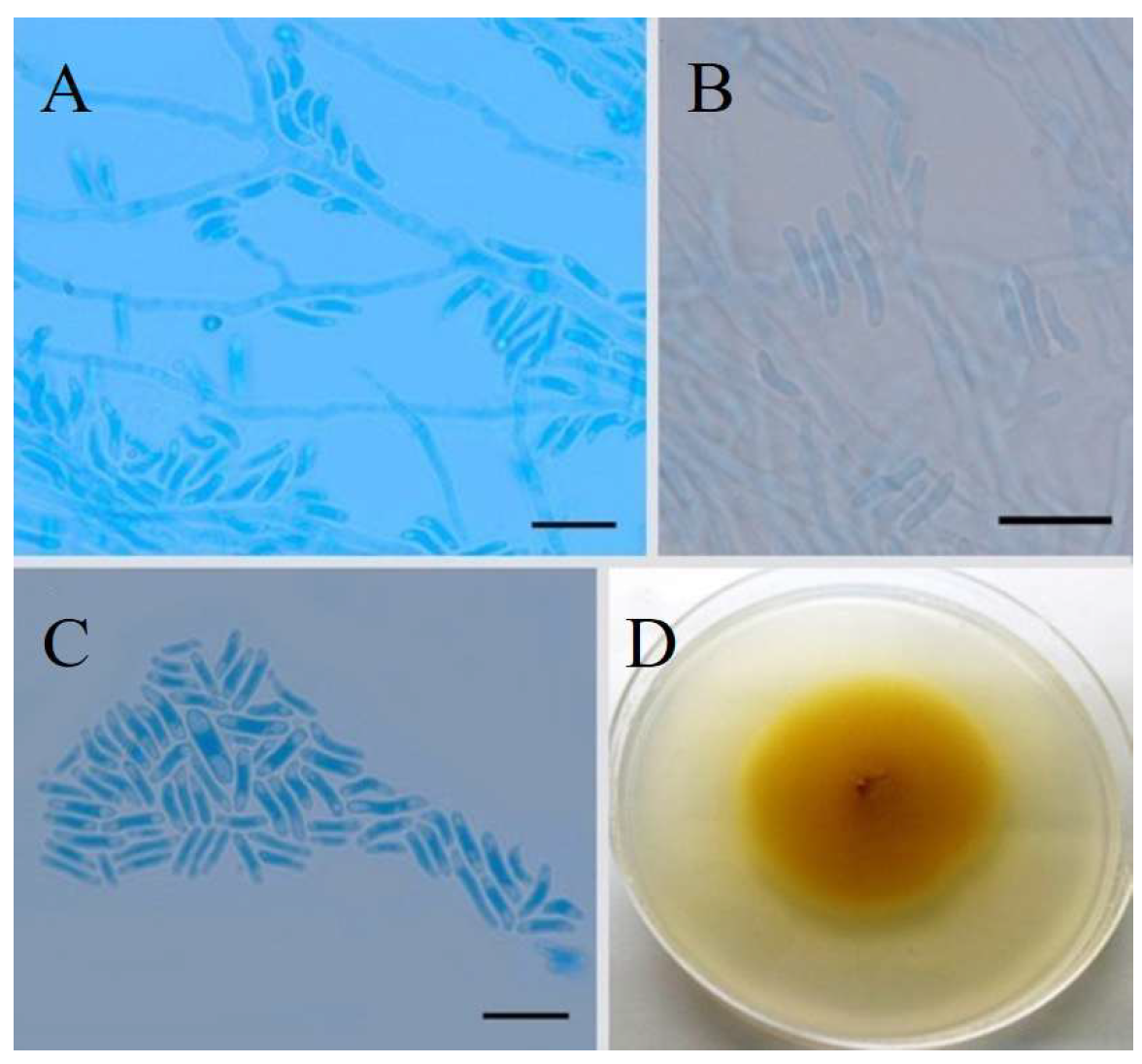
Fungal Extract Preparation

The fungal strain Coniochaeta dendrobiicola was cultured in three independent 500 mL Erlenmeyer flasks, each containing 250 mL of Potato Dextrose Broth (PDB). The flasks were inoculated and incubated at 28 °C on a rotary shaker at 120 rpm for 15 days [12]. After the incubation, the fungal mycelium and culture broth were separated by filtration process using Whatman filter paper (grade 1:11μm).
The culture broth was extracted three times with ethyl acetate in a 1:3 (v/v) ratio. The combined organic layers were concentrated and dried using a rotary evaporator. The resulting crude extract was re-dissolved in 2 mL of HPLC-grade methanol.
The separated fungal mycelium was homogenized using a mortar and pestle, soaked in methanol, and left to extract at room temperature for 48 hours. The methanolic extract was then filtered, and the filtrate was collected. Both the ethyl acetate extract of the broth and the methanol extract of the mycelium were stored separately in sterile vials.
Before analysis, both extracts were syringe-filtered using 0.22 µm filters. The filtered samples were subsequently used for antioxidant activity assays and metabolite profiling via high-resolution mass spectrometry (HRMS).

Estimation of Total Phenolic and Total Flavonoid Content of Fungal Extracts

The Total Phenolic Content (TPC) in the fungal mycelium extract and fungal broth extract was evaluated using the Folin-Ciocalteu (FC) technique with minor modifications [13]. In this method, 1 mL of fungal mycelium extract or fungal broth extract was added to 100 μL of 0.5 N FC reagent and allowed to stand for 15 minutes. Thereafter, 2.5 mL of sodium carbonate was added. Absorbance at 765 nm was measured using a UV spectrophotometer, with methanol as a blank. The total phenolic content of the fungal extract was quantified using a standard curve prepared with concentrations ranging from 5 to 200 μg/mL gallic acid. Results were expressed as micrograms of gallic acid equivalents (GAE) per milligram of fungal extract (μg GAE/mg extract).
The total flavonoid content (TFC) was determined using the aluminum chloride (AlCl3·H2O) method [13]. For this assay, 1 mL of fungal extract in 3 mL of methanol was mixed with 200 μL of 10% AlCl3 and 200 μL of 9.8% potassium acetate. The mixture was then diluted with 5.6 mL of distilled water and incubated for 30 minutes at room temperature. Absorbance was measured at 420 nm using a UV spectrophotometer, with methanol serving as the blank.
The total flavonoid content of the fungal extract was quantified using a standard curve prepared with quercetin at concentrations ranging from 5 to 200 μg/mL. Results were expressed as micrograms of quercetin equivalents (μg QE) per milligram of fungal extract.

Free Radical Scavenging Assay

The free radical scavenging activity of the fungal mycelium and broth methanolic extracts was evaluated using the 2,2-diphenyl-1-picrylhydrazyl (DPPH) radical scavenging assay as described [13]. In this assay, 2 mL of fungal methanolic extract at concentrations ranging from 5 μg/mL to 500 μg/mL was used. After incubating the sample in the dark for 30 minutes at room temperature, 2 mL of a 50 g/mL DPPH solution (SRL, India) was added. After vertex for a brief period, absorbance was measured at 517 nm (UV-spectrophotometer, SHIMADZU, UV-1800, Japan). In this assay, methanol (Merck, India) was used as a solvent, and DPPH (SRL, India) was used as a control and blank. L-ascorbic acid (SRL, India) was taken as a positive control at a range of 5 to 100 g/mL. The percentage of free radical scavenging potential was calculated using the following formula:
Scavenging activity (%) = Acontrol−Asample x 100/Acontrol, where Acontrol is the absorbance of the DPPH solution without the sample, and Asample is the absorbance of the DPPH solution with the fungal extract

High-Resolution Accurate Mass Spectrometry (HRMS) System Parameter

Metabolite profiling was performed on fungal extracts using high-resolution mass spectrometry (HRMS) [12]. The instrument used for metabolite identification was the Orbitrap Eclipse Tribrid Mass Spectrometer (Thermo Fisher Scientific). The samples were analyzed for 12 minutes in both positive and negative ionization modes. A) Instrument Parameters- Ionization Source: H-ESI; Spray Voltage: Positive ion mode: 3500 V and Negative ion mode: 2500 V; Sheath Gas (Arb): 50; Auxiliary Gas (Arb): 10; Sweep Gas (Arb): Not specified; Ion Transfer Tube Temperature: 325 °C; Vaporizer Temperature: 350 °C.
Mass Spectrometry Global Settings-Infusion Mode: Liquid Chromatography; Expected LC Peak Width: 8s; Default Charge State: 1; Orbitrap Resolution: 60,000; Mass Range: Normal; Quadrupole Isolation: Enabled; Scan Range: 100–1000 m/z; RF Lens: 35%; AGC Target: Custom; Normalized AGC Target: 25%; Maximum Injection Time Mode: Custom; Maximum Injection Time: 50 ms; Microscans: 1; Data Type: Profile; Polarity: Positive. Intensity and Exclusion Settings- Filter Type: Intensity threshold; Intensity Threshold: 2 × 10⁵; Dynamic Exclusion: Disabled; Exclude After n Times: 1; Exclusion Duration: 1 s; Mass Tolerance: 5 ppm (low level) – 5 ppm (high level).
Data-Dependent Acquisition (DDA) Settings-Cycle Time-Defined; Time Between Master Scans: 0.6 s; Isolation Window: 1.5 m/z; Isolation Offset: Off; Activation Type: HCD; Collision Energy Mode: Stepped; HCD Collision Energies: 30%, 45%, 60%; Detector Type: Orbitrap; Orbitrap Resolution: 15,000; Mass Range: Normal; AGC Target: Custom; Normalized AGC Target: 20%; Maximum Injection Time Mode: Custom; Maximum Injection Time: 22 ms; Microscans: 1; Data Type: Centroid. For the compound identification were performed by the default parameters of “Compound discoverer 3.2.0.421”using online databases.

Statistical Analysis

All experiments were carried out in triplicate for broth versus mycelium extract. The broth and mycelium extracts for total flavonoid and phenolic content, percentage of inhibition of free radicals, were compared using one-way analysis of variance (ANOVA) at the level of p≤ 0.05 (Tukey’s HSD test). Generated data from HRMS were analyzed using R version 4.3.1 (R Core Team, 2023) using the RStudio IDE (RStudio Team, 2023).

3. Result

Coniochaeta dendrobiicola is an endophytic fungus isolated from the root of Dendrobium longicornu. The fungi were isolated and identified as C. dendrobiicola by morphoplogical and molecular phylogenetic analysis [14]. Morphologically it bears cylindrical to allantoid-shaped conidia and conidiogenous cells arising laterally from hyphae. Culture morphology appears to be pale brown with radiating furrows; the colony surface appears shiny, smooth, and flat, circular with regular margins as shown in Figure 1.

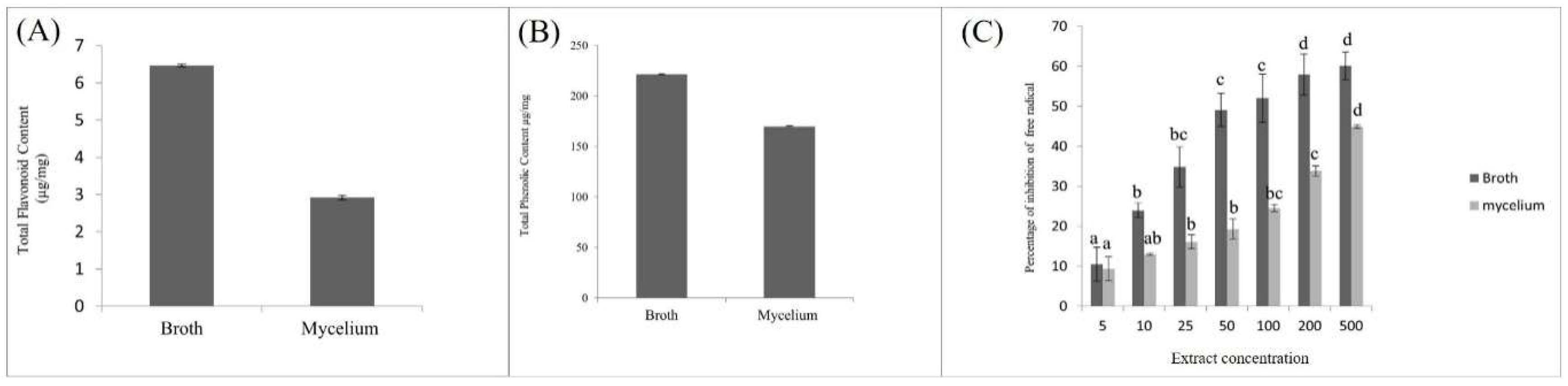
Total Phenolic Content, Total Flavonoid Content, and Antioxidant Activity of Broth and Mycelium

The total flavonoid content in the broth’s methanolic extract was 6.50 µg/mg, whereas in the mycelium extract, it was 2.9 µg/mg as shown in Figure 2A. Similarly, the total phenolic content in the broth’s methanolic extract was 221.706 µg/mg, while in the mycelium extract, it was 169.7 µg/mg as shown in Figure 2B. The significant difference in total flavonoid and phenolic content between the broth and mycelium extracts suggests that the extract possesses higher antioxidant properties.

Comparative Antioxidant Activity of Fungal Mycelium and Broth Extract

The antioxidant activity of fungal mycelium and broth extracts was evaluated using the DPPH free radical scavenging assay. The results indicated that antioxidant activity increased with increasing concentrations of both broth and mycelium extracts. At concentrations of 200 µg/mL and 500 µg/mL, the antioxidant activity of both extracts became comparable as shown in Figure 2C. However, the highest antioxidant activity was observed at 500 µg/mL, with broth extract showing 60% activity and mycelium extract showing 43% activity.

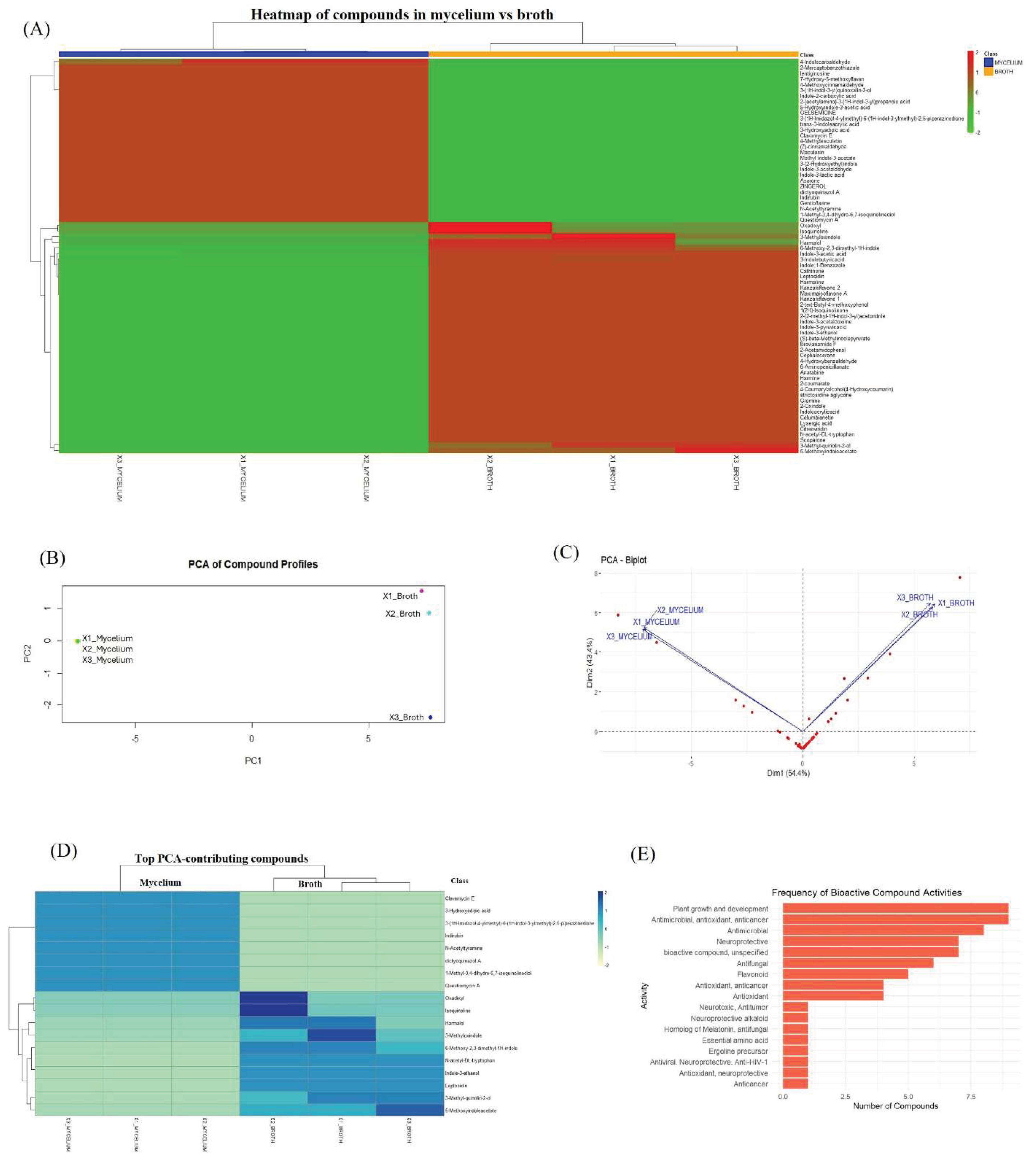
Comparative Analysis of Metabolites Produced by Intracellular Mycelium and Extracellular Broth

High-resolution mass spectrometry (HRMS) analysis, both broth and mycelium extracts contain diverse array of bioactive metabolites that include indole derivatives, alkaloids, terpenoids, flavonoids, phenolic compounds and others as shown in Table 1. The Heatmap clustering of compounds detected in mycelium and broth extract forms distinct clusters representing significantly different compounds as shown in Figure 3A. The predominantly accumulated compound in mycelium are 4-Indolecarbaldehyde acid, Lentiginosine, 7-Hydroxy-5-methoxyflavan, 4-Methoxycinnamaldehyde, 3-(1H-indole-3-yl) quinoxaline-2-ol, Indole-2-caboxylic acid, Gelsemicine, 3-(1H-Imidazol-4-ylmethyl)-6-(1H-indol-3-ylmethyl)-2,5-piperazinedione, trans-3-Indole acrylic acid, 3-Hydeoxyadipic acid, Clavamycin E, 4-Methylesculetin, Indole 3 acetate, indole-3-acetyldehye, indole-3-lactic acid, Gentioflavine, Zingerol, Cinnamaldehyde. In contrast, the broth extract contained: Cathinole, Leptosidin, Harmaline, Kanzaki flavone 1 and flavone 2, Maxima isoflavone, Coumarate and its derivatives, Harmalol, Gramine, Lysergic acid, Citreoviridin, Scoparone, Tryptophan.
The PCA plot strongly suggests compositional distinction between intracellular metabolites (mycelium) and extracellular metabolites (broth). The PCA biplot further supports distinguishing the metabolites from mycelium and broth extract clustering within each group with 95% confidence ellipse as shown in Figure 3B-C. This suggests consistency within the group and between the groups. Therefore, the metabolic profile of mycelium and broth extract reflects the two different environments (intracellular and extracellular) responsible for the differential metabolic profile. The top PCA -contributing compounds from mycelium included: 3-Hydroxyadipic acid, Indirubin, Questiomycin A, 1-Methyl-3,4-dihydro-6,7-isoquinolinediol, dictyoquinazol. Whereas the top compounds that have significant abundance in broth: Oxadixyl, Isoquinoline, Harmalol, 3-Methyloxindole, 6-Methoxy-2,3-dimethyl-1H-indole, N-acetyl-DL-tryptophan, Indole-3-ethanol, Leptosidin, 3-Metyl-quinolin-2-ol, 5-Methoxyindoleacetate in Figure 3D. The clusters for the two groups, mycelium and broth, are distinctive and may correlate with potential functional differences of their compounds. Overall, compounds were most of the compounds were indole compounds that may influence the plant growth and development activities, along with other compounds that have been reported to have antimicrobial, antioxidant, and anticancer activities. Similarly, some compounds have neuroprotective activities as shown in Figure 3E.

4. Discusssion

Coniochaeta is a diverse and pleomorphic genus of ascomycetous fungi that may be animal or plant pathogenic or endophytic. Increasing evidence suggests that members of this genus produce a broad spectrum of biologically active secondary metabolites, including compounds with antioxidant, cytotoxic, antimicrobial, and plant growth–promoting properties.
In the present study, we comparatively evaluated the antioxidant activity, flavonoid content, and phenolic content of broth and mycelial extracts of Coniochaeta. The broth extract exhibited significantly higher antioxidant activity, total flavonoid content, and phenolic content compared to the mycelial extract (Figure 1). This suggests that a substantial proportion of bioactive metabolites are secreted extracellularly into the liquid medium during incubation under shaking conditions. Such secretion dynamics align with previous reports demonstrating that endophytic fungi actively release secondary metabolites into surrounding environments, potentially facilitating ecological interactions and host modulation [2,8].
Metabolomic analysis revealed that the broth extract was enriched with diverse antioxidant-related compounds, including indole derivatives, flavonoids (Kanzakiflavones, Maximaisoflavone A), and phenolics such as 4-Hydroxybenzaldehyde and Scoparone, explaining its significantly higher antioxidant, flavonoid, and phenolic content. In contrast, the mycelial extract contained comparatively fewer phenolic constituents, including 4-Methylesculetin, 4-Methoxycinnamaldehyde, N-Acetyltyramine, and Zingerol.
Notably, indole derivatives was detected in the broth, including Indole-3-acetic acid (IAA), Indole-3-lactic acid, Indole-3-acetaldoxime, and Gramine [15,34]. Indole compounds are well documented for their roles in plant growth regulation, immune modulation, and antioxidant activity [70]. The abundance of these indole-related metabolites supports the hypothesis that Coniochaeta possesses intrinsic plant growth–promoting potential, other than IAA production. Our findings therefore support earlier reports that endophytic fungi synthesize phytohormone-like molecules capable of influencing host development.
The detection of antifungal and antimicrobial agents such as Oxadixyl [18], 2-Mercaptobenzothiazole [25], Clavamycin E, 3-Methyl-quinolin-2-ol [17], and Questiomycin A [76] highlights the defensive ecological role of Coniochaeta metabolites. Additionally, flavonoids including Gentioflavine [58,59], Maximaisoflavone A, and Kanzakiflavones [60,61], along with coumarin derivatives such as 4-Methylesculetin, 4-Coumaryl alcohol, and 2-Coumarate [78], and phenolics like 4-Hydroxybenzaldehyde [20] and 4-Methoxycinnamaldehyde [73], have documented antioxidant, anti-inflammatory, and anticancer properties. These findings are consistent with prior studies reporting antioxidant and cytotoxic metabolites from Coniochaeta sp. F-8, including Phomoxanthone A and Penialidin A, both of which exhibited strong in vitro antioxidant activity, with Phomoxanthone A showing notable cytotoxicity toward Balb/c3T3 cells [79].
Beyond metabolite production, previous transcriptomic studies demonstrated that Coniochaeta exhibits trophic plasticity, functioning as a benign endophyte in living host tissue while switching to saprotrophic metabolism during host senescence [80]. This dual functionality suggests ecological versatility and supports its role in both plant growth promotion and nutrient recycling. Our previous studies further support this concept, demonstrating that Coniochaeta africana and C. dendrobicola significantly enhanced in vitro growth of orchid protocorms and plantlets. The presence of auxin-related indole derivatives and diverse phytochemicals in the present metabolomic dataset provides biochemical evidence that may explain the plant growth–promoting effects observed in orchids. In addition to ecological and plant-associated roles, certain species such as Coniochaeta ligniaria have demonstrated industrial importance due to their tolerance to acetate and capacity to detoxify fermentation inhibitors in biomass hydrolysates [81]. Overall, the present comparative analysis demonstrates that extracellular (broth) extracts of Coniochaeta contain a richer profile of antioxidant, flavonoid, phenolic, indole, and antimicrobial metabolites compared to intracellular mycelial extracts. When considered alongside prior studies on cytotoxic metabolites, trophic plasticity, plant growth promotion, and industrial detoxification capacity, our findings emphasize the broad ecological, agricultural, pharmaceutical, and biotechnological potential of Coniochaeta species.

5. Conclusion

The present study demonstrates the potential antioxidant activity and detection of important bioactive compounds. Antioxidant activities may have direct correlation with detected bioactive compounds. Significantly higher abundance of excretory bioactive compounds detected in the broth extract may explains its higher antioxidant activity, flavonoid and phenolic contents. These finding higlight the importance to explore the excretory compounds that may have plant growth promoting actitivties, symbiosis cofactor or therapeutic application. However, several challenges are associated with formentation, large-scale mycelium cultivation, bioacitive compound profiling, as these processes are strongly influenced by temperature and require careful optimation of environmental conditions. Moreover, proper post-processing of the bioactive compound important and challenging to presever their activity.

Author Contributions

Sujit Shah: Manuscript writing: original draft, investigation, data collection, data analysis, data visualization and interpretation, conceptualization. Gajanan Mane: Manuscript editing and methodology. Biva Shah: Manuscript editing and data interpretation. Vibhav Gautam: Manuscript editing and methodology. Mukti Ram Paudel: Manuscript editing and data interpretation. Bijaya Pant: Manuscript editing and data interpretation. Rohit Sharma: Manuscript editing and data interpretation. Deepak Kumar: Writing – review & editing, Supervision, Project administration, Investigation, Funding acquisition, Formal analysis, Conceptualization.

Funding

This work was financially supported by the Institute of Eminence (IOE) seed grant (Dev. No. 6031(B)), Banaras Hindu University, and India Science and Research Fellowship (ISRF), Department of Science & Technology (DST), Government of India.

Institutional Review Board Statement

Not applicable for this study, no involvement of humans or animals.

Informed Consent Statement

Not applicable for this study, no involvement of humans or animals.

Data Availability Statement

Data supporting the findings of this study are accessible from the corresponding author upon request.

Acknowledgments

The authors would like to acknowledge the High-Resolution Mass Spectroscopy (HRMS) facility at the Sophisticated Analytical & Technical Help Institute (SATHI) in the Central Discovery Centre (CDC), BHU, for providing the instrumentation facility.

Conflicts of Interests

No conflict of interest reported by authors.

References

  1. Alam, B.; Lǐ, J.; Gě, Q.; Khan, M.A.; Gōng, J.; Mehmood, S.; Yuán, Y.; Gǒng, W. Endophytic fungi: From symbiosis to secondary metabolite communications or vice versa? Front. Plant Sci. 2021, 12, 791033. [Google Scholar] [CrossRef]
  2. Baron, N.C.; Rigobelo, E.C. Endophytic fungi: A tool for plant growth promotion and sustainable agriculture. Mycology 2021, 13, 39–55. [Google Scholar] [CrossRef]
  3. Kashyap, A.K.; Dubey, S.K.; Shah, S.; Kumar, A. A short review on genes regulating biosynthesis of major secondary metabolites. In Phytochemical Genomics: Plant Metabolomics and Medicinal Plant Genomics; Elsevier: Amsterdam, The Netherlands, 2023; pp. 501–519. [Google Scholar]
  4. Pant, B.; Shah, S.; Shrestha, R.; Pandey, S.; Joshi, P.R. An overview on orchid endophytes. In Mycorrhiza—Nutrient Uptake, Biocontrol, Ecorestoration; Varma, A., Prasad, R., Tuteja, N., Eds.; Springer: Cham, Switzerland, 2017; pp. 435–453. [Google Scholar] [CrossRef]
  5. Phukhamsakda, C.; Nilsson, R.H.; Bhunjun, C.S.; et al. The numbers of fungi: Contributions from traditional taxonomic studies and challenges of metabarcoding. Fungal Divers. 2022, 114, 327–386. [Google Scholar] [CrossRef]
  6. Saini, H.P.; Meena, M.; Sahoo, A.; Mehta, T. A review on fungal endophytes of the family Fabaceae, their metabolic diversity and biological applications. Heliyon 2025, 11, e42153. [Google Scholar] [CrossRef] [PubMed]
  7. Wen, J.; Okyere, S.K.; Wang, S.; et al. Endophytic fungi: An effective alternative source of plant-derived bioactive compounds for pharmacological studies. J. Fungi 2022, 8, 205. [Google Scholar] [CrossRef] [PubMed]
  8. Gupta, A.; Meshram, V.; Gupta, M.; et al. Fungal endophytes: Microfactories of novel bioactive compounds with therapeutic interventions. Biomolecules 2023, 13, 1038. [Google Scholar] [CrossRef]
  9. Shah, S.; Shah, B.; Sharma, R.; et al. Colonization with non-mycorrhizal culturable endophytic fungi enhances orchid growth and indole acetic acid production. BMC Microbiol. 2022, 22, 101. [Google Scholar] [CrossRef]
  10. Pant, B.; Joshi, P.R.; Maharjan, S.; et al. Comparative cytotoxic activity of wild harvested stems and in vitro-raised protocorms of Dendrobium chryseum. Adv. Pharmacol. Pharm. Sci. 2021, 2021, 8839728. [Google Scholar] [CrossRef]
  11. Li, T.; Yang, W.; Wu, S.; Selosse, M.A.; Gao, J. Progress and prospects of mycorrhizal fungal diversity in orchids. Front. Plant Sci. 2021, 12, 646325. [Google Scholar] [CrossRef]
  12. Garg, A.; Shah, S.; Patel, T.B.B.; Gautam, V.; Kumar, D. Contrasting effects of melatonin on Brassica juncea and Alternaria brassicae. Plant Stress 2025, 100845. [Google Scholar] [CrossRef]
  13. Miliauskas, G.; Venskutonis, P.R.; Van Beek, T.A. Screening of radical scavenging activity of some medicinal and aromatic plant extracts. Food Chem. 2004, 85, 231–237. [Google Scholar] [CrossRef]
  14. Crous, P.W.; Carnegie, A.J.; Wingfield, M.J.; et al. Fungal Planet description sheets: 868–950. Persoonia 2019, 42, 291–473. [Google Scholar] [CrossRef] [PubMed]
  15. Zhao, Z.H.; Xin, F.Z.; Xue, Y.; et al. Indole-3-propionic acid inhibits gut dysbiosis. Exp. Mol. Med. 2019, 51, 1–14. [Google Scholar] [CrossRef]
  16. Chua, R.W.; Song, K.P.; Ting, A.S.Y. Characterization of antimicrobial compounds from endophytic Fusarium incarnatum. Int. Microbiol. 2024, 27, 977–992. [Google Scholar] [CrossRef] [PubMed]
  17. Mehta, T.; Meena, M.; Nagda, A. Bioactive compounds of Curvularia species. Front. Microbiol. 2022, 13, 1069095. [Google Scholar] [CrossRef]
  18. Chen, M.; Liang, J.; Wang, Y.; et al. A new benzaldehyde from Aspergillus terreus. J. Zhejiang Univ. Sci. B 2022, 23, 230–240. [Google Scholar] [CrossRef]
  19. Reveglia, P.; Pacetti, A.; Masi, M.; et al. Phytotoxic metabolites produced by Diaporthe eres. Nat. Prod. Res. 2021, 35, 2872–2880. [Google Scholar] [CrossRef]
  20. Zang, X.; Wang, Y.; Han, C.; et al. 2-Acetamidophenol suppresses atherosclerosis. Mar. Drugs 2024, 22, 513. [Google Scholar] [CrossRef]
  21. Xue, M.; Hou, X.; Fu, J.; et al. Bioactive secondary metabolites from fungi triggered by epigenetic modifiers. J. Fungi 2023, 9, 172. [Google Scholar] [CrossRef]
  22. Xu, D.; Xue, M.; Shen, Z.; et al. Phytotoxic secondary metabolites from fungi. Toxins 2021, 13, 261. [Google Scholar] [CrossRef]
  23. Roager, H.M.; Licht, T.R. Microbial tryptophan catabolites in health and disease. Nat. Commun. 2018, 9, 3294. [Google Scholar] [CrossRef] [PubMed]
  24. Zhang, Y.H.; Du, H.F.; Liu, Y.F.; et al. Novel anti-inflammatory diketopiperazine alkaloids. Appl. Microbiol. Biotechnol. 2024, 108, 194. [Google Scholar] [CrossRef] [PubMed]
  25. Okano, T.; Kobayashi, N.; Izawa, K.; et al. Genes responsible for citreoviridin biosynthesis. Toxins 2020, 12, 125. [Google Scholar] [CrossRef] [PubMed]
  26. Auckloo, B.N.; Pan, C.; Akhter, N.; et al. Stress-driven discovery of cryptic antibiotics. Front. Microbiol. 2017, 8, 1450. [Google Scholar] [CrossRef]
  27. Shiratori, N.; Kobayashi, N.; Tulayakul, P.; et al. Occurrence of Penicillium brocae and P. citreonigrum. Toxins 2017, 9, 194. [Google Scholar] [CrossRef]
  28. Jastrzębski, M.K.; Kaczor, A.A.; Wróbel, T.M. Methods of lysergic acid synthesis. Molecules 2022, 27, 7322. [Google Scholar] [CrossRef]
  29. Xu, X.; Zhang, X.; Nong, X.; et al. Oxindole alkaloids from Penicillium commune. Tetrahedron 2015, 71, 1640–1645. [Google Scholar] [CrossRef]
  30. Sharma, S.; Monga, Y.; Gupta, A.; Singh, S. 2-Oxindole and related heterocycles. RSC Adv. 2023, 13, 14249–14267. [Google Scholar] [CrossRef]
  31. Cabutaje, E.M.; Ueno, K.; Osaki-Oka, K.; et al. Phthalide derivatives and an oxindole compound from Pleurotus ostreatus. J. Pestic. Sci. 2023, 48, 156–167. [Google Scholar] [CrossRef]
  32. Fu, S.F.; Wei, J.Y.; Chen, H.W.; et al. Indole-3-acetic acid in fungal interactions. Plant Signal. Behav. 2015, 10, e1048052. [Google Scholar] [CrossRef]
  33. Kumar, P.; Lee, J.H.; Lee, J. Diverse roles of microbial indole compounds. Biol. Rev. 2021, 96, 2522–2545. [Google Scholar] [CrossRef]
  34. Tang, Z.; Wang, Y.; Yang, J.; et al. Flavonoid-producing endophytic fungi. PeerJ 2020, 8, e8978. [Google Scholar] [CrossRef] [PubMed]
  35. Kong, M.; Liang, J.; Ali, Q.; et al. 5-Methoxyindole affects Fusarium graminearum. Int. J. Mol. Sci. 2021, 22, 10991. [Google Scholar] [CrossRef] [PubMed]
  36. Chung, K.R.; Shilts, T.; Ertürk, U.; et al. Indole derivatives produced by Colletotrichum acutatum. FEMS Microbiol. Lett. 2003, 226, 23–30. [Google Scholar] [CrossRef] [PubMed]
  37. Kowalczyk, J.; Budzyńska, B.; Kurach, Ł.; et al. Neuropsychopharmacological profiling of scoparone. Sci. Rep. 2022, 12, 822. [Google Scholar] [CrossRef]
  38. Liu, C.M.; Liang, X.; Yao, F.H.; Qi, S.H. Aromatic polyketides and isoquinoline alkaloids. Tetrahedron 2022, 112, 133067. [Google Scholar] [CrossRef]
  39. Shang, X.F.; Yang, C.J.; Morris-Natschke, S.L.; et al. Biologically active isoquinoline alkaloids. Med. Res. Rev. 2020, 40, 2212–2289. [Google Scholar] [CrossRef]
  40. Nord, C.; Levenfors, J.J.; Bjerketorp, J.; et al. Antibacterial isoquinoline alkaloids. Molecules 2019, 24, 4616. [Google Scholar] [CrossRef]
  41. Umashankar, P.; Nygård, Y. Filamentous fungi as emerging cell factories. Fungal Biol. Biotechnol. 2024, 11, 19. [Google Scholar] [CrossRef]
  42. Nag, S.; Chowdhury, C.; Das, N. Antioxidant potency of kanzakiflavone-2. J. Mol. Struct. 2021, 1231, 129928. [Google Scholar] [CrossRef]
  43. Murthy, M.S.R.; Rao, E.V. Maxima isoflavone. J. Phytochemistry 1985, 24, 767–768. [Google Scholar] [CrossRef]
  44. El Gendy, M.A.; Soshilov, A.A.; Denison, M.S.; El-Kadi, A.O.S. Harmaline and harmalol inhibit CYP1A1. Toxicol. Lett. 2012, 213, 306–313. [Google Scholar]
  45. Hu, Y.; Yu, X.; Yang, L.; et al. Antitumor effects of harmine. Front. Oncol. 2024, 14, 1382142. [Google Scholar] [CrossRef] [PubMed]
  46. Bhatt, S.K.; Saxena, V.K.; Singh, K.V. A leptosidin glycoside. Phytochemistry 1981, 20, 2605. [Google Scholar] [CrossRef]
  47. Caturegli, P.; De Remigis, A.; Ferlito, M.; et al. Anatabine ameliorates autoimmune thyroiditis. Endocrinology 2012, 153, 4580–4587. [Google Scholar] [CrossRef]
  48. Boyce, G.R.; Gluck-Thaler, E.; Slot, J.C.; et al. Psychoactive alkaloids from cicada pathogens. Fungal Ecol. 2019, 41, 147–164. [Google Scholar] [CrossRef]
  49. Zhang, L.; Ge, Y.; Li, J.; et al. Simultaneous determination of columbianetin derivatives. J. Pharm. Biomed. Anal. 2018, 153, 221–231. [Google Scholar] [CrossRef]
  50. Robinson, M.; Riov, J.; Sharon, A. Indole-3-acetic acid biosynthesis in Colletotrichum gloeosporioides. Appl. Environ. Microbiol. 1998, 64, 5030–5032. [Google Scholar] [CrossRef]
  51. Peng, J.; Wang, X.; Wang, H.; et al. Advances in understanding grapevine downy mildew. Mol. Plant Pathol. 2024, 25, e13401. [Google Scholar] [CrossRef]
  52. Tang, J.; Li, Y.; Zhang, L.; et al. Biosynthetic pathways and functions of indole-3-acetic acid in microorganisms. Microorganisms 2023, 11, 2077. [Google Scholar] [CrossRef]
  53. King, H.D.; Langhärig, J.; Sanglier, J.J. Clavamycins. J. Antibiot. 1986, 39, 510–515. [Google Scholar] [CrossRef]
  54. Kozlovskiĭ, A.G.; Zhelifonova, V.P.; Antipova, T.V.; et al. Penicillium expansum producing xanthocillin X. Prikl. Biokhim. Mikrobiol. 2004, 40, 344–349. [Google Scholar] [PubMed]
  55. Pan, H.; Li, H.; Wu, S.; et al. De novo biosynthesis of N-acetyltyramine. Enzyme Microb. Technol. 2023, 162, 110149. [Google Scholar] [CrossRef] [PubMed]
  56. Karagianni, E.P.; Kontomina, E.; Davis, B.; et al. Xenobiotic metabolizing N-acetyltransferases in fungi. Sci. Rep. 2015, 5, 12900. [Google Scholar] [CrossRef] [PubMed]
  57. Correia, J.; Borges, A.; Simões, M.; Simões, L.C. Beyond penicillin: Filamentous fungi for drug discovery. Antibiotics 2023, 12, 1250. [Google Scholar] [CrossRef]
  58. Singh, V.K.; Kumar, A. Secondary metabolites from endophytic fungi. Symbiosis 2023, 1–15. [Google Scholar] [CrossRef]
  59. Wu, X.; Zou, K.; Liu, X.; et al. Intracellular and extracellular flavonoids from Aspergillus sp. Front. Microbiol. 2022, 13, 972294. [Google Scholar] [CrossRef]
  60. Ponnusamy, K.; Petchiammal, C.; Mohankumar, R.; Hopper, W. Antifungal activity of indirubin. J. Ethnopharmacol. 2010, 132, 349–354. [Google Scholar] [CrossRef]
  61. Lee, I.K.; Yun, B.S.; Han, G.; et al. Dictyoquinazols A–C. J. Nat. Prod. 2002, 65, 1769–1772. [Google Scholar] [CrossRef]
  62. Lizarme, Y.; Wangsahardja, J.; Marcolin, G.M.; et al. Synthesis and neuroprotective activity of dictyoquinazol A. Bioorg. Med. Chem. 2016, 24, 1480–1487. [Google Scholar] [CrossRef]
  63. Mao, Q.Q.; Xu, X.Y.; Cao, S.Y.; et al. Bioactive compounds of ginger. Foods 2019, 8, 185. [Google Scholar] [CrossRef]
  64. Bai, X.; Sheng, Y.; Tang, Z.; et al. Polyketides from Aspergillus. J. Fungi 2023, 9, 261. [Google Scholar] [CrossRef]
  65. Wang, P.; Ma, L.; Jin, J.; et al. Anti-aflatoxigenic mechanism of cinnamaldehyde. Sci. Rep. 2019, 9, 10499. [Google Scholar] [CrossRef] [PubMed]
  66. Peng, Y.L.; Liang, J.J.; Xue, Y.; et al. Gelsemium and its endophytic fungi. Curr. Top. Med. Chem. 2023, 23, 2452–2487. [Google Scholar] [CrossRef] [PubMed]
  67. Kumla, J.; Suwannarach, N.; Matsui, K.; Lumyong, S. Biosynthetic pathway of indole-3-acetic acid. PLoS ONE 2020, 15, e0227478. [Google Scholar] [CrossRef] [PubMed]
  68. Sakurai, T.; Odamaki, T.; Xiao, J.Z. Production of indole-3-lactic acid. Microorganisms 2019, 7, 340. [Google Scholar] [CrossRef]
  69. Que, Y.; Huang, D.; Gong, S.; et al. Indole-3-carboxylic acid from Lasiodiplodia pseudotheobromae. Front. Cell. Infect. Microbiol. 2022, 12, 898500. [Google Scholar] [CrossRef]
  70. Sun, P.; Huang, Y.; Yang, X.; et al. Role of indole derivatives in plant growth. Front. Plant Sci. 2023, 13, 1120613. [Google Scholar] [CrossRef]
  71. Ye, X.; Li, H.; Anjum, K.; et al. Dual role of indoles from intestinal microbiota. Front. Immunol. 2022, 13, 903526. [Google Scholar] [CrossRef]
  72. Paudel, B.; Maharjan, R.; Rajbhandari, P.; et al. Maculosin from Streptomyces sp. Pharm. Biol. 2021, 59, 933–936. [Google Scholar] [CrossRef]
  73. Morozumi, S. o-Methoxycinnamaldehyde from cinnamon. Appl. Environ. Microbiol. 1978, 36, 577–583. [Google Scholar] [CrossRef]
  74. Pina, J.; de Castro, C.S.; Delgado-Pinar, E.; Seixas de Melo, J.S. 4-Methylesculetin derivatives. J. Mol. Liq. 2019, 278, 616–626. [Google Scholar] [CrossRef]
  75. Pastuszak, I.; Molyneux, R.J.; James, L.F.; Elbein, A.D. Lentiginosine inhibits amyloglucosidase. Biochemistry 1990, 29, 1886–1891. [Google Scholar] [CrossRef] [PubMed]
  76. Machihara, K.; Tanaka, H.; Hayashi, Y.; et al. Questiomycin A stimulates sorafenib-induced cell death. Biochem. Biophys. Res. Commun. 2017, 492, 396–401. [Google Scholar] [CrossRef] [PubMed]
  77. Zhang, J.; Jia, Q.; Li, N.; et al. Recent developments of gramine. Molecules 2023, 28, 5695. [Google Scholar] [CrossRef]
  78. Tsivileva, O.M.; Koftin, O.V.; Evseeva, N.V. Coumarins as fungal metabolites. Antibiotics 2022, 11, 1156. [Google Scholar] [CrossRef]
  79. Fu, J.; Hu, L.; Shi, Z.; Sun, W.; Yue, D.; Wang, Y.; Ma, X.; Ren, Z.; Zuo, Z.; Peng, G.; Zhong, Z.; Deng, J.; Cao, S.; Hu, Y. Two metabolites isolated from endophytic fungus Coniochaeta sp. F-8 in Ageratina adenophora exhibit antioxidative activity and cytotoxicity. Nat. Prod. Res. 2021, 35, 2840–2848. [Google Scholar] [CrossRef]
  80. Chen, K.H.; Liao, H.L.; Arnold, A.E.; Korotkin, H.B.; Wu, S.H.; Matheny, P.B.; Lutzoni, F. Comparative transcriptomics of fungal endophytes in co-culture with their moss host Dicranum scoparium reveals fungal trophic lability and moss unchanged to slightly increased growth rates. New Phytol. 2022, 234, 1832–1847. [Google Scholar] [CrossRef]
  81. Nichols, N.N.; Mertens, J.A.; Frazer, S.E.; Hector, R.E. Growth of Coniochaeta Species on Acetate in Biomass Sugars. Fermentation 2022, 8, 721. [Google Scholar] [CrossRef]
Figure 1. The endophytic fungi Coniochaeta dendrobiicola isolated from the Dendrobium longicornu, (A-C) Conidia cylindrical to allantoids shape and conidiogenous cells arising laterally from hyphae; (D) PDA fungal culture pale yellow, circular with regular margins.
Preprints 199663 g001
Figure 2. Total flavonoid content measured in µg/mg in broth and mycelium of the methanolic extract. The broth content is 6.5 µg per mg whereas the mycelium content 2.9 µg per mg (A). The total phenolic content 221.7 µg per mg and 169.7 µg per mg in broth and mycelium respectively (B). The percentage inhibition of free radical with the DPPH assay showed antioxidant activity (C). Bar represents mean ± SE (n = 3). The Values with different letters are significantly different at the level of p ≤ 0.05.
Preprints 199663 g002
Figure 3. Heatmap of metabolite profiles (A) of mycelium and broth extracts. Color gradients indicate abundance levels (red: high, green: low). Hierarchical clustering reveals distinct metabolic patterns between mycelium and broth, suggesting differential biosynthesis and secretion of secondary metabolites. The PCA plot (B) reveals a distinct separation between mycelium (green dot) and broth (cyan, pink, blue dots), indicating substantial differences in their metabolite compositions. c-PCA biplot shows the separation of mycelium and broth samples based on metabolite profiles with ellipses representing 95% confidence intervals for each group. Altogether, indicating distinctive metabolic profiling based on two different environmental backgrounds, intracellular (mycelium) and extracellular (broth). d-The corresponding heatmap displays the top PCA-contributing compounds, highlighting the distinct clustering of mycelium and broth samples based on compound abundance. e-Mycelium is enriched in indole derivatives and organic acids, while broth contains more quinolines, alkaloids, and bioactive secondary metabolites. This pattern underscores functional divergence between intracellular and extracellular metabolomes in fungal cultures. f-The bar chart shows the number of compounds that have biological activities based on prior studies and database annotations. Common multifunctional activities include antimicrobial, antioxidant and anticancer along with plant growth and development, neuroprotection indicating potential therapeutic and agricultural applications.
Preprints 199663 g003
Table 1. List of the compounds detected in broth and mycelium extract by HRMS technique.
Compound Name Enriched In Nature of compound Reported Source Reported Activity References
3-Indolepropionic acid Broth Indole compound Unreported from fungi Antioxidant, neuroprotective [15]
3-Methyl-quinolin-2-ol Broth Isoquinoline Fusarium incarnatum Antifungal [16]
4-Hydroxybenzaldehyde Broth Phenolic compound Curvularia, Diaporthe, Aspergillus Antimicrobial [17,18,19]
2-Acetamidophenol Broth Phenolic compound Penicillium chrysogenum, Aspergillus sp., Pseudomons chlororaphis Antimicrobial, antioxidant, anticancer [20]
N-acetyl-DL-tryptophan Broth Derivative of amino acid Fungi, Bacteria Essential amino acid [21,22,23]
Brevianamide F Broth 2,5-diketopiperazine Aspergillus, Penicillium Antimicrobial [24]
Citreoviridin Broth Mycotoxin Penicillium, Aspergillus Neurotoxic, Antitumor [25,26,27]
Lysergic acid Broth Ergoline alkaloids Claviceps purpurea, Periglandula spp. Ergoline precursor [28]
2-Oxindole Broth Indole compound Colletotrichum fragariae, Aspergillus versicolor, and Penicillium commune, plant and human gut Antimicrobial, antioxidant, anticancer [29,30,31]
Indole 3 ethanol Broth Indole compound Endophytic, mycorrhizal fungi Plant growth and development [32,33,34]
Indole 3 butyric acid Broth Indole compound Endophytic, mycorrhizal fungi Plant growth and development [32,33,34]
Indole 3 pyruvic acid Broth Indole compound Endophytic, mycorrhizal fungi Plant growth and development [32,33,34]
5-Methoxyindole Broth Indole compound Unreported from fungi Homolog of Melatonin, antifungal [35]
Indole acrylic acid Broth Indole compound Endophytic, mycorrhizal fungi Plant growth and development [32,33,34]
3-Methyloxindole Broth Indole compound Unreported from fungi Bioactive compound, unspecified [33,34,36]
Indole;1-Benzazole Broth Indole compound Unreported from fungi Bioactive compound, unspecified [33,34,36]
2-(2-methyl-1H-indol-3-yl)acetonitrile Broth Indole compound Unreported from fungi Bioactive compound, unspecified
Scoparone Broth Phenolic compound Plant derivatives Neuroprotective [37]
5-Methoxyindoleacetate Broth Indole compound Plant derivatives Antioxidant [34,35,37]
Indole-3-acetaldoxime Broth Indole compound Plant derivatives Antioxidant [34,35,37]
6-Methoxy-2,3-dimethyl-1H-indole Broth Indole compound Plant derivatives Antioxidant [33,34,36]
Gramine Broth Indole alkaloid Plant derivatives Antimicrobial, antioxidant, anticancer [33,34,36]
1(2H)-Isoquinolinone Broth Isoquinolinone Plant derivatives Neuroprotective [38,39,40]
4-Coumarylalcohol(4-Hydroxycoumarin) Broth Coumarin Plant derivatives Antimicrobial, antioxidant, anticancer [41]
Kanzakiflavone 1 Broth Flavonoid Flavonoid Flavonoid [42]
Maximaisoflavone A Broth Flavonoid Flavonoid Flavonoid [43]
Kanzakiflavone 2 Broth Flavonoid Flavonoid Flavonoid [42]
2- coumarate Broth Flavonoid Flavonoid Flavonoid [42]
Isoquinoline Broth Alkaloid Plant derivatives Neuroprotective [39,40,41]
Harmalol Broth Alkaloid Plant derivatives Antioxidant, anticancer [44]
Harmaline Broth Alkaloid Plant derivatives Antioxidant, anticancer [44]
Harmine Broth Alkaloid Plant derivatives Antioxidant, anticancer [45]
Leptosidin Broth Flavonoid Plant derivatives Antioxidant, anticancer [46]
Anatabine Broth Alkaloid Plant derivatives Neuroprotective [47]
Cathinone Broth Alkaloid Plant-mushroom derivatives Neuroprotective [48]
Columbianetin Broth phenolic furanocoumarin Plant Antifungal [49]
Indole-3-acetic acid Broth/ Mycelium Indole compound Endophytic, mycorrhizal fungi Plant growth and development [32,50]
Oxadixyl Broth/Mycelium Oxazolidinone Compound Fungi Antifungal [51]
Methyl 2,3-dihydro-3-hydroxy-2-oxo-1H-indole-3-acetate Mycelium Indole compound Unreported from fungi Bioactive compound, unspecified [52]
1-Methyl-3,4-dihydro-6,7-isoquinolinediol Mycelium Isoquinoline Plant derivatives Neuroprotective [39,40,41]
Clavamycin E Mycelium Clavams group of compounds Fungi Antimicrobial [53]
Questiomycin A Mycelium Phenazine derivative Penicillium expansum Antimicrobial [54]
N-Acetyltyramine Mycelium
Phenolic
Tyramine alkaloid Fungi (Aspergillus) Antimicrobial, antioxidant, anticancer [55,56]
Gentioflavine Mycelium
Alkaloid
Fungi (Aspergillus endophytes) Flavonoid [57,58,59]
Indirubin Mycelium Indole compound Wrightia tinctoria, Schizophyllum commune, Malassezia spp. Anticancer [60]
Dictyoquinazol A Mycelium Quinazolines compound Dicyophora indusiata Neuroprotective [61,62]
Zingerol Mycelium
Phenolic
Phenolic compound Plant derivatives Antimicrobial [63]
Asarone Mycelium Phenylpropanoid Aspergillus sp. Antimicrobial [64]
trans-3-Indoleacrylic acid Mycelium Indole compound Lactiplantibacillus plantarum LPP95 Antifungal [65]
Gelsemicine Mycelium Indole compound Gelsemium spp., Endophytic fungi Neuroprotective alkaloid [66]
3-(1H-indol-3-yl)quinoxalin-2-ol Mycelium Indole compound Fungi Antimicrobial, antioxidant, anticancer [31]
Indole-3-lactic acid Mycelium Indole compound Ectomycorrhizal fungi Plant growth and development [67,68]
Indole-3-acetaldehyde Mycelium Indole compound Ectomycorrhizal fungi Plant growth and development [67]
Indole-3-carboxylic acid Mycelium Indole compound Lasiodiploda pseudotheobromae Antifungal [69]
3-(2-Hydroxyethyl)indole Mycelium Indole compound Sporormiella minimoides Antiviral, Neuroprotective, Anti-HIV-1 [70]
(S)-beta-Methylindolepyruvate Mycelium Indole compound Unreported from fungi Antimicrobial, antioxidant, anticancer [24,71]
4-Indolecarbaldehyde acid Mycelium Indole compound Unreported from fungi Bioactive compound, unspecified [37,38,39]
2-(acetylamino)-3-(1H-indol-3-yl)propanoic acid Mycelium Indole compound Unreported from fungi Bioactive compound, unspecified
5-Hydroxyindole-3-acetic acid Mycelium Indole compound Endophytic, mycorrhizal fungi Plant growth and development [51]
Methyl indole-3-acetate Mycelium Indole compound Endophytic, mycorrhizal fungi Plant growth and development [51]
3-(1H-Imidazol-4-ylmethyl)-6-(1H-indol-3-ylmethyl)-2,5-piperazinedione Mycelium Indole compound Unreported from fungi Bioactive compound, unspecified
Maculosin Mycelium dipeptide, pyrrolopyrazine Bacterial derived Antioxidant [72]
4-Methoxycinnamaldehyde Mycelium Phenolic compound Plant derivatives Antimicrobial [73]
(Z)-cinnamaldehyde Mycelium Flavonoid Plant derivatives Antimicrobial [73]
4-Methylesculetin Mycelium
Phenolic
Phenolic compound Plant derivatives Antimicrobial, antioxidant, anticancer [74]
7-Hydroxy-5-methoxyflavan Mycelium Flavonoid Flavonoid Antimicrobial, antioxidant, anticancer [39,67]
lentiginosine Mycelium Alkaloid Plant derivatives Antifungal [75]
Disclaimer/Publisher’s Note: The statements, opinions and data contained in all publications are solely those of the individual author(s) and contributor(s) and not of MDPI and/or the editor(s). MDPI and/or the editor(s) disclaim responsibility for any injury to people or property resulting from any ideas, methods, instructions or products referred to in the content.
Copyright: This open access article is published under a Creative Commons CC BY 4.0 license, which permit the free download, distribution, and reuse, provided that the author and preprint are cited in any reuse.
Prerpints.org logo

Preprints.org is a free preprint server supported by MDPI in Basel, Switzerland.

Subscribe

Disclaimer

Terms of Use

Privacy Policy

Privacy Settings

© 2026 MDPI (Basel, Switzerland) unless otherwise stated