Preprint
Review

This version is not peer-reviewed.

Autophagy: A Double-Edged Sword in the Aging of C. elegans

Submitted:

11 February 2026

Posted:

13 February 2026

You are already at the latest version

Abstract

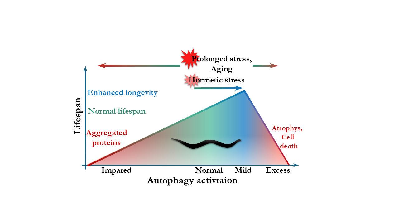
Autophagy is a tightly regulated catabolic process essential for cellular homeostasis, stress adaptation, and regeneration. In the nematode Caenorhabditis elegans, with its short lifespan, transparent body, and well-defined genetics, the process can be investigated at tissue- and age-specific manner, making it an excellent model to study the connection between autophagy and longevity. While autophagy is indispensable for development and homeostasis, recent studies have revealed that its role in aging is more complex than previously thought. During post-reproductive life, autophagic flux and the degradative capacity of lysosomes decline, resulting in the accumulation of undegraded material and cellular stress. Several studies have demonstrated that the experimental modulation of core autophagy in aged or post-reproductive C. elegans, particularly in neurons, can improve proteostasis, preserve tissue integrity, and extend lifespan. Here we review the current results obtained using the genetic model system Caenorhabditis elegans that link autophagy to lifespan regulation. We focus on studies that investigate unexpected, context-dependent, or deleterious effects of inhibiting autophagy-related genes during aging. We also discuss how age- and tissue-specific modulation of autophagy could define the most effective strategies for promoting healthy aging. This could provide relevant insights for the therapeutic targeting of autophagy in humans.

Keywords: 
;  ;  ;  ;  ;  

1. Introduction

Macroautophagy, or autophagy, is a conserved catabolic process in eukaryotic cells that delivers misfolded proteins, aggregates, and defective organelles to lysosomes via double-membraned autophagosomes for degradation and recycling [1]. Autophagy occurs at a basal level in most tissues under normal physiological conditions and is rapidly upregulated in response to various intra- and extracellular stressors such as nutrient deprivation, or environmental stress [2]. Dysfunctional autophagy or impaired lysosomal breakdown has been implicated in a wide range of diseases, including inflammation, cardiovascular disorders, metabolic diseases such as diabetes, cancer, and neurodegeneration. Beyond disease, autophagy is also recognized as a critical regulator of organismal lifespan [3].
This conserved cellular machinery operates via well-defined molecular steps. Upon induction, a double-membraned structure called the phagophore (also referred to as the isolation membrane or pre-autophagosomal membrane) forms at multiple sites of the cytoplasm [4,5]. The membrane elongates and engulfs the targeted cytoplasmic components, resulting in autophagosome formation. This structure then fuses with a lysosome to form an autolysosome, where the sequestered material is degraded by acid hydrolases and recycled [6]. Bulk autophagy is a non-selective process that provides the cell with essential macromolecules, especially under nutrient-deprived conditions [7]. In contrast, selective autophagy, such as mitophagy, aggrephagy, or lipophagy involves cargo recognition by specific receptors and plays a key role in maintaining cellular quality control and homeostasis [8].
Given its short lifespan, genetic tractability, and conserved autophagy machinery, Caenorhabditis elegans has emerged as a powerful in vivo model to dissect the role of autophagy in lifespan regulation [9]. Genetic screens in C. elegans identified several metazoan-specific autophagy genes (e.g. epg-2, epg-3, epg-4, and epg-5), expanding our understanding beyond yeast models [10]. Studies in this organism have also revealed not only the longevity-promoting functions of autophagy, but also unexpected age- and tissue-specific detrimental effects, providing a unique framework to understand the context-dependent nature of autophagy during aging [11,12,13,14].

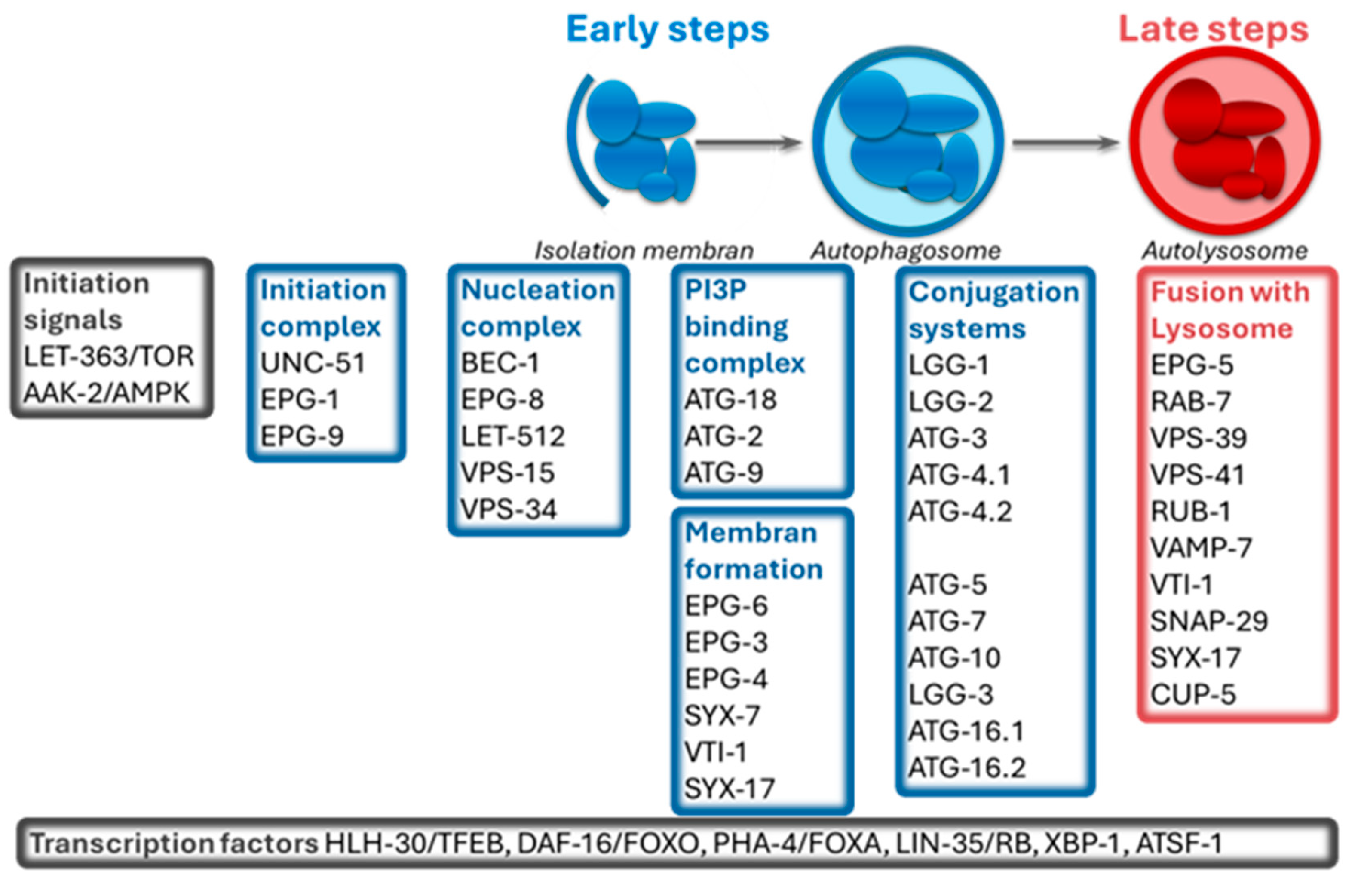
2. C. elegans as a Model Organism to Study Autophagy

Autophagy is broadly conserved: many mammalian autophagy-related proteins have a single ortholog in C. elegans, and genetic studies support the existence of a conserved core machinery [15]. However, some of these components have not yet been characterized experimentally in C. elegans, so their roles are inferred solely in their homology with orthologs in other organisms. The core molecular machinery of macroautophagy has been thoroughly reviewed elsewhere [16]; therefore, its mechanism is summarized only schematically in Figure 1.
Several forms of selective autophagy have been described in C. elegans, including aggrephagy, which is mediated by the adaptor protein SQST-1/p62, and mitophagy, which is mediated by the protein DCT-1/BNIP3. These findings highlight the fact that cargo selectivity adds another layer to autophagy-aging interactions [17,18].
Key signaling pathways and processes involved in longevity regulate autophagy in worms. The LET-363/TOR (target of rapamycin) pathway coordinates nutrient sensing, protein synthesis, and autophagy [19,20]. The insulin/IGF-1 pathway influences metabolism, stress resistance, and lifespan [21]. Under conditions of mitochondrial stress or mitochondrial dysfunction, autophagy can be upregulated, emphasizing the integration of stress responses and recycling systems [22]. Dietary restriction (DR) robustly induces autophagy via multiple conserved mechanisms including the suppression of the LET-363/TOR and the insulin/IGF-1 signaling, sirtuin activation, epigenetic modulation, and mitochondrial remodeling [23]. In C. elegans, eating-defective mutants, such as eat-2, provide a genetic model of dietary restriction [24]. Increased autophagic activity has been observed in these mutants, particularly in intestinal tissues. These findings support a role for autophagy in mediating DR-induced longevity [25]. The helix-loop-helix transcription factor HLH-30, an ortholog of mammalian TFEB, is a central transcriptional regulator of autophagy-related genes in C. elegans. Under conditions such as starvation or infection, HLH-30 translocates into the nucleus, where it activates the expression of genes involved in autophagy and lysosomal function [24,26]. Furthermore, the transcription factor PHA-4/FOXA acts in the adult stages to regulate the expression of autophagy-related genes, especially under dietary restriction or TOR suppression [25]. Autophagy gene regulation is also modulated by additional pathways including TGF-β (Sma/Mab), Rb (lin-35), the unfolded protein response (XBP-1), and mitochondrial stress response (ATFS-1) [27].
To monitor autophagy dynamics in vivo, the transparent C. elegans offers robust molecular tools such as the endogenously inserted gfp::mCherry::lgg-1 reporter, which enables reliable and artifact-free visualization of autophagy progression. This dual-fluorescent system reveals life stage- and stress-dependent regulation of autophagic flux [28].
In addition to mutational inactivation, silencing of autophagy genes is also highly accessible in this animal model. RNA interference (RNAi) can be induced by feeding animals bacteria expressing double-stranded RNA that target specific genes [29]. Sensitivity to RNAi can be enhanced in rrf-3 mutant backgrounds [30], whereas tissue-specific knockdown is achieved using sid-1 mutants, defective in systemic RNA interference, complemented with transgenic, cell-type–specific expression of the SID-1 dsRNA receptor [13,31,32]. The auxin-inducible degradation (AID) system provides a recently developed approach in C. elegans for conditional, tissue-specific and temporally controlled protein depletion, complementing genetic and RNAi-based perturbations [33]. This ability to monitor and manipulate autophagy in a tissue-specific and temporally resolved manner grants powerful opportunities to elucidate how autophagy contributes to organismal aging.
C. elegans has also become a cornerstone of aging research due to its short lifespan and conserved aging hallmarks, including muscle deterioration, intestinal and gonadal atrophy, lipid accumulation, and loss of proteostasis and mobility; features that parallel human aging [34].
Taken together, C. elegans offers an unexampled model for studying autophagy contributions in lifespan regulation in vivo because of its short lifespan, genetic tractability, conserved autophagy machinery, and powerful tools for tissue- and age-specific analyses.

3. Autophagy as a Conserved Driver of Lifespan Extension in C. elegans

Since aging is considered a genetically regulated process, C. elegans has become an effective model for investigating the molecular mechanisms underlying lifespan regulation. The first long-lived mutant identified carried the daf-2(e1370) allele, which encodes a partial loss-of-function variant of the insulin/IGF-1 receptor [21]. Meléndez and colleagues demonstrated that the autophagy gene bec-1 (homologous to BECN1/ATG6) is essential for the extended lifespan observed in daf-2 mutants [35]. This finding was later supported by studies showing that the inactivation of atg-18, or the RNAi-mediated silencing of atg-7 and atg-12 partially suppresses the longevity phenotype of daf-2 mutants Table 1.
Autophagy is required for lifespan extension across multiple genetically and environmentally induced longevity pathways in C. elegans. Beyond reduced insulin/IGF-1 signaling, dietary restriction, TOR inhibition, and mild mitochondrial dysfunction all extend lifespan in an autophagy-dependent manner. For example, lifespan extension in let-363/TOR mutants, eat-2 dietary-restriction models, and mitochondrial mutants such as clk-1 or atp-3 are suppressed by genetic or RNAi-mediated inhibition of core autophagy genes including bec-1, unc-51, and atg-18 [11,18,20,25,36,37]. Collectively, the increased autophagic activity is not merely correlated with extended lifespan but is functionally required for longevity across a broad spectrum of long-lived C. elegans models Table 1.
Interestingly, many of the cytoprotective mechanisms of autophagy are concentrated in the intestine. Intestine-specific overexpression of the Kruppel-like transcription factor klf-3, which regulates cell proliferation and metabolic homeostasis, promotes autophagy by upregulating autophagy-related genes and thereby extends lifespan [38]. In addition, the paracaspase MALT-1, known for its proinflammatory role in mammals, was recently identified as a negative regulator of the early steps of autophagy in C. elegans. Loss of malt-1 enhances lifespan via increased autophagic activity in the intestine, an effect reversed by atg-13, bec-1, and lgg-2 knockdown Table 1 [39].
Notably, elevated autophagic activity in the intestine leads to the formation of tubular lysosomes (TLs), a specialized lysosomal network, necessary for the lifespan extension of eat-2 mutants. Although TL formation alone is not sufficient to extend lifespan, their abundance strongly correlates with improved healthspan. This suggests that TLs play a role in maintaining cellular homeostasis during aging [40,41]. Moreover, pexophagy, the selective degradation of peroxisomes, becomes more active in the intestine of young adults and starved worms. In prx-11 mutants, where this process is impaired, lifespan is markedly shortened, underscoring the physiological relevance of peroxisome turnover [42]. These findings emphasize that the role of autophagy is highly tissue-dependent and must be studied with appropriate tissue-specific resolution.
Beyond endogenous regulatory circuits, autophagy can be directly modulated through targeted genetic manipulation. Overexpression of the transcription factor HLH-30/TFEB and the selective autophagy receptor SQST-1/p62 has been shown to extend the lifespan of C. elegans by enhancing autophagy and improving protein homeostasis Table 1 [17,24,43,44]. In addition, external environmental factors, such as low temperature conditions (under 15 °C) [45], dietary exposure to specific amino acids [46], and in response to stress modulated by microRNA-9 [47], can dynamically modulate autophagy and thereby influence lifespan in C. elegans. Mild, transient heat stress during early adulthood can activate the heat-shock response via HSF-1 and stimulate HLH-30/TFEB-dependent autophagy, restoring proteostasis and enhancing lifespan [43]. This hormetic effect exemplifies how temporal stress can beneficially modulate autophagy and counteract age-related proteotoxic damage and illustrating that maintaining optimal autophagic flux is essential for sustaining proteostasis and delaying the aging process [48,49,50,51].

4. Dysregulation of Autophagy Can Lead to Disadvantageous Effects in C. elegans

While autophagy is crucial for cytoprotection under hormetic stress [43], increasing evidence suggests that persistent overactivation can lead to deleterious effects, including cellular damage or death [49,52,53].
Toxic mutations affecting MEC-4, DEG-1, and DEG-3 ion channels induce neurodegeneration through excessive cellular stress and excessive autophagy. Genetic inhibition of key autophagy genes, such as unc-51, bec-1, and lgg-1, suppresses neuronal death, demonstrating that autophagy was found not protective but instead actively contributes to neuronal degeneration [54]. Besides, the mitogen-activated protein kinase MPK-1/MAPK functions in the pharyngeal muscles of C. elegans during starvation [19]. In mpk-1 mutants, both autophagy levels and lifespan are diminished during regeneration after refeeding; however, MPK-1 direct role in modulating autophagy is not proved. Still, constitutive activation of MPK-1 is associated with excessive autophagy and pharyngeal tissue damage, leading to lethality [55]. Moreover, prolonged opening of the mitochondrial permeability transition pore (mPTP) also leads to mitochondrial dysfunction and cell death [56]. In C. elegans lacking SGK-1, main component in the regulation of the cellular ionhomeostasis [57], Zhou et al. (2019) showed that elevated autophagy, in the context of impaired mitochondrial homeostasis and increased mPTP opening, contributes to a shortened lifespan. Suppression of autophagy via RNAi (e.g. bec-1, lgg-1, and unc-51) can restore or partially rescue this phenomenon [56]. Furthermore, under a high-glucose diet, HLH-30–dependent autophagy is overactivated, leading to lipid dysregulation and reduced lifespan. Inhibition of HLH-30 suppresses autophagy and restores longevity Table 1 [58].
Taken together, that chronic autophagy in diverse tissues and stress contexts can lead to the accumulation of cellular damage or even necrotic cell death.

5. Antagonistic Pleiotropy and Age-Dependent Reversal of Autophagy Function

Several lines of evidence suggest that autophagy genes can negatively affect the lifespan of C. elegans. Loss of epg-6 (a paralog of atg-18) extends lifespan, in contrast to the shortened lifespan observed in atg-18 mutants, despite both genes contributing to the early steps of autophagosome formation [59]. EPG-6 functions as a PI3P effector that regulates the progression from omegasomes to autophagosomes [60,61], whereas ATG-18 plays broader, pleiotropic roles, including metabolic regulation and dauer fat accumulation [59]. These observations reflect functional differences between distinct autophagy-related genes. Several subsequent studies demonstrated that even the same autophagy genes can exert opposing effects on lifespan depending on the timing of their inactivation.
The first direct evidence demonstrating that autophagy genes with an overall protecting role can become detrimental later in life was provided by Hashimoto et al. (2009) [62]. Although lifelong autophagy inactivation reduces lifespan, RNAi silencing of certain autophagy genes, such as unc-51, bec-1, atg-7, and atg-9, in young adults slightly enhances it [13,62,63]. This effect is independent of longevity pathways, such as dietary restriction, insulin/IGF-1 signaling, and reduced mitochondrial respiration. The TOR signaling pathway is suggested to contribute to this lifespan extension partially upon adult-specific autophagy gene silencing Table 1 [62].
These observations raised an important mechanistic question: how can inhibition of the same process reduce lifespan in one context, yet extend it in another? One possible mechanism involves age-related hyperfunction of intestinal autophagy. In C. elegans, yolk proteins are synthesized by autophagy conversion of intestinal tissue, partly regulated by the transcription factor PHA-4 [64]. Mechanistically, this reflects a failure to downregulate reproduction-associated autophagy after reproductive maturity. The excessive vitellogenesis drives progressive intestinal atrophy and accumulation of pseudocoelomic lipoprotein pools (PLPs) [14]. Importantly, post-reproductive inhibition of autophagy by RNA interference targeting early acting autophagy genes (e.g., atg-13, bec-1), suppresses yolk overproduction, alleviates intestinal degeneration, and significantly extends lifespan [14]. These findings demonstrate that late-life intestinal autophagy is a detrimental process rather than a protective recycling mechanism. This supports the theory of antagonistic pleiotropy, which states that mechanisms that enhance early-life reproductive fitness can also drive late-life degeneration [13]. Notably, the antagonistic effects of autophagy are not restricted to the intestine Table 1.
Furthermore, neuron-specific knockdown of core autophagy genes, such as atg-7 and bec-1, in adult C. elegans was also shown to extend lifespan by up to ~30% [13]. The inhibition of neuronal autophagy improved motor functions and maintained muscle structure in a non-cell-autonomous manner Table 1. These findings suggest that, in late life, sustained neuronal autophagy may represent a maladaptive process whose attenuation promotes longevity and functional health, highlighting the tissue- and age-specific antagonistic roles of autophagy with potential relevance for neurodegenerative disease intervention [13].

6. Spatiotemporal and Compensatory Regulation of Autophagy During Aging

Building on the observations described above, autophagy emerges as a process with strongly context-dependent effects on aging. In the nematode Caenorhabditis elegans autophagy supports development and cellular homeostasis; however, its chronic dysregulation, whether by overactivation or inhibition, can lead to cellular dysfunction, atrophy, tissue degeneration, and reduced lifespan [12,65,66,67]. Lifespan optimization likely depends on the spatiotemporal control of autophagy. Inhibition of the early steps of autophagy in older animals has been shown to improve overall health and longevity [12,65,66,67]. However, targeting pleiotropic autophagy genes that play roles in multiple cellular processes, such as lgg-1 [68,69] can also have negative effects [14].
Among the different tissues, the intestine emerges as a central hub for coordinating systemic aging processes in C. elegans. As a multifunctional organ responsible for metabolism, innate immunity, and stress responses, the intestine integrates various longevity pathways [70]. Several long-lived mutants, including those affecting insulin signaling, dietary restriction, or mitochondrial respiration, require intestinal autophagy for lifespan extension [37]. Age-related intestinal pathologies, such as yolk overproduction and lipoprotein accumulation, further highlight the gut as a primary site of aging-related deterioration [14]. This observation naturally leads to the concept that tissue-specific modulation of autophagy, especially in the intestine, may be a promising strategy for enhancing longevity in C. elegans.
In certain contexts, autophagy inhibition triggers compensatory mechanisms to preserve proteostasis and organismal health, suggesting the existence of backup systems that safeguard longevity. For example, neurons under proteotoxic or mitochondrial stress, and impaired lysosomal activity increase exopher biogenesis, extruding aggregates and organelles from the cell, which correlates with better neuronal function [64]. Suppression of autophagy genes acting in early steps of the process, such as unc-51, atg-13, atg-7, atg-4.1, and lgg-1, leads to enhanced exopher biogenesis in neurons. These exophers facilitate the removal of toxic protein aggregates, including aggregation-prone proteins such as polyglutamine-containing species, thereby reducing proteotoxic stress and contributing to improved neuronal function and longevity [71].
Besides neurons, pharyngeal muscles also exhibit a protective ‘safety mechanism’ that suppresses protein aggregation when core degradation pathways are compromised [72]. Chaperone-mediated autophagy (CMA) has recently been identified in C. elegans and shows strong cross-regulation with macroautophagy under starvation conditions in different cell-types [73]. However, other mechanisms, such as the canonical endosomal microautophagy have not been established in C. elegans [74]. Compensatory activation of alternative clearance mechanisms may reflect another adaptive response to declining autophagic efficiency with age, mitigating the deleterious effects of persistent or dysfunctional autophagy.
Declining lysosomal capacity, particularly in neuronal cells, has profound consequences for cellular homeostasis and is closely linked to the development of neurodegenerative diseases. In aged or chronically stressed cells, lysosomal function becomes compromised, characterized by impaired acidification, defective transport, and reduced fusion of autophagosomes and endosomes with lysosomes. Consequently, autophagic flux is disrupted, leading to the accumulation of protein aggregates, damaged organelles, and other toxic cellular components [75]. Recent work by Murley et al. demonstrated that macroautophagy can itself induce lysosomal damage even in the young, arrested larvae that impedes quiescent cell re-entry, highlighting a context-dependent detrimental role of autophagy beyond its canonical protective functions. This confirms the notion that autophagy’s impact on organismal health and aging depends critically on temporal and cellular state [76]. These findings highlight that the effects of autophagy modulation on lifespan are highly context-dependent and shaped by compensatory pathways, tissue identity, and stress conditions.
Zhong at al. (2025) recently demonstrated that in C. elegans neurons, HLH-30 actively expands lysosomal degradative capacity in early adulthood, and that this capacity declines with age. Loss of HLH-30 leads to impaired proteostasis and accelerated dendritic degeneration, highlighting lysosomal dysfunction as a key driver of age-associated neuronal decline [77]. These results support the notion that preserving lysosomal competence, rather than uniformly enhancing autophagy, is critical for maintaining tissue health during aging.
It is worth noting that most of the findings mentioned in this review were derived primarily from lifespan measurements. Although lifespan often correlates with healthspan, it does not always accurately capture the functional and physiological aspects of aging. In C. elegans, several well-established approaches enable direct assessment of healthspan, including the quantification of lipofuscin accumulation as a marker of cellular aging, measurements of locomotion and paralysis, pharyngeal pumping assays, and behavioral or neuromuscular tests that monitor tissue functionality [78,79,80]. Incorporating such parameters can refine our understanding of how autophagy influences not only lifespan but also the quality of aging in this model.

7. Relevance to Human Disease

The dual role of autophagy has also been observed in human diseases. The metaphor of the “double-edged sword” refers to the paradoxical role of autophagy in cancer, acting both as a tumor suppressor and a survival mechanism for malignant cells [81,82,83]. Similar contradictory roles have been found in liver disorders, muscle degeneration, and infectious diseases [84]. In neurodegenerative disorders including Alzheimer’s and Parkinson’s diseases, defects in autophagosome–lysosome fusion and the accumulation of autolysosomal intermediates represent key pathogenic drivers, mirroring the age-related decoupling of autophagic flux described in C. elegans [75,85]. One therapeutic strategy involves activating autophagy precisely at the point of blockage using small molecules [86,87,88,89,90], while another considers the directed inhibition of autophagy to treat age-related diseases.
Accumulating evidence from mammalian model systems indicates that autophagy is not universally protective and can exert detrimental effects under specific pathological conditions. In models of metabolic and cardiovascular disease, excessive or dysregulated autophagy has been shown to promote tissue damage rather than confer protection. For example, in diabetic cardiomyopathy, hyperactivation of autophagy contributes to cardiomyocyte dysfunction and ventricular remodeling, while pharmacological inhibition of autophagy ameliorates disease progression [91]. Similarly, in pancreatic β cells, excessive autophagy and ferritinophagy drive ferroptotic cell death and metabolic dysfunction [92]. Moreover, recent studies in the central nervous system demonstrate that hyperactivation of secretory autophagy promotes neuroinflammation and neurodegeneration by enhancing cytokine release and pyroptotic cell death, contributing to Alzheimer’s disease pathology [93]. Together, these findings indicate that, as observed in C. elegans, autophagy in mammals can shift from a cytoprotective to a pathogenic process depending on cellular context, stress intensity, and lysosomal competence.
Human studies also support the concept that autophagy dysfunction is highly tissue-dependent. Impaired autophagy manifests in distinct pathological outcomes depending on cellular context, including proteotoxic stress in neurons, metabolic dysregulation in hepatocytes, and contractile dysfunction in skeletal muscle, rather than a uniform proteostasis collapse across tissues [94,95]. All these findings support the main conclusion drawn from nematode research: safe and effective modulation of autophagy is likely to require both spatiotemporal precision and careful calibration, rather than uniform activation or inhibition across the whole organism [49,94]. Furthermore, hormetic interventions such as intermittent fasting, mild heat stress, or phytochemical-induced stress responses have shown to modestly enhance autophagy and improve proteostasis in mammals, echoing the beneficial effects of moderate autophagic activation observed in C. elegans [84]. These findings suggest that fine-tuning autophagic activity, rather than maximizing it, may be the most promising strategy for promoting human healthspan.

8. Conclusions

Together, these findings support a view of autophagy as a context-dependent regulator of aging rather than a uniformly beneficial process. Studies in C. elegans highlight how its effects on longevity depend on timing, tissue specificity, and physiological state, providing a framework to guide future efforts to modulate autophagy. Emerging genetic tools, such as auxin-inducible degron (AID)-based protein depletion, offer unprecedented opportunities to manipulate autophagy with high temporal and spatial precision, thereby improving our ability to distinguish beneficial versus detrimental autophagy activity during aging.

Author Contributions

Conceptualization, writing—original draft preparation, T.S.; writing—review and editing, J.B.; visualization, T.S.; supervision, J.B.; funding acquisition, J.B. All authors have read and agreed to the published version of the manuscript.

Funding

This research has been supported by the National Research Development and Innovation Office (NKFIH) through the OTKA Grants FK131944. J.B. is also supported by the HUN-REN-ELTE Genetics Research Group (01062).

Institutional Review Board Statement

Not applicable.

Informed Consent Statement

Not applicable.

Data Availability Statement

This study is a review article and does not contain any original data.

Acknowledgments

The author is grateful to colleagues for insightful discussions and constructive feedback that contributed to the development of this review.

Conflicts of Interest

The authors declare no competing financial interest.

Abbreviations

The following abbreviations are used in this manuscript:
AID Auxin-inducible degradation
atg Autophagy-related genes
C. elegans Caenorhabditis elegans
DR Dietary restriction
FOXA Forkhead box A
HLH-30 Helix-loop-helix protein 30
IGF-1 Insulin growth factor 1
PHA-4 Pharynx Defective protein 4
PLPs Pseudocoelomic lipoprotein pools
RNAi RNA interference
SID Systemic RNA interference defective
TFEB Transcription factor EB
TOR Target of Rapamycin
TLs Tubular lysosomes

References

  1. Mizushima, N. A brief history of autophagy from cell biology to physiology and disease. Nat. Cell Biol. 2018, 20, 521–527. [Google Scholar] [CrossRef]
  2. Mizushima, N.; Komatsu, M. Autophagy: renovation of cells and tissues. Cell. 2011, 147(4), 728–41. [Google Scholar] [CrossRef]
  3. Levine, B.; Kroemer, G. Biological functions of autophagy genes: A disease perspective. Cell 2019, 176, 11–42. [Google Scholar] [CrossRef]
  4. Hollenstein, D.M.; Kraft, C. Autophagosomes are formed at a distinct cellular structure. Curr. Opin. Cell Biol. 2020, 65, 50–57. [Google Scholar] [CrossRef]
  5. Mizushima, N. Autophagy: Process and function. Genes Dev. 2007, 21, 2861–2873. [Google Scholar] [CrossRef] [PubMed]
  6. Ohsumi, Y. Historical landmarks of autophagy research. Cell Res. 2014, 24, 9–23. [Google Scholar] [CrossRef] [PubMed]
  7. Kuma, A.; et al. The role of autophagy during the early neonatal starvation period. Nature 2004, 432, 1032–1036. [Google Scholar] [CrossRef] [PubMed]
  8. Gatica, D.; Lahiri, V.; Klionsky, D.J. Cargo recognition and degradation by selective autophagy. Nat. Cell Biol. 2018, 20, 233–242. [Google Scholar] [CrossRef]
  9. Palmisano, N.J.; Meléndez, A. Autophagy in C. elegans development. Dev. Biol. 2019, 447, 103–125. [Google Scholar] [CrossRef]
  10. Tian, Y.; et al. C. elegans screen identifies autophagy genes specific to multicellular organisms. Cell 2010, 141, 1042–1055. [Google Scholar] [CrossRef]
  11. Tóth, M.L.; et al. Longevity pathways converge on autophagy genes to regulate life span in Caenorhabditis elegans. Autophagy 2008, 4, 330–338. [Google Scholar] [CrossRef] [PubMed]
  12. Hars, E.S.; et al. Autophagy regulates ageing in C. elegans. Autophagy 2007, 3, 93–95. [Google Scholar] [CrossRef] [PubMed]
  13. Wilhelm, T.; et al. Neuronal inhibition of autophagy extends lifespan. Genes Dev. 2017, 31, 1561–1572. [Google Scholar] [CrossRef] [PubMed]
  14. Ezcurra, M.; et al. Intestinal autophagy in aging. Curr. Biol. 2018, 28, 2544–2556.e5. [Google Scholar] [CrossRef]
  15. Alberti, A.; Michelet, X.; Djeddi, A.; Legouis, R. The autophagosomal protein LGG-2 acts synergistically with LGG-1 in dauer formation and longevity in C. elegans. Autophagy 2010, 6, 622–633. [Google Scholar] [CrossRef]
  16. Palikaras, K.; Tavernarakis, N. In vivo mitophagy monitoring in Caenorhabditis elegans to determine mitochondrial homeostasis. Bio Protoc. 2017, 7, e2215. [Google Scholar] [CrossRef]
  17. Kumsta, C.; et al. The autophagy receptor p62/SQST-1 promotes proteostasis and longevity in C. elegans. Nat. Commun. 2019, 10, 5648. [Google Scholar] [CrossRef]
  18. Palikaras, K.; Lionaki, E.; Tavernarakis, N. Coordination of mitophagy and mitochondrial biogenesis during ageing in C. elegans. Nature 2015, 521, 525–528. [Google Scholar] [CrossRef]
  19. Blackwell, T.K.; Sewell, A.K.; Wu, Z.; Han, M. TOR signaling in Caenorhabditis elegans development, metabolism, and aging. Genetics 2019, 213, 329–360. [Google Scholar] [CrossRef]
  20. Vellai, T.; et al. Influence of TOR kinase on lifespan in C. elegans. Nature 2003, 426, 620. [Google Scholar] [CrossRef]
  21. Kenyon, C.; et al. A C. elegans mutant that lives twice as long as wild type. Nature 1993, 366, 461–464. [Google Scholar] [CrossRef] [PubMed]
  22. Lan, J.; et al. Translational regulation of non-autonomous mitochondrial stress response promotes longevity. Cell Rep. 2019, 28, 1050–1062.e6. [Google Scholar] [CrossRef] [PubMed]
  23. Panowski, S.H.; et al. PHA-4/Foxa mediates diet-restriction-induced longevity of C. elegans. Nature 2007, 447, 550–555. [Google Scholar] [CrossRef] [PubMed]
  24. O’Rourke, E.J.; Ruvkun, G. MXL-3 and HLH-30 link lipolysis and autophagy to nutrient availability. Nat. Cell Biol. 2013, 15, 668–676. [Google Scholar] [CrossRef]
  25. Visvikis, O.; et al. Innate host defense requires TFEB-mediated transcription. Immunity 2014, 40, 896–909. [Google Scholar] [CrossRef]
  26. Hansen, M.; et al. Autophagy in lifespan extension by dietary restriction in C. elegans. PLoS Genet. 2008, 4, e24. [Google Scholar] [CrossRef]
  27. Guo, B.; et al. Genome-wide screen identifies regulators of autophagy in C. elegans. EMBO Rep. 2014, 15, 705–713. [Google Scholar] [CrossRef]
  28. Bördén, K.; Vellai, T.; Sigmond, T. Developing endogenous autophagy reporters in C. elegans. Int. J. Mol. Sci. 2025, 26, 10178. [Google Scholar] [CrossRef]
  29. Conte, D.; et al. RNA interference in C. elegans. Curr. Protoc. Mol. Biol. 2015, 109, 26.3.1–26.3.30. [Google Scholar] [CrossRef]
  30. Maher, K.N.; et al. Large-scale gene knockdown in C. elegans. J. Vis. Exp. 2013, 79, e50693. [Google Scholar] [CrossRef]
  31. Calixto, A.; et al. Enhanced neuronal RNAi in C. elegans. Nat. Methods 2010, 7, 554–559. [Google Scholar] [CrossRef] [PubMed]
  32. Whangbo, J.S.; et al. SID-1 domains important for dsRNA import. G3 2017, 7, 3887–3899. [Google Scholar] [CrossRef] [PubMed]
  33. Vicencio, J.; et al. Engineering the auxin-inducible degron system. Nat. Commun. 2025, 16, 10848. [Google Scholar] [CrossRef] [PubMed]
  34. Son, H.G.; et al. Age-dependent biomarkers in C. elegans. Aging Cell 2019, 18, e12853. [Google Scholar] [CrossRef]
  35. Meléndez, A.; et al. Autophagy genes are essential for dauer development. Science 2003, 301, 1387–1391. [Google Scholar] [CrossRef]
  36. Long, X.; et al. TOR deficiency causes developmental arrest. Curr. Biol. 2002, 12, 1448–1461. [Google Scholar] [CrossRef]
  37. Gelino, S.; et al. Intestinal autophagy improves healthspan. PLoS Genet. 2016, 12, e1006135. [Google Scholar] [CrossRef]
  38. Hsieh, P.N.; et al. Conserved KLF-autophagy pathway. Nat. Commun. 2017, 8, 914. [Google Scholar] [CrossRef]
  39. Vérièpe-Salerno, J.; et al. MALT-1 shortens lifespan. Autophagy Rep. 2023, 2, 2277584. [Google Scholar] [CrossRef]
  40. Villalobos, T.V.; et al. Tubular lysosome induction. Nat. Aging 2023, 3, 1091–1106. [Google Scholar] [CrossRef]
  41. Sigmond, T.; Vellai, T. Lysosomal alteration links food limitation to longevity. Nat. Aging 2023, 3, 1048–1050. [Google Scholar] [CrossRef]
  42. Dolese, D.A.; et al. Tubular lysosomes link pexophagy and aging. Autophagy 2021, 1–12. [Google Scholar] [CrossRef]
  43. Kumsta, C.; et al. Hormetic heat stress induces autophagy. Nat. Commun. 2017, 8, 14337. [Google Scholar] [CrossRef] [PubMed]
  44. Lapierre, LR; De Magalhaes Filho, CD; McQuary, PR; Chu, CC; Visvikis, O; Chang, JT; Gelino, S; Ong, B; Davis, AE; Irazoqui, JE; Dillin, A; Hansen, M. The TFEB orthologue HLH-30 regulates autophagy and modulates longevity in Caenorhabditis elegans. Nat Commun. 2013, 4, 2267. [Google Scholar] [CrossRef] [PubMed]
  45. Edwards, C.; et al. Amino acid-mediated lifespan extension. BMC Genet. 2015, 16, 8. [Google Scholar] [CrossRef]
  46. Yang, J.; et al. miR-34 regulates lifespan via atg9. Age 2013, 35, 11–22. [Google Scholar] [CrossRef]
  47. Hansen, M.; Rubinsztein, D.C.; Walker, D.W. Autophagy and longevity. Nat. Rev. Mol. Cell Biol. 2018, 19, 579–593. [Google Scholar] [CrossRef]
  48. Madeo, F.; et al. Essential role for autophagy in lifespan extension. J. Clin. Invest. 2015, 125, 85–93. [Google Scholar] [CrossRef]
  49. Castillo-Quan, J.I.; et al. Genetics of longevity. Adv. Genet. 2015, 90, 1–101. [Google Scholar] [CrossRef]
  50. Gelino, S.; Hansen, M. Autophagy and aging. J. Clin. Exp. Pathol. 2012, Suppl. 4. [Google Scholar] [CrossRef]
  51. Maiuri, M.C.; Kroemer, G. Autophagy in stress and disease. Cell Death Differ. 2015, 22, 365–366. [Google Scholar] [CrossRef] [PubMed]
  52. Levine, B.; Kroemer, G. Autophagy in aging and disease. Cell Death Differ. 2009, 16, 1–2. [Google Scholar] [CrossRef] [PubMed]
  53. Tóth, M.L.; et al. Autophagy genes and neurodegeneration. J. Cell Sci. 2007, 120, 1134–1141. [Google Scholar] [CrossRef] [PubMed]
  54. Kang, C.; You, Y.; Avery, L. Dual roles of autophagy in starvation. Genes Dev. 2007, 21, 2161–2171. [Google Scholar] [CrossRef]
  55. Zhou, B.; et al. Mitochondrial permeability and lifespan. Cell 2019, 177, 299–314.e16. [Google Scholar] [CrossRef]
  56. Sakai, N.; et al. TORC2 signaling in learning. PLoS ONE 2017, 12, e0177900. [Google Scholar] [CrossRef]
  57. Franco-Juárez, B.; et al. High glucose induces autophagy. Aging 2018, 10, 2657–2667. [Google Scholar] [CrossRef]
  58. Takacs, Z.; et al. ATG-18 and EPG-6 in lifespan control. Cells 2019, 8, 236. [Google Scholar] [CrossRef]
  59. Lu, Q.; et al. EPG-6 regulates autophagosome formation. Dev. Cell 2011, 21, 343–357. [Google Scholar] [CrossRef]
  60. Polson, H.E.J.; et al. WIPI2 regulates LC3 lipidation. Autophagy 2010, 6, 506–522. [Google Scholar] [CrossRef]
  61. Hashimoto, Y.; et al. Suppression of autophagy extends lifespan. Genes Cells 2009, 14, 717–726. [Google Scholar] [CrossRef] [PubMed]
  62. Byrne, J.; Wilhelm, T.; Richly, H. Neuronal autophagy and longevity. Aging 2017, 9, 1953–1954. [Google Scholar] [CrossRef] [PubMed]
  63. Perez, M.F.; Lehner, B. Vitellogenins in C. elegans. Front. Physiol. 2019, 10, 1067. [Google Scholar] [CrossRef]
  64. Mizunuma, M.; et al. mTORC2-SGK-1 in longevity. Aging Cell 2014, 13, 869–878. [Google Scholar] [CrossRef] [PubMed]
  65. Meléndez, A.; et al. Monitoring autophagy in aging. Methods Enzymol. 2008, 451, 493–520. [Google Scholar] [CrossRef]
  66. Palmisano, N.J.; et al. RAB-10 promotes autophagy. Autophagy 2017, 13, 1742–1753. [Google Scholar] [CrossRef]
  67. Jenzer, C.; et al. Autophagy in apoptosis. Autophagy 2019, 15, 228–241. [Google Scholar] [CrossRef]
  68. Jenzer, C.; et al. Human GABARAP rescues autophagy. Autophagy 2014, 10, 1868–1872. [Google Scholar] [CrossRef]
  69. McGhee, J.D. The C. elegans intestine. WormBook 2007, 1–36. [Google Scholar] [CrossRef]
  70. Yang, Y.; et al. ATG-16.2 mediates neuronal autophagy. Nat. Aging 2024, 4, 198–212. [Google Scholar] [CrossRef]
  71. Jung, R.; et al. Tissue-specific resistance to aggregation. PLoS Biol. 2023, 21, e3002284. [Google Scholar] [CrossRef] [PubMed]
  72. Gallardo-Campos, M.; et al. KFERQ-selective autophagy. PLoS ONE 2025, 20, e0330339. [Google Scholar] [CrossRef]
  73. Mukherjee, A.; et al. Selective microautophagy in Drosophila. Autophagy 2016, 12, 1984–1999. [Google Scholar] [CrossRef] [PubMed]
  74. Nixon, R.A. Autophagy in neurodegeneration. Nat. Med. 2013, 19, 983–997. [Google Scholar] [CrossRef]
  75. Murley, A.; et al. Macroautophagy and lysosomal damage. Cell 2025, 188, 2670–2686.e14. [Google Scholar] [CrossRef]
  76. Zhong, R.; Richardson, C.E. Lysosomal expansion protects neurons. PLoS Biol. 2025, 23, e3002957. [Google Scholar] [CrossRef]
  77. Bansal, A.; et al. Lifespan vs. healthspan. Proc. Natl. Acad. Sci. USA 2015, 112, E277–E286. [Google Scholar] [CrossRef]
  78. Huang, C.; et al. Physiological predictors of lifespan. Proc. Natl. Acad. Sci. USA 2004, 101, 8084–8089. [Google Scholar] [CrossRef]
  79. Podshivalova, K.; et al. Aging and late-life decline. Cell Rep. 2017, 19, 441–450. [Google Scholar] [CrossRef]
  80. Kondapuram, S.K.; et al. Targeting autophagy in cancer. JCMT 2019. [Google Scholar] [CrossRef]
  81. Thorburn, A.; et al. Autophagy and cancer therapy. Mol. Pharmacol. 2014, 85, 830–838. [Google Scholar] [CrossRef] [PubMed]
  82. Shintani, T.; Klionsky, D.J. Autophagy in health and disease. Science 2004, 306, 990–995. [Google Scholar] [CrossRef] [PubMed]
  83. Wong, S.Q.; et al. Autophagy in aging. Hum. Genet. 2020, 139, 277–290. [Google Scholar] [CrossRef] [PubMed]
  84. Stavoe, A.K.H.; Holzbaur, E.L.F. Autophagy in neurons. Annu. Rev. Cell Dev. Biol. 2019, 35, 477–500. [Google Scholar] [CrossRef]
  85. Djajadikerta, A.; et al. Autophagy induction as therapy. J. Mol. Biol. 2020, 432, 2799–2821. [Google Scholar] [CrossRef]
  86. Rahman, M.A.; Rhim, H. Autophagy in neurodegeneration. BMB Rep. 2017, 50, 345–354. [Google Scholar] [CrossRef]
  87. Kovács, T.; et al. AUTEN-99 protects against neurodegeneration. Sci. Rep. 2017, 7, 42014. [Google Scholar] [CrossRef]
  88. Papp, D.; et al. AUTEN-67 and neuroprotection. Autophagy 2016, 12, 273–286. [Google Scholar] [CrossRef]
  89. Billes, V.; et al. AUTEN-67 in Huntington’s disease. J. Huntingtons Dis. 2016, 5, 133–147. [Google Scholar] [CrossRef]
  90. Madonna, R.; et al. Empagliflozin and autophagy. Cardiovasc. Res. 2023, 119, 1175–1189. [Google Scholar] [CrossRef]
  91. Wei, S.; et al. Arsenic-induced autophagy. J. Hazard. Mater. 2020, 384, 121390. [Google Scholar] [CrossRef]
  92. Hartmann, J.; et al. Secretory autophagy and neurodegeneration. Nat. Commun. 2024, 15, 2635. [Google Scholar] [CrossRef]
  93. Kaushik, S.; Cuervo, A.M. Proteostasis and aging. Nat. Med. 2015, 21, 1406–1415. [Google Scholar] [CrossRef]
  94. Rubinsztein, D.C.; Mariño, G.; Kroemer, G. Autophagy and aging. Cell 2011, 146, 682–695. [Google Scholar] [CrossRef]
Figure 1. Mechanism and regulation of macroautophagy in Caenorhabditis elegans. Autophagy is primarily regulated by the nutrient-sensing kinases mTOR (LET-363) and AMPK (AAK-2). Upon induction, portions of the cytoplasm are sequestered within a double-layered isolation membrane. Closure of this membrane generates an autophagosome, which subsequently matures and fuses with acidic lysosomes to form autolysosomes, where cytoplasmic material is degraded. This process is orchestrated in a hierarchical manner by autophagy-related proteins that assemble into functional complexes, including initiation, nucleation, PI3P-binding, and conjugation systems. Membrane expansion is supported by ATG9-containing vesicles, and additional factors regulate vesicle trafficking and membrane fusion. Transcription of most autophagy-related genes (atg) is governed by stress-responsive transcription factors such as HLH-30/TFEB, DAF-16/FOXO, and PHA-4/FOXA.
Preprints 198555 g001
Table 1. Beneficial and context-dependent effects of autophagy on lifespan regulation in Caenorhabditis elegans. The table summarizes autophagy-modulating conditions, regulators, tissue specificity, and animals’ age.
Conditions Activation trigger category Animal age Autophagy gene studied Inactivation/
overexpression
Tissue Ref.
Benefical effets of autophagy
TOR pathway inhibition Endogene activation Early adulthood bec-1, unc-51, atg-18 Mutants/RNAi1 Multiple tissues [11,20,54]
Reduced insulin/IGF-1 signaling Endogene activation Early adulthood bec-1, atg-7, lgg-1 Mutants/RNAi1 Multiple tissues [11,26,35]
Mitochondrial dysfunction Mitochondrial stress Early adulthood unc-51, bec-1, atg-18, dct-1 Mutants/RNAi1 Multiple tissues [11,18]
Dietary restriction Mild stress activation Early adulthood Multiple autophagy genes Mutants/RNAi1 Intestine [11,37]
MALT-1 inhibition Direct autophagy activation Early adulthood hlh-30 Mutants Intestine [39]
TFEB/HLH-30 overexpression Direct transcriptional activation Early adulthood hlh-30 Overexpression Intestine [44]
SQST-1/p62 overexpression Activation of selective autophagy Early adulthood sqst-1 Overexpression Intestin, neurons [24]
Normal homeostasis Neutral / homeostatic Lifelong Core autophagy genes RNAi1 Multiple tissues [11,52]
Prolonged stress induced overexpression of autophagy
Proteotoxicity in mec-4(d) mutants Proteotoxic stress Adulthood bec-1, atg-7 RNAi1 Neurons [53]
Constitutive MPK-1 activation Starvation Early adulthood bec-1 RNAi1 Pharynx [54]
sgk-1 inhition mPTP activation Adulthood bec-1, atg-16 RNAi1 Intestine [55]
High glucose diet Metabolic stress Adulthood hlh-30 RNAi1 Intestine [58]
Age-dependent negative effects of autophagy
Aging-related dysregulation Neutral / homeostatic Postreproductive unc-51, bec-1, atg-7, atg-9 RNAi1 Multiple tissues [62]
Aging-related dysregulation Proteotoxic stress with aging Postreproductive atg-7, bec-1 Tissue-specific RNAi Neurons [13]
Aging-related hyperfunction Eccessive autophagy with aging Postreproductive bec-1, atg-13, atg-18 RNAi1 Intestine [14]
Cellular toxicity Progressive lysosomal overload Late adulthood Multiple autophagy genes Mutants Multiple tissues [76]
1It should be noted that systemic RNAi in C. elegans is typically inefficient in neurons because of limited dsRNA transport and processing, which may affect conclusions regarding neuronal autophagy functions.
Disclaimer/Publisher’s Note: The statements, opinions and data contained in all publications are solely those of the individual author(s) and contributor(s) and not of MDPI and/or the editor(s). MDPI and/or the editor(s) disclaim responsibility for any injury to people or property resulting from any ideas, methods, instructions or products referred to in the content.
Copyright: This open access article is published under a Creative Commons CC BY 4.0 license, which permit the free download, distribution, and reuse, provided that the author and preprint are cited in any reuse.
Prerpints.org logo

Preprints.org is a free preprint server supported by MDPI in Basel, Switzerland.

Subscribe

Disclaimer

Terms of Use

Privacy Policy

Privacy Settings

© 2026 MDPI (Basel, Switzerland) unless otherwise stated