Preprint
Article

This version is not peer-reviewed.

In Vitro and In Planta Evaluation of Trichoderma spp. for the Control of Neopestalotiopsis rosae on Strawberry

Submitted:

06 February 2026

Posted:

09 February 2026

You are already at the latest version

Abstract

Neopestalotiopsis rosae is an emerging fungal pathogen causing leaf blight and fruit rot on strawberry. Due to limited fungicide availability and the small number of substances confirmed to be effective against this pathogen, alternative disease control strategies have become a focus of current research. This study aimed to assess, quantify and compare the efficacy of extracts and inocula of Trichoderma spp. with the conventional fungicide Switch in controlling N. rosae. While T. harzianum T16 and T. asperellum T23 extracts showed moderate inhibitory effects against N. rosae, T. koningiopsis T10 extracts were able to inhibit mycelial growth and conidia germination as effectively as the reference fungicide. Under greenhouse conditions, extracts from T. koningiopsis T10 were as effective as Switch in controlling leaf spots incited by N. rosae. Our findings highlight T. koningiopsis T10 extracts as a promising alternative to chemical fungicides in the integrated management of N. rosae on strawberry.

Keywords: 
;  ;  ;  ;  ;  

1. Introduction

Strawberries are among the most economically important berry crops worldwide [1,2]. Cultivation of this crop is highly challenging, requiring significant investment of resources and labor, resulting in high production costs, particularly with regard to the persistent threat of pathogen infestation [3,4,5]. Multiple plant pathogens can infect strawberry plants and pose a serious threat to strawberry production [6,7]. Leaf spots, fruit, crown, and root rots as well as wilt are particularly deleterious diseases, incited by fungal pathogens such as Ramularia sp., Botrytis sp., Phytophthora sp., Colletotrichum sp., Fusarium sp. and Verticillium sp. [8,9,10,11,12]. Whilst it is crucial to distinguish between these different pathogens for the implementation of targeted control strategies, the emergence of new pathogens further reinforces the need for proactive monitoring systems and dynamic management responses to safeguard strawberry production [5].
Newly emerging pathogens such as Neopestalotiopsis rosae pose a substantial risk to strawberry production [5]. Early-stage infections by N. rosae may be misidentified, and even when correctly diagnosed, effective control measures are rather limited [13,14,15,16]. N. rosae is a fungal pathogen known to cause leaf spot, crown rot and fruit rot in strawberries [5,17,18,19,20]. Recently, multiple outbreaks of N. rosae have been reported in the United States, where no chemical control strategies are available [13,19], leading to significant crop losses with yield reduction of up to 70% [13,17,19,21]. In Germany, N. rosae has not yet been included in the national list of quarantine plant pathogens, thus no officially approved methods of control have been established yet [22].
A limited number of chemical fungicides have demonstrated partial efficacy against N. rosae, and options for effective management are even more restricted [13,14,15,16]. Cyprodinil and Fludioxonil either individually or in combination were able to completely inhibit mycelial growth of N. rosae [13]. Both fungicides are currently approved and widely utilized in Germany for the control of Botrytis cinerea and other fungal pathogens in strawberries [22]. Nevertheless, the application of chemical fungicides is associated with concerns regarding environmental and public health, particularly due to residue accumulation and the subsequent decline in biodiversity [23]. Furthermore, the emergence of fungicide resistance poses a significant risk to the long-term effectiveness of this control measure [24,25].
The sustainable management of phytopathogens can be achieved through integrated pest management (IPM) including the utilization of biocontrol agents (BCAs) [26,27]. Research on BCAs has been ongoing for several decades [28,29,30,31,32]. Members of the genus Trichoderma are among the most extensively studied, owing to their diverse mechanisms in suppressing fungal pathogens [33,34,35,36,37], including mycoparasitism [38,39,40], induction of plant resistance to pathogens [41,42], and cell wall-degrading capabilities [43,44]. Another notable mode of action is the high competition capability for space and nutrients [45]. Moreover, the production of antifungal secondary metabolites by Trichoderma spp. has been well characterized [46,47,48,49]. Despite numerous studies investigating secondary metabolite production by Trichoderma spp., their efficacy in controlling fungal pathogens remains insufficiently understood [50]. Trichoderma secondary metabolites have shown significant antagonistic effects against Fusarium moniliforme in maize and Phakopsora pachyrihzi on soybean [51,52]. The germination of B. cinerea conidia has been shown to be inhibited by gliotoxin, a metabolite produced by Trichoderma spp. [53]. Additionally, 6-pentyl-alpha-pyrone (6PAP) has been reported not only to reduce mycelial growth of F. oxysporum, B. cinerea and R. solani, but also to promote plant growth and induce systemic resistance [53,54]. There are currently limited reports describing the control of N. rosae using culture extracts or secondary metabolites of Trichoderma spp. Therefore, the present study explored the potential of Trichoderma spp. and their secondary metabolites as a biological control measure for N. rosae.
The specific objectives of this study were (1) to investigate and quantify the antifungal effects of culture extracts of Trichoderma spp. on N. rosae and to compare their efficacy with that of the conventional fungicide Switch (cyprodinil + fludioxonil), and (2) to compare their effectiveness to the BCA treatments utilizing conidial suspensions as well as to Switch in a greenhouse setting.

2. Materials and Methods

2.1. Micobial and Plant Materials

The fungal strains Trichoderma koningiopsis T10 (GenBank ACNO: OQ822265), T. harzianum T16 (GenBank ACNO: MW520837), T. asperellum T23 (GenBank ACNO: MW509067) and Neopestalotiopsis rosae AETS11 (GenBank NCBI: PQ511123) were obtained from the Institute of Phytomedicine at the University of Hohenheim. All microorganisms were cultivated on glucose-medium-7 (GM7) according to Rieker et al. 2023 [55]. Strawberry plants of the cultivated variety Herzle were provided by Hummel InVitro (Reinhold Hummel GmbH & Co. KG, Stuttgart, Germany).

2.2. Preparation of Conidial Suspensions from Trichoderma spp. and N. rosae

GM7-agar plates (90 mm ⍉) were inoculated with an agar plug (5 mm ⍉) of 14 days-old cultures of Trichoderma spp. or N. rosae. The cultures were sealed airtight with Parafilm and incubated for 14 d at 21 ± 2°C and 14/10 h (light/dark) cycle. After addition of 4 mL sterile water to each culture, fungal biomass was completely removed with a spatula. To separate conidia from mycelial fragments, the resulting suspension was filtered through two layers of sterile gauze (Hartmann, Neuhausen am Rheinfall, Switzerland) for N. rosae and four layers for Trichoderma spp. The conidial density was determined using a Fuchs-Rosenthal counting chamber (Brand, Wertheim, Germany) and adjusted to 107 and 105 conidia mL-1 for Trichoderma spp. and N. rosae, respectively.

2.3. Extraction of Secondary Metabolites from Trichoderma spp.

Secondary metabolites from cultures of Trichoderma spp. were obtained according to the procedure described by El-Hasan et al. [52]. Briefly, 100 mL of potato dextrose broth (PDB) (Merck, Darmstadt, Germany) in 250 mL Erlenmeyer flasks were inoculated with 15 agar plugs (5 mm ⍉) from fully grown and sporulating T. koningiopsis T10, T. harzianum T16, or T. asperellum T23. As a negative control, agar plugs without fungal growth were used. Each treatment was performed in triplicate. Inoculated Erlenmeyer flasks were incubated at 150 rpm and 21 ± 2°C in the dark. Based on our previous studies, incubation time was set to 2 d for T. koningiopsis T10 (unpublished data) and 12 d for T. harzianum T16 and T. asperellum T23 [29]. Fungal biomass was filtered through sterile filter paper, and the filtrates were extracted twice with 100 mL ethyl acetate (EtOAc; Merck KGaA, Darmstadt, Germany). Organic phases were collected, dehydrated with Na2SO4 and subsequently evaporated in a rotary evaporator under reduced pressure (150-170 mbar) at 40°C. Dried residues were redissolved in 1 mL acetone. Extracts were subsequently sealed airtight and stored at 7°C in the dark.

2.4. Bioautography Assay

To estimate the antifungal potential of the crude extracts, thin layer chromatography (TLC) and bioautography procedures described in our previous study [46] were used with modifications. Briefly, 50 µL of the crude extract of Trichoderma spp. T10, T16 and T23 were applied to TLC plates (5 x 5 cm) (Machery-Nagel, Düren, Germany). GM7 extracts and acetone served as controls. Each treatment was done in triplicate. TLC plates were sprayed with a total of 30 mL GM7 at approximately 50°C, followed by a conidial suspension of N. rosae with a concentration of 105 conidia mL-1, using an airbrush compressor (N026, KNF, Freiburg, Germany). Subsequently, TLC plates were incubated in a humidity chamber, at 25°C in the dark for 24 h. Each plate was photographed and fungal growth was determined using ImageJ (v. 1.54d) software (National Institute of Health, USA).

2.5. Poisoned Agar Plug Assay

To further assess the inhibitory effects of crude extracts on growth and sporulation of N. rosae, GM7 petri dishes (35 mm ⍉, Sardstedt, Nümbrecht, Germany) were each inoculated with one centered agar plug (5 mm ⍉) of an actively growing N. rosae culture. Each agar plug was then treated separately with 50 µL crude extract of Trichoderma spp. T10, T16, or T23, respectively. GM7 extract and acetone were used as controls. Switch (Syngenta, Basel, Switzerland) (a.i.: 375 mg g-1 Cyprodinil + 250 mg g-1 Fludioxonil) in the recommended application dosage of 2.5 µg µL-1 was used as a positive control. Each treatment was done in triplicate. Inoculated petri dishes were incubated for 5 d at 21 ± 2°C and 14/10 h (light/dark) cycle. Fungal colonies were then photographed, and fungal growth and agar coverage rate was determined with the software ImageJ. To assess the effect on sporulation, the initial agar plugs were removed, and conidia formation was determined by adding 2 mL of sterile water to each petri dish. Fungal biomass was scraped off with a sterile spatula and filtered through two layers of sterile gauze, and conidial density was determined using a Fuchs-Rosenthal counting chamber.

2.6. Inhibition of Conidia Germination of N. rosae

To assess the inhibitory effect of crude extracts on conidial germination of N. rosae, a 2-(3,5-diphenyltetrazol-2-ium-2-yl)-4,5-dimethyl-1,3-thiazole bromide (MTT) assay was utilized. Trichoderma spp. T10, T16, and T23 extracts, and acetone were diluted in liquid GM7 to final concentrations of 1- 5% (v/v), in 1 % increments. GM7 medium served as negative control, while Switch (2.5 µg µL-1) in liquid GM7 served as positive control. Each treatment (100 µL) was pipetted into a flat-bottom 96 well microplate (Greiner, Frickenhausen, Germany). Subsequently, 100 µL of a conidial suspension (3.1 × 10⁵ conidia mL-1) from N. rosae containing 0.2 µL mL-1 Break-Thru S 301 (Evonik Industries, Essen, Germany) was added to each well, resulting in a final volume of 200 µL per well. Three wells were filled with 100 µL of liquid GM7 and 100 µL of conidia suspension from N. rosae without Break-Thru S 301. The remaining wells were filled with 200 µL of liquid GM7 and were used as blanks. The microplate was sealed airtight with Parafilm, placed inside a plastic box with transparent lid and incubated for 24 h at 21 ± 2°C and 14/10 h (light/dark) cycle.
Then, 50 µL MTT solution (2 mg µL -1) prepared in salt buffer were added to each well. The salt buffer consisted of 8 g L-1 NaCl, 0.2 g L-1 KCl, 1,78 g L-1 Na2PO4 · 2 H2O and 0.27 g L-1 KH2PO4 in sterile deionized water. After adding the MTT to the salt buffer, the solution was filtered through a 0.2 µm microfilter.
After 1 h of incubation at 37°C with shaking in double-orbital mode at 100 rpm inside a CLARIOstar microplate reader (BMG Labtech, Ortenberg, Germany), the absorbance was measured at 570 nm using the orbital (6 mm) mode with 106 reading points per well.

2.7. Greenhouse Trial

To assess the efficacy of crude extracts of Trichoderma spp. towards N. rosae on strawberry plants and compare their efficacy with the conventional-used live spores biocontrol, two months old strawberry plants cv. Herzle were planted in pots (0.34 L) containing soil (CLT; Einheitserde Werkverband, Sinntal-Altengronau, Germany). Plants were fertilized every other week with 0.2 % Wuxal® Super (Aglukon, Duesseldorf, Germany).
Crude extracts of Trichoderma spp. T10, T16 and T23 as well as acetone were diluted to 3% (v/v) in sterile water. Five healthy plants were placed in a tray and 2 mL of the respective treatment were pipetted onto the rhizome of each plant. Sterile water and Switch (2.5 µg µL-1) served as negative and positive control, respectively. Conidial suspensions of Trichoderma spp. T10, T16 and T23 (107 conidia mL-1) and N. rosae (105 conidia mL-1) were prepared as described in Section 2.3. All conidial suspensions contained 0.02% Break-Thru S 301. To maintain appropriate relative humidity, trays were placed on fleece saturated with water and covered with translucent plastic boxes (l x w x h; 1.8 m x 0.6 m x 0.6 m). Trays were arranged randomly inside the greenhouse cabin. At 24 h intervals, boxes were removed for 8 h, to simulate a daily cycle of humidity. After seven days, each plant was sprayed with 2 mL of their respective BCA-treatment using an airbrush. Two days after the second BCA application, every plant was sprayed with 2 mL of the N. rosae conidial suspension. The experiment was done in triplicate. Plants were incubated for 37 days at 21 ± 2°C under 14/10 h (light/dark) conditions.
Leaves showing visible signs of infection were scored and the total number of infected and intact leaves of each plant was counted starting two weeks after the inoculation with N. rosae. Scoring was repeated at 7 day-intervals for 21 d. Disease symptoms were examined using a digital microscope (VHX-X1; Keyence Deutschland GmbH, Leinfelden-Echterdingen, Germany). The area under disease progress curve (AUDPC) for the developed leaf lesions was calculated using the formula:
A U D P C = i = 1 n 1 y i + y i + 1 2 t i + 1 t 1
where t is the time of each assessment, y is the percentage of infested plant parts in each assessment, and n is the number of readings. The variable t stands for days [56].
Subsequently, the relative AUDPC (rAUDPC) was derived from the AUDPC using the formula:
r A U D P C = A U D P C t m a x t 1 × 100

2.8. Statistical Analysis

Statistical analyses were performed using the GLM procedure in SAS software (version 9.4). A one-factorial analysis of variance (ANOVA) or two-factorial analysis of variance (ANCOVA), followed by a least-significant difference (LSD) test were used to compare treatment means. Differences between treatments were considered significant at a probability level of p = 0.05.

3. Results

3.1. Mycelial Growth Inhibition of N. rosae on TLC Plates Amended with Trichoderma Extracts

The control and acetone treatments showed no fungal growth inhibition and were statistically similar, confirming that the solvent itself had no antifungal effect within this set up (Figure 1).
Among the Trichoderma extracts, T. asperellum T23 exhibited a pronounced inhibitory effect, producing a small but statistically significant inhibition area ranging between 171.1 ± 13.64 mm². This was followed by the fungicide Switch, which showed a moderate inhibition of about 366.96 ± 59.67 mm². A stronger inhibitory effect was observed with T. harzianum T16, with an inhibition area of 484.42 ± 5.55 mm², while T. koningiopsis T10 produced the highest inhibition of mycelial growth reaching 860.39 ± 68.04 mm².

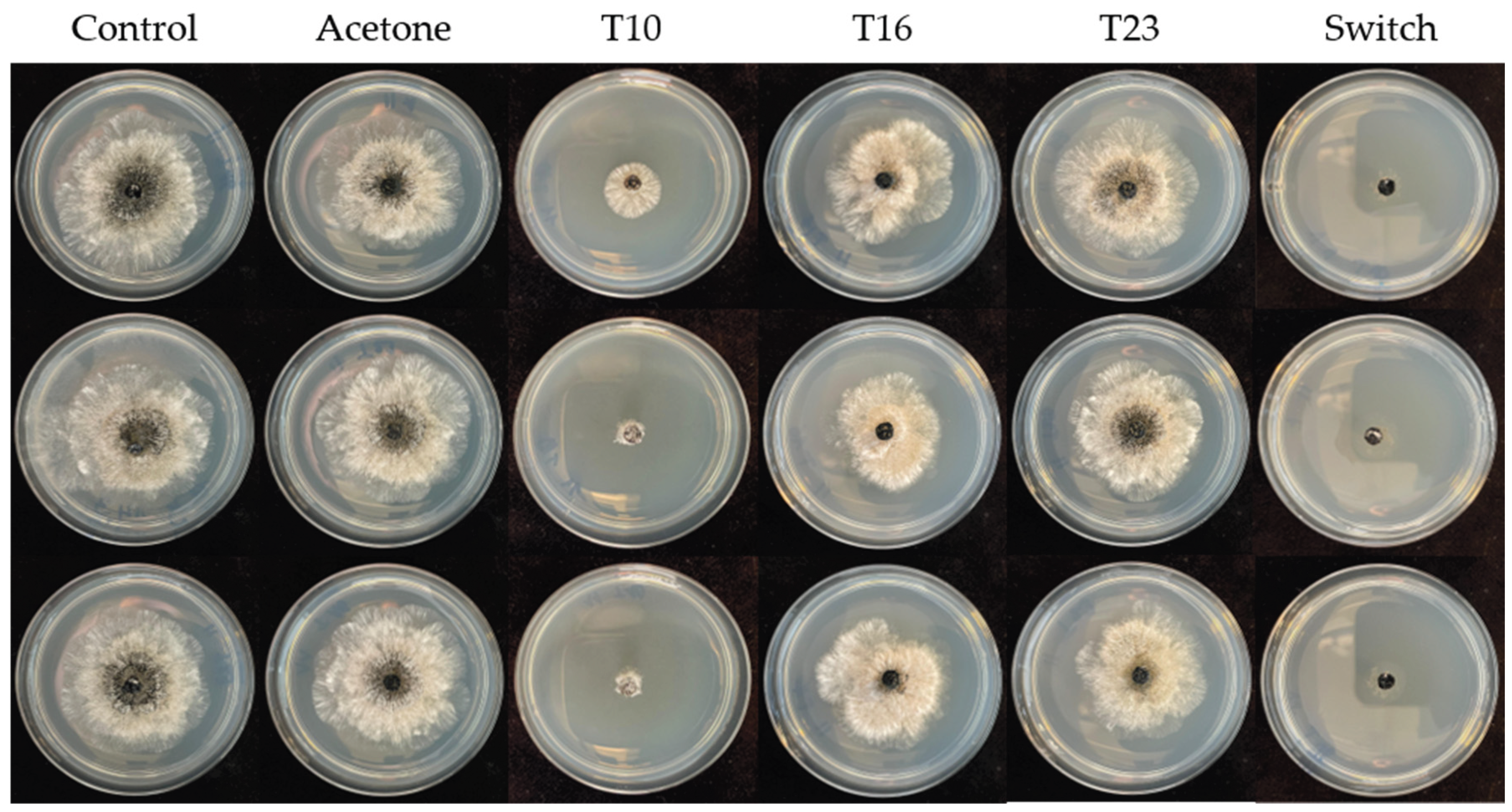
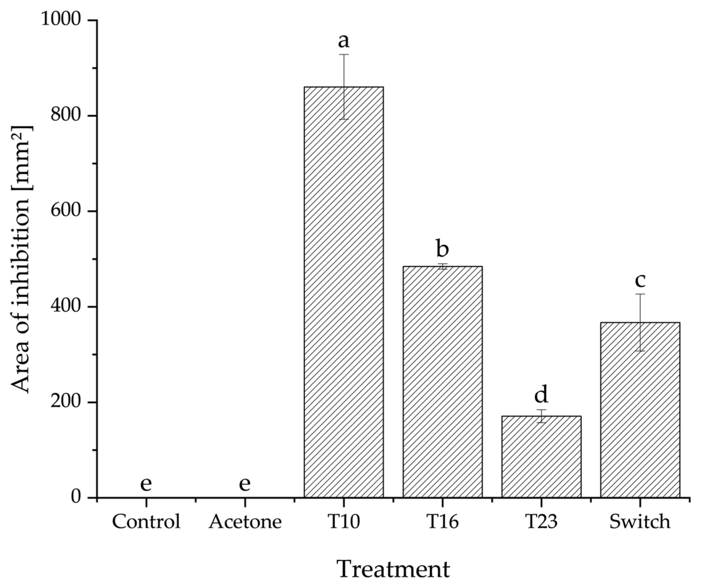
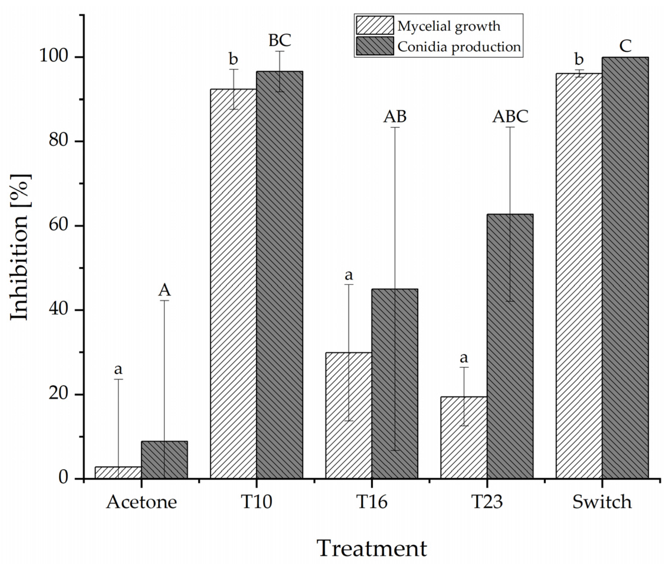
3.2. Poisoned Agar Plug Assay

The results show clear differences among treatments in their inhibitory effects on mycelial growth and conidia production of N. rosae (Figure 2, Appendix). Acetone insignificantly inhibited mycelial growth by 2.8 ± 20.9%. When treated with T. asperellum T23 or T. harzianum T16, N. rosae showed low growth inhibition with no significant differences compared to the acetone treatment. On the other hand, in the presence of T. koningiopsis T10 or Switch, N. rosae showed the highest growth inhibition measured by 92.3 ± 4.7% and 96.1 ± 0.9%, respectively. Both latter treatments did not differ significantly from each other.
Concentrations of conidia of N. rosae were highest in the GM7 control and acetone treatments at 4.19 × 10⁶ conidia mL-1 and 3.87 × 10⁶ conidia mL-1, respectively. While T. harzianum T16 and T. asperellum T23 led to a moderate spore formation inhibition (45 ± 38.3% and 62.7 ± 20.6%, respectively), N. rosae sporulation was almost completely inhibited when treated with T. koningiopsis T10 (96.5 ± 4.8%) or Switch (99.9 ± 0.04 %).
In general, conidia production appeared to be more sensitive than mycelial growth to T. harzianum T16 and T. asperellum T23 extracts, while T. koningiopsis T10 extract and Switch were highly effective against both parameters.

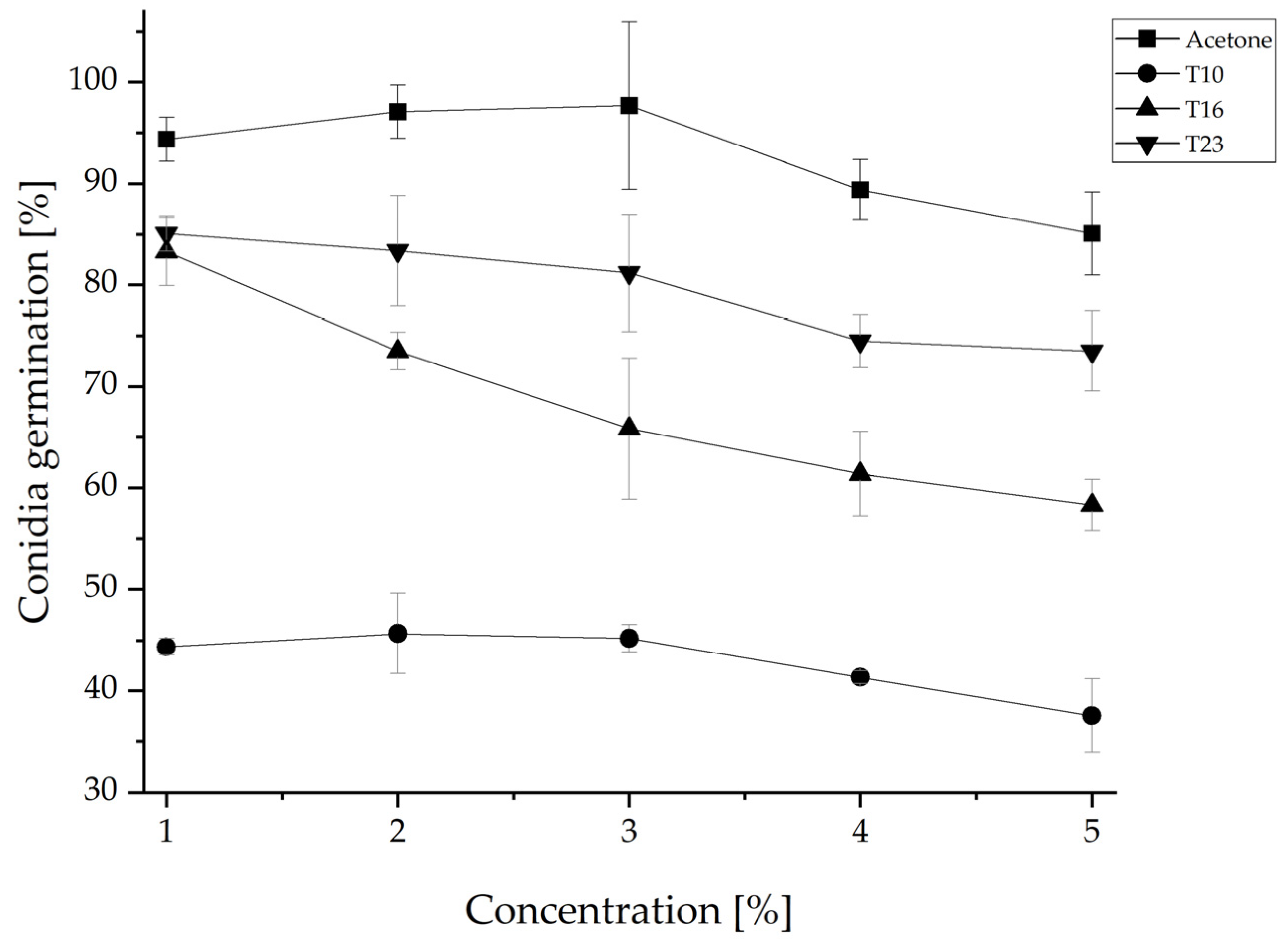
3.3. Inhibition of Conidia Germination of N. rosae by Trichoderma Crude Extracts

The effect of Trichoderma spp. extracts on conidia germination of N. rosae in liquid culture was evaluated using the MTT assay at concentrations ranging from 1- 5% and compared with the acetone control at equivalent concentrations. Conidia germination in the acetone control remained consistently high across all tested concentrations, with values above 90% at 1 - 3% acetone and only a slight decrease observed at higher concentrations (85.1 ± 4.1% at 5%, Figure 3). This indicates that acetone, even at increasing concentrations, had minimal inhibitory effects on conidia germination of N. rosae. In contrast, all Trichoderma spp. extracts significantly reduced conidia germination relative to the acetone control in a concentration-dependent manner. Extracts of T. asperellum T23 caused a moderate but consistent inhibition, with germination decreasing from 85.1 ± 1.7% at 1% to 73.5 ± 3.9% at 5%. T. harzianum T16 extracts proved to be significantly more effective in inhibiting conidia germination, showing a progressive reduction in germination, decreasing from 83.3 ± 3.4% at 1% to 58.3 ± 2.5% at 5%. However, extracts of T. koningiopsis T10 exhibited the strongest inhibitory effect, ranging between 44.4 ± 0.8% and 37.5 ± 3.6% depending on their concentration.

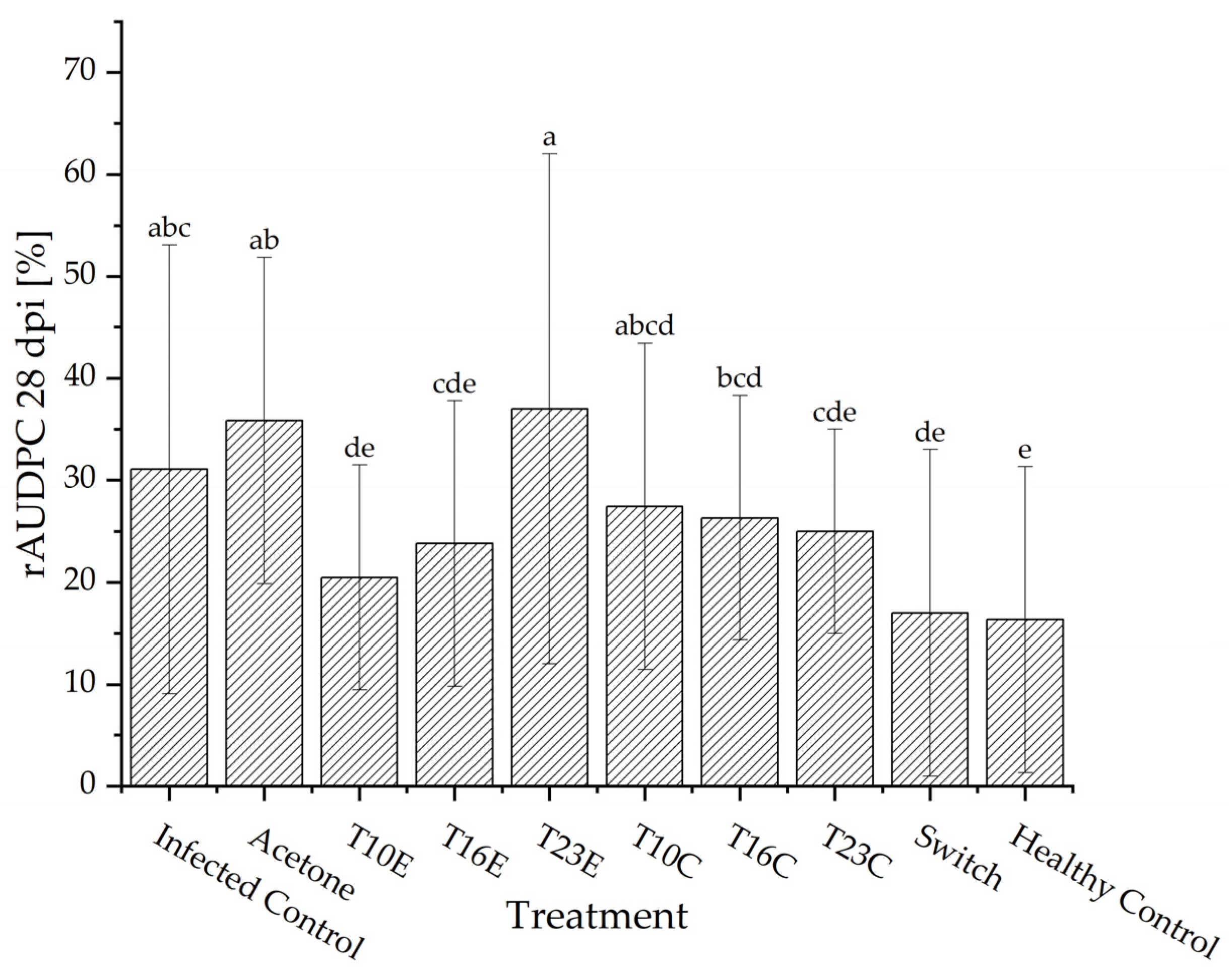
3.4. In Planta Assesment of the Effects of Crude Extracts and Conidia of Trichoderma spp. Against N. rosae

Leaves infected with N. rosae exhibited characteristic disease symptoms including brown leaf spots, discoloration of leaf tissue, leaf curling (Figure 4 A-C) and formation of characteristic black acervuli (Figure 4 D, E) and conidia (Figure 4 E, F), confirming the efficacy of the inoculation method and the identity of N. rosae.
Leaf infestation by N. rosae, expressed as rAUDPC (%) at 28 days post inoculation (dpi), was significantly affected by the different treatments. The infected control showed a high level of disease development (31.1 ± 22%), whereas the healthy control exhibited the lowest rAUDPC values (16.3 ± 15%; Figure 5).
Conidial (C) treatments of Trichoderma spp. (T10C, T16C, and T23C), as well as the extract (E) treatment T16E, produced intermediate rAUDPC values ranging between 23.8% and 27.5%. These treatments insignificantly reduced leaf infestation and remained significantly higher than the healthy control.
Among the extract treatments, clear differences were observed. Application of acetone and T23E resulted in relatively high rAUDPC values (35.8 ± 16% and 37 ± 25%, respectively) and did not differ significantly from the infected control, indicating no suppressive effect on disease development. In contrast, treatment with T10E significantly (20.5 ± 11%) reduced disease severity and was among the most effective extract treatments. The fungicide treatment (Switch) resulted in a strong reduction of disease development (17 ± 16%) and differed insignificantly from the healthy control, indicating high efficacy under the experimental conditions.

4. Discussion

The control of Neopestalotiopsis species, particularly N. rosae and N. clavispora, has become a critical priority due to their emergence as destructive pathogens causing root, crown, and fruit rot in strawberries and flower blight in ornamental plants [57]. Conventional management of these pathogens relies largely on chemical fungicides, which often fail to provide complete control and are associated with resistance development and potential health and environmental risks [58]. Consequently, research is shifting toward more effective and sustainable alternatives for integrated disease management [30,37,59,60,61].
In this study, we assessed the antifungal activity of Trichoderma-derived metabolites in vitro and evaluated their performance in planta.
In the direct-contact TLC assay, extracts of T. koningiopsis T10, T. harzianum T16, and T. asperellum T23 all inhibited mycelial growth of N. rosae, confirming antibiosis as an important mode of action (Figure 1). Among these, T. koningiopsis T10 produced inhibition zones that were 2.5-fold larger than those of the reference fungicide Switch, indicating a particularly strong antifungal effect. Bacterial BCAs including Bacillus cereus, B. amyloliquefaciens, and B. methylotrophicus inhibited N. clavispora growth by about 60–79% in vitro [62], which is lower than the very strong inhibition observed for T. koningiopsis T10 extracts in our experiments. Similarly, cell-free culture filtrates of three Bacillus strains (two B. velezensis and one B. subtilis) significantly suppressed N. clavispora in vitro, with inhibition ranging from 63.3% to 73.6%. In planta, application of these antagonists markedly reduced root rot severity (up to 67%) and enhanced superoxide dismutase, peroxidase, and catalase activities in leaves, indicating that their cell-free metabolites act in combination with induced host resistance [63].
Comparable patterns of strong in vitro inhibition have been observed with botanical extracts: clove (Syzygium aromaticum) and turmeric (Curcuma longa) extracts completely suppressed mycelial growth of Neopestalotiopsis sp. and Pseudopestalotiopsis sp., whereas ginger, lemongrass, and roselle caused only partial inhibition and mangosteen extract only had a minimal effect [64].
When mycelial growth was quantified following the amendment of the agar plugs of the pathogen with Trichoderma extracts, T. koningiopsis T10 achieved 92.3% inhibition of N. rosae after five days, approaching the 96.2% inhibition obtained with the reference fungicide (Figure 2, Appendix). In contrast, T. harzianum T16 and T. asperellum T23 extracts caused only moderate growth suppression. In vitro, volatile and non-volatile metabolites produced by T. asperellum CMT10 exhibited strong antifungal activity against N. clavispora, inhibiting mycelial growth by 79.7% and 69.8%, respectively [65]. Comparable strain-dependent variability has been reported for Trichoderma isolates antagonizing Fusarium sp., Phytophthora sp., and Botrytis spp. [46,66].
Using botanicals, a high in vitro efficacy has been observed: cinnamon essential oil nanoemulsions inhibited N. rosae growth at high dilutions and completely suppressed leaf spot severity [67].
We also quantified the effect of the Trichoderma extracts on N. rosae sporulation. T. harzianum T16 and T. asperellum T23 extracts reduced conidia production by 45% and 62.7%, respectively, whereas T. koningiopsis T10 extracts suppressed conidiation by 96.5%, close to the 99.9% reduction achieved by the reference fungicide (Figure 2). Since conidia are the primary propagules driving Neopestalotiopsis epidemics [41], such strong inhibition of conidia formation is highly relevant for disease management. Similar effects on sporulation have been documented for other Trichoderma-based BCAs: extracts of Trichoderma spp., for example, markedly inhibited sporulation of Fusarium moniliforme and Phakopsora pachyrhizi, resulting in reduced lesion development and lower disease severity [51,52]. Although explicit quantification of conidia reduction is less commonly reported, B. cereus reduced sporulation indirectly by impairing mycelial growth [68]. Compared with botanical agents, our data are similar in magnitude to clove and turmeric extracts, which fully suppressed Neopestalotiopsis and Pseudopestalotiopsis growth at high concentrations [64].
In the conidia germination assay, T. koningiopsis T10 extracts inhibited N. rosae germination by 62.5%, whereas T. harzianum T16 and T. asperellum T23 showed a different efficacy ranking than in the sporulation assay, indicating that conidia production and germination are differentially sensitive targets. Similar discrepancies between sporulation and germination responses have been reported for other pathosystems and with other BCAs, where fungicides or extracts can strongly suppress sporulation while having more moderate effects on germination, or vice versa [52]. At the same time, we observed that acetone concentrations of 4% or higher significantly suppressed germination in the MTT assay, so Trichoderma extract efficacy could be reliably evaluated only up to 3% extract concentration. By comparison, Switch was the only treatment in our study that completely inhibited N. rosae conidial germination, which is consistent with its known multi-stage activity against several strawberry pathogens [1,13,14,56,57]. Complete inhibition of conidial germination of Neopestalotiopsis spp. is rarely reported. For example, cinnamon essential oil nanoemulsions markedly reduce germination and infection structures formation but do not achieve full suppression [67]. Similarly, Bacillus-based BCAs often limit germination indirectly through induced resistance rather than through direct and complete inhibition [68].
Considering the limitations of in vitro assays in capturing the complexity of pathogen–host interactions, both live inocula and crude extracts of Trichoderma spp. were evaluated in planta under greenhouse conditions. Our results show that conidial suspensions only moderately and not significantly reduced N. rosae leaf spot. This outcome suggests that under our specific conditions (Trichoderma strain, inoculum density, timing, environmental parameters, and host cultivar), live inocula did not establish or act sufficiently to provide robust protection. In contrast, T. koningiopsis T10 crude extracts significantly suppressed N. rosae leaf spot severity, reaching control levels comparable to the fungicide Switch (Figure 5). This finding is particularly notable because Switch is regarded as one of the most effective fungicides against Neopestalotiopsis and other strawberry pathogens, with proven efficacy in reducing mycelial growth, sporulation, and infection in field and greenhouse settings [15,56]. Our results show that crude extracts from T. koningiopsis T10 can translate their pronounced in vitro activity into effective in planta disease control. This stands in line with several reports where Trichoderma spp. have shown strong in planta effects. For example, T. asperellum inhibited N. clavispora by 66.7% in vitro and reduced disease severity by 75.8% on strawberry, performing similarly to chemical fungicides [69]. Acosta-González et al. [56] also demonstrated that Trichoderma spp. significantly suppressed N. rosae incidence in strawberry. Similarly, CMT10 of T. asperellum achieved 63.1% biocontrol efficacy against strawberry root rot caused by N. clavispora and concurrently promoted plant growth under greenhouse conditions [70].
Similar extract-based in planta efficacy has also been reported for botanicals: cinnamon essential oil nanoemulsions reduced leaf spot severity by nearly 80% without phytotoxicity [67], and clove and turmeric extracts have shown strong protection of fruits and foliage against Pestalotiopsis-like fungi at high concentrations [64]. Moreover, bacterial BCAs such as B. cereus and B. amyloliquefaciens also reduced disease incidence and severity e.g. B. cereus Bce-2 lowered N. clavispora leaf spot while strongly activating host defense enzymes, and B. amyloliquefaciens isolates reduced strawberry root rot by >57% and promoted plant growth, but their suppression levels are often slightly lower than those achieved by the synthetic fungicides [68].
Taken together, our results show that crude extract of T. koningiopsis T10 exhibits very strong in vitro activity against N. rosae (on mycelial growth, sporulation, and, to a lesser extent, conidial germination) and can provide in planta disease control equivalent to a commercial fungicide, whereas Trichoderma inocula were not significantly effective under the conditions tested. The effective control of N. rosae by fungicides such as Switch may contribute to its currently low incidence in German strawberry cultivation. However, problems could arise in the future if fungicide performance declines, or N. rosae develops resistance [24,25]. Nevertheless, T. koningiopsis T10 extracts were equally effective as Switch in suppressing N. rosae, suggesting its potential as a suitable alternative to conventional chemical fungicides.

5. Conclusions

The emergence of novel pathogens poses a significant threat to the cultivation of strawberries. Continuous reliance on chemical fungicides has detrimental environmental consequences and is not a sustainable practice over an extended period. The present study thus focused on the identification of biological control agents against N. rosae. The results of this study demonstrate that extracts from Trichoderma spp. can effectively inhibit growth and spore formation of N. rosae. Among the isolates tested, T. koningiopsis T10 exhibited efficacy comparable to the reference fungicide (Switch) in terms of its impact on mycelial growth, spore formation, and conidia germination of N. rosae. In greenhouse trials with strawberry plants, T. koningiopsis T10 was likewise as effective as Switch in preventing strawberry leaf infestation.

Author Contributions

Conceptualization, T.E.S and A.E.-H.; methodology, T.E.S and A.E.-H.; software, T.E.S.; validation, A.E.-H. and R.T.V.; formal analysis, T.E.S.; investigation, T.E.S.; resources, A.E.-H. and R.T.V.; data curation, T.E.S.; writing—original draft preparation, T.E.S.; writing—review and editing, A.E.-H. and R.T.V.; visualization, T.E.S.; supervision, A.E.-H.; project administration, A.E.-H.; funding acquisition, A.E.-H. and R.T.V. All authors have read and agreed to the published version of the manuscript.

Funding

This research was funded by Deutsche Bundesstiftung Umwelt, grant number DBU AZ38050/01 and APC were covered by a 100% feature paper discount for T.E.S.

Data Availability Statement

Data is contained within the article; further inquiries can be directed at the corresponding authors.

Acknowledgments

The Deutsche Bundesstiftung Umwelt is greatly acknowledged for the financial support (Grant No. 38050/01). We also thank Isabelle Wölper for her help in executing the experiments.

Conflicts of Interest

The authors declare no conflicts of interest.

Abbreviations

The following abbreviations are used in this manuscript:
ANOVA Analysis of Variance
ANCOVA Analysis of Covariance
AUDPC Area Under The Disease Progress Curve
BCA Biological Control Agent
EtOAc Ethyl acetate
GM7 Glucose-Medium-7
IPM Integrated Pest Management
LSD Least Significant Difference
MDPI Multidisciplinary Digital Publishing Institute
MTT 2-(3,5-diphenyltetrazol-2-ium-2-yl)-4,5-dimethyl-1,3-thiazole bromide
rAUDPC Relative Area Under The Disease Progress Curve
6 PAP 6-pentyl-alpha-pyrone

Appendix A

Poisoned Agar Plug Assay with N. rosae

Figure A1. Mycelial growth of N. rosae after five days on GM7 medium amended with solvent (Acetone), extracts of Trichoderma spp., or Switch.
Preprints 197845 g0a1

References

  1. Ávila-Hernández, J.G.; León-Ramírez, C.G.; Abraham-Juárez, M. del R.; Tlapal-Bolaños, B.; Olalde-Portugal, V.; Délano-Frier, J.P.; Martínez-Antonio, A.; Aguilar-Zárate, P. Neopestalotiopsis Spp.: A Threat to Strawberry Production and Management. Horticulturae 2025, 11, 288. [Google Scholar] [CrossRef]
  2. Simpson, D. The Economic Importance of Strawberry Crops. In The Genomes of Rosaceous Berries and Their Wild Relatives; Hytönen, T., Graham, J., Harrison, R., Eds.; Springer International Publishing: Cham, 2018; pp. 1–7. ISBN 978-3-319-76020-9. [Google Scholar]
  3. Rajnish, K.; Gautam, H.R. Prevalence and Management of Pestalotia Leaf Spot (Pestalotia sp.) of Strawberry. International Journal of Economic Plants 2022, 9, 250–254. [Google Scholar] [CrossRef]
  4. Garrido, C.; González-Rodríguez, V.E.; Carbú, M.; Husaini, A.M.; Cantoral, J.M. Fungal Diseases of Strawberry and Their Diagnosis. In Strawberry: Growth, Development and Diseases; Husaini, A.M., Neri, D., Eds.; Cabi Publishing: Wallingford, UK, 2016; pp. pp. 157–195. ISBN 978-1-78064-663-3. [Google Scholar]
  5. Schierling, T.E.; Voegele, R.T.; El-Hasan, A. First Report on the Emergence of Neopestalotiopsis Rosae as a Severe Economic Threat to Strawberry Production in Germany. Microorganisms 2025, 13, 6. [Google Scholar] [CrossRef] [PubMed]
  6. Xiong, J.; Peng, S.; Liu, Y.; Yin, H.; Zhou, L.; Zhou, Z.; Tan, G.; Gu, Y.; Zhang, H.; Huang, J.; Meng, D. Soil Properties, Rhizosphere Bacterial Community, and Plant Performance Respond Differently to Fumigation and Bioagent Treatment in Continuous Cropping Fields. Frontiers in Microbiology 2022, 13, 923405. [Google Scholar] [CrossRef]
  7. Zhang, S.; Wu, J.; Chen, J.; Jun, S.; Yuan, Y.; Dai, X.; Wang, F.; Ma, Y. The Biological Control Effect of Bacillus cereus on Strawberry Leaf Spot Disease Caused by Neopestalotiopsis clavispora. Scientia Horticulturae 2024, 327, 112841. [Google Scholar] [CrossRef]
  8. Aljawasim, B.D.; Samtani, J.B.; Rahman, M. New Insights in the Detection and Management of Anthracnose Diseases in Strawberries. Plants 2023, 12, 3704. [Google Scholar] [CrossRef] [PubMed]
  9. Chung, P.-C.; Wu, H.-Y.; Wang, Y.-W.; Ariyawansa, H.A.; Hu, H.-P.; Hung, T.-H.; Tzean, S.-S.; Chung, C.-L. Diversity and Pathogenicity of Colletotrichum Species Causing Strawberry Anthracnose in Taiwan and Description of a New Species, Colletotrichum miaoliense sp. nov. Scientific Reports 2020, 10, 14664. [Google Scholar] [CrossRef]
  10. Gunnell, P.S.; Gubler, W.D. Taxonomy and Morphology of Colletotrichum Species Pathogenic to Strawberry. Mycologia 1992, 84, 157–165. [Google Scholar] [CrossRef]
  11. Koike, S.T.; Gordon, T.R. Management of Fusarium Wilt of Strawberry. Crop Protection 2015, 73, 67–72. [Google Scholar] [CrossRef]
  12. Basu, P.K. Verticillium Disease of Strawberries. Canadian Journal of Botany 1961, 39, 165–196. [Google Scholar] [CrossRef]
  13. Baggio, J.S.; Rebello, C.S.; de Morais, M.B.; Marin, M.V.; Gama, A.B.; Forcelini, B.B.; Mertely, J.C.; Peres, N.A. Efficacy of Single- and Multi-Site Fungicides Against Neopestalotiopsis spp. of Strawberry. Plant Disease 2023, 107, 2177–2184. [Google Scholar] [CrossRef]
  14. Essa, T.A.; Kamel, S.M.; Ismail, A.M.; El-Ganainy, S. Characterization and Chemical Control of Neopestalotiopsis rosae the Causal Agent of Strawberry Root and Crown Rot in Egypt. Egyptian Journal of Phytopathology 2018, 46, 1–19. [Google Scholar] [CrossRef]
  15. Rebollar-Alviter, A.; Silva-Rojas, H.V.; Fuentes-Aragón, D.; Acosta-González, U.; Martínez-Ruiz, M.; Parra-Robles, B.E. An Emerging Strawberry Fungal Disease Associated with Root Rot, Crown Rot and Leaf Spot Caused by Neopestalotiopsis rosae in Mexico. Plant Disease 2020, 104, 2054–2059. [Google Scholar] [CrossRef]
  16. Ara, M.; Monzur, S.; Saand, M.; Islam, R.; Alam, S.; Hossain, M. The First Report of Pestalotiopsis sp. Causing Crown Rot Disease on Strawberry (Fragaria X ananassa Duch.) in Bangladesh and Evaluation of Fungicide Activity. International Journal of Biosciences 2017, 11, 350–358. [Google Scholar]
  17. Baggio, J.S.; Forcelini, B.B.; Wang, N.-Y.; Ruschel, R.G.; Mertely, J.C.; Peres, N.A. Outbreak of Leaf Spot and Fruit Rot in Florida Strawberry Caused by Neopestalotiopsis spp. Plant Disease 2021, 105, 305–315. [Google Scholar] [CrossRef]
  18. Chandana, R.; Poonacha, T.T.; Chethan, D.; Karan, R.; Kruthika, R.; Khan, F.; Ashwini, K.S.; Bevanur, A.; Vani, Y.; Ramesh, G.V.; Palanna, K.B. Neopestalotiopsis rosae, a Novel Pathogen Causing Leaf Blight and Crown Rot of Strawberries in India. Physiological and Molecular Plant Pathology 2024, 133, 102377. [Google Scholar] [CrossRef]
  19. Kaur, H.; Gelain, J.; Marin, M.V.; Peres, N.A.; Schnabel, G. Development of a Molecular Tool for Identification of a New Neopestalotiopsis sp. Associated with Disease Outbreaks on Strawberry. Plant Disease 2023, 107, 1544–1549. [Google Scholar] [CrossRef]
  20. Sun, Q.; Harishchandra, D.; Jia, J.; Zuo, Q.; Zhang, G.; Wang, Q.; Yan, J.; Zhang, W.; Li, X. Role of Neopestalotiopsis rosae in Causing Root Rot of Strawberry in Beijing, China. Crop Protection 2021, 147, 105710. [Google Scholar] [CrossRef]
  21. Rotondo, F.; Klass, T.L.; Scott, K.; McCartney, M.; Jacobs, J.M.; Lewis Ivey, M.L. First Report of Neopestalotiopsis Disease in Ohio Caused by an Emerging and Novel Species of Neopestalotiopsis on Strawberry. Plant Disease 2023, 107, 940. [Google Scholar] [CrossRef]
  22. Bund. Verzeichnis Zugelassener Pflanzenschutzmittel. Available online: https://psm-zulassung.bvl.bund.de/psm/jsp/ (accessed on 9 October 2025).
  23. Gupta, R.C; Gupta, P.K. Chapter 45 - Toxicity of Fungicides. In Veterinary Toxicology (Third Edition); Gupta, R.C., Ed.; Academic Press: London, UK, 2025; pp. 581–593. ISBN 978-0-12-811410-0. [Google Scholar]
  24. Amiri, A.; Heath, S.M.; Peres, N.A. Phenotypic Characterization of Multifungicide Resistance in Botrytis cinerea Isolates from Strawberry Fields in Florida. Plant Disease 2013, 97, 393–401. [Google Scholar] [CrossRef]
  25. Forcelini, B.B.; Seijo, T.E.; Amiri, A.; Peres, N.A. Resistance in Strawberry Isolates of Colletotrichum acutatum from Florida to Quinone-Outside Inhibitor Fungicides. Plant Disease 2016, 100, 2050–2056. [Google Scholar] [CrossRef]
  26. Karuppuchamy, P.; Venugopal, S. Integrated Pest Management. In Ecofriendly Pest Management for Food Security; Omkar, Ed.; Academic Press: San Diego, 2016; pp. 651–684. ISBN 978-0-12-803265-7. [Google Scholar]
  27. Stenberg, J.A. A Conceptual Framework for Integrated Pest Management. Trends in Plant Science 2017, 22, 759–769. [Google Scholar] [CrossRef]
  28. Ghisalberti, E.L.; Rowland, C.Y. Antifungal Metabolites from Trichoderma harzianum. Journal of Natural Products 1993, 56, 1799–1804. [Google Scholar] [CrossRef]
  29. El-Hasan, A.; Walker, F.; Schöne, J.; Buchenauer, H. Antagonistic Effect of 6-Pentyl-Alpha-Pyrone Produced by Trichoderma harzianum toward Fusarium moniliforme. Journal of Plant Disease and Protection 2007, 114, 62–68. [Google Scholar] [CrossRef]
  30. Abbey, J.A.; Percival, D.; Abbey, Lord; Asiedu, S.K.; Prithiviraj, B.; Schilder, A. Biofungicides as Alternative to Synthetic Fungicide Control of Grey Mould (Botrytis cinerea) – Prospects and Challenges. Biocontrol Science and Technology 2019, 29, 207–228. [Google Scholar] [CrossRef]
  31. Anand, T.; Chandrasekaran, A.; Kuttalam, S.; Senthilraja, G.; Samiyappan, R. Integrated Control of Fruit Rot and Powdery Mildew of Chilli Using the Biocontrol Agent Pseudomonas fluorescens and a Chemical Fungicide. Biological Control 2010, 52, 1–7. [Google Scholar] [CrossRef]
  32. Jochum, C.C.; Osborne, L.E.; Yuen, G.Y. Fusarium Head Blight Biological Control with Lysobacter enzymogenes Strain C3. Biological Control 2006, 39, 336–344. [Google Scholar] [CrossRef]
  33. Vinale, F.; Sivasithamparam, K.; Ghisalberti, E.L.; Marra, R.; Woo, S.L.; Lorito, M. Trichoderma–Plant–Pathogen Interactions. Soil Biology and Biochemistry 2008, 40, 1–10. [Google Scholar] [CrossRef]
  34. Benítez, T.; Rincón, A.M.; Limón, M.C.; Codón, A.C. Mecanismos de Biocontrol de Cepas de Trichoderma. International Microbiology 2004, 7, 249–260. [Google Scholar]
  35. Verma, M.; Brar, S.K.; Tyagi, R.D.; Surampalli, R.Y.; Valéro, J.R. Antagonistic Fungi, Trichoderma spp.: Panoply of Biological Control. Biochemical Engineering Journal 2007, 37, 1–20. [Google Scholar] [CrossRef]
  36. Gruber, S.; Seidl-Seiboth, V. Self versus Non-Self: Fungal Cell Wall Degradation in Trichoderma. Microbiology 2012, 158, 26–34. [Google Scholar] [CrossRef]
  37. Schierling, T.E.; Vogt, W.; Voegele, R.T.; El-Hasan, A. Efficacy of Trichoderma spp. and Kosakonia sp. Both Independently and Combined with Fungicides against Botrytis cinerea on Strawberries. Antibiotics 2024, 13, 912. [Google Scholar] [CrossRef] [PubMed]
  38. Schuster, A.; Schmoll, M. Biology and Biotechnology of Trichoderma. Applied Microbiology and Biotechnology 2010, 87, 787–799. [Google Scholar] [CrossRef] [PubMed]
  39. Steyaert, J.M.; Ridgway, H.J.; Elad, Y.; Stewart, A. Genetic Basis of Mycoparasitism: A Mechanism of Biological Control by Species of Trichoderma. New Zealand Journal of Crop and Horticultural Science 2003, 31, 281–291. [Google Scholar] [CrossRef]
  40. Mukherjee, P.K.; Mendoza-Mendoza, A.; Zeilinger, S.; Horwitz, B.A. Mycoparasitism as a Mechanism of Trichoderma-Mediated Suppression of Plant Diseases. Fungal Biology Reviews 2022, 39, 15–33. [Google Scholar] [CrossRef]
  41. Nawrocka, J.; Małolepsza, U. Diversity in Plant Systemic Resistance Induced by Trichoderma. Biological Control 2013, 67, 149–156. [Google Scholar] [CrossRef]
  42. Bisen, K.; Keswani, C.; Patel, J.S.; Sarma, B.K.; Singh, H.B. Trichoderma spp.: Efficient Inducers of Systemic Resistance in Plants. In Microbial-mediated Induced Systemic Resistance in Plants; Choudhary, D.K., Varma, A., Eds.; Springer Nature: Singapore, 2016; pp. 185–195. ISBN 978-981-10-0388-2. [Google Scholar]
  43. Sivan, A.; Chet, I. Degradation of Fungal Cell Walls by Lytic Enzymes of Trichoderma harzianum. Microbiology 1989, 135, 675–682. [Google Scholar] [CrossRef]
  44. Metcalf, D.A.; Wilson, C.R. The Process of Antagonism of Sclerotium cepivorum in White Rot Affected Onion Roots by Trichoderma koningii. Plant Pathology 2001, 50, 249–257. [Google Scholar] [CrossRef]
  45. Santos, A.J.C.; Divina, C.C.; Pineda, F.G. In Vitro Evaluation of the Antagonistic Activity of Trichoderma sp. against Fusarium verticillioides. International Journal of Agricultural Technology 2017, 13, 2539–2548. [Google Scholar]
  46. El-Hasan, A.; Schöne, J.; Höglinger, B.; Walker, F.; Voegele, R.T. Assessment of the Antifungal Activity of Selected Biocontrol Agents and Their Secondary Metabolites against Fusarium graminearum. European Journal of Plant Pathology 2018, 150, 91–103. [Google Scholar] [CrossRef]
  47. Vinale, F.; Sivasithamparam, K.; Ghisalberti, E.L.; Woo, S.L.; Nigro, M.; Marra, R.; Lombardi, N.; Pascale, A.; Ruocco, M.; Lanzuise, S.; Manganiello, G.; Lorito, M. Trichoderma Secondary Metabolites Active on Plants and Fungal Pathogens. The Open Mycology Journal 2014, 8, 127–139. [Google Scholar] [CrossRef]
  48. Vinale, F.; Marra, R.; Scala, F.; Ghisalberti, E.L.; Lorito, M.; Sivasithamparam, K. Major Secondary Metabolites Produced by Two Commercial Trichoderma Strains Active against Different Phytopathogens. Letters in Applied Microbiology 2006, 43, 143–148. [Google Scholar] [CrossRef] [PubMed]
  49. Hermosa, R.; Cardoza, R.E.; Rubio, M.B.; Gutiérrez, S.; Monte, E. Secondary Metabolism and Antimicrobial Metabolites of Trichoderma. In Biotechnology and Biology of Trichoderma; Gupta, V.K., Schmoll, M., Herrera-Estrella, A., Upadhyay, R.S., Druzhinina, I., Tuohy, M.G., Eds.; Elsevier: Amsterdam, NL, 2014; pp. 125–137. ISBN 978-0-444-59576-8. [Google Scholar]
  50. Keswani, C.; Mishra, S.; Sarma, B.K.; Singh, S.P.; Singh, H.B. Unraveling the Efficient Applications of Secondary Metabolites of Various Trichoderma spp. Applied Microbiology and Biotechnology 2014, 98, 533–544. [Google Scholar] [CrossRef]
  51. El-Hasan, A. Potential of Trichoderma harzianum Rifai and Its Secondary Metabolites in Controlling Seedling Blight of Maize Incited by Fusarium moniliforme Sheld; Cuvillier Verlag, 2008; ISBN 978-3-7369-2800-8. [Google Scholar]
  52. El-Hasan, A.; Walker, F.; Klaiber, I.; Schöne, J.; Pfannstiel, J.; Voegele, R.T. New Approaches to Manage Asian Soybean Rust (Phakopsora pachyrhizi) Using Trichoderma spp. or Their Antifungal Secondary Metabolites. Metabolites 2022, 12, 507. [Google Scholar] [CrossRef]
  53. Khan, R.A.A.; Najeeb, S.; Hussain, S.; Xie, B.; Li, Y. Bioactive Secondary Metabolites from Trichoderma spp. against Phytopathogenic Fungi. Microorganisms 2020, 8, 817. [Google Scholar] [CrossRef] [PubMed]
  54. Vinale, F.; Sivasithamparam, K.; Ghisalberti, E.L.; Marra, R.; Barbetti, M.J.; Li, H.; Woo, S.L.; Lorito, M. A Novel Role for Trichoderma Secondary Metabolites in the Interactions with Plants. Physiological and Molecular Plant Pathology 2008, 72, 80–86. [Google Scholar] [CrossRef]
  55. Rieker, M.E.G.; Lutz, M.A.; El-Hasan, A.; Thomas, S.; Voegele, R.T. Hyperspectral Imaging and Selected Biological Control Agents for the Management of Fusarium Head Blight in Spring Wheat. Plants 2023, 12, 3534. [Google Scholar] [CrossRef]
  56. Forbes, G.A.; Pérez, W.; Adrade-Piedra, J. Field Assessment of Resistance in Potato to Phytophthora infestans: International Cooperators Guide. In International Potato Center; Lima, PE, 2014; ISBN 978-92-9060-440-2. [Google Scholar]
  57. Maharachchikumbura, S.S.N.; Hyde, K.D.; Groenewald, J.Z.; Xu, J.; Crous, P.W. Pestalotiopsis Revisited. Studies in Mycology 2014, 79, 121–186. [Google Scholar] [CrossRef]
  58. Elmer, P. a. G.; Reglinski, T. Biosuppression of Botrytis cinerea in Grapes. Plant Pathology 2006, 55, 155–177. [Google Scholar] [CrossRef]
  59. Vreysen, M.J.B.; Robinson, A.S.; Hendrichs, J.; Kenmore, P. Area-Wide Integrated Pest Management (AW-IPM): Principles, Practice and Prospects. In Proceedings of the Area-Wide Control of Insect Pests; Vreysen, M.J.B., Robinson, A.S., Hendrichs, J., Eds.; Springer Netherlands: Dordrecht, 2007; pp. 3–33. [Google Scholar]
  60. He, D.; Zhan, J.; Xie, L. Problems, Challenges and Future of Plant Disease Management: From an Ecological Point of View. Journal of Integrative Agriculture 2016, 15, 705–715. [Google Scholar] [CrossRef]
  61. National Research Council. Ecologically Based Pest Management: New Solutions for a New Century. In National Academic Press; Washington DC, USA, 1996; ISBN 978-0-309-05330-3. [Google Scholar]
  62. Ramallo, A.; Namen, S.; Salazar, S.; Heredia, A.; Kirschbaum, D. New Approaches for the Sustainable Management of Neopestalotiopsis clavispora, the Causal Agent of Strawberry Crown and Root Rot in Tucumán, Argentina. Revista Agronómica del Noroeste Argentino 2024, 44, 10–14. [Google Scholar] [CrossRef]
  63. Ning, J.; Ning, T.; Jin, L.; Li, Q.; Niu, Y.; Chen, Z.; Han, C.; Tang, Y.; Deng, C.; Xie, Y.; Zhao, M.; Cui, X.; Li, J. Biocontrol Effects of Bacillus velezensis and Bacillus subtilis against Strawberry Root Rot Caused by Neopestalotiopsis clavispora. Frontiers in Microbiology 2025, 16, 1683291. [Google Scholar] [CrossRef]
  64. Darapanit, A.; Boonyuen, N.; Leesutthiphonchai, W.; Nuankaew, S.; Piasai, O. Identification, Pathogenicity and Effects of Plant Extracts on Neopestalotiopsis and Pseudopestalotiopsis Causing Fruit Diseases. Scientific Reports 2021, 11, 22606. [Google Scholar] [CrossRef]
  65. Liu, P.; Yang, R.; Wang, Z.; Ma, Y.; Ren, W.; Wei, D.; Ye, W. Biocontrol Potential of Trichoderma asperellum CMT10 against Strawberry Root Rot Disease. Horticulturae 2024, 10, 246. [Google Scholar] [CrossRef]
  66. El-Hasan, A.; Walker, F.; Schöne, J.; Buchenauer, H. Detection of Viridiofungin A and Other Antifungal Metabolites Excreted by Trichoderma harzianum Active against Different Plant Pathogens. European Journal of Plant Pathology 2009, 124, 457–470. [Google Scholar] [CrossRef]
  67. Tran, T.-N.-M.; Vu, N.-B.-D.; Nguyen, M.-H. Antifungal Activity of Essential Oil-Encapsulated Lipid Nanoemulsions against Neopestalotiopsis rosae Causing Leaf Spot on Strawberry. Journal of Plant Diseases and Protection. 2023, 130, 823–832. [Google Scholar] [CrossRef]
  68. Zhang, S.; Wu, J.; Chen, J.; Jun, S.; Yuan, Y.; Dai, X.; Wang, F.; Ma, Y. The Biological Control Effect of Bacillus cereus on Strawberry Leaf Spot Disease Caused by Neopestalotiopsis clavispora. Scientia Horticulturae 2024, 327, 112841. [Google Scholar] [CrossRef]
  69. Amrutha, P.; Vijayaraghavan, R. Evaluation of Fungicides and Biocontrol Agents against Neopestalotiopsis clavispora Causing Leaf Blight of Strawberry (Fragaria x ananassa Duch.). International Journal of Current Microbiology and Applied Sciences 2018, 7, 622–628. [Google Scholar] [CrossRef]
  70. Liu, P.; Yang, R.; Wang, Z.; Ma, Y.; Ren, W.; Wei, D.; Ye, W. Biocontrol Potential of Trichoderma asperellum CMT10 against Strawberry Root Rot Disease. Horticulturae 2024, 10, 246. [Google Scholar] [CrossRef]
Figure 1. Mycelial growth inhibition of N. rosae by Trichoderma spp. extracts and Switch on TLC plates. Bars represent means and error bars indicate standard deviation. Treatments with the same letter are not significantly different at a probability level of p = 0.05.
Preprints 197845 g001
Figure 2. Inhibition of mycelial growth and conidia production of N. rosae following treatment with Trichoderma spp. extracts, or the fungicide Switch. Bars represent means and error bars indicate standard deviation. Treatments with the same letter for mean values are not significantly different from each other at a probability of p = 0.05.
Preprints 197845 g002
Figure 3. Effect of Trichoderma spp. extracts on conidia germination of N. rosae in liquid culture using the MTT assay. Bars represent standard deviation.
Preprints 197845 g003
Figure 4. (A-C) Symptoms of strawberry leaves due to N. rosae infection. (C, D) Acervuli on strawberry leaf. (E) Conidia on strawberry leaf. (F) typical conidia under light microscope.
Preprints 197845 g004
Figure 5. Effect of extracts (E) and conidia (C) of Trichoderma spp. on the infestation of leaves with N. rosae. Bars represent means and error bars indicate the standard deviation. Treatments with the same letter are not significantly different from each other at a probability of p = 0.05.
Preprints 197845 g005
Disclaimer/Publisher’s Note: The statements, opinions and data contained in all publications are solely those of the individual author(s) and contributor(s) and not of MDPI and/or the editor(s). MDPI and/or the editor(s) disclaim responsibility for any injury to people or property resulting from any ideas, methods, instructions or products referred to in the content.
Copyright: This open access article is published under a Creative Commons CC BY 4.0 license, which permit the free download, distribution, and reuse, provided that the author and preprint are cited in any reuse.
Prerpints.org logo

Preprints.org is a free preprint server supported by MDPI in Basel, Switzerland.

Subscribe

Disclaimer

Terms of Use

Privacy Policy

Privacy Settings

© 2026 MDPI (Basel, Switzerland) unless otherwise stated