
Submitted:
06 February 2026
Posted:
09 February 2026
You are already at the latest version
Abstract
This article presents electron paramagnetic resonance (EPR) studies of commercial polyester resin-based powder coatings before and after laser irradiation. Two industrial powder coatings were examined. The main objective was to evaluate whether localized laser irradiation leads to measurable changes in the behavior of paramagnetic centers and whether these changes are comparable to effects observed under thermal treatment. EPR spectroscopy was chosen due to its high sensitivity to unpaired electrons and paramagnetic defects in complex polymer systems. Measurements were carried out in the temperature range of 300 to 500 K for nonirradiated samples and after laser irradiation. The analysis is focused on EPR parameters, including signal intensity, linewidth, resonance field, g_eff and integrated intensity. After laser irradiation, clear and reproducible differences in the EPR spectra were observed in comparison to the nonirradiated state. Changes in signal amplitude, linewidth, and resonance field were observed, suggesting changes in the temperature-dependent behavior of signals associated with paramagnetic centers. Temperature-dependent measurements revealed different trends before and after laser exposure, indicating that localized laser treatment produces measurable effects of predominantly thermal character in polyester resin–based powder coatings. The obtained results confirm that EPR spectroscopy is a sensitive and effective tool for monitoring laser- and temperature-induced changes in powder coatings.
Keywords:
powder coatings
; polyester resin-based coatings
; electron paramagnetic resonance
; laser irradiation
; thermal effects
; laser-assisted processing
1. Introduction
The undertaken research on the influence of laser radiation on powder coatings results from the need to better understand how external energy input affects materials commonly used as protective and decorative finishes in industrial applications. Powder coatings are widely applied in the architectural, automotive, appliance, and structural sectors [1,2] due to their good mechanical durability, chemical resistance, and aesthetic properties [3]. However, their final performance strongly depends on the processing conditions and, in particular, on the way thermal energy is delivered during curing, which determines the degree of polymer-network development and the stability of the coating.
Polyester resin–based coatings belong to the most commonly used powder coating systems [4,5]. Polyester systems are also known to respond to UV and laser treatment through measurable physical and chemical changes [6]. Their polymer structure, combined with pigments and functional additives, makes them sensitive to external stimuli, such as temperature and radiation. Exposure to additional energy can modify the local environment of structural defects or other paramagnetic centers present in the material. Understanding how such changes manifest themselves in measurable physical parameters is essential for assessing coating behavior under processing or service conditions. Electron paramagnetic resonance (EPR) spectroscopy is one of the most sensitive experimental techniques [7] for detecting and characterizing paramagnetic centers in solids and complex materials. Because EPR probes unpaired electrons, the recorded spectrum represents a overlapping of signals [8] originating from different types of paramagnetic centers that may be present in the coating. These centers can be associated with structural defects, radical species formed in the organic matrix, pigment-related components, or trace paramagnetic ions introduced with fillers and additives. As a result, EPR spectra provide a direct experimental fingerprint of the paramagnetic structure of powder coatings. In practical terms, EPR parameters are used to describe the character of EPR signals and to compare different materials or processing conditions. Changes in linewidth and intensity reflect modifications in relaxation processes and interactions within the material [9], while shifts of the resonance field and indicate changes in the local magnetic environment of the paramagnetic centers. Therefore, systematic monitoring of these quantities allows us to describe the overall character of changes that occur in the coating without imposing a specific microscopic mechanism that is not directly confirmed by experiment. Temperature-dependent EPR measurements constitute an important extension of this approach [10]. Increasing the temperature affects the molecular mobility in polymer systems and may influence the balance between different relaxation pathways and paramagnetic interactions. Consequently, recording EPR spectra as a function of temperature enables observation of how parameters evolve under controlled thermal conditions. Such measurements are widely used to assess the behavior of paramagnetic centers in solid materials and to compare their response to different types of energy input.
Previous EPR studies of powder coatings have shown that their spectra may contain multiple overlapping components [11] and that the relative contribution of these components depends on the composition of the coating and the processing history. In particular, differences between coatings and changes induced by external treatments can be detected by variations in parameters and their temperature dependence. This makes EPR a suitable diagnostic tool for comparative studies of powder coatings subjected to different processing conditions. In the present work, laser irradiation is employed as a controlled method to locally deliver energy to the powder coating material. The laser used operates in the visible range and is applied under well-defined conditions [12]. Because the detailed electronic energy level structure of the complex polymer–pigment system is not known [13], laser treatment is considered primarily as a source of localized thermal input, as also demonstrated in alkoxyamine-based laser-triggered systems [14] rather than as a trigger for specific electron transitions. Therefore, the discussion is restricted to the experimentally observable effects reflected in the EPR parameters and their temperature evolution, without invoking detailed photoelectronic mechanisms. The motivation for this approach is related to the technological context of powder coating processing. Conventional curing requires prolonged heating in industrial ovens, which is energy-intensive and limits flexibility, especially for large or fixed structures [15,16]. Localized laser treatment represents a potential alternative or complementary method of supplying energy directly to the coating [17]. From an experimental perspective, an essential first step is to verify whether laser exposure leads to measurable and systematic changes [18] in EPR spectra that can be compared with changes induced by temperature.
Therefore, the purpose of the present study is to evaluate how controlled laser treatment and temperature influence the EPR parameters of paramagnetic centers in commercial polyester resin–based powder coatings. The analysis is based on a comparative approach, involving two different coatings and measurements performed before and after laser irradiation over a wide temperature range. By focusing on changes in intensity, linewidth, resonance field, and , the study seeks to establish a clear experimental basis for further investigations of laser-assisted approaches to powder coating processing. This work provides a methodological basis for further studies aimed at evaluating laser-assisted approaches as potential alternatives or complements to conventional thermal curing processes used in industrial powder coating technologies.
2. Materials and Methods
In this study, two commercial polyester resin–based powder coatings were examined in their original powder form, differing in color. Both were manufactured by Inver Polska Sp. z o.o., (Dębica, Poland), a subsidiary of the global corporation Sherwin-Williams (Cleveland, USA). The samples are further referred to in this work as Sample 1 (red powder coating) and Sample 2 (black powder coating).
EPR measurements were performed using a Bruker Elexsys E580 spectrometer (Bruker, Rheinstetten, Germany) operating in the X-band (≈9.5 GHz).. The resonance is determined by the EPR equation:
where h is the Planck constant, ν is the microwave frequency, is the Bohr magneton, g is the spectroscopic splitting factor, and is the resonance field. EPR spectra were recorded under identical instrumental settings. A 10 dB microwave attenuation was applied to ensure stable and comparable signal amplitudes and to minimize saturation effects. The magnetic field modulation amplitude was set to 5 G, providing a compromise between sensitivity and resolution and limiting artificial modulation broadening of the recorded lines. Measurements were carried out in the temperature range of 300 to 500 K using a quartz nitrogen cryostat with a 41131 VT digital controller (Bruker, Massachusetts, USA).
For laser treatment, each powder sample was placed in a quartz capillary tube and irradiated using a continuous-wave semiconductor laser (λ = 532 nm, output power 300 mW, beam diameter ≈1.5 mm, class 3B) for 150 s. The beam was directed perpendicularly to the capillary axis to provide reproducible irradiation geometry. Because the irradiation is localized, only a part of the powder volume in the capillary is exposed to the laser beam. Laser treatment is considered primarily as a localized energy input of predominantly thermal character, and its effect is experimentally evaluated by changes in the EPR spectra and extracted parameters.
After irradiation, the quartz capillaries containing the samples were placed on the spectrometer. The analysis was based on temperature-dependent changes of measurable EPR parameters, including signal intensity, peak-to-peak linewidth (Δ), resonance field (), effective g factor (), and integrated intensity of the spectra.
3. Results & Discussion
3.1. EPR Measurements of Sample 1
To establish the effect of laser irradiation on powder paints, the EPR spectra of Sample 1 were measured at 300 K, before and after laser exposure. A direct comparison of the spectra enables identification of changes in signal shape and amplitude induced by laser treatment. Figure 1 presents the EPR spectra of Sample 1 recorded before and after laser irradiation.
EPR spectra are dominated by a single broad line. The shape of the recorded line shape is relatively smooth and does not show any narrow features that could be clearly separated. Small deviations from the ideal theoretical Lorentzian shapes are visible in the lower part of the line. Because these deviations are small, no line-shaped decomposition was performed in this study, although it is clear that the observed broad line is a sum of at least two lines. Additionally, we observe a narrow line in the field around 3300 Gauss. The EPR spectra of the sample were also measured after 150 s of exposure to green laser radiation. Measurements were taken as a function of temperature. Figure 2 shows the temperature-dependent EPR spectra of Sample 1 measured before and after laser irradiation.
For the nonirradiated sample, the intensity of the EPR spectra increases with temperature up to about 370 K. Above this value, the intensity decreases slightly, and the line becomes broader. Shifts of the resonance field toward lower magnetic field values were also observed. In the EPR spectra for the sample after laser irradiation, the EPR line generally shows a larger amplitude. The shape and position of the line remain comparable across the temperature range, with minor broadening.
To characterize the observed changes, all parameters of the EPR line were obtained, such as intensity, peak-to-peak linewidth, resonance field position, , integrated intensity, and inverse integrated intensity. Their systematic evolution with temperature provides a detailed characterization of the behavior of paramagnetic centers. Figure 3 summarizes the temperature dependence of the EPR parameters of Sample 1 before and after laser irradiation.
With increasing temperature, a general increase in signal intensity is observed. For the nonirradiated sample, this increase is up to a temperature of approximately 370 K, which appears to be a characteristic temperature above which noticeable changes occur in all parameters. For the sample after laser irradiation, the intensity is higher and its increase with temperature is more pronounced. The linewidth shows moderate changes. The resonance field shows a slight shift toward lower magnetic field values. These small but consistent shifts in the recorded spectra indicate measurable temperature-dependent modifications resulting from temperature. The effective g-factor remains almost constant up to around 350 K and then shows an increase at higher temperatures]. The overall changes in geff are small but clearly visible. At temperatures above approximately 450 K, an additional EPR line becomes visible in the field of approximately 3100 Gs. Its intensity and peak-to-peak linewidth increase with increasing temperature. The parameters of this additional line are summarized in Table 1.
The resonance field remains stable at around 3100 G, confirming the stability of this parameter. The increase in intensity with temperature indicates that the line becomes more distinct in the EPR spectra, suggesting a connection between this line and powder coating hardening processes.
3.2. EPR Measurements of Sample 2
The EPR spectra of Sample 2 at 300 K compared to the spectrum of Sample 1, and they result from differences in the composition of these samples. Figure 4 presents the EPR spectra of Sample 2 recorded before and after laser irradiation, measured at temperature 300 K.
The broad line is symmetric and moderately wide, with a smooth shape and no signs of splitting or additional components. The intensity of the signal is uniform, and its position remains stable. For the sample after laser irradiation, the intensity increases. The line becomes slightly asymmetric, and a slight broadening is visible on the right side of the signal. The comparison of both spectra (Figure 4a and Figure 4b) shows that the laser treatment mainly affects the intensity and line shape of the EPR signal.
To analyze the temperature-dependent behavior of the paramagnetic centers in Sample 2 and to assess the influence of laser irradiation, EPR measurements were performed. The evolution of the spectra with temperature enables a direct comparison of thermal effects in samples before and after laser exposure. Figure 5 shows the temperature dependence of the EPR spectra of Sample 2.
In the EPR spectra for samples after laser irradiation, systematic changes were observed with increasing temperature. In particular, above 400 K, the spectra exhibit noticeable broadening and shifts in the resonance field, indicating changes in the behavior of paramagnetic centers. At higher temperatures, the asymmetric signal shapes and partial splitting become more pronounced, suggesting the presence of dynamic and thermally activated defect-related centers. As a result, we observe the superposition of two or more components of the EPR line, each of which is associated with a different paramagnetic center.
To characterize the observed changes, all parameters of the EPR line were obtained, such as intensity, peak-to-peak linewidth, resonance field value, , integrated intensity, and inverse integrated intensity. Figure 6 presents the temperature dependencies of the EPR parameters for Sample 2.
The EPR spectra show temperature-related changes between the nonirradiated and irradiated samples. For the nonirradiated one, the broad line exhibits a gradual increase in intensity with rising temperature, most notably in the temperature range of 350–420 K. Above this temperature, the intensity remains relatively stable, with only minor variations at higher temperatures. Sample after laser irradiation, the intensity of the broad line is generally higher, and its temperature dependence becomes more pronounced. The linewidth shows moderate fluctuations with temperature. In both samples, it remains relatively stable at lower temperatures and increases gradually at higher temperatures, indicating a temperature dependence broadening of the recorded line. The resonance field also changes slightly with temperature, showing a small but consistent shift toward lower magnetic field values. The effective g factor remains nearly constant up to approximately 340–360 K, followed by a gradual increase towards higher temperatures.
The temperature dependence evolution of the EPR parameters in Sample 2 indicates systematic variations in the nonirradiated and irradiated samples, with laser treated material showing higher signal intensity and subtle differences in the behavior of linewidth, resonance field, and geff. Similar to Sample 1, Sample 2 showed characteristic changes in shape for a broad line at around 370 K, combined with changes in its parameters. These changes suggest the start of the powder paint hardening process at this temperature. To confirm this thesis, a practical verification test was carried out by applying the same red powder coating to an aluminum panel and heating it at approximately 360 K. After this treatment, the coating exhibited visible hardening and adhesion to the substrate, although it had not yet reached its final mechanical strength and surface properties expected after full industrial curing. This observation suggests that the start of hardening occurs at around 360 K, which corresponds to the temperature at which distinct EPR changes were recorded for both samples.
4. Conclusions
This study investigated the influence of green laser irradiation and temperature on the EPR response of two commercial polyester resin–based powder coatings. The analysis was based on measurements of temperature dependencies of the EPR spectra in the temperature range of 300-500 K, carried out before and after laser radiation, with particular attention paid to changes in signal intensity, linewidth, resonance field, , and integrated intensity.
For Sample 1, after laser irradiation, clear and reproducible changes in the EPR parameters were observed. In addition, a clearly visible EPR line appears around 370 K, which is probably related to the paramagnetic center associated with powder paint hardening processes.
For Sample 2, the EPR spectra revealed a more complex behavior. The main EPR line shows a temperature dependence of the linewidth and resonance field. A comparison of samples indicates that the response to laser irradiation strongly depends on the initial defect structure of the powder coatings. While laser exposure leads to clear and systematic changes in the EPR parameters of Sample 1, the spectrum of Sample 2 likely contains more line components, which is why any changes in one of the components are less visible.
The observed changes in the EPR spectrum for both samples exposed to laser irradiation in the range of 360-450K reflect the processes associated with the early stages of thermal hardening of the polyester coating. The results confirm that EPR spectroscopy is a sensitive method to detect and monitoring laser- and temperature-induced changes in polyester resin–based powder coatings. The observed variations of parameters provide experimental evidence that laser irradiation can alter the paramagnetic structure of powder coatings in a measurable way. These findings form a basis for further studies aimed at evaluating laser-assisted approaches as potential alternatives to conventional thermal curing processes.
Author Contributions
Conceptualization, X.X. and Y.Y.; methodology, X.X.; software, X.X.; validation, X.X., Y.Y. and Z.Z.; formal analysis, X.X.; investigation, X.X.; resources, X.X.; data curation, X.X.; writing—original draft preparation, X.X.; writing—review and editing, X.X.; visualization, X.X.; supervision, X.X.; project administration, X.X.; funding acquisition, Y.Y. All authors have read and agreed to the published version of the manuscript.
Funding
This research received no external funding.
Institutional Review Board Statement
Not applicable.
Informed Consent Statement
Not applicable.
Data Availability Statement
Data available on request from the authors.
Conflicts of Interest
The authors declare no conflicts of interest.
References
- Kuzio, O.R.; Hornak, J.P. An Introduction to the Electron Paramagnetic Resonance Spectral Library of Pigments. Heritage 2022, 5, 545–566. [CrossRef]
- Du, Z.; Wen, S.; Wang, J.; Yin, C.; Yu, D.; Luo, J. A Review of Powder Coatings: Materials, Preparation, Properties and Applications. Journal of Materials Science and Chemical Engineering 2016, 4(3), 54–59. [CrossRef]
- Skoczylas, A.; Zaleski, K. Study on the Surface Layer Properties and Fatigue Life of a Workpiece Machined by Centrifugal Shot Peening and Burnishing. Materials 2022, 15, 6677. [CrossRef]
- Pisiak, P.; Cieniek, B.; Stefaniuk, H. Unconventional EPR Studies of Powder Coatings. Ochrona przed Korozją 2024, 9, 5. [CrossRef]
- Karde, V.; Khale, M.; Kisuka, F.; Heng, J.Y.Y.; Hare, C. A Review of Dry Powder Coating: Techniques, Theory, and Applications. KONA Powder and Particle Journal 2025, 42, Advance Publication. [CrossRef]
- Ayesh, M.; Horrocks, A.R.; Kandola, B.K. The Impact of Atmospheric Plasma/UV Laser Treatment on the Chemical and Physical Properties of Cotton and Polyester Fabrics. Fibers 2022, 10(8), 66. [CrossRef]
- Eaton, G.R.; Eaton, S.S.; Barr, D.P.; Weber, R.T. Electron Paramagnetic Resonance: Elementary Theory and Practical Applications. Wiley: Hoboken, NJ, USA, 2010.
- Abragam, A.; Bleaney, B. Electron Paramagnetic Resonance of Transition Ions. Oxford University Press: Oxford, UK, 2012.
- Weil, J.A.; Bolton, J.R. Electron Paramagnetic Resonance. Wiley: Hoboken, NJ, USA, 2007.
- Stefaniuk, I.; Rogalska, I.; Potera, P.; Wróbel, D. EPR Measurements of Ceramic Cores Used in the Aircraft Industry. Nukleonika 2013, 58, 391–395.
- Stefaniuk, I.; Cieniek, B.; Rogalska, I. Electron Magnetic Resonance Study of Multiwalled Carbon Nanotubes and Carbon Nanohorns. EPJ Web of Conferences 2017, 133, 02003. [CrossRef]
- Berger, R.; Kliava, J.; Yahiaoui, E.-M.; Bissey, J.-C.; Zinsou, P.K.; Béziade, P. Diluted and Non-Diluted Ferric Ions in Borate Glasses Studied by Electron Paramagnetic Resonance. Journal of Non-Crystalline Solids 1995, 189, 1–12. [CrossRef]
- Raulin, K.; Gobeltz, N.; Vézin, H.; Touati, N.; Ledé, B.; Moissette, A. Identification of the EPR Signal of S₂⁻ in Green Ultramarine Pigments. Physical Chemistry Chemical Physics 2011, 13, 9253–9259. [CrossRef]
- Bonardi, A.H.; Dumur, F.; Gigmes, D.; Xu, Y.Y.; Lalevée, J.Light-Induced Thermal Decomposition of Alkoxyamines upon Infrared CO₂ Laser: Toward Spatially Controlled Polymerization of Methacrylates. ACS Omega 2020, 5(1), 510–518. [CrossRef]
- Moretto, L.M.; Orsega, E.F.; Mazzocchin, G.A. Spectroscopic Methods for the Analysis of Celadonite and Glauconite in Roman Green Wall Paintings. Journal of Cultural Heritage 2011, 12, 514–523. [CrossRef]
- Baxevani, A.; Lamprou, E.; Mavropoulos, A.; Stergioudi, F.; Michailidis, N.; Tsoulfaidis, I. Investigation of Corrosion Resistance in Powder-Coated 6060 Aluminum Alloy: Effects of Powder Coating and Pre-Anodizing Followed by Powder Coating. Metals 2025, 15(10), 1062. [CrossRef]
- Ameri, R.; Hsu, C.-C.; Band, S.S. A Systematic Review of Deep Learning Approaches for Surface Defect Detection in Industrial Applications. Engineering Applications of Artificial Intelligence 2024, 130, 107717. [CrossRef]
- Stoll, A.; Schweiger, A. EasySpin, a Comprehensive Software Package for Spectral Simulation and Analysis in EPR. Journal of Magnetic Resonance 2006, 178, 42–55. [CrossRef]
- Qin, H.; Chen, X.; Zhang, J.; Song, Y.; Zhang, L.; Liu, Q.; Wang, F.; Wang, D.; Sang, Y.; Liu, H. Electronic Paramagnetic Resonance Analysis of Point Defects in Lithium Niobate: Progress and Prospects. Journal of Materials Chemistry C 2024. [CrossRef]
- Son, N.T.; Hai, P.N.; Huy, P.T.; Gregorkiewicz, T.; Ammerlaan, C.A.J.; Lindström, J.L.; Chen, W.M.; Monemar, B.; Janzén, E. Electron-Paramagnetic-Resonance Studies of Defects in Electron-Irradiated p-Type 4H and 6H SiC. Physica B: Condensed Matter 2000, 273, 21–33. [CrossRef]
- Barklie, R.C. Characterisation of Defects in Amorphous Carbon by Electron Paramagnetic Resonance. Diamond and Related Materials 2003, 12, 1427–1434. [CrossRef]
- Zaleski, K.; Skoczylas, A. Selected Properties of the Surface Layer of C45 Steel Samples after Slide Burnishing. Materials 2023, 16, 6513. [CrossRef]
- Xu, H.; Gao, M.; Li, J.; Zhang, X.; Wang, Q.; Chen, S.; Li, H. Selective Metallization with a Sn₂P₂O₇ Oxygen Vacancy Laser Sensitizer for Multifunctional Metal Circuits on Polymer Substrates. ACS Applied Materials & Interfaces 2025, 17(39), 55542–55553. [CrossRef]
- Kłonica, M.; Zaleski, K. Effect of Technological Parameters on Surface Roughness and Microhardness of Stainless Steel after Slide Burnishing. Materials 2019, 12, 3707. [CrossRef]
- Tsegay, N.M.; Du, X.-Y.; Ma, K.; Li, Q.; Wang, C.-F.; Chen, S. Infrared Laser-Ignited Horizontal Frontal Polymerization of Versatile Unsaturated Polyester Resins. Journal of Applied Polymer Science 2018, 135(10), 45935. [CrossRef]
- Ashrafi, S.; Teymouri, S.; Khoramdel, J.; Borhani, Y. Steel Surface Defect Detection and Segmentation Using Deep Neural Networks. Results in Engineering 2025, 25, 103972. [CrossRef]
- Skoczylas, A.; Zaleski, K.; Matuszak, J. Evaluation of the Effectiveness of Surface Defect Removal by Slide Burnishing. Applied Sciences 2025, 15, 7398. [CrossRef]
Figure 1.
EPR spectra of Sample 1 recorded (a) before and (b) after laser irradiation at 300 K.
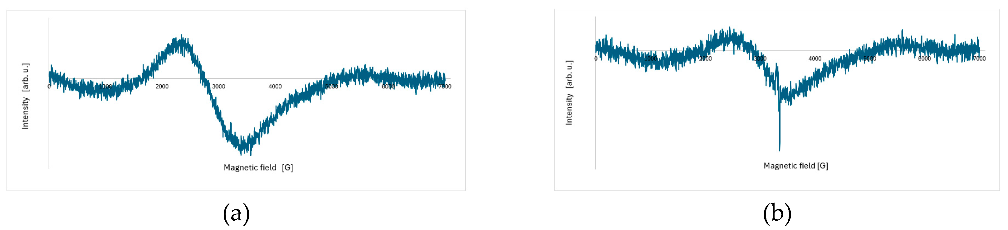
Figure 2.
Temperature dependence of the EPR spectra of Sample 1 recorded (a) before and (b) after laser irradiation in the temperature range of 300–500 K.
Figure 2.
Temperature dependence of the EPR spectra of Sample 1 recorded (a) before and (b) after laser irradiation in the temperature range of 300–500 K.
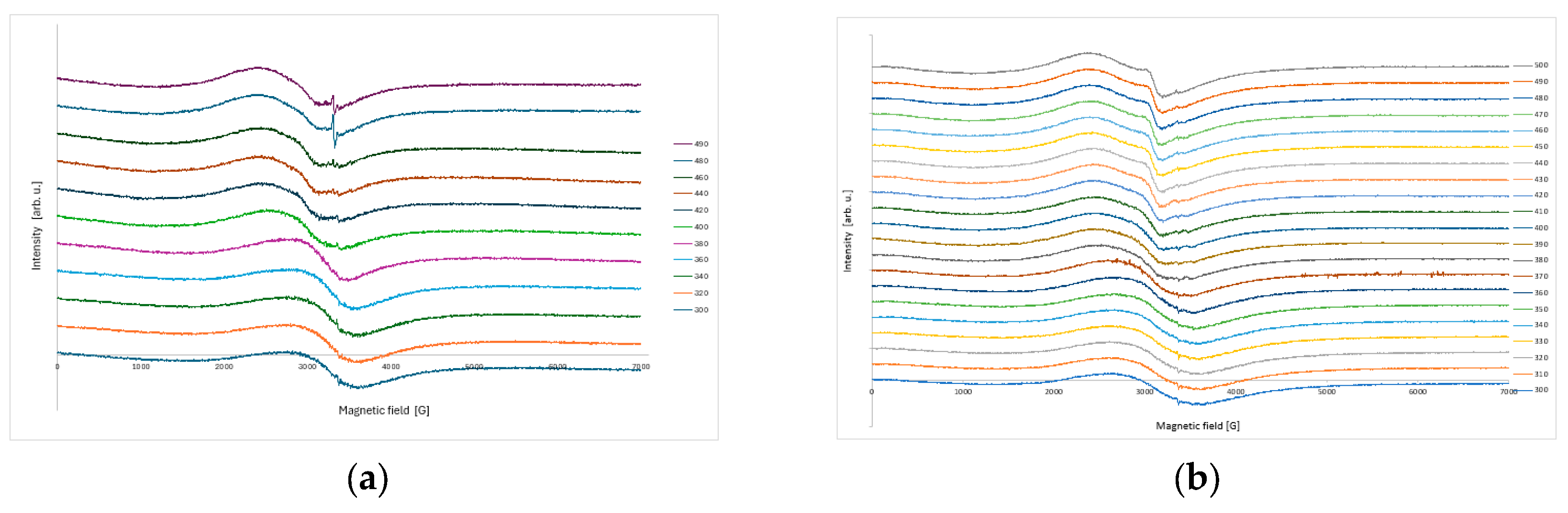
Figure 3.
Temperature dependence of the EPR parameters for the polyester resin-based powder coating Sample 1, measured before and after laser irradiation.
Figure 3.
Temperature dependence of the EPR parameters for the polyester resin-based powder coating Sample 1, measured before and after laser irradiation.
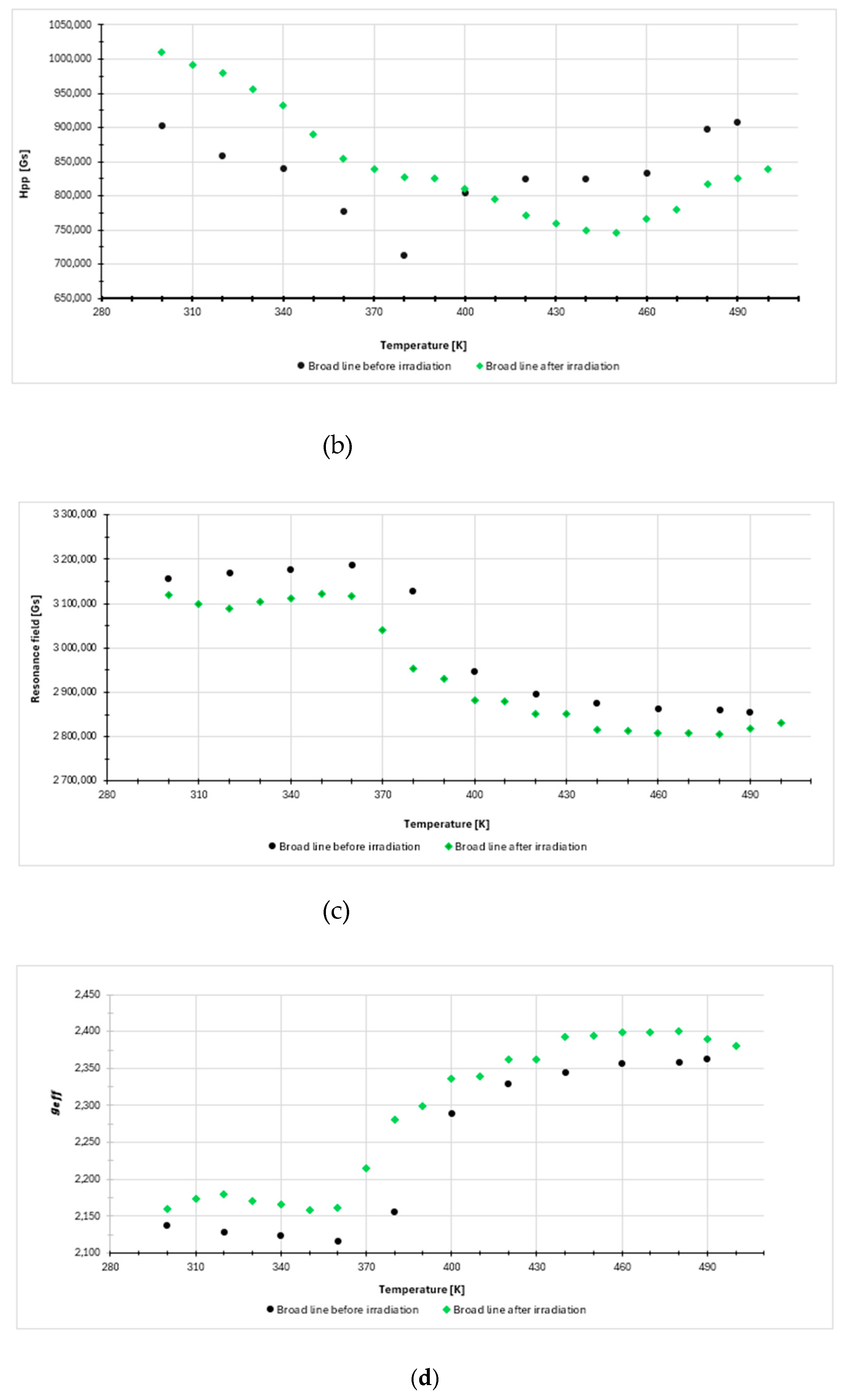
Figure 4.
EPR spectra of Sample 2 recorded (a) before and (b) after laser irradiation at 300 K.
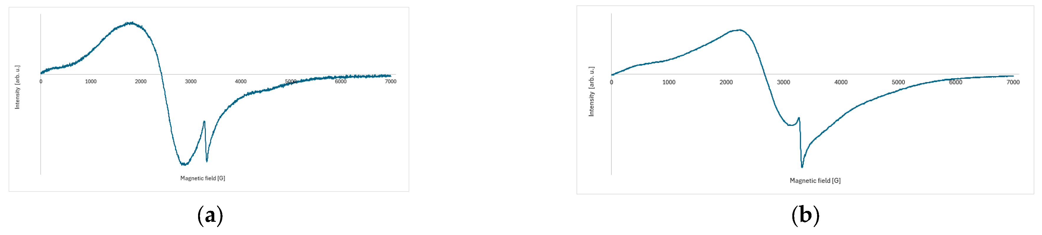
Figure 5.
Temperature dependence of the EPR spectra of Sample 2 recorded (a) before and (b) after laser irradiation in the temperature range 315–500 K.
Figure 5.
Temperature dependence of the EPR spectra of Sample 2 recorded (a) before and (b) after laser irradiation in the temperature range 315–500 K.
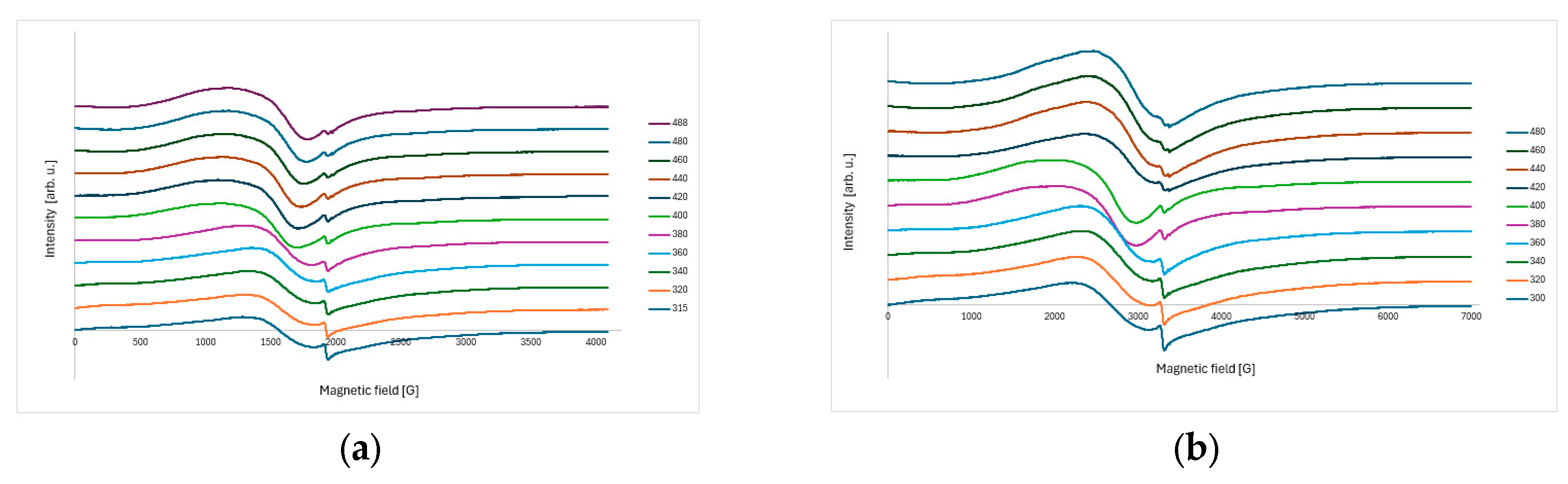
Figure 6.
Temperature dependence of the EPR parameters for powder coating Sample 2, measured before and after laser irradiation.
Figure 6.
Temperature dependence of the EPR parameters for powder coating Sample 2, measured before and after laser irradiation.
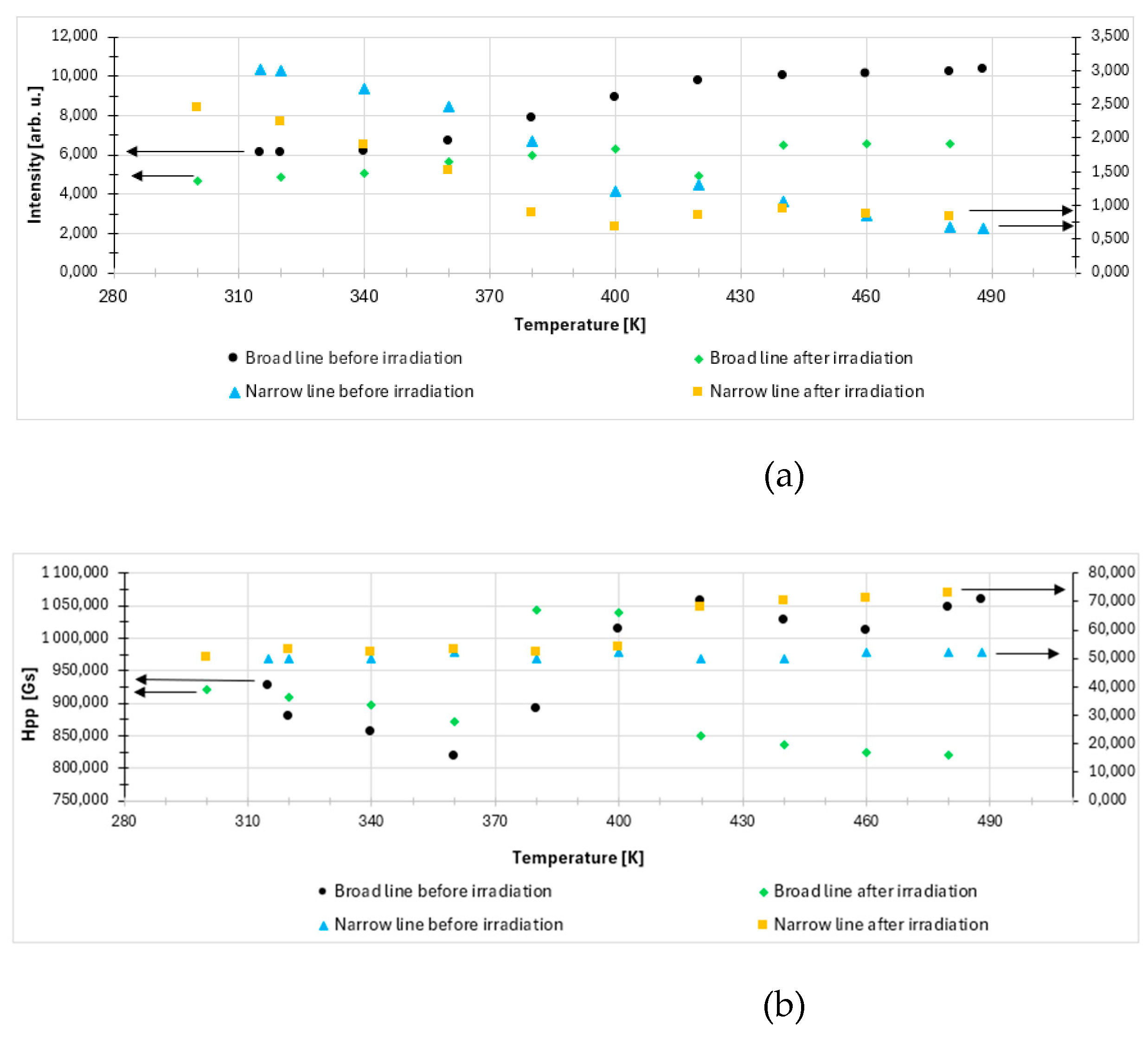


Table 1.
Temperature dependence of the EPR parameters for the additional EPR line observed in Sample 1 after laser irradiation.
Table 1.
Temperature dependence of the EPR parameters for the additional EPR line observed in Sample 1 after laser irradiation.
| Temperature [K] | Intensity [arb. u.] | Hpp [G] | Resonance field [G] | geff | ||||
| 500 | 0.857 | 189.69 | 3100.25 | 2.172 | ||||
| 490 | 0.855 | 186.28 | 3060.15 | 2.201 | ||||
| 480 | 0.857 | 193.11 | 3084.85 | 2.183 | ||||
| 470 | 0.877 | 199.95 | 3086.42 | 2.182 | ||||
| 460 | 0.831 | 176.96 | 3076.18 | 2.189 | ||||
| 450 | 0.832 | 181.15 | 3072.67 | 2.192 | ||||
Disclaimer/Publisher’s Note: The statements, opinions and data contained in all publications are solely those of the individual author(s) and contributor(s) and not of MDPI and/or the editor(s). MDPI and/or the editor(s) disclaim responsibility for any injury to people or property resulting from any ideas, methods, instructions or products referred to in the content. |
© 2026 by the authors. Licensee MDPI, Basel, Switzerland. This article is an open access article distributed under the terms and conditions of the Creative Commons Attribution (CC BY) license (http://creativecommons.org/licenses/by/4.0/).
Copyright: This open access article is published under a Creative Commons CC BY 4.0 license, which permit the free download, distribution, and reuse, provided that the author and preprint are cited in any reuse.


