Preprint
Communication

This version is not peer-reviewed.

A Universal Gatekeeper Unloading Nutrients to Seeds

Submitted:

04 February 2026

Posted:

06 February 2026

You are already at the latest version

Abstract

formation is an important mechanism in plant evolution that serves as the material basis for human survival. After double fertilization, the development of both the embryo and the endosperm requires a large amount of nutrients, and long-distance sucrose transportation is indispensable. We recently discovered a ring-shaped structure that can affect the unloading of nutrients into seeds. This structure is formed by the deposition of callose, which blocks the transport of nutrients by reducing the pore size of the plasmodesmata (PD). If fertilization is successful, the PD gate will form, but will remain open; if fertilization fails, the PD gate will be gradually closed—a strategy that efficiently prevents energy loss. A similar gating mechanism exists in rice, indicating that this strategy has substantial potential for agricultural production.

Keywords: 
;  ;  ;  

1. Introduction

In angiosperms, double fertilization is necessary for seed formation, and seeds serve as intergenerational carriers of genetic information. Seed development is a highly energy-intensive process: as a primary “sink,” it must acquire photoassimilates from distant photosynthetic “sources” [1]. The pressure-flow hypothesis describes the mechanism of osmotically generated pressure differentials that drive the long-distance movement of sugars in the phloem. However, sucrose unloading is more finely regulated. Recently, Liu et al. [2] revealed a callose-based structure, termed the Kasahara Gateway (KGW), at the chalazal end that modulates nutrient entry into the ovule [3,4]. Essentially, it is the plasmodesmata between the sieve tube cells at the end of the vascular bundle and the surrounding cells that are affected by callose deposition. In this paper, we will explore the universality of KGW from a broader perspective.
The callose-based structure known as the KGW has been identified in Arabidopsis ovule, where it functions as a nutrient gatekeeper [2,3,4]. Using an aniline blue staining experiment, a ring-shaped structure composed of callose deposits was discovered. The amount of callose deposition at the chalazal end was lower in wild type (WT) ovules at 2 DAP (Figure 1A) than in gcs1 (fertilization defective mutant) [5,6] ovules at 2 DAP (Figure 1B). Sucrose tracer Carboxyfluorescein diacetate (CFDA) was used to test whether callose deposition obstructs nutrient influx. CFDA can successfully enter fertilized ovules, but cannot enter gcs1 ovules, most likely because it is blocked by the callose, as mentioned in Liu et al. [2].

2. Materials and Methods

2.1. The Aniline Blue Staining

For aniline blue staining of silique tissue, Arabidopsis WT flowers were emasculated at stage 12c and pollinated with WT, gcs1/gcs1 pollen grans. Siliques were collected at 2 DAP. The pistils of Pseudoroegneria stipifolia, Oxalis pes-caprae L. and Oxalis debilis Kunth were collected at 2 or 3 days after flowering. The pistils were fixed and bleached in ethanol: acetic acid (3: 1) solution, rinsed with ddH2O, and then stained with aniline blue solution [0.1% (w/v) aniline blue and 0.1 M K3PO4] for more than 3 h. Confocal/two-photon images were acquired using a laser scanning inverted microscope (LSM880, Zeiss). The images were processed using the ZEN software (Zeiss).

2.2. Observation of Rice Pistils

The pistils after 2 or 3 days of flowering of rice “Nipponbare” (Oryza sativa ssp. japonica) were fixed in 4% paraformaldehyde, 5% acetic acid, and 50% ethanol. Then these were dehydrated, embedded in Technovit 7100 sliced and observed as described as Liu et al. (2025).

3. Results and Discussion

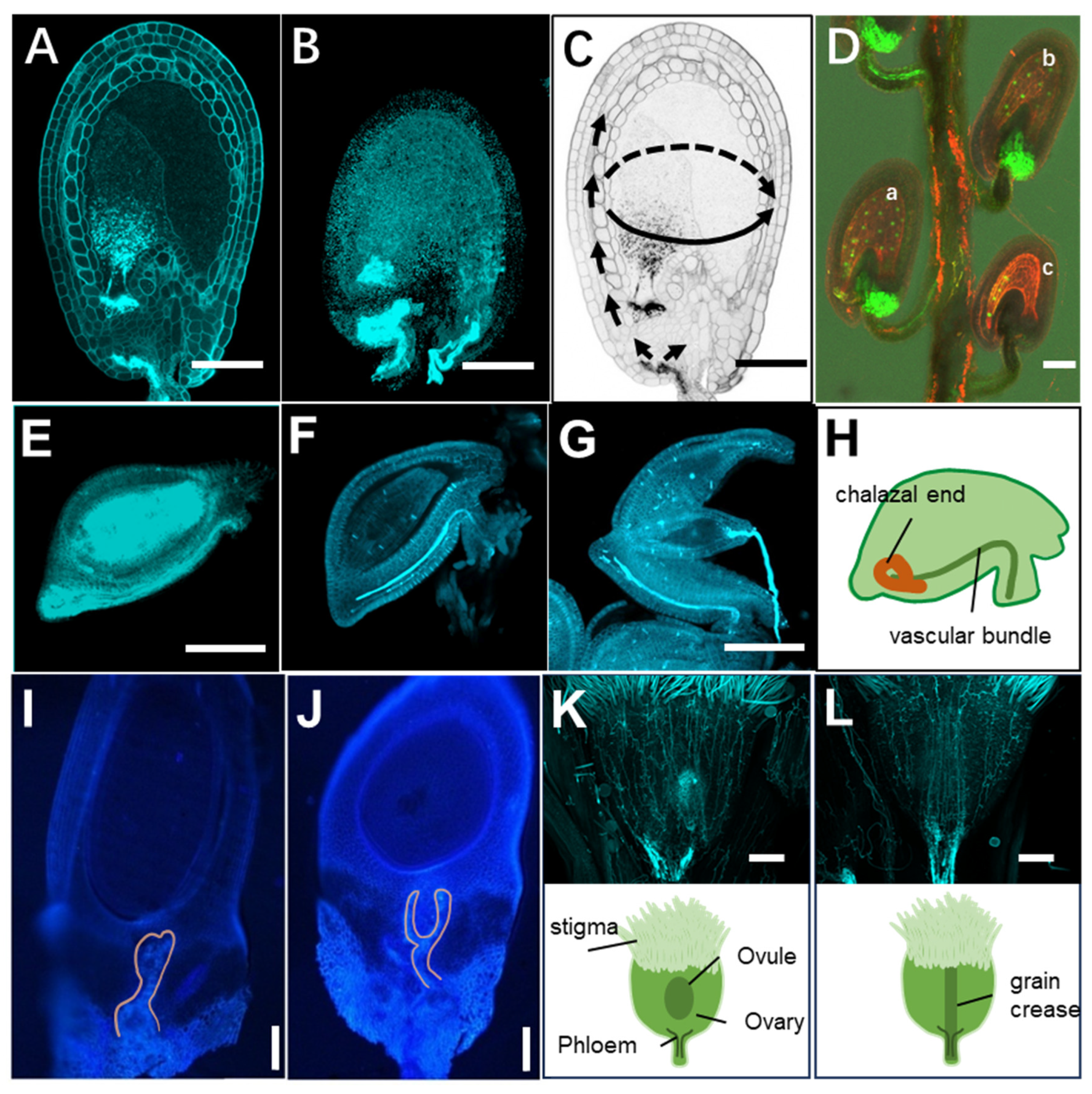
3.1. Dicotyledonous Plants Oxalis pes-caprae L. and Sterile Oxalis debilis Kunth

Since KGW is involved in seed development, we speculate that this structure is common to seed plants. Therefore, two other dicotyledonous and monocotyledonous plants were used to test whether the presence of KGW is universal.
In addition to the Arabidopsis, we also used fertile Oxalis pes-caprae L. [7] and sterile Oxalis debilis Kunth [8] to verify, as they are also dicotyledonous plants. From the structure of the ovules, they are anatropous ovules, with long vascular bundles visible extending to the chalazal end. The results of aniline blue staining of Oxalis pes-caprae L. showed that there was no significant deposition of callose either before or after fertilization (Figure 1G and H). However, the sterile Oxalis debilis Kunth shows significant differences before pollination (Figure 1E) and after flower withering (Figure 1F), with a large deposition of callose at the chalazal end and inside the ovules after the flowers have wilted.

3.2. Oryza sativa L ssp. japonica

Rice, a monocot, has also shown KGW [2]. In this study, we show callose contained KGW from rice “Nipponbare” (Oryza sativa ssp. japonica) by the fluorescence signal of anillin blue which can stains callose. KGW is known to contain callose as a deposition or nutrient, and it will be hydrolyzed after fertilization of the central cell [2,3,4]. To avoid the callose degradation by fertilization, we observed osgcs1 (generative-cell specific 1) mutant rice which could induce failure of fertilization [9], resulting that the KGW of rice is distributed at pedicle side (Figure 1I and J).

3.3. Pseudoroegneria stipifolia (Trautv.) Á.Löve

Pseudoroegneria stipifolia (Trautv.) Á.Löve [10], a monocot of the Triticeae, represents a distant lineage relative to wheat. Its ovules are enclosed within the ovary, which ultimately develops into a grain after fertilization. Aniline blue staining revealed that the vascular bundles extend to the base of the ovary, where they terminate and accumulate abundant callose (Figure 1K). Moreover, the ovary is distinctly patterned with root-like vascular traces, which is denser at the grain crease (Figure 1L).
Comparative studies of dicot and monocot ovules show that this region consistently contains a KGW structure, indicating such a mechanism is conserved throughout angiosperms. Evolutionarily, this structure likely emerged concomitantly with the origin of seed plants and has since been retained as the primordial device governing nutrient flow between maternal tissue and the ovule.
Studies show that the earliest seed plants emerged unobtrusively during the Late Devonian, evolving from a paraphyletic group known as progymnosperms. The progymnosperms “folded” the gametophyte inside the spore wall and fed it from the parental sporophyte—this pivotal transition forged the maternal–offspring nutrient conduit and took the decisive stride toward the seed. Fossil seeds are several mm to several cm long. The integument is three-layered, comprising an epidermis and an outer fleshy layer [11]. Although these plants are extinct, there is no way to know whether they possessed KGW or its rudiment. However, based on the seed structures of extant gymnosperms and angiosperms, their seeds all obtain nutrients from the maternal plant via vascular tissues. Aniline blue staining of ovules from diverse angiosperms also revealed that they all possess KGW in various morphologies. It is therefore highly likely that KGW is widespread among seed plants, yet verification across a broader range of extant species remains necessary.

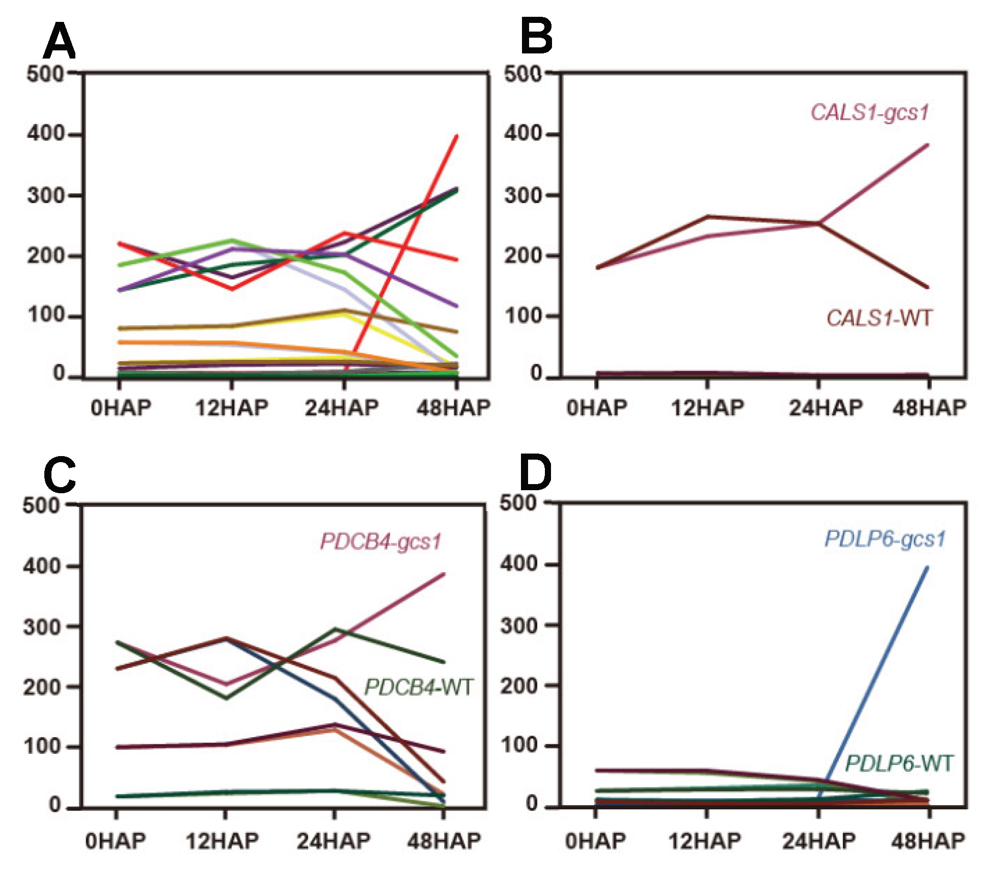
3.4. Fertilization of the Central Cell Unlocks the KGW

Although double fertilization is widely considered an obligatory trigger of seed development, the key question is what signal triggers nutrient influx. This study demonstrated that fertilization of the central cell alone was sufficient to initiate early nutrient influx. The kpl mutant (18% single fertilization of the central cell and 23% single fertilization of the egg cell) [12] was used to verify which cell fertilization could activate nutrient influx. Carboxyfluorescein diacetate was used as a tracer to indicate nutrient inflow. The embryo and endosperm were labeled with WOX2p::H2B-GFP + WOX2p::LTI-tdTomato double marker and AGL62::GFP [13,14], respectively. The results showed that an intense CF signal was observed above the phloem end in double-fertilized and central-cell single-fertilized ovules (Figure 1D-a and b). However, this signal was not observed above the phloem end of egg cell single-fertilized ovules (Figure 1D-c), indicating that nutrient transport did not occur if central cell fertilization failed.

3.5. On/off Mechanism of the KGW

Plasmodesmata are dynamic structures regulated by callose deposition. Although the specific signal released during endosperm development that activates the callose gate remains unknown, it is plausible that the enzymes involved in callose synthesis and degradation, as well as plasmodesmata-associated proteins [15], are regulated by unidentified signals that control the opening and closing of the callose gate. In the study by Liu et al. [2], transcriptome profiling revealed pronounced up-regulation of AtBG_ppap—a β-1,3-glucanase specialized for callose degradation—in fertilized ovules, indicating that callose is actively dismantled upon fertilization. Overexpression of AtBG_ppap enlarges seeds, implying that elevated expression of this β-1,3-glucanase enhances assimilate import.
In addition to the recently reported AtBG_ ppap [2], many other proteins can also affect the permeability of plasmodesmata. We reanalyzed the transcriptome data from WT and POEM (pollen tube-dependent ovule enlargement morphology) ovules and found that several gene families may be involved in callose-mediated POEM ovule death [16], including the callose synthase genes (CALSs), plasmodesmata callose-binding protein (PDCBs) and plasmodesmata-located protein(PDLPs). The CALS1, PDCB and PDLP6 are up-regulated in 48HAP gcs1 ovules, contrary to WT. This shows that fertilization failure induces the expression of these genes, promoting callose deposition (Figure 2).
In addition, the enzymatic activity of AtBG_ppap may be affected by protein interactions that ultimately modulate plasmodesmatal conductivity. Upon viral invasion, PDLP7 physically interacts with and suppresses BG10 β-glucosidase activity, triggering callose encasement of plasmodesmata and thereby severing the intercellular highway exploited by the virus [17]. It remains unknown whether the KGW in the ovule is also subject to such regulation.

Author Contributions

Conceptualization, L.X. and R.D.K.; writing—original draft preparation, L.X.; writing—review and editing, L.X. and R.D.K.; supervision, R.D.K.; project administration, R.D.K.; funding acquisition, R.D.K. T. K.; providing rice ovule observation data. Thanks to Yu Zhenhao from Sichuan Agricultural University for providing the seeds of Pseudoroegneria stipifolia (Trautv.) Á.Löve (PI675310). All authors have read and agreed to the published version of the manuscript.

Funding

This work was supported by Japan Society for the Promotion of Science Grants-in-Aid for Scientific Research (grant no. 22K21366 to R.D.K.). This work was partially supported by the Carbon Recycle Foundation (grant no. 5-01-2024 to R.D.K.), Cannon Foundation (to R.D.K.) and.the 34th Botanical Research Grant of ICHIMURA Foundation for New Technology. (to R.D.K.).

Conflicts of Interest

The authors declare that they have no competing interests.

References

  1. Münch, E. Material Flow in Plants; Milburn, J. A.; Kreeb, K. H., Translators; University of Bremen: Germany; Gustav Fischer Verlag: Jena Germany.
  2. Liu, X.; Nakajima, K.P.; Adhikari, P.B.; Wu, X.; Zhu, S.; Okada, K.; Kagenishi, T.; Kurotani, K.; Ishida, T.; Nakamura, M.; Sato, Y; Kawakatsu, Y.; Xie, L.; Huang, C.; He, J.; Yokawa, K.; Sawa, S.; Higashiyama, T.; Bradford, K.J.; Notaguchi, M.; Kasahara, R.D. Fertilization-dependent phloem end gate regulates seed size. Curr Biol. 2025, 35, 2049–63.e3. [Google Scholar] [CrossRef] [PubMed]
  3. Kasahara, R. D. Fertilization initiates seed nutrition via phloem end by a callose degradation enzyme. DNA Cell Biol. 2025, 44, 407–410. [Google Scholar] [CrossRef] [PubMed]
  4. Nakajima, Kohdai P.; Kasahara, Ryushiro D. How Do Seeds Get Bigger? A Newly Identified Phloem-Terminal Tissue Controls Seed Enlargement through Callose Degradation. Academia Molecular Biology and Genomics 2025. [Google Scholar] [CrossRef]
  5. Mori, T.; Kuroiwa, H.; Higashiyama, T.; Kuroiwa, T. GENERATIVE CELL SPECIFIC 1 is essential for angiosperm fertilization. Nat. Cell Biol. 2006, 8, 64–71. [Google Scholar] [CrossRef] [PubMed]
  6. von Besser, K.; Frank, A. C.; Johnson, M. A.; Preuss, D. Arabidopsis HAP2 (GCS1) is a sperm-specific gene required for pollen tube guidance and fertilization. Development 2006, 133, 4761–4769. [Google Scholar] [CrossRef] [PubMed]
  7. Castro, S.; Castro, M.; Ferrero, V.; Costa, J.; Tavares, D.; Navarro, L.; Loureiro, J. Invasion fosters change: independent evolutionary shifts in reproductive traits after Oxalis pes-caprae L. introduction. Front. Plant Sci. 2016, 7, 874. [Google Scholar] [CrossRef] [PubMed]
  8. Luo, S.; Zhang, D.; Renner, S. S. Oxalis debilis in China: distribution of flower morphs, sterile pollen and polyploidy. Ann Bot. 2006, 98, 459–64. [Google Scholar] [CrossRef] [PubMed]
  9. Honma, Y; Adhikari, P. B.; Kuwata, K.; Kagenishi, K.; Yo kawa, K.; Notaguchi, M.; Kurotani, K.; Toda, E.; Bessho-Uehara, K.; Liu, X.; Zhu, S.; Wu, X.; Kasahara, R. D. High-quality sugar production by osgcs1 rice. Commun. Biol. 2020, 3, 617. [Google Scholar] [CrossRef] [PubMed]
  10. Ji, Y.; Khan, N.; Chaudhary, R.; Perumal, S.; Wang, Z.; Hucl, P.; Biligetu, B.; Sharpe, A.G.; Jin, L. Genomic insights into wheatgrass: unravelling genetic diversity, population structure, and evolutionary dynamics in Pseudoroegneria species. Mol. Phylogenet. Evol. 2025, 211, 108397. [Google Scholar] [CrossRef] [PubMed]
  11. Linkies, A.; Graeber, K.; Knight, C.; Leubner-Metzger, G. The evolution of seeds. New Phytol. 2010, 186, 817–831. [Google Scholar] [CrossRef] [PubMed]
  12. Ron, M.; Saez, M. A.; Williams, L. E.; Fletcher, J. C.; McCormick, S. Proper regulation of a sperm-specific cis-nat-siRNA is essential for double fertilization in Arabidopsis. Genes Dev. 2010, 24, 1010–1021. [Google Scholar] [CrossRef] [PubMed]
  13. Gooh, K.; Ueda, M.; Aruga, K.; Park, J.; Arata, H.; Higashiyama, T.; Kurihara, D. Live-cell imaging and optical manipulation of Arabidopsis early embryogenesis. Dev. Cell 2015, 34, 242–251. [Google Scholar] [CrossRef] [PubMed]
  14. Kang, I. H.; Steffen, J. G.; Portereiko, M. F.; Lloyd, A.; Drews, G. N. The AGL62 MADS domain protein regulates cellularization during endosperm development in Arabidopsis. Plant Cell 2008, 20, 635–647. [Google Scholar] [CrossRef] [PubMed]
  15. Bayer, E. M.; Benitez-Alfonso, Y. Plasmodesmata: Channels under pressure. Annual Review of Plant Biology 2024, 75, 291–317. [Google Scholar] [CrossRef] [PubMed]
  16. Kasahara, R. D; Notaguchi, M.; Honma, Y. Discovery of pollen tube-dependent ovule enlargement morphology phenomenon, a new step in plant reproduction. Commun. Integr. Biol. 2017, 5, e1338989. [Google Scholar] [CrossRef]
  17. Chen, X.; Li, W.; Gao, J.; Wu, Z.; Du, J.; Zhang, X.; Zhu, Y. Arabidopsis PDLP7 modulated plasmodesmata function is related to BG10-dependent glucosidase activity required for callose degradation. Sci. Bull. 2024, 69, 3075–3088. [Google Scholar] [CrossRef] [PubMed]
Figure 1. KGW at the chalazal end of the ovule regulates nutrient transport.A and B. Aniline blue staining of WT and gcs1 ovules. Callose is deposited in gcs1 ovules. C. Nutrient transport pattern. D. Carboxyfluorescein diacetate (CFDA)-based tracing of sucrose transport in ovules pollinated by kpl pollen. Triangles indicate CF entry into the ovule (optimum excitation: 490 nm; emission: 515 nm). a. Ovules that have completed double fertilization. b. Single-fertilized ovules with central cell fertilization. c. Single-fertilized ovules with egg cell fertilization. The embryo and endosperm were labeled with the WOX2p::H2B-GFP + WOX2p::LTI-tdTomato double marker and AGL62::GFP, respectively. E. Aniline blue staining of Oxalis debilis Kunth., callose is deposited in the ovules of the faded flower. F and G. Oxalis pes-caprae L., before pollination (F), after pollination (G). H. cartoon of Oxalis. I and J. Structure of KGWs from diagonal vertical sections of rice (gcs1) ovules. Fluorescence signals by aniline blue indicates callose containing organs KGW. This seems to come from pedicle side and curved within the base of ovules. Scale bar is 10 µm. K and L show the two faces of the grain (Pseudoroegneria stipifolia). Callose accumulates at the base of the ovary, and vein-like callose signals are distributed across the ovary.
Preprints 197456 g001
Figure 2. Genes associated with callose deposition are expressed in the ovule.(A) Genes associated with callose deposition are expressed in the ovules of WT and gcs1 according to transcriptome data (Kasahara et al. 2016). (B) The callose synthase gene CALS1 is highly expressed in gcs1 ovules, contrary to WT. (C) The plasmodesmata callose-binding protein (PDCBs) is expressed in ovules. (D) The expression of plasmodesmata-located protein(PDLPs) in ovules.
Preprints 197456 g002
Disclaimer/Publisher’s Note: The statements, opinions and data contained in all publications are solely those of the individual author(s) and contributor(s) and not of MDPI and/or the editor(s). MDPI and/or the editor(s) disclaim responsibility for any injury to people or property resulting from any ideas, methods, instructions or products referred to in the content.
Copyright: This open access article is published under a Creative Commons CC BY 4.0 license, which permit the free download, distribution, and reuse, provided that the author and preprint are cited in any reuse.
Prerpints.org logo

Preprints.org is a free preprint server supported by MDPI in Basel, Switzerland.

Subscribe

Disclaimer

Terms of Use

Privacy Policy

Privacy Settings

© 2026 MDPI (Basel, Switzerland) unless otherwise stated