Submitted:
31 January 2026
Posted:
03 February 2026
You are already at the latest version
Abstract
Several receptors have received considerable attention as therapeutic targets in gastric cancer (GC), and numerous receptor inhibitors have been developed. However, the development of novel gastric cancer therapeutics is time-consuming. Therefore, this study aimed to identify drugs effective against gastric cancer from existing anticancer agents originally developed for other malignancies. In this study, the cancer-related genomic profiles of 286 genes were analyzed in 14 gastric cancer cell lines using targeted DNA sequencing, and these cell lines were utilized as models to evaluate the efficacy of 35 anticancer drugs. The 14 cell lines were assessed for 286 gene alterations, copy number variations, amplification of 14 gastric cancer-related therapeutic targets, and sensitivity to 35 drugs. p-MET and MET were overexpressed in the SNU5, SNU620, MKN45, and Hs746T cell lines, while p-EGFR was overexpressed in the NCI-N87 cell line. FGFR2 overexpression was observed in the Kato III and SNU16 cell lines. TGFβR1 was overexpressed in the MKN7 cell line. HER2 and CDK12 were overexpressed in the NCI-N87 and MKN7 cell lines. PD-L1 overexpression was detected in the Hs746T and MKN7 cell lines. CD44 was overexpressed in the SNU5 and Hs746T cell lines, and CLDN18 overexpression was observed in the MKN7 cell line. Well-characterized gastric cancer cell lines are essential for drug development research. This study provides a framework for selecting cell lines that are responsive to each of the 35 anticancer drugs and for elucidating their underlying therapeutic mechanisms through follow-up studies. Ultimately, clinical studies are required to confirm the therapeutic efficacy of the selected drugs.
Keywords:
targeted therapy
; gastric cancer
; biomarker
; cell line
1. Introduction
Gastric cancer (GC) is the fourth most common cancer worldwide in terms of both incidence and mortality, with the highest incidence rates observed in Eastern Asia [1,2]. Despite advances in chemotherapy, the prognosis for unresectable GC/gastroesophageal junction cancer remains poor, with a 5-year relative survival rate of less than 10% among patients with advanced disease (Cancer Stat Facts: Stomach cancer. National Cancer Institute [2025], https://seer.cancer.gov/statistics-network/explorer/). Therefore, additional treatment strategies are urgently needed to improve clinical outcomes.
Drug discovery typically requires 10–15 years to develop a new therapeutic agent; however, the overall success rate is only approximately 2.01% [3]. To address this limitation, drug repositioning has recently been widely explored. Unlike traditional drug development approaches, drug repositioning, defined as identifying new therapeutic uses for existing drugs, represents an efficient strategy for discovering novel indications for approved agents. This approach enables a more rapid route to drug development because the clinical safety profiles and pharmacokinetic data of existing drugs have already been established [4]. Recent advances in genomics have further enabled the elucidation of disease-specific molecular mechanisms at a personalized level. Precision medicine has therefore become increasingly important in the management of GC. In many cases, clinically relevant molecular targets for which approved drugs already exist can be identified, creating opportunities for drug repurposing. Moreover, identifying therapeutic agents that target specific molecular alterations may shorten the time required to initiate effective treatment for individual patients.
Receptor tyrosine kinases (RTKs) and other receptors have been extensively investigated in GC. Among RTKs, amplification of the mesenchymal-epithelial transition factor proto-oncogene (MET), fibroblast growth factor receptor 2 (FGFR2), epidermal growth factor receptor (EGFR/ERBB1/HER1), and HER2 genes has been identified in approximately 2–24%, 2–9%, 27–64%, and 7–17% of patients with GC, respectively [5,6,7,8]. Upon activation, aberrant MET, FGFR2, EGFR, and HER2 signaling initiates complex intracellular cascades that promote cancer cell proliferation, migration, inhibition of apoptosis, and angiogenesis through the upregulation of vascular endothelial growth factor (VEGF) [9,10,11,12,13]. With recent improvements in the efficacy of targeted therapies, comprehensive tumor profiling, including the analysis of somatic mutations and copy-number variations (CNVs), has become increasingly necessary [14,15]. In particular, high-level CNVs play a critical role in tumorigenesis across multiple cancer types [15,16]. Cell line models with target gene overexpression have consistently demonstrated concordance with clinical responses, showing susceptibility to inhibitors directed against the corresponding molecular targets.
This study aimed to identify drugs that could serve as novel targeted therapies for GC through a drug repositioning approach using clinically approved agents. To this end, we identified therapeutic targets in 14 GC cell lines and evaluated the effects of 35 candidate drugs on cell viability.
2. Results
2.1. Characteristics of 14 GC Cell Lines
Fourteen GC cell lines (SNU16, SNU5, AGS, SNU719, MKN45, SNU1, N87, MKN74, MKN28, Kato III, MKN7, SNU638, SNU620, and Hs746T) were analyzed using targeted sequencing performed at the Theragen Bio Institute (Seongnam, Korea). The 286 GC panel genes and their individual genetic aberrations are presented in Figure 1.
To evaluate amplification of the 286 cancer-related genes in the 14 GC cell lines, CNV analysis was performed using targeted DNA sequencing data (Figure 2, Table S1). FGFR2 amplification, defined as a copy number greater than 20, was identified in 2/14 (14.3%) cell lines (Figure 2). MET amplification was observed in 4/14 (28.6%) cell lines, MYC amplification in 1/14 (7.1%), CDK12 amplification in 2/14 (14.3%), and ERBB2 amplification in 2/14 (14.3%) cell lines.
2.2. Patterns of RTKs and GC-Related Targets in the 14 GC Cell Lines
Additional information on the 14 GC cell lines, including concomitant overexpression of RTKs or GC-related targets, is presented in Figure 3 and Figure 4. Among the RTKs and GC-related targets, overexpression of MET, FGFR2, MYC, and CD44 was predominantly observed in diffuse-type GC cells, whereas overexpression of HER2, CDK12, TGFβR1, and CLDN18 was mainly observed in intestinal-type GC cells.
2.3. Sensitivity of 35 Drugs in the 14 GC Cell Lines
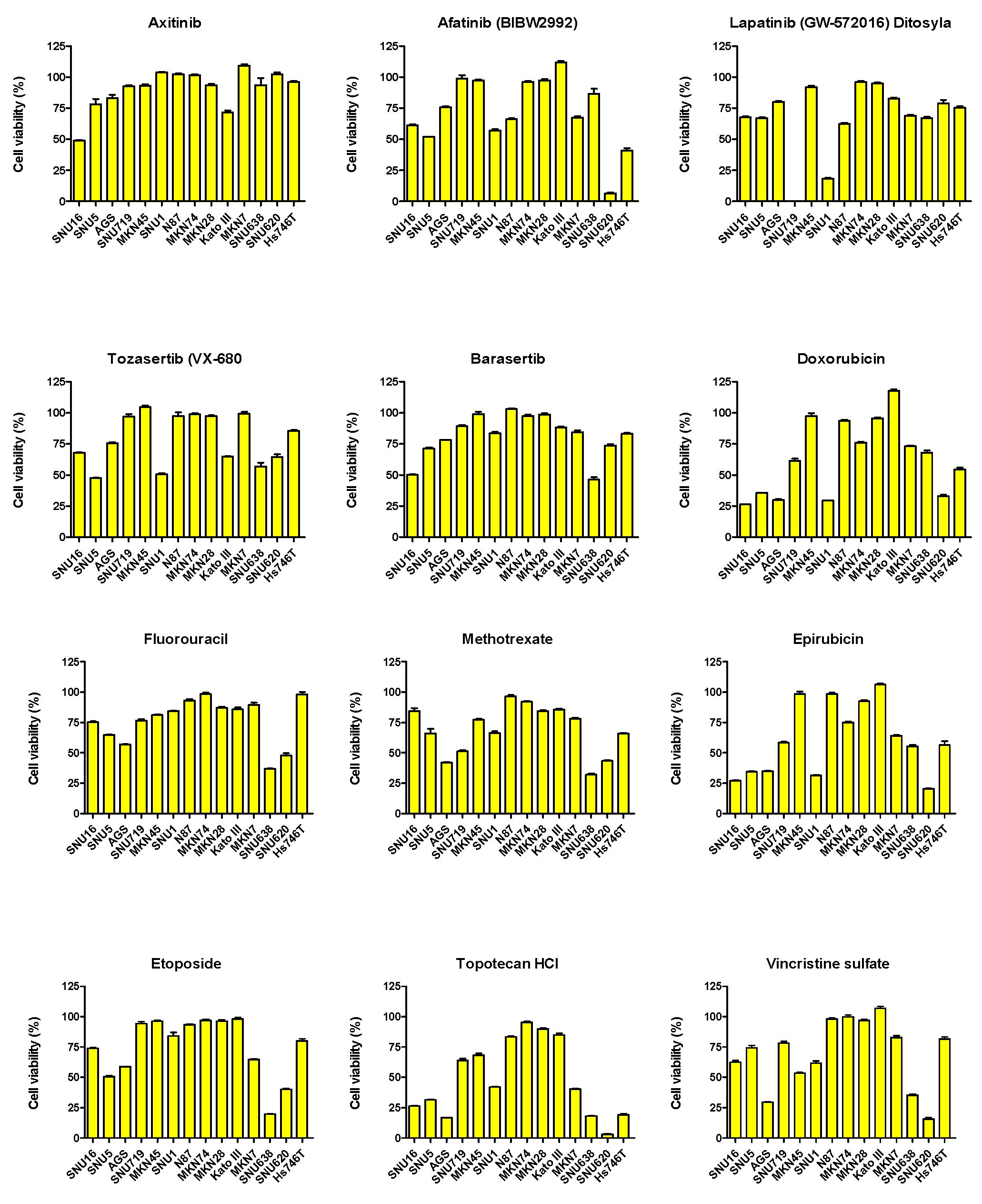
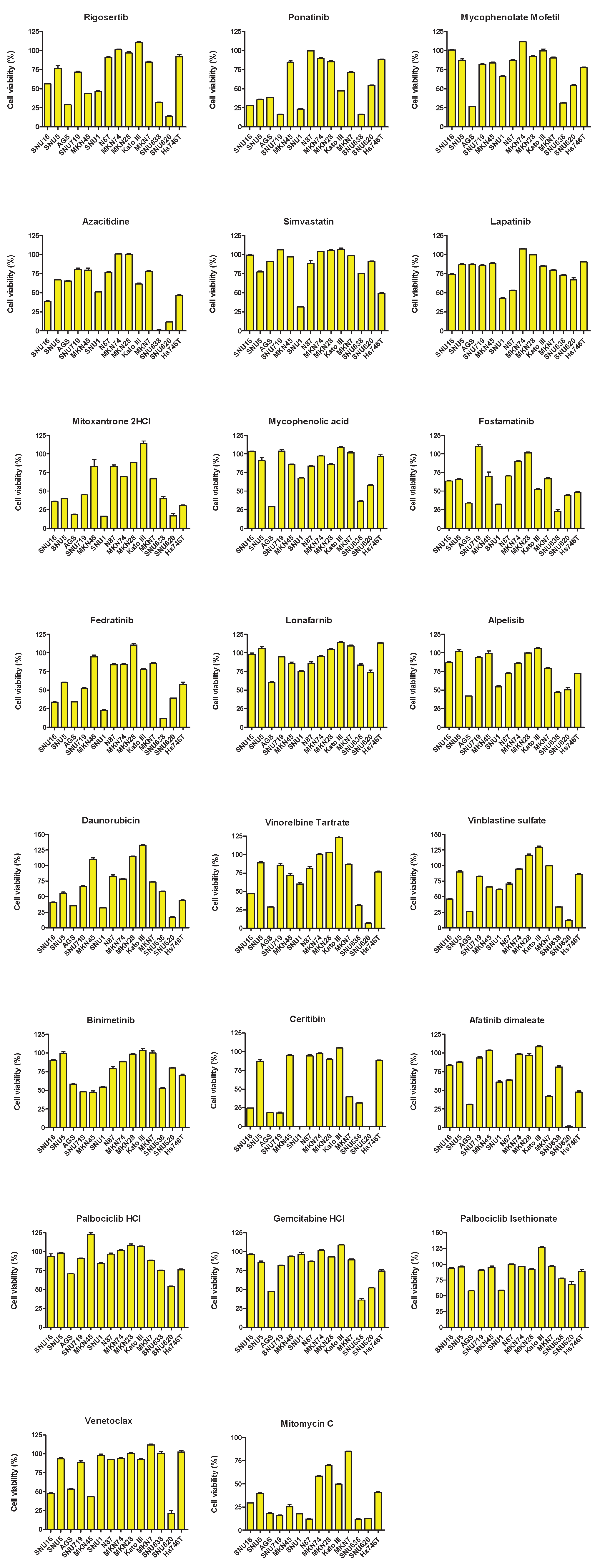
To evaluate drug repositioning potential in the 14 GC cell lines, sensitivity to 35 drugs was assessed (Figure 5). Drug sensitivity was analyzed based on the viability rate (%) in the 14 GC cell lines. Among these cell lines, the drugs that reduced cell viability to 50% or less in more than 7 cell lines were epirubicin, doxorubicin, topotecan HCl, ponatinib, mitoxantrone 2HCl, fostamatinib, fedratinib, daunorubicin, ceritinib, and mitomycin C.


3. Discussion
Traditional GC drug development requires a significant amount of time, typically 10–15 years [18]. In contrast, drug repurposing involves the identification of existing drugs for new indications, different from their original purpose. By leveraging drugs with established safety and efficacy profiles, the need to discover entirely new candidates is eliminated, thereby shortening the safety and efficacy evaluation period and markedly reducing the time and cost associated with new drug development [4,19]. Accordingly, we aimed to identify effective therapeutic agents for GC among existing anticancer drugs developed for other malignancies. In addition, appropriate preclinical studies, particularly those using patient-derived cancer cell lines, are critically important for successful drug development. In this study, we analyzed 286 genetic alterations and CNVs in 14 GC cell lines using targeted DNA sequencing to identify potential GC therapeutic targets for each cell type and evaluated drug sensitivity to 35 agents in these cell lines.
Among RTKs, MET, EGFR, HER2, VEGFR, and FGFR2, and among non-RTKs, TGFβR1, CDK12, PD-L1, CD44, and CLDN18, are currently being considered as potential targets for GC treatment [20,21,22,23]. RTKs catalyze the transfer of phosphate groups from ATP to tyrosine residues on intracellular proteins, thereby transducing extracellular signals into the cell, whereas non-RTKs act primarily on intracellular signaling pathways [24,25]. In particular, upregulation of RTKs has been frequently observed in GC, suggesting that RTK inhibitors may play an important role in its treatment. Drugs that reduced the viability of p-MET- and MET-overexpressing SNU5, SNU620, MKN45, and Hs746T cell lines by approximately 25% included methotrexate, topotecan HCl, fostamatinib, and mitomycin C. Ponatinib and mitomycin C reduced the viability of FGFR2-overexpressing Kato III and SNU16 cell lines by more than 50%. In the p-EGFR-overexpressing NCI-N87 cell line, lapatinib and mitomycin C reduced cell viability by more than 50%. Afatinib, afatinib dimaleate, alpelisib, azacitidine, lapatinib ditosylate, daunorubicin, and fostamatinib reduced the viability of HER2-overexpressing NCI-N87 and MKN7 cell lines by more than 25%.
Non-RTK targets also play important roles in GC. Among the 14 GC cell lines, MKN7, MKN28, MKN74, N87, SNU16, and SNU620 harbored mutations in CDK12; however, only MKN7 and N87 exhibited CDK12 overexpression. CDK12 promotes angiogenesis in GC by activating the PI3K/AKT/mTOR signaling pathway [26]. Afatinib, afatinib dimaleate, alpelisib, azacitidine, lapatinib ditosylate, daunorubicin, and fostamatinib reduced the viability of CDK12-overexpressing NCI-N87 and MKN7 cell lines by more than 25%. In advanced cancers, TGFBR2 promotes tumor progression, further emphasizing its importance as a potential therapeutic target [27,28]. In addition, isoform 2 of CLDN18 (CLDN18.2) is expressed exclusively in differentiated epithelial cells of the gastric mucosa and primary GC, highlighting its potential as a therapeutic target [29]. Topotecan HCl, ceritinib, and afatinib dimaleate reduced the viability of the MKN7 cell line, which overexpressed TGFβR1 and CLDN18, by more than 50%. PD-L1 is expressed in GC and is significantly more prevalent in males, HER2/neu-positive tumors, proximal GC, unclassified and papillary GC, MSI(define) GC, and EBV-associated GC [30]. Afatinib dimaleate and topotecan HCl reduced viability by more than 50% in PD-L1-overexpressing Hs746T and MKN7 cell lines. High CD44 expression in GC has been associated with increased recurrence and metastasis rates and poor prognosis [31,32]. Afatinib, doxorubicin, daunorubicin, mitoxantrone 2HCl, epirubicin, topotecan HCl, and mitomycin C reduced the viability of CD44-overexpressing Hs746T and SNU5 cell lines by more than 50%.
4. Materials and Methods
4.1. Reagents
Axitinib, afatinib, lapatinib ditosylate, tozasertib, barasertib, doxorubicin HCl, fluorouracil, methotrexate, epirubicin HCl, etoposide, topotecan HCl, vincristine sulfate, rigosertib, ponatinib, mycophenolate mofetil, azacitidine, simvastatin, lapatinib, mitoxantrone 2HCl, mycophenolic acid, fostamatinib, fedratinib, lonafarnib, alpelisib, daunorubicin HCl, vinorelbine tartrate, vinblastine sulfate, binimetinib, ceritinib, afatinib dimaleate, palbociclib HCl, gemcitabine HCl, palbociclib isethionate, venetoclax, and mitomycin C were obtained from Selleck Chemicals (Houston, TX, USA).
4.2. Cell Lines and Cell Culture
The GC cell lines SNU16, SNU5, AGS, SNU719, MKN45, SNU1, N87, MKN74, MKN28, Kato III, MKN7, SNU638, SNU620, and Hs746T were obtained from the Korean Cell Line Bank (Seoul, Republic of Korea). All cell lines except Hs746T were cultured in RPMI 1640 medium (Thermo Fisher Scientific), while Hs746T cells were maintained in Dulbecco’s Modified Eagle Medium (Thermo Fisher Scientific). Both media were supplemented with 10% (v/v) fetal bovine serum and 1% (w/v) penicillin/streptomycin. All cells were cultured under standard conditions.
4.3. Target DNA-Sequencing Analysis
The target DNA-sequencing methodology has been described in detail previously [17]. Fourteen GC cell lines were subjected to target DNA-sequencing analysis.
4.4. Cell Viability Assay
To evaluate the effects of 35 drugs on cell viability, an MTS(define) assay was performed using the CellTiter 96 Aqueous One Solution Cell Proliferation Assay kit (Promega, Madison, WI, USA). Briefly, 14 GC cell lines were seeded into 96-well plates at approximately 50% confluence and incubated for 24 h. The cells were subsequently treated with the indicated drugs at a concentration of 10 µM for 48 h. Cell viability was then assessed using the MTS assay according to the manufacturer’s instructions.
4.5. Western Blotting
Western blotting was performed using standard procedures. The primary antibodies used were anti-MET (#4560; 1:1000; Cell Signaling Technology [CST], Danvers, MA, USA), anti-phospho-MET (#3077; 1:1000; CST), anti-TGFβR1 (sc518018; 1:1000; Santa Cruz Biotechnology), anti-phospho-EGFR (#2234; 1:1000; CST), anti-EGFR (ab32077; 1:1000; Abcam), anti-CD44 (#3570; 1:1000; CST), anti-c-MYC (sc40; 1:1000; Santa Cruz Biotechnology, Dallas, TX, USA), anti-FGFR2 (ab10648; 1:1000; Abcam), anti-Frizzled 5 (ab75234; 1:1000; Abcam), anti-CLDN18 (#38-800; 1:1000; Invitrogen), anti-HER2 (#2165; 1:1000; CST), anti-CDK12 (ab317746; 1:1000; Abcam), anti-VEGFR2 (#9698; 1:1000; CST), anti-PD-L1 (#13684; 1:1000; CST), and anti-GAPDH (sc32233; 1:4000; Santa Cruz Biotechnology).
5. Conclusions
This study identified GC cell lines that are responsive to each of the 35 tested drugs and provided a basis for elucidating their underlying therapeutic mechanisms through subsequent follow-up studies. Ultimately, clinical trials will be required to confirm the therapeutic efficacy of the selected drugs.
Supplementary Materials
The following supporting information can be downloaded at the website of this paper posted on Preprints.org.
Author Contributions
S.-H.S. performed some of the molecular experiments, analyzed the data, and drafted the article. H.J.S. performed the other molecular experiments. B.J.K. critically revised the manuscript in terms of important intellectual content. D.Y.Z. supervised the study, obtained funding, guided the data analyses, and edited the manuscript. All authors have read and agreed to the published version of the manuscript.
Funding
The research was funded by the Korea Health Technology R&D Project (grant number grant number RS-2022-KH129464), the Patient-Centered Clinical Research Coordinating Center (PACEN, grant number RS-2020-KH095267) of the Ministry of Health and Welfare, and Hallym University Research Fund. No funder played any role in the study design; data collection, analyses, or interpretation; manuscript preparation; or the decision to publish the results.
Institutional Review Board Statement
Not applicable.
Informed Consent Statement
Not applicable.
Data Availability Statement
All the data are presented in the body of the manuscript. The data are available from the corresponding author upon reasonable request.
Conflicts of Interest
The authors declare no conflicts of interest.
Abbreviations
The following abbreviations are used in this manuscript:
| GC | gastric cancer |
| GEJC | gastric ductal carcinoma in situ |
| RTKs | receptor tyrosine kinases |
| MET | mesenchymal–epithelial transition |
| FGFR2 | fibroblast growth factor receptor 2 |
| EGFR/ERBB1/HER1 | epidermal growth factor receptor |
| CNV | copy number variations |
| CLDN18.2 | isoform 2 of CLDN18 |
References
- Jung, K.W.; Won, Y.J.; Kong, H.J.; Lee, E.S.; Community of Population-Based Regional Cancer, R. Cancer Statistics in Korea: Incidence, Mortality, Survival, and Prevalence in 2015. Cancer Res Treat 2018, 50, 303–316. [Google Scholar] [CrossRef]
- Sitarz, R.; Skierucha, M.; Mielko, J.; Offerhaus, G.J.A.; Maciejewski, R.; Polkowski, W.P. Gastric cancer: epidemiology, prevention, classification, and treatment. Cancer Manag Res 2018, 10, 239–248. [Google Scholar] [CrossRef]
- Yeu, Y.; Yoon, Y.; Park, S. Protein localization vector propagation: a method for improving the accuracy of drug repositioning. Mol Biosyst 2015, 11, 2096–2102. [Google Scholar] [CrossRef]
- Xue, H.; Li, J.; Xie, H.; Wang, Y. Review of Drug Repositioning Approaches and Resources. Int J Biol Sci 2018, 14, 1232–1244. [Google Scholar] [CrossRef]
- Maroun, C.R.; Rowlands, T. The Met receptor tyrosine kinase: a key player in oncogenesis and drug resistance. Pharmacol Ther 2014, 142, 316–338. [Google Scholar] [CrossRef] [PubMed]
- Matsumoto, K.; Arao, T.; Hamaguchi, T.; Shimada, Y.; Kato, K.; Oda, I.; Taniguchi, H.; Koizumi, F.; Yanagihara, K.; Sasaki, H.; et al. FGFR2 gene amplification and clinicopathological features in gastric cancer. Br J Cancer 2012, 106, 727–732. [Google Scholar] [CrossRef] [PubMed]
- Arienti, C.; Pignatta, S.; Tesei, A. Epidermal Growth Factor Receptor Family and its Role in Gastric Cancer. Front Oncol 2019, 9, 1308. [Google Scholar] [CrossRef] [PubMed]
- Tanner, M.; Hollmen, M.; Junttila, T.T.; Kapanen, A.I.; Tommola, S.; Soini, Y.; Helin, H.; Salo, J.; Joensuu, H.; Sihvo, E.; et al. Amplification of HER-2 in gastric carcinoma: association with Topoisomerase IIalpha gene amplification, intestinal type, poor prognosis and sensitivity to trastuzumab. Ann Oncol 2005, 16, 273–278. [Google Scholar] [CrossRef]
- Lang, S.A.; Klein, D.; Moser, C.; Gaumann, A.; Glockzin, G.; Dahlke, M.H.; Dietmaier, W.; Bolder, U.; Schlitt, H.J.; Geissler, E.K.; et al. Inhibition of heat shock protein 90 impairs epidermal growth factor-mediated signaling in gastric cancer cells and reduces tumor growth and vascularization in vivo. Mol Cancer Ther 2007, 6, 1123–1132. [Google Scholar] [CrossRef]
- Sohn, S.H.; Sul, H.J.; Kim, B.J.; Zang, D.Y. Responses to the Tepotinib in Gastric Cancers with MET Amplification or MET Exon 14 Skipping Mutations and High Expression of Both PD-L1 and CD44. Cancers (Basel) 2022, 14. [Google Scholar] [CrossRef]
- Kawakami, H.; Okamoto, I.; Arao, T.; Okamoto, W.; Matsumoto, K.; Taniguchi, H.; Kuwata, K.; Yamaguchi, H.; Nishio, K.; Nakagawa, K.; et al. MET amplification as a potential therapeutic target in gastric cancer. Oncotarget 2013, 4, 9–17. [Google Scholar] [CrossRef] [PubMed]
- Turner, N.; Grose, R. Fibroblast growth factor signalling: from development to cancer. Nat Rev Cancer 2010, 10, 116–129. [Google Scholar] [CrossRef] [PubMed]
- Moasser, M.M. The oncogene HER2: its signaling and transforming functions and its role in human cancer pathogenesis. Oncogene 2007, 26, 6469–6487. [Google Scholar] [CrossRef] [PubMed]
- Wang, Y.; Yao, X.; Li, S.N.; Suo, A.L.; Tian, T.; Ruan, Z.P.; Guo, H.; Yao, Y. Detection of prostate cancer related copy number variations with SNP genotyping array. Eur Rev Med Pharmacol Sci 2013, 17, 2916–2922. [Google Scholar]
- Shlien, A.; Malkin, D. Copy number variations and cancer. Genome Med 2009, 1, 62. [Google Scholar] [CrossRef]
- Xu, H.; Zhu, X.; Xu, Z.; Hu, Y.; Bo, S.; Xing, T.; Zhu, K. Non-invasive Analysis of Genomic Copy Number Variation in Patients with Hepatocellular Carcinoma by Next Generation DNA Sequencing. J Cancer 2015, 6, 247–253. [Google Scholar] [CrossRef]
- Sohn, S.H.; Sul, H.J.; Kim, B.J.; Zang, D.Y. Comparison of Tepotinib, Paclitaxel, or Ramucirumab Efficacy According to the Copy Number or Phosphorylation Status of the MET Gene: Doublet Treatment versus Single Agent Treatment. Int J Mol Sci 2024, 25. [Google Scholar] [CrossRef]
- Sertkaya, A.; Beleche, T.; Jessup, A.; Sommers, B.D. Costs of Drug Development and Research and Development Intensity in the US, 2000-2018. JAMA Netw Open 2024, 7, e2415445. [Google Scholar] [CrossRef]
- Jourdan, J.P.; Bureau, R.; Rochais, C.; Dallemagne, P. Drug repositioning: a brief overview. J Pharm Pharmacol 2020, 72, 1145–1151. [Google Scholar] [CrossRef]
- Panahizadeh, R.; Panahi, P.; Asghariazar, V.; Makaremi, S.; Noorkhajavi, G.; Safarzadeh, E. A literature review of recent advances in gastric cancer treatment: exploring the cross-talk between targeted therapies. Cancer Cell Int 2025, 25, 23. [Google Scholar] [CrossRef]
- Becker, J.C.; Muller-Tidow, C.; Serve, H.; Domschke, W.; Pohle, T. Role of receptor tyrosine kinases in gastric cancer: new targets for a selective therapy. World J Gastroenterol 2006, 12, 3297–3305. [Google Scholar] [CrossRef]
- Hashimoto, I.; Oshima, T. Claudins and Gastric Cancer: An Overview. Cancers (Basel) 2022, 14. [Google Scholar] [CrossRef]
- Agnarelli, A.; Vella, V.; Samuels, M.; Papanastasopoulos, P.; Giamas, G. Incorporating Immunotherapy in the Management of Gastric Cancer: Molecular and Clinical Implications. Cancers (Basel) 2022, 14. [Google Scholar] [CrossRef] [PubMed]
- Qin, S.; Li, A.; Yi, M.; Yu, S.; Zhang, M.; Wu, K. Recent advances on anti-angiogenesis receptor tyrosine kinase inhibitors in cancer therapy. J Hematol Oncol 2019, 12, 27. [Google Scholar] [CrossRef] [PubMed]
- Zhong, L.; Li, Y.; Xiong, L.; Wang, W.; Wu, M.; Yuan, T.; Yang, W.; Tian, C.; Miao, Z.; Wang, T.; et al. Small molecules in targeted cancer therapy: advances, challenges, and future perspectives. Signal Transduct Target Ther 2021, 6, 201. [Google Scholar] [CrossRef] [PubMed]
- Gao, L.Z.; Wang, J.Q.; Chen, J.L.; Zhang, X.L.; Zhang, M.M.; Wang, S.L.; Zhao, C. CDK12 Promotes the Proliferation, Migration, and Angiogenesis of Gastric Carcinoma via Activating the PI3K/AKT/mTOR Signaling Pathway. Appl Biochem Biotechnol 2023, 195, 6913–6926. [Google Scholar] [CrossRef]
- Ikushima, H.; Miyazono, K. TGFbeta signalling: a complex web in cancer progression. Nat Rev Cancer 2010, 10, 415–424. [Google Scholar] [CrossRef]
- Jin, Z.; Huang, Z.; Wu, C.; Zhang, F.; Gao, Y.; Guo, S.; Tao, X.; Lu, S.; Zhang, J.; Huang, J.; et al. Molecular insights into gastric cancer: The impact of TGFBR2 and hsa-mir-107 revealed by microarray sequencing and bioinformatics. Comput Biol Med 2024, 172, 108221. [Google Scholar] [CrossRef]
- Sahin, U.; Koslowski, M.; Dhaene, K.; Usener, D.; Brandenburg, G.; Seitz, G.; Huber, C.; Tureci, O. Claudin-18 splice variant 2 is a pan-cancer target suitable for therapeutic antibody development. Clin Cancer Res 2008, 14, 7624–7634. [Google Scholar] [CrossRef]
- Boger, C.; Behrens, H.M.; Mathiak, M.; Kruger, S.; Kalthoff, H.; Rocken, C. PD-L1 is an independent prognostic predictor in gastric cancer of Western patients. Oncotarget 2016, 7, 24269–24283. [Google Scholar] [CrossRef]
- Jung, W.Y.; Kang, Y.; Lee, H.; Mok, Y.J.; Kim, H.K.; Kim, A.; Kim, B.H. Expression of moesin and CD44 is associated with poor prognosis in gastric adenocarcinoma. Histopathology 2013, 63, 474–481. [Google Scholar] [CrossRef]
- Hou, W.; Kong, L.; Hou, Z.; Ji, H. CD44 is a prognostic biomarker and correlated with immune infiltrates in gastric cancer. BMC Med Genomics 2022, 15, 225. [Google Scholar] [CrossRef]
Figure 1.
Gene mutations in human gastric cancer cell lines measured by targeted next-generation sequencing.
Figure 1.
Gene mutations in human gastric cancer cell lines measured by targeted next-generation sequencing.
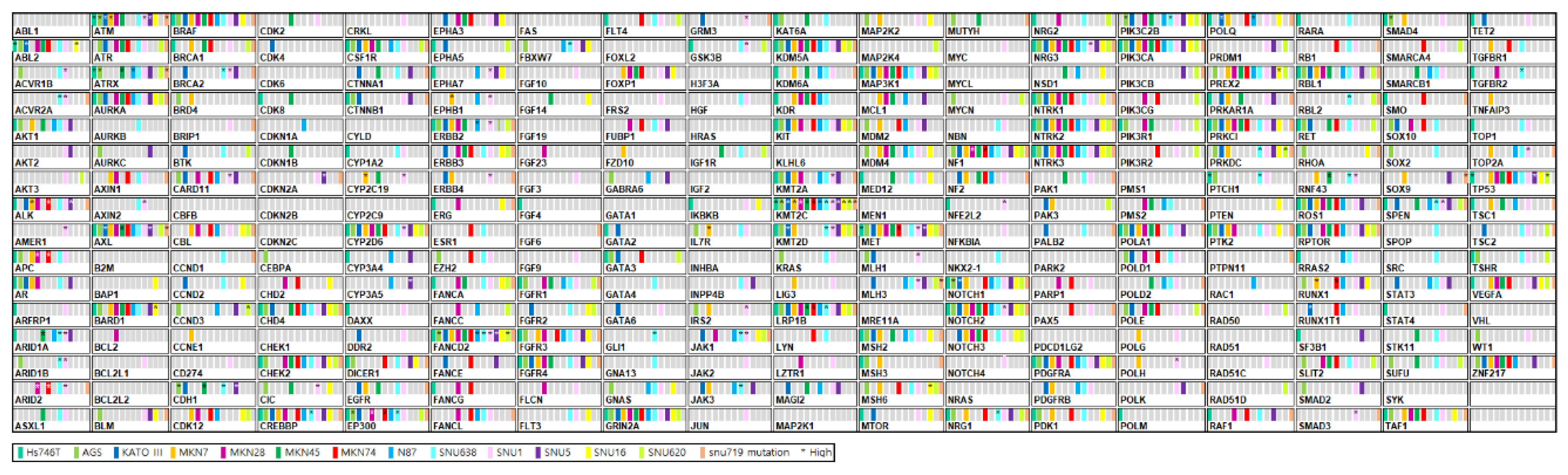
Figure 2.
Analysis of genes with CNVs of three or higher in at least six of the 14 gastric cancer cell lines, as determined by targeted next-generation sequencing.
Figure 2.
Analysis of genes with CNVs of three or higher in at least six of the 14 gastric cancer cell lines, as determined by targeted next-generation sequencing.
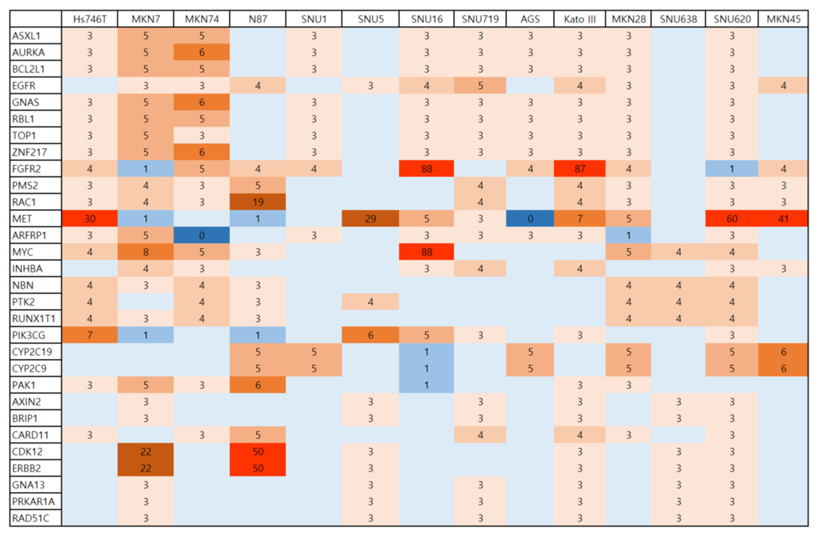
Figure 3.
Major protein expression profiles related to RTKs and cancer therapeutic targets in the 14 gastric cancer cell lines.
Figure 3.
Major protein expression profiles related to RTKs and cancer therapeutic targets in the 14 gastric cancer cell lines.
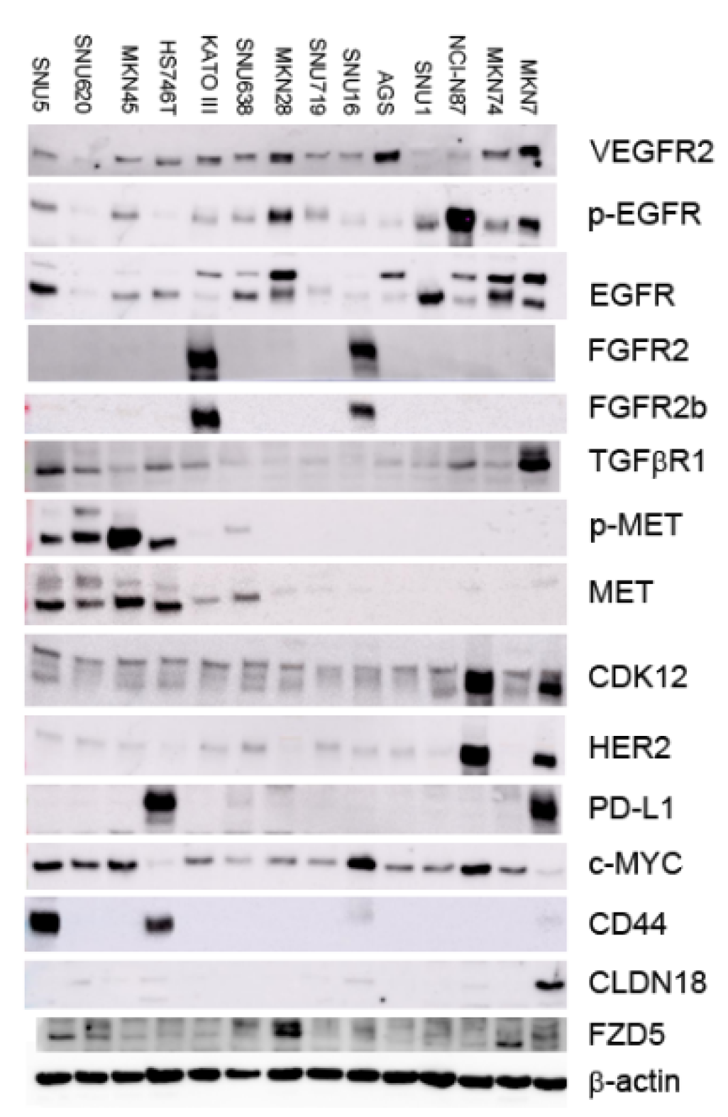
Figure 4.
Clinicopathological and biological characteristics of the 14 gastric cancer cell lines according to cancer therapeutic targets, including CNVs or protein overexpression. Lauren classification was compared with other biomarkers using targeted DNA sequencing or Western blot analysis.
Figure 4.
Clinicopathological and biological characteristics of the 14 gastric cancer cell lines according to cancer therapeutic targets, including CNVs or protein overexpression. Lauren classification was compared with other biomarkers using targeted DNA sequencing or Western blot analysis.
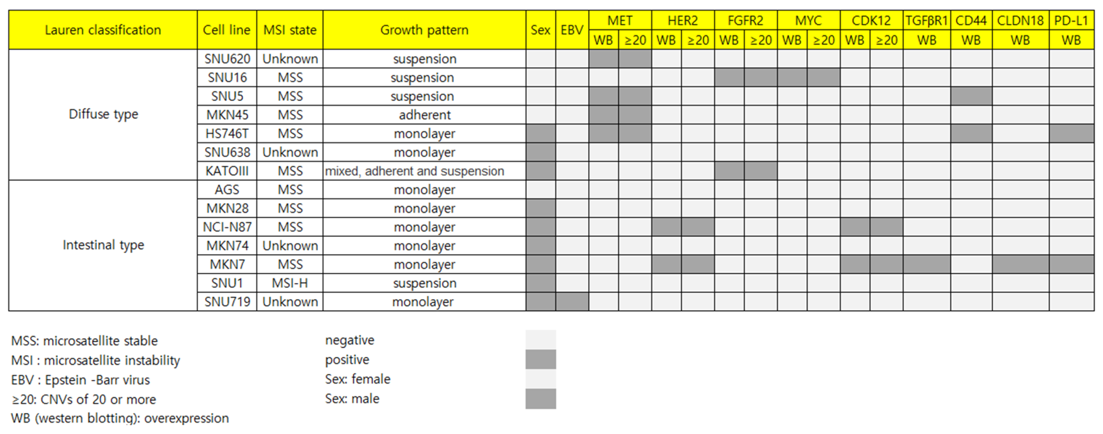
Figure 5.
Sensitivity of the 35 drugs tested in the 14 gastric cancer cell lines. Inhibition rate (%) of each drug is presented at a concentration of 10 μM.
Figure 5.
Sensitivity of the 35 drugs tested in the 14 gastric cancer cell lines. Inhibition rate (%) of each drug is presented at a concentration of 10 μM.
Disclaimer/Publisher’s Note: The statements, opinions and data contained in all publications are solely those of the individual author(s) and contributor(s) and not of MDPI and/or the editor(s). MDPI and/or the editor(s) disclaim responsibility for any injury to people or property resulting from any ideas, methods, instructions or products referred to in the content. |
© 2026 by the authors. Licensee MDPI, Basel, Switzerland. This article is an open access article distributed under the terms and conditions of the Creative Commons Attribution (CC BY) license.
Copyright: This open access article is published under a Creative Commons CC BY 4.0 license, which permit the free download, distribution, and reuse, provided that the author and preprint are cited in any reuse.


