Submitted:
30 January 2026
Posted:
03 February 2026
You are already at the latest version
Abstract
Sonodynamic therapy (SDT) is a non-invasive treatment modality that leverages the deep tissue penetration and spatiotemporal controllability of ultrasound to activate sonosensitizers and induce cytotoxic bioeffects, predominantly through reactive oxygen species (ROS)-mediated oxidative stress and cavitation-associated mechanical injury. Despite encouraging progress, early SDT development has been limited by poor aqueous solubility and nonspecific biodistribution of many sonosensitizers, inadequate intratumoral accumulation, and challenges in standardizing and monitoring ultrasound dosimetry. Nanocarrier technologies have substantially expanded the therapeutic design space of SDT by improving pharmacokinetics, reducing off-target exposure, preserving sensitizer activity (e.g., mitigating aggregation-related quenching), and enabling tumor- and organelle-directed delivery. This review summarizes the historical evolution of SDT and nanocarriers and synthesizes current mechanistic understanding, including cavitation-centered biophysics, multi-route ROS generation (sonochemistry, conditional sonoluminescence pathways, and mechano-electronic charge-redox processes), and downstream regulated cell death and immunogenic cell death. We further organize SDT-nanocarrier synergy into key modules: pharmacokinetic/spatial control, ultrasound-enhanced transport (sonoporation and barrier modulation), hypoxia relief and redox reprogramming to amplify ROS, engineered cavitation/energy transduction, and immune reprogramming enabling sono-immunotherapy. Finally, we discuss emerging application strategies-targeted delivery, tumor microenvironment modulation, and multimodal combination regimens-alongside translational progress and remaining barriers such as delivery heterogeneity, real-time exposure verification, long-term biosafety, manufacturability, and clinical trial design. Future advances are expected to prioritize simplified GMP-compatible platforms, delivery diagnostics for patient stratification, and closed-loop ultrasound control for consistent, safe, and precision SDT.
Keywords:
sonodynamic therapy
; nanocarriers
; targeting
; efficacy
; ultrasound
; immune
1. Historical Evolution of Sonodynamic Therapy
1.1. Early Research and Discoveries
Sonodynamic therapy (SDT) has emerged as a promising non-invasive therapeutic modality [1,2,3], leveraging the synergistic interaction between ultrasound and sonosensitizers to induce cytotoxic effects in tumor cells [4,5,6]. This approach has been particularly effective due to its ability to penetrate deeply into tissues, offering a significant advantage over photodynamic therapy (PDT), which is limited by the shallow penetration of light [7,8].
The earliest experimental evidence for sonodynamic effects was reported by Yumita and colleagues in 1989, showing that hematoporphyrin could sensitize cells to ultrasound-induced damage in the absence of light [9]. Subsequent work by Umemura et al. (1990) further elucidated the mechanism of this cytotoxicity, laying the foundation for what would become sonodynamic therapy [10]. The early research and discoveries in SDT have laid the groundwork for its application in various medical fields, including tumor therapy and antimicrobial therapy [4,11,12].
1.2. Progression and Challenges
During the early 1990s, the term “sonodynamic therapy” was gradually introduced to describe this ultrasound-activated, sensitizer-mediated therapeutic approach, explicitly drawing an analogy to photodynamic therapy while emphasizing its independence from optical energy sources. Initial mechanistic investigations suggested that acoustic cavitation played a central role in SDT-induced cytotoxicity [13]. The oscillation and collapse of cavitation bubbles generated localized extreme conditions, including transient high temperatures and pressures, which were believed to activate sonosensitizers and initiate biological damage [14,15]. Concurrently, early evidence indicated the involvement of ROS, such as singlet oxygen and free radicals, linking SDT mechanistically to oxidative stress pathways similar to those observed in PDT, albeit triggered through fundamentally different physical processes [16,17,18,19].
In parallel with mechanistic exploration, early SDT research focused on identifying suitable sonosensitizers. Porphyrin-based compounds, including hematoporphyrin derivatives and protoporphyrin IX, were among the agents evaluated due to their established use in PDT and favorable redox properties [20,21]. These studies demonstrated that certain sensitizers could be selectively activated by ultrasound to enhance cytotoxicity, providing proof-of-concept for sensitizer-dependent SDT [22,23]. Beyond porphyrins, early investigations also explored the synergistic effects of ultrasound with chemotherapeutic agents, suggesting the potential for combination strategies that could amplify therapeutic outcomes [5].
Despite these encouraging advances, early-generation SDT faced several critical limitations, including poor water solubility, non-specific biodistribution, and systemic toxicity of sonosensitizers, as well as a lack of precise control over ultrasound parameters. These challenges significantly constrained therapeutic efficacy and hindered clinical translation [24,25,26].
Figure 1.
Historical Milestones of SDT and Nanocarriers: Why SDT x Nanocarriers? Created in BioRender.
Figure 1.
Historical Milestones of SDT and Nanocarriers: Why SDT x Nanocarriers? Created in BioRender.
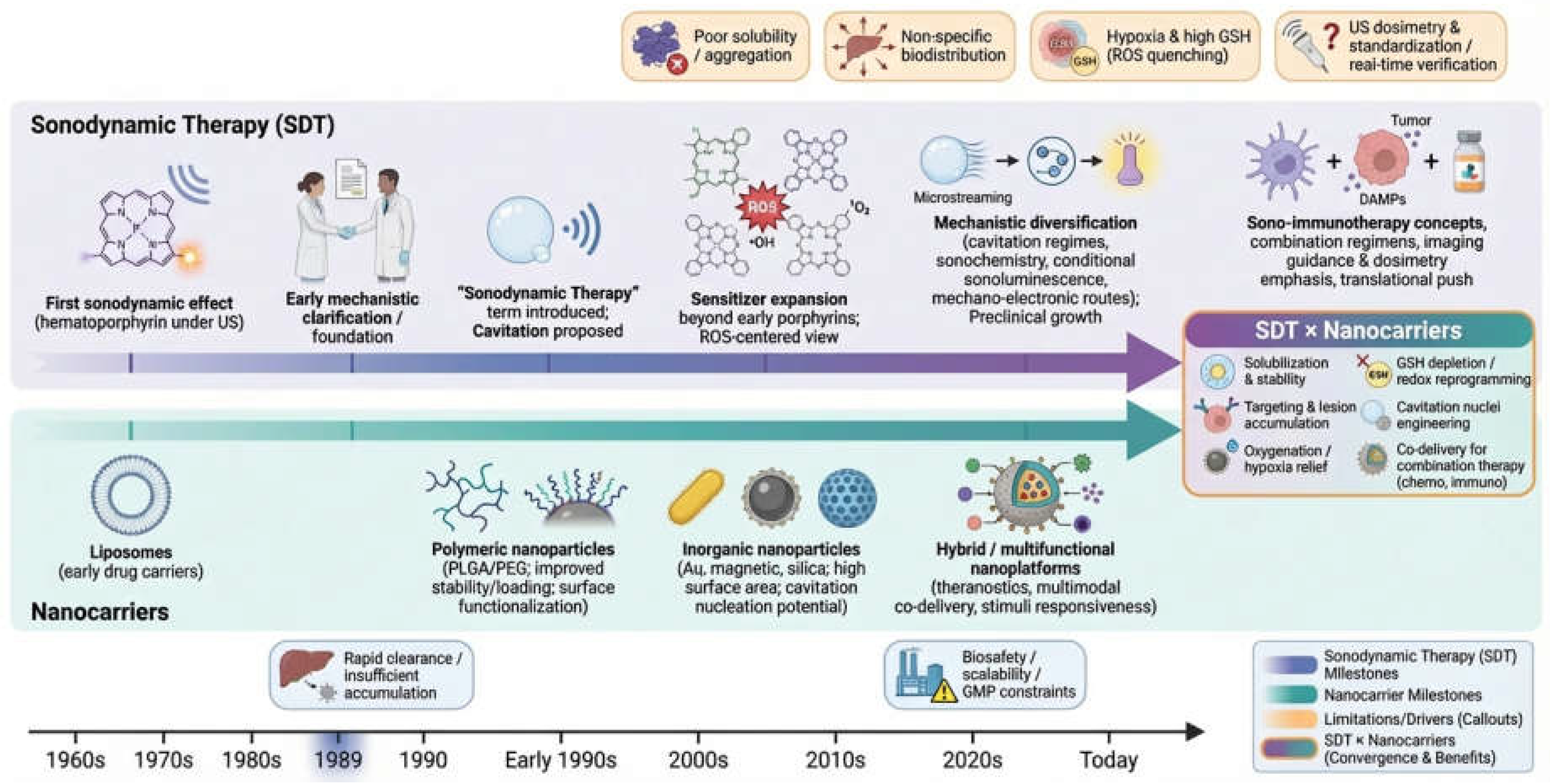
2. Historical Evolution of Nanocarriers
Nanocarrier technology has undergone significant advancements over the past few decades, dramatically improving the delivery and efficacy of therapeutic agents, including sonosensitizers used in SDT [27,28]. These innovations have addressed several limitations associated with early sonosensitizers, such as poor solubility, limited tissue penetration, non-specific biodistribution, and systemic toxicity. By encapsulating or conjugating sonosensitizers with nanocarriers, researchers have enhanced their stability, controlled their release, and facilitated targeted delivery to tumor sites, which is critical for increasing the therapeutic efficacy of SDT [4,29]. The evolution of nanocarriers can be divided into four key phases: liposomes, polymeric nanoparticles, inorganic nanoparticles, and multifunctional nanoplatforms [30,31,32,33]. Each phase represents a leap in nanocarrier design, contributing to the refinement of SDT and expanding its therapeutic potential.
3. Mechanisms of Sonodynamic Therapy and Nanocarrier Synergy
3.1. Mechanisms of Sonodynamic Therapy in Disease Treatment
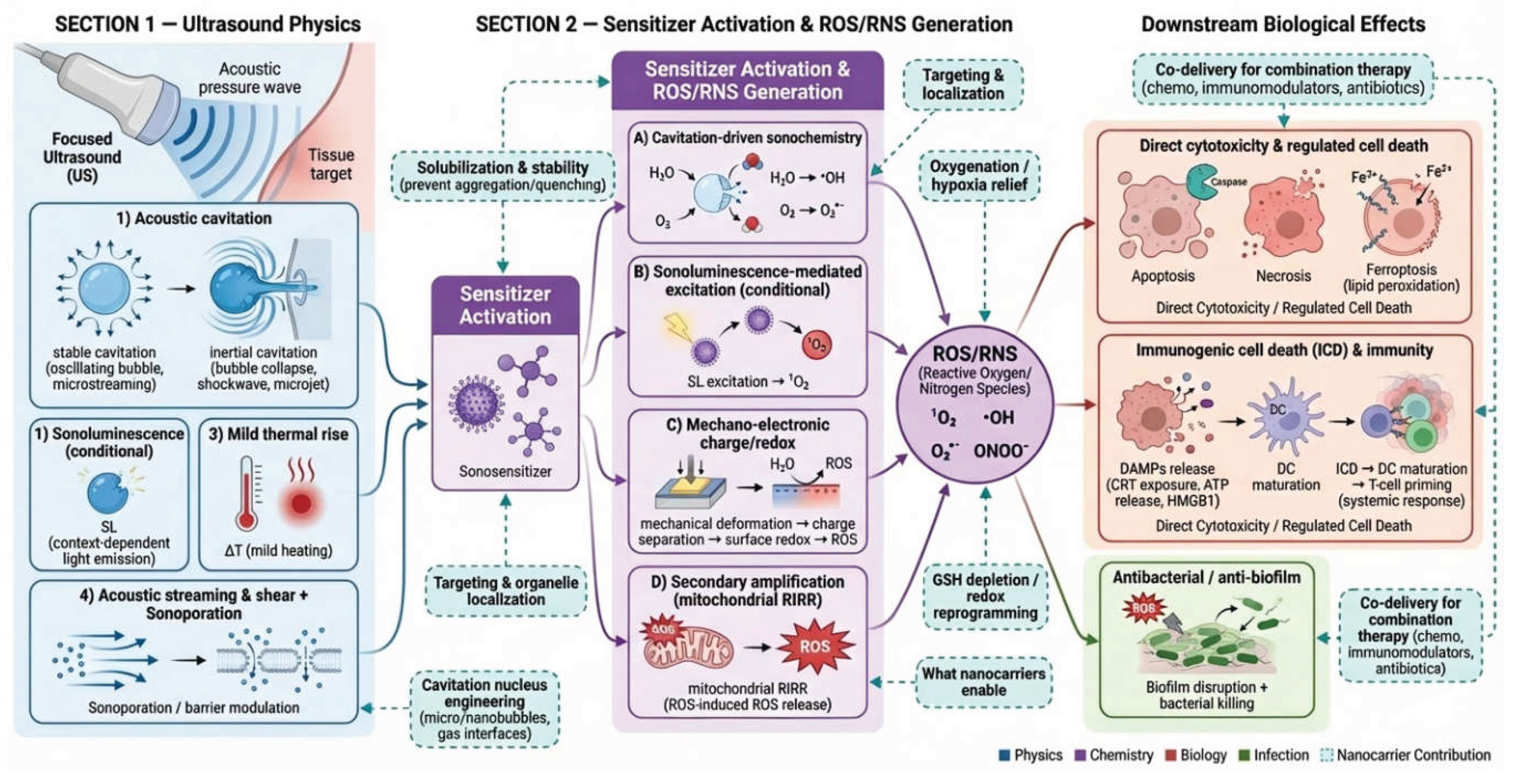
Sonodynamic therapy is generally understood as a spatiotemporally controlled oxidative (and partly mechanical) insult produced when focused ultrasound (US) interacts with a sonosensitizer in biological media [6,23,126,127,128,129,130]. The therapeutic outcome is therefore not dictated by a single pathway, but by the coupling among (i) ultrasound physics, (ii) sensitizer photophysics/sonochemistry (or mechano-chemistry), and (iii) cellular stress-response programs and immunity. Reviews increasingly converge on four major contributors: acoustic cavitation, reactive oxygen species (ROS) generation, mild thermal effects, and (in some models) sonoluminescence [126,127,128,129,130]. Reviews increasingly converge on four major contributors: acoustic cavitation, ROS generation, mild thermal effects, and (in some models) sonoluminescence [6,12,28,94,112].
3.1.1. Ultrasound-Tissue Interactions: Cavitation-Centered Biophysics
In SDT-relevant regimes, typically “low-intensity” compared with ablative high intensity focused ultrasound (HIFU), US propagates through tissue as a mechanical wave, generating pressure oscillations that can (a) deform membranes and cytoskeleton [131,132,133], (b) induce microstreaming and shear forces in fluids [134,135], and (c) trigger cavitation when pre-existing gas nuclei or introduced gas bodies (e.g., microbubbles) undergo oscillation [136,137,138]. Cavitation is usually categorized as stable cavitation and inertial (transient) cavitation [136].
Stable cavitation refers to sustained oscillation of bubbles that produces microstreaming and localized shear, enhancing membrane permeability and mass transport, while inertial (transient) cavitation means violent collapse of bubbles that can produce shock waves, microjets, localized high temperature/pressure, and radical chemistry at the bubble interface [139,140,141]. Stable cavitation can induce microstreaming and shear forces, leading to enhanced permeability of biological membranes, while inertial cavitation generates microjets, shock waves, and high local temperature and pressure, which can drive radical chemistry at the bubble interface [142,143,144]. These effects can directly damage cellular membranes and organelles [72,139,145], and also create conditions that amplify sonochemical ROS formation and/or sensitizer activation [130,146,147].
3.1.2. ROS Generation: Multiple Co-Existing Routes
Most SDT frameworks place ROS as the central cytotoxic intermediate (e.g., singlet oxygen 1O2, hydroxyl radicals •OH, superoxide O2•−), but the origin of ROS under ultrasound can vary across sensitizer classes and acoustic conditions [6,89,94,120,148,149,150].
For cavitation-driven sonochemistry (water/oxygen activation), inertial cavitation can yield highly reactive species through extreme microenvironments at bubble collapse, including water sonolysis and oxygen activation near the bubble interface [147,151,152]. These radicals can diffuse short distances to attack lipids, proteins, and nucleic acids [153,154], and they may also initiate secondary oxidative cascades such aslipid peroxidation and mitochondrial ROS amplification [155,156].
For sonoluminescence-mediated excitation, there exists debated contribution. A long-standing hypothesis proposes that light emitted during cavitation (sonoluminescence, SL) can excite sensitizers in a photodynamic-therapy-like manner, leading to ROS generation [28,157,158,159]. Recent literature shows active debate: some analyses argue SL may be a major mechanistic focus in certain contexts, while other experimental work suggests the emitted light/ROS levels under typical regimens may be insufficient to explain therapeutic effects, implying sonoluminescence may be minor or conditional [160,161].
Taken together, these mechanistic uncertainties translate into actionable nanocarrier design rules: even if SL is not universally dominant, formulations that introduce or co-localize sensitizers with cavitation nuclei (e.g., micro-/nanobubbles, gas-stabilizing interfaces) can bias SDT toward more inertial-cavitation–like, ROS-producing events. In fact, microbubble cavitation under therapeutic ultrasound has been shown to generate detectable SL, and the SL intensity correlates with broadband cavitation emissions—supporting the idea that “more cavitation activity” can shift the mechanistic balance [160]. Because SL exhibits threshold-like behavior and depends on exposure parameters (e.g., duty cycle, temperature), its contribution is plausibly conditional rather than constant across regimens [161]. Consistent with this view, recent SDT reviews still present ROS generation as a multi-route outcome of cavitation/SL/pyrolysis-type processes, rather than a single pathway [89].
Beyond SL-mediated excitation, many inorganics or hybrid nanosonosensitizer systems support “mechano-electronic” ROS routes: ultrasound-driven mechanical deformation can generate internal electric fields and charge separation, enabling surface redox reactions that yield ROS without requiring optical excitation [130,162,163]. Finally, even when primary ROS output is modest, it can trigger secondary biological amplification, most notably mitochondrial ROS-induced ROS release (RIRR)—a positive-feedback process that escalates oxidative stress and broadens downstream damage [156].
3.1.3. Downstream Cell Death and Immunological Consequences
Once oxidative and mechanical stress surpasses cellular repair capacity, SDT can engage multiple regulated cell death programs [72,129]. Apoptosis is frequently observed, but resistance to apoptosis in aggressive tumors has motivated strategies that bias SDT toward ferroptosis (iron-dependent lipid peroxidation) or mixed death phenotypes to improve efficacy [163].
A particularly important modern view is that SDT can induce immunogenic cell death (ICD)—characterized by danger-associated molecular patterns (DAMPs) such as calreticulin exposure, ATP release, and HMGB1 release—thereby promoting dendritic cell maturation and T-cell priming [164,165,166]. This provides a mechanistic bridge from “local ROS killing” to systemic antitumor effects, but it also highlights why spatial control matters for safety and for immune programming [167,168,169].
3.1.4. Key Determinants of SDT Efficacy and Mechanistic Levers
Mechanistically, SDT outcome can be viewed as a function of three coupled “dials”: acoustic parameters, sensitizer properties and oxygen/redox microenvironment [170]. Acoustic parameters include frequency, pressure amplitude, duty cycle, exposure time, focusing geometry (which set cavitation probability, microstreaming, and heating) [149,171,172]. Sensitizer properties include ROS quantum yield, subcellular localization, aggregation state, and stability [94,173,174,175,176]. Reviews on sonosensitizer development emphasize that sensitizer chemistry and microenvironment strongly influence ROS pathways [148,170]. Oxygen/redox microenvironment include hypoxia and high glutathione (GSH) in tumors can quench ROS and limit SDT; therefore, oxygen supply and redox modulation are recurring design targets [177,178,179,180].
Figure 2.
Mechanisms of Sonodynamic Therapy (US Physics → Chemical Activation → Biological Effects). Created in BioRender.
Figure 2.
Mechanisms of Sonodynamic Therapy (US Physics → Chemical Activation → Biological Effects). Created in BioRender.
3.2. Mechanisms of Nanocarrier-Mediated Drug Delivery
Nanocarriers improve SDT not only by “delivering more sensitizer,” but by reshaping where the sensitizer resides, how long it persists, and what microenvironmental constraints it experiences (oxygen, pH, enzymes, redox) [104,173,181,182,183]. Mechanistically, nanocarrier delivery is a multi-stage process with distinct bottlenecks [184,185,186].
3.2.1. Systemic Fate: Protein Corona, Clearance, and “Stealth”
Upon entering biological fluids, nanoparticles rapidly adsorb proteins to form a protein corona [187,188,189], which can alter colloidal stability [190,191], cellular recognition [192], targeting ligand availability [193,194], and cellular uptake pathways [195]. Reviews on corona biology emphasize that corona composition and dynamics strongly influence in vivo fate and pharmacokinetics [187,196,197]. Therefore, design implications for SDT nanocarriers include: (i) surface chemistry to control corona formation [198,199,200]; (ii) “stealth” layers (e.g., PEGylation or zwitterionic coatings) to reduce opsonization [183,201,202,203]; and (iii) biomimetic cloaks (cell membranes) to modulate biocompatibility, tumor enrichment, and immunological effects [48,204,205,206].
3.2.2. Tumor Accumulation: EPR, Transcytosis, and Heterogeneity
The classical explanation for tumor nanomedicine accumulation is the enhanced permeability and retention (EPR) effect, wherein leaky vasculature and impaired lymphatic drainage favor macromolecule retention [207,208]. However, modern perspectives stress that tumor delivery is heterogeneous and may involve additional mechanisms such as active trans-endothelial transport (transcytosis) and microenvironment-dependent vascular behavior [49,209,210,211,212]; a 2024 Nature-hosted review discusses delivery mechanisms and strategies beyond a simplistic EPR-only model [208]. For SDT, this heterogeneity is critical: insufficient accumulation lowers efficacy [94,149,170,183]. Besides, SDT efficacy is strongly dependent on local sonosensitizer availability and ROS generation, so insufficient intratumoral accumulation can markedly reduce therapeutic response. In such settings, the relative contribution of ultrasound-only bioeffects, thermal/mechanical effects such as cavitation and sonoporation may become more pronounced [127,145,213,214].
3.2.3. Cellular Internalization and Subcellular Trafficking
After reaching the tumor interstitium, nanoparticles must cross cellular membranes via endocytosis or, less commonly, direct fusion or penetration depending on material type [215,216,217,218]. Intracellular trafficking often leads to endosomal or lysosomal compartments; for sensitizers, this localization can be beneficial, as lysosomal rupture can amplify stress, or limiting, like quenching or enzymatic degradation [216,218,219,220]. Thus, organelle-targeting motifs and endosomal escape strategies can reshape which death pathways dominate, like apoptosis, ferroptosis or ICD [163,164,220,221].
3.2.4. Release Mechanisms: Diffusion, Degradation, and Stimuli Responsiveness
Nanocarriers can be engineered for controlled drug release through both passive and triggerable mechanisms. In passive modes, payloads are liberated by diffusion/desorption (including surface-bound drug), by gradual matrix erosion/degradation of the carrier material, or—especially for lipid-based systems—via lipid exchange with surrounding membranes/lipoproteins that perturbs bilayer composition and promotes cargo leakage/transfer [222,223,224].
Endogenous, disease-associated cues then enable “smart” on-site release, leveraging hallmarks of the tumor microenvironment and intracellular trafficking routes such as acidic pH (tumor interstitium/lysosomes), elevated GSH and ROS, overexpressed enzymes (notably matrix metalloproteinases, MMPs), and hypoxia, typically via cleavable linkers or stimulus-labile materials [225,226]. Exogenous ultrasound—highly compatible with sonodynamic-therapy workflows—can further provide spatiotemporally controlled activation, where acoustic fields promote release by mechanical disruption of carrier shells, cavitation-mediated permeabilization/rupture, acoustic radiation forces that enhance carrier–tissue interactions, and phase transitions of perfluorocarbon (PFC) cores (acoustic droplet vaporization) that rapidly destabilize the nanocarrier and “burst” payloads on demand [35,227,228,229]. Recent reviews emphasize that ultrasound-responsive designs deliberately couple these physicochemical triggers to tune release thresholds and improve on-target activation while limiting off-target leakage [227,230].

Table 1.
Comparison of Sonosensitizer Families and Key Properties Favorable for Nanodelivery.
| Category (Representative Examples) | Key ROS Types and Mechanistic Features | Advantages / Limitations | Standard Delivery Methods | Representative References |
| Porphyrins / PpIX-precursors | Mainly 1O2 plus radicals depending on microenvironment | Advantages: Clinically familiar; strong redox activity; benefits from encapsulation to improve PK Limitations: Hydrophobicity/aggregation; potential dark toxicity; oxygen dependence |
Liposomes; polymeric NPs; albumin-based carriers | [348,349] |
| Chlorin derivatives (e.g., Ce6, Photochlor) | Predominantly 1O2; can be boosted by oxygenation modules | Advantages: Strong ROS yield; easy co-loading with O2 modulators Limitations: Aggregation quenching; hypoxia sensitivity |
Microbubbles/nanobubbles; lipid NPs; membrane camouflage | [205,300] |
| Cyanine / heptamethine dyes (e.g., IR780, iodinated cyanine) | Mixed with radical and 1O2; often coupled to hypoxia relief or catalytic ROS loops | Advantages: NIR imaging/theranostics-ready; mitochondrial affinity Limitations: Photothermal/sonothermal crosstalk; instability; higher off-target risk without “stealth” |
Hollow MnO2 shells; mesoporous silica; antibody conjugates | [75,98,307] |
| AIEgens / AIE-active sonosensitizers | Often designed toward radical under hypoxia; aggregation-tolerant | Advantages: “Anti-ACQ” by design; good for high-loading nanoformulations Limitations: Chemistry varies; activation threshold needs tuning |
Polymeric micelles; amphiphilic assemblies; targeted ligands | [74,237] |
| Inorganic semiconductors / sonocatalysts / piezoelectric | •OH / O2•− via mechano-electronic charge separation / catalytic surfaces | Advantages: High stability; oxygen-independent (partly); can lower cavitation threshold Limitations: Potential long-term biosafety; clearance; surface defects variability |
Inorganic core–shell; metal-doped catalysts; membrane camouflage | [150,162,231] |
| MOF / COF framework-based | Frequently radical + catalytic cascades (Fenton-like, GSH-responsive) | Advantages: High payload capacity; modular catalytic nodes; can integrate imaging + TME triggers Limitations: Stability/ion release; reproducibility; biodegradation products |
MOF/COF nanoparticles; bacteria/OMV modification; HA coating | [109,114,116,297] |
3.3. Synergy Between Sonodynamic Therapy and Nanocarriers
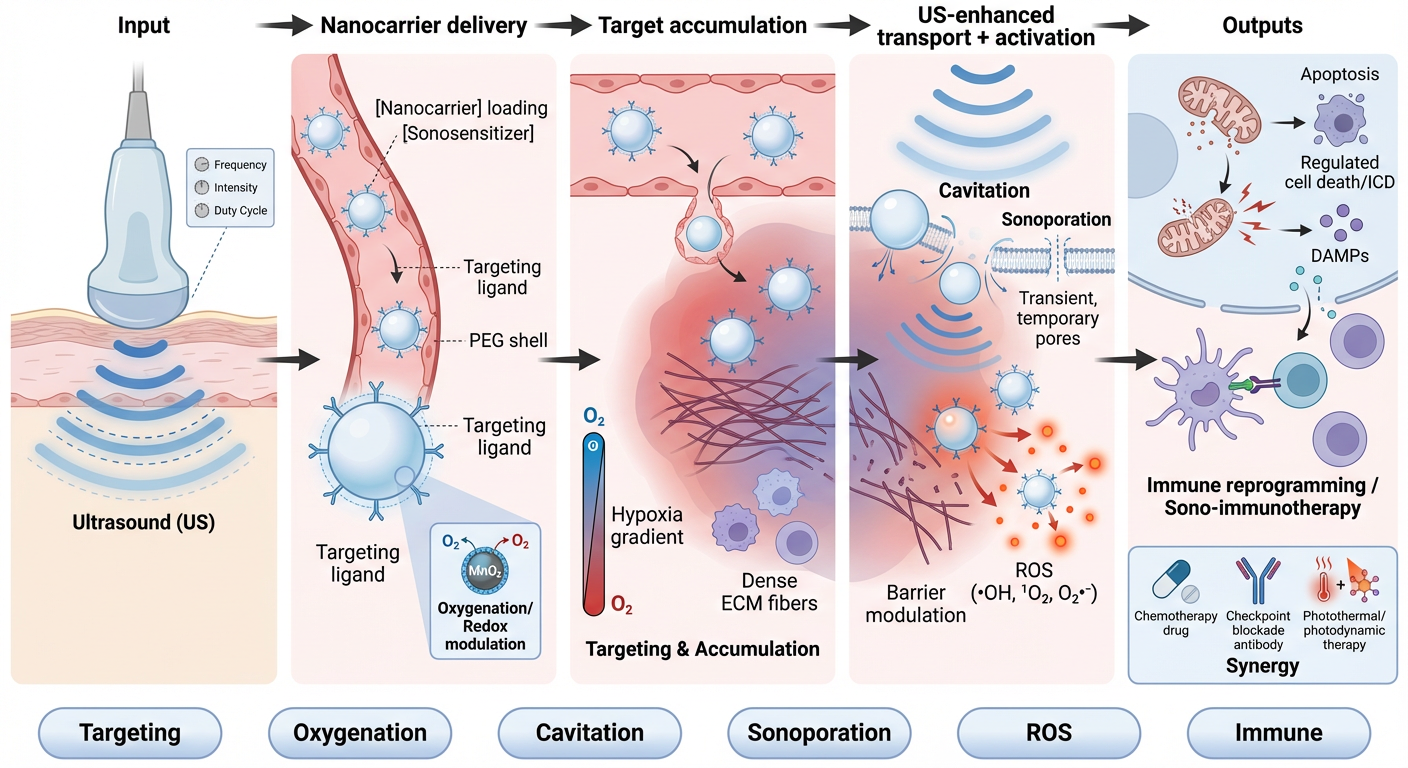
The “SDT + nanocarrier” synergy can be mechanistically organized into five cooperating modules: pharmacokinetic control, ultrasound-enhanced transport, ROS amplification, cavitation/energy transduction engineering, and immune reprogramming.
3.3.1. Pharmacokinetic and Spatial Control: Concentrating the Sensitizer Where Ultrasound Will Act
Nanocarriers extend circulation time and reduce premature clearance/metabolism of sensitizers, increasing the probability that sensitizer and focused ultrasound overlap in space and time [12,231,232,233]. This is a uniquely important safety lever in SDT: ultrasound exposure is localized, but off-target sensitizer distribution can still raise risks if broad-field ultrasound is used or if sensitizer persists in sensitive tissues [12,48,72,232]. Encapsulation also mitigates aggregation-induced quenching for hydrophobic sensitizers, preserving ROS competence [234,235,236,237].
3.3.2. Ultrasound-Enhanced Delivery: Sonoporation and Barrier Modulation
Ultrasound can transiently increase membrane and vascular permeability, often termed sonoporation, particularly when cavitation nuclei (microbubbles) are present [238,239,240,241]. This can improve extravasation, interstitial penetration, and intracellular uptake of nanocarriers [101,242,243,244,245,246], creating a positive feedback loop: more nanocarrier uptake → more sensitizer in cells → more SDT effect [101,247]. Foundational work on sonoporation mechanisms emphasizes transient pore formation and enhanced endocytosis driven by microbubble oscillation and associated stresses [136,246,248,249]. From a design standpoint, pairing SDT nanocarriers with (i) microbubbles, (ii) gas-generating components, or (iii) phase-change droplets can convert ultrasound from a “trigger” into a “delivery accelerator” would make sense [101,245,250,251,252,253].
3.3.3. ROS Amplification and Hypoxia Mitigation: Oxygen-Carrying and Oxygen-Generating Nanoplatforms
Because ROS production often requires molecular oxygen, tumor hypoxia is a frequent SDT bottleneck [89,178,254]. A major nanocarrier synergy strategy is oxygen supply: Perfluorocarbon (PFC)-based oxygen carriers can dissolve and transport oxygen, alleviating hypoxia and improving ROS yield in oxygen-dependent therapies [180,254,255,256].
Oxygen-sufficient or oxygen-loaded core-shell platforms have been demonstrated in chemo-sonodynamic settings, explicitly using oxygen-carrying cores to boost singlet oxygen generation and reduce hypoxia-associated resistance [118,182,257]. Complementary approaches include catalase- or MnO₂-like components that convert endogenous H₂O₂ into O₂, simultaneously lowering oxidative buffering and increasing available oxygen [258,259,260,261]. These “self-oxygenating” concepts are also attractive for strengthening ICD, since robust oxidative stress can enhance DAMP release [164,221,254,259].
3.3.4. Engineering Cavitation and Energy Transduction: Making Ultrasound “Work Harder” at the Tumor
Nanocarriers can be engineered to increase the probability of acoustic cavitation and to concentrate ultrasound energy deposition within tumors, thereby amplifying both direct mechanical injury (e.g., microjets/shock effects) and sonochemistry-driven ROS production [14,72]. Because cavitation is widely regarded as a key prerequisite underpinning SDT efficacy, “cavitation-first” nanodesigns offer a practical route to intensify local bioeffects without relying solely on higher external power [167,262].
Mechanistically, representative strategies include: (i) cavitation-nuclei incorporation, such as micro-/nanobubbles, solid gas-trapping or porous/concave architectures, and in situ gas-generating chemistries (e.g., oxygen- or CO₂-forming reactions) that seed bubble nucleation [250,262,263,264,265,266,267]; (ii) phase-change droplets, typically PFC nanodroplets that undergo acoustic droplet vaporization to form local bubbles, enhancing cavitation while enabling ultrasound-triggered “burst” release [228,253,254,268]; and (iii) mechano-catalytic sensitizers, including piezoelectric or semiconductor nanomaterials that convert ultrasound-induced stress into charge separation and subsequent ROS formation [269,270]. These designs are especially relevant for deep tissues, where acoustic intensity must remain within safety constraints; improving nanoscale “energy conversion efficiency” (lowering cavitation thresholds and strengthening stress-to-chemistry transduction) can help preserve efficacy without escalating macroscopic exposure [89,159].
3.3.5. Immunological Synergy: From Local SDT to Systemic Control
When nanocarriers concentrate SDT damage in tumors and boost oxidative stress, they can increase the likelihood of ICD and subsequent adaptive immune activation (CRT/ATP/HMGB1 signaling, dendritic cell maturation, T-cell priming) [129,254,271,272]. This creates a mechanistic rationale for combining SDT nanomedicines with checkpoint blockade or other immunomodulators (often co-loaded in the same nanocarrier): SDT provides antigen release and inflammatory cues, while immunotherapy prevents T-cell exhaustion and immunosuppressive rebound [48,254,272,273].
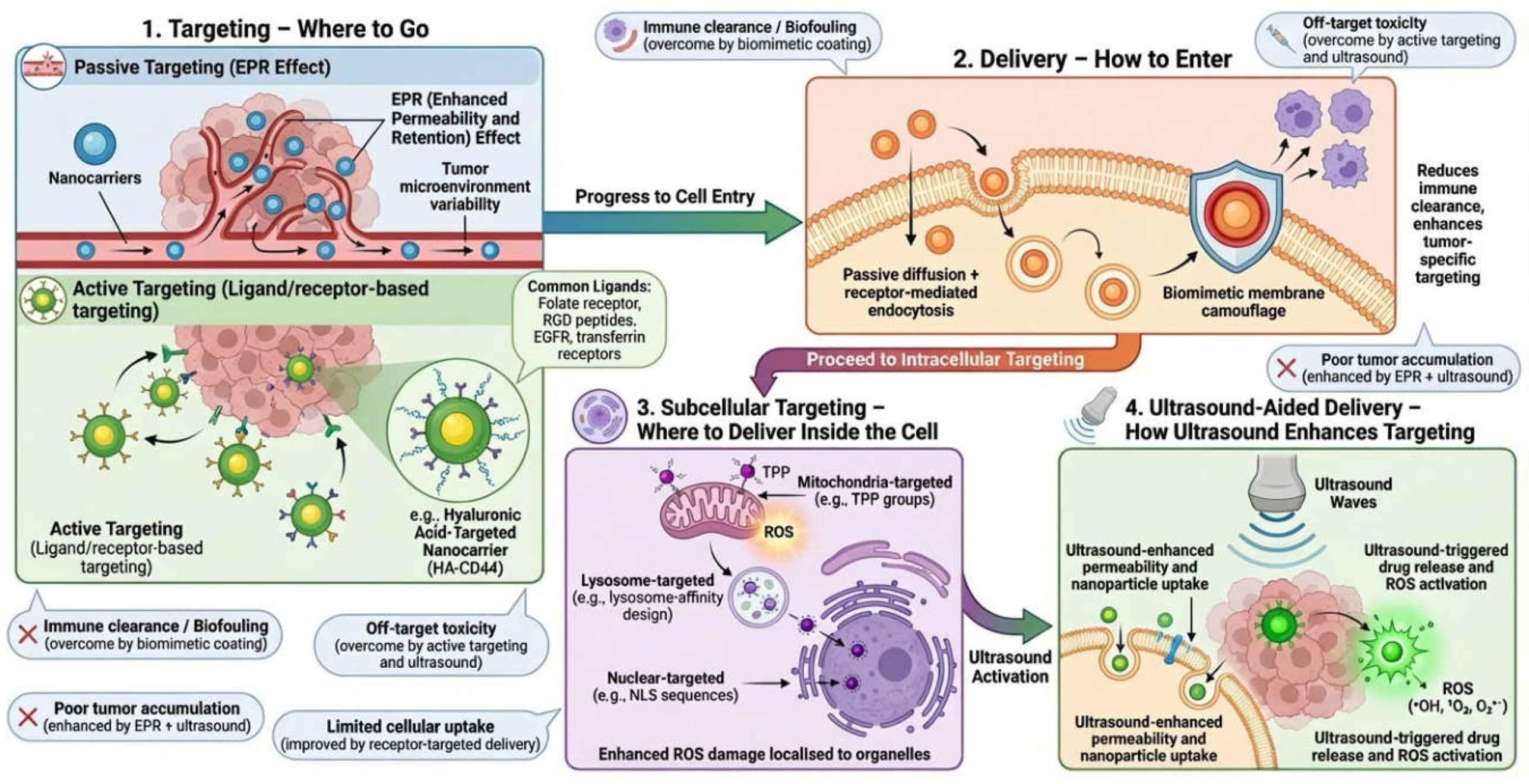
4. Nanocarrier-Based Strategies for Enhancing SDT Targeting and Efficacy
Nanocarriers have become central to modern SDT research because they directly address the key biological and engineering bottlenecks that limit standalone SDT [72,89,274]: (i) insufficient tumor accumulation and heterogeneous intratumoral penetration of sonosensitizers [146,275,276], (ii) premature clearance and off-target distribution [48,146,275], (iii) tumor-microenvironment (TME) suppression of reactive oxygen species (ROS) through hypoxia and antioxidant systems (e.g., glutathione, GSH) [89,146,181], and (iv) the need for spatiotemporal control of sensitizer activation and co-therapeutic release [28,38,275]. Recent reviews emphasize that “nanotechnology-enabled SDT” is evolving from simply “loading a sonosensitizer” into designing multi-stage, responsive delivery systems that coordinate pharmacokinetics, TME modulation, and ultrasound-triggered actuation [72,89,104,275].
4.1. Targeted Drug Delivery Systems in SDT
4.1.1. Passive Targeting: Circulation Engineering and Transport Optimization
Although the EPR effect can facilitate nanoparticle accumulation in some tumors, its variability across tumor types, stages, and patients makes passive targeting alone unreliable [73,277,278]. Consequently, contemporary SDT nanocarriers increasingly optimize system-level transport: prolonging circulation (e.g., PEGylation or alternative “stealth” coatings), controlling size (typically balancing extravasation vs. renal clearance), and tuning surface charge to reduce opsonization while preserving tumor uptake [72,278,279]. The design logic is to maximize the area-under-the-curve for tumor exposure, then leverage ultrasound to achieve local activation and/or release [72,227,280]. These principles are discussed prominently in recent SDT nanotechnology reviews and in the broader ultrasound-responsive nanocarrier literature [183,278].
4.1.2. Active Targeting: Ligand/Receptor Recognition and Multi-Receptor Strategies
Active targeting adds a molecular recognition layer on top of passive accumulation. In SDT, active targeting is not only about increasing uptake; it also improves selectivity of ultrasound-triggered ROS damage by enriching sensitizers at the tumor site and reducing sensitizer exposure in normal tissues [281,282,283]. Common ligand-receptor axes include: (i) Hyaluronic acid (HA)-CD44 targeting (frequent in breast, ovarian, and other CD44-high tumors) [75]; (ii) RGD/iRGD peptides-integrins (tumor endothelium and invasive tumor cells, can also support deeper tissue penetration) [284]; (iii) Folate receptor, transferrin receptor, EGFR/HER2 antibodies, and aptamers, depending on tumor biology and intended clinical indication [281]. A representative 2025 example is an HA-modified hollow MnO₂ platform loaded with IR780 (IR780@H-MnO₂@HA), which uses CD44 targeting to enhance tumor delivery and couples TME-triggered MnO₂ decomposition with imaging/therapy functions for SDT [75].
Active targeting is also being integrated with immunomodulatory payloads to convert SDT from a purely cytotoxic modality to a “tumor vaccination”-like process. An Acta Biomaterialia 2025 study reported a targeted nanosensitizer-augmented sono-immunotherapy platform co-delivering a STING agonist (MSA-2) with an RGD-targeting element to enhance immune activation alongside SDT [285]. MSA-2 is a recognized small-molecule STING agonist with the potential to be combined with SDT to enhance immune responses [286]. Additional studies exploring strategies for the local release of STING agonists co-encapsulated with photosensitizers, among other agents, may serve as supplementary background information [287].
4.1.3. Biomimetic Targeting: Cell-Membrane Coating and Endogenous Trafficking
Biomimetic strategies, such as cancer-cell membrane, platelet, red-blood-cell (RBC), or immune-cell membrane camouflage, are increasingly used to (i) reduce immune clearance, (ii) exploit homologous targeting, and (iii) improve vascular margination or immune-cell-mediated trafficking into tumors [48,288,289,290,291,292]. A 2025 review highlights how “sonodynamic biomimetic nanomedicine” can tackle off-target toxicity and TME barriers by integrating biological interfaces with ultrasound-activated therapeutics [48].
4.1.4. Subcellular Targeting: Mitochondria/Lysosome/Nucleus-Directed SDT
Because SDT efficacy depends on the proximity of ROS generation to vulnerable subcellular structures, recent nanocarriers increasingly incorporate organelle-targeting motifs (e.g., mitochondrial targeting groups such as triphenylphosphonium, nuclear localization sequences, or lysosome-affinity designs) [220,293,294,295,296]. The rationale is: ROS are short-lived and diffusion-limited, so organelle-localized sensitizers can produce disproportionately higher damage per ROS unit [175,297].
In summary, the most efficacious approach to targeting within the context of sonodynamic therapy (SDT) is frequently characterized by a multi-layered strategy. This involves a combination of systemic transport, tumor selectivity, and, in certain instances, organelle localization. This multi-faceted targeting is further enhanced by the application of ultrasound-triggered activation, which serves to refine spatial precision [104,117,220,293].
Figure 3.
Scientific mechanism map: Targeted Delivery Strategies in SDT: From ‘Where to Go’ to ‘Where to Enter’. Created in BioRender.
Figure 3.
Scientific mechanism map: Targeted Delivery Strategies in SDT: From ‘Where to Go’ to ‘Where to Enter’. Created in BioRender.
4.2. Tumor Microenvironment Targeting
The TME is frequently hostile to SDT because ROS-based killing is attenuated by hypoxia, high GSH, and immunosuppressive cell populations [177,178,181,298]. Thus, many state-of-the-art SDT nanocarriers are explicitly designed to reprogram the TME rather than merely deliver sensitizers [180,181,255,258,299].
4.2.1. Hypoxia Alleviation: Oxygen Delivery and Oxygen Generation
Hypoxia reduces oxygen-dependent ROS pathways and can also promote therapy resistance [113,300,301]. Nanocarriers address this via oxygen carriers (e.g., perfluorocarbon-based droplets) that physically transport O₂ into tumors [113,121,254,301]. Some systems combine oxygen delivery with ultrasound-triggered phase transitions or cavitation to improve local release and perfusion [253,300,302]. In situ oxygen generation, often using catalytic components that convert endogenous H₂O₂ into O₂ (e.g., MnO₂-based shells) or oxygen-releasing compounds (e.g., CaO₂) [121,181,258,303]. A 2025 Journal of Materials Chemistry B report describes a cascade strategy (including CaO₂-based oxygenation and antioxidant disruption) to counter hypoxia and improve SDT outcomes [181]. A compelling “integrated oxygenation + immunomodulation” approach is the Chemical Engineering Journal 2025 ImmunoSonogen platform, which combines sonodynamic oxygenation with immune activation to overcome hypoxia-linked immunosuppression and promote stronger antitumor responses [113].
4.2.2. Redox Targeting: GSH Depletion and ROS Amplification Loops
Elevated GSH in tumor cells and the tumor microenvironment can rapidly quench SDT-generated ROS, thereby weaken oxidative damage and ultimately reduce SDT efficacy [304,305]. Accordingly, redox-targeting nanocarriers are often designed to undergo GSH-responsive disassembly/decomposition—for example, MnO₂ is reduced by GSH (Mn(IV)→Mn(II)), which can both trigger on-site release or activation of the sonosensitizer and simultaneously consume GSH to diminish antioxidant buffering capacity [259,304]. To further boost killing, many platforms construct cascade ROS-amplification loops, where SDT-initiated ROS and/or endogenous H₂O₂ is funneled into secondary radical-generating reactions (e.g., Mn²⁺- or Fe-based Fenton/Fenton-like processes) to continuously produce highly cytotoxic •OH and related oxidants [259,306]. In parallel, coupling SDT with ferroptosis can reinforce lipid-peroxidation “positive feedback” by depleting GSH and/or disabling GPX4/FSP1 defenses, enabling lipid peroxides to accumulate and sustain ROS-driven membrane damage [304,305,306,307].
4.2.3. ECM/Stromal Targeting: Penetration Enhancement and Immune Infiltration
Dense ECM can limit both nanoparticle penetration and immune-cell trafficking [308,309,310]. Beyond enzyme-based ECM degradation, recent work demonstrates physical/thermal modulation to soften barriers and enhance delivery [311,312,313]. The SnSNP “denaturation-and-penetration” strategy is notable because it directly addresses stromal restriction and links improved penetration to improved cytotoxic T lymphocyte infiltration and antitumor immunity [183].
4.2.4. Immune Microenvironment Targeting: ICD, STING, and Macrophage Reprogramming
A major advance in SDT nanocarrier research is the shift from viewing SDT as purely localized cytotoxicity to treating it as an immunogenic therapy that can induce ICD and remodel the tumor immune microenvironment [129,164]. A 2025 review in Frontiers in Immunology summarizes how SDT-oriented nanoparticle delivery systems can be engineered to enhance ICD hallmarks and improve immune-contexture outcomes (e.g., dendritic cell activation and T-cell infiltration) by optimizing sensitizer delivery and stimulus responsiveness [164].
Two representative immuno-TME-targeting nanocarrier routes have emerged. First, STING-pathway activation plus SDT uses co-delivery of STING agonists (e.g., MSA-2-based designs) with nanosensitizers so that ultrasound-triggered SDT can synergize with innate immune activation, amplifying dendritic cell priming and downstream T-cell responses [285,314]. Second, tumor-associated macrophage (TAM) reprogramming employs ultrasound-responsive nanocarriers to deliver immunomodulators (such as siRNA or pathway inhibitors), shifting macrophages from an immunosuppressive M2-like state toward a pro-inflammatory M1-like phenotype to enhance antitumor immunity; a 2024 open-access study in Journal of Nanobiotechnology exemplifies this approach using ultrasound-responsive carriers integrating siRNA and Fe₃O₄ to modulate macrophage polarization and strengthen immune control of tumors [315].
Overall, modern SDT nanocarriers increasingly target tumor-microenvironment vulnerabilities (oxygen/redox imbalance, ECM barriers, and immune suppression) as deliberately as they target tumor cells, thereby improving targeting specificity and therapeutic efficacy [48,316].
Table 2.
A Checklist of Tumor Microenvironment (TME) Modulation Strategies for Enhanced SDT.
| TME bottleneck | Nanoengineering Techniques | How it Boosts SDT | Key Risk Points | Representative References |
| Hypoxia | O2 carriers (e.g., PFC-based) | Restores O2-dependent ROS; improves response consistency | Gas embolism concerns; formulation complexity | [180,301] |
| Hypoxia | Catalytic O2 generation (MnO2, CAT-like) | Converts H2O2 to O2; supports sustained ROS production | Metal ion release; H2O2 dependency | [258,300] |
| Hypoxia | O2-releasing compounds (CaO2) | Local O2 supply under hypoxia; “always-on” oxygenation | Local alkalinization; Ca2+ overload/toxicity | [181,303] |
| High GSH / strong antioxidant buffering | GSH-consuming shells (MnO2, pMOF) | Depletes GSH leading to less ROS quenching; can unlock cascade catalysis | Oxidative stress to normal tissues; Mn-related safety | [179,235] |
| Redox resilience | Self-amplifying ROS loops (CDT/SDT cascades) | SDT initiates ROS leading to feeds secondary radical generation | Off-target inflammation; metal-catalyst byproducts | [177,306] |
| Dense ECM / high interstitial resistance | ECM degradation (e.g., collagenase) | Improves penetration + intratumoral distribution | Vascular leakage; inflammation; metastasis concern if uncontrolled | [311,313] |
| Mechanical barriers | Mechanical microenvironment modulation | Lowers transport resistance; improves perfusion/uptake | Edema; unpredictable perfusion changes | [308] |
| Immunosuppression | STING agonist co-delivery / sono-STING | SDT→ICD + innate activation → stronger T cell priming | Systemic cytokine risk; autoimmunity-like toxicity | [314,343] |
| Immune resistance (checkpoint dominance) | SDT + ICB / nanovaccine | SDT-induced ICD supplies antigens; ICB prevents T cell exhaustion | Immune-related adverse events | [344,345] |
| TAM polarization / poor phagocytosis | siRNA / magneto-acoustic immunomodulation | Reprograms macrophages; increases antigen presentation & clearance | Off-target gene silencing; RES accumulation | [315] |
4.3. Combining SDT with Other Modalities Using Nanocarriers
Nanocarriers are uniquely suited to SDT combination therapy because they can co-load multiple agents, control their ratios, and coordinate sequential or conditional release under TME cues and/or ultrasound [12,104,298,317,318]. This has led to a surge of “SDT+X” platforms where SDT serves as the deep-penetrating trigger and ROS generator, while co-therapies address complementary resistance mechanisms [48,72,89,104,119,250,298,317,319].
4.3.1. SDT + Chemotherapy/Molecular Inhibitors
4.3.2. SDT + Gene/RNA Therapy (RNAi/CRISPR-Adjacent Strategies)
Gene silencing can remove intrinsic SDT resistance pathways such as antioxidant defenses [325,326,327], survival signaling [328,329], autophagy/mitophagy [325,330]. A 2025 Theranostics paper reported a reduction-responsive RNAi nanoplatform co-delivering Nrf2 siRNA, a mitophagy inhibitor (3-MA), and a sonosensitizer (purpurin-18), aiming to enhance cancer sonoimmunotherapy by dual inhibition of mitophagy and Nrf2 pathways [325].
4.3.3. SDT + Immunotherapy (Checkpoint Blockade, STING Agonists, Nanovaccines)
SDT can trigger ICD and the release of tumor antigens/DAMPs, while nanocarriers enable co-delivery of immune adjuvants or pathway agonists to convert local tumor ablation into systemic antitumor immunity [112,129]. Targeted STING-agonist co-delivery nanosystems, such as liposomes co-loading a sonosensitizer with the non-nucleotide STING agonist MSA-2—combine SDT-triggered antigen liberation with STING activation to enhance dendritic cell maturation and cytotoxic T-cell priming [285,331]. Oxygenation–immunomodulation designs (e.g., the ImmunoSonogen platform) integrate ultrasound-driven oxygen generation with immunomodulatory components to relieve hypoxia and overcome hypoxia-associated immune suppression, thereby strengthening downstream immune activation [113,332]. Consistently, recent reviews summarize ICD-oriented SDT nanomedicines as both ICD inducers and tumor immune microenvironment remodelers, and emphasize their compatibility with immune checkpoint blockade and other immunotherapeutic regimens [72,112,129].
4.3.4. SDT + Ferroptosis / Chemodynamic Therapy (CDT) / Catalytic Therapies
Because lipid peroxidation is central to ferroptosis, SDT-generated ROS can synergize strongly with ferroptosis induction and iron-catalyzed reactions [119,173,260,304,333]. A 2025 Journal of Nanobiotechnology study described an SDT-boosted biomimetic nanoplatform targeting ferroptosis and immune microenvironment remodeling, reporting macrophage polarization effects and durable immune memory in preclinical models [304].
4.3.5. SDT + Phototherapy (PDT/PTT) and Multi-Trigger Platforms
Hybrid sonosensitizer/photosensitizer systems can exploit complementary activation windows (light for superficial or intraoperative contexts; ultrasound for deep lesions) [72,89,334,335,336,337]. The SnSNP work is an instructive example of combining mild photothermal effects with SDT to improve penetration and immune activation [183].
Table 3.
The Combination Works in SDT-Based Regimens.
| Combination direction | Core logic of synergy | Carrier Design Notes | Representative References |
| Chemo / metabolic inhibitors | SDT increases permeability + ROS stress → sensitizes to chemo; chemo can weaken repair pathways | Co-loading vs. sequential release; ultrasound-triggered burst to align timing | [108,171,182,293] |
| Gene / RNA therapeutics | Knock down antioxidant/escape pathways → SDT ROS becomes “unbuffered”; can rewire immune context | Protect nucleic acids; endosomal escape; redox- or US-triggered unpacking | [296,315,325] |
| Immunotherapy (STING, ICB, vaccines) | SDT → ICD/DAMPs + antigen release; immune adjuvants/ICB sustain systemic response | Keep immunostimulant shielded systemically; tumor-local activation; avoid cytokine burst | [183,295,314,343,344,345] |
| Ferroptosis / CDT | SDT ROS seeds lipid peroxidation; CDT supplies •OH; ferroptosis disables GSH/GPX4/FSP1 defenses → positive feedback | Metal/catalyst nodes + GSH depletion; membrane/mitochondria targeting improves efficiency | [177,304,305,306,307,352] |
| PDT/PTT | PTT improves perfusion/oxygenation and accelerates kinetics; PDT adds orthogonal ROS modality; multimodal imaging-ready | Avoid overheating; choose trigger hierarchy (US-first vs. light-first); spatial co-localization | [116,300,301] |
5. Clinical Applications and Translational Research
5.1. Preclinical Studies
Over the past decade, nanocarrier-assisted SDT has progressed from proof-of-concept ROS generation to disease-oriented therapeutic engineering, especially for deep-seated tumors where light-based photodynamic therapy is limited by penetration [12,89]. Contemporary SDT typically employs low-intensity focused ultrasound (LIFU/FUS) to activate sonosensitizers and induce oxidative stress–dominated cytotoxicity; recent summaries place commonly used ultrasound settings broadly within MHz frequencies and low W/cm² intensities depending on the application and device configuration [158,338].
5.1.1. Cancer Models (Solid Tumors, Orthotopic Tumors)
Preclinical efficacy has been repeatedly demonstrated in murine xenograft and orthotopic systems by packaging sonosensitizers (e.g., porphyrin/chlorin derivatives, ALA/PpIX precursors, inorganic sonosensitizers, or hybrid catalysts) into liposomes, polymeric nanoparticles, MOFs, exosome-like vesicles, and inorganic-organic composites to improve pharmacokinetics and tumor accumulation, mitigate premature clearance, and enable stimulus-responsive release [72,89,94,109,231,339,340,341]. It is emphasized that nanomaterials can (i) increase intratumoral sensitizer concentration, (ii) relieve hypoxia or amplify ROS chemistry, and (iii) provide theranostic functions (MRI/PA/US imaging) that are particularly valuable for orthotopic disease where dose delivery is otherwise uncertain [72,89,94,109,231,339,340,341,342].
5.1.2. Immunological “Second Wave” of SDT Efficacy
A major translational trend is to treat SDT not only as a local ablation-like approach but also as a method to induce ICD and reshape the tumor immune microenvironment [112,164,167]. Multiple preclinical reports indicate that SDT-mediated oxidative injury can promote antigen release and immune activation; recent work continues to refine this concept via nanocarriers that co-deliver immune adjuvants, ferroptosis amplifiers, or checkpoint blockade partners [183,255,298,304,343,344,345].
Notably, a 2025 study combining 5-ALA-mediated SDT with a novel high-intensity focused ultrasound (HIFU) approach reported effective tumor suppression at reduced acoustic intensities while enhancing anti-tumor immune responses, supporting the idea that ultrasound engineering and sensitizer biology can be co-optimized to expand the therapeutic window [346].
5.1.3. Brain Tumor Relevant Preclinical Evidence
5.1.4. Anti-Bacterial / Anti-Biofilm Applications
Beyond oncology, SDT has been explored as a non-antibiotic antimicrobial strategy, leveraging ultrasound penetration and ROS-mediated multi-target damage to reduce resistance pressure [351,352,353]. Recent syntheses specifically highlight nanoplatform-based antibacterial SDT (nanocarriers delivering organic sonosensitizers or nanosonosensitizers themselves) for deep infections and biofilm-associated disease [351,352,353,354].
5.2. Clinical Trials and Progress
Despite extensive preclinical activity, SDT’s clinical footprint remains small, consistent with earlier translational analyses noting that only limited clinical reporting existed historically and that standardization and dosimetry were major barriers [170,172,347,355,356]. The situation is now changing, driven largely by drug repurposing (5-ALA) and the availability of clinically deployed ultrasound platforms [348]. A key recent milestone is the first-in-human early clinical study of 5-ALA (SONALA-001) + MRgFUS SDT in recurrent high-grade glioma (NCT04559685). The report describes assessment of safety and biological efficacy across ascending MRgFUS energy doses in nine patients, supporting feasibility of this drug–device approach in humans [348]. Besides, there are still some ongoing or registered clinical trials. Conference updates also indicate ongoing maturation of these programs. For example, CTNI-76 is a phased "trial update" for the SDT-202 (SONALA-001 + Exablate) project of rGBM [357].
5.3. Limitations and Challenges in Clinical Translation
5.3.1. Biological Delivery Variability
Nanocarrier-enhanced SDT often assumes passive tumor accumulation via the EPR effect, yet multiple clinical-focused reviews emphasize that EPR is heterogeneous and dynamic across tumor types, stages, and even regions within one tumor, driving inconsistent nanoparticle deposition and variable efficacy [277,359,360,361]. For SDT, this is especially problematic because therapeutic effect depends on co-localization of (a) sonosensitizer/nanocarrier and (b) a sufficiently dosed ultrasound field [89,172]. Translation will likely require patient stratification (imaging or biomarkers of nanoparticle delivery) and/or “EPR-enhancing” strategies (vascular normalization, ECM modulation, ultrasound-assisted transport), rather than relying on passive targeting alone [89,172].
5.3.2. Ultrasound Dosimetry, Standardization, and Real-Time Monitoring
Unlike pharmacologic dosing, ultrasound dose depends on parameters (frequency, duty cycle, intensity, pulse structure), tissue acoustics, and cavitation dynamics [172,363]. Lack of standardized reporting and real-time verification can impede reproducibility across sites [172]. Emerging approaches that combine real-time cavitation monitoring with imaging (e.g., passive cavitation imaging/mapping integrated with B-mode guidance) illustrate how SDT could move toward closed-loop control and safer, more consistent exposures [363,364].
5.3.3. Safety of Complex Nanocarriers: Immunogenicity, Long-Term Fate, and “Silent” Accumulation
Many advanced SDT nanoplatforms incorporate heavy atoms, inorganic lattices, catalytic metals, or persistent polymers [89,338]. Even when acute toxicity appears low, clinical translation requires addressing: complement activation / infusion reactions, RES accumulation (liver/spleen), degradation pathways and metabolite safety, potential interactions between ultrasound and nanoparticle surfaces (heating, fragmentation, altered biodistribution) [280,365,366]. These concerns push the field toward biodegradable or clinically precedent materials (lipids, PLGA, albumin-like systems) and to simplified designs compatible with scalable GMP [365,367].
5.3.4. Clinical trial design challenges
6. Future Perspectives and Research Directions
6.1. Nanocarrier Design for Personalized SDT
Personalized SDT will likely develop along two coupled axes: The first is personalizing nanocarrier exposure. Given EPR variability, future trials may incorporate delivery diagnostics. For example, contrast-enhanced imaging, radiolabel tracing, or ultrasound/PA reporters embedded in the carrier, to confirm tumor deposition before therapeutic ultrasound is applied [105,277,370,371,372]. The second is personalizing ultrasound dose. Integrating real-time cavitation mapping, thermometry (where relevant), and patient-specific acoustic modeling could enable patient-tailored sonication plans and reduce under- or over-treatment [373,374,375,376].
6.2. SDT + Blood Brain Barrier (BBB) Modulation / Enhanced Brain Delivery
For central nervous system (CNS) disease, one compelling direction is pairing SDT with ultrasound-mediated BBB permeabilization (often with microbubbles), either to increase nanocarrier entry or to combine SDT with systemically delivered drugs [371,376,377]. Current clinical and translational reviews of BBB opening by focused ultrasound provide a roadmap for safety monitoring, workflow integration, and agent selection [317,347,378].
6.3. Closed-Loop Ultrasound Control
A central limitation of SDT is that the delivered ultrasound settings (e.g., nominal acoustic power, duty cycle, duration) are not equivalent to the biologically effective dose at the tumor [170,172,379]. Variations in acoustic coupling, tissue attenuation, patient anatomy (especially transcranial pathways), and microbubble/nanocarrier interactions can shift the therapy from sub-therapeutic activation to excessive cavitation or unintended bioeffects [72,378,379,380,381,382]. Consequently, next-generation SDT, particularly nanocarrier-enabled SDT will likely require closed-loop ultrasound control to ensure consistent activation while maintaining safety margins across patients and centers [72,378,380,382]. This direction parallels focused-ultrasound fields where monitoring modalities such as passive cavitation detection/mapping and MR thermometry are used to improve treatment controllability and reproducibility [374,378,383,384,385,386].
Figure 4.
From Materials to Clinical Loop: Translational and Engineering Roadmap for SDT. Created in BioRender.
Figure 4.
From Materials to Clinical Loop: Translational and Engineering Roadmap for SDT. Created in BioRender.
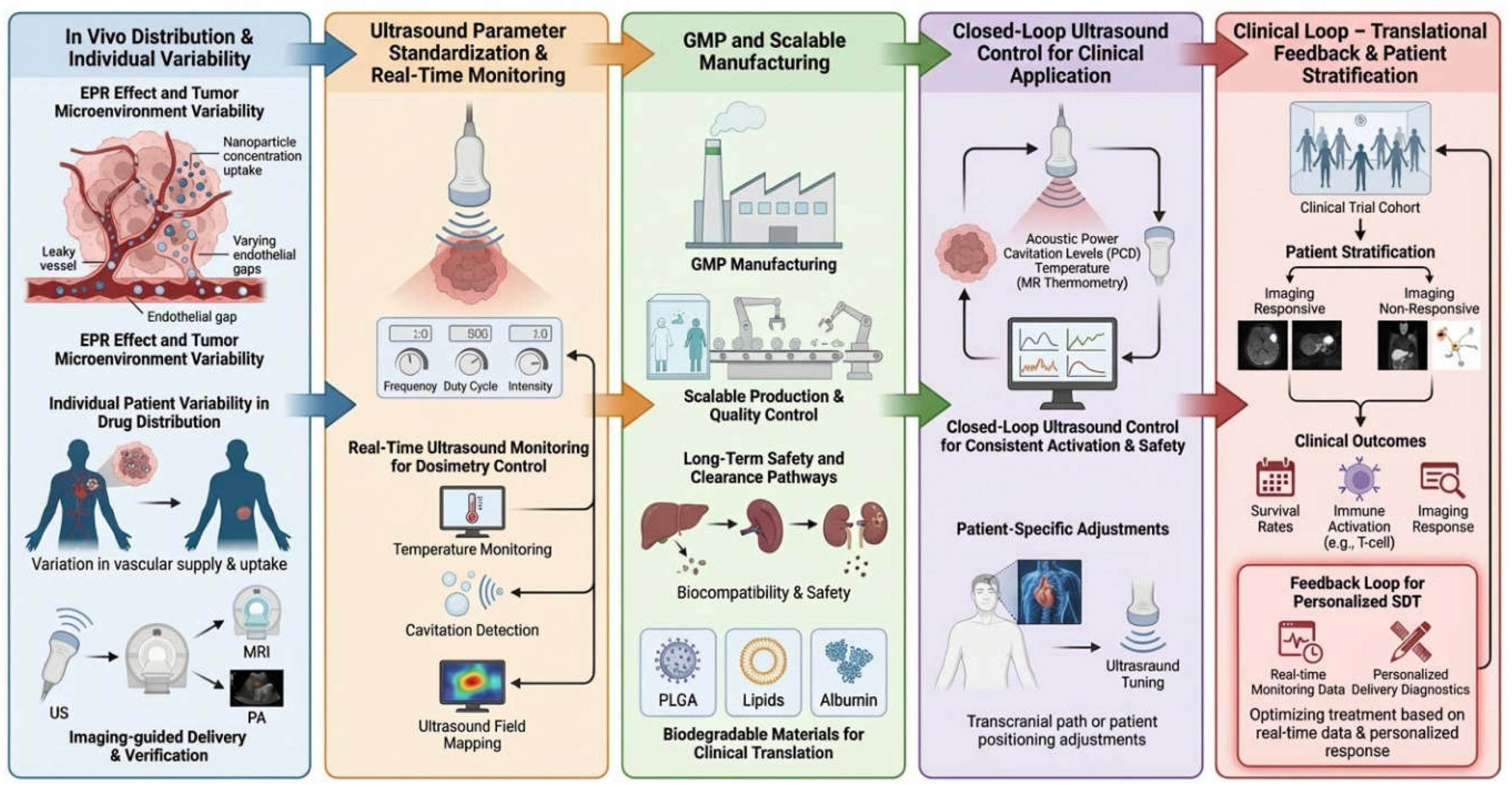
7. Conclusion
SDT has emerged as a promising non-invasive therapeutic strategy that leverages the deep tissue penetrability and spatiotemporal controllability of ultrasound to activate sonosensitizers and generate ROS for disease treatment. However, early SDT development was constrained by key translational barriers, including the poor solubility and non-specific biodistribution of many sensitizers, systemic toxicity concerns, and limited standardization and control of ultrasound exposure.
The integration of nanocarriers has substantially expanded the therapeutic design space of SDT. By encapsulating or conjugating sonosensitizers, nanocarriers can improve aqueous dispersibility, pharmacokinetics, and local retention while reducing off-target exposure. Importantly, modern SDT nanoplatforms go beyond “delivering more sensitizer”: they enable microenvironment-aware engineering (e.g., hypoxia relief, redox modulation, and ROS amplification), multimodal imaging/theranostic guidance, and rational co-delivery of synergistic agents. These capabilities support combination regimens (e.g., chemo-SDT, catalytic/ferroptotic amplification, and sono-immunotherapy) and offer a path toward more precise and reproducible SDT activation in vivo.
Despite rapid progress, several challenges remain before nanocarrier-assisted SDT can be widely translated, including inter- and intra-tumoral variability in nanoparticle delivery, the need for quantitative ultrasound dosimetry and real-time monitoring, and rigorous evaluation of long-term biosafety and manufacturability for complex formulations. Future advances will likely center on simplified yet functional and GMP-compatible designs, delivery diagnostics for patient stratification, and closed-loop ultrasound control to ensure consistent activation and safety across patients and clinical sites. With continued interdisciplinary collaboration across nanomedicine, ultrasound engineering, and clinical research, nanocarrier-enabled SDT is poised to evolve from a promising concept into a clinically feasible, precision-targeted therapeutic platform.
References
- Tachibana, K., Feril Jr, L. B., & Ikeda-Dantsuji, Y. (2008). Sonodynamic therapy. Ultrasonics, 48(4), 253-259.
- Shibaguchi, H., Tsuru, H., Kuroki, M., & Kuroki, M. (2011). Sonodynamic cancer therapy: a non-invasive and repeatable approach using low-intensity ultrasound with a sonosensitizer. Anticancer research, 31(7), 2425-2429.
- Ouyang, J., Tang, Z., Farokhzad, N., Kong, N., Kim, N. Y., Feng, C., ... & Tao, W. (2020). Ultrasound mediated therapy: recent progress and challenges in nanoscience. Nano Today, 35, 100949. [CrossRef]
- Yan, P., Liu, L. H., & Wang, P. (2020). Sonodynamic therapy (SDT) for cancer treatment: advanced sensitizers by ultrasound activation to injury tumor. ACS applied bio materials, 3(6), 3456-3475. [CrossRef]
- Hirschberg, H., & Madsen, S. J. (2017). Synergistic efficacy of ultrasound, sonosensitizers and chemotherapy: a review. Therapeutic Delivery, 8(5), 331-342. [CrossRef]
- Rosenthal, I., Sostaric, J. Z., & Riesz, P. (2004). Sonodynamic therapy––a review of the synergistic effects of drugs and ultrasound. Ultrasonics sonochemistry, 11(6), 349-363. [CrossRef]
- Rengeng, L., Qianyu, Z., Yuehong, L., Zhongzhong, P., & Libo, L. (2017). Sonodynamic therapy, a treatment developing from photodynamic therapy. Photodiagnosis and photodynamic therapy, 19, 159-166. [CrossRef]
- Pan, X., Wang, H., Wang, S., Sun, X., Wang, L., Wang, W., ... & Liu, H. (2018). Sonodynamic therapy (SDT): a novel strategy for cancer nanotheranostics. Science China Life sciences, 61(4), 415-426. [CrossRef]
- Yumita, N., Nishigaki, R., Umemura, K., & Umemura, S. I. (1989). Hematoporphyrin as a sensitizer of cell-damaging effect of ultrasound. Japanese Journal of Cancer Research, 80(3), 219-222. [CrossRef]
- Umemura, S. I., Yumita, N., Nishigaki, R., & Umemura, K. (1990). Mechanism of cell damage by ultrasound in combination with hematoporphyrin. Japanese Journal of Cancer Research, 81(9), 962-966. [CrossRef]
- Wang, R., Liu, Q., Gao, A., Tang, N., Zhang, Q., Zhang, A., & Cui, D. (2022). Recent developments of sonodynamic therapy in antibacterial application. Nanoscale, 14(36), 12999-13017. [CrossRef]
- Yang, N., Li, J., Yu, S., Xia, G., Li, D., Yuan, L., ... & Li, J. (2024). Application of nanomaterial-based sonodynamic therapy in tumor therapy. Pharmaceutics, 16(5), 603. [CrossRef]
- Choi, V., Rajora, M. A., & Zheng, G. (2020). Activating drugs with sound: mechanisms behind sonodynamic therapy and the role of nanomedicine. Bioconjugate Chemistry, 31(4), 967-989. [CrossRef]
- Wu, X., Chen, F., Zhang, Q., & Tu, J. (2024). What Is the Magical Cavitation Bubble: A Holistic Perspective to Trigger Advanced Bubbles, Nano-Sonocatalysts, and Cellular Sonosensitizers. BME frontiers, 5, 0067. [CrossRef]
- Huang, H., Zheng, Y., Chang, M., Song, J., Xia, L., Wu, C., ... & Chen, Y. (2024). Ultrasound-based micro-/nanosystems for biomedical applications. Chemical Reviews, 124(13), 8307-8472. [CrossRef]
- Lin, J., Li, D., Li, C., Zhuang, Z., Chu, C., Ostrikov, K. K., ... & Wang, P. (2023). A review on reactive oxygen species (ROS)-inducing nanoparticles activated by uni-or multi-modal dynamic treatment for oncotherapy. Nanoscale, 15(28), 11813-11833. [CrossRef]
- Zhou, Z., Song, J., Nie, L., & Chen, X. (2016). Reactive oxygen species generating systems meeting challenges of photodynamic cancer therapy. Chemical society reviews, 45(23), 6597-6626. [CrossRef]
- Przygoda, M., Bartusik-Aebisher, D., Dynarowicz, K., Cieślar, G., Kawczyk-Krupka, A., & Aebisher, D. (2023). Cellular mechanisms of singlet oxygen in photodynamic therapy. International Journal of Molecular Sciences, 24(23), 16890. [CrossRef]
- Yang, B., Chen, Y., & Shi, J. (2019). Reactive oxygen species (ROS)-based nanomedicine. Chemical reviews, 119(8), 4881-4985. [CrossRef]
- Chen, J., Zhou, Q., & Cao, W. (2024). Multifunctional porphyrin-based sonosensitizers for sonodynamic therapy. Advanced Functional Materials, 34(40), 2405844. [CrossRef]
- Tsolekile, N., Nelana, S., & Oluwafemi, O. S. (2019). Porphyrin as diagnostic and therapeutic agent. Molecules, 24(14), 2669. [CrossRef]
- Xiong, W., Wang, P., Hu, J., Jia, Y., Wu, L., Chen, X., ... & Wang, X. (2015). A new sensitizer DVDMS combined with multiple focused ultrasound treatments: an effective antitumor strategy. Scientific reports, 5(1), 17485. [CrossRef]
- McHale, A. P., Callan, J. F., Nomikou, N., Fowley, C., & Callan, B. (2016). Sonodynamic therapy: concept, mechanism and application to cancer treatment. Therapeutic ultrasound, 429-450.
- Zhou, Y., Wang, M., & Dai, Z. (2020). The molecular design of and challenges relating to sensitizers for cancer sonodynamic therapy. Materials Chemistry Frontiers, 4(8), 2223-2234. [CrossRef]
- Zhu, K., Wang, J., Wang, Z., Chen, Q., Song, J., & Chen, X. (2025). Ultrasound-Activated Theranostic Materials and Their Bioapplications. Angewandte Chemie International Edition, 64(22), e202422278. [CrossRef]
- Wang, X., Zhong, X., Gong, F., Chao, Y., & Cheng, L. (2020). Newly developed strategies for improving sonodynamic therapy. Materials Horizons, 7(8), 2028-2046. [CrossRef]
- Son, S., Kim, J. H., Wang, X., Zhang, C., Yoon, S. A., Shin, J., ... & Kim, J. S. (2020). Multifunctional sonosensitizers in sonodynamic cancer therapy. Chemical Society Reviews, 49(11), 3244-3261. [CrossRef]
- Wan, G. Y., Liu, Y., Chen, B. W., Liu, Y. Y., Wang, Y. S., & Zhang, N. (2016). Recent advances of sonodynamic therapy in cancer treatment. Cancer biology & medicine, 13(3), 325-338. [CrossRef]
- Ding, Y., Yang, Y., Aras, O., An, F., Zhou, M., & Chai, Y. (2025). Development and Application of Organic Sonosensitizers in Cancer Therapy. Aggregate, e70032.
- Rao, T. R., Sravani, B., & Aarthi, R. (2025). Nanocarriers in Drug Delivery Systems: An Overview. Journal of Advanced Scientific Research, 16(03), 8-14. [CrossRef]
- Karmaker, S., Rosales, P. D., Tirumuruhan, B., Viravalli, A., & Boehnke, N. (2025). More than a delivery system: the evolving role of lipid-based nanoparticles. Nanoscale. [CrossRef]
- Kibuuka, R. S. (2024). Nanomedicine and Targeted Drug Delivery: Advances and Challenges. [CrossRef]
- Yanar, F., Carugo, D., & Zhang, X. (2023). Hybrid nanoplatforms comprising organic nanocompartments encapsulating inorganic nanoparticles for enhanced drug delivery and bioimaging applications. Molecules, 28(15), 5694. [CrossRef]
- Sharma, V. K., & Agrawal, M. K. (2021). A historical perspective of liposomes-a bio nanomaterial. Materials Today: Proceedings, 45, 2963-2966. [CrossRef]
- Bahutair, W. N., Abuwatfa, W. H., & Husseini, G. A. (2022). Ultrasound triggering of liposomal nanodrugs for cancer therapy: a review. Nanomaterials, 12(17), 3051. [CrossRef]
- Jahangir, M. A., Mohanty, D., Choudhury, A., & Imam, S. S. (2023). Theranostic Applications of Functionalized Vesicular Carriers: Theranostic Applications of Functionalized Vesicular Carriers (Liposomes, Niosomes, Virosomes, Ethosomes, Phytosomes). In Multifunctional And Targeted Theranostic Nanomedicines: Formulation, Design And Applications (pp. 49-76). Singapore: Springer Nature Singapore.
- Fateh, S. T., Moradi, L., Kohan, E., Hamblin, M. R., & Dezfuli, A. S. (2021). Comprehensive review on ultrasound-responsive theranostic nanomaterials: mechanisms, structures and medical applications. Beilstein Journal of Nanotechnology, 12(1), 808-862. [CrossRef]
- Entzian, K., & Aigner, A. (2021). Drug delivery by ultrasound-responsive nanocarriers for cancer treatment. Pharmaceutics, 13(8), 1135. [CrossRef]
- Liu, F., Wu, L., Chen, L., Qi, X., Ge, Y., & Shen, S. (2016). Ultrasound-guided tumor sonodynamic therapy based on sonosensitizer liposome. Chemistry Letters, 45(11), 1304-1306. [CrossRef]
- Souri, S., Jadidi, M., Hasanzadeh, H., Khani, T., & Semnani, V. (2023). An In-Vivo Study of Sonodynamic Therapy with Encapsulated Hematoporphyrin. Frontiers in Biomedical Technologies, 10(2), 140-149. [CrossRef]
- Emiliani, C., Delmelle, M., Cannistraro, S., & Van de Vorst, A. (1983). Solubility of hematoporphyrin and photodynamic damages in liposomal systems: optical and electron spin resonance studies. Photobiochemistry and Photobiophysics, 5(2), 119-128. [CrossRef]
- Allen, T. M., & Cullis, P. R. (2013). Liposomal drug delivery systems: from concept to clinical applications. Advanced drug delivery reviews, 65(1), 36-48. [CrossRef]
- Sercombe, L., Veerati, T., Moheimani, F., Wu, S. Y., Sood, A. K., & Hua, S. (2015). Advances and challenges of liposome assisted drug delivery. Frontiers in pharmacology, 6, 286. [CrossRef]
- Pattni, B. S., Chupin, V. V., & Torchilin, V. P. (2015). New developments in liposomal drug delivery. Chemical reviews, 115(19), 10938-10966. [CrossRef]
- Chelliah, R., Rubab, M., Vijayalakshmi, S., Karuvelan, M., Barathikannan, K., & Oh, D. H. (2025). Liposomes for drug delivery: Classification, therapeutic applications, and limitations. Next Nanotechnology, 8, 100209. [CrossRef]
- Sun, Y., Wang, H., Wang, P., Zhang, K., Geng, X., Liu, Q., & Wang, X. (2019). Tumor targeting DVDMS-nanoliposomes for an enhanced sonodynamic therapy of gliomas. Biomaterials science, 7(3), 985-994. [CrossRef]
- Zhao, H., Du, F., Huang, J., Guo, R., Feng, Z., Wang, Z., & Qiu, L. (2025). Biomimetic liposomal nanovesicles remodel the tumor immune microenvironment to augment sono-immunotherapy. Journal of Controlled Release, 113830. [CrossRef]
- Zhang, A., Zheng, X., Yan, G., Liu, X., Xie, D., Xu, X., ... & Liu, Z. (2025). Sonodynamic biomimetic-nanomedicine fight cancers. Journal of Nanobiotechnology, 23(1), 548. [CrossRef]
- Wilhelm, S., Tavares, A. J., Dai, Q., Ohta, S., Audet, J., Dvorak, H. F., & Chan, W. C. (2016). Analysis of nanoparticle delivery to tumours. Nature reviews materials, 1(5), 1-12. [CrossRef]
- Sun, R., Xiang, J., Zhou, Q., Piao, Y., Tang, J., Shao, S., ... & Shen, Y. (2022). The tumor EPR effect for cancer drug delivery: Current status, limitations, and alternatives. Advanced drug delivery reviews, 191, 114614. [CrossRef]
- Shi, Y., Van Der Meel, R., Chen, X., & Lammers, T. (2020). The EPR effect and beyond: Strategies to improve tumor targeting and cancer nanomedicine treatment efficacy. Theranostics, 10(17), 7921. [CrossRef]
- Inglut, C. T., Sorrin, A. J., Kuruppu, T., Vig, S., Cicalo, J., Ahmad, H., & Huang, H. C. (2020). Immunological and toxicological considerations for the design of liposomes. Nanomaterials, 10(2), 190. [CrossRef]
- Ishida, T., Harashima, H., & Kiwada, H. (2002). Liposome clearance. Bioscience reports, 22(2), 197-224. [CrossRef]
- Zahednezhad, F., Saadat, M., Valizadeh, H., Zakeri-Milani, P., & Baradaran, B. (2019). Liposome and immune system interplay: Challenges and potentials. Journal of Controlled Release, 305, 194-209. [CrossRef]
- van Etten, E. W., ten Kate, M. T., Snijders, S. V., & Bakker-Woudenberg, I. A. (1998). Administration of liposomal agents and blood clearance capacity of the mononuclear phagocyte system. Antimicrobial Agents and Chemotherapy, 42(7), 1677-1681. [CrossRef]
- Basak, S., Das, T. K., & Ganguly, S. (2025). A review on tumor-targeting liposomes: Fabrication, mechanism and applications. Biochemical Pharmacology, 117460. [CrossRef]
- Liu, Y., Wang, T., Chi, X., Yu, S., He, W., He, H., ... & Zhang, J. (2025). Modeling based dynamics mechanism and pathway of liposome penetration in multicellular tumor spheroid for liposome optimization. International Journal of Pharmaceutics, 671, 125237. [CrossRef]
- Maji, I., Mahajan, S., Sriram, A., Mehra, N. K., & Singh, P. K. (2023). Polymeric nanomaterials: Fundamentals and therapeutic applications. In Nanomaterial-based drug Delivery systems: therapeutic and theranostic applications (pp. 33-64). Cham: Springer International Publishing.
- Locatelli, E., & Comes Franchini, M. (2012). Biodegradable PLGA-b-PEG polymeric nanoparticles: synthesis, properties, and nanomedical applications as drug delivery system. Journal of Nanoparticle Research, 14(12), 1316. [CrossRef]
- Elmowafy, E. M., Tiboni, M., & Soliman, M. E. (2019). Biocompatibility, biodegradation and biomedical applications of poly (lactic acid)/poly (lactic-co-glycolic acid) micro and nanoparticles. Journal of Pharmaceutical Investigation, 49(4), 347-380. [CrossRef]
- Makadia, H. K., & Siegel, S. J. (2011). Poly lactic-co-glycolic acid (PLGA) as biodegradable controlled drug delivery carrier. Polymers, 3(3), 1377-1397. [CrossRef]
- Bhardwaj, H., & Jangde, R. K. (2023). Current updated review on preparation of polymeric nanoparticles for drug delivery and biomedical applications. Next Nanotechnology, 2, 100013. [CrossRef]
- Eltaib, L. (2025). Polymeric Nanoparticles in Targeted Drug Delivery: Unveiling the Impact of Polymer Characterization and Fabrication. Polymers, 17(7), 833. [CrossRef]
- Floyd, T. G., Gurnani, P., & Rho, J. Y. (2025). Characterisation of polymeric nanoparticles for drug delivery. Nanoscale, 17(13), 7738-7752. [CrossRef]
- Parveen, S., Gupta, P., Kumar, S., & Banerjee, M. (2023). Lipid polymer hybrid nanoparticles as potent vehicles for drug delivery in cancer therapeutics. Medicine in drug discovery, 20, 100165. [CrossRef]
- Moreno-Lanceta, A., Medrano-Bosch, M., Edelman, E. R., & Melgar-Lesmes, P. (2022). Polymeric Nanoparticles for Targeted Drug and Gene Delivery Systems. In Pharmaceutical Nanobiotechnology for Targeted Therapy (pp. 561-608). Cham: Springer International Publishing.
- Geszke-Moritz, M., & Moritz, M. (2024). Biodegradable polymeric nanoparticle-based drug delivery systems: comprehensive overview, perspectives and challenges. Polymers, 16(17), 2536.
- Mares, A. G., Pacassoni, G., Marti, J. S., Pujals, S., & Albertazzi, L. (2021). Formulation of tunable size PLGA-PEG nanoparticles for drug delivery using microfluidic technology. PLoS One, 16(6), e0251821. [CrossRef]
- He, C., Hu, Y., Yin, L., Tang, C., & Yin, C. (2010). Effects of particle size and surface charge on cellular uptake and biodistribution of polymeric nanoparticles. Biomaterials, 31(13), 3657-3666. [CrossRef]
- Lin, S., Zhu, L., Li, Z., Yue, S., Wang, Z., Xu, Y., ... & Geng, J. (2023). Ultrasound-responsive glycopolymer micelles for targeted dual drug delivery in cancer therapy. Biomaterials Science, 11(18), 6149-6159. [CrossRef]
- Zhang, Y., Khan, A. R., Yang, X., Shi, Y., Zhao, X., & Zhai, G. (2021). A sonosensitiser-based polymeric nanoplatform for chemo-sonodynamic combination therapy of lung cancer. Journal of Nanobiotechnology, 19(1), 57. [CrossRef]
- Huang, Y., Ouyang, W., Lai, Z., Qiu, G., Bu, Z., Zhu, X., ... & Liu, J. (2024). Nanotechnology-enabled sonodynamic therapy against malignant tumors. Nanoscale advances, 6(8), 1974-1991. [CrossRef]
- Fan, D., Cao, Y., Cao, M., Wang, Y., Cao, Y., & Gong, T. (2023). Nanomedicine in cancer therapy. Signal Transduction and Targeted Therapy, 8(1), 293.
- Deng, K., Yu, Y., Zhao, Y., Li, J., Li, K., Zhao, H., ... & Huang, S. (2023). Tumor-targeted AIE polymeric micelles mediated immunogenic sonodynamic therapy inhibits cancer growth and metastasis. Nanoscale, 15(17), 8006-8018. [CrossRef]
- Ning, Y., Zhang, Z., Yasen, A., Sun, J., Zhou, Y., Tian, Q., & Cai, Q. (2025). Hyaluronic Acid-Modified Hollow MnO₂-Loaded IR780 Smart Theranostic Platform for Dual Imaging Guided Sonodynamic Therapy. Colloids and Surfaces A: Physicochemical and Engineering Aspects, 138224. [CrossRef]
- Zheng, F., Zhang, P., Zhang, Y., Long, H., Zhu, F., Chen, H., & Gao, Y. (2025). Aptamer-Modified Mesoporous Silica Nanoparticle for Nitric Oxide-Enhanced Targeted Sonodynamic Therapy against Lung Cancer. ACS Applied Nano Materials, 8(10), 5179-5192. [CrossRef]
- Herdiana, Y., Wathoni, N., Shamsuddin, S., & Muchtaridi, M. (2022). Scale-up polymeric-based nanoparticles drug delivery systems: Development and challenges. OpenNano, 7, 100048. [CrossRef]
- Rao, J. P., & Geckeler, K. E. (2011). Polymer nanoparticles: Preparation techniques and size-control parameters. Progress in polymer science, 36(7), 887-913. [CrossRef]
- Nel, A. E., Mädler, L., Velegol, D., Xia, T., Hoek, E. M., Somasundaran, P., ... & Thompson, M. (2009). Understanding biophysicochemical interactions at the nano–bio interface. Nature materials, 8(7), 543-557. [CrossRef]
- Owens III, D. E., & Peppas, N. A. (2006). Opsonization, biodistribution, and pharmacokinetics of polymeric nanoparticles. International journal of pharmaceutics, 307(1), 93-102.
- Alexis, F., Pridgen, E., Molnar, L. K., & Farokhzad, O. C. (2008). Factors affecting the clearance and biodistribution of polymeric nanoparticles. Molecular pharmaceutics, 5(4), 505-515. [CrossRef]
- Dobrovolskaia, M. A., & McNeil, S. E. (2007). Immunological properties of engineered nanomaterials. Nature nanotechnology, 2(8), 469-478. [CrossRef]
- Peer, D., Karp, J. M., Hong, S., Farokhzad, O. C., Margalit, R., & Langer, R. (2020). Nanocarriers as an emerging platform for cancer therapy. Nano-enabled medical applications, 61-91. [CrossRef]
- Unnikrishnan, G., Joy, A., Megha, M., Kolanthai, E., & Senthilkumar, M. (2023). Exploration of inorganic nanoparticles for revolutionary drug delivery applications: a critical review. Discover Nano, 18(1), 157. [CrossRef]
- Eker, F., Duman, H., Akdaşçi, E., Bolat, E., Sarıtaş, S., Karav, S., & Witkowska, A. M. (2024). A comprehensive review of nanoparticles: from classification to application and toxicity. Molecules, 29(15), 3482. [CrossRef]
- Patil, T., Gambhir, R., Vibhute, A., & Tiwari, A. P. (2023). Gold nanoparticles: synthesis methods, functionalization and biological applications. Journal of Cluster Science, 34(2), 705-725. [CrossRef]
- Stiufiuc, G. F., & Stiufiuc, R. I. (2024). Magnetic nanoparticles: synthesis, characterization, and their use in biomedical field. Applied Sciences, 14(4), 1623. [CrossRef]
- Gordel-Wójcik, M., Pietrzak, M., Kołkowski, R., & Zych, E. (2024). Silica-coated gold nanoshells: Surface chemistry, optical properties and stability. Journal of Luminescence, 270, 120565. [CrossRef]
- Yang, M., Wang, X., Peng, M., Wang, F., Hou, S., Xing, R., & Chen, A. (2025). Nanomaterials enhanced sonodynamic therapy for multiple tumor treatment. Nano-Micro Letters, 17(1), 157. [CrossRef]
- Hang, Y., Wang, A., & Wu, N. (2024). Plasmonic silver and gold nanoparticles: shape-and structure-modulated plasmonic functionality for point-of-caring sensing, bio-imaging and medical therapy. Chemical Society Reviews, 53(6), 2932-2971. [CrossRef]
- Alguno, A. C., Capangpangan, R. Y., Dumancas, G. G., Lubguban, A. A., Malaluan, R. M., & Rivera, R. B. P. (2025). Properties of Gold Nanoparticles and Their Functionalization. In Gold Nanoparticles: Green Synthesis, Characterization, and Multifaceted Applications (pp. 65-75). Singapore: Springer Nature Singapore.
- Shanei, A., & Shanei, M. M. (2017). Effect of gold nanoparticle size on acoustic cavitation using chemical dosimetry method. Ultrasonics Sonochemistry, 34, 45-50. [CrossRef]
- McLaughlan, J. R. (2018). Controllable nucleation of cavitation from plasmonic gold nanoparticles for enhancing high intensity focused ultrasound applications. Journal of Visualized Experiments: JoVE, (140), 58045.
- Chen, P., Zhang, P., Shah, N. H., Cui, Y., & Wang, Y. (2023). A comprehensive review of inorganic sonosensitizers for sonodynamic therapy. International journal of molecular sciences, 24(15), 12001. [CrossRef]
- Qin, L., Wei, W., Wang, K., Shi, X., Ling, G., & Zhang, P. (2024). Ultrasound-responsive heterojunction sonosensitizers for multifunctional synergistic sonodynamic therapy. Chinese Chemical Letters, 110777. [CrossRef]
- Brazzale, C., Canaparo, R., Racca, L., Foglietta, F., Durando, G., Fantozzi, R., ... & Serpe, L. (2016). Enhanced selective sonosensitizing efficacy of ultrasound-based anticancer treatment by targeted gold nanoparticles. Nanomedicine, 11(23), 3053-3070. [CrossRef]
- Żelechowska-Matysiak, K., Wawrowicz, K., Wierzbicki, M., Budlewski, T., Bilewicz, A., & Majkowska-Pilip, A. (2023). Doxorubicin-and trastuzumab-modified gold nanoparticles as potential multimodal agents for targeted therapy of HER2+ cancers. Molecules, 28(6), 2451. [CrossRef]
- Kobzev, D., Semenova, O., Aviel-Ronen, S., Kulyk, O., Carmieli, R., Mirzabekov, T., ... & Patsenker, L. (2024). Sonodynamic Therapy for HER2+ Breast Cancer with Iodinated Heptamethine Cyanine–Trastuzumab Conjugate. International Journal of Molecular Sciences, 25(18), 10137. [CrossRef]
- Zhou, Y., Cao, Z., Jiang, L., Chen, Y., Cui, X., Wu, J., ... & Ying, T. (2024). Magnetically actuated sonodynamic nanorobot collectives for potentiated ovarian cancer therapy. Frontiers in Bioengineering and Biotechnology, 12, 1374423. [CrossRef]
- Wang, D., Li, T., Lin, L., Meng, M., Hao, K., Guo, Z., ... & Chen, X. (2024). Magnetic covalent organic framework-based nanoadjuvant for multi-amplify sonodynamic antitumor therapy effect. Nano Today, 54, 102088. [CrossRef]
- Zhu, Y., Arkin, G., He, T., Guo, F., Zhang, L., Wu, Y., ... & Xie, Z. (2024). Ultrasound imaging guided targeted sonodynamic therapy enhanced by magnetophoretically controlled magnetic microbubbles. International journal of pharmaceutics, 655, 124015. [CrossRef]
- Sun, L., Wang, P., Zhang, J., Sun, Y., Sun, S., Xu, M., ... & Cui, L. (2021). Design and application of inorganic nanoparticles for sonodynamic cancer therapy. Biomaterials science, 9(6), 1945-1960. [CrossRef]
- Lim, S. H., Wong, T. W., & Tay, W. X. (2024). Overcoming colloidal nanoparticle aggregation in biological milieu for cancer therapeutic delivery: Perspectives of materials and particle design. Advances in colloid and interface science, 325, 103094. [CrossRef]
- Jiang, Z., Xiao, W., & Fu, Q. (2023). Stimuli responsive nanosonosensitizers for sonodynamic therapy. Journal of Controlled Release, 361, 547-567. [CrossRef]
- Zhang, Z., Yuan, Y., Xue, Y., Zhang, W., Sun, X., Xu, X., & Liu, C. (2024). Nanomaterials for ultrasound imaging-guided sonodynamic therapy. Technology in Cancer Research & Treatment, 23, 15330338241263197. [CrossRef]
- Ang, M. J. Y., Chan, S. Y., Goh, Y. Y., Luo, Z., Lau, J. W., & Liu, X. (2021). Emerging strategies in developing multifunctional nanomaterials for cancer nanotheranostics. Advanced Drug Delivery Reviews, 178, 113907. [CrossRef]
- Huang, J., Liu, F., Han, X., Zhang, L., Hu, Z., Jiang, Q., ... & Li, P. (2018). Nanosonosensitizers for highly efficient sonodynamic cancer theranostics. Theranostics, 8(22), 6178. [CrossRef]
- Yang, Y., Fan, Z., Zheng, K., Shi, D., Su, G., Ge, D., ... & Hou, Z. (2021). A novel self-targeting theranostic nanoplatform for photoacoustic imaging-monitored and enhanced chemo-sonodynamic therapy. Journal of Materials Chemistry B, 9(27), 5547-5559. [CrossRef]
- Jiang, Q., Xu, H., Zhang, W., Wang, Y., Xia, J., & Chen, Z. (2023). Mn (ii)–hemoporfin-based metal–organic frameworks as a theranostic nanoplatform for MRI-guided sonodynamic therapy. Biomaterials Science, 11(24), 7838-7844. [CrossRef]
- Ma, J., Mei, W., Hu, H., Xu, Z., & Xu, Q. (2025). Enhanced sonodynamic therapy and theranostic integration for breast cancer treatment: nanomaterial-driven multifunctional platforms. Journal of Materials Chemistry B, 13(45), 14639-14659. [CrossRef]
- Xu, M., Zhou, L., Zheng, L., Zhou, Q., Liu, K., Mao, Y., & Song, S. (2021). Sonodynamic therapy-derived multimodal synergistic cancer therapy. Cancer letters, 497, 229-242. [CrossRef]
- Feng, Z., Wang, Z., Xiang, X., Wang, L., Du, F., Xiao, X., ... & Qiu, L. (2024). Progress in nanomedicine for sonodynamic immunotherapy of tumors. EngMedicine, 1(2), 100027. [CrossRef]
- Tang, J., Chen, Y., Lai, H., Zeng, Q., Li, Y., Feng, J., ... & Liang, Z. (2025). Innovative immunosonogen nanoplatform combining sonodynamic oxygenation and immunomodulation for superior cancer therapy. Chemical Engineering Journal, 163582. [CrossRef]
- Wang, Z., Yu, N., Zhang, J., Ren, Q., Li, M., & Chen, Z. (2022). Nanoscale Hf-hematoporphyrin frameworks for synergetic sonodynamic/radiation therapy of deep-seated tumors. Journal of colloid and interface science, 626, 803-814. [CrossRef]
- Li, G., Zhang, Y., & Li, J. (2023). A hybrid nanoassembly for ultrasound-inducible cytosolic siRNA delivery and cancer sono-gene therapy. Ultrasonics Sonochemistry, 92, 106262. [CrossRef]
- Pan, Y., Fang, L., Gu, S., Chen, L., Wu, C., Xue, S., ... & Liu, C. (2025). Carbon dot decorated covalent organic framework for mild NIR-II photothermal and heterojunction amplified sonodynamic and chemodynamic therapy. Carbon, 236, 119987. [CrossRef]
- Hu, C., Hou, B., & Xie, S. (2022). Application of nanosonosensitizer materials in cancer sono-dynamic therapy. RSC advances, 12(35), 22722-22747. [CrossRef]
- Zhou, Y., Gao, Y., Yao, N., Lu, G., Dong, C., Wang, K., ... & Li, X. (2024). Multi-modal triggered-release sonodynamic/chemo/phototherapy synergistic nanocarriers for the treatment of colon cancer. Frontiers in Bioengineering and Biotechnology, 12, 1439883. [CrossRef]
- Wu, M., Zhang, Z., Li, D., Ruan, X., Yang, J., Chen, S., ... & Ling, W. (2025). Integrating oxygen-boosted sonodynamic therapy and ferroptosis via engineered exosomes for effective cancer treatment. Theranostics, 15(1), 68. [CrossRef]
- Liang, Y., Zhang, M., Zhang, Y., & Zhang, M. (2023). Ultrasound sonosensitizers for tumor sonodynamic therapy and imaging: a new direction with clinical translation. Molecules, 28(18), 6484.
- Xu, Y., Tan, W., Chen, M., Chen, S., Tang, K., Liao, H., & Niu, C. (2022). MnO2 coated multi-layer nanoplatform for enhanced sonodynamic therapy and MR imaging of breast cancer. Frontiers in Bioengineering and Biotechnology, 10, 955127. [CrossRef]
- Yang, T., Lou, Y., & Ying, Z. P. (2025). Nanoparticle-Ultrasound Synergy: An Emerging Theranostic Paradigm for Breast and Gynecologic Cancers. Frontiers in Medical Technology, 7, 1617939. [CrossRef]
- Omidian, H., & Gill, E. J. (2025). Multifunctional Nanoplatforms Bridging Diagnostics and Therapeutics in Cancer. Micromachines, 16(12), 1323. [CrossRef]
- Li, Y., Tao, T., Xiong, Y., Guo, W., & Liang, Y. (2025). Multifunctional PLGA nanosystems: enabling integrated diagnostic and therapeutic strategies. Frontiers in Pharmacology, 16, 1670397. [CrossRef]
- Biglari, N., Ghomi, M., Zare, E. N., Mahmoudi, E., Shen, J., & Makvandi, P. (2026). Toward multifunctional nanoplatforms based on layered double hydroxides (LDHs) for cancer therapy: From structural design to application. Bioactive Materials, 55, 602-622. [CrossRef]
- Yamaguchi, T., Kitahara, S., Kusuda, K., Okamoto, J., Horise, Y., Masamune, K., & Muragaki, Y. (2021). Current landscape of sonodynamic therapy for treating cancer. Cancers, 13(24), 6184. [CrossRef]
- Wang, C., Tian, Y., Wu, B., & Cheng, W. (2022). Recent progress toward imaging application of multifunction sonosensitizers in sonodynamic therapy. International Journal of Nanomedicine, 17, 3511. [CrossRef]
- Yumita, N., Iwase, Y., Nishi, K., Komatsu, H., Takeda, K., Onodera, K., ... & Momose, Y. (2012). Involvement of reactive oxygen species in sonodynamically induced apoptosis using a novel porphyrin derivative. Theranostics, 2(9), 880. [CrossRef]
- Wang, T., Peng, W., Du, M., & Chen, Z. (2023). Immunogenic sonodynamic therapy for inducing immunogenic cell death and activating antitumor immunity. Frontiers in Oncology, 13, 1167105. [CrossRef]
- Wang, G., Qi, Y., Liu, Z., & Wang, R. (2025). Emerging piezoelectric sonosensitizer for ROS-Driven sonodynamic Cancer therapy. Inorganics, 13(3), 71. [CrossRef]
- Wang, M., Zhang, Y., Cai, C., Tu, J., Guo, X., & Zhang, D. (2018). Sonoporation-induced cell membrane permeabilization and cytoskeleton disassembly at varied acoustic and microbubble-cell parameters. Scientific reports, 8(1), 3885. [CrossRef]
- Mizrahi, N., Zhou, E. H., Lenormand, G., Krishnan, R., Weihs, D., Butler, J. P., ... & Kimmel, E. (2012). Low intensity ultrasound perturbs cytoskeleton dynamics. Soft matter, 8(8), 2438-2443. [CrossRef]
- Du, M., Li, Y., Zhang, Q., Zhang, J., Ouyang, S., & Chen, Z. (2022). The impact of low intensity ultrasound on cells: Underlying mechanisms and current status. Progress in biophysics and molecular Biology, 174, 41-49. [CrossRef]
- Collis, J., Manasseh, R., Liovic, P., Tho, P., Ooi, A., Petkovic-Duran, K., & Zhu, Y. (2010). Cavitation microstreaming and stress fields created by microbubbles. Ultrasonics, 50(2), 273-279. [CrossRef]
- Inserra, C., Regnault, G., Cleve, S., Mauger, C., & Blanc-Benon, P. (2021). Induction of microstreaming by nonspherical bubble oscillations in an acoustic levitation system. Journal of Visualized Experiments (JoVE), (171), e62044. [CrossRef]
- Lentacker, I., De Cock, I., Deckers, R., De Smedt, S. C., & Moonen, C. T. W. (2014). Understanding ultrasound induced sonoporation: definitions and underlying mechanisms. Advanced drug delivery reviews, 72, 49-64. [CrossRef]
- Kooiman, K., Roovers, S., Langeveld, S. A., Kleven, R. T., Dewitte, H., O'Reilly, M. A., ... & Holland, C. K. (2020). Ultrasound-responsive cavitation nuclei for therapy and drug delivery. Ultrasound in medicine & biology, 46(6), 1296-1325. [CrossRef]
- Feril, L. B., & Kondo, T. (2004). Biological effects of low intensity ultrasound: the mechanism involved, and its implications on therapy and on biosafety of ultrasound. Journal of radiation research, 45(4), 479-489.
- Paranjape, A. N., Chen, X., & Villanueva, F. S. (2025). Alteration of Essential Cell Function by Ultrasound-Targeted Microbubble Cavitation. Current Cardiology Reports, 27(1), 155. [CrossRef]
- Tan, Z. Q., Ooi, E. H., Chiew, Y. S., Foo, J. J., Ng, Y. K., & Ooi, E. T. (2024). Modelling the dynamics of microbubble undergoing stable and inertial cavitation: Delineating the effects of ultrasound and microbubble parameters on sonothrombolysis. Biocybernetics and Biomedical Engineering, 44(2), 358-368. [CrossRef]
- Wei, L., Liu, S., & Dong, F. (2024). Mechanism study of micro-jet generation induced by acoustic cavitation. Journal of Hydrodynamics, 36(6), 1104-1117. [CrossRef]
- Zhang, Y., Zhang, X., Zhang, S., Ying, J., Yang, Y., Wang, H., ... & Zhang, Y. (2023). A review of the dynamics progress of bubble collapse within droplet and droplet splash. Applied Sciences, 13(13), 7822. [CrossRef]
- Wang, C., Tao, R., Wu, J., Jiang, H., Hu, Z., Wang, B., & Yang, Y. (2025). Sonochemistry: Materials science and engineering applications. Coordination Chemistry Reviews, 526, 216373. [CrossRef]
- Qin, D., Lei, S., Chen, B., Li, Z., Wang, W., & Ji, X. (2023). Numerical investigation on acoustic cavitation characteristics of an air-vapor bubble: Effect of equation of state for interior gases. Ultrasonics sonochemistry, 97, 106456. [CrossRef]
- Przystupski, D., & Ussowicz, M. (2022). Landscape of cellular bioeffects triggered by ultrasound-induced sonoporation. International journal of molecular sciences, 23(19), 11222. [CrossRef]
- Liu, C., Liu, X., Zha, L., Zhang, Y., Ouyang, R., Sun, D., & Miao, Y. (2025). Recent exploration of inorganic sonosensitizers for sonodynamic therapy of tumors. RSC advances, 15(25), 19762-19785. [CrossRef]
- Yasui, K. (2022). Production of O radicals from cavitation bubbles under ultrasound. Molecules, 27(15), 4788. [CrossRef]
- Liu, X., Pan, X., Wang, C., & Liu, H. (2023). Modulation of reactive oxygen species to enhance sonodynamic therapy. Particuology, 75, 199-216. [CrossRef]
- Zheng, Y., Wang, J., Chen, H., & Gao, Y. (2024). Exploring Different Ultrasonic Parameters and Treatment Conditions to Optimize In Vitro Sonodynamic Therapeutic Effects in Cancer Cells. Cell Biochemistry and Biophysics, 82(1), 303-314. [CrossRef]
- Tsurunishi, T., Furui, Y., & Kawasaki, H. (2025). Ultrasonic Activation of Au Nanoclusters/TiO2: Tuning Hydroxyl Radical Production Through Frequency and Nanocluster Size. Molecules, 30(3), 541. [CrossRef]
- Suslick, K. S. (1990). Sonochemistry. science, 247(4949), 1439-1445.
- Suslick, K. S., & Flannigan, D. J. (2008). Inside a collapsing bubble: sonoluminescence and the conditions during cavitation. Annu. Rev. Phys. Chem., 59(1), 659-683. [CrossRef]
- Peng, K., Tian, S., Zhang, Y., He, Q., & Wang, Q. (2022). Penetration of hydroxyl radicals in the aqueous phase surrounding a cavitation bubble. Ultrasonics Sonochemistry, 91, 106235. [CrossRef]
- Peng, K., Qin, F. G., Jiang, R., Qu, W., & Wang, Q. (2022). Production and dispersion of free radicals from transient cavitation Bubbles: An integrated numerical scheme and applications. Ultrasonics Sonochemistry, 88, 106067. [CrossRef]
- Wang, B., Wang, Y., Zhang, J., Hu, C., Jiang, J., Li, Y., & Peng, Z. (2023). ROS-induced lipid peroxidation modulates cell death outcome: mechanisms behind apoptosis, autophagy, and ferroptosis. Archives of toxicology, 97(6), 1439-1451. [CrossRef]
- Zorov, D. B., Juhaszova, M., & Sollott, S. J. (2006). Mitochondrial ROS-induced ROS release: an update and review. Biochimica et Biophysica Acta (BBA)-Bioenergetics, 1757(5-6), 509-517. [CrossRef]
- Canaparo, R., Foglietta, F., Barbero, N., & Serpe, L. (2022). The promising interplay between sonodynamic therapy and nanomedicine. Advanced drug delivery reviews, 189, 114495. [CrossRef]
- Cressey, P., Abd Shukor, S. B., & Thanou, M. (2025). Sonodynamic therapy: transforming sound into light for hard-to-treat tumours. Advanced Drug Delivery Reviews, 115696. [CrossRef]
- Datta, P., Moolayadukkam, S., Sahu, R. P., Ganguly, R., Sen, S., & Puri, I. K. (2024). Deciphering the hydrodynamics of lipid-coated microbubble sonoluminescence for sonodynamic therapy. Ultrasonics sonochemistry, 111, 107090. [CrossRef]
- Beguin, E., Shrivastava, S., Dezhkunov, N. V., McHale, A. P., Callan, J. F., & Stride, E. (2019). Direct evidence of multibubble sonoluminescence using therapeutic ultrasound and microbubbles. ACS applied materials & interfaces, 11(22), 19913-19919. [CrossRef]
- Pickworth, M. J. W., Dendy, P. P., Leighton, T. G., & Walton, A. J. (1988). Studies of the cavitational effects of clinical ultrasound by sonoluminescence: 2. Thresholds for sonoluminescence from a therapeutic ultrasound beam and the effect of temperature and duty cycle. Physics in Medicine & Biology, 33(11), 1249. [CrossRef]
- Chen, Z., Sang, L., Liu, Y., & Bai, Z. (2025). Sono-Piezo Dynamic Therapy: Utilizing Piezoelectric Materials as Sonosensitizer for Sonodynamic Therapy. Advanced Science, 12(12), 2417439. [CrossRef]
- Zhou, L., Dong, C., Ding, L., Feng, W., Yu, L., Cui, X., & Chen, Y. (2021). Targeting ferroptosis synergistically sensitizes apoptotic sonodynamic anti-tumor nanotherapy. Nano Today, 39, 101212. [CrossRef]
- Du, Y., Yang, J., & Fu, D. (2025). Research progress on the induction of immunogenic cell death in tumor immunotherapy using a sonodynamic therapy nanoparticle delivery system. Frontiers in Immunology, 16, 1681773. [CrossRef]
- Zhou, Y., Jiao, J., Yang, R., Wen, B., Wu, Q., Xu, L., ... & Yan, H. (2023). Temozolomide-based sonodynamic therapy induces immunogenic cell death in glioma. Clinical Immunology, 256, 109772. [CrossRef]
- Martins, I., Wang, Y., Michaud, M., Ma, Y., Sukkurwala, A. Q., Shen, S., ... & Kroemer, G. (2014). Molecular mechanisms of ATP secretion during immunogenic cell death. Cell Death & Differentiation, 21(1), 79-91. [CrossRef]
- Yang, Y., Cheng, Y., & Cheng, L. (2024). The emergence of cancer sono-immunotherapy. Trends in Immunology, 45(7), 549-563.
- Cheng, D., Wang, X., Zhou, X., & Li, J. (2021). Nanosonosensitizers with ultrasound-induced reactive oxygen species generation for cancer sonodynamic immunotherapy. Frontiers in Bioengineering and Biotechnology, 9, 761218. [CrossRef]
- Sun, S., Tang, Q., Sun, L., Zhang, J., Zhang, L., Xu, M., ... & Liang, X. (2022). Ultrasound-mediated immune regulation in tumor immunotherapy. Materials Today Advances, 14, 100248. [CrossRef]
- Wang, X., Xu, X., Yang, Z., Xu, X., Han, S., & Zhang, H. (2023). Improvement of the effectiveness of sonodynamic therapy: by optimizing components and combination with other treatments. Biomaterials Science, 11(23), 7489-7511. [CrossRef]
- Gong, Z., Hou, D., Xu, Y., Wang, M., Lin, S., Zheng, Y., & Dai, Z. (2025). Enhancing SDT Efficacy of Doxorubicin-Loaded Sonosensitizer Micelles to Overcome Resistance of Cancer Therapy by Optimizing Acoustic Parameters. Aggregate, 6(5), e70005. [CrossRef]
- Martins, Y. A., Pavan, T. Z., & Lopez, R. F. V. (2021). Sonodynamic therapy: Ultrasound parameters and in vitro experimental configurations. International Journal of Pharmaceutics, 610, 121243. [CrossRef]
- Li, R., Wang, X., Shi, J., Kang, Y., & Ji, X. (2023). Sonocatalytic cancer therapy: theories, advanced catalysts and system design. Nanoscale, 15(48), 19407-19422. [CrossRef]
- Shan, Q., Li, R., Ying, B., Zhu, W., Wu, X., Xu, S., ... & Chen, J. (2025). Organic Sonosensitizers-based SDT with enhanced ROS generation. Ultrasonics Sonochemistry, 107625. [CrossRef]
- Hwang, E., Yun, M., & Jung, H. S. (2023). Mitochondria-targeted organic sonodynamic therapy agents: concept, benefits, and future directions. Frontiers in Chemistry, 11, 1212193. [CrossRef]
- Xing, X., Zhao, S., Xu, T., Huang, L., Zhang, Y., Lan, M., ... & Wang, P. (2021). Advances and perspectives in organic sonosensitizers for sonodynamic therapy. Coordination Chemistry Reviews, 445, 214087. [CrossRef]
- Liu, Y., Wang, L., Wei, F., Tian, Y., Mou, J., Yang, S., & Wu, H. (2023). Modulation of hypoxia and redox in the solid tumor microenvironment with a catalytic nanoplatform to enhance combinational chemodynamic/sonodynamic therapy. Biomaterials Science, 11(5), 1739-1753. [CrossRef]
- Wang, X., Li, M., Cheng, R., Zhao, L., Xi, Y., Wang, J., ... & Sang, S. (2025). Nuclear-targeted reactive oxygen species burst: a self-amplifying nanoplatform that overcomes hypoxia and redox barriers for enhanced sonodynamic cancer therapy. Bio-Design and Manufacturing, 8(5), 776-799. [CrossRef]
- Zheng, N., Li, D., Hu, X., Yan, L., Ding, L. Y., Feng, J., ... & Hu, J. (2025). Enhanced Sonodynamic Cancer Therapy through Boosting Reactive Oxygen Species and Depleting Glutathione. Nano Letters, 25(14), 5908-5915. [CrossRef]
- Wei, P., Chen, S., Shi, J., & Du, J. (2023). Oxygen-generating polymer vesicles for enhanced sonodynamic tumor therapy. Journal of Controlled Release, 353, 975-987. [CrossRef]
- Zhang, M., Wang, X., Zhang, C., Sun, D., Wu, Z., Yang, D., ... & Jing, X. (2025). A versatile nanoplatform for enhanced sonodynamic therapy via hypoxia alleviation, glutathione depletion, and calcium overload. Journal of Materials Chemistry B, 13(31), 9559-9575. [CrossRef]
- Huang, B., Chen, S., Pei, W., Xu, Y., Jiang, Z., Niu, C., & Wang, L. (2020). Oxygen-sufficient nanoplatform for chemo-sonodynamic therapy of hypoxic tumors. Frontiers in Chemistry, 8, 358. [CrossRef]
- Li, Y., Chen, W., Kang, Y., Zhen, X., Zhou, Z., Liu, C., ... & Tao, W. (2023). Nanosensitizer-mediated augmentation of sonodynamic therapy efficacy and antitumor immunity. Nature communications, 14(1), 6973. [CrossRef]
- Xu, C., Sun, Y., Yu, Y., Hu, M., Yang, C., & Zhang, Z. (2019). A sequentially responsive and structure-transformable nanoparticle with a comprehensively improved ‘CAPIR cascade’for enhanced antitumor effect. Nanoscale, 11(3), 1177-1194. [CrossRef]
- Blanco, E., Shen, H., & Ferrari, M. (2015). Principles of nanoparticle design for overcoming biological barriers to drug delivery. Nature biotechnology, 33(9), 941-951. [CrossRef]
- Li, X., Hu, Y., Zhang, X., Shi, X., Parak, W. J., & Pich, A. (2024). Transvascular transport of nanocarriers for tumor delivery. Nature Communications, 15(1), 8172. [CrossRef]
- Tenzer, S., Docter, D., Kuharev, J., Musyanovych, A., Fetz, V., Hecht, R., ... & Stauber, R. H. (2013). Rapid formation of plasma protein corona critically affects nanoparticle pathophysiology. Nature Nanotechnology, 8(10), 772-781. [CrossRef]
- Cedervall, T., Lynch, I., Lindman, S., Berggård, T., Thulin, E., Nilsson, H., ... & Linse, S. (2007). Understanding the nanoparticle–protein corona using methods to quantify exchange rates and affinities of proteins for nanoparticles. Proceedings of the National Academy of Sciences, 104(7), 2050-2055. [CrossRef]
- Monopoli, M. P., Åberg, C., Salvati, A., & Dawson, K. A. (2012). Biomolecular coronas provide the biological identity of nanosized materials. Nature Nanotechnology, 7(12). [CrossRef]
- Walkey, C. D., & Chan, W. C. (2012). Understanding and controlling the interaction of nanomaterials with proteins in a physiological environment. Chemical Society Reviews, 41(7), 2780-2799. [CrossRef]
- Gebauer, J. S., Malissek, M., Simon, S., Knauer, S. K., Maskos, M., Stauber, R. H., ... & Treuel, L. (2012). Impact of the nanoparticle–protein corona on colloidal stability and protein structure. Langmuir, 28(25), 9673-9679. [CrossRef]
- Bashiri, G., Padilla, M. S., Swingle, K. L., Shepherd, S. J., Mitchell, M. J., & Wang, K. (2023). Nanoparticle protein corona: from structure and function to therapeutic targeting. Lab on a Chip, 23(6), 1432-1466. [CrossRef]
- Salvati, A., Pitek, A. S., Monopoli, M. P., Prapainop, K., Bombelli, F. B., Hristov, D. R., ... & Dawson, K. A. (2013). Transferrin-functionalized nanoparticles lose their targeting capabilities when a biomolecule corona adsorbs on the surface. Nature nanotechnology, 8(2), 137-143. [CrossRef]
- Stordy, B., Zhang, Y., Sepahi, Z., Khatami, M. H., Kim, P. M., & Chan, W. C. (2022). Conjugating ligands to an equilibrated nanoparticle protein corona enables cell targeting in serum. Chemistry of Materials, 34(15), 6868-6882. [CrossRef]
- Lesniak, A., Fenaroli, F., Monopoli, M. P., Åberg, C., Dawson, K. A., & Salvati, A. (2012). Effects of the presence or absence of a protein corona on silica nanoparticle uptake and impact on cells. ACS nano, 6(7), 5845-5857. [CrossRef]
- Morbidelli, M., Papini, E., & Tavano, R. (2024). Essential protocols for decoding the composition and the functional effects of the nanoparticle protein corona. Frontiers in Nanotechnology, 6, 1500567. [CrossRef]
- Owens III, D. E., & Peppas, N. A. (2006). Opsonization, biodistribution, and pharmacokinetics of polymeric nanoparticles. International journal of pharmaceutics, 307(1), 93-102.
- Yu, Q., Zhao, L., Guo, C., Yan, B., & Su, G. (2020). Regulating protein corona formation and dynamic protein exchange by controlling nanoparticle hydrophobicity. Frontiers in bioengineering and biotechnology, 8, 210. [CrossRef]
- Lee, H. (2024). Recent Advances in Simulation Studies on the Protein Corona. Pharmaceutics, 16(11), 1419. [CrossRef]
- Mayordomo, N. M., Zatarain-Beraza, A., Valerio, F., Álvarez-Méndez, V., Turegano, P., Herranz-García, L., ... & Fanarraga, M. L. (2025). The Protein Corona Paradox: Challenges in Achieving True Biomimetics in Nanomedicines. Biomimetics, 10(5), 276. [CrossRef]
- Li, J., Hu, Z., Zhu, J., Lin, X., Gao, X., & Lv, G. (2023). Antitumor effects of pegylated zinc protoporphyrin-mediated sonodynamic therapy in ovarian cancer. Pharmaceutics, 15(9), 2275. [CrossRef]
- King, B. M., & Fiegel, J. (2022). Zwitterionic polymer coatings enhance gold nanoparticle stability and uptake in various biological environments. The AAPS journal, 24(1), 18. [CrossRef]
- Lucana, M. C., Pandey, S., Borrós, S., & Oller-Salvia, B. (2025). Development of simplified poly (β-aminoester)-zwitterion nanovehicles for controlled cancer cell transfection and enhanced gene delivery across a cell-based model of the blood-brain barrier. Drug Delivery and Translational Research, 1-13. [CrossRef]
- Li, C., Yang, X. Q., An, J., Cheng, K., Hou, X. L., Zhang, X. S., ... & Zhao, Y. D. (2020). Red blood cell membrane-enveloped O2 self-supplementing biomimetic nanoparticles for tumor imaging-guided enhanced sonodynamic therapy. Theranostics, 10(2), 867. [CrossRef]
- Zhang, L., Yin, T., Zhang, B., Yan, C., Lu, C., Liu, L., ... & Cai, L. (2022). Cancer-macrophage hybrid membrane-camouflaged photochlor for enhanced sonodynamic therapy against triple-negative breast cancer. Nano Research, 15(5), 4224-4232. [CrossRef]
- Guo, W., Wang, T., Huang, C., Ning, S., Guo, Q., Zhang, W., ... & Wang, X. (2023). Platelet membrane-coated C-TiO2 hollow nanospheres for combined sonodynamic and alkyl-radical cancer therapy. Nano Research, 16(1), 782-791. [CrossRef]
- Matsumura, Y., & Maeda, H. (1986). A new concept for macromolecular therapeutics in cancer chemotherapy: mechanism of tumoritropic accumulation of proteins and the antitumor agent smancs. Cancer research, 46(12_Part_1), 6387-6392.
- Nguyen, L. N., Ngo, W., Lin, Z. P., Sindhwani, S., MacMillan, P., Mladjenovic, S. M., & Chan, W. C. (2024). The mechanisms of nanoparticle delivery to solid tumours. Nature Reviews Bioengineering, 2(3), 201-213. [CrossRef]
- Hobbs, S. K., Monsky, W. L., Yuan, F., Roberts, W. G., Griffith, L., Torchilin, V. P., & Jain, R. K. (1998). Regulation of transport pathways in tumor vessels: role of tumor type and microenvironment. Proceedings of the National Academy of Sciences, 95(8), 4607-4612.
- Jain, R. K., & Stylianopoulos, T. (2010). Delivering nanomedicine to solid tumors. Nature reviews Clinical oncology, 7(11), 653-664. [CrossRef]
- Sindhwani, S., Syed, A. M., Ngai, J., Kingston, B. R., Maiorino, L., Rothschild, J., ... & Chan, W. C. (2020). The entry of nanoparticles into solid tumours. Nature materials, 19(5), 566-575. [CrossRef]
- Zhou, Q., Li, J., Xiang, J., Shao, S., Zhou, Z., Tang, J., & Shen, Y. (2022). Transcytosis-enabled active extravasation of tumor nanomedicine. Advanced Drug Delivery Reviews, 189, 114480. [CrossRef]
- Izadifar, Z., Babyn, P., & Chapman, D. (2019). Ultrasound cavitation/microbubble detection and medical applications. Journal of Medical and Biological Engineering, 39(3), 259-276. [CrossRef]
- Quarato, C. M. I., Lacedonia, D., Salvemini, M., Tuccari, G., Mastrodonato, G., Villani, R., ... & Sperandeo, M. (2023). A review on biological effects of ultrasounds: key messages for clinicians. Diagnostics, 13(5), 855. [CrossRef]
- Foroozandeh, P., & Aziz, A. A. (2018). Insight into cellular uptake and intracellular trafficking of nanoparticles. Nanoscale research letters, 13(1), 339. [CrossRef]
- Desai, N., Rana, D., Salave, S., Benival, D., Khunt, D., & Prajapati, B. G. (2024). Achieving endo/lysosomal escape using smart nanosystems for efficient cellular delivery. Molecules, 29(13), 3131. [CrossRef]
- Nakamura, H., & Watano, S. (2018). Direct permeation of nanoparticles across cell membrane: a review. KONA Powder and Particle Journal, 35, 49-65. [CrossRef]
- Lin, J., Miao, L., Zhong, G., Lin, C. H., Dargazangy, R., & Alexander-Katz, A. (2020). Understanding the synergistic effect of physicochemical properties of nanoparticles and their cellular entry pathways. Communications biology, 3(1), 205. [CrossRef]
- Li, Y., Han, W., Gong, D., Luo, T., Fan, Y., Mao, J., ... & Lin, W. (2023). A self-assembled nanophotosensitizer targets lysosomes and induces lysosomal membrane permeabilization to enhance photodynamic therapy. Chemical Science, 14(19), 5106-5115. [CrossRef]
- Pang, E., Xing, X., Zhao, S., Tan, Q., Pan, T., Yu, T., ... & Lan, M. (2023). Lysosome-and plasma membrane-accumulative and tumor-targetable polythiophene nanoparticles for enhanced sonodynamic therapy. Journal of Materials Chemistry B, 11(26), 6123-6130. [CrossRef]
- Yuan, H., Ma, J., Huang, W., Gong, P., Shi, F., Xu, X., ... & Wang, J. (2023). Antitumor effects of a distinct sonodynamic nanosystem through enhanced induction of immunogenic cell death and ferroptosis with modulation of tumor microenvironment. JACS Au, 3(5), 1507-1520. [CrossRef]
- Contri, R. V., Gazzi, R. P., Pohlmann, A. R., Guterres, S. S., & Frank, L. A. (2022). Drug release from pharmaceutical nanocarriers. In The ADME Encyclopedia: A Comprehensive Guide on Biopharmacy and Pharmacokinetics (pp. 419-428). Cham: Springer International Publishing.
- Zhdanov, V. P. (2023). Release of molecules from nanocarriers. Physical Chemistry Chemical Physics, 25(42), 28955-28964.
- Scherphof, G. L., Maruyama, K., van Borssum Waalkes, M., Hoekstra, D., Damen, J., Kennel, S. J., & Huang, L. (1992). Lipid flow phenomena between liposomes, lipoproteins and cell membranes; Applications in drug delivery. In Liposome Dermatics: Griesbach Conference (pp. 11-19). Berlin, Heidelberg: Springer Berlin Heidelberg.
- Zhang, Q., Kuang, G., Li, W., Wang, J., Ren, H., & Zhao, Y. (2023). Stimuli-responsive gene delivery nanocarriers for cancer therapy. Nano-Micro Letters, 15(1), 44. [CrossRef]
- Wang, Q., Cui, H., Gan, N., Ma, X., Ren, W., & Wu, A. (2022). Recent advances in matrix metalloproteinases-responsive nanoprobes for cancer diagnosis and therapy. Reviews in Analytical Chemistry, 41(1), 198-216. [CrossRef]
- Al Refaai, K. A., AlSawaftah, N. A., Abuwatfa, W., & Husseini, G. A. (2024). Drug release via ultrasound-activated nanocarriers for cancer treatment: a review. Pharmaceutics, 16(11), 1383. [CrossRef]
- Wilson, M. G., Parikh, A., Dara, A., Beaver, A. S., & Kubanek, J. (2024). Targeted drug release from stable and safe ultrasound-sensitive nanocarriers. Frontiers in Molecular Biosciences, 11, 1408767. [CrossRef]
- Cao, Y., Chen, Y., Yu, T., Guo, Y., Liu, F., Yao, Y., ... & Ran, H. (2018). Drug release from phase-changeable nanodroplets triggered by low-intensity focused ultrasound. Theranostics, 8(5), 1327. [CrossRef]
- Wei, P., Cornel, E. J., & Du, J. (2021). Ultrasound-responsive polymer-based drug delivery systems. Drug delivery and translational research, 11(4), 1323-1339. [CrossRef]
- Yang, S., Hu, T., Williams, G. R., Yang, Y., Zhang, S., Shen, J., ... & Lyu, L. (2024). Boosting the sonodynamic performance of CoBiMn-layered double hydroxide nanoparticles via tumor microenvironment regulation for ultrasound imaging-guided sonodynamic therapy. Journal of Nanobiotechnology, 22(1), 317. [CrossRef]
- Jeong, Y. G., Park, J. H., & Khang, D. (2024). Sonodynamic and acoustically responsive nanodrug delivery system: cancer application. International journal of nanomedicine, 11767-11788. [CrossRef]
- Cong, X., Zhang, Z., Li, H., Yang, Y. G., Zhang, Y., & Sun, T. (2024). Nanocarriers for targeted drug delivery in the vascular system: focus on endothelium. Journal of Nanobiotechnology, 22(1), 620. [CrossRef]
- Zhao, G., Yu, Q., Li, M., Lu, G., Li, H., Wei, L., ... & Lu, M. (2025). De-aggregation of sonosensitizers enhances bactericidal activity in sonodynamic therapy of deep-seated bacterial osteomyelitis. Chemical Engineering Journal, 164431. [CrossRef]
- Wu, L., Zhu, Y., Liu, X., Hu, Q., Jing, Y., Song, J., & Nong, W. (2025). Hyaluronic acid coated GSH-responsive Mn (III)-pMOF-based nanosonosensitizers for cascade-catalytic enhancement of sonodynamic therapy against hepatocellular carcinoma. International Journal of Biological Macromolecules, 145269. [CrossRef]
- Sun, Y., Luo, K., Zhu, X., Chen, M., Sun, Y., Wang, H., ... & Zhao, Y. (2025). Cyclodextrin polyrotaxane based self oxygen supplying nanoparticles for enhancing the sonodynamic therapy mammary cancer performance with a “one-stone-two-birds” strategy. Carbohydrate Polymers, 124439. [CrossRef]
- Zhao, W., Fu, C., Gao, H., Zhou, Y., Yan, C., Yin, Y., ... & Tang, B. Z. (2023). A high performance AIE-active sonosensitizer for efficient sonodynamic tumor therapy. Materials Chemistry Frontiers, 7(24), 6229-6235. [CrossRef]
- Padilla, F., Brenner, J., Prada, F., & Klibanov, A. L. (2023). Theranostics in the vasculature: bioeffects of ultrasound and microbubbles to induce vascular shutdown. Theranostics, 13(12), 4079. [CrossRef]
- Hu, Q., Zhang, Y., Fu, L., Xi, Y., Ye, L., Yang, X., ... & Zhai, G. (2024). Progress and preclinical application status of ultrasound microbubbles. Journal of Drug Delivery Science and Technology, 92, 105312. [CrossRef]
- Bismuth, M., Eck, M., & Ilovitsh, T. (2023). Nanobubble-mediated cancer cell sonoporation using low-frequency ultrasound. Nanoscale, 15(44), 17899-17909. [CrossRef]
- Omata, D., Munakata, L., Maruyama, K., & Suzuki, R. (2021). Enhanced vascular permeability by microbubbles and ultrasound in drug delivery. Biological and Pharmaceutical Bulletin, 44(10), 1391-1398. [CrossRef]
- Holman, R., & McDannold, N. (2025). Identifying new therapeutics for focused ultrasound-enhanced drug delivery in the management of glioblastoma. Frontiers in Oncology, 15, 1507940.
- Chen, L., Liu, J., Chen, Q., Li, Y., He, Y., Jin, H., ... & Hu, Z. (2025). Modulating tumor interstitial fluid pressure using ultrasound and microbubble therapy: a preclinical study for enhanced drug delivery in cancer treatment. BMC cancer. [CrossRef]
- Shakya, G., Cattaneo, M., Guerriero, G., Prasanna, A., Fiorini, S., & Supponen, O. (2024). Ultrasound-responsive microbubbles and nanodroplets: A pathway to targeted drug delivery. Advanced drug delivery reviews, 206, 115178. [CrossRef]
- Chapla, R., Huynh, K. T., & Schutt, C. E. (2022). Microbubble–nanoparticle complexes for ultrasound-enhanced cargo delivery. Pharmaceutics, 14(11), 2396. [CrossRef]
- van Elburg, B., Deprez, J., van den Broek, M., De Smedt, S. C., Versluis, M., Lajoinie, G., ... & Segers, T. (2023). Dependence of sonoporation efficiency on microbubble size: An in vitro monodisperse microbubble study. Journal of Controlled Release, 363, 747-755. [CrossRef]
- Feng, Z., Yao, Y., Wang, Z., Xiang, X., Wang, L., Xiao, X., ... & Qian, Z. (2026). A multimodal imaging nanobubble enhancing sonodynamic therapy by cell membrane disruption for effective anti-melanoma. Biomaterials, 324, 123450. [CrossRef]
- Meijlink, B., van der Kooij, H. R., Wang, Y., Li, H., Huveneers, S., & Kooiman, K. (2024). Ultrasound-activated microbubbles mediate F-actin disruptions and endothelial gap formation during sonoporation. Journal of Controlled Release, 376, 1176-1189. [CrossRef]
- Yang, Y., Li, Q., Guo, X., Tu, J., & Zhang, D. (2020). Mechanisms underlying sonoporation: Interaction between microbubbles and cells. Ultrasonics Sonochemistry, 67, 105096. [CrossRef]
- Pan, M., Hu, D., Yuan, L., Yu, Y., Li, Y., & Qian, Z. (2023). Newly developed gas-assisted sonodynamic therapy in cancer treatment. Acta Pharmaceutica Sinica B, 13(7), 2926-2954. [CrossRef]
- Ma, G., Cheng, K., Wang, X., Zeng, Y., Hu, C., He, L., ... & Huang, P. (2025). Dual oxygen supply system of carbon dot-loaded microbubbles with acoustic cavitation for enhanced sonodynamic therapy in diabetic wound healing. Biomaterials, 318, 123145. [CrossRef]
- Zhao, Y., Feng, Y., & Wu, L. (2025). Process, dynamics and bioeffects of acoustic droplet vaporization induced by dual-frequency focused ultrasound. Ultrasonics Sonochemistry, 113, 107234. [CrossRef]
- Dorvashi, M., Harrison, O. J., Sultan, H. H., Zhang, G., Thanou, M., Ghavami, N., ... & Harput, S. (2024). A meta-analysis of the effect of ultrasound activation parameters on phase-change nanodroplets in imaging and therapy. Frontiers in Acoustics, 2, 1483731. [CrossRef]
- Huang, T., Wu, W., Wu, J., Tan, Y., Zhang, M., Long, H., ... & Zhang, C. (2023). Perfluorocarbon nanodrug induced oxygen self-enriching sonodynamic therapy improves cancer immunotherapy after insufficient radiofrequency ablation. Frontiers in Immunology, 14, 1124152. [CrossRef]
- Tang, C., Tang, X., Tang, J., Hu, J., Wan, L., Chen, J., ... & Li, R. (2025). An oxygen-generating nanoplatform remodels the immunosuppressive tumor microenvironment via synergistic lactate depletion and sonodynamic therapy. Journal of Nanobiotechnology, 23(1), 458. [CrossRef]
- Zhao, M., Liu, H., & Jahr, J. S. (2024). Perfluorocarbon-based oxygen carriers: What is new in 2024?. Journal of Anesthesia and Translational Medicine, 3(1), 10-13. [CrossRef]
- Huang, D., Zhao, C., Wen, B., Fu, X., Shang, L., Kong, W., & Zhao, Y. (2022). Oxygen-carrying microfluidic microcapsules for enhancing chemo-sonodynamic therapy on patient-derived tumor organoid models. Chemical Engineering Journal, 435, 134871. [CrossRef]
- Gong, Q., Huo, L., Xiong, S., Han, Y., Zhao, X., Wen, L., ... & Liang, C. (2025). Engineering BSA-CAT-Ir nanoparticles for enhanced sonodynamic therapy via alleviating tumor hypoxia. Nanoscale Advances. [CrossRef]
- Liao, H., Chen, M., Liao, Z., Luo, Y., Chen, S., Wang, L., ... & Niu, C. (2025). MnO2-based nanoparticles remodeling tumor micro-environment to augment sonodynamic immunotherapy against breast cancer. Biomaterials Science, 13(10), 2767-2782. [CrossRef]
- Shen, Q., Zhu, X., Huo, M., Lin, Y., Zhang, W., Yang, M., ... & Gai, Y. (2025). A hollow nanozyme-based multifunctional platform enhances sonodynamic–chemodynamic-induced ferroptosis for cancer therapy. RSC advances, 15(12), 9408-9419. [CrossRef]
- Fan, Z., Ding, L., Zhu, F., Yang, N., Shi, Y., Sun, Q., ... & Hou, Z. (2025). Copper-coordinated engineered nanoreactors with self-amplifying O2 and cascade chem-drug synthesis for enhanced sonodynamic-mediated oncotherapy. Journal of Nanobiotechnology. [CrossRef]
- Barmin, R. A., Moosavifar, M., Dasgupta, A., Herrmann, A., Kiessling, F., Pallares, R. M., & Lammers, T. (2023). Polymeric materials for ultrasound imaging and therapy. Chemical Science, 14(43), 11941-11954. [CrossRef]
- Terlikowska, K. M., Dobrzycka, B., & Terlikowski, S. J. (2024). Modifications of nanobubble therapy for cancer treatment. International Journal of Molecular Sciences, 25(13), 7292. [CrossRef]
- Pang, E., Li, B., Zhou, C., Zhao, S., Tang, Y., Tan, Q., ... & Lan, M. (2023). Catalase-like pleated niobium carbide MXene loaded with polythiophene for oxygenated sonodynamic therapy in solid tumor. Nanoscale, 15(40), 16466-16471. [CrossRef]
- Chuzeville, L., Aissani, A., Manisekaran, A., Fleming, Y., Grysan, P., Contal, S., ... & Thomann, J. S. (2024). Size and phase preservation of amorphous calcium carbonate nanoparticles in aqueous media using different types of lignin for contrast-enhanced ultrasound imaging. Journal of Colloid and Interface Science, 658, 584-596. [CrossRef]
- Wu, Z., Liu, X., Guo, H., Huang, J., He, G., Chen, H., & Liu, X. (2024). Promoting ultrasonic cavitation via Negative-Curvature nanoparticles. Ultrasonics Sonochemistry, 107, 106924. [CrossRef]
- Xu, J., Pei, Z., Wang, Y., Jiang, N., Gong, Y., Gong, F., ... & Cheng, L. (2025). Bioactive microspheres to enhance sonodynamic-embolization-metalloimmune therapy for orthotopic liver cancer. Biomaterials, 317, 123063. [CrossRef]
- Du, M., Wang, T., Feng, R., Zeng, P., & Chen, Z. (2023). Establishment of ultrasound-responsive SonoBacteriaBot for targeted drug delivery and controlled release. Frontiers in bioengineering and biotechnology, 11, 1144963. [CrossRef]
- Zuo, T., Dewanjee, S., Zhang, C., Chakraborty, P., Lu, W., Jha, N. K., ... & Chen, Z. S. (2025). Biopiezoelectric-based nanomaterials; a promising strategy in cancer therapy. Journal of Experimental & Clinical Cancer Research, 44(1), 171. [CrossRef]
- Huang, H., Miao, Y., & Li, Y. (2025). Recent advances of piezoelectric materials used in sonodynamic therapy of tumor. Coordination Chemistry Reviews, 523, 216282. [CrossRef]
- Wang, J., Ma, J., Xie, F., Miao, F., Lv, L., Huang, Y., ... & Bao, L. (2024). Immunogenic cell death-based cancer vaccines: promising prospect in cancer therapy. Frontiers in Immunology, 15, 1389173. [CrossRef]
- Wei, M., Wang, X., Mo, Y., Kong, C., Zhang, M., Qiu, G., ... & Wu, F. (2024). Combined effects of Anti-PD-L1 and nanosonodynamic therapy on HCC immune activation in mice: an investigation. International Journal of Nanomedicine, 7215-7236. [CrossRef]
- Collins, V. G., Hutton, D., Hossain-Ibrahim, K., Joseph, J., & Banerjee, S. (2025). The abscopal effects of sonodynamic therapy in cancer. British Journal of Cancer, 132(5), 409-420. [CrossRef]
- Yuan, F., Peng, D., Lu, M., Zhang, K., Mi, P., & Xu, J. (2025). Ultrasound-responsive nanocarriers for cancer therapy: Physiochemical features-directed design. Journal of Controlled Release, 114353.
- Yu, Q., Zhou, Y., Zhang, Q., Li, J., Yan, S., Xu, J., ... & Sun, Y. (2025). NIR-II imaging guided on-site size variable clustered nanosystem to potentiate sonodynamic therapy in deep-seated tumors. Biomaterials, 123381. [CrossRef]
- Zhao, P., Deng, Y., Xiang, G., & Liu, Y. (2021). Nanoparticle-assisted sonosensitizers and their biomedical applications. International journal of nanomedicine, 4615-4630. [CrossRef]
- Dasgupta, A., Sofias, A. M., Kiessling, F., & Lammers, T. (2024). Nanoparticle delivery to tumours: from EPR and ATR mechanisms to clinical impact. Nature reviews bioengineering, 2(9), 714-716. [CrossRef]
- Kim, J., Cho, H., Lim, D. K., Joo, M. K., & Kim, K. (2023). Perspectives for improving the tumor targeting of nanomedicine via the EPR effect in clinical tumors. International journal of molecular sciences, 24(12), 10082. [CrossRef]
- Deng, X., Zhang, Z., Ren, T., & Chen, L. (2025). Regulation of oxidative stress and inflammation caused by drug accumulation in the TME based on EPR-passive strategy and active targeting. Cancer Nanotechnology, 16(1), 40. [CrossRef]
- Kashkooli, F. M., Jakhmola, A., Hornsby, T. K., Tavakkoli, J. J., & Kolios, M. C. (2023). Ultrasound-mediated nano drug delivery for treating cancer: Fundamental physics to future directions. Journal of Controlled Release, 355, 552-578. [CrossRef]
- Hong, L., Li, W., Li, Y., & Yin, S. (2023). Nanoparticle-based drug delivery systems targeting cancer cell surfaces. RSC advances, 13(31), 21365-21382. [CrossRef]
- Al Jayoush, A. R., Haider, M., Khan, S. A., & Hussain, Z. (2025). Hyaluronic acid-functionalized nanomedicines for CD44-receptors-mediated targeted cancer therapy: A review of selective targetability and biodistribution to tumor microenvironment. International Journal of Biological Macromolecules, 142486. [CrossRef]
- Spada, A., & Gerber-Lemaire, S. (2025). Surface functionalization of nanocarriers with anti-EGFR ligands for cancer active targeting. Nanomaterials, 15(3), 158. [CrossRef]
- Bogdanović, B., Fagret, D., Ghezzi, C., & Montemagno, C. (2024). Integrin targeting and beyond: enhancing cancer treatment with dual-targeting RGD (arginine–glycine–aspartate) strategies. Pharmaceuticals, 17(11), 1556. [CrossRef]
- Yang, H., Li, R., Jin, S., Tian, Y., Wang, C., Sun, Y., ... & Cheng, W. (2025). Targeted Nanosensitizer-Augmented Sono-Immunotherapy with STING Agonist to Remodel the Immune Microenvironment in Hepatocellular Carcinoma. Acta Biomaterialia. [CrossRef]
- Yang, J., Luo, Z., Ma, J., Wang, Y., & Cheng, N. (2024). A next-generation STING agonist MSA-2: From mechanism to application. Journal of Controlled Release, 371, 273-287. [CrossRef]
- Jia, Y., Jia, W., Tang, Z., Wu, Y., Yang, W., Ye, W., ... & Yu, M. (2025). Dual-Locked Polymeric STING Nano-Agonist/Sonosensitizer Augments Spatiotemporally Controlled Cancer Sono-Immunotherapy. Angewandte Chemie International Edition, 64(44), e202514516. [CrossRef]
- Zhang, Y., Zhang, X., Li, H., Liu, J., Wei, W., & Gao, J. (2022). Membrane-coated biomimetic nanoparticles: a state-of-the-art multifunctional weapon for tumor immunotherapy. Membranes, 12(8), 738. [CrossRef]
- Coradduzza, D., Vecciu, B., Cadoni, M. P. L., Azara, E. G., Carru, C., & Medici, S. (2026). Platelet-derived membranes as biomimetic interfaces for engineering functional nanocarriers in targeted drug delivery and diagnostics: a systematic review. Biomaterials Science. [CrossRef]
- Guo, Q., Wang, S., Xu, R., Tang, Y., & Xia, X. (2024). Cancer cell membrane-coated nanoparticles: a promising anti-tumor bionic platform. RSC advances, 14(15), 10608-10637. [CrossRef]
- Zhu, L., Yu, X., Cao, T., Deng, H., Tang, X., Lin, Q., & Zhou, Q. (2023). Immune cell membrane-based biomimetic nanomedicine for treating cancer metastasis. Acta Pharmaceutica Sinica B, 13(6), 2464-2482. [CrossRef]
- Zou, S., Zhang, Y., Mao, S., Yuan, M., & Yang, J. (2025). Sonodynamic and magnetic targeting platelet-membrane biomimetic platform for glioblastoma therapy. Frontiers in Bioengineering and Biotechnology, 13, 1648167. [CrossRef]
- Cao, T. G. N., Hoang, Q. T., Hong, E. J., Kang, S. J., Kang, J. H., Ravichandran, V., ... & Shim, M. S. (2023). Mitochondria-targeting sonosensitizer-loaded extracellular vesicles for chemo-sonodynamic therapy. Journal of Controlled Release, 354, 651-663. [CrossRef]
- Cao, T. G. N., Hoang, Q. T., Kang, J. H., Kang, S. J., Ravichandran, V., Rhee, W. J., ... & Shim, M. S. (2023). Bioreducible exosomes encapsulating glycolysis inhibitors potentiate mitochondria-targeted sonodynamic cancer therapy via cancer-targeted drug release and cellular energy depletion. Biomaterials, 301, 122242. [CrossRef]
- Chen, J., Duan, Z., Zhan, Q., Li, Q., Qu, J., & Liu, R. (2024). Nucleus-Targeted Sonosensitizer Activates the cGAS-STING Pathway for Tumor Sonodynamic Immunotherapy. ACS Applied Bio Materials, 7(11), 7183-7193. [CrossRef]
- Wan, G., Chen, X., Wang, H., Hou, S., Wang, Q., Cheng, Y., ... & Zhang, Q. (2021). Gene augmented nuclear-targeting sonodynamic therapy via Nrf2 pathway-based redox balance adjustment boosts peptide-based anti-PD-L1 therapy on colorectal cancer. Journal of Nanobiotechnology, 19(1), 347. [CrossRef]
- Zhang, T., Sun, Y., Cao, J., Luo, J., Wang, J., Jiang, Z., & Huang, P. (2021). Intrinsic nucleus-targeted ultra-small metal–organic framework for the type I sonodynamic treatment of orthotopic pancreatic carcinoma. Journal of Nanobiotechnology, 19(1), 315. [CrossRef]
- Hu, H., Yan, J., Zhu, H., Wang, X., Zhao, Y., Li, S., ... & Zhang, G. (2024). Self-delivered sonodynamic nanomedicine for enhanced tumor immunotherapy by simultaneously reversing the immunosuppression and immune resistance. Chemical Engineering Journal, 501, 157580. [CrossRef]
- Xu, M., Hu, Y., Wu, J., Liu, J., & Pu, K. (2024). Sonodynamic nano-LYTACs reverse tumor immunosuppressive microenvironment for cancer immunotherapy. Journal of the American Chemical Society, 146(50), 34669-34680. [CrossRef]
- Li, P., Tan, X., Dan, Q., Hu, A., Hu, Z., Yang, X., ... & Zheng, T. (2024). MnO2/Ce6 microbubble-mediated hypoxia modulation for enhancing sono-photodynamic therapy against triple negative breast cancer. Biomaterials Science, 12(6), 1465-1476. [CrossRef]
- Xie, X., Zhang, J., Sun, L., Xu, S., Ma, S. S., Wang, H., ... & Liang, X. (2025). Ultrasound-triggered topical oxygen delivery enhances synergistic sonodynamic and antibody therapies against hypoxic gastric cancer. Journal of Controlled Release, 380, 736-750. [CrossRef]
- Zhang, W., Shi, Y., Abd Shukor, S., Vijayakumaran, A., Vlatakis, S., Wright, M., & Thanou, M. (2022). Phase-shift nanodroplets as an emerging sonoresponsive nanomaterial for imaging and drug delivery applications. Nanoscale, 14(8), 2943-2965. [CrossRef]
- Zhang, S., He, C., & Zhang, C. (2024). The progress and prospect of calcium peroxide nanoparticles in cancer therapy. Journal of Drug Delivery Science and Technology, 101, 106114. [CrossRef]
- Liao, H., Chen, M., Liao, Z., Luo, Y., Chen, S., Tan, W., ... & Niu, C. (2025). Sonodynamic therapy-boosted biomimetic nanoplatform targets ferroptosis and CD47 as vulnerabilities for cancer immunotherapy. Journal of Nanobiotechnology, 23(1), 432. [CrossRef]
- Chen, J., Zhan, Q., Li, L., Xi, S., Cai, L., Liu, R., & Chen, L. (2025). Cell-membrane targeting sonodynamic therapy combination with FSP1 inhibition for ferroptosis-boosted immunotherapy. Materials Today Bio, 30, 101407. [CrossRef]
- Tian, Y., Li, P., Wang, L., Ye, X., Qu, Z., Mou, J., ... & Wu, H. (2024). Glutathione-triggered release of SO2 gas to augment oxidative stress for enhanced chemodynamic and sonodynamic therapy. Biomaterials Science, 12(9), 2341-2355. [CrossRef]
- Lai, Y., Lu, N., Ouyang, A., Zhang, Q., & Zhang, P. (2022). Ferroptosis promotes sonodynamic therapy: a platinum (ii)–indocyanine sonosensitizer. Chemical science, 13(34), 9921-9926. [CrossRef]
- Zhang, X., Zhang, X., Yong, T., Gan, L., & Yang, X. (2024). Boosting antitumor efficacy of nanoparticles by modulating tumor mechanical microenvironment. EBioMedicine, 105. [CrossRef]
- Hu, Q., Zhu, Y., Mei, J., Liu, Y., & Zhou, G. (2025). Extracellular matrix dynamics in tumor immunoregulation: from tumor microenvironment to immunotherapy. Journal of Hematology & Oncology, 18(1), 65. [CrossRef]
- Du, W., Xia, X., Hu, F., & Yu, J. (2024). Extracellular matrix remodeling in the tumor immunity. Frontiers in Immunology, 14, 1340634. [CrossRef]
- Yin, X., Zhao, X., Shen, Y., Xie, W., He, C., Guo, J., ... & Fang, C. (2025). Nanoparticle-mediated dual targeting of stromal and immune components to overcome fibrotic and immunosuppressive barriers in hepatocellular carcinoma. Journal of Controlled Release, 113783. [CrossRef]
- Cassani, M., Fernandes, S., Pagliari, S., Cavalieri, F., Caruso, F., & Forte, G. (2025). Unraveling the role of the tumor extracellular matrix to inform nanoparticle design for nanomedicine. Advanced Science, 12(2), 2409898. [CrossRef]
- Gong, P., Wang, F., Hua, Y., Ying, J., Chen, J., & Qiao, Y. (2025). Collagenase-mediated extracellular matrix targeting for enhanced drug penetration and therapeutic efficacy in nanoscale delivery systems for cancer therapy. Journal of Nanobiotechnology, 23(1), 733. [CrossRef]
- Jiang, J., Zhang, M., Lyu, T., Chen, L., Wu, M., Li, R., ... & Zhen, X. (2023). Sono-Driven STING Activation using Semiconducting Polymeric Nanoagonists for Precision Sono-Immunotherapy of Head and Neck Squamous Cell Carcinoma. Advanced Materials, 35(30), 2300854. [CrossRef]
- Li, M., Li, Y., Zheng, J., Ma, Z., Zhang, J., Wu, H., ... & Nie, F. (2024). Ultrasound-responsive nanocarriers with siRNA and Fe3O4 regulate macrophage polarization and phagocytosis for augmented non-small cell lung cancer immunotherapy. Journal of Nanobiotechnology, 22(1), 605. [CrossRef]
- Yang, Y., Yan, F., & Shi, Z. (2024). Regulation of tumor microenvironment under malignant conditions: application of nMOFs in sonodynamic therapy. Chemical Research in Chinese Universities, 40(4), 611-626. [CrossRef]
- Qian, C., Zhao, G., Huo, M., Su, M., Hu, X., Liu, Q., & Wang, L. (2024). Tumor microenvironment-regulated drug delivery system combined with sonodynamic therapy for the synergistic treatment of breast cancer. RSC advances, 14(25), 17612-17626. [CrossRef]
- Sun, L., Li, Z., Lan, J., Wu, Y., Zhang, T., & Ding, Y. (2024). Better together: nanoscale co-delivery systems of therapeutic agents for high-performance cancer therapy. Frontiers in Pharmacology, 15, 1389922. [CrossRef]
- Zhang, Z., Tu, J., Kuang, X., Shi, M., Zhang, Y., Li, H., ... & Yuan, H. (2024). Bacterial outer membrane vesicle-modified metal–organic frameworks for sonodynamic therapy–immunotherapy of breast cancer. New Journal of Chemistry, 48(1), 367-376. [CrossRef]
- Hornsby, T. K., Kashkooli, F. M., Jakhmola, A., Kolios, M. C., & Tavakkoli, J. (2023). Kinetic modelling of ultrasound-triggered chemotherapeutic drug release from the surface of gold nanoparticles. Scientific Reports, 13(1), 21301. [CrossRef]
- Wang, K., Li, L., Liang, G., Xiao, H., Zhang, L., & Liu, T. (2025). Sonodynamic activated nanoparticles with Glut1 inhibitor and cystine-containing polymer stimulate disulfidptosis for improved immunotherapy in bladder cancer. Biomaterials, 319, 123178. [CrossRef]
- Hong, S., Kim, J., Chung, G., Lee, D., & Song, J. M. (2025). Revolutionizing drug delivery: low-intensity pulsed ultrasound (LIPUS)-driven deep penetration into hypoxic tumor microenvironments of cholangiocarcinoma. Theranostics, 15(1), 30. [CrossRef]
- Tang, N., Tang, J., Tang, J., Zhu, Q., Dong, X., Zhang, Y., ... & Liu, Z. (2023). Sononeoperfusion: a new therapeutic effect to enhance tumour blood perfusion using diagnostic ultrasound and microbubbles. Cancer Imaging, 23(1), 29. [CrossRef]
- Qiu, D., He, Y., Feng, Y., Lin, M., Lin, Z., Zhang, Z., ... & Liu, J. (2024). Tumor perfusion enhancement by microbubbles ultrasonic cavitation reduces tumor glycolysis metabolism and alleviate tumor acidosis. Frontiers in Oncology, 14, 1424824. [CrossRef]
- Fang, J., Xu, R., Cao, Y., Zhao, Z., Li, W., Lin, L., ... & Saw, P. E. (2025). Reduction-responsive RNAi nanoplatform for enhanced cancer sonoimmunotherapy via dual inhibition of mitophagy and Nrf2 pathways. Theranostics, 15(16), 7973. [CrossRef]
- Sun, N., Lei, Q., Wu, M., Gao, S., Yang, Z., Lv, X., ... & Cai, L. (2024). Metal-organic framework-mediated siRNA delivery and sonodynamic therapy for precisely triggering ferroptosis and augmenting ICD in osteosarcoma. Materials Today Bio, 26, 101053. [CrossRef]
- Wang, H., Liu, X., Yan, X., Fan, J., Li, D., Ren, J., & Qu, X. (2022). A MXene-derived redox homeostasis regulator perturbs the Nrf2 antioxidant program for reinforced sonodynamic therapy. Chemical Science, 13(22), 6704-6714. [CrossRef]
- Wang, G., Lu, H., Pan, Y., Qi, Y., & Huang, Y. (2024). Ultrasound-Sensitive Targeted Liposomes as a Gene Delivery System for the Synergistic Treatment of Hepatocellular Carcinoma. Small, 20(47), 2406182. [CrossRef]
- Glaviano, A., Foo, A. S., Lam, H. Y., Yap, K. C., Jacot, W., Jones, R. H., ... & Kumar, A. P. (2023). PI3K/AKT/mTOR signaling transduction pathway and targeted therapies in cancer. Molecular cancer, 22(1), 138. [CrossRef]
- Wang, M. Y., Deng, X. N., Dong, H., Feng, X. Y., Zhu, F. R., Yin, D., ... & Zhu, X. M. (2025). Enhancing the efficacy of sonodynamic therapy through modulation of autophagy. Ultrasonics Sonochemistry, 107527. [CrossRef]
- Chen, X., Xu, Z., Li, T., Thakur, A., Wen, Y., Zhang, K., ... & Yan, Y. (2024). Nanomaterial-encapsulated STING agonists for immune modulation in cancer therapy. Biomarker Research, 12(1), 2. [CrossRef]
- Fan, P., Zhang, N., Candi, E., Agostini, M., Piacentini, M., Centre, T. O. R., ... & Melino, G. (2023). Alleviating hypoxia to improve cancer immunotherapy. Oncogene, 42(49), 3591-3604. [CrossRef]
- Wang, J., Liu, Y., Cui, T., Yang, H., & Lin, L. (2024). Current progress in the regulation of endogenous molecules for enhanced chemodynamic therapy. Chemical Science, 15(26), 9915-9926. [CrossRef]
- Dinakaran, D., & Wilson, B. C. (2023). The use of nanomaterials in advancing photodynamic therapy (PDT) for deep-seated tumors and synergy with radiotherapy. Frontiers in Bioengineering and Biotechnology, 11, 1250804. [CrossRef]
- Jung, H. S., & Kim, H. J. (2022). Definitive surgery and intraoperative photodynamic therapy for locally advanced non-small cell lung cancer: a case report. World Journal of Surgical Oncology, 20(1), 265. [CrossRef]
- Rodriguez, B., Rivera, D., Zhang, J. Y., Brown, C., Young, T., Williams, T., ... & Hadjpanayis, C. (2025). Innovations in intraoperative therapies in neurosurgical oncology: a narrative review. Journal of Neuro-Oncology, 171(3), 549-557. [CrossRef]
- Hu, H., Zhao, J., Ma, K., Wang, J., Wang, X., Mao, T., ... & Wang, S. (2023). Sonodynamic therapy combined with phototherapy: novel synergistic strategy with superior efficacy for antitumor and antiinfection therapy. Journal of Controlled Release, 359, 188-205. [CrossRef]
- Sosnik, A., Zlotver, I., & Potthuri, H. (2025). Inorganic sonosensitizer nanomaterials for sonodynamic therapy of diseases beyond cancer. Progress in Materials Science, 148, 101384. [CrossRef]
- Ebeling, A., & Prada, F. (2025). Sonodynamic Therapy Using 5-Aminolevulinic Acid for Malignant Gliomas: A Review. Life, 15(5), 718. [CrossRef]
- Li, X., Tang, W. J., Wang, W., Yue, S., Yao, H., & Zhu, J. J. (2024). Acid-responsive liposomal nanodrug with promoted tumor penetration for photoacoustic imaging-guided sonodynamic therapy. Chemical Communications, 60(100), 15023-15026. [CrossRef]
- Cao, T. G. N., Kang, J. H., Kim, W., Lim, J., Kang, S. J., You, J. Y., ... & Shim, M. S. (2022). Engineered extracellular vesicle-based sonotheranostics for dual stimuli-sensitive drug release and photoacoustic imaging-guided chemo-sonodynamic cancer therapy. Theranostics, 12(3), 1247. [CrossRef]
- Wang, M., Zhang, N., Li, R., Foiret, J., Ferrara, K. W., Yue, X., & Dai, Z. (2025). Multimodal imaging-guided sonodynamic therapy for orthotopic liver cancer using a functionalized sonosensitizer. Nano Today, 61, 102618. [CrossRef]
- Huang, H., Du, L., Su, R., Li, Z., Shao, Y., Yuan, Y., ... & Zhang, C. (2024). Albumin-based co-loaded sonosensitizer and STING agonist nanodelivery system for enhanced sonodynamic and immune combination antitumor therapy. Journal of Controlled Release, 375, 524-536. [CrossRef]
- Wen, M., Qiu, P., Meng, J., Zhao, W., Wang, X., Niu, S., ... & Xie, D. (2025). Multifunctional nanozymes for sonodynamic-enhanced immune checkpoint blockade therapy by inactivating PI3K/AKT signal pathway. Biomaterials, 318, 123125. [CrossRef]
- Zhan, G., Xu, Q., Zhang, Z., Wei, Z., Yong, T., Bie, N., ... & Yang, X. (2021). Biomimetic sonodynamic therapy-nanovaccine integration platform potentiates Anti-PD-1 therapy in hypoxic tumors. Nano Today, 38, 101195. [CrossRef]
- Satoh, T., Tada, R., Yamaguchi, T., Endo-Takahashi, Y., Kanno, T., Hayakawa, M., ... & Masamune, K. (2025). Induction of an anti-tumor immune response by sonodynamic therapy, combining 5-aminolevulinic acid and high-intensity focused ultrasound using the trigger pulse sonication. WFUMB Ultrasound Open, 100088. [CrossRef]
- Hutton, D. L., Burns, T. C., & Hossain-Ibrahim, K. (2024). A review of sonodynamic therapy for brain tumors. Neurosurgical focus, 57(3), E7. [CrossRef]
- Sanai, N., Tovmasyan, A., Tien, A. C., Chang, Y. W., Margaryan, T., Knight, W., ... & Mehta, S. (2025). An early clinical trial of 5-ALA sonodynamic therapy in recurrent high-grade glioma. Science Translational Medicine, 17(826), eads5813. [CrossRef]
- Suehiro, S., Ohnishi, T., Yamashita, D., Kohno, S., Inoue, A., Nishikawa, M., ... & Kunieda, T. (2018). Enhancement of antitumor activity by using 5-ALA–mediated sonodynamic therapy to induce apoptosis in malignant gliomas: significance of high-intensity focused ultrasound on 5-ALA-SDT in a mouse glioma model. Journal of neurosurgery, 129(6), 1416-1428. [CrossRef]
- Keenlyside, A., Marples, T., Gao, Z., Hu, H., Nicely, L. G., Nogales, J., ... & Joseph, J. (2023). Development and optimisation of in vitro sonodynamic therapy for glioblastoma. Scientific Reports, 13(1), 20215. [CrossRef]
- Yi, S., Gao, Y., Yu, L., & Chen, Y. (2025). Antibacterial sonodynamic nanomedicine: mechanism, category, and applications. Biomaterials Translational, 6(1), 24. [CrossRef]
- Zheng, G., Tang, Z., & Peng, F. (2025). Ultrasound-activated inorganic nanomaterials to generate ROS for antibacterial applications. Biomaterials Science, 13(10), 2628-2641. [CrossRef]
- Xue, L., Ran, S., Huang, J., Wei, X., Yan, X., He, T., ... & Gu, M. (2025). Antimicrobial sonodynamic therapy: recent advances and challenges in new therapeutic approaches to antimicrobials. Journal of Pharmaceutical Analysis, 101375. [CrossRef]
- Zhang, Z., Wang, Y., Qu, J., Ding, D., Wang, M., Yue, X., ... & Shen, J. (2024). Highly effective DPA-SCP sonosensitizers for biofilm removal in infected root canals via sonodynamic therapy. Materials Chemistry Frontiers, 8(23), 3906-3918. [CrossRef]
- Lafond, M., Yoshizawa, S., & Umemura, S. I. (2019). Sonodynamic therapy: advances and challenges in clinical translation. Journal of Ultrasound in Medicine, 38(3), 567-580. [CrossRef]
- Bonosi, L., Marino, S., Benigno, U. E., Musso, S., Buscemi, F., Giardina, K., ... & Maugeri, R. (2023). Sonodynamic therapy and magnetic resonance-guided focused ultrasound: new therapeutic strategy in glioblastoma. Journal of neuro-oncology, 163(1), 219-238. [CrossRef]
- Placantonakis, D., Grabowski, M., Burns, T. C., Butowski, N. A., Fenn, P., Clanton, R., ... & Benaim, E. (2024). A phase 1/2 dose escalation and expansion study of sonodynamic therapy with SONALA-001 in combination with Exablate 4000 Type 2.0 MR-guided focused ultrasound in patients with progressive or recurrent glioblastoma (rGBM). [CrossRef]
- Sanai, N., Tien, A., Tovmasyan, A., Chang, Y., Margaryan, T., Hendrickson, K., ... & Mehta, S. (2025, October). A CLINICAL UPDATE OF A FIRST-IN-HUMAN PHASE 0/1 TRIAL OF 5-AMINOLEVULINIC ACID SONODYNAMIC THERAPY (5-ALA SDT) IN RECURRENT GLIOBLASTOMA. In NEURO-ONCOLOGY (Vol. 27). JOURNALS DEPT, 2001 EVANS RD, CARY, NC 27513 USA: OXFORD UNIV PRESS INC. [CrossRef]
- Rama, E., May, J. N., Rix, A., Lammers, T., & Kiessling, F. (2025). Image-guided strategies to improve drug delivery to tumors beyond using the enhanced permeability and retention (EPR) effect. Biochemical and Biophysical Research Communications, 152346. [CrossRef]
- Belyaev, I. B., Griaznova, O. Y., Yaremenko, A. V., Deyev, S. M., & Zelepukin, I. V. (2025). Beyond the EPR effect: Intravital microscopy analysis of nanoparticle drug delivery to tumors. Advanced Drug Delivery Reviews, 115550. [CrossRef]
- Khan, M. S., Alqahtani, T., Al Shmrany, H., Gupta, G., Goh, K. W., Sahebkar, A., & Kesharwani, P. (2025). Enhanced permeability and retention (EPR) effect: Advances in nanomedicine for improved tumor targeting. Biomaterials Advances, 214636. [CrossRef]
- Ikeda-Imafuku, M., Wang, L. L. W., Rodrigues, D., Shaha, S., Zhao, Z., & Mitragotri, S. (2022). Strategies to improve the EPR effect: A mechanistic perspective and clinical translation. Journal of Controlled Release, 345, 512-536. [CrossRef]
- Hoang, T. N., Lin, H. C., Tsai, C. H., Jan, C. K., & Liu, H. L. (2022). Passive Cavitation Enhancement Mapping via an Ultrasound Dual-Mode phased array to monitor blood-brain barrier opening. Journal of Medical and Biological Engineering, 42(6), 757-766. [CrossRef]
- Zhu, Y., Zhang, G., Zhang, Q., Luo, L., Ding, B., Guo, X., ... & Tu, J. (2024). Real-time passive cavitation mapping and B-mode fusion imaging via hybrid adaptive beamformer with modified diagnostic ultrasound platform. Ultrasonics, 142, 107375. [CrossRef]
- Joyce, P., Allen, C. J., Alonso, M. J., Ashford, M., Bradbury, M. S., Germain, M., ... & Santos, H. A. (2024). A translational framework to DELIVER nanomedicines to the clinic. Nature nanotechnology, 19(11), 1597-1611. [CrossRef]
- Saxena, S., Sharma, S., Kumar, G., & Thakur, S. (2025). Unravelling the complexity of CARPA: a review of emerging advancements in therapeutic strategies. Archives of Dermatological Research, 317(1), 439. [CrossRef]
- Atre, P., & Rizvi, S. A. (2024). A brief overview of quality by design approach for developing pharmaceutical liposomes as nano-sized parenteral drug delivery systems. RSC Pharmaceutics. [CrossRef]
- Niyazi, M., Andratschke, N., Bendszus, M., Chalmers, A. J., Erridge, S. C., Galldiks, N., ... & Minniti, G. (2023). ESTRO-EANO guideline on target delineation and radiotherapy details for glioblastoma. Radiotherapy and Oncology, 184, 109663. [CrossRef]
- Zhang, D. Y., Pearce, J. J., Mazza, J., Petrosyan, E., Borghei, A., Patel, N., & Sani, S. (2023). Initiating a magnetic resonance-guided focused ultrasound program: comprehensive workflow and lessons learned from the initial 116 cases. Stereotactic and Functional Neurosurgery, 101(2), 101-111. [CrossRef]
- Pallares, R. M., Mottaghy, F. M., Schulz, V., Kiessling, F., & Lammers, T. (2022). Nanoparticle diagnostics and theranostics in the clinic. Journal of Nuclear Medicine, 63(12), 1802-1808. [CrossRef]
- Zhao, P., Wu, T., Tian, Y., You, J., & Cui, X. (2024). Recent advances of focused ultrasound induced blood-brain barrier opening for clinical applications of neurodegenerative diseases. Advanced Drug Delivery Reviews, 209, 115323. [CrossRef]
- Zeng, S., Chen, J., Gao, R., Chen, R., Xue, Q., Ren, Y., ... & Fang, C. (2024). NIR-II photoacoustic imaging-guided oxygen delivery and controlled release improves photodynamic therapy for hepatocellular carcinoma. Advanced Materials, 36(4), 2308780. [CrossRef]
- Zhang, Q., Zhu, Y., Zhang, G., Xue, H., Ding, B., Tu, J., ... & Guo, X. (2024). 2D spatiotemporal passive cavitation imaging and evaluation during ultrasound thrombolysis based on diagnostic ultrasound platform. Ultrasonics Sonochemistry, 110, 107051. [CrossRef]
- Mattay, R. R., Kim, K., Shah, L., Shah, B., Sugrue, L., Safoora, F., ... & Narsinh, K. H. (2024). MR thermometry during transcranial MR imaging–guided focused ultrasound procedures: a review. American Journal of Neuroradiology, 45(1), 1-8. [CrossRef]
- Kong, D., Liu, G., Cheng, B., Qi, X., Zhu, J., He, Q., ... & Gong, Q. (2024). A novel transcranial MR Guided focused ultrasound method based on the ultrashort echo time skull acoustic model and phase retrieval techniques. Scientific Reports, 14(1), 11876. [CrossRef]
- Chien, C. Y., Yang, Y., Gong, Y., Yue, Y., & Chen, H. (2022). Blood-brain barrier opening by individualized closed-loop feedback control of focused ultrasound. BME frontiers, 2022, 9867230. [CrossRef]
- Kong, C., & Chang, W. S. (2023). Preclinical research on focused ultrasound-mediated blood–brain barrier opening for neurological disorders: a review. Neurology international, 15(1), 285-300. [CrossRef]
- Durham, P. G., Butnariu, A., Alghorazi, R., Pinton, G., Krishna, V., & Dayton, P. A. (2024). Current clinical investigations of focused ultrasound blood-brain barrier disruption: A review. Neurotherapeutics, 21(3), e00352. [CrossRef]
- Chien, C. Y., Xu, L., Pacia, C. P., Yue, Y., & Chen, H. (2022). Blood–brain barrier opening in a large animal model using closed-loop microbubble cavitation-based feedback control of focused ultrasound sonication. Scientific reports, 12(1), 16147. [CrossRef]
- Chien, C. Y., Xu, L., Yuan, J., Fadera, S., Stark, A. H., Athiraman, U., ... & Chen, H. (2024). Quality assurance for focused ultrasound-induced blood-brain barrier opening procedure using passive acoustic detection. EBioMedicine, 102. [CrossRef]
- Fan, C. H., Wu, N., & Yeh, C. K. (2023). Enhanced sonodynamic therapy by carbon dots-shelled microbubbles with focused ultrasound. Ultrasonics sonochemistry, 94, 106342. [CrossRef]
- Lea-Banks, H., Wu, S. K., Lee, H., & Hynynen, K. (2022). Ultrasound-triggered oxygen-loaded nanodroplets enhance and monitor cerebral damage from sonodynamic therapy. Nanotheranostics, 6(4), 376. [CrossRef]
- Gray, M. D., Elbes, D., Paverd, C., Lyka, E., Coviello, C. M., Cleveland, R. O., & Coussios, C. C. (2020). Dual-array passive acoustic mapping for cavitation imaging with enhanced 2-D resolution. IEEE Transactions on Ultrasonics, Ferroelectrics, and Frequency Control, 68(3), 647-663. [CrossRef]
- Kamimura, H. A., Wu, S. Y., Grondin, J., Ji, R., Aurup, C., Zheng, W., ... & Konofagou, E. E. (2020). Real-time passive acoustic mapping using sparse matrix multiplication. IEEE transactions on ultrasonics, ferroelectrics, and frequency control, 68(1), 164-177. [CrossRef]
- Blackwell, J., Kraśny, M. J., O'Brien, A., Ashkan, K., Galligan, J., Destrade, M., & Colgan, N. (2022). Proton resonance frequency shift thermometry: a review of modern clinical practices. Journal of Magnetic Resonance Imaging, 55(2), 389-403. [CrossRef]
- deSouza, N. M., Gedroyc, W., Rivens, I., & Ter Haar, G. (2022). Tissue specific considerations in implementing high intensity focussed ultrasound under magnetic resonance imaging guidance. Frontiers in Oncology, 12, 1037959. [CrossRef]
Disclaimer/Publisher’s Note: The statements, opinions and data contained in all publications are solely those of the individual author(s) and contributor(s) and not of MDPI and/or the editor(s). MDPI and/or the editor(s) disclaim responsibility for any injury to people or property resulting from any ideas, methods, instructions or products referred to in the content. |
© 2026 by the authors. Licensee MDPI, Basel, Switzerland. This article is an open access article distributed under the terms and conditions of the Creative Commons Attribution (CC BY) license (http://creativecommons.org/licenses/by/4.0/).
Copyright: This open access article is published under a Creative Commons CC BY 4.0 license, which permit the free download, distribution, and reuse, provided that the author and preprint are cited in any reuse.


