Submitted:
28 January 2026
Posted:
29 January 2026
You are already at the latest version
Abstract
The interaction between probiotic bacteria and the innate immune system is of in-creasing interest due to its capacity to modulate inflammatory and antimicrobial re-sponses. The murine macrophage cell line RAW 264.7 is widely used to investigate the immunomodulatory effects of probiotic bacteria and their cell-free derivatives, such as membrane vesicles (MVs). In this study, we evaluated whether MVs derived from Lactobacillus acidophilus promote superior modulation of cytokine production and antimicrobial signaling in RAW 264.7 macrophages compared with whole cells (W.C). Our results show that L. acidophilus MVs exhibited direct bactericidal activity against Escherichia coli and induced a more selective and balanced cytokine profile than whole cells. These findings highlight the potential of probiotic-derived membrane vesicles as acellular immunomodulatory effectors for the development of novel cell-free biotherapeutic strategies.
Keywords:
membrane vesicles
; Lactobacillus acidophilus
; cytokine modulation
; whole cells
; antimicrobial signaling
1. Introduction
The interaction between probiotic bacteria and the innate immune system has garnered considerable interest due to its potential to modulate inflammatory and antimicrobial responses in mucosal epithelial tissues [1,2]. In this context, macrophages play a crucial role as effector cells that recognize molecular patterns associated with microorganisms via pattern recognition receptors (PRRs), including Toll-like receptors (TLRs), thereby regulating cytokine production and antibody responses and enhancing non-specific immune mechanisms [3,4]. In particular, the murine cell line RAW 264.7 has been widely used as an in vitro model to investigate the immunomodulatory mechanisms induced by probiotics and their cell-free derivatives, such as membrane vesicles (MVs) [5].
MVs have emerged as actively secreted nanostructures produced by both Gram-positive and Gram-negative bacteria, which transport a variety of biologically active components, including membrane proteins, lipids, polysaccharides, toxins, and nucleic acids, capable of interacting with host cells and mediating specific immune responses [6,7]. The secretion of MVs allows for the concentrated and targeted delivery of microbial antigens, thereby avoiding some of the risks associated with the administration of whole cells (W.C), including excessive inflammatory stimulation or systemic bacterial translocation [8].
Lactobacillus acidophilus is one of the most extensively studied probiotic species and has demonstrated beneficial immunomodulatory effects in both animal models and cell-based systems, including the regulation of pro-inflammatory and anti-inflammatory cytokines and the enhancement of innate antimicrobial mechanisms [9,10]. Experimental evidence indicates that L. acidophilus MVs activate RAW 264.7 macrophages, inducing morphological changes associated with cellular activation and significantly increasing the expression of proinflammatory cytokines, such as IL-1β and TNF-α [11,12]. This activation profile indicates that MVs can elicit a robust immune response that may be modulated more effectively and with greater control than stimulation with whole bacteria. This has been demonstrated in studies using probiotic vesicles, such as those derived from Lactobacillus helveticus, which modulate cytokine production in RAW 264.7 cells [13], as well as in investigations describing comparable immunomodulatory mechanisms induced by probiotic Escherichia coli Nissle 1917-derived outer membrane vesicles [14].
The objective of the present study was to determine whether MVs derived from Lactobacillus acidophilus promote superior modulation of cytokine production and antimicrobial signaling in RAW 264.7 macrophages compared with their W.C counterparts. Our findings revealed that MVs obtained from L. acidophilus isolated from the ileum of free-living rats exhibited both direct bactericidal activity against Escherichia coli and the capacity to modulate macrophage immune responses. Compared with W.C, MVs induced a more selective and balanced cytokine profile, characterized by the coordinated expression of proinflammatory and regulatory mediators. The transport and delivery of antigenic molecules via vesicles represent a versatile biological platform that could overcome the limitations of conventional probiotic therapies by enabling more precise delivery of immunomodulatory molecules. Taken together, these findings suggest that L. acidophilus membrane vesicles constitute acellular effectors with a distinctive capacity to modulate cytokine production and activate antimicrobial signaling pathways in RAW 264.7 macrophages. A detailed understanding of these mechanisms not only expands current knowledge of probiotic–host communication but also opens new perspectives for the development of therapeutic strategies based on acellular microbial agents, with applications in immunology and inflammatory diseases, and as a measure to counteract antimicrobial resistance.
2. Results
2.1. Lactobacillus Acidophilus Isolated from the Ileum of Free-Living Rats Releases MVs
To characterize and confirm the release of membrane vesicles (MVs) from Lactobacillus acidophilus isolated from the ileum of free-living rats, transmission electron microscopy (TEM) was performed. Negative-staining TEM analysis revealed the formation of multiple spherical MVs surrounding the peptidoglycan layer of L. acidophilus (Figure 1A and B). Close-up images further demonstrated the double membrane of the vesicles and a diameter of 100-200 nm (Figure 1C and D).
2.2. Antimicrobial Effect of L. acidophilus MVs is Higher than Their Whole Cells (W.C) Against Escherichia coli
Once MVs were isolated, antimicrobial inhibition assays were performed using disk diffusion tests. The size of the inhibition zones depended on the protein concentration administered from both MVs and whole cells (W.C.), with the most significant effect observed at 100 µg of protein (Figure 2A). Notably, 100 µg of L. acidophilus MVs produced significantly greater inhibition compared with PBS (**p = 0.043). In contrast, W.C at the same protein concentration showed a minor difference compared to PBS (*p = 0.062) (Figure 2B).
2.3. Administration of W.C and MVs of L. Acidophilus Triggers Activation of RAW 264.7cells
Once the antimicrobial activity of Lactobacillus plantarum MVs was confirmed, their impact on the immune response was evaluated. Because macrophages are antigen-presenting cells that play a key role in the gastrointestinal tract, their response was assessed using the murine macrophage cell line RAW 264.7. Before stimulation, these cells displayed a typical spherical morphology, which was preserved after PBS treatment (Figure 3A). In contrast, stimulation with the different therapies induced notable morphological changes, characterized by an elongated, spindle-like shape and the formation of pseudopodia (Figure 3B–D). In that regard, no morphological differences were observed between RAW 264.7 cells stimulated with commercial LPS and those treated with W.C or MVs. Based on these findings, quantitative PCR (qPCR) analysis was performed to evaluate the expression of selected cytokines.
2.4. RAW 264.7 Cells Stimulated with W.C or MVs of L. acidophilus Showed Differences in Cytokine Expression
Following the morphological changes observed in RAW264.7 cells after MVs administration, the expression levels of the cytokines IL-1β, TNF-α, IL-10, IL-12, and TLR2 were evaluated by quantitative PCR (qPCR).
IL-1β expression peaked at 5 h in cells stimulated with either MVs or W.C. Notably, MVs-stimulated cells exhibited significantly higher IL-1β expression compared with PBS-treated cells (****p < 0.0001) and W.C-stimulated cells (**p = 0.0085). In contrast, stimulation with W.C resulted in a statistically significant increase in IL-1β expression only when compared with PBS (***p = 0.001) (Figure 4A).
TNF-α expression peaked at 8 h in cells stimulated with MVs or W.C, corresponding to 3 h post-E coli challenge. In contrast, LPS-stimulated cells maintained elevated TNF-α expression from 1 to 5 h, followed by a reduction at 8 h after the E. coli challenge. Notably, cells stimulated with LPS or W.C exhibited significantly higher TNF-α expression compared with PBS-treated cells (****p < 0.0001 for both). Conversely, MVs-stimulated cells showed substantially lower TNF-α expression relative to PBS-treated cells (***p = 0.0006) (Figure 4B).
IL-10 expression peaked at 8 h in MVs-stimulated RAW 264.7 cells at the end of the E. coli challenge. In contrast, cells stimulated with W.C maintained a relatively constant level of IL-10 expression up to 8 h. Notably, W.C.-stimulated cells exhibited significantly higher IL-10 expression compared with PBS-treated cells (****p < 0.0001) and also relative to MVs-stimulated cells (*p = 0.0522). In contrast, MVs-stimulated RAW 264.7 cells showed significantly increased IL-10 expression only when compared with PBS-treated cells (***p = 0.0007) (Figure 4C).
IL-12 expression showed a similar pattern in RAW 264.7 cells stimulated with MVs or W.C, reaching peak levels at 8 h at the end of the E. coli challenge. Notably, MVs-stimulated cells exhibited significantly higher IL-12 expression compared with PBS-treated cells (****p < 0.0001), LPS-stimulated cells (****p < 0.0001), and W.C-stimulated cells (**p = 0.0043). Although W.C stimulation did not surpass the effect induced by MVs, W.C.-stimulated cells still displayed significantly higher IL-12 expression compared with PBS-treated cells (****p < 0.0001) and LPS-stimulated cells (****p < 0.0001) (Figure 4D).
TLR2 expression peaked at 3 h in RAW 264.7 cells stimulated with either MVs or W.C. Notably, only W.C-stimulated cells exhibited a statistically significant increase in TLR2 expression compared with PBS-treated cells (**p = 0.003) (Figure 4E).
Overall, the results shown in Figure 4 demonstrate that stimulation of RAW 264.7 macrophages with MVs and W.C from Lactobacillus acidophilus induces distinct cytokine expression profiles. MVs stimulation was characterized by an early induction of IL-1β, a marked increase in IL-12 expression, a restrained TNF-α response, and moderate but sustained IL-10 expression. This cytokine pattern suggests a selectively regulated immune response that combines pro-inflammatory and antimicrobial signaling with preserved regulatory control. In contrast, W.C stimulation promoted a cytokine profile dominated by IL-10 expression, accompanied by increased TNF-α and limited IL-12 induction, as well as a significant upregulation of TLR2. Collectively, these findings indicate that MVs elicit a more specific and finely tuned immunomodulatory response, whereas W.C induces a broader, predominantly regulatory response, likely mediated through TLR2-dependent pathways.
For this reason, the next step was to evaluate the expression of pro- and anti-inflammatory cytokines in RAW 264.7 macrophages under basal conditions and following an E. coli challenge. This approach allowed us to determine whether the immunological profiles induced by MVs and W.C were maintained, enhanced, or differentially regulated in the presence of a pathogenic stimulus, thereby providing insight into their capacity to modulate macrophage responses during bacterial challenge.
2.4. E. coli Challenge Enhances the Immunological Profile of Macrophages Stimulated with MVs
The results revealed marked differences in cytokine expression in RAW 264.7 macrophages following E. coli challenge, depending on whether cells had been previously stimulated with MVs or W.C.
When comparing the MVs + E. coli and MVs groups, the bacterial challenge significantly enhanced the expression of the pro-inflammatory cytokines IL-1β and TNF-α (***p = 0.0002 and ****p < 0.0001, respectively). Notably, this response was accompanied by a selectively regulated cytokine profile, with IL-10 expression maintained and IL-12 expression markedly increased (**p = 0.0013). In addition, the MVs + E. coli group exhibited higher TNF-α expression compared with the LPS + E. coli group (***p = 0.0002) and higher IL-10 expression compared with the PBS + E. coli group (**p = 0.0334) (Figure 5).
A distinct pattern was observed in macrophages stimulated with W.C. Following E. coli challenge, W.C-stimulated cells showed a tendency toward increased TNF-α, IL-10, and TLR2 expression. However, these changes did not reach statistical significance when compared with the non-challenged W.C group. The concomitant increase in TLR2 and IL-10 expression suggests that TLR2-mediated signaling may represent a major immunomodulatory pathway activated by W.C. However, the lack of a concomitant rise in IL-12 indicates a more generalized and less specialized immunomodulatory response. Consistently, the W.C + E. coli group exhibited higher TNF-α and TLR2 expression compared with the LPS + E. coli group (***p = 0.0006 and **p = 0.0037, respectively), as well as higher IL-10 and TLR2 expression compared with the PBS + E. coli group (*p = 0.0522 and **p = 0.0185, respectively) (Figure 5).
Notably, the sustained IL-10 expression observed in the MVs + E. coli group, together with the preserved or enhanced expression of IL-1β, TNF-α, and IL-12 compared with W.C and W.C + E. coli groups, supports the notion that MVs elicit a more selective and finely tuned immunomodulatory response. This effect appears to be largely independent of TLR2 signaling, further highlighting the distinct and potentially advantageous immunological properties of MVs compared with W.C.
3. Discussion
The administration of lactic acid bacteria (LAB) as probiotics has been widely associated with beneficial effects on host health, primarily due to their capacity to enhance immune responses and exert antimicrobial activity [15]. However, because LAB retain the ability to replicate, increasing evidence has highlighted potential risks associated with their use in both immunocompromised individuals [16] and clinically healthy subjects [17]. Consequently, membrane vesicles (MVs) derived from LAB have recently emerged as a promising and safer alternative to conventional therapy antibiotics. Similar to their parental bacteria, these MVs carry bactericidal components that can stimulate the immune response; however, these components are often present at higher concentrations within MVs and, unlike LAB, lack replicative capacity [18].
Therefore, studying these biological agents is a vital strategy to reduce antibiotic resistance while minimizing potential risks to host health.
In the present study, we focused on MVs derived from L. acidophilus isolated from the ileum of wild rats inhabiting urban settlements, given their ability to survive, proliferate, and reproduce in environments with a high burden of pathogenic microorganisms detrimental to other species, including humans. In addition, previous studies have demonstrated that these MVs reduce the adhesion of Haemonchus contortus L3 larvae to abomasal explants [12] and exhibit antimicrobial and immunomodulatory properties against Salmonella Typhimurium and Escherichia coli [19].
In this work, transmission electron microscopy (TEM) analysis confirmed that MVs derived from Lactobacillus acidophilus exhibit a spherical, double-membrane structure with diameters ranging from approximately 50 to 200 nm, consistent with previous reports for this bacterial genus [20].
Furthermore, both whole cells (W.C) and MVs displayed antimicrobial activity against Escherichia coli in a dose-dependent manner, in agreement with previous studies reported for the genus L. plantarum [19,21,22]. Notably, the inhibition halos generated by MVs were consistently larger than those produced by W.C at both evaluated concentrations (75 and 100 µg), with the most substantial effect observed at 100 µg of MVs, yielding an inhibition zone of 1.4 cm (Figure 2). This enhanced activity may be attributed, at least in part, to the smaller size of MVs compared with W.C, as observed by TEM (Figure 1). Their reduced size may facilitate more efficient diffusion through the semi-solid medium, allowing MVs to establish direct contact with the target bacteria, promote membrane fusion, and subsequently release their bioactive components [23,24].
In addition, MVs have been reported to contain higher concentrations of bioactive molecules and functional components compared with their parental bacteria [24,25]. Accordingly, MVs may transport unique factors that enhance antimicrobial activity. Moreover, the composition of MVs content is influenced by the surrounding microenvironment and by stimuli that trigger bacterial stress responses [26,27]. In this context, the MVs analyzed in the present study were derived from a field strain of Lactobacillus acidophilus isolated from the ileum of free-living rats, an ecological niche characterized by continuous exposure to a diverse array of microorganisms. This environmental pressure may promote the incorporation of a broader range of antigens and antimicrobials into the vesicles, potentially enhancing their microbicidal capacity. In contrast, Dean et al. (2019) characterized MVs from the reference strain Lactobacillus acidophilus ATCC 53544 using proteomic analyses and reported the presence of ABC transporters associated with bacteriocin secretion [20]. These antimicrobial peptides exert their effects by inhibiting cell wall synthesis through binding to lipid II and interfering with peptidoglycan transport to the bacterial cell wall. Moreover, due to their net positive charge, bacteriocins can interact with the negatively charged lipopolysaccharide (LPS), leading to a charge imbalance, increased membrane permeability through pore formation, allowing an influx of water, and leakage of intracellular substrates, resulting in bacterial death and growth inhibition [28].
However, Chiba et al. (2024) reported, through proteomic analysis, differences in the components present in cell-free supernatant (CFS) compared with MVs isolated from the same supernatants of Ligilactobacillus salivarius UO.C249. In this study, the bacteriocin Abp118 was detected exclusively in the CSF. Nevertheless, the authors demonstrated that MVs exhibited bactericidal activity against Campylobacter jejuni ATCC BAA-1153, despite lacking Abp118. This effect was attributed to a higher abundance of components involved in proteolysis, hydrolysis, peptidase activity, as well as a significantly increased presence of peptidoglycan recognition proteins (PGRPs) and ABC transporters. PGRPs have been shown to induce bacterial death by triggering depolarization and the generation of hydroxyl radicals (OH), thereby inhibiting macromolecular biosynthesis. In Gram-positive bacteria, PGRPs bind to peptidoglycan in the cell wall, leading to wall rupture during cell division. In contrast, in Gram-negative bacteria, they interact directly with the outer membrane, compromising LPS integrity [29].
Similarly, Zhang et al. (2025) describe the transmembrane protein FS25, which exhibits broad antimicrobial activity against both Gram-positive (Staphylococcus aureus, Listeria monocytogenes) and Gram-negative (Escherichia coli, Salmonella Enteritidis) bacteria. This effect was shown to be dose-dependent, and the authors proposed that the presence of multiple transmembrane domains may facilitate FS25 anchoring during MVs biogenesis, enabling MVs to act as delivery vehicles that exert direct antimicrobial effects following membrane fusion with the target bacterium [23].
Therefore, although W.C has been reported to exert antimicrobial activity through bacteriocins, metabolic products, short-chain fatty acids, and indole compounds [29], MVs possess a distinct, enriched arsenal of bioactive components at higher concentrations, which collectively enhance and amplify their antimicrobial effects [18].
Based on these observations, we hypothesize that one or more of the aforementioned components underlie the superior inhibitory effect of MVs compared with their corresponding whole cells. After demonstrating the antimicrobial effect of L. acidophilus MVs, the next step was to stimulate the murine macrophage cell line RAW 264.7 with these MVs to evaluate cytokine expression. In this context, morphological changes were observed following the administration of commercial Escherichia coli LPS, as well as MVs and W.C of L. acidophilus, with cells transitioning from a regular spherical morphology to a fusiform shape accompanied by the presence of pseudopodia. Consistent with these observations, Xiaoyan et al. (2022) reported similar morphological changes in RAW 264.7 cells after stimulation with 10 ng/mL of commercial LPS, which they considered indicative of macrophage activation [30]. Taken together, these findings suggest that the morphological features observed in the present study are likely associated with the activation of this cell line.
In that regard, macrophage activation is essential for mounting an appropriate response to diverse stimuli. Because they can modify their functional phenotype in response to microenvironmental signals, this process is known as polarization. Macrophages can polarize toward an M1 (classically activated) or an M2 (alternatively activated) phenotype, each characterized by distinct markers, cytokines, and chemokines that are directly involved in their functional roles [31].
M1 macrophages are highly effective against intracellular pathogens, and their activation promotes T lymphocyte polarization toward a Th1 profile. Consequently, they secrete proinflammatory cytokines such as TNF-α, IL-6, IL-1β, IL-12, and type I interferons. In contrast, although several M2 subtypes have been described (M2a, M2b, M2c, and M2d), M2 macrophages generally produce cytokines such as IL-4, IL-10, TNF-α, and IL-6. These cells are therefore involved in allergic responses, anti-inflammatory activity, fibrosis induction, Th2 lymphocyte polarization, and immune regulation [31].
In the present study, stimulation of RAW 264.7 cells with MVs or W.C resulted in a gradual increase in IL-1β expression, reaching a peak at 5 h. This response may be associated with prior restimulation at 0, 2, and 4 h. In this regard, it has been reported that previous stimulation of pattern recognition receptors (PRRs) of innate immune cells with LAB or non-pathogenic ligands induces a “booster” effect, enabling a faster and more robust response upon a second stimulus, such as an infectious agent [32,33].
In addition, macrophages stimulated with MVs and challenged with E. coli demonstrated a selective induction of IL-1β and an absence of a parallel increase in TNF-α, in contrast with W.C, suggesting that MVs promote efficient innate immune activation while limiting excessive inflammatory signaling. IL-1β is a pivotal early pro-inflammatory cytokine that orchestrates innate immune responses to microbial stimuli, enhances neutrophil recruitment, and promotes pathogen clearance, thereby contributing to host defense (e.g., inflammasome activation leads to antimicrobial responses)[34]. In contrast, TNF-α, although also pro-inflammatory, is more strongly associated with systemic inflammation and tissue damage when produced in excess, and differential expression of these cytokines has been shown to lead to distinct macrophage activation states and immune outcomes [35]. Therefore, the observed cytokine pattern may favor antimicrobial defense and immune priming without triggering uncontrolled inflammatory cascades that could be detrimental to the host.
In contrast, cells stimulated with W.C before the E. coli challenge exhibited a more general immunomodulatory effect, characterized by relatively stable IL-10 expression and a transient TLR2 peak at 3 h. The transient increase in TLR2 expression may activate downstream signaling pathways that modulate cytokine production at the end of the assay.
TLR2 activation has been widely associated with the induction of regulatory and anti-inflammatory responses, including the upregulation of IL-10 in macrophages and dendritic cells (e.g., peptidoglycan-induced IL-10 via TLR2 in APCs) [36]. In line with previous reports, enhanced TLR2 engagement by bacterial cell wall components such as lipoteichoic acid and teichoic acids may account for the pronounced IL-10 expression observed in W.C-stimulated cells, and TLR2 ligands have been shown to suppress proinflammatory signaling in immune cells [37,38]. This TLR2–IL-10 axis has been proposed as a key mechanism by which commensal and probiotic bacteria promote immune tolerance and limit excessive inflammation [39].
In contrast, cells stimulated with LPS before the E. coli challenge displayed sustained TNF-α expression, indicative of a predominantly proinflammatory and poorly regulated response, which has been associated with tissue damage [35].
Following the E. coli challenge, cells pre-stimulated with MVs exhibited increased expression of IL-1β, TNF-α, and IL-12. Notably, this response was accompanied by sustained IL-10 expression throughout the challenge period, with both TNF-α and IL-12 reaching peak levels during this phase. These findings further support the precise and finely coordinated immunomodulatory effect induced by MVs, as discussed above. In this context, the peak in TNF-α expression, together with the presence of IL-10 at the end of the challenge, suggests that TNF-α may play a role more closely associated with tissue repair rather than excessive proinflammatory activity [40,41].
Similarly, following the E. coli challenge, MVs stimulation induced a marked upregulation of IL-12, indicative of a proinflammatory and Th1-polarizing response, which was accompanied by a more moderate increase in IL-10 expression. This cytokine profile may favor an efficient antimicrobial response while preventing excessive inflammatory damage [42]. The simultaneous induction of IL-12 and IL-10 highlights a balanced immune response, in which proinflammatory signaling is counter-regulated by anti-inflammatory mechanisms to maintain immune homeostasis. This represents an advantage for MVs, as the effectiveness of the immune response against different diseases and pathogens relies on a finely tuned balance between M1 and M2 macrophage polarization, which is essential for an appropriate inflammatory response and subsequent tissue repair [31]. At the same time, this coordinated cytokine response has been described as a hallmark of the immunomodulatory effects exerted by probiotics [43]. It may contribute to the protective activity of Lactobacillus acidophilus against enteric pathogens.
In contrast, W.C stimulation following the E. coli challenge promoted a stronger IL-10 response, accompanied by a moderate increase in IL-12, consistent with a more regulatory or inflammation-resolving macrophage phenotype. In this context, the pronounced induction of IL-10 in W.C-stimulated cells may be partially explained by enhanced TLR2 engagement, as TLR2 activation has been associated with anti-inflammatory and regulatory macrophage responses [2,44]. Conversely, the stronger IL-12 response elicited by MVs, despite the lack of significant TLR2 upregulation, suggests that MVs-mediated signaling may engage additional pattern recognition receptors or intracellular sensing pathways beyond TLR2. This interpretation is consistent with observations reported by Morishita et al (2022) for MVs derived from other LAB, who proposed NOD2 and endosomal Toll-like receptors, including TLR3, TLR7, and TLR9, as potential candidates [45].
Together, these findings support a model in which W.C primarily activates TLR2-dependent pathways leading to a regulatory cytokine profile dominated by IL-10. In contrast, MVs induce a more pronounced IL-12–driven response through alternative or complementary signaling routes. Receptors other than TLR2 may underlie the different immunomodulatory effects observed between W.C and their MVs.
The limited interaction between MVs and TLR2 may be related to their compositional characteristics, as MVs are formed by budding of the cytoplasmic membrane and subsequent transit across the peptidoglycan layer, a process that may limit the incorporation of particular cell wall–associated components [18,24,25]. Several studies have reported the absence of peptidoglycan, teichoic acids, and lipoteichoic acids in MVs [46,47], whereas others have described the presence of peptidoglycan-associated components [48,49]. These discrepancies highlight the influence of the biogenesis pathway and the nature of the stimulus on MVs cargo composition. Therefore, it cannot be ruled out that MVs derived from Lactobacillus acidophilus isolated from the ileum of free-living rats may carry trace amounts of these cell wall components, which may be responsible for their immunomodulatory and antimicrobial effects.
Overall, these findings highlight MVs as a particularly advantageous immunomodulatory strategy compared with W.C. By inducing a finely tuned cytokine profile characterized by the coordinated expression of proinflammatory mediators, such as IL-1β and IL-12, together with regulatory signals like IL-10, MVs promote an effective antimicrobial and Th1-oriented immune response while preventing excessive or uncontrolled inflammation. This balanced activation contrasts with the broader and more TLR2-dependent regulatory profile elicited by W.C and the predominantly proinflammatory response induced by LPS. Importantly, the ability of MVs to engage multiple innate immune sensing pathways without requiring bacterial replication positions them as a safer and more controllable alternative to live probiotics. Collectively, these properties underscore the potential of L. acidophilus-derived MVs as next-generation acellular probiotics that can enhance host defense while preserving immune homeostasis.
4. Materials and Methods
4.1. Bacterial Strains
For this study, a strain of Lactobacillus acidophilus was isolated and purified from the ileum of clinically healthy rats captured in urban settlements of Mexico City. These rats underwent a quarantine period, followed by euthanasia and necropsy, in accordance with NOM-062-ZOO-1999 and the protocol approved by the Institutional Subcommittee for the Care and Use of Experimental Animals (SICUAE) of the Faculty of Higher Studies Cuautitlán, UNAM, under approval number MC-2022/1-1, dated March 27, 2022. The strain was molecularly characterized by endpoint PCR and biochemically identified using the APIweb 50CHL system (BioMérieux, Lyon, France) [19].
The field strain of Escherichia coli was donated by the Institute of Agricultural, Forestry, and Livestock Research (CENID-INIFAP, Mexico) for antimicrobial assays with L. acidophilus.
4.2. Isolation and Quantification of L. acidophilus MVs
L. acidophilus strain was centrifuged at 1,400 × g for 3 min. The supernatant was discarded, and the pellet was resuspended in 500 mL of Lactobacilli MRS medium and incubated aerobically for 24 h at 37°C to enhance MVs production. The culture was centrifuged at 9,000 × g for 15 min, and the resulting pellet, corresponding to whole cells (W.C), was stored at -4 °C for subsequent assays. The supernatant was sequentially filtered through nitrocellulose membranes with pore sizes of 0.45 µm and 0.22 µm, followed by ultracentrifugation at 150,000 × g for 3 h at 4 °C. The resulting pellet, corresponding to the membrane vesicles (MVs), was resuspended in 500 µL of sterile phosphate-buffered saline (PBS) and stored at -80 °C until use [12].
Protein quantification was performed using the Bradford method with linear regression and a bovine serum albumin (BSA) standard curve. Experiments were conducted in triplicate with independent samples [50].
4.3. Transmission Electron Microscopy (TEM) of MVs
L. acidophilus samples and MVs were placed on 200-mesh copper grids coated with formvar (Electron Microscopy Sciences, Pennsylvania, USA) and shadowed with carbon to confirm MVs formation and assess purification. A 10 µL sample was applied to the grid and stained with 1% phosphotungstic acid (pH 6.0) (Sigma-Aldrich, Massachusetts, USA) for 1 min. Samples were visualized using a JEM 1400 (JEOL, Peabody, Massachusetts, USA) transmission electron microscope at the Research and Advanced Studies Center of the National Polytechnic Institute (CINVESTAV), Zacatenco Unit [51].
4.4. Antimicrobial Inhibition Assays of MVs or W.C from L. acidophilus on Enteropathogenic Bacterial Cultures
After confirming the formation and purification of MVs, an antimicrobial inhibition assay was performed. The methodology of Vanegas et al. (2017) was followed with some modifications. Petri dishes were prepared with a base layer of Mueller-Hinton agar (Dibico®, State of Mexico, Mexico) and allowed to solidify at room temperature. Subsequently, a layer of previously sterilized and tempered Sulfide, Indole, Motility (SIM) semi-solid medium (BD, New Jersey, USA) containing a dilution of Escherichia coli (1×10⁸) was added. The plates were refrigerated at 4°C for 2 hours [52].
Once solidified, sensi-disks were treated with 75 or 100 µg of protein from MVs and W.C to compare their bactericidal effects. W.C was used as the positive control for inhibition, while sterile PBS was used as the negative control. After applying the treatments at different concentrations, the culture plates were incubated for 18-24 hours at 37°C. These assays were performed in triplicate with independent samples.
4.5. Stimulation of RAW 264.7 Cells with L. acidophilus MVs and Challenge with E. coli
To evaluate the stimulation and possible activation of macrophages with L. acidophilus MVs, the methodology of Gutiérrez et al. (2023) was followed with some modifications. The RAW 264.7 murine macrophage cell line (ATCC, Virginia, USA) was cultured in 24-well plates for 8 hours at a concentration of 1×10⁵ cells per well in high-glucose DMEM medium (4.5 g/L) (Biowest, Nuaillé, France) supplemented with 10% fetal bovine serum (HyClone, Utah, USA). The cells were incubated for 24 hours at 37°C with 5% CO₂ to ensure adherence to the culture plates. RAW 264.7 cells were stimulated by adding 10 µg of MVs or W.C from L. acidophilus. Additionally, 2 µg of lipopolysaccharide (LPS) (Escherichia coli O111:B4, Sigma-Aldrich, Massachusetts, USA) or 1X PBS was used as an experimental control [53].
Throughout the 8-hour experiment, cells were stimulated at 0, 2, and 4 hours. At 5 hours, they were challenged with 10 µL of a dilution containing E. coli (1×10⁸). The cultures were incubated for an additional 3 hours, bringing the total to 8 hours. Samples were collected at 0, 1, 3, 5, and 8 hours for qPCR analysis, performing three independent experimental replicates.
4.6. qPCR Quantification of IL-1β, TNFα, IL-10, IL-12, and TLR2 in RAW 264.7 Cells Stimulated with MVs of L. acidophilus and Challenged with E.coli
To determine TLR and interleukin expression, RNA was extracted from cultured cells at 0, 1, 3, 5, and 8 hours using TRIzol Reagent (Thermo Scientific, Massachusetts, USA). RNA was purified using the chloroform-isopropanol-ethanol method [53]. The resulting precipitate was resuspended in 50 µL of RNase-free water. RNA concentration was measured by spectrophotometry using a NanoDrop (NanoDrop Lite, Thermo Scientific, Massachusetts, USA). Subsequently, cDNA was synthesized using the FastGene Scriptase Basic cDNA Synthesis Kit (Nippon Genetics, Tokyo, Japan). The concentration of the obtained cDNA was determined by spectrophotometry. Primers were synthesized by T4Oligo (Irapuato, Guanajuato, Mexico) based on published sequences from GenBank (TLR2 #NM_011905.3, IL-1β #NM_008361.4, TNFα #NM_001278601.1, IL-10 #NM_010548.2, and IL-12 #NM_001303244.1). Primers were designed using Primer3 (v.0.4.0) and aligned using BioEdit (v7.2.5, Ibis Bioscience, California, USA). The sequences are listed in Table 1. qPCR was performed in triplicate, with independent samples for each interleukin, using 10 ng of cDNA and the RealQ Plus Master Mix Green Without ROX (AMPLIQON, Odense, Denmark). Amplification was carried out using an Agilent Technologies Mx3005P system (Stratagene Mx3000P, Thermo Scientific, Massachusetts, USA).
The housekeeping gene hypoxanthine-guanine phosphoribosyltransferase (HPRT) was used as an internal control. The amplification protocol consisted of the following steps: Enzyme activation: 1 cycle at 95°C for 15 min; Denaturation: 95°C for 30 s; Annealing: 50°C for 30 s; Elongation: 72°C for 30 s (40 cycles). The amplification conditions for cytokines were identical, except for the annealing temperature, which is detailed in Table 1. Amplification and dissociation curves were generated to verify the specific expression of the gene of interest (Figure S1).
Relative expression quantification was performed using the ΔΔCt method, applying the following equations:
Cp (sample)−Cp (HPRT)=ΔCp
ΔCp (sample)−ΔCp (calibrator)=ΔΔCp
Relative quantity=2−ΔΔCp
4.7. Statistical Analysis
Data were analyzed using a one-tailed Student’s t-test and analysis of variance (ANOVA), followed by Tukey’s test. Statistical analyses were conducted using GraphPad Prism 8.0.2 (GraphPad, California, USA). Differences were considered significant when p ≤ 0.1 (*), p ≤ 0.05 (**), p ≤ 0.001 (***), and p ≤ 0.0001 (****).
5. Conclusions
In conclusion, MVs derived from Lactobacillus acidophilus isolated from the ileum of free-living rats exhibited both direct bactericidal activity against E. coli and the ability to modulate macrophage immune responses. Compared with W.C, MVs induced a more selective and balanced cytokine profile, characterized by the coordinated expression of proinflammatory and regulatory mediators. This dual antimicrobial and immunomodulatory capacity highlights the potential of L. acidophilus-derived MVs as a promising acellular probiotic for the prevention and control of infectious diseases, offering a novel and potentially safer alternative to conventional probiotic-based strategies.
Supplementary Materials
The following supporting information can be downloaded at the website of this paper posted on Preprints.org.
Author Contributions
Conceptualization: CGR and CDLS; methodology: CDLS, HRA, AVR and FRGD; validation: RIHP, HACC, JACO, GRR, ENA and MRL; formal analysis: CDLS, CGR and AVR; investigation and resources: CGR and JACO; data curation: CDLS; writing—original draft preparation: CDLS and CGR; writing—review and editing: CDLS and CGR; visualization, supervision: CDLS, HRA and CGR; project administration: CGR and funding acquisition, CGR and JACO. All authors have read and agreed to the published version of the manuscript.” All authors have read and agreed to the published version of the manuscript.”.
Funding
This research was funded by the Programa de Apoyo a Proyectos de Investigación e Innovación Tecnológica (PAPIIT/ IT201824). The first author received scholarships from CONAHCYT No: 1184738.
Institutional Review Board Statement
The research project was carried out in accordance with the ethical and humanitarian guidelines that govern experimentation with animals, which are described in NOM-062-ZOO-1999. The animal study protocol was approved by SICUAE—Institutional Subcommittee for the Care and Use of Experimental Animals of Autonomous National University of Mexico (UNAM), approval number MC-2022/1-1, dated March 27, 2022.
Informed Consent Statement
“Not applicable.” for studies not involving humans.
Data Availability Statement
Data contained within the article.
Acknowledgments
We thank Biol. María de Lourdes Rojas Morales and Dra. Christian Ávalos Gómez for her support in obtaining the transmission electron microscopy micrographs in the Advanced Microscopy Laboratory from Centro de Investigación y de Estudios Avanzados del IPN (CINVESTAV). In addition, we thank Dra. Marisela Leal Hernández from the Centro Nacional de Investigación Disciplinaria en Salud Animal e Inocuidad from Instituto Nacional de Investigaciones Forestales, Agrícolas y Pecuarias, for generously providing the Escherichia coli strain used in this study.
Conflicts of Interest
The authors declare no conflicts of interest.
Abbreviations
The following abbreviations are used in this manuscript:
| ANOVA | Analysis of variance |
| APCs | Antigen-Presenting Cells |
| BSA | Bovine serum albumin |
| cDNA | Complementary DNA |
| CFS | Cell-free supernatant |
| DMEM | Dulbecco’s Modified Eagle Medium |
| HPRT | Hypoxanthine-Guanine Phosphoribosyltransferase |
| IL-1β | Interleukin 1 beta |
| IL-4 | Interleukin 4 |
| IL-6 | Interleukin 6 |
| IL-10 | Interleukin 10 |
| IL-12 | Interleukin 12 |
| LAB | Lactic Acid Bacteria |
| LPS | Lipopolysaccharide |
| MRS | Man–Rogosa–Sharpe medium |
| MVs | Membrane vesicles |
| NOD2 | Nucleotide-binding oligomerization domain-containing protein 2 |
| OH | Hydroxyl radical |
| PBS | Phosphate-buffered saline |
| PGRPs | Peptidoglycan recognition proteins |
| PRRs | Pattern recognition receptors |
| qPCR | Quantitative Polymerase Chain Reaction |
| RNA | Ribonucleic Acid |
| SIM | Sulfide, Indole, Motility medium |
| TEM | Transmission electron microscopy |
| TLR2 | Toll-like receptor 2 |
| TLR3 | Toll-like receptor 3 |
| TLR7 | Toll-like receptor 7 |
| TLR9 | Toll-like receptor 9 |
| TNF-α | Tumor Necrosis Factor Alpha |
| W. C | Whole cells |
References
- Bron, P.A.; Kleerebezem, M.; Brummer, R.-J.; Cani, P.D.; Mercenier, A.; MacDonald, T.T.; Garcia-Ródenas, C.L.; Wells, J.M. Can probiotics modulate human disease by impacting intestinal barrier function?. Br. J. Nutr. 2017, 117, 93–107. [CrossRef]
- Lebeer, S.; Vanderleyden, J.; De Keersmaecker, S.C.J. Host interactions of probiotic bacterial surface molecules: comparison with commensals and pathogens. Nat. Rev. Microbiol. 2010, 8, 171–184. [CrossRef]
- Akira, S.; Uematsu, S.; Takeuchi, O. Pathogen recognition and innate immunity. Cell 2006, 124, 783–801. [CrossRef]
- Medzhitov, R., Toll-like receptors and innate immunity. Nat Rev Immunol, 2001. 1(2): p. 135-45.
- Mosser, D.M.; Edwards, J.P. Exploring the full spectrum of macrophage activation. Nat. Rev. Immunol. 2008, 8, 958–969. [CrossRef]
- Brown, L.; Wolf, J.M.; Prados-Rosales, R.; Casadevall, A. Through the wall: extracellular vesicles in Gram-positive bacteria, mycobacteria and fungi. Nat. Rev. Microbiol. 2015, 13, 620–630. [CrossRef]
- Kim, J.H.; Lee, J.; Park, J.; Gho, Y.S. Gram-negative and Gram-positive bacterial extracellular vesicles. Semin. Cell Dev. Biol. 2015, 40, 97–104. [CrossRef]
- Kaparakis-Liaskos, M.; Ferrero, R.L. Immune modulation by bacterial outer membrane vesicles. Nat. Rev. Immunol. 2015, 15, 375–387. [CrossRef]
- Marco, M.L.; Heeney, D.; Binda, S.; Cifelli, C.J.; Cotter, P.D.; Foligné, B.; Gänzle, M.; Kort, R.; Pasin, G.; Pihlanto, A.; et al. Health benefits of fermented foods: microbiota and beyond. Curr. Opin. Biotechnol. 2017, 44, 94–102. [CrossRef]
- Mohamadzadeh, M.; Olson, S.; Kalina, W.V.; Ruthel, G.; Demmin, G.L.; Warfield, K.L.; Bavari, S.; Klaenhammer, T.R. Lactobacilli activate human dendritic cells that skew T cells toward T helper 1 polarization. Proc. Natl. Acad. Sci. 2005, 102, 2880–2885. [CrossRef]
- Shen, Y.; Torchia, M.L.G.; Lawson, G.W.; Karp, C.L.; Ashwell, J.D.; Mazmanian, S.K. Outer Membrane Vesicles of a Human Commensal Mediate Immune Regulation and Disease Protection. Cell Host Microbe 2012, 12, 509–520. [CrossRef]
- Pérez-Martínez, P.I.; Gutiérrez-Espinosa, V.; Ávalos-Gómez, C.; De la Garza-Amaya, M.; Vargas-Ruíz, A.; Higuera-Piedrahita, R.I.; Marín-Flamand, E.; Lonngi-Sosa, C.D.; González-Díaz, F.R.; Ramírez-Álvarez, H.; et al. Evaluation of the Immunostimulant Effect of Microvesicles of Lactobacillus acidophilus Isolated from Wild Rats. Microorganisms 2025, 13, 1341. [CrossRef]
- Olovo, C.V.; Ji, Y.; Ocansey, D.K.W.; Huang, X.; Xu, M. Lactobacillus helveticus R0052-derived membrane vesicles ameliorate DSS-induced inflammatory bowel disease by modulating the gut microbiota and activating the cholinergic anti-inflammatory pathway. Int. Immunopharmacol. 2026, 171, 116058. [CrossRef]
- Hu, R.; Lin, H.; Li, J.; Zhao, Y.; Wang, M.; Sun, X.; Min, Y.; Gao, Y.; Yang, M. Probiotic Escherichia coli Nissle 1917-derived outer membrane vesicles enhance immunomodulation and antimicrobial activity in RAW264.7 macrophages. BMC Microbiol. 2020, 20, 1–13. [CrossRef]
- Deng, Z.; Hou, K.; Zhao, J.; Wang, H. The Probiotic Properties of Lactic Acid Bacteria and Their Applications in Animal Husbandry. Curr. Microbiol. 2021, 79, 1–11. [CrossRef]
- González-Lozano, E.; García-García, J.; Gálvez, J.; Hidalgo-García, L.; Rodríguez-Nogales, A.; Rodríguez-Cabezas, M.E.; Sánchez, M. Novel Horizons in Postbiotics: Lactobacillaceae Extracellular Vesicles and Their Applications in Health and Disease. Nutrients 2022, 14, 5296. [CrossRef]
- García, J.P.; Hoyos, J.A.; Alzate, J.A.; Cristancho, E. Bacteremia after Bacillus clausii administration for the treatment of acute diarrhea: A case report. Veter- Parasitol. Reg. Stud. Rep. 2021, 41, 13–20. [CrossRef]
- Toyofuku, M.; Nomura, N.; Eberl, L. Types and origins of bacterial membrane vesicles. Nat. Rev. Microbiol. 2019, 17, 13–24. [CrossRef]
- Sosa, C.D.L.; Díaz, F.R.G.; Álvarez, H.R.; Ruíz, A.V.; Hernández, J.L.M.; Piedrahita, R.I.H.; Cruz, H.A.d.l.C.; Hernández, M.L.; Ramírez-Rico, G.; Ordaz, J.A.C.; et al. Lactiplantibacillus plantarum Membrane Vesicles (MVs) exhibit immunomodulatory and bactericidal effects against Escherichia coli and Salmonella Typhimurium. PLOS ONE 2026, 21, e0332017. [CrossRef]
- Dean, S.N.; Leary, D.H.; Sullivan, C.J.; Oh, E.; Walper, S.A. Isolation and characterization of Lactobacillus-derived membrane vesicles. Sci. Rep. 2019, 9, 1–11. [CrossRef]
- Lee, B.-H.; Wu, S.-C.; Shen, T.-L.; Hsu, Y.-Y.; Chen, C.-H.; Hsu, W.-H. The applications of Lactobacillus plantarum-derived extracellular vesicles as a novel natural antibacterial agent for improving quality and safety in tuna fish. Food Chem. 2021, 340, 128104. [CrossRef]
- Yadav, P.; Debnath, N.; Mehta, P.K.; Kumar, A.; Yadav, A.K. Assessment of Antimicrobial Potential of Lactiplantibacillus plantarum and Their Derived Extracellular Vesicles. Mol. Nutr. Food Res. 2025, 69. [CrossRef]
- Yubo, Z., et al., Discovery of a novel antibacterial protein from Lactobacillus acidophilus using integrated genomic mining, molecular dynamics, and functional assays. Food Bioscience, 2025. 73.
- Toyofuku, M.; Schild, S.; Kaparakis-Liaskos, M.; Eberl, L. Composition and functions of bacterial membrane vesicles. Nat. Rev. Microbiol. 2023, 21, 415–430. [CrossRef]
- Briaud, P.; Carroll, R.K. Extracellular Vesicle Biogenesis and Functions in Gram-Positive Bacteria. Infect. Immun. 2020, 88. [CrossRef]
- Sandanusova, M.; Turkova, K.; Pechackova, E.; Kotoucek, J.; Roudnicky, P.; Sindelar, M.; Kubala, L.; Ambrozova, G. Growth Phase Matters: Boosting immunity via Lacticasebacillus-derived membrane vesicles and their interactions with TLR2 pathways. J. Extracell. Biol. 2024, 3, e169. [CrossRef]
- Orench-Rivera, N.; Kuehn, M.J. Environmentally controlled bacterial vesicle-mediated export. Cell. Microbiol. 2016, 18, 1525–1536. [CrossRef]
- Huang, F.; Teng, K.; Liu, Y.; Cao, Y.; Wang, T.; Ma, C.; Zhang, J.; Zhong, J. Bacteriocins: Potential for Human Health. Oxidative Med. Cell. Longev. 2021, 2021. [CrossRef]
- Chiba, M.; Miri, S.; Yousuf, B.; Esmail, G.A.; Leao, L.; Li, Y.; Hincke, M.; Minic, Z.; Mottawea, W.; Hammami, R. Dual bacteriocin and extracellular vesicle-mediated inhibition of Campylobacter jejuni by the potential probiotic candidate Ligilactobacillus salivarius UO.C249. Appl. Environ. Microbiol. 2024, 90, e0084524. [CrossRef]
- Xiaoyan, X.; Hongxia, S.; Jiamin, G.; Huicheng, C.; Ye, L.; Qiang, X. Antimicrobial peptide HI-3 from Hermetia illucens alleviates inflammation in lipopolysaccharide-stimulated RAW264.7 cells via suppression of the nuclear factor kappa-B signaling pathway. Microbiol. Immunol. 2022, 67, 32–43. [CrossRef]
- Gharavi, A.T.; Hanjani, N.A.; Movahed, E.; Doroudian, M. The role of macrophage subtypes and exosomes in immunomodulation. Cell. Mol. Biol. Lett. 2022, 27, 1–18. [CrossRef]
- Pellon, A., et al., The commensal bacterium. Gut Microbes, 2021. 13(1): p. 1939598.
- Negi, S.; Das, D.K.; Pahari, S.; Nadeem, S.; Agrewala, J.N. Potential Role of Gut Microbiota in Induction and Regulation of Innate Immune Memory. Front. Immunol. 2019, 10, 2441. [CrossRef]
- Elena, C. and B. Gabriele, THE ROLE OF INTERLEUKIN-1 IN BACTERIAL INFECTIONS. International Journal of Infection, 2022. 6(3).
- Batra, R.; Suh, M.K.; Carson, J.S.; Dale, M.A.; Meisinger, T.M.; Fitzgerald, M.; Opperman, P.J.; Luo, J.; Pipinos, I.I.; Xiong, W.; et al. IL-1β (Interleukin-1β) and TNF-α (Tumor Necrosis Factor-α) Impact Abdominal Aortic Aneurysm Formation by Differential Effects on Macrophage Polarization. Arter. Thromb. Vasc. Biol. 2018, 38, 457–463. [CrossRef]
- Frodermann, V.; Chau, T.A.; Sayedyahossein, S.; Toth, J.M.; Heinrichs, D.E.; Madrenas, J. A Modulatory Interleukin-10 Response to Staphylococcal Peptidoglycan Prevents Th1/Th17 Adaptive Immunity to Staphylococcus aureus. J. Infect. Dis. 2011, 204, 253–262. [CrossRef]
- Kaji, R.; Kiyoshima-Shibata, J.; Nagaoka, M.; Nanno, M.; Shida, K. Bacterial Teichoic Acids Reverse Predominant IL-12 Production Induced by Certain Lactobacillus Strains into Predominant IL-10 Production via TLR2-Dependent ERK Activation in Macrophages. J. Immunol. 2010, 184, 3505–3513. [CrossRef]
- Saito, S.; Okuno, A.; Cao, D.-Y.; Peng, Z.; Wu, H.-Y.; Lin, S.-H. Bacterial Lipoteichoic Acid Attenuates Toll-Like Receptor Dependent Dendritic Cells Activation and Inflammatory Response. Pathogens 2020, 9, 825. [CrossRef]
- Gómez-Llorente, C.; Muñoz, S.; Gil, A. Role of Toll-like receptors in the development of immunotolerance mediated by probiotics. Proc. Nutr. Soc. 2010, 69, 381–389. [CrossRef]
- Wang, L.X.; Zhang, S.X.; Wu, H.J.; Rong, X.L.; Guo, J. M2b macrophage polarization and its roles in diseases. J. Leukoc. Biol. 2019, 106, 345–358. [CrossRef]
- Aggarwal, B.B. Signalling pathways of the TNF superfamily: a double-edged sword. Nat. Rev. Immunol. 2003, 3, 745–756. [CrossRef]
- Couper, K.N.; Blount, D.G.; Riley, E.M. IL-10: The master regulator of immunity to infection. J. Immunol. 2008, 180, 5771–5777. [CrossRef]
- Kaji, R.; Kiyoshima-Shibata, J.; Tsujibe, S.; Nanno, M.; Shida, K. Short communication: Probiotic induction of interleukin-10 and interleukin-12 production by macrophages is modulated by co-stimulation with microbial components. J. Dairy Sci. 2018, 101, 2838–2841. [CrossRef]
- Netea, M.G.; Sutmuller, R.; Hermann, C.; Van der Graaf, C.A.A.; Van der Meer, J.W.M.; van Krieken, J.H.; Hartung, T.; Adema, G.; Kullberg, B.J. Toll-Like Receptor 2 Suppresses Immunity against Candida albicans through Induction of IL-10 and Regulatory T Cells. J. Immunol. 2004, 172, 3712–3718. [CrossRef]
- Morishita, M.; Sagayama, R.; Yamawaki, Y.; Yamaguchi, M.; Katsumi, H.; Yamamoto, A. Activation of Host Immune Cells by Probiotic-Derived Extracellular Vesicles via TLR2-Mediated Signaling Pathways. Biol. Pharm. Bull. 2022, 45, 354–359. [CrossRef]
- Kurata, A.; Kiyohara, S.; Imai, T.; Yamasaki-Yashiki, S.; Zaima, N.; Moriyama, T.; Kishimoto, N.; Uegaki, K. Characterization of extracellular vesicles from Lactiplantibacillus plantarum. Sci. Rep. 2022, 12, 1–12. [CrossRef]
- Bajic, S.S.; Cañas, M.-A.; Tolinacki, M.; Badia, J.; Sánchez, B.; Golic, N.; Margolles, A.; Baldomá, L.; Ruas-Madiedo, P. Proteomic profile of extracellular vesicles released by Lactiplantibacillus plantarum BGAN8 and their internalization by non-polarized HT29 cell line. Sci. Rep. 2020, 10, 1–12. [CrossRef]
- Jeong, D.; Kim, M.J.; Park, Y.; Chung, J.; Kweon, H.-S.; Kang, N.-G.; Hwang, S.J.; Youn, S.H.; Hwang, B.K.; Kim, D. Visualizing extracellular vesicle biogenesis in gram-positive bacteria using super-resolution microscopy. BMC Biol. 2022, 20, 1–14. [CrossRef]
- Li, M.; Mao, B.; Tang, X.; Zhang, Q.; Zhao, J.; Chen, W.; Cui, S. Lactic acid bacteria derived extracellular vesicles: emerging bioactive nanoparticles in modulating host health. Gut Microbes 2024, 16, 2427311. [CrossRef]
- Bradford, M.M., A rapid and sensitive method for the quantitation of microgram quantities of protein utilizing the principle of protein-dye binding. Anal Biochem, 1976. 72: p. 248-54.
- Rico, G.R.; Martínez-Castillo, M.; González-Ruíz, C.; Luna-Castro, S.; de la Garza, M. Mannheimia haemolytica A2 secretes different proteases into the culture medium and in outer membrane vesicles. Microb. Pathog. 2017, 113, 276–281. [CrossRef]
- Vanegas, M.F., et al., Capacidad antimicrobiana de bacterias ácido lácticas autóctonas aisladas de queso doble crema y quesillo colombiano. . Biotecnoloía en el Sector Agropecuario y.
- Agroindustrial, 2017. 15(1): p. 45.
- Gutiérrez, V., Evaluación in vitro del efecto inmunoestimulante de Microvesículas de Bacterias Ácido-Lácticas de Rattus norvegicus de vida libre, sobre la línea celular RAW 264.7 gamma NO (-) ATCC CRL 2278. . Tesis de maestría. Universidad Nacional Autónoma de México., 2023.
- Raymaekers, M.; Smets, R.; Maes, B.; Cartuyvels, R. Checklist for optimization and validation of real-time PCR assays. J. Clin. Lab. Anal. 2009, 23, 145–151. [CrossRef]
- Pfaffl, M.W. A new mathematical model for relative quantification in real-time RT-PCR. Nucleic Acids Res. 2001, 29, e45. [CrossRef]
Figure 1.
Negative-stained transmission electron microscopy of L. acidophilus membrane vesicles (MVs) is shown with yellow arrows. (A) The formation of multiple MVs surrounding the peptidoglycan layer of the bacterial cell. (B) Close-up of the peptidoglycan layer with spherical MVs. C) MVs show an approximate diameter of 100 nm (D) MVs (yellow arrows) show a bilayer and a diameter less than 200 nm.
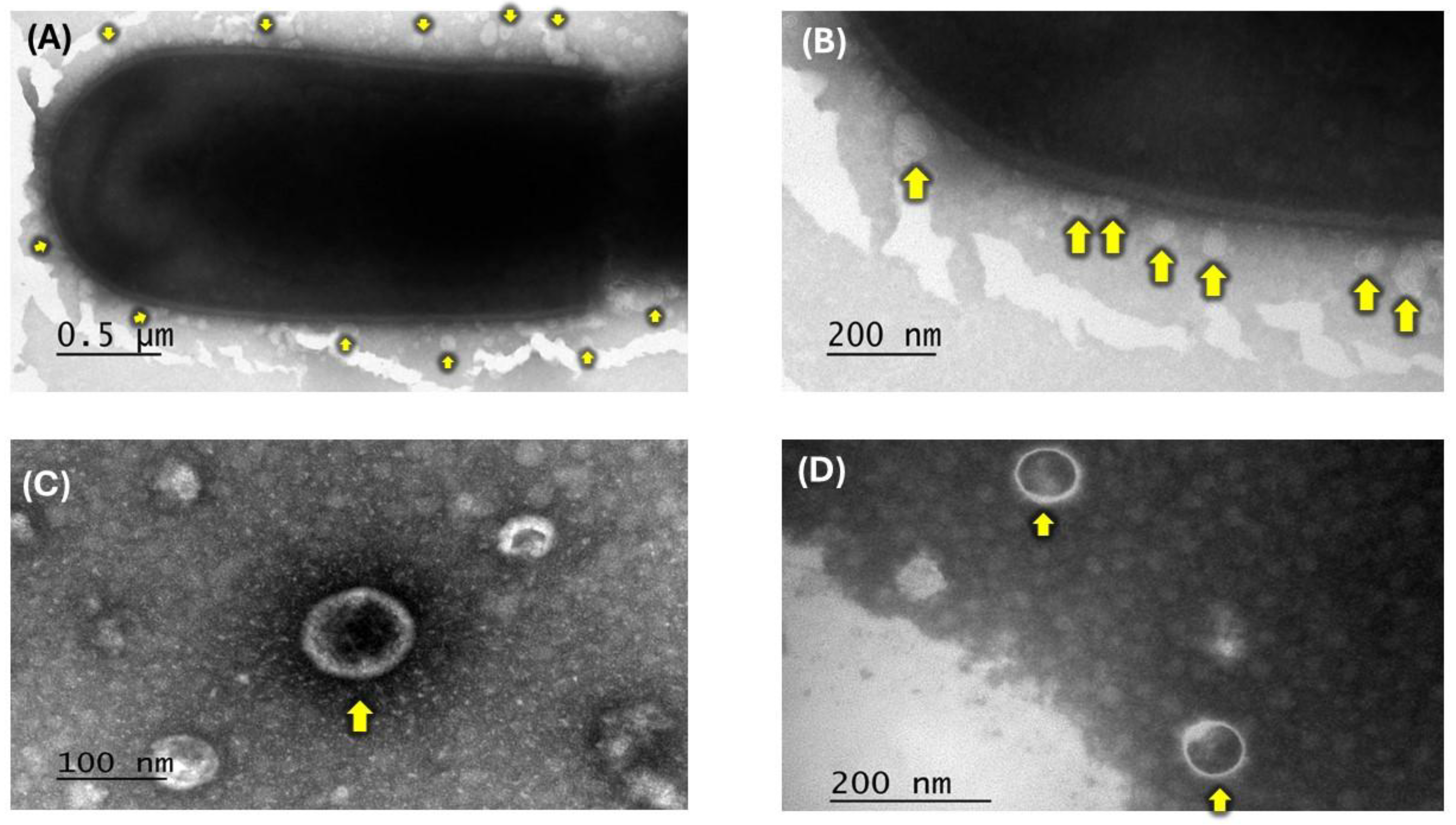
Figure 2.
Inhibition assay performed in SIM medium inoculated with E. coli (1×10⁸) after the addition of different concentrations of whole cells (W.C) or membrane vesicles (MVs) derived from Lactobacillus acidophilus. (A) Representative inhibition zones observed after treatment with PBS and 75 or 100 µg of W.C or MVs. (B) Quantification of inhibition zones obtained with different concentrations of W.C and MVs. Statistical analysis was performed using one-way ANOVA. Significance is indicated as *p ≤ 0.1 and **p ≤ 0.05.
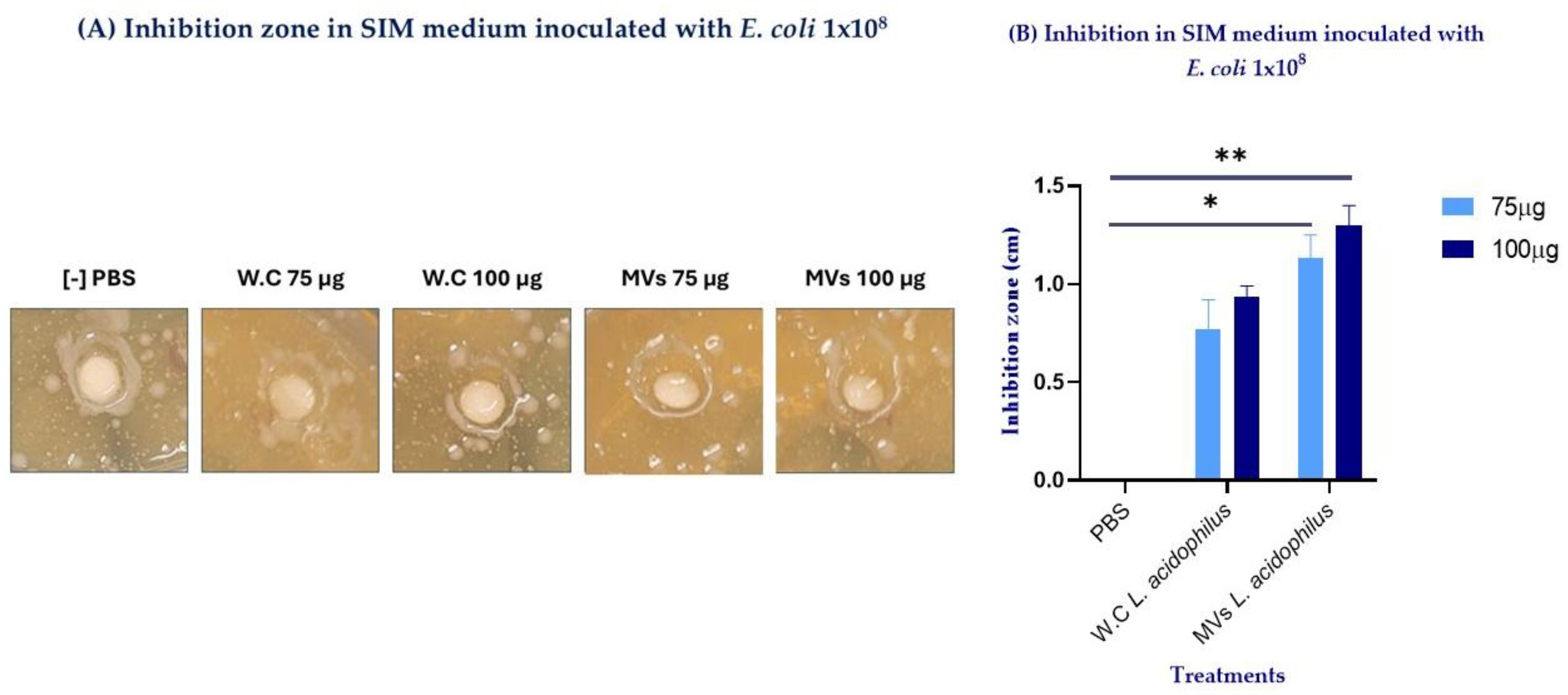
Figure 3.
Morphological changes observed in RAW 264.7 cells at 5 h following stimulation at 0, 2, and 4 h with the different treatments: (A) PBS-stimulated cells; (B) cells stimulated with 2 µg of commercial Escherichia coli LPS; (C) cells stimulated with 10 µg of whole cells (W.C) of L. acidophilus; and (D) cells stimulated with 10 µg of membrane vesicles (MVs) of L. acidophilus.
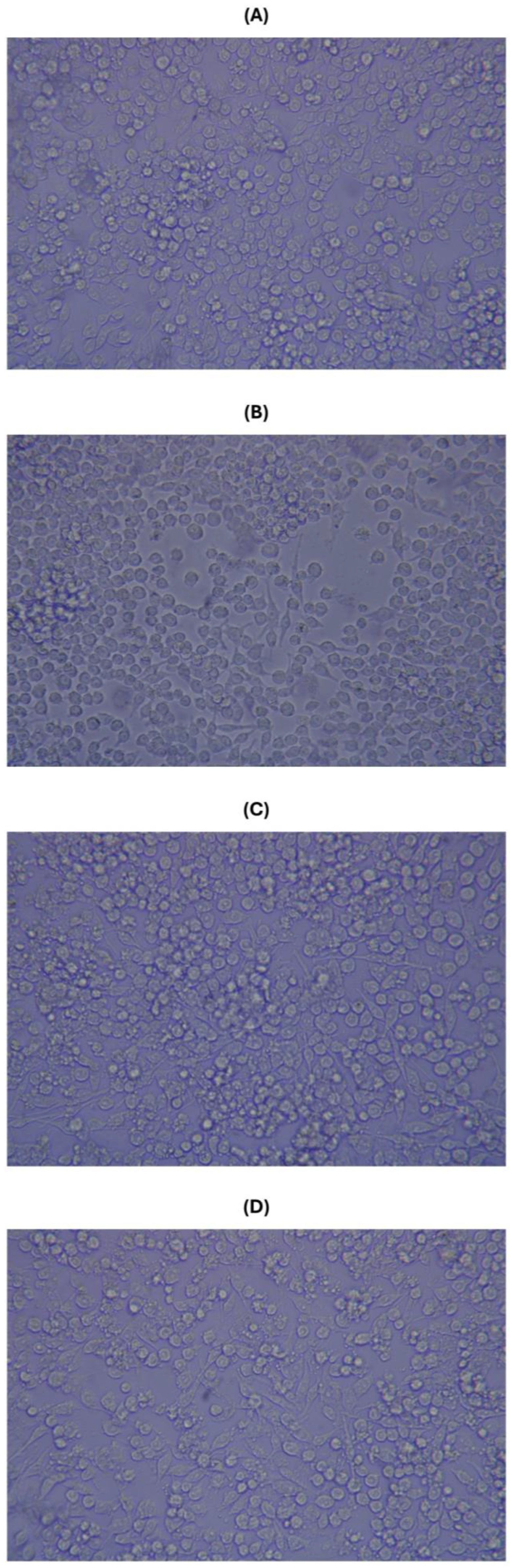
Figure 4.
Quantitative PCR (qPCR) analysis of cytokine expression kinetics in RAW 264.7 macrophages stimulated at 0, 2, and 4 h (*) with PBS, LPS, whole cells (W.C) of Lactobacillus acidophilus, or membrane vesicles (MVs) of L. acidophilus. Subsequently, cells were challenged with Escherichia coli at 5 h, and cytokine expression was evaluated up to 8 h. The expression of (A) IL-1β, (B) TNF-α, (C) IL-10, (D) IL-12, and (E) TLR2 is shown. Statistical analysis was performed using two-way ANOVA. Significance is indicated as * p ≤ 0.1, ** p ≤ 0.05, *** p ≤ 0.001, and **** p ≤ 0.0001.
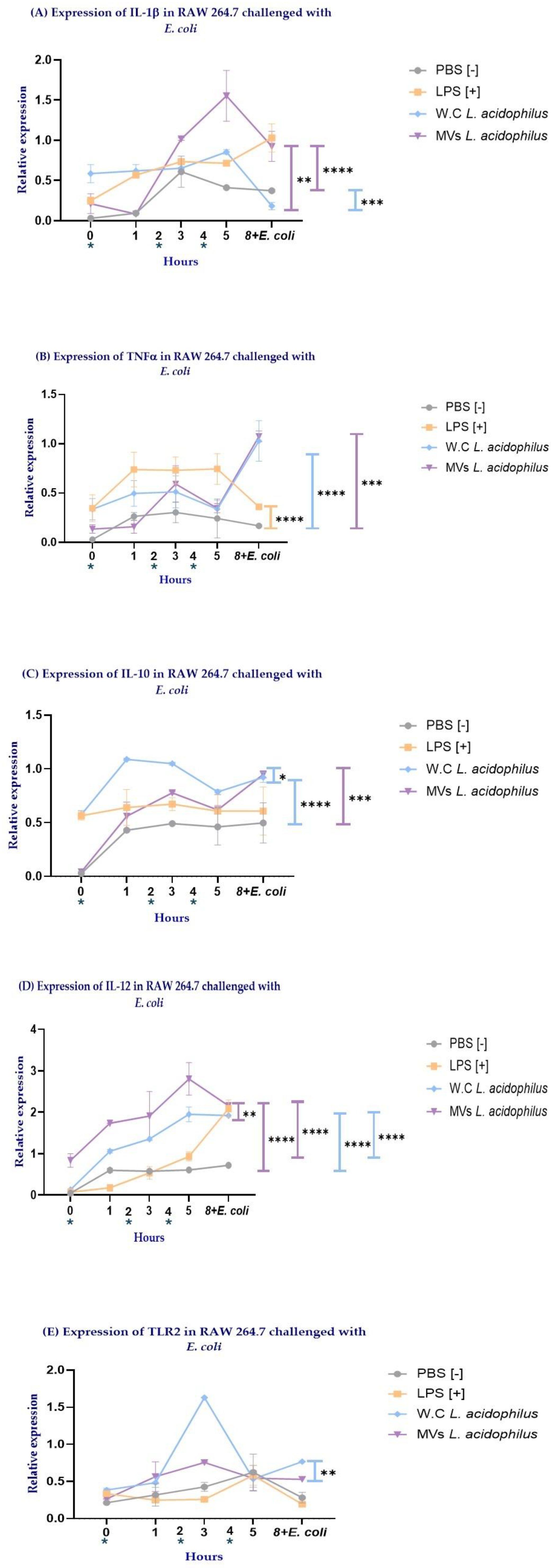
Figure 5.
Quantitative PCR (qPCR) analysis of cytokine expression in RAW 264.7 macrophages at 8 h following stimulation with PBS, LPS, whole cells of Lactobacillus acidophilus (W.C), or membrane vesicles of Lactobacillus acidophilus (MVs), in the presence or absence of Escherichia coli challenge. The expression of IL-1β, TNF-α, IL-10, IL-12, and TLR2 is shown. Statistical analysis was performed using two-way ANOVA. Significance is indicated as * p ≤ 0.1, ** p ≤ 0.05, *** p ≤ 0.001, and **** p ≤ 0.0001.
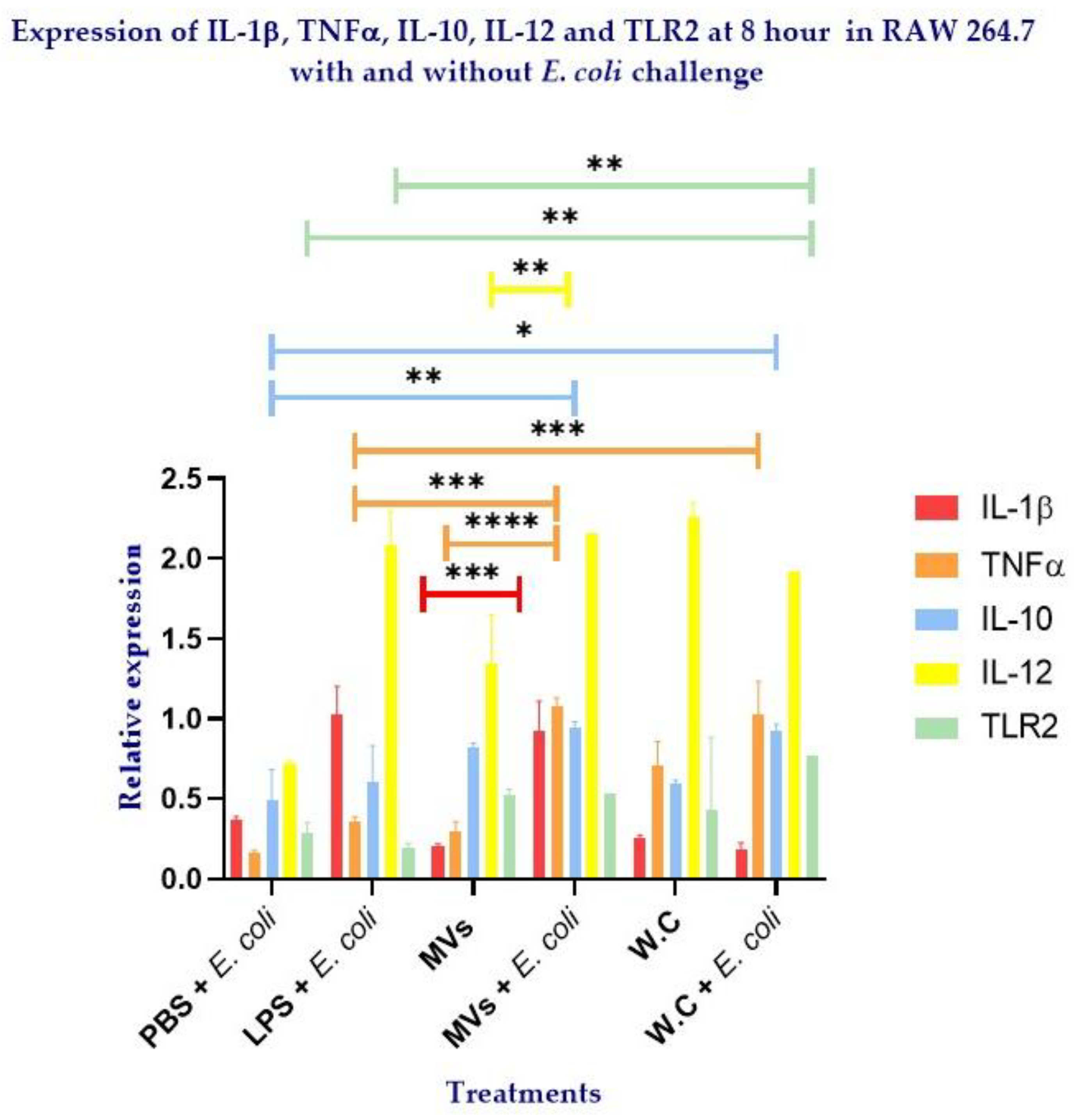

Table 1.
Primer Sequences and Annealing Parameters for IL-1β, TNFα, IL-10, IL-12 and TLR2
| Citokines | Secuences |
Temperature of alignment |
Expected size |
| IL-1β | Fw: GGTGTGTGACGTTCCCATTA | 62°C | 170pb |
| Rv: CGTTGCTTGGTTCTCCTTGT | |||
| TNFα | Fw: TATGGCTCAGGGTCCAACTC | 59°C | 174pb |
| Rv: CTCCCTTTGCAGAACTCAGG | |||
| IL-10 | Fw: GCCTTATCGGAAATGATCC | 56°C | 176pb |
| Rv: TCCACTGCCTTGCTCTTATT | |||
| IL-12 | Fw: ACAGCACCAGCTTCTTCATC | 57°C | 165pb |
| Rv: GCTGGATTCGAACAAAGAACT | |||
| TLR2 | Fw: CTCCCACTTCAGGCTCTTTG | 61°C | 223pb |
| Rv: GAAGTCAGGAACTGGGTGGA |
Disclaimer/Publisher’s Note: The statements, opinions and data contained in all publications are solely those of the individual author(s) and contributor(s) and not of MDPI and/or the editor(s). MDPI and/or the editor(s) disclaim responsibility for any injury to people or property resulting from any ideas, methods, instructions or products referred to in the content. |
© 2026 by the authors. Licensee MDPI, Basel, Switzerland. This article is an open access article distributed under the terms and conditions of the Creative Commons Attribution (CC BY) license.
Copyright: This open access article is published under a Creative Commons CC BY 4.0 license, which permit the free download, distribution, and reuse, provided that the author and preprint are cited in any reuse.


