Submitted:
24 January 2026
Posted:
26 January 2026
You are already at the latest version
Abstract
Background: The optimal fixation strategy for hallux valgus correction using the Minimally Invasive Chevron and Akin (MICA) procedure remains debated despite ongoing advances and active discussion at recent international surgical congresses. While du-al-screw fixation is commonly employed to prevent malunion, single-screw fixation may suffice in select cases. Methods: From 2019 to November 2023, a single-surgeon case series was conducted, including 105 patients with mild to moderate HV who had failed nonoperative management and underwent MICA correction. Patients were divided into two groups: Group 1 (n = 49) received single-screw fixation; Group 2 (n = 56) received du-al-screw fixation. Pre- and postoperative weight-bearing radiographs were used to assess hallux valgus angle, intermetatarsal angle, and distal metatarsal articular angle. Postoperative CT scans were obtained when complications were suspected. Outcomes included complication rates, operative time, fluoroscopy duration, and radiation exposure, with clinical follow-up of at least 3 months for all patients. Results: Clinical outcomes were comparable between groups. A significant difference in osteotomy translation was observed between single- and dual-screw cohorts. One case of delayed union occurred in the single-screw group, and one case of delayed union with loss of correction was noted in the dual-screw group. Operative time, fluoroscopy duration, and radiation exposure were significantly lower with single-screw fixation. Conclusions Effective and reproducible correction with low complication rates can be achieved with both fixation methods. Single-screw fixation is generally safe and sufficient for moderate cases, but dual-screw fixation remains mandatory when large osteotomy translation or poor bone quality is present.
Keywords:
hallux valgus
; minimally invasive chevron-akin (MICA) procedure
; screw fixation
; percutaneous osteotomy
; fixation stability
1. Introduction
Determining the optimal procedure for correcting hallux valgus deformity after failure of nonsurgical treatments remains a subject of ongoing debate. In recent years, minimally invasive surgery has gained significant traction, with outcomes shown to be comparable to those of the traditional open approach for hallux valgus correction [1,2,3,4,5,6].
Reported advantages of minimally invasive surgery include a smaller scar, preservation of soft tissue, reduced postoperative pain, and a lower incidence of wound complications [2,4,5,6,7,8,9]. Collectively, these benefits contribute to faster recovery and rehabilitation [2,4,5,6,7,8,9].
One of the most widely adopted percutaneous techniques in forefoot surgery is the minimally invasive Chevron-Akin (MICA) procedure [4,6,7,10,11]. Routinely indicated for moderate hallux valgus deformities—typically defined as a hallux valgus angle (HVA) less than 40° and an intermetatarsal angle (IMA) less than 20°, without first metatarsal instability—the procedure involves a chevron-type osteotomy at the distal diaphyseal-metaphyseal junction of the first metatarsal bone and, in most cases, an Akin-type osteotomy of the first phalanx [4,6,7,10,11]. Recent studies have highlighted the MICA procedure’s success, demonstrating stable, effective, and reproducible correction of hallux valgus, with a learning curve comparable to traditional open surgery [3,6,7,8,11,12]. Internal fixation with compression screws is considered essential to provide stability and compression at the chevron osteotomy site.
The history of percutaneous hallux valgus correction reflects a gradual refinement of technique and fixation. The initial first-generation Reverdin-Isham procedure, characterized by lack of fixation, gave way to the second-generation Bösch technique, which similarly left the osteotomy unfixed [6]. Subsequent modification by Magnan introduced K-wire fixation, but this approach lost favor due to associated superficial and deep infections. The advent of third-generation techniques marked a major evolution, incorporating fixation methods such as the MICA procedure, percutaneous extra-articular reverse-L Chevron (PERC), percutaneous Chevron-Akin with straight transverse subcapital osteotomy (PECA), and percutaneous intra-articular Chevron osteotomy (PeICO). Early third-generation techniques employed a single screw from the dorsal side (PERC), but best results were achieved with two parallel screws from the medial side, one proximal and one distal, as in the MICA and PECA procedures [3,4,5,10,11]. Alternative fixation solutions have also emerged, including intramedullary devices such as the Endolog, ISO Plate, V-TEK Plate, and LINK Internal Hallux Fixator [5,13], as well as the recently described fourth-generation metaphyseal extra-articular transverse and Akin osteotomy (META), which differs from MICA by employing a linear rather than chevron-type cut of the metatarsal, potentially enhancing stability and distal screw fixation [14]. Recent studies further emphasize the need for bi-cortical fixation of the proximal screw to prevent osteotomy movement [4,5,10].
Despite these advances, the optimal fixation strategy for the MICA procedure remains unresolved. Notably, this issue was the focus of expert panel discussions and multiple presentations at the 7th International Congress of Foot & Ankle Minimally Invasive Surgery (MIFAS), as well as other recent surgical meetings, underscoring its continued relevance and controversy within the surgical community.
At present, there is no clear consensus regarding which type of fixation may be unreliable for specific patient populations or which could be considered overtreatment. Nevertheless, in osteotomies requiring metatarsal head translation, internal fixation is generally recommended to avoid loss of correction or malposition, and to enable early full weight-bearing and range of motion exercises—facilitating early rehabilitation [15].
The present study aims to compare the clinical and radiographic outcomes of single versus dual-screw fixation of distal chevron osteotomy in the MICA procedure at 3 months’ follow-up. We hypothesized that both fixation strategies would yield comparable results, provided that single-screw fixation is inserted bicortically and directed medio-laterally.
2. Materials and Methods
Between 2019 and November 2023, a consecutive case series was conducted at a single tertiary center, with all procedures performed by a fellowship-trained foot and ankle surgeon with extensive experience in percutaneous hallux valgus correction, including the minimally invasive chevron and Akin (MICA) procedure. This design ensured consistency of surgical technique, perioperative care, and follow-up assessment.
Patients with mild to moderate hallux valgus who failed nonoperative treatments and were scheduled for distal chevron osteotomy using the MICA technique were included. Exclusion criteria were severe hallux valgus, hypermobility of the first metatarsal, osteoarthritis of the first metatarsophalangeal joint, prior correction of the first metatarsal bone, or the need for additional operative treatment of second ray pathology. This strict application of inclusion and exclusion criteria minimized heterogeneity and potential confounding.
A total of 105 patients met the criteria and were divided into two groups according to fixation method: Group 1 (n = 49; 2 males, 47 females) received single-screw fixation, and Group 2 (n = 56; 4 males, 52 females) received dual-screw fixation of the chevron osteotomy (Figure 1).
Pre- and postoperative weight-bearing anteroposterior and lateral radiographs were systematically obtained for each patient to assess deformity severity and the degree of correction achieved. Radiological evaluation included measurement of hallux valgus angle (HVA), intermetatarsal angle (IMA), and distal metatarsal articular angle (DMAA). Postoperative radiographs were further analyzed for osteotomy translation (expressed as a percentage) and first metatarsal shortening. For patients with suspected complications such as non-union, loss of correction, or angular deformation, postoperative CT scans were performed at 3 months to clarify the diagnosis.
Clinical follow-up was a minimum of 3 months for all patients and was extended in cases of complications or when screw removal was required. This approach facilitated comprehensive identification of both radiographic and clinical outcomes.
The study was approved by the Commission for Ethics in Medicine of UKC Maribor (UKC-MB-KME-33/19) and conducted in accordance with the Helsinki Declaration, the Oviedo Convention on Human Rights and Biomedicine, and the Slovene Code of Medical Deontology.
Statistical analysis was performed using IBM SPSS Statistics 29.0. The Student t-test was used to compare observed parameters between the groups.
Surgical Technique
The minimally invasive chevron and Akin (MICA) procedure was performed through a medial punctiform incision, 3–5 mm in length, located over the base of the medial eminence of the first metatarsal. Under fluoroscopic guidance, a chevron osteotomy was created using a 2.2 mm diameter, 20 mm length burr. The metatarsal head was then translated laterally as much as possible, employing Kocher forceps or a specialized elevator introduced into the first metatarsal metaphyseal canal at the level of the osteotomy.
Temporary fixation was achieved using a “K-wire first” technique: a 2 mm K-wire was inserted at the level of the interphalangeal (IP) joint, directed toward the osteotomy site and placed into the intramedullary canal, consistent with the Bösch technique. For definitive fixation, a second and, if required, a third punctiform medial incision was made. Under image intensifier control, a 1 mm diameter guidewire was advanced bicortically through the metatarsal diaphysis, crossing the osteotomy and ending in the metatarsal head. A specialized 3 mm, 45° beveled profile head screw (FH Ortho®) was inserted over the guidewire, traversing both cortices to secure the metatarsal head (Figure 2). At this point, the stability of the fixation was reassessed. A second screw was added between the first screw and the tarsometatarsal (TMT) joint if lateral translation exceeded four-fifths of the metatarsal cortical diameter, or if the initial screw could not achieve sufficient tightening due to poor bone quality. The temporary K-wire was removed following completion of screw fixation.
Both groups followed a standardized postoperative protocol, including immediate full weight-bearing using a ready-made (Darco®) hallux valgus flat sole orthosis during ambulation for six weeks. Physiotherapy was initiated with passive motion of the hallux on the third postoperative day, progressing to active motion after one week. Full, unrestricted weight-bearing was allowed after six weeks.
3. Results
A total of 105 MICA procedures were performed (49 single-screw fixation, 56 dual-screw fixation), with all patients completing a minimum of 3 months’ follow-up and no loss to follow-up. Clinical outcomes were comparable between the two groups (Table 1). Due to surgical selection—where single-screw fixation (Figure 3) was preferentially used for cases requiring less metatarsal translation—a statistically significant difference in translation achieved was observed between groups (mean translation: 59 ± 12% for single-screw vs. 67 ± 14% for dual-screw, p < 0.05). There was one case of loss of fixation with malunion in each group. (Figure 4 and Figure 5) The rate of screw removal was 6% (n = 3) in the single-screw group and 9% (n = 5) in the dual-screw group. No other major complications were observed.
Operative time, fluoroscopy duration, number of scans, and total radiation exposure were all statistically significantly lower in the single-screw fixation group (operative time: 36 ± 12 min vs. 43 ± 13 min, p < 0.05; fluoroscopy time: 56 ± 19 sec vs. 83 ± 30 sec, p < 0.05; number of scans: 58 ± 19 vs. 87 ± 31, p < 0.05; radiation exposure: 0.46 ± 0.18 mGy vs. 0.69 ± 0.28 mGy, p < 0.05).
Table 1 summarizes the pre- and postoperative radiological measurements and complication rates for both groups. Notably, both groups demonstrated significant correction in hallux valgus angle, intermetatarsal angle, and distal metatarsal articular angle, with no statistically significant difference in metatarsal shortening or change in IMA and DMAA between groups. There were no cases of deep infection or nonunion in either group
4. Discussion
The wider adoption of minimally invasive techniques for hallux valgus correction began with the second-generation Bösch technique, first published in 1990 [16]. At that time, temporary single K-wire intraoperative fixation was considered sufficient for moderate deformities without significant complications [16,17]. In 2011, Oliva et al. described a percutaneous technique using an oscillating saw to perform a linear osteotomy nearly perpendicular to the first metatarsal, with fixation achieved using one or occasionally two K-wires [18]. Subsequently, Trnka et al. compared the Bösch method with K-wire fixation to the open chevron procedure and reported comparable clinical outcomes [19]. However, the minimally invasive group demonstrated a notably higher rate of superficial and deep infections [6], and K-wire fixation has since been shown to be inferior to screw fixation in terms of stability and complication profile [20]. Despite this, some authors continue to advocate K-wire-only fixation as sufficient in selected cases [21,22].
From a biomechanical perspective, screw fixation provides superior stability, allowing early mobilization with full weight-bearing and thereby improving patient safety while reducing infection risk and revision rates [21,23]. Constructs stabilized with screws are better able to withstand rotational forces acting on the first metatarsal [24] and tolerate greater axial loads [20]. In the same year as Trnka’s publication, Vernois and Redfern described a percutaneous chevron technique with dorsal single-screw fixation [7]. They demonstrated that even severe deformities (IMA > 20°) could be addressed, achieving metatarsal head translation of up to 80% or more; however, they recommended the use of two screws for larger displacements [7]. More recently, Harasser et al. evaluated radiographic correction, patient satisfaction, range of motion, and pain outcomes in 50 cases undergoing MICA with either single- or dual-screw fixation [25]. Both fixation strategies resulted in stable anchoring, full consolidation, and favorable clinical outcomes, with average translations ranging from 70% to 90%. The authors suggested that routine placement of a second screw may not be necessary [25]. These findings align with our results, which demonstrate that when metatarsal head translation is moderate, dual-screw fixation does not confer a clear advantage over single-screw fixation. Moreover, dual-screw constructs were associated with increased fluoroscopy use, higher radiation exposure, greater overall cost, and notably longer operative times, the latter still being one of the most predominant factors contributing to a higher risk of infection [26]. Further reduction in radiation exposure may be achieved by routinely employing temporary K-wire fixation during MICA, which has been shown to significantly decrease fluoroscopy time compared with traditional perforator, drill, and joystick techniques (p < 0.001) [27].
Consistent with our findings, the incidence of implant removal due to soft tissue irritation increases with the number of implanted screws [28]. When osteotomy translation is moderate (approximately 80% of the metatarsal diameter), bone quality is adequate, and stability is achieved after placement of the first screw, a proximal screw may be unnecessary. Dual-screw fixation has also been associated with an increased risk of first metatarsal fracture, particularly when limited cortical bone surrounds the proximal screw, predisposing the bone to fragmentation [29]. Additionally, when space is insufficient for a second screw, attempts to reposition the initial screw may compromise cortical bone integrity due to repeated drilling or close screw proximity. Nevertheless, for larger translations exceeding 80% of the metatarsal diameter and in cases of compromised bone quality—such as osteoporosis or rheumatoid arthritis—dual-screw fixation remains advisable to reduce the risk of non-union, pseudoarthrosis, or loss of correction. Similarly, in high-demand patients, including athletes or those undergoing simultaneous bilateral hallux valgus correction, dual-screw fixation may offer additional construct security.
The heterogeneity of minimally invasive techniques and fixation strategies has been further highlighted in the literature. In 2018, Malagelada et al. conducted a comprehensive review of 23 studies involving 2,279 procedures in 1,762 patients, comparing the Bösch technique, MICA, Reverdin-Isham procedure, and Endolog [5]. Despite the diversity of fixation methods, the authors were unable to determine whether one technique was more effective than the others [5]. More recently, Zaveri et al. found no functional or radiological advantages to using intramedullary devices over other fixation systems [13], suggesting that both intramedullary fixation and routine use of dual-screw fixation may represent overtreatment in many cases.
Over the past decade, there has also been a gradual transition from chevron to straight transverse osteotomy [14,30]. While the transverse osteotomy is intrinsically less stable, it allows improved correction of first metatarsal head malrotation and, importantly, preserves more of the lateral cortical bone. This preservation facilitates placement of dual screws with a greater interval, reducing the risk of shattering the lateral wall during fixation [14,30]. In 1981, there were over 150 known osteotomies for hallux valgus correction, with little consensus on which method was superior [19,31]. It appears that the situation remains similar today, with limited agreement on which minimally invasive technique or type of fixation is most effective [5,6,8,9].
This study has several important strengths and limitations. Its primary strengths include the consistency of operative technique, as all procedures were performed by a single fellowship-trained foot and ankle surgeon using standardized pre- and postoperative radiographic protocols and well-defined inclusion and exclusion criteria. The consecutive patient cohort and complete follow-up further reduce the risk of selection and attrition bias. However, the retrospective design and lack of randomization introduce the possibility of allocation bias, particularly as fixation type was determined intraoperatively based on the required degree of osteotomy translation. The minimum three-month follow-up, while adequate to assess radiographic healing and early complications, may not fully capture late recurrences or hardware-related issues. Additionally, the single-center, single-surgeon nature of the study may limit generalizability to broader clinical practice. Despite these limitations, the study provides relevant real-world data on the comparative effectiveness and safety of single- versus dual-screw fixation in minimally invasive chevron and Akin osteotomies for hallux valgus correction.
The choice between single- and dual-screw fixation often depends on the degree of osteotomy translation and intraoperative stability. Even with the introduction of modified techniques such as PECA and META, no clear consensus has emerged regarding the superiority of any minimally invasive method or fixation strategy, underscoring the continuing need for further research and standardization in this field.
5. Conclusions
The MICA procedure offers effective and reproducible correction of hallux valgus, providing reduced postoperative pain, faster recovery, and a low complication rate compared to open techniques. For moderate deformities with good bone quality, these advantages are independent of whether single- or dual-screw metatarsal fixation is used. Single-screw fixation confers the additional benefits of lower cost, reduced operative and radiation time, and a decreased incidence of implant removal. However, in cases of severe hallux valgus with large metatarsal translations or poor bone quality, dual-screw fixation is mandatory to minimize the risk of revision surgery due to complications such as pseudoarthrosis, malunion, or loss of correction. Further studies are needed to refine and standardize indications for single- versus dual-screw fixation in minimally invasive hallux valgus surgery.
Author Contributions
Conceptualization, M.M. and J.Z.; methodology, M.M.; software, M.M.; validation, M.M., J.Z; formal analysis, M.M and J.Z.; investigation, M.M.; resources, M.M and J.Z.; data curation, M.M.; writing—original draft preparation, J.Z.; writing—review and editing, M.M.; visualization, M.M. and J.Z.; supervision, M.M.; project administration, M.M. All authors have read and agreed to the published version of the manuscript.
Funding
This research received no external funding.
Institutional Review Board Statement
The study was conducted in accordance with the Declaration of Helsinki, the Oviedo Convention on Human Rights and Biomedicine, and the Slovene Code of Medical Deontology, and approved by the Commission for ethics in medicine of UKC Maribor (UKC-MB-KME-33/19) on 28 March 2019.
Informed Consent Statement
Informed written consent was obtained from all subjects involved in the study.
Data Availability Statement
Data supporting the reported results can be found in the physical databases of UKC Maribor and are available from the corresponding author on reasonable request.
Conflicts of Interest
The authors declare no conflicts of interest.
Abbreviations
The following abbreviations are used in this manuscript:
| HV | Hallux valgus |
| MIS | Minimally invasive surgery |
| MICA | Minimally invasive Chevron and Akin |
| HVA | Hallux valgus angle |
| IMA | Intermetatarsal angle |
| DMAA | Distal metatarsal articular angle |
| CT | Computed tomography |
| PERC | Percutaneous extra-articular reverse-L Chevron |
| PECA | Percutaneous Chevron-Akin with straight transverse subcapital osteotomy |
| PeICO | Percutaneous intra-articular Chevron osteotomy |
| META | Metaphyseal extra-articular transverse and Akin osteotomy |
| TMT | Tarsometatarsal (joint) |
References
- Giannini, S.; Faldini, C.; Nanni, M.; Di Martino, A.; Luciani, D.; Vannini, F. A Minimally Invasive Technique for Surgical Treatment of Hallux Valgus: Simple, Effective, Rapid, Inexpensive (SERI). Int. Orthop. 2013, 37, 1805. [Google Scholar] [CrossRef] [PubMed]
- Redfern, D.; Perera, A.M. Minimally Invasive Osteotomies. Foot Ankle Clin. 2014, 19, 181–189. [Google Scholar] [CrossRef] [PubMed]
- Bauer, T. Percutaneous Forefoot Surgery. Orthop. Traumatol. Surg. Res. 2014, 100. [Google Scholar] [CrossRef] [PubMed]
- Botezatu, I.; Marinescu, R.; Laptoiu, D. Minimally Invasive–Percutaneous Surgery – Recent Developments of the Foot Surgery Techniques. J. Med. Life 2015, 8, 87. [Google Scholar]
- Malagelada, F.; Sahirad, C.; Dalmau-Pastor, M.; Vega, J.; Bhumbra, R.; Manzanares-Céspedes, M.C.; Laffenêtre, O. Minimally Invasive Surgery for Hallux Valgus: A Systematic Review of Current Surgical Techniques. Int. Orthop. 2019, 43, 625–637. [Google Scholar] [CrossRef]
- Del Vecchio, J.J.; Ghioldi, M.E. Evolution of Minimally Invasive Surgery in Hallux Valgus. Foot Ankle Clin. 2020, 25, 79–95. [Google Scholar] [CrossRef]
- Vernois, J.; Redfern, D. Percutaneous Chevron; the Union of Classic Stable Fixed Approach and Percutaneous Technique. Fuß Sprunggelenk 2013, 11, 70–75. [Google Scholar] [CrossRef]
- Jowett, C.R.J.; Bedi, H.S. Preliminary Results and Learning Curve of the Minimally Invasive Chevron Akin Operation for Hallux Valgus. J. Foot Ankle Surg. 2017, 56, 445–452. [Google Scholar] [CrossRef]
- Lai, M.C.; Rikhraj, I.S.; Woo, Y.L.; Yeo, W.; Ng, Y.C.S.; Koo, K. Clinical and Radiological Outcomes Comparing Percutaneous Chevron-Akin Osteotomies vs Open Scarf-Akin Osteotomies for Hallux Valgus. Foot ankle Int. 2018, 39, 311–317. [Google Scholar] [CrossRef]
- Lam, K.L.K.; Kong, S.W.; Chow, Y.H. Percutaneous Chevron Osteotomy in Treating Hallux Valgus: Hong Kong Experience and Mid-Term Results. J. Orthop. Trauma Rehabil. 2015, 19, 25–30. [Google Scholar] [CrossRef]
- Kaufmann, G.; Mörtlbauer, L.; Hofer-Picout, P.; Dammerer, D.; Ban, M.; Liebensteiner, M. Five-Year Follow-up of Minimally Invasive Distal Metatarsal Chevron Osteotomy in Comparison with the Open Technique: A Randomized Controlled Trial. J. Bone Joint Surg. Am. 2020, 102, 873–879. [Google Scholar] [CrossRef]
- Merc, M.; Fokter, S.K.; I.S., I. Learning Curve in Relation to Radiation Exposure, Procedure Duration and Complications Rate for Minimally Invasive Chevron Akin (MICA) Osteotomy. BMC Musculoskelet. Disord. 2023, 24. [Google Scholar] [CrossRef]
- Zaveri, A.; Katmeh, R.; Patel, S.; Malhotra, K.; Cullen, N.; Welck, M. The Use of Intramedullary Devices for Fixation of Metatarsal Osteotomies in Hallux Valgus Surgery - A Systematic Review. Foot Ankle Surg. 2022, 28, 483–491. [Google Scholar] [CrossRef] [PubMed]
- Lewis, T.L.; Lau, B.; Alkhalfan, Y.; Trowbridge, S.; Gordon, D.; Vernois, J.; Lam, P.; Ray, R. Fourth-Generation Minimally Invasive Hallux Valgus Surgery With Metaphyseal Extra-Articular Transverse and Akin Osteotomy (META): 12 Month Clinical and Radiologic Results. Foot ankle Int. 2023, 44, 178–191. [Google Scholar] [CrossRef] [PubMed]
- Robinson, P.W.; Lam, P. Percutaneous Surgery for Mild to Severe Hallux Valgus. Tech. Foot Ankle Surg. 2020, 19, 76–83. [Google Scholar] [CrossRef]
- Bösch, P.; Markowski, H.; Rannicher, V. Technik Und Erste Ergebnisse Der Subkutanen Distalen Metatarsale-I-Osteotomie. Orthopädische Prax. 1990, 26, 51–6. [Google Scholar]
- Magnan, B.; Pezzè, L.; Rossi, N.; Bartolozzi, P. Percutaneous Distal Metatarsal Osteotomy for Correction of Hallux Valgus. J. Bone Joint Surg. Am. 2005, 87, 1191–1199. [Google Scholar] [CrossRef]
- Oliva, F.; Longo, U.G.; Maffulli, N. Minimally Invasive Hallux Valgus Correction. In Minimally Invasive Surgery of the Foot and Ankle; Maffulli, N., Easley, M., Eds.; Springer Verlag, 2011; pp. 123–131. [Google Scholar]
- Trnka, H.J.; Krenn, S.; Schuh, R. Minimally Invasive Hallux Valgus Surgery: A Critical Review of the Evidence. Int. Orthop. 2013, 37, 1731–1735. [Google Scholar] [CrossRef]
- Xie, Q.; Li, X.; Wang, P. Three Dimensional Finite Element Analysis of Biomechanics of Osteotomy Ends with Three Different Fixation Methods after Hallux Valgus Minimally Invasive Osteotomy. J. Orthop. Surg. (Hong Kong) 2023, 31. [Google Scholar] [CrossRef]
- Bİlgİn, E.; KeÇecİ, T.; Turgut, A.; Adiyeke, L.; Kİlİnc, B.E. Comparison of Clinical and Radiological Results of Two Fixation Materials after Distal Chevron Osteotomy for Hallux Valgus? - Two Kirschner Wires versus Single Screw Fixation. Acta Chir. Orthop. Traumatol. Cech. 2020, 87, 350–355. [Google Scholar] [CrossRef]
- Xavier, N.R.; Lara, L.C.R.; Vaz, G.A.R.; de Lima, F.P.; Bordignon, G.; Lancia, L.F.; de Carvalho, M.M.D.; Gonçalves, A.H.S.; da Silva, M.F.; Costa, L.T. da S. Fixation Methods of Chevron Osteotomy in Percutaneous Surgery for Hallux Valgus: A Comparative Study. J. Foot Ankle 2024, 18, 75–82. [Google Scholar] [CrossRef]
- Huang, S.H.; Cheng, Y.M.; Chen, C.H.; Huang, P.J. Modified Mitchell Osteotomy with Screw Fixation for Correction of Hallux Valgus. 2012, 33, 1098–1102. [Google Scholar] [CrossRef] [PubMed]
- Xu, C.; Liu, H.; Li, M.; Li, H.; Pan, C. Biomechanical Study of Minimally Invasive Correction of Hallux Valgus Fixation with Absorbable Screws: A Finite Element Analysis. 2023. [Google Scholar] [CrossRef]
- Harrasser, N.; Hinterwimmer, F.; Baumbach, S.F.; Pfahl, K.; Glowalla, C.; Walther, M.; Hörterer, H. The Distal Metatarsal Screw Is Not Always Necessary in Third-Generation MICA: A Case-Control Study. Arch. Orthop. Trauma Surg. 2023, 143, 4633–4639. [Google Scholar] [CrossRef] [PubMed]
- Li, X.; Zhang, J.; Fu, S.; Wang, C.; Yang, F.; Shi, Z. First Metatarsal Single-Screw Minimally Invasive Chevron-Akin Osteotomy: A Cost Effective and Clinically Reliable Technique. Front. Surg. 2023, 9. [Google Scholar] [CrossRef]
- Espinosa-Uribe, A.G.; Fernández-Garza, F.A.; Muñoz-Leija, D.; Vílchez-Cavazos, J.F.; Quiroga-Garza, A.; Peña-Martínez, V.M.; Elizondo-Omaña, R.E.; Gutiérrez-de la O, J. A Comparison of Three Techniques for the Osteosynthesis after Minimal Invasive Osteotomies for Hallux Valgus. Int. Orthop. 2024, 48, 2137–2143. [Google Scholar] [CrossRef]
- Jentzsch, T.; Renner, N.; Niehaus, R.; Farei-Campagna, J.; Deggeller, M.; Scheurer, F.; Palmer, K.; Wirth, S.H. The Influence of the Number of Screws and Additional Surgical Procedures on Outcome in Hallux Valgus Treatment. J. Orthop. Surg. Res. 2018, 13, 99. [Google Scholar] [CrossRef]
- Blitz, N.M.; Wong, D.T.; Baskin, E.S. Patterns of Metatarsal Explosion After New Modern Minimally Invasive Bunion Surgery. A Retrospective Review and Case Series of 16 Feet. J. Minim. Invasive Bunion Surg. 2024, 1, 92774. [Google Scholar] [CrossRef]
- Lewis, T.L.; Robinson, P.W.; Ray, R.; Dearden, P.M.C.; Goff, T.A.J.; Watt, C.; Lam, P. Five-Year Follow-up of Third-Generation Percutaneous Chevron and Akin Osteotomies (PECA) for Hallux Valgus. Foot ankle Int. 2023, 44, 104–117. [Google Scholar] [CrossRef]
- Helal, B. Surgery for Adolescent Hallux Valgus. Clin. Orthop. Relat. Res. 1981, 50–63. [Google Scholar] [CrossRef]
Figure 1.
Postoperative weightbearing X-ray of foot after MICA with dual-screw fixation; newly formed bone can be seen after 3 months at the level of osteotomy.
Figure 1.
Postoperative weightbearing X-ray of foot after MICA with dual-screw fixation; newly formed bone can be seen after 3 months at the level of osteotomy.
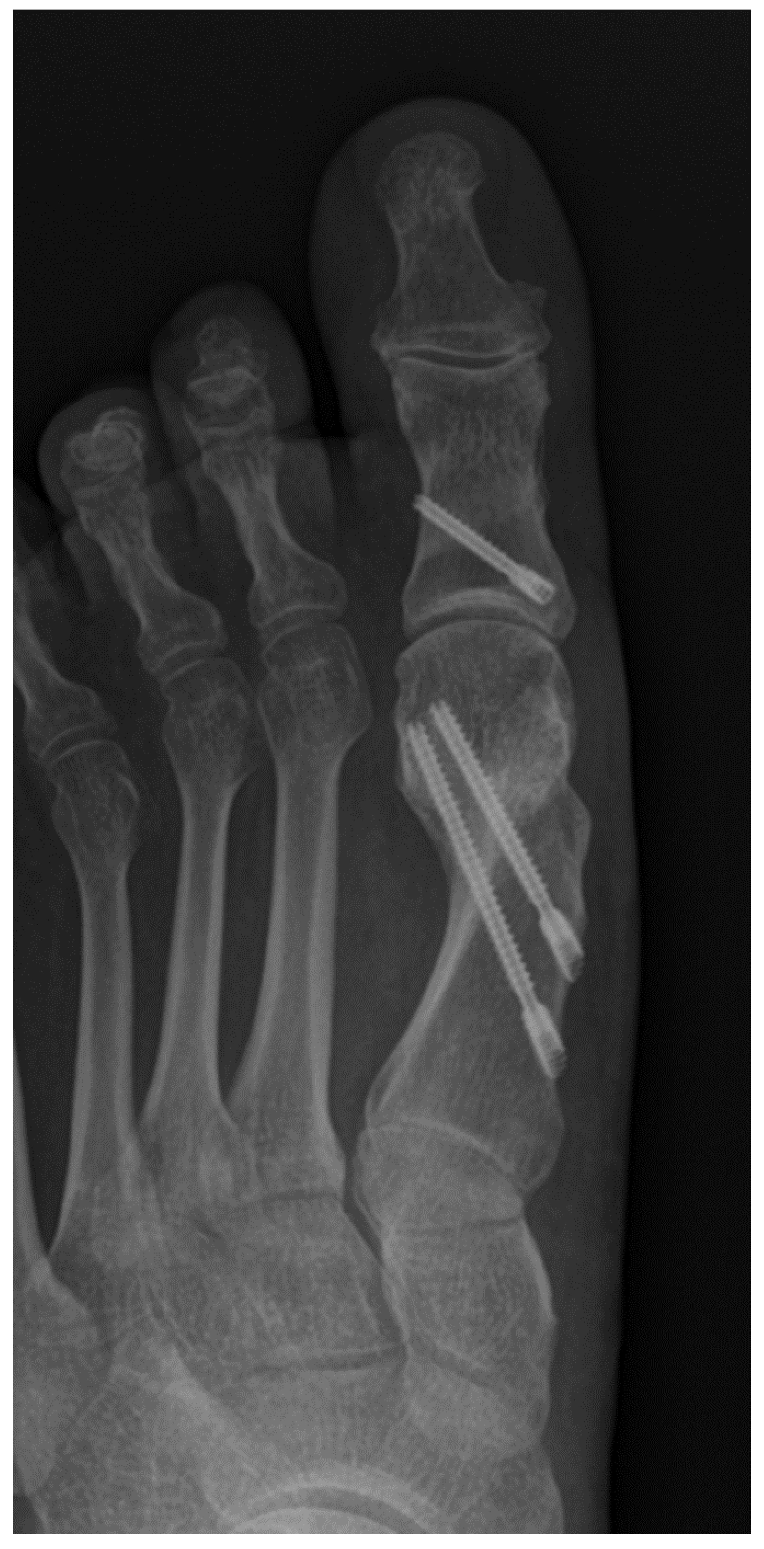
Figure 2.
Figure 2. Postoperative weightbearing X-ray of foot after MICA with single-screw fixation; newly formed bone can be seen after 3 months at the level of osteotomy.
Figure 2.
Figure 2. Postoperative weightbearing X-ray of foot after MICA with single-screw fixation; newly formed bone can be seen after 3 months at the level of osteotomy.
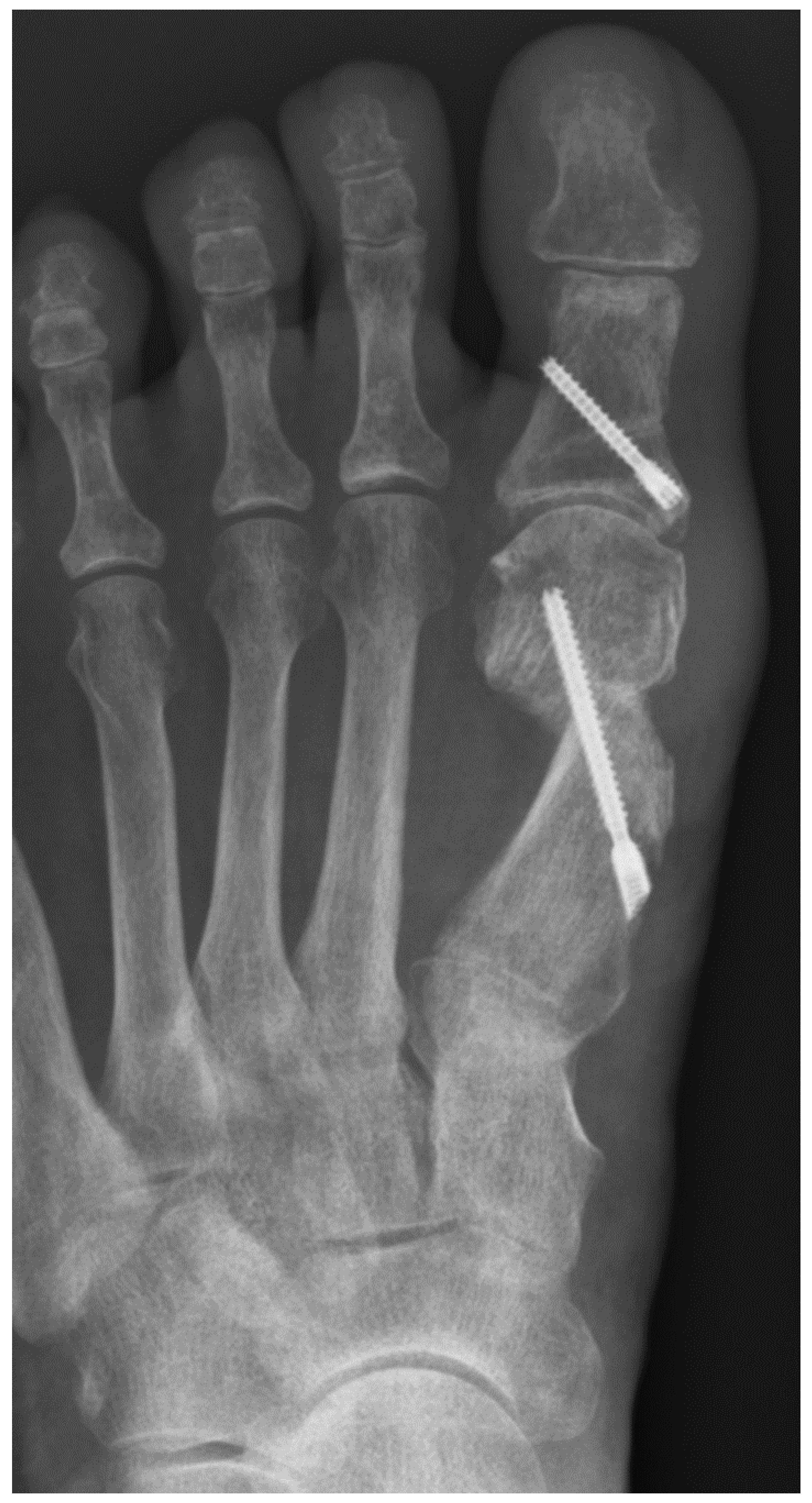
Figure 3.
X-ray showing both feet after MICA. Left right postoperatively using a single-screw fixation and right after bone healing and screw removal due to implant irritation.
Figure 3.
X-ray showing both feet after MICA. Left right postoperatively using a single-screw fixation and right after bone healing and screw removal due to implant irritation.
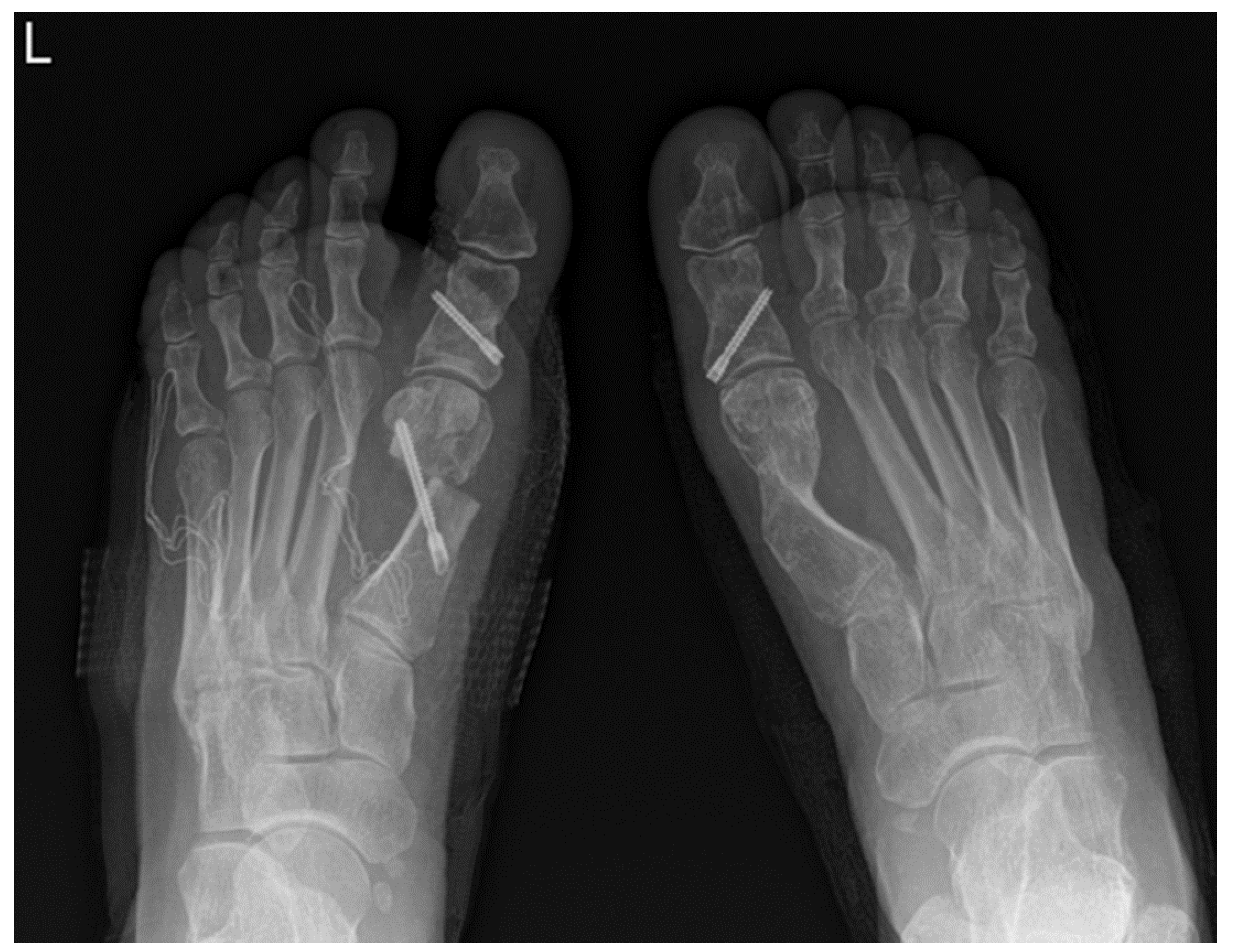
Figure 4.
Figure 4. X-ray scan presents loss of correction and implant failure after single screw fixation.
Figure 4.
Figure 4. X-ray scan presents loss of correction and implant failure after single screw fixation.
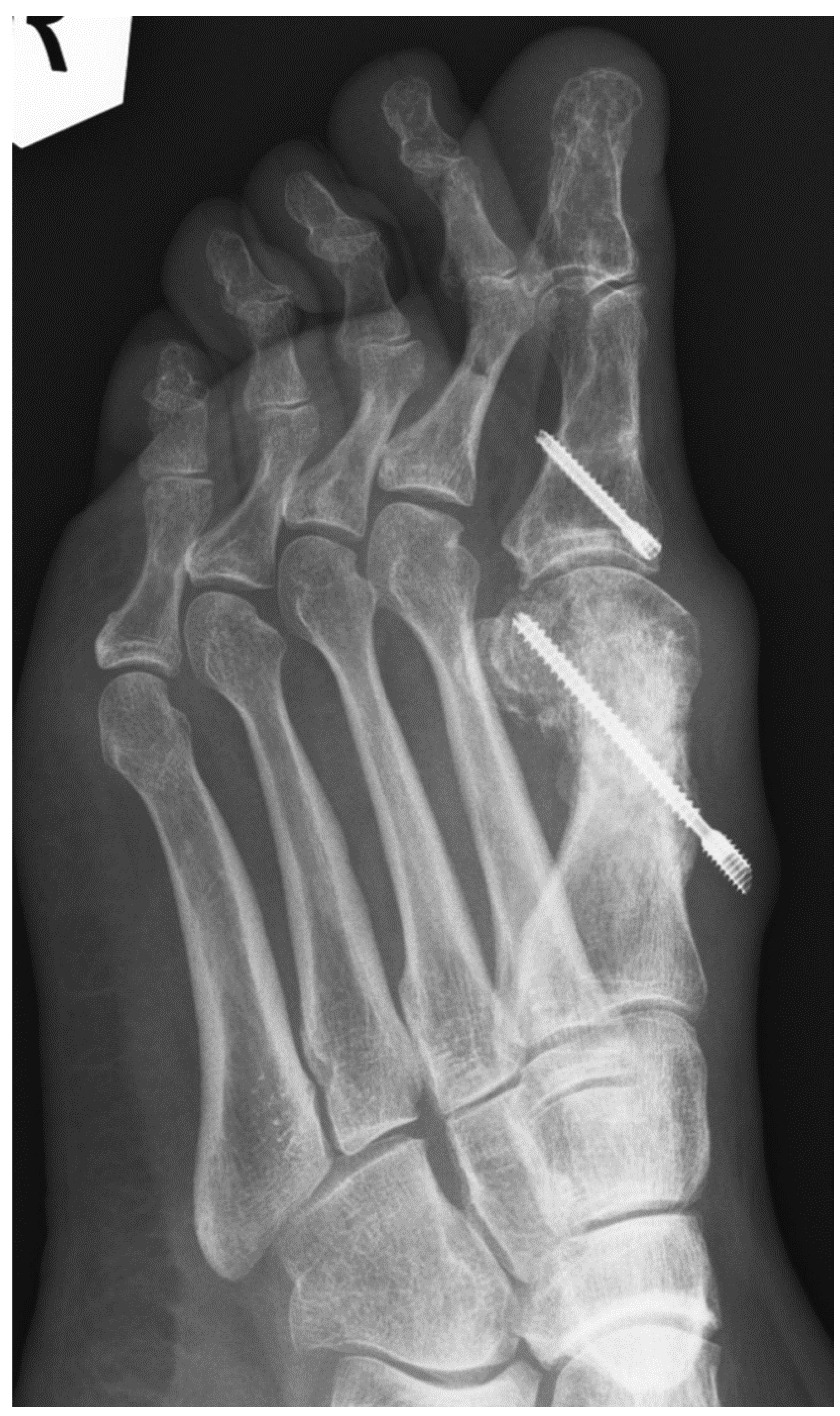
Figure 5.
Loss of correction and pseudoarthrosis on CT scan after dual screw fixation.
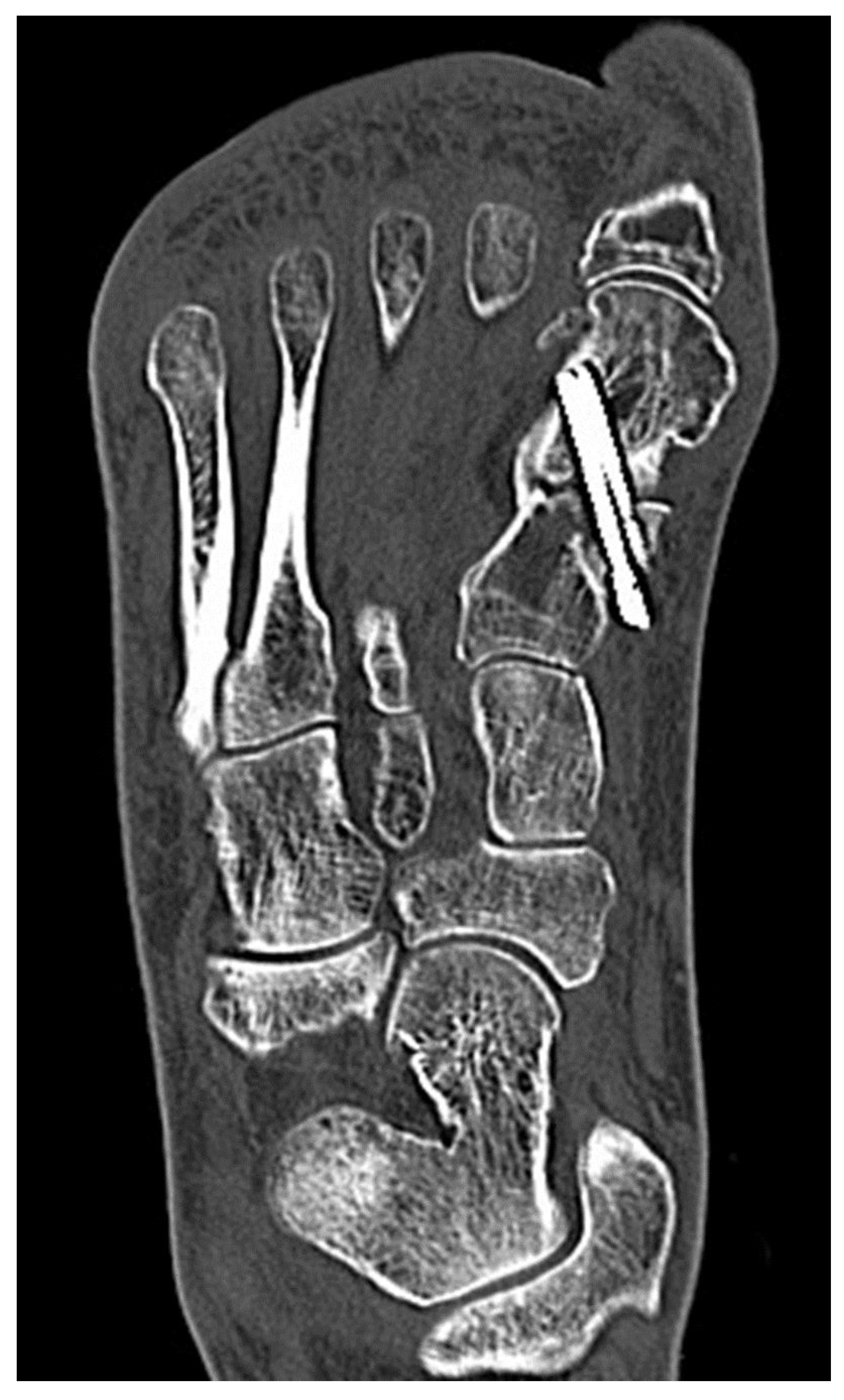

Table 1.
Table showing pre- and post- operative radiological measurements and complications for both groups.
Table 1.
Table showing pre- and post- operative radiological measurements and complications for both groups.
| 1 screw | 2 screws | ||
|---|---|---|---|
| No. patients | 49 | 56 | |
| age (years) | 53 ± 11 | 50 ± 17 | NS |
| pre-HVA (°) | 28 ± 11 | 35 ± 9 | < 0.05 |
| pre-IMA (°) | 14 ± 4 | 15 ± 4 | NS |
| pre-DMA (°) | 14 ± 6 | 17 ± 7 | < 0.05 |
| ∆HVA (°) | 22 ± 11 | 27 ± 12 | 0.05 |
| ∆IMA (°) | 11 ± 5 | 11 ± 4 | NS |
| ∆DMA (°) | 14 ± 6 | 12 ± 5 | NS |
| Translation (%) | 59 ± 12 | 67 ± 14 | < 0.05 |
| Shortening (mm) | 2 ± 1 | 2 ± 2 | NS |
| Complications | 1 loss of correction with malunion |
1 loss of correction with malunion |
|
| Screw removal (No) | 3 (6%) | 5 (9%) | |
| Operative time (min) | 36 ± 12 | 43 ± 13 | < 0.05 |
| Fluoroscopy time (sec) | 56 ± 19 | 83 ± 30 | < 0.05 |
| No. of scans | 58 ± 19 | 87 ± 31 | < 0.05 |
| Exposure (mGy) | 0,46 ± 0, 18 | 0,69 ± 0, 28 | < 0.05 |
Disclaimer/Publisher’s Note: The statements, opinions and data contained in all publications are solely those of the individual author(s) and contributor(s) and not of MDPI and/or the editor(s). MDPI and/or the editor(s) disclaim responsibility for any injury to people or property resulting from any ideas, methods, instructions or products referred to in the content. |
© 2026 by the authors. Licensee MDPI, Basel, Switzerland. This article is an open access article distributed under the terms and conditions of the Creative Commons Attribution (CC BY) license (http://creativecommons.org/licenses/by/4.0/).
Copyright: This open access article is published under a Creative Commons CC BY 4.0 license, which permit the free download, distribution, and reuse, provided that the author and preprint are cited in any reuse.


