
Submitted:
08 January 2026
Posted:
26 January 2026
You are already at the latest version
Abstract
The selection of the impression technique is an essential process that must be carried out according to the needs of each patient and the complexity of each clinical case. Impression methods have specific advantages, and their selection can directly influence the quality of the final result, the stability and long-term success of the prosthetic restoration, but also the patient's comfort. Objectives: The purpose of this study was to compare conventional and digital impression techniques, highlighting the advantages and limitations of each and their clinical implications. Materials and Methods: The study was conducted based on the results of an online questionnaire comprising 16 items, distributed to dentists in the Oltenia region of Romania. The collected data were statistically analyzed, and correlations among variables were established. Results: The study highlights a high acceptance rate of digital impression techniques among dentists in Oltenia, with preference among doctors from urban areas and the 36-45-year age group. Digital impression techniques are associated with superior clinical accuracy, increased patient comfort, and high levels of professional satisfaction despite their high initial costs. The results confirm that direct experience with digital technologies significantly influences doctors' willingness to adopt and recommend this method. Conclusions: Although digital impression techniques are increasingly adopted, conventional techniques remain indispensable in numerous clinical scenarios owing to their proven reliability and adaptability.
Keywords:
conventional impression techniques
; digital impression techniques
; intraoral scanner
; questionnaire
1. Introduction
Dental impressions are an essential stage in the planning and execution of dental treatments, directly influencing the adaptation, stability, and functionality of the restorations [1,2,3]. Technological advances in dentistry have led to significant transformations in impression techniques, which are essential for achieving high-precision prosthetic restorations and optimal functional adaptability [4,5,6,7,8,9,10]. Dental impression techniques play a crucial role across multiple branches of dentistry, including dental prosthetics, implantology, and orthodontics, and directly affect the long-term success of treatments [11,12,13,14]. The accuracy of the impression influences the fidelity of the replication of dental arches and adjacent structures and represents a determining factor in achieving optimal clinical outcomes [15,16,17,18,19,20,21,22,23,24].
The choice of impression technique should be based on each patient’s needs and the complexity of each clinical case. Both conventional and digital impression techniques play essential roles in contemporary dentistry. Each method has specific advantages and can directly influence the quality of the final result. The traditional impression technique has been the gold standard in dentistry, with materials such as alginate, polyvinyl siloxane, and polyether used to capture the anatomical details of the oral cavity [25,26,27,28,29,30,31]. These materials have been improved over time to provide greater dimensional stability and increased accuracy, but their physicochemical characteristics impose certain limitations [32,33]. In contrast, the development of digital impression techniques has opened new perspectives in modern dentistry. This method employs high-precision intraoral scanners that generate three-dimensional models of dental arches, thereby eliminating the need for conventional impression materials. The integration of this technology into dental practice has led to workflow optimization, including reduced execution time and improved patient comfort. In addition, digital impression techniques enable electronic data storage, facilitating rapid communication with dental laboratories and reducing the risk of degradation or loss of conventional impressions [1,2,3].
In a medical context characterized by accelerated digitization, understanding the impact of these technologies on dental practice is essential for determining optimal use scenarios and assessing the degree to which digital innovations can replace or complement traditional methods. A detailed analysis of these techniques is necessary to develop a scientific, evidence-based framework that facilitates informed decision-making in everyday clinical practice [34,35,36,37,38,39,40]. Therefore, it is essential to identify the advantages and limitations of each method in order to develop optimal clinical strategies tailored to the characteristics of each case [32,33,41,42,43,44,45,46,47,48,49,50,51,52,53].
Comparing the two methods, the choice between conventional and digital impression techniques depends on several factors, including the complexity of the clinical case, available resources, and the preferences of the patient and clinician. While digital methods are increasingly adopted for their obvious advantages, conventional impression techniques remain a valuable and indispensable option in many clinical contexts, owing to their proven effectiveness over time [1,2,3,22,54,55].
The impact of digitization on modern dentistry extends beyond impression taking. Still, it extends to aspects of diagnosis and various treatments, including digital design in specialized CAD software, the performance of dental restorations using dental CAM software and dedicated production equipment, and the analysis of the quality of these restorations before clinical use [56,57,58]. In this context, an in-depth study of the differences between conventional and digital impression techniques is essential to optimize therapeutic strategies and improve the patient experience. Thus, the present study aims to compare conventional and digital impression techniques, highlighting the advantages and limitations of each and their implications for the clinical and long-term success of prosthetic restorations. Through this analysis, the study seeks to deepen understanding of the efficiency and applicability of each method, providing a solid scientific basis for the optimal selection of the impression technique according to each patient’s particularities.
2. Materials and Methods
2.1. Study Design
The present study evaluated the frequency of use of conventional versus digital impression techniques and dentists’ confidence in their use of modern technologies. The study was approved by the Ethics Committee of the University of Medicine and Pharmacy of Craiova, Romania (approval reference no. 65/29.01.2024), in accordance with the ethical guidelines for research with human participants from the University of Medicine and Pharmacy in Craiova, Romania. Informed consent was obtained from all participants via voluntary access to the link provided in the online questionnaire.
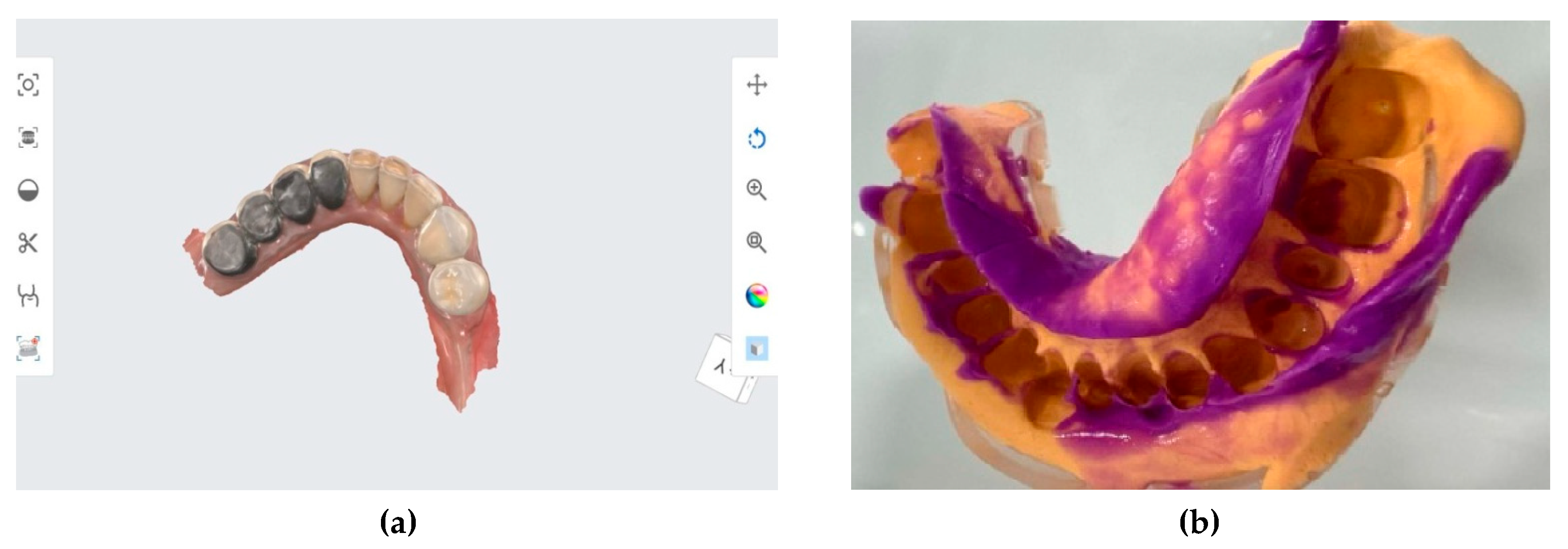
Figure 1.
(a) Digital impression technique (b) Conventional impression technique.
The data were collected by distributing an online questionnaire to dentists practicing in the Oltenia region of Romania. The questionnaire included both single-answer questions and multiple-choice questions. Participant selection was designed to reflect demographic and professional characteristics, thereby enabling a rigorous comparative analysis. Respondents were aged 18-65 years and practiced dentistry in urban, rural, or both areas.
Participation in the study was voluntary, and anonymity and data confidentiality were guaranteed. The questionnaire included the following questions and response options:
- What is your age? (18-23/24-35/36-45/46-55)
- In which environment do you practice dentistry? (Rural/Urban/Both)
- What is your professional degree? (Dentist/Resident dentist/Specialist dentist /Primary dentist)
- Which impression technique do you use most frequently? (Conventional impression/Digital impression /Both methods)
- What advantages do you think digital impression techniques offer compared to conventional impression techniques? (Efficient communication and increased patient comfort/Higher accuracy/Shorter working time/Reduced risk of impression distortion/More efficient communication with the dental laboratory)
- What are the disadvantages of digital impression techniques? (High initial equipment costs/Need for a learning curve/Technical problems (software compatibility)/Limited accessibility in some dental offices/None)
- What advantages do you consider that conventional impression techniques offer compared to digital impression techniques? (Lower initial costs/Accessibility in any dental office/Possibility to use preferred materials/Familiarity with traditional techniques/None)
- In which situations do you prefer conventional impression techniques over digital impression techniques? (Complex prosthetic cases/Situations in which digital impression techniques are not available/Patients unfamiliar with digital technology/Limited clinical space /Never)
- Have you experienced significant difficulties when using impression techniques? (Yes/No/if yes, specify the challenges)
- Do you think digital impression techniques will completely replace conventional impression techniques in the future? (Yes/No/Not sure)
- In your opinion, which method provides more accurate clinical results? (Conventional impression techniques/Digital impression techniques/Both approaches are equally accurate/Depends on the clinical case)
- Would you recommend digital impression techniques to other colleagues? (Yes/No /if yes, please specify why/if not, please specify why)
- What impact do you think digital impression techniques have on patient comfort and satisfaction? (Increase patient comfort and satisfaction/Decrease patient comfort and satisfaction/Has no significant impact/I don’t know)
- Which digital technologies are you currently using in your practice? (Intraoral scanner/computer-aided design (CAD) software/3D printer/I do not use digital technologies/All of the above)
- To what extent are you satisfied with the results obtained using digital impression techniques? (Very satisfied/Satisfied/Dissatisfied/Very dissatisfied)
- Do you think that specialized training is necessary for the effective use of digital impression techniques? (Yes/No)
2.2. Statistical Analysis
The data obtained from this questionnaire were collected and organized using Google Forms, and the statistical analysis was performed using Microsoft Excel 365, version 2503 (San Francisco, CA, USA) and SPSS (Statistical Package for the Social Sciences) version 20 (SPSS Inc., Armonk, NY, USA), platforms that allowed the application of relevant statistical tests and the generation of detailed graphical representations for an optimal interpretation of the results. For data analysis, appropriate statistical methods were used, tailored to the type of variables and their distributions. The chi-square test was applied to examine relationships between categorical variables, including the association between professional specialization and the predominantly used impression technique. The choice of this test was motivated by the need to identify significant associations among these variables. To establish the relevance of the results, a significance threshold of p < 0.05 was used, in accordance with biomedical standards.
3. Results
The questionnaire was distributed to a representative sample of dentists in the Oltenia region of Romania, and the results provide a detailed perspective on the degree of implementation and acceptance of digital impressions in daily practice. Statistical correlations were performed between the data obtained from the 16 questions (Table 1).
The age group distribution indicates that the majority of respondents were aged between 24 and 35 years (18.8%) (Table 2). Most respondents practiced dentistry in urban areas (70.3%), whereas 16.8% practiced in rural areas; the remaining participants practiced in both settings (Table 2).
Digital impression techniques were preferred by 52.5% of respondents, while conventional impression techniques were used by 23.8%. In addition, 27% of respondents reported using both methods. The statistical correlation between Question 1 and Question 4 showed distinct preferences across age groups. Conventional impression techniques were mainly preferred in the 24-35 age group (76.19%). Digital impression techniques were predominantly preferred by dentists aged 36-45 years (84.21%). Dentists aged 46-65 years most frequently reported using both impression techniques (36%). Statistical analysis indicated that the preference for digital impression techniques differs significantly between age groups (p = 0.007), confirming a more pronounced tendency among younger dentists to adopt this technology (Table 3).
There was also a statistically significant difference (p = 0.005) between urban and rural areas in the use of digital impression techniques, indicating greater use in urban areas (Table 4).
About digital impressions, the advantages listed by respondents included: greater accuracy (56.4%), efficient communication and increased patient comfort (48.5%), reduced working time (33.7%), more efficient communication with the dental laboratory (31.7%), and finally, reduced risk of re-impressing (27.7%) (Figure 2a). The disadvantages listed by respondents were primarily related to the high cost of the equipment (47.5%), followed by limited accessibility in some practices (22.8%), technical problems (21.8%), and the need for a learning curve (6.9%) (Figure 2b).
Regarding conventional impressions, respondents reported the following advantages: availability in any dental office (40.6%), lower initial costs (28.7%), the option to use preferred materials (16.8%), and familiarity with traditional techniques (12.9%) (Figure 3).
Conventional impressions are primarily used when digital methods are unavailable (43.6%), in complex prosthetic cases (30.7%), and in situations where patients are unfamiliar with digital technology (23.8%). These responses reflect a pragmatic approach by dentists, who choose the optimal method depending on the circumstances (Figure 4).
On the other hand, most respondents (74.3%) reported no significant difficulties with impression techniques, whereas 20.8% reported problems. This relatively small percentage indicates that the technology is generally well accepted and used without significant complications. 59.4% of respondents believe that digital impression provides more accurate results; 75.2% believe that digital impression techniques improve patient comfort and satisfaction; 61.4% are very satisfied with the results obtained with digital impression techniques; and 36.6% are satisfied. This result confirms the effectiveness of the digital method and its high level of acceptance in practice.
Regarding the accuracy and precision of the two impression techniques, more accurate clinical results were obtained with the digital impression technique (67.65%), whereas 17.65% of responding physicians reported that conventional impression techniques yielded accurate clinical results. The degree of satisfaction among dentists with digital impression techniques is favorable, with 63% satisfied and 37% dissatisfied (Table 5).
Regarding dentists’ satisfaction with the use of the digital impression technique, the correlation analysis indicates that most respondents among those who are very satisfied (72.06%) believe that the digital impression technique has the potential to replace the conventional impression technique entirely. At the same time, only satisfied dentists show a lower percentage (26.47%) in this direction, whereas dissatisfied dentists constitute a very small proportion. These data clearly show that satisfaction levels influence dentists’ confidence in the widespread adoption of digital technology (Table 6).
Regarding the association between the perceived impact of digital impression techniques on patient comfort and dentists’ satisfaction, the data show a strong correlation between these variables. Among dentists who believe that digital impression techniques increase patient comfort and satisfaction (71.43%), a similar proportion (71.43%) also reports being very satisfied with the results obtained using this technique. In contrast, among those who believe the impact of digitization is not significant, only 44.44% report being very satisfied, and dentists who believe that the digital impression technique reduces patient comfort report an average level of satisfaction (100%). These results indicate that a positive perception of the patient experience directly influences dentists’ satisfaction with digital technology, confirming the correspondence between patients’ clinical benefits and dentists’ professional assessments (Table 7).
Regarding the relationship between the use of digital technologies in dental practice and the recommendation of digital impressions, the results show a clear trend: dentists who frequently use digital technologies are also those who most often recommend this method to their colleagues. Among intraoral scanner users, 70.79% would recommend digital impressions, whereas the percentages are lower among those who use 3D printers (10.11%) or CAD software (19.1%). These data show that direct and consistent experience with digital technology, particularly the intraoral scanner, is associated with greater confidence in this method and a stronger inclination to promote it to other professionals. Thus, the extent of integration of digital technologies into daily practice significantly influences dentists’ willingness to recommend this technique (Table 8).
4. Discussion
After analyzing the questionnaire responses, a clear trend toward the use of digital impressions in dental practice was observed, but with the conventional method being retained in certain specific situations. Most study participants fall within the 36-45 age group (24.8%), followed by those aged 24-35 (18.8%). A significant proportion (70.3%) practices in urban areas, suggesting easier access to modern technologies. In terms of specialisation, 49.5% of respondents are dentists and 34.7% are resident doctors, indicating a growing interest in new technologies among both experienced professionals and those at the beginning of their careers.
In the study conducted by Lee et al., it was found that the group represented by students rated digital impression techniques as the most effective impression technique, while the group represented by clinicians showed a uniform distribution in their choice of impression techniques. Their study concluded that, from the operator’s perspective, digital impressions are easier to perform for inexperienced clinicians [59]. Other studies in the literature have also obtained the same results [60,61,62]. Digital impressions were the most frequently selected method, selected by 52.5% of respondents. On the other hand, 23.8% continue to use conventional impressions, and 27% combine both techniques, suggesting that there are still situations where the traditional method remains a viable option.
Among the main benefits of digital impressions are greater accuracy (56.4%), efficient communication and increased patient comfort (48.5%), reduced working time (33.7%), and decreased risk of re-impression (27.7%). Another important advantage mentioned by 31.7% of respondents is improved communication with the dental laboratory, which can lead to better results in prosthetic work. The main obstacle to the adoption of digital impressions is the high initial cost of the equipment, as mentioned by 47.5% of respondents. Other issues identified include limited access to the technology in certain practices (22.8%) and technical difficulties, such as software incompatibilities (21.8%). These issues may limit the widespread implementation of digital technology, especially in smaller practices. Although digital impressions are gaining ground, the traditional method remains popular due to its accessibility (40.6%), lower initial costs (28.7%), and the option of using preferred materials (16.8%). These aspects suggest that the traditional method remains a valid option, particularly in settings without advanced digital technology. Digital impressions are considered more accurate by 59.4% of respondents, confirming the technological advantages of this method. In addition, 78.2% of participants would recommend digital impressions to colleagues, indicating high confidence in this technology. A total of 75.2% of respondents believe that digital impressions improve patient comfort and satisfaction, which may contribute to greater acceptance of this method. In addition, 61.4% of respondents reported being very satisfied with the results, and 36.6% were satisfied, indicating a high level of satisfaction with this technology. Other studies in the literature have also found that digital impression techniques offer greater time efficiency and fewer sources of error than conventional methods. The technology is a more comfortable and less invasive method for patients, facilitating rapid and cost-effective information transmission and simplifying data storage [63,64,65,66,67,68,69,70,71,72].
Respondents indicated that conventional impression techniques are used primarily when the digital option is unavailable (43.6%), in complex prosthetic cases (30.7%), or when patients are unfamiliar with digital technology (23.8%). This indicates that, although technology is advancing, there are still situations in which the traditional method remains preferable. A large proportion of respondents (74.3%) reported no significant difficulties with using digital impressions, indicating a high level of adaptability to this technology. However, 20.8% reported difficulties, suggesting that more training or improvements in the equipment and software used are needed. Regarding the possibility of digital impression techniques permanently replacing the traditional method, only 6.3% of respondents believe this will occur, whereas 16.8% believe the conventional method will continue to be used. These results indicate that, although digital technology is becoming increasingly popular, the conventional method will not disappear completely.
- Only 16.3% of respondents believe specialized training is required to use digital impression techniques, suggesting that most dentists adapt to this technology relatively easily. However, perception may vary with each user’s experience and the complexity of the system used.
This study presents limitations for interpreting the results: it was conducted exclusively in the Oltenia region, which may limit the generalizability of the findings at the national level. A larger and more diverse sample would have allowed a broader analysis of how digitization is being adopted in different regions of the country; since the data were collected through a self-administered questionnaire, there is a risk of self-reporting biases, such as subjectivity of responses, overestimation of digital skills or a tendency to provide socially acceptable answers; given that the majority of participants practice in the urban environment, the results may overestimate the acceptance and use of digital technologies, since urban cabinets generally have easier access to modern; the study is based strictly on the perception of doctors and does not include objective assessments of the accuracy of the two types of impression techniques, not analyzing concrete clinical results, such as marginal adaptation or recovery rate of prosthetic works; respondents use different models of intraoral scanners and CAD software, which can generate significant variations in reported experiences (the study does not differentiate results according to brands or technological generations); although high costs were cited as a disadvantage, a detailed cost-benefit analysis comparing the two techniques from a financial and profitability perspective was not carried out.
Based on the identified results and limitations, the following future research directions can be proposed: expanding the research in several regions of Romania or in other countries would allow a broader understanding of the level of digitization and the factors influencing the adoption of digital impression technologies; it is necessary to assess under controlled conditions the accuracy of impression, the adaptation of restorations and the long-term success rate in order to validate the perceptions reported by dentists; investigate how dentists’ attitude and competencies towards evolve over time, with the accumulation of experience and the acceleration of digitization; future studies should compare initial costs, maintenance costs, cost-effectiveness and the impact; since the current study is based only on the perspective of dentists, future research could examine the satisfaction, convenience and acceptance of digital technologies from the perspective of patients; evaluating the performance of existing models on the market would provide helpful information on optimal investments and support the standardization of the use of impression techniques; a study focused on the efficiency of full digital flow, from intraoral scanning to prosthetic production in the laboratory, would clarify how new technologies optimize communication and reduce laboratory errors.
5. Conclusions
The questionnaire results indicate a clear trend toward digitization in dental impression techniques, with the majority of respondents reporting appreciation for the accuracy, efficiency, and increased patient comfort offered by digital methods. However, specific barriers to the full adoption of this technology persist, such as high equipment costs and accessibility issues in some dental offices. Although digital impression techniques are considered superior in terms of accuracy and patient satisfaction, conventional impression techniques are still used in some situations due to their accessibility and lower cost. In this context, it can be concluded that the two methods will continue to coexist, at least in the medium term, depending on the available resources and the needs of each dental office.
Author Contributions
Conceptualization, M.O.A, S.-M.-S.P., A.G.I., A.D.T. and A.S.K.; methodology, M.O.A., S.-M.-S.P., A.S.K. and R.G.R.; validation, A.G.I, A.D.T. and L.D.; resources, S.-M.-S.P., N.A.P., C.E.A. and A.M.P.; data curation, A.S.K. and A.M.P.; writing—original draft preparation, M.O.A. and S.-M.-S.P.; writing—review and editing, M.O.A., S.-M.-S.P., A.G.I, A.D.T. and A.S.K.; visualization, M.O.A., R.G.R., L.D., N.A.P. and C.E.A.; supervision, M.O.A., S.-M.-S.P., and A.S.K.; project administration, M.O.A., S.-M.-S.P., and A.S.K.. All authors have read and agreed to the published version of the manuscript.
Funding
The Article Processing Charges were funded by the University of Medicine and Pharmacy of Craiova, Romania.
Institutional Review Board Statement
The study was conducted according to the guidelines of the Declaration of Helsinki and approved by the Ethics Committee of the University of Medicine and Pharmacy of Craiova (approval reference no. 65/29.01.2024).
Informed Consent Statement
Informed consent was obtained from all the subjects involved in the study.
Data Availability Statement
The authors declare that the data of this research are available from the corresponding authors upon reasonable request. The data are not publicly available due to privacy.
Conflicts of Interest
The authors declare no conflict of interest.
References
- De La Cruz, J.E.; Funkenbusch, P.D.; Ercoli, C.; Moss, M.E.; Graser, G.N.; Tallents, R.H. Verification jig for implant-supported prostheses: A comparison of standard impressions with verification jigs made of different materials. J Prosthet Dent. 2002, 88(3), 329–36. [Google Scholar] [CrossRef]
- Khan, S.A.; Singh, S.; Neyaz, N.; Jaiswal, M.M.; Tanwar, A.S.; Singh, A. Comparison of Dimensional Accuracy of Three Different Impression Materials Using Three Different Techniques for Implant Impressions: An In Vitro Study. J Contemp Dent Pract. 2021, 22(2), 172–178. [Google Scholar] [CrossRef]
- Wong, A.W.H.; Nedelcu, R.; Hamilton, A. Clinical Recommendations for Implant Verification Jigs in Analogue and Digital Workflows. A Narrative Review. Aust Dent J. 2025, 70 Suppl 1(Suppl 1), S35–S49. [Google Scholar] [CrossRef]
- Millar, B. How to make a good impression (crown and bridge). Br Dent J. 2001, 191(7), 402–3, 405. [Google Scholar] [CrossRef]
- Al-Odinee, N.M.; Al-Hamzi, M.; Al-Shami, I.Z.; Madfa, A.; Al-Kholani, A.I.; Al-Olofi, Y.M. Evaluation of the quality of fixed prosthesis impressions in private laboratories in a sample from Yemen. BMC Oral Health 2020, 20(1), 304. [Google Scholar] [CrossRef]
- Tamini, F.; Hirayama, H. Digital Restorative Dentistry – A Guide to Materials, Equipment and Clinical Procedures. Springer Nature Switzerland AG. 2019, 115-136.
- Naderi, R.K.; Patel, T.J.; Thompson, M.A. A comparison study: The use of digital and conventional impression techniques in dental hygiene education. J Dent Educ. 2024, 88(5), 518–523. [Google Scholar] [CrossRef]
- Alqutaibi, A.Y.; Alghauli, M.A.; Aboalrejal, A.N. Digital scans, compared to conventional impressions, may expedite fabrication time for full-arch implant-supported prostheses while maintaining a comparable level of marginal bone loss. J Evid Based Dent Pract. 2024, 24(2), 101987. [Google Scholar] [CrossRef] [PubMed]
- Alfaraj, A.; Alqudaihi, F.; Khurshid, Z.; Qadiri, O.; Lin, W.S. Comparative analyses of accuracy between digital and conventional impressions for complete-arch implant-supported fixed dental prostheses-A systematic review and meta-analysis. J Prosthodont 2025. [Google Scholar] [CrossRef] [PubMed]
- Reis, I.N.R.D.; Chamma-Wedemann, C.N.; Silva, I.A.O.; Spin-Neto, R.; Sesma, N.; da Silva, E.V.F. Clinical outcomes of digital scans versus conventional impressions for implant-supported fixed complete arch prostheses: A systematic review and meta-analysis. J Prosthet Dent. 2025, 134(2), 346–355. [Google Scholar] [CrossRef]
- Tylman, S.D.; Malone, W.F.; Koth, D.L. Tylman’s theory and practice of fixed prosthodontics. (No Title). 1978.
- Pachiou, A.; Zervou, E.; Sykaras, N.; Tortopidis, D.; Ioannidis, A.; Jung, R.E.; Strauss, F.J.; Kourtis, S. Patient-Reported Outcomes of Digital Versus Conventional Impressions for Implant-Supported Fixed Dental Prostheses: A Systematic Review and Meta-Analysis. J Pers Med. 2025, 15(9), 427. [Google Scholar] [CrossRef]
- Alqahtani, W.M.S.; Yousief, S.A.; Demachkia, A.M.R.; Demachkia, M.R.; Barakat, A.; Dimashkieh, M.R.; Mekkey, M.A.M.; Abdelglel, A.M.S.; Waly, A.S.; Bamanie, R.M.S.; Alnafisah, D.A. Assessing Precision in All-Ceramic Fixed Restorations: Unveiling the Marginal Fit Through Digital and Traditional Impressions-A Comprehensive Systematic Review and Meta-Analysis. Eur J Dent. 2025, 19(4), 903–918. [Google Scholar] [CrossRef]
- Popa, D.A.; Mercuț, V.; Popescu, A.M.; Târtea, D.A.; Castravete, Ș.; Rîcă, G.R.; Scrieciu, M.; Amărăscu, M.O.; Vlăduțu, D.E. Assessing the effects of an occlusal disharmony on the dento-maxillary system using the finite element method. Romanian Journal of Oral Rehabilitation 2024, 16, 482–502. [Google Scholar] [CrossRef]
- Christensen, G.J. The challenge to conventional impressions. J Am Dent Assoc. 2008, 139(3), 347–9. [Google Scholar] [CrossRef]
- Burzynski, J.A.; Firestone, A.R.; Beck, F.M.; Fields, H.W., Jr.; Deguchi, T. Comparison of digital intraoral scanners and alginate impressions: Time and patient satisfaction. Am J Orthod Dentofacial Orthop. 2018, 153(4), 534–541. [Google Scholar] [CrossRef]
- Punj, A.; Bompolaki, D.; Garaicoa, J. Dental Impression Materials and Techniques. Dent Clin North Am. 2017, 61(4), 779–796. [Google Scholar] [CrossRef]
- Runkel, C.; Güth, J.F.; Erdelt, K.; Keul, C. Digital impressions in dentistry-accuracy of impression digitalisation by desktop scanners. Clin Oral Investig. 2020, 24(3), 1249–1257. [Google Scholar] [CrossRef] [PubMed]
- Flügge, T.; van der Meer, W.J.; Gonzalez, B.G.; Vach, K.; Wismeijer, D.; Wang, P. The accuracy of different dental impression techniques for implant-supported dental prostheses: A systematic review and meta-analysis. Clin Oral Implants Res. 2018, 29 Suppl 16, 374–392. [Google Scholar] [CrossRef] [PubMed]
- Park, J.S.; Alshehri, Y.F.A.; Kruger, E.; Villata, L. Accuracy of digital versus conventional implant impressions in partially dentate patients: A systematic review and meta-analysis. J Dent. 2025, 160, 105918. [Google Scholar] [CrossRef]
- Mahmoud, R.A.; Hakim, A.A.A.; Rady, N.A. Effect of different impression techniques on marginal integrity of CAD-CAM milled all-on-four mandibular frameworks: an in vitro study. BMC Oral Health 2025, 25(1), 497. [Google Scholar] [CrossRef]
- Bandiaky, O.N.; Le Bars, P.; Gaudin, A.; Hardouin, J.B.; Cheraud-Carpentier, M.; Mbodj, E.B.; Soueidan, A. Comparative assessment of complete-coverage, fixed tooth-supported prostheses fabricated from digital scans or conventional impressions: A systematic review and meta-analysis. J Prosthet Dent. 2022, 127(1), 71–79. [Google Scholar] [CrossRef] [PubMed]
- Farzin, M.; Derafshi, R.; Giti, R.; Kalantari, M.H. Effect of Core Materials on the Dimensional Accuracy of Casts Made of Two Different Silicone Impression Materials: An Experimental Study. J Int Soc Prev Community Dent. 2020, 10(2), 196–204. [Google Scholar] [CrossRef]
- Pozzi, A.; Arcuri, L.; Carosi, P.; Laureti, A.; Londono, J.; Wang, H.L. Photogrammetry Versus Intraoral Scanning in Complete-Arch Digital Implant Impression: A Systematic Review and Meta-Analysis. Clin Implant Dent Relat Res. 2025, 27(3), e70059. [Google Scholar] [CrossRef] [PubMed]
- Benic, G.I.; Mühlemann, S.; Fehmer, V.; Hämmerle, C.H.; Sailer, I. Randomized controlled within-subject evaluation of digital and conventional workflows for the fabrication of lithium disilicate single crowns. Part I: digital versus conventional unilateral impressions. J Prosthet Dent. 2016, 116(5), 777–782. [Google Scholar] [CrossRef]
- Sedky, A.; Abd-Elwahab Radi, I. Limited evidence suggests complete arch digital scans are less time efficient than conventional impression. Evid Based Dent. 2020, 21(4), 138–139. [Google Scholar] [CrossRef] [PubMed]
- Gartshore, J.; Glavin, C.; Jackson, G.; Bonsor, S. Lithium disilicate, full coverage crowns: what is the effect of using conventional impressions compared to digital impression with respect to the internal fit of the restoration? A systematic review. Evid Based Dent. 2025, 26(1), 67. [Google Scholar] [CrossRef]
- Jánosi, K.M.; Cerghizan, D.; Mártha, K.I.; Elekes, É.; Szakács, B.; Elekes, Z.; Kovács, A.; Szász, A.; Mureșan, I.; Hănțoiu, L.G. Evaluation of Intraoral Full-Arch Scan versus Conventional Preliminary Impression. J Clin Med. 2023, 12(17), 5508. [Google Scholar] [CrossRef]
- Chen, L.; Chen, C.; Li, Z.Y.; Zhang, Q. Clinical performance of intraoral digital impression for fixed prosthodontics: a Meta-analysis. Hua Xi Kou Qiang Yi Xue Za Zhi 2021, 39(3), 306–312. [Google Scholar]
- Abuduwaili, K.; Huang, R.; Song, J.; Liu, Y.; Chen, Z.; Huang, B.; Li, Z. Comparison of photogrammetric imaging, intraoral scanning and conventional impression accuracy of full-arch dental implant rehabilitation: an in vitro study. BMC Oral Health 2025, 25(1), 753. [Google Scholar] [CrossRef]
- Siqueira, R.; Galli, M.; Chen, Z.; Mendonça, G.; Meirelles, L.; Wang, H.L.; Chan, H.L. Intraoral scanning reduces procedure time and improves patient comfort in fixed prosthodontics and implant dentistry: a systematic review. Clin Oral Investig. 2021, 25(12), 6517–6531. [Google Scholar] [CrossRef]
- Basker, R.M.; Davenport, J.C.; Thomason, J.M. Prosthetic treatment of the edentulous patient. John Wiley & Sons. 2011.
- Ribeiro, C.S.C.; Brandão, M.T.O.; Moreira, G.C.; Bitencourt, S.B.; de Carvalho, R.F.; Lemos, C.A.A. Intraoral Scanning Versus Conventional Impression for Implant Prostheses: A Systematic Review. Eur J Prosthodont Restor Dent. 2025, 33(2), 206–218. [Google Scholar] [PubMed]
- Megremis, S.; Tiba, A.; Vogt, K.; Geary, R.; Kuehne, J. An evaluation of selected vinyl polysiloxane and vinyl polysiloxane-hybrid elastomeric impression materials. J Am Dent Assoc. 2012, 143(4), 405–6. [Google Scholar] [CrossRef]
- Mangano, F.G.; Veronesi, G.; Hauschild, U.; Mijiritsky, E.; Mangano, C. Trueness and Precision of Four Intraoral Scanners in Oral Implantology: A Comparative in Vitro Study. PLoS One 2016, 11(9), e0163107. [Google Scholar] [CrossRef]
- Kim, R.J.; Park, J.M.; Shim, J.S. Accuracy of 9 intraoral scanners for complete-arch image acquisition: A qualitative and quantitative evaluation. J Prosthet Dent. 2018, 120(6), 895–903.e1. [Google Scholar] [CrossRef]
- Rutkūnas, V.; Gedrimienė, A.; Al-Haj Husain, N.; Pletkus, J.; Barauskis, D.; Jegelevičius, D.; Özcan, M. Effect of additional reference objects on accuracy of five intraoral scanners in partially and completely edentulous jaws: An in vitro study. J Prosthet Dent. 2023, 130(1), 111–118. [Google Scholar] [CrossRef]
- Park, J.M.; Kim, R.J.; Lee, K.W. Comparative reproducibility analysis of 6 intraoral scanners used on complex intracoronal preparations. J Prosthet Dent. 2020, 123(1), 113–120. [Google Scholar] [CrossRef] [PubMed]
- Alenezi, A.; Yehya, M.; Alkhodary, M. Effect of full arch two scanning techniques on the accuracy of overdenture conventional and CAD/CAM Co-Cr bars. Saudi Dent J. 2022, 34(7), 553–564. [Google Scholar] [CrossRef] [PubMed]
- García-Gil, I.; Cortés-Bretón-Brinkmann, J.; Jiménez-García, J.; Peláez-Rico, J.; Suárez-García, M.J. Precision and practical usefulness of intraoral scanners in implant dentistry: A systematic literature review. J Clin Exp Dent. 2020, 12(8), e784–e793. [Google Scholar] [CrossRef]
- Zarbakhsh, A.; Jalalian, E.; Samiei, N.; Mahgoli, M.H.; Kaseb Ghane, H. Accuracy of Digital Impression Taking Using Intraoral Scanner versus the Conventional Technique. Front Dent. 2021, 18, 6. [Google Scholar] [CrossRef] [PubMed]
- Giachetti, L.; Sarti, C.; Cinelli, F.; Russo, D.S. Accuracy of Digital Impressions in Fixed Prosthodontics: A Systematic Review of Clinical Studies. Int J Prosthodont. 2020, 33(2), 192–201. [Google Scholar] [CrossRef]
- Kihara, H.; Hatakeyama, W.; Komine, F.; Takafuji, K.; Takahashi, T.; Yokota, J.; Oriso, K.; Kondo, H. Accuracy and practicality of intraoral scanner in dentistry: A literature review. J Prosthodont Res. 2020, 64(2), 109–113. [Google Scholar] [CrossRef]
- Miranda Ataíde, I.C.; Serna-Muñoz, C.; Ferreira Guimaraes Pereira Areias, C.M.; Ferreira de Azevedo, Á.A.; Pereirinha Henriques Ferreira de Andrade, R.E.; Ortiz-Ruiz, A.J. From Alginate to Pixel: Comparing the Effect of Two Dental Impression Methods on Children’s Anxiety. Children (Basel) 2025, 12(7), 866. [Google Scholar] [CrossRef]
- Giuliodori, G.; Rappelli, G.; Aquilanti, L. Intraoral Scans of Full Dental Arches: An In Vitro Measurement Study of the Accuracy of Different Intraoral Scanners. Int J Environ Res Public Health 2023, 20(6), 4776. [Google Scholar] [CrossRef] [PubMed]
- Albayrak, B.; Sukotjo, C.; Wee, A.G.; Korkmaz, İ.H.; Bayındır, F. Three-Dimensional Accuracy of Conventional Versus Digital Complete Arch Implant Impressions. J Prosthodont. 2021, 30(2), 163–170. [Google Scholar] [CrossRef] [PubMed]
- Jaiswal, T.; Bahadur, S.; Sharma, P.; Gupta, S.; Bhushan, P.; Sharma, B.; Meharwade, V. Comparative analysis of digital versus conventional impression techniques for dental implant placement. Bioinformation 2025, 21(7), 2140–2144. [Google Scholar] [CrossRef] [PubMed]
- Albanchez-González, M.I.; Brinkmann, J.C.; Peláez-Rico, J.; López-Suárez, C.; Rodríguez-Alonso, V.; Suárez-García, M.J. Accuracy of Digital Dental Implants Impression Taking with Intraoral Scanners Compared with Conventional Impression Techniques: A Systematic Review of In Vitro Studies. Int J Environ Res Public Health 2022, 19(4), 2026. [Google Scholar] [CrossRef]
- Hasanzade, M.; Aminikhah, M.; Afrashtehfar, K.I.; Alikhasi, M. Marginal and internal adaptation of single crowns and fixed dental prostheses by using digital and conventional workflows: A systematic review and meta-analysis. J Prosthet Dent. 2021, 126(3), 360–368. [Google Scholar] [CrossRef]
- Hasanzade, M.; Shirani, M.; Afrashtehfar, K.I.; Naseri, P.; Alikhasi, M. In Vivo and In Vitro Comparison of Internal and Marginal Fit of Digital and Conventional Impressions for Full-Coverage Fixed Restorations: A Systematic Review and Meta-analysis. J Evid Based Dent Pract. 2019, 19(3), 236–254. [Google Scholar] [CrossRef]
- Porto, A.M.; Nascimento, M.V.; Garcia, B.A.; de Freitas Pontes, K.M.; Costa, F.W.G.; de Negreiros, W.A. Marginal adaptation of tooth-supported fixed restorations fabricated using digital scanning versus conventional impression techniques: An overview of systematic reviews. J Prosthet Dent. 2025, 134(3), 629.e1–629.e13. [Google Scholar] [CrossRef]
- Lo Russo, L.; Caradonna, G.; Biancardino, M.; De Lillo, A.; Troiano, G.; Guida, L. Digital versus conventional workflow for the fabrication of multiunit fixed prostheses: A systematic review and meta-analysis of vertical marginal fit in controlled in vitro studies. J Prosthet Dent. 2019, 122(5), 435–440. [Google Scholar] [CrossRef]
- Manisha, J.; Srivastava, G.; Das, S.S.; Tabarak, N.; Choudhury, G.K. Accuracy of single-unit ceramic crown fabrication after digital versus conventional impressions: A systematic review and meta-analysis. J Indian Prosthodont Soc. 2023, 23(2), 105–111. [Google Scholar]
- Mahat, N.S.; Shetty, N.Y.; Kohli, S.; Jamayet, N.B.; Patil, P. Clinical outcomes of implant-supported and tooth-supported fixed prostheses fabricated from digital versus analogue impression: a systematic review and meta-analysis. Evid Based Dent. 2023, 24(3), 142. [Google Scholar] [CrossRef]
- Eggmann, F.; Blatz, M.B. Recent Advances in Intraoral Scanners. J Dent Res. 2024, 103(13), 1349–1357. [Google Scholar] [CrossRef]
- Târtea, D.A.; Manolea, H.O.; Ionescu, M.; Gîngu, O.; Amărăscu, M.O.; Popescu, A.M.; Mercuţ, V.; Popescu, S.M. A Microscopy Evaluation of Emergence Profile Surfaces of Dental Custom CAD-CAM Implant Abutments and Dental Implant Stock Abutments. J Pers Med. 2024, 14(7), 699. [Google Scholar] [CrossRef]
- Târtea, D.A.; Popescu, S.M.; Manolea, H.O.; Ionescu, M.; Amărăscu, O.M.; Mărășescu, P.C.; Gîngu, O. Microscope evaluation of micro-gaps at implant-abutment connection. Romanian Journal of Oral Rehabilitation 2024, 16(2). [Google Scholar] [CrossRef]
- Popescu, A.M.; Vlăduțu, D.E.; Mărășescu, P.C.; Amărăscu, M.O.; Târtea, D.A.; Popescu, S.M.; Mercuț, V. Applications of 3d printing techniques for occlusal splints used in bruxism. Romanian Journal of Oral Rehabilitation 2024, 16(1). [Google Scholar] [CrossRef]
- Lee, S.J.; Macarthur, R.X., 4th; Gallucci, G.O. An evaluation of student and clinician perception of digital and conventional implant impressions. J Prosthet Dent. 2013, 110(5), 420–3. [Google Scholar] [CrossRef]
- Papaspyridakos, P.; Vazouras, K.; Chen, Y.W.; Kotina, E.; Natto, Z.; Kang, K.; Chochlidakis, K. Digital vs Conventional Implant Impressions: A Systematic Review and Meta-Analysis. J Prosthodont. 2020, 29(8), 660–678. [Google Scholar] [CrossRef] [PubMed]
- Róth, I.; Czigola, A.; Joós-Kovács, G.L.; Dalos, M.; Hermann, P.; Borbély, J. Learning curve of digital intraoral scanning - an in vivo study. BMC Oral Health 2020, 20(1), 287. [Google Scholar] [CrossRef]
- Ciocan, L.T.; Pantea, M.; Vasilescu, V.G.; Țâncu, A.M.C.; Sfeatcu, R.; Didilescu, A.C.; Ripszky, A.; Popa, A.; Pițuru, S.M.; Imre, M. Evaluation of Undergraduate Dental Students’ Opinions on the Use of Digital Versus Conventional Design in Prosthodontics. Dent J (Basel) 2025, 13(6), 242. [Google Scholar] [CrossRef] [PubMed]
- Yuzbasioglu, E.; Kurt, H.; Turunc, R.; Bilir, H. Comparison of digital and conventional impression techniques: evaluation of patients’ perception, treatment comfort, effectiveness and clinical outcomes. BMC Oral Health 2014, 14, 10. [Google Scholar] [CrossRef]
- Yilmaz, H.; Eglenen, M.N.; Cakmak, G.; Yilmaz, B. Effect of Impression Technique and Operator Experience on Impression Time and Operator-Reported Outcomes. J Prosthodont. 2021, 30(8), 676–683. [Google Scholar] [CrossRef]
- Ahmed, S.; Hawsah, A.; Rustom, R.; Alamri, A.; Althomairy, S.; Alenezi, M.; Shaker, S.; Alrawsaa, F.; Althumairy, A.; Alteraigi, A. Digital Impressions Versus Conventional Impressions in Prosthodontics: A Systematic Review. Cureus 2024, 16(1), e51537. [Google Scholar] [CrossRef] [PubMed]
- Bernburg, M.; Gebhardt, J.S.; Groneberg, D.A.; Mache, S. Impact of Digitalization in Dentistry on Technostress, Mental Health, and Job Satisfaction: A Quantitative Study. Healthcare (Basel) 2025, 13(1), 72. [Google Scholar] [CrossRef]
- Ahlholm, P.; Sipilä, K.; Vallittu, P.; Jakonen, M.; Kotiranta, U. Digital Versus Conventional Impressions in Fixed Prosthodontics: A Review. J Prosthodont. 2018, 27(1), 35–41. [Google Scholar]
- Lee, S.J.; Jamjoom, F.Z.; Le, T.; Radics, A.; Gallucci, G.O. A clinical study comparing digital scanning and conventional impression making for implant-supported prostheses: A crossover clinical trial. J Prosthet Dent. 2022, 128(1), 42–48. [Google Scholar]
- Papaspyridakos, P.; Gallucci, G.O.; Chen, C.J.; Hanssen, S.; Naert, I.; Vandenberghe, B. Digital versus conventional implant impressions for edentulous patients: accuracy outcomes. Clin Oral Implants Res. 2016, 27(4), 465–72. [Google Scholar] [CrossRef] [PubMed]
- Bi, C.; Wang, X.; Tian, F.; Qu, Z.; Zhao, J. Comparison of accuracy between digital and conventional implant impressions: two and three dimensional evaluations. J Adv Prosthodont. 2022, 14(4), 236–249. [Google Scholar]
- Dauti, R.; Cvikl, B.; Franz, A.; Schwarze, U.Y.; Lilaj, B.; Rybaczek, T.; Moritz, A. Comparison of marginal fit of cemented zirconia copings manufactured after digital impression with lava™ C.O.S and conventional impression technique. BMC Oral Health 2016, 16(1), 129. [Google Scholar] [CrossRef]
- Tabesh, M.; Nejatidanesh, F.; Savabi, G.; Davoudi, A.; Savabi, O.; Mirmohammadi, H. Marginal adaptation of zirconia complete-coverage fixed dental restorations made from digital scans or conventional impressions: A systematic review and meta-analysis. J Prosthet Dent. 2021, 125(4), 603–610. [Google Scholar] [CrossRef] [PubMed]
Figure 2.
(a) Advantages of the digital impression method. (b) Disadvantages of the digital impression method.
Figure 2.
(a) Advantages of the digital impression method. (b) Disadvantages of the digital impression method.
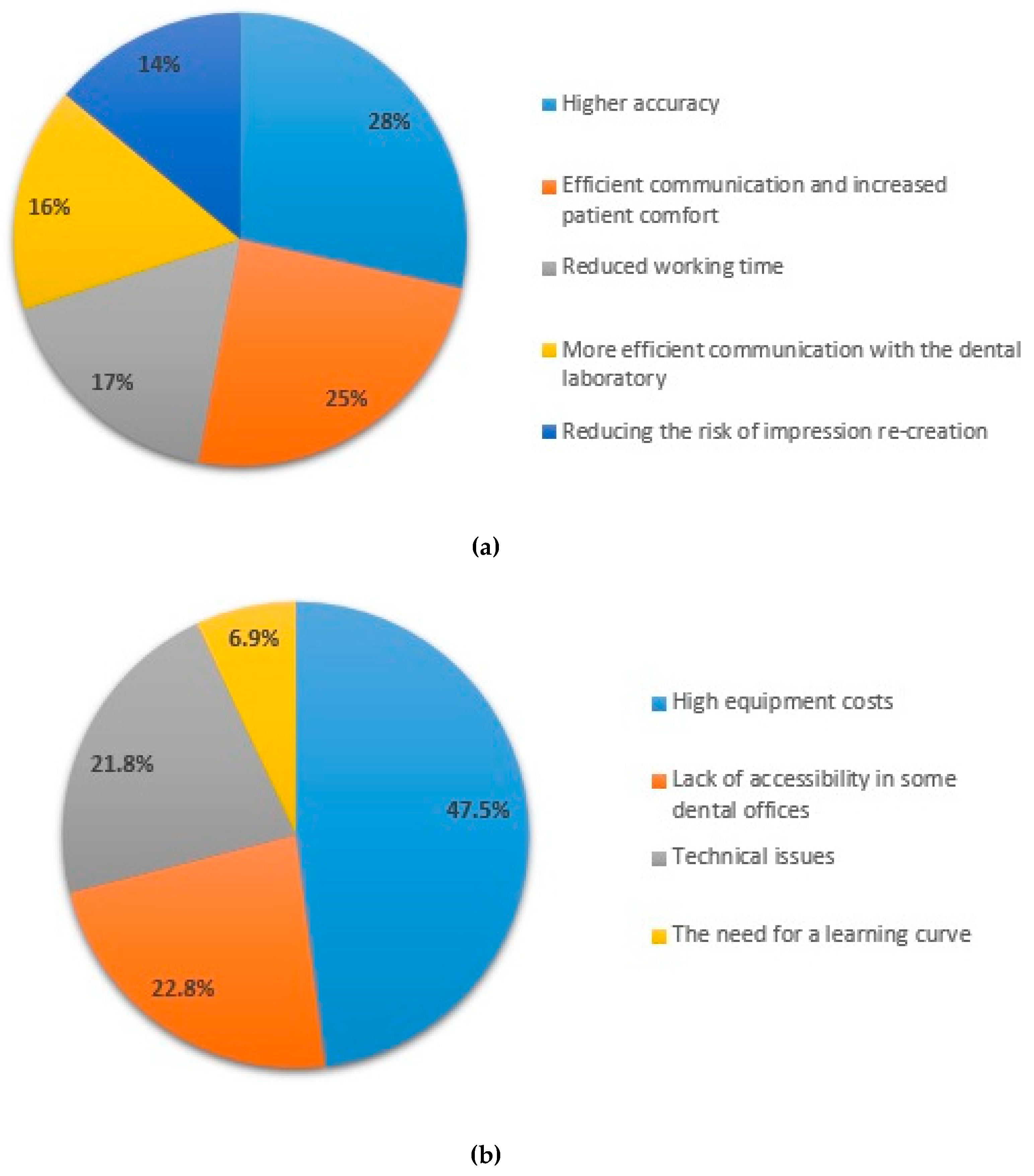
Figure 3.
Advantages of the conventional impression method.
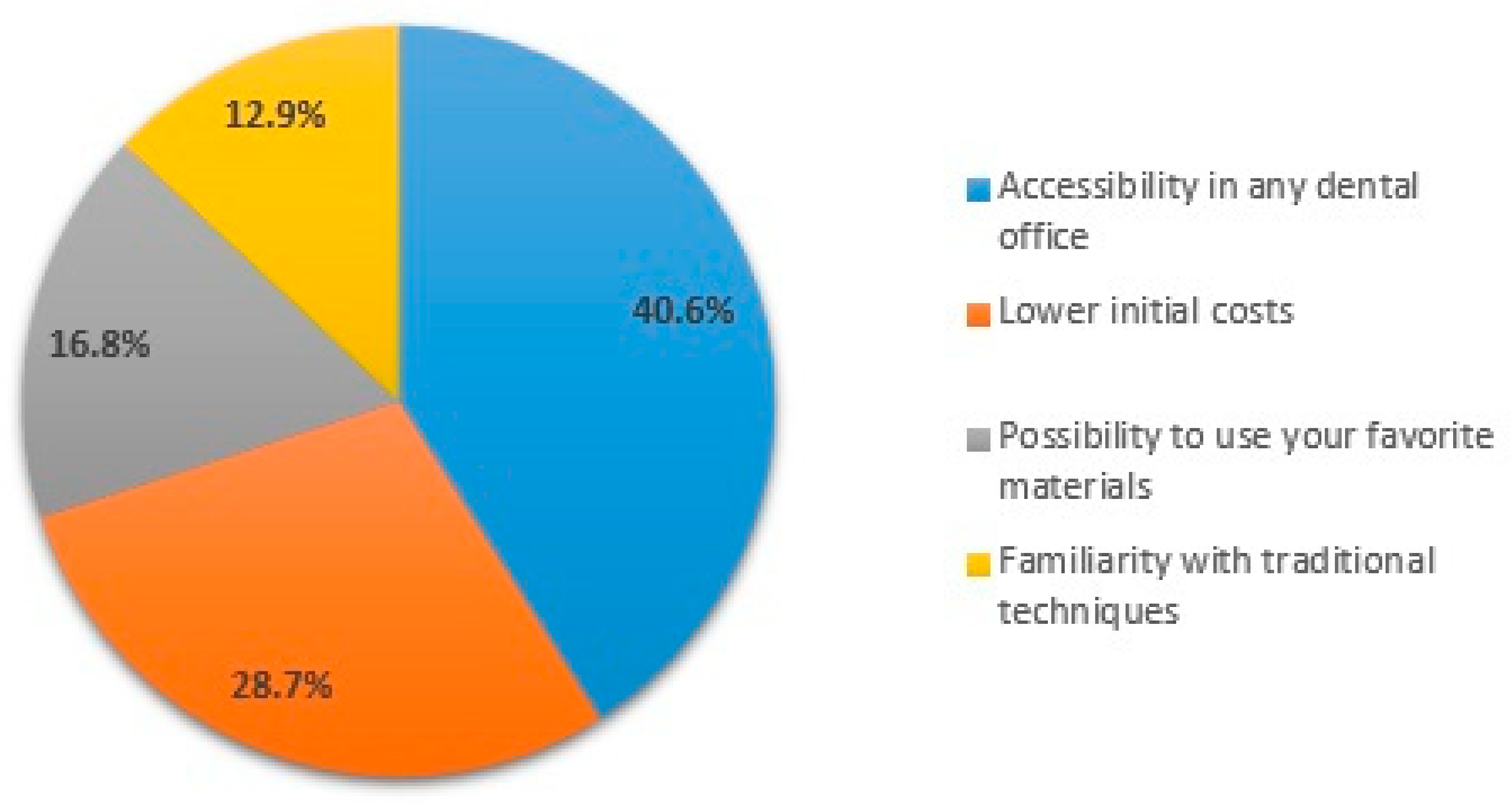
Figure 4.
Cases in which the conventional impression method is chosen.
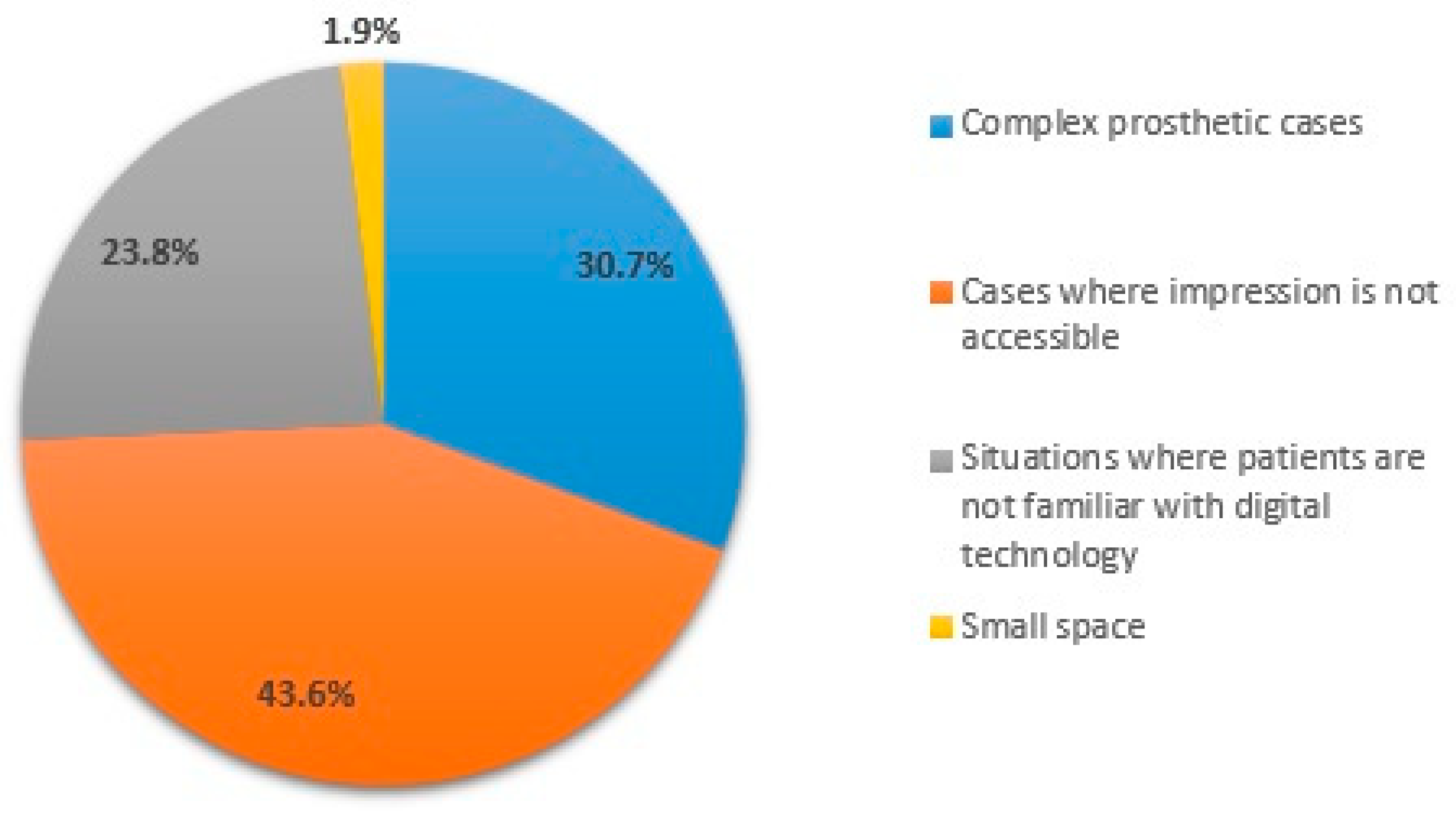

Table 1.
Statistical correlations.
| Q1 | Q2 | Q3 | Q4 | Q5_1 | Q5_2 | Q5_3 | Q5_4 | Q5_5 | Q6 | Q7 | Q8 | Q9 | Q10 | Q11 | Q12 | Q13 | Q14 | Q15 | Q16 | |
| Q1 | 1 | 0.017 | 0.090 | 0.007 | 0.072 | 0.696 | 0.010 | 0.015 | < 0.0 | 0.029 | 0.133 | 0.050 | 0.074 | 0.033 | 0.123 | < 0.0 | 0.038 | 0.142 | 0.033 | 0.686 |
| Q2 | 1 | 0.004 | 0.005 | 0.016 | 0.030 | 0.100 | 0.524 | 0.150 | 0.242 | 1.136 | 0.031 | 0.949 | 0.349 | 0.151 | < 0.0 | 0.094 | 0.091 | 0.452 | 0.478 | |
| Q3 | 1 | 0.060 | 0.381 | 0.074 | 0.675 | 0.064 | 0.021 | 0.371 | 0.957 | 0.274 | 0.629 | 0.496 | 0.819 | 0.003 | 0.195 | 0.756 | 0.852 | 0.741 | ||
| Q4 | 1 | 0.285 | 0.004 | 0.110 | 0.625 | 0.438 | 0.160 | 0.422 | 0.897 | 0.188 | 0.299 | < 0.0 | 0.113 | 0.087 | 0.633 | 0.375 | 0.496 | |||
| Q5_1 | 1 | 0.567 | 0.161 | 0.313 | 0.023 | 0.269 | 0.986 | 0.913 | 0.212 | 0.738 | 0.879 | 0.415 | 0.306 | 0.058 | 0.413 | 0.759 | ||||
| Q5_2 | 1 | 0.480 | 0.629 | 0.437 | 0.225 | 0.013 | 0.228 | 0.719 | 0.368 | 0.047 | 0.118 | 0.801 | 0.254 | 0.091 | 0.642 | |||||
| Q5_3 | 1 | < 0.0 | 0.016 | 0.837 | 0.709 | 0.830 | 0.199 | 0.189 | 0.395 | 0.401 | 0.463 | 0.912 | 0.581 | 1 | ||||||
| Q5_4 | 1 | < 0.0 | 0.133 | 0.361 | 0.188 | 0.110 | 0.027 | 0.268 | 0.705 | 0.598 | 0.770 | 0.650 | 0.178 | |||||||
| Q5_5 | 1 | 0.649 | 0.244 | 0.378 | 0.012 | 0.136 | 0.018 | 0.184 | 0.404 | 0.105 | 0.672 | 0.457 | ||||||||
| Q6 | 1 | 0.056 | 0.376 | 0.117 | 0.164 | 0.076 | < 0.0 | 0.219 | 0.552 | 0.162 | 0.226 | |||||||||
| Q7 | 1 | 0.007 | 0.234 | 0.022 | 0.898 | 0.348 | 0.102 | 0.196 | 0.310 | 0.843 | ||||||||||
| Q8 | 1 | 0.127 | 0.964 | 0.016 | 0.771 | 0.360 | 0.672 | 0.226 | 0.049 | |||||||||||
| Q9 | 1 | 0.058 | 0.257 | 0.371 | 0.186 | 0.445 | 0.325 | 0.069 | ||||||||||||
| Q10 | 1 | 0.008 | 0.069 | 0.319 | 0.307 | 0.027 | 0.065 | |||||||||||||
| Q11 | 1 | 0.227 | 0.646 | 0.393 | 0.116 | 0.067 | ||||||||||||||
| Q12 | 1 | 0.071 | 0.042 | 0.264 | 0.443 | |||||||||||||||
| Q13 | 1 | 0.073 | 0.004 | 0.974 | ||||||||||||||||
| Q14 | 1 | 0.412 | 0.360 | |||||||||||||||||
| Q15 | 1 | 0.085 | ||||||||||||||||||
| Q16 | 1 |
Table 2.
Demographic data.
| Parameter | Age group | Total | p | |||
|---|---|---|---|---|---|---|
| 24-35 Years | 36-45 Years | 46-65 Years | ||||
| Area of practice | Rural | 46 (63.89%) | 9 (12.5%) | 17 (23.61%) | 72 (100%) | 0.017 |
| 79.31% | 47.37% | 68% | ||||
| Urban | 5 (29.41%) | 8 (47.06%) | 4 (23.53%) | 17 (100%) | ||
| 8.62% | 42.11% | 16% | ||||
| Both | 7 (53.85%) | 2 (15.38%) | 4 (30.77%) | 13 (100%) | ||
| 12.07% | 10.53% | 16% | ||||
| Total | 58 (100%) | 19 (100%) | 25 (100%) | |||
Table 3.
Impression technique used according to the age group.
| Parameter | Age group | Total | p | |||
|---|---|---|---|---|---|---|
| 24-35 Years | 36-45 Years | 46-65 Years | ||||
| Impression Technique | Conventional | 16 (76.19%) | 2 (9.52%) | 3 (14.29%) | 21 (100%) | 0.007 |
| 27.59% | 10.53% | 12% | ||||
| Digital | 22 (43.14%) | 16 (31.37%) | 13 (25.49%) | 51 (100%) | ||
| 37.93% | 84.21% | 52% | ||||
| Both methods | 20 (66.67%) | 1 (3.33%) | 9 (30%) | 30 (100%) | ||
| 34.48% | 5.26% | 36% | ||||
Table 4.
Impression technique used according to the practice area.
| Parameter | Practice area | Total | p | |||
|---|---|---|---|---|---|---|
| Urban | Rural | Both | ||||
| Impression Technique | Conventional | 14 (66.67%) | 1 (4.76%) | 6 (28.57%) | 21 (100%) | 0.005 |
| 19.44% | 5.88% | 46.15% | ||||
| Digital | 35 (68.63%) | 14 (27.45%) | 2 (3.92%) | 51 (100%) | ||
| 48.61% | 82.35% | 15.38% | ||||
| Both methods | 23 (76.67%) | 2 (6.67%) | 5 (16.67%) | 30 (100%) | ||
| 31.94% | 11.76% | 38.46% | ||||
| Total | 72 (100%) | 17 (100%) | 13 (100%) | |||
Table 5.
Impression method with accurate clinical results.
| Parameter | Responses | p | ||||
|---|---|---|---|---|---|---|
| Yes | No | Not sure | ||||
| Impression method | Conventional impression | 12 (66.67%) | 4 (22.22%) | 2 (11.11%) | 18 (100%) | 0.008 |
| 17.65% | 23.53% | 11.76% | ||||
| Digital impression | 46 (75.41%) | 8 (13.11%) | 7 (11.48%) | 61 (100%) | ||
| 67.65% | 47.06% | 41.18% | ||||
| Both methods | 6 (60%) | 3 (30%) | 1 (10%) | 10 (100%) | ||
| 8.82% | 17.65% | 5.88% | ||||
| Depending on the case | 4 (30.77%) | 2 (15.38%) | 7 (53.85%) | 13 (100%) | ||
| 5.88% | 11.76% | 41.18% | ||||
| 68 (100%) | 17 (100%) | 17 (100%) | ||||
Table 6.
Degree of satisfaction among physicians based on the impression method.
| Parameter | Responses | P | ||||
|---|---|---|---|---|---|---|
| Yes | No | Not sure | ||||
| Degree of satisfaction | Very satisfied | 49 (77.78%) | 6 (9.52%) | 8 (12.7%) | 63 (100%) | 0.027 |
| 72.06% | 35.29% | 47.06% | ||||
| Satisfied | 18 (48.65%) | 10 (27.03%) | 9 (24.32%) | 37 (100%) | ||
| 26.47% | 58.82% | 52.94% | ||||
| Dissatisfied | 1 (50%) | 1 (50%) | 0 (0%) | 2 (100%) | ||
| 1.47% | 5.88% | 0% | ||||
| 68 (100%) | 17 (100%) | 17 (100%) | ||||
Table 7.
The degree of satisfaction of doctors in terms of patient comfort and satisfaction.
| Parameter | Responses | P | |||||
|---|---|---|---|---|---|---|---|
| Increase patient comfort and satisfaction | Decrease patient comfort and satisfaction | Has no significant impact | I don’t know | ||||
| Degree of satisfaction | Very satisfied | 55 (87.3%) | 0 (0%) | 8 (12.7%) | 0 (0%) | 63 (100%) | 0.004 |
| 71.43% | 0% | 44.44% | 0% | ||||
| Satisfied | 20 (54.05%) | 3 (8.11%) | 10 (27.03%) | 4 (10.81%) | 37 (100%) | ||
| 25.97% | 100% | 55.56% | 100% | ||||
| Dissatisfied | 2 (100%) | 0 (0%) | 0 (0%) | 0 (0%) | 2 (100%) | ||
| 2.6% | 0% | 0% | 0% | ||||
| 77 (100%) | 3 (100%) | 18 (100%) | 4 (100%) | 102 (100%) | |||
Table 8.
The correlation between the use of digital technologies in dental practice and the recommendation of digital impressions.
Table 8.
The correlation between the use of digital technologies in dental practice and the recommendation of digital impressions.
| Parameters | Responses | P | |||
|---|---|---|---|---|---|
| Da | Nu | ||||
| Digital technologies | 3D printers | 9 (69.23%) | 4 (30.77%) | 13 (100%) | 0.042 |
| 10.11% | 30.77% | ||||
| Intraoral scanner | 63 (92.65%) | 5 (7.35%) | 68 (100%) | ||
| 70.79% | 38.46% | ||||
| CAD software | 17 (80.95%) | 4 (19.05%) | 21 (100%) | ||
| 19.1% | 30.77% | ||||
| 89 (100%) | 13 (100%) | 102 (100%) | |||
Disclaimer/Publisher’s Note: The statements, opinions and data contained in all publications are solely those of the individual author(s) and contributor(s) and not of MDPI and/or the editor(s). MDPI and/or the editor(s) disclaim responsibility for any injury to people or property resulting from any ideas, methods, instructions or products referred to in the content. |
© 2026 by the authors. Licensee MDPI, Basel, Switzerland. This article is an open access article distributed under the terms and conditions of the Creative Commons Attribution (CC BY) license (http://creativecommons.org/licenses/by/4.0/).
Copyright: This open access article is published under a Creative Commons CC BY 4.0 license, which permit the free download, distribution, and reuse, provided that the author and preprint are cited in any reuse.


