
Submitted:
16 January 2026
Posted:
19 January 2026
You are already at the latest version
Abstract
Background/Objectives: Dementia remains one of the major global health challenges of the modern era. Researchers worldwide continue to seek effective therapeutic strategies to combat this neurodegenerative condition. Silymarin, a natural compound with strong neuroprotective and antioxidant properties, holds great potential for dementia management; however, its poor aqueous solubility and limited ability to cross the blood–brain barrier (BBB) have restricted its clinical use. This study focused on the formulation and evaluation of a heparin–pullulan silymarin liposomal nano-gel (HPSL) to enhance silymarin’s bioavailability and brain delivery. Methods: The HPSL nano-gel was synthesized using the thin-film hydration technique and optimized based on entrapment efficiency, particle size, zeta potential, and in-vitro release kinetics. Neuroprotective efficacy of HPSL nano-gel was assessed in mice through behavioral evaluations, biochemical estimation of oxidative stress, analysis of cholinergic enzyme activity and histopathological examination of brain tissues. Results: Morphological characterization using scanning electron microscopy (SEM) confirmed uniform nanoscale structure. The optimized formulation (HPSL-3) exhibited a particle size of 406.07 ± 19.33 nm, zeta potential of –23.72 ± 7.64 mV, and entrapment efficiency of 73.53 ± 12.05%, indicating good stability and efficient drug loading. The in-vitro release followed non-Fickian diffusion, suggesting a sustained drug-release profile. Behavioral studies in scopolamine-induced amnesic mice (elevated plus maze, hole board, and light/dark paradigms) demonstrated significant (p ≤ 0.001) improvements in learning and memory retention. Biochemical analyses revealed elevated levels of ChAT, SOD, CAT, and GSH, along with reduced AChE and MDA levels, supporting the formulation’s neuroprotective potential. Histopathological evaluation showed marked attenuation of neuronal degeneration, inflammation, and edema (HAI = 4) compared to the scopolamine group (HAI = 11). Conclusions: Overall, the HPSL formulation effectively enhanced silymarin delivery across the BBB, providing potent antioxidant, neuroprotective, and cholinergic modulatory effects. These findings suggest that HPSL represents a promising nano-carrier system for the treatment of dementia and other oxidative stress–related neurological disorders.
Keywords:
nano-carrier system
; controlled drug release
; neuroprotection
; oxidative stress
; cognition
1. Introduction
Currently the modern World is under the threat of Dementia. Dementia is a progressively occurring neurological disorder which is characterized by cognitive impairment along with loss of memory and other intellectual functions of the human brain [1]. There are multiple causes of dementia, including Alzheimer’s dementia being on the top. Vascular, Frontotemporal, Lewy Body and Alzheimer’s dementia are some famous types of dementia [2]. The pathophysiology of dementia is very complex depending upon the particular cause [3]. Among all, the cholinergic hypothesis best described the pathogenesis of it, which stated that the loss of cholinergic neurons due to neurodegeneration lowers the acetylcholine below the normal level in human brain [4]. Ageing and oxidative stress are two leading causes of neurodegeneration in brain [5]. Dementia has become the significant health issue for the whole World with the prevalence of almost 55 million peoples around the Globe. This situation is becoming alarming day by day and WHO has estimated that by 2050 this figure will cross 150 million [6,7]. Dementia is not only having very bad impact on the quality of lives of the people suffering from this condition but also affecting the routine functioning of their families and care takers. The progressive cognitive decline of the patients ultimately leads to the physical, emotional, behavioral and financial disturbance of the families [8]. One of the leading hindrance towards the management and care of dementia is lack of in time, accurate diagnosis. The healthcare professionals are facing difficulties in ruling out of dementia, since no definitive diagnostic tools are available for comprehensive cognitive assessment [9]. Second major problem is unavailability of disease modifying remedies which could minimize the progression of dementia. The medicines which are currently in practice for the management of dementia could only provide temporary symptomatic relief along with improving some quality of life of the patients [10]. Currently, anticholinergic drugs, (galantamine, donepezil, rivastigmine and tacrine), NMDA receptor blockers (memantine), few antidepressants and antipsychotics are prescribed to improve memory and other cognitive functionality along with psychological and behavioral symptoms [11,12]. One major problem with use of these medications is that the patients observe serious sides effects with their use and they loss their compliance towards the therapy which further worsen the symptoms. Thus the scientists are in continuous search of such regimens which provide better therapeutic profile with minimum chance of side effects. They are thus focusing the natural products which are most of the chances considered safe and free of toxicities when used in therapeutic doses. Silymarin is one of the very famous herbal supplement which was originally extracted from Silybum marianum, commonly known as milk thistle plant [13]. Silymarin is most commonly famous for its hepato-protective activity in management of fatty liver disease, cirrhosis and hepatitis etc [14]. It exhibits best antioxidant activity due to this fact it is widely used in management of chronic inflammation (skin ulcers, inflammatory bowel disease and arthritis), neurodegenerative disorders, cancers (lung, liver, colon, prostate, breast etc), diabetes and skin health (rosacea, acne and healing wounds) [15]. Silymarin is a very potent antioxidant and its role in amelioration of dementia has been well explored in scopolamine induced-hyper amnesic rat models [16]. The major hindrance in the use of silymarin in treatment of neurological disorders is its poor bioavailability across the blood brain barrier (BBB) [17]. Similarly, the pharmacotherapy of neurological disorders including dementia is facing a big challenge of blood brain barrier hindrance, which prevents the transportation of many therapeutic agents to brain parenchymal cells. Thus the use of conventional drug delivery systems results in sub-therapeutic drug concentrations at the target sites. To achieve the desired therapeutic effects, the dose is increased which produces systemic side effects. To overcome this problem, the nano-carrier based techniques like liposomes can be used to promote not only the biocompatibility but also the bioavailability of drugs [18,19]. However, hydrophilic polymers like pullulan can also be incorporated to facilitate the receptor mediated transcytosis across the blood brain barrier. The liposomal core is merged with polysaccharide based shell to construct a versatile nano-gel platform. This improves the stability and release of the drug along with providing desired therapeutic concentrations of drugs in brain for the effective management of dementia [20].
The current study is designed to develop and characterize a heparin–pullulan liposomal nano-gel encapsulating silymarin for effective drug delivery in brain. The detailed behavioral studies along with biochemical and histopathological studies in mice is incorporated to assess the brain-targeting efficiency and therapeutic potential in a dementia model. This research provides valuable insights into nano-gel-based strategies for overcoming blood brain barrier limitations and offers a promising platform for the development of effective therapies for dementia.
2. Materials and Methods
2.1. Chemicals
Silymarin (purity ≥98%), heparin (low molecular weight, ~6 kDa, pharmaceutical grade), pullulan (molecular weight ~200 kDa), phospholipids (soy lecithin, DSPC), cholesterol, ethanol (analytical grade), chloroform, and dialysis membranes (MWCO 10 kDa) were procured from Sigma-Aldrich (USA). Cell culture media (DMEM), fetal bovine serum (FBS), and other cell culture reagents were also obtained from the same supplier. All other chemicals and solvents used in this study were of analytical grade and utilized without further purification.
2.2. Preparation of HPSL Nano-Gel
The HPSL nano-gel was prepared using a modified thin-film hydration method followed by integration into a heparin-pullulan hydrogel network. Initially, phospholipids (DSPC, 100 mg), cholesterol (25 mg), and silymarin (10 mg) were dissolved in a 10 mL mixture of chloroform and ethanol (2:1, v/v) [21,22].
The organic solvent was evaporated under reduced pressure using rotary evaporation at 40°C and 100 rpm to form a thin, uniform lipid film on the inner wall of a round-bottom flask. To ensure complete removal of residual solvents, the flask was kept under vacuum for 12 hours. The dried lipid film was then hydrated with 10 mL of phosphate-buffered saline (PBS, pH 7.4) preheated to 37°C, under continuous stirring at 200 rpm for 30 minutes. The resulting multilamellar vesicles (MLVs) were subsequently sonicated using a probe sonicator (Stalwart, Van Nuys, L.A. USA) set at 40% amplitude, applying 10 cycles of 30 seconds ON and 30 seconds OFF, for a total of 5 minutes, leading to the formation of nano-sized unilamellar liposomes with an approximate size of 150 nm.
For nano-gel formation, pullulan (50 mg) and heparin (30 mg) were dissolved in 5 mL of PBS (pH 7.4) at 25°C under continuous stirring at 500 rpm for 1 hour, allowing the formation of a uniform polymeric solution. The prepared liposomal dispersion was then gradually introduced into the polymer matrix under constant stirring at 300 rpm for 3 hours to facilitate electrostatic interactions between the negatively charged heparin and the positively charged liposomes. This process resulted in the stable integration of liposomes within the nano-gel network. Finally, HPSL were centrifuged for 20 min at 8000x g by using centrifuge machine (Hettich EBA 200S, Singapore) and extracted HPSL nano-gel was lyophilized in a single chamber LSCplus Martin ChristT.M freezer Germany by putting them at -40°C, overnight. The final HPSL nano-gel formulation was stored at 4°C for further characterization.
2.3. Entrapment Efficiency
The procedure that was previously established undergone only minor modifications for the calculation of the EE (entrapment efficiency) percentage [23]. Total of 500 mg of HPSL nano-gel, which was equivalent to 5 mg silymarin, was solubilized in 5 mL of PBS (Phosphate buffered saline) having pH of 7.4. After that, the mixture was introduced into a dialysis membrane and was agitated for 1 hour at 37 ºC. The magnetic stirrer was used for this purpose at a speed of 100 rpm. At planned time span (0 and 1 hr), 5 mL of sample was drawn from the mixture and the absorbance was read on UV-visible spectrophotometer (Shimadzu UV 1900i, Tokyo, Japan) at a wavelength of 239 nm.
The % EE was calculated by using the equation,
2.4. Assessing Hydrodynamic Dimension and Zeta Potential: A Measurement investigation
Ultrapure distilled water was used to the find the hydrodynamic size of HPSL nano-gel. The particle sizes and zeta potential was examined by using Malvern Zetasizer Nano ZS (Cambridge, UK) [24].
2.5. Visual Examination Using Scanning Electron Microscopy (SEM)
The SEM (scanning electron microscopy) was performed by scanning electron microscope Hitachi S-4700 (Houghton, MI, USA). It was activated at an acceleration voltage between 10 to 20 kV and the sample was solubilized in ethanol and was loaded as a fast dispersion. It was then deposited onto freshly washed silicon wafers for the purpose of desiccation. To enhance the conductivity of the material, a gold-sputter covering was used on the HPSL nano-gel samples [25].
2.6. Release kinetics of Silymarin
The liberation of Silymarin from HPSL nano-gel was observed by using a well-known methodology with the aim of developing kinetic models [26]. In short, 500 mg Silymarin-NS was dispersed in 5 mL of pH 7.4 phosphate-buffered saline. The mixture was placed on a dialysis membrane and immersed in 100 mL of phosphate-buffered saline (pH 7.4) with constant agitation by adjusting the agitator at 75-rpm and temperature at 37°C. The release of Silymarin was determined periodically by measuring the absorbance at 239 nm by using UV-Vis spectrophotometer (Shimadzu UV 1900i, Tokyo, Japan). The release process of pure Silymarin from NS was determined by using DDSolver software, and the release of kinetics-based mathematical models.
2.7. Animals
Male Swiss albino mice having weight 27 ± 3 g were used for assessment of behavioral and biochemical parameters. Freshly breaded healthy animals were selected and arranged in different groups according to study design. They were kept in polycarbonate cages under standard environmental conditions (temperature 25 ± 2 ℃; humidity 50-55 % and light/dark ratio of 12 hrs each). The animals were first acclimatized with lab environment for one week and supplied with standard palette diet and water ad-libitum throughout the study. All the animals were initially trained for one week before the start of behavioral studies. Formal approval regarding ethics of animals was obtained vide voucher no AEC/Pharm-GCU/0071-1A. The study design mentioned in Table-1, indicates the details of how the animals were treated for behavioral and biochemical studies.
2.8. Behavioral Studies
Three well known paradigms; elevated plus maze, light/dark test and hole board phenomenon, were selected for the assessment of behavioral activity. Animals were given treatment according to study design (Table 1) for one week and then all the animals were individually subjected to above said paradigms on day 7th and 8th day of treatment. Initial transfer latencies, retention transfer latencies and inflexion ratios were calculated through experimentation of elevated plus maze. Similarly, time spent in light and dark compartment by each animal was recorded by using light/dark test apparatus. Hole board test was used to record the number of pokings through hole by each animal. The detailed procedure along with protocols are explained in our previous studies [27,28,29].
2.9. Biochemical Studies
Once the behavioral studies were done on 8th day of the treatment then all the animals were sacrificed by using chloroform. The mice brains were preserved and later on the brain homogenates were used to estimate the levels of two famous enzymes; ChAT (choline acetyl transferase) and AChE (acetylcholinesterase). Similarly, the antioxidant enzymes; CAT (catalase), GSH (reduced glutathione), MDA (malondialdehyde) and SOD (superoxide dismutase) were also quantified in the brain homogenates. The methods used for estimation of all these said enzymes along with detailed procedures are available in our previously published studies [29].
2.10. Histopathological Studies
Histopathological studies were performed on mice brain. Two out of six mice from each group were specified for the histopathological studies. The mice were sacrificed by using chloroform, once the treatment span was over. Brains were washed with normal saline and preserved in 10 % buffered formalin. Individual slides were prepared by cutting the brain from the middle and by fixing it in paraffin. The frozen paraffin embedded brain tissue was subjected to the microtome and slices of 5 μm thickness were made. Very fine slice from each group was first washed with ethanol and then rehydrated with distilled water and was put on clean glass slide. The slides were stained by using hematoxylin-eosin dye and were properly fixed with xylene [30]. A digital microscope of resolution power (100 X) was used for microscopic study of slides. The photomicrograph of each slide was taken by top mounted microscopic camera and histopathological evaluation of all the slides was done by rendering them a particular score for the presence of pathological indications like necrosis, inflammation, fibrosis, edema and neuronal degeneration as shown in Table. Finally the histology activity index (HAI) of each photomicrograph was calculated as per Table 2 [31].
2.11. Statistical Analysis
Data were expressed as mean ± Standard error of mean (SEM), and one-way ANOVA (Dunnett’s test) was applied by using Graph Pad Prism Version.8. Amnesic control group was compared with normal control group and all other treated groups were compared with amnesic control. The P value ≤ 0.05 was considered as significant.
3. Results
3.1. Characterization and Optimization of HPSL: A Physical Perspective
The particle size increases as the concentrations of heparin and pullulan increase. The largest particle size was observed for HPSL-3 (406.07 ± 19.33 nm), making it beneficial for drug delivery applications requiring stability and slower release rates. HPSL-4 (319.76 ± 21.57 nm) also showed a relatively large particle size, whereas HPSL-1 and HPSL-2 had smaller particle sizes of 189.73 ± 14.29 nm and 233.81 ± 39.64 nm, respectively. Regarding loading efficiency, HPSL-4 displayed the highest value (42.74 ± 6.02%), followed by HPSL-2 (39.16 ± 7.45%). HPSL-3 exhibited a moderate loading efficiency (30.91 ± 4.08%), indicating a balance between drug loading and other properties such as release profile and stability. HPSL-1 had the lowest loading efficiency (27.55 ± 6.89%).
The entrapment efficiency of HPSL-4 (93.67 ± 3.21%) was the highest among all formulations, suggesting effective drug encapsulation. Although HPSL-3 did not have the highest entrapment efficiency (73.53 ± 12.05%), it still demonstrated significant retention of the drug within the particles. HPSL-2 also showed a high entrapment efficiency (89.31 ± 7.74%), whereas HPSL-1 had the lowest value (67.22 ± 11.37%).
In terms of particle size distribution, HPSL-1 had the lowest polydispersity index (PDI) of 0.306 ± 0.025, indicating the most uniform size distribution. HPSL-3 exhibited a higher PDI (0.504 ± 0.061) compared to HPSL-1 and HPSL-2 (0.427 ± 0.014) but was lower than HPSL-4 (0.596 ± 0.071). This suggests moderate uniformity, which may be acceptable depending on application requirements.
All formulations exhibited a negative zeta potential, which is significant for stability due to electrostatic repulsion. The zeta potential of HPSL-3 was the least negative (-23.72 ± 7.64 mV) among all formulations, which might enhance interactions with biological membranes. In comparison, HPSL-1, HPSL-2, and HPSL-4 had zeta potentials of -26.39 ± 4.14 mV, -29.88 ± 5.62 mV, and -25.69 ± 7.98 mV, respectively. The details are represented in Table 3 and Figure 1.
3.2. Scanning Electron Microscopy
The SEM (scanning electron microscopy) image presents a cluster of HPSL nano-gel. The image is captured at a magnification power of 74,900x, revealing the complex details of the nano-gels (Figure 2).
3.3. Silymarin Release Kinetics
The in vitro release profile of Silymarin from the HPSL nano-gel is shown in the Figure 3, where the cumulative percentage of Silymarin released is plotted against time. The graph demonstrates a comparatively faster release rate of Silymarin from the HPSL nano-gel than from the pure drug.
3.4. Findings of Behavioral Studies
The results of behavioral studies conducted by elevated plus maze, indicated that the administration of animals with HPSL nano-gel 40 mg/Kg produced significant (P ≤ 0.001) increase in inflexion ratio as compared to scopolamine treated hyper amnesic mice. Similarly, the findings of hole poking paradigm, significantly (P ≤ 0.001) presented a higher no of poking through holes by HPSL nano-gel 40 mg/Kg treated mice. The 3rd paradigm used was light-dark test, the results of which indicated that the HPSL nano-gel treated mice retained their memory of exploration more efficiently and remained maximum of the time in dark compartment as compared to other groups (piracetam treated group, silymarin treated group and scopolamine treated mice), which lost their memory of finding the entrance hole and remained most of the time in light area (Table 4, Table 5 and Table 6).
3.5. Findings of Biochemical Studies
The findings of biochemical studies conducted on mice brain indicated that the level of AChE was significantly reduced by the use of HPSL nano-gel while the ChAT level was found to be increased in HPSL nano-gel treated mice in comparison to scopolamine treated animals. Similarly, antioxidant markers (CAT, GSH and SOD) were increased significantly (P ≤ 0.001) after treatment with HPSL nano-gel. The MDA level was lowered in HPSL nano-gel treated mice in comparison to scopolamine control group. The mice treated with silymarin produced less efficient results in comparison of HPSL nano-gel treated mice (Table 7).
3.6. Results of Histopathologiocal studies
Histopathological studies of mice brains revealed significant findings following the treatment protoclos. The animals exhibited pronounced inflammation and edema, with the highest Histopathology Activity Index (HAI) score observed at 11 in the toxic control group. In contrast, the standard control group showed lower HAI scores compared to the toxic control group. Among the experimental groups, animals treated with a silymarin showed HAI 7 in comparison to HPSL nano-gel 20 mg/Kg and HPSL nano-gel 40 mg/Kg treated groups which exhibited a lower HAI score of 5 and 4 respectively. The results are summarized in the accompanying Table 8 and Figure 4. for detailed reference.
4. Discussion
The prevalence of dementia is increasing day by day and the experts are continuously forecasting very high damages from this disorders. The people above sixty years of the age are at high risk [32]. There is very strong need to search out new moieties which may be helpful in the prevention of dementia. Keeping in view the current demand of research in this area, and to overcome the problems associated to drug delivery across the blood brain barrier, we designed the present study to explore the benefits of advanced, hybrid nanocarrier drug-delivery system for prevention or cure of dementia. Before that silymarin has been scientifically well proved for its hepato-protective activity and it is widely in practice for the management of numerous hepatic dysfunctions [14]. Its efficacy against dementia is well supported by scientific evidence [16] however, poor aqueous solubility and limited bioavailability across the blood brain barrier pose major challenges for the researchers [17]. It poses hepato-protection by scavenging of highly reactive free radicals along with detoxification of toxins. It mitigates the tissue injury by preventing the activation of certain pro-inflammatory mediators and cytokines. It stabilizes the hepatic cell membranes and prevents enzymatic leakage in blood pool of the body. Moreover, its anti-fibrotic action and modulation of hepatic cells regeneration are two principle mechanisms by which it presents the promising hepato-protective action [33]. The protective effects of silymarin against scopolamine-induced dementia in rats are mediated through its potent antioxidant and anti-inflammatory properties, along with the enhancement of cholinergic neurotransmission in the brain [16].
In the present study, a Heparin–Pullulan Liposomal Nano-gel of silymarin was designed to enhance colloidal stability and promote effective penetration across the blood brain barrier. The formulated HPSL nano gel of silymarinn was subsequently evaluated for its modulatory effects on key brain enzymes, including choline acetyltransferase (ChAT), acetylcholinesterase (AChE), catalase (CAT), reduced glutathione (GSH), and superoxide dismutase (SOD).
The findings of physical characteristics indicated that increasing heparin and pullulan concentrations led to larger particle sizes, with HPSL-3 showing the highest size, while HPSL-1 and HPSL-2 remained smaller. HPSL-4 exhibited the greatest drug loading and entrapment efficiency, whereas HPSL-3 demonstrated moderate values, suggesting a balance between stability and release. HPSL-1 showed the most uniform size distribution, while HPSL-3 displayed acceptable polydispersity. All formulations carried a negative zeta potential, supporting colloidal stability. Notably, the relatively less negative surface charge of HPSL-3 may enhance biological membrane interactions.
Based on these findings, HPSL-3 was selected as the optimized formulation due to its well-balanced properties. Its larger particle size (406.07 ± 19.33 nm) is advantageous for controlled and sustained drug release. Though its loading efficiency (30.91 ± 4.08%) is moderate, it indicates sufficient drug loading. The entrapment efficiency (73.53 ± 12.05%) ensures that a significant amount of the drug is retained within the particles. Additionally, HPSL-3 exhibits an acceptable PDI (0.504 ± 0.061) and a less negative zeta potential (-23.72 ± 7.64 mV), which may enhance cellular interactions and improve drug delivery efficiency.
In conclusion, HPSL-3's combination of adequate loading and entrapment efficiencies, large particle size, appropriate zeta potential, and acceptable uniformity makes it a favorable candidate for further experimentation in drug delivery systems.
The nano-gels exhibit a predominantly spherical morphology, which is a typical characteristic of successfully self-assembled polysaccharide-based nanocarriers [34]. This indicates that the self-assembly process of the Heparin-Pullulan (HPSL) has led to the formation of well-defined, uniform structures.
The relatively smooth surface of the nano-gels suggests that the liposomal membrane is intact and free from significant irregularities. A key feature observed is the close packing of the nano-gels. This characteristic tight packing, combined with their internal network, is highly advantageous for achieving high drug loading capacity and efficient delivery [35].
The smooth surface, spherical morphology, and close packing recommend these nano-gels as appropriate platforms for efficient drug delivery [34,35]. Specifically, the Heparin-Pullulan (HPSL) carrier system is designed to enhance the bioavailability and targeted delivery of challenging drugs like Silymarin, potentially leading to improved therapeutic outcomes [36]. The use of Pullulan as a backbone provides biocompatibility and facilitates nanoparticle formation [37] while Heparin can contribute to targeting capabilities [36].
In conclusion, the SEM image confirms morphological features that propose a favorable platform for targeted drug delivery. However, as is common with novel nanocarrier systems, more research is required to fully analyze the improvement in potential therapeutic efficacy and safety of silymarin delivered via this HPSL nano-gel [35].
This enhanced release kinetics can be attributed to the nanoscale size and liposomal encapsulation of Silymarin within the HPSL matrix, which significantly increases the effective surface area for diffusion and dissolution, a well-documented phenomenon in nanocarrier systems [38,39].
HPSL is a nano-gel comprising a cross-linked network of the natural polysaccharides, heparin and pullulan. These polymers are widely recognized for their excellent biodegradability and biocompatibility, making them safe and effective for biomedical applications [40,41]. This inherent safety profile is a critical quality, suggesting a low risk of toxicity for potential clinical use.
The liposomal component of HPSL is specifically designed to encapsulate Silymarin, shielding it from premature degradation in the biological environment [42]. This protection ensures that a greater proportion of the drug remains intact until it reaches the target site. The synergistic combination of the protective liposomes and the tunable, biocompatible nano-gel network creates a drug delivery system that is both efficacious and safe. Therefore, HPSL presents itself as a highly favorable candidate for the targeted delivery of Silymarin and other challenging therapeutic compounds [38,42].
The R² coefficient provides a quantitative measure for analyzing the kinetics and release patterns of silymarin from HPSL nano-gel. [43]. The R² value derived from first-order analysis (0.9438) indicates that the concentration of silymarin directly influences its release from the nano-gel within a specific timeframe. This suggests that HPSL nano-gel exhibits a concentration-dependent drug release profile. Additionally, the Peppas model yielded a high R² value (0.9817, n = 0.865), indicating that the release mechanism of silymarin from HPSL nano-gel is governed by erosion, swelling, and a non-Fickian super case II transport. These findings confirm that HPSL nano-gel facilitates sustained drug release, making it a promising system for controlled drug delivery applications [44].The use of HPSL nano-gel in drug delivery offers several advantages. One of the key benefits is sustained drug release, as the nano-gel acts as a drug reservoir, gradually releasing the drug over an extended period. This controlled release mechanism ensures prolonged therapeutic effects while reducing the frequency of drug administration. Additionally, HPSL nano-gel enhances drug delivery by enabling targeted drug release at specific sites in the body. The presence of liposomes within the nano-gel protects the drug from premature degradation, ensuring its availability at the intended location for optimal therapeutic efficacy.
Another significant advantage of HPSL nano-gel is the reduction in drug-related side effects. By restricting the rapid absorption of the drug, the nano-gel minimizes the risk of toxicity, making the treatment safer for patients. Furthermore, HPSL nano-gel improves patient compliance by offering a more convenient mode of administration. Designed for both oral and intravenous delivery, HPSL nano-gel formulations provide flexibility in treatment, making drug therapy more accessible and user-friendly. In summary, HPSL nano-gel represents a novel drug delivery system with the potential to revolutionize therapeutic approaches by improving drug stability, targeting efficiency, and patient adherence to treatment.
The results of in-vivo testing indicated that the treatment of mice with HPSL nano-gel produced significant changes in their behavior. The inflexion ratio found by elevated plus maze paradigm indicated a significant (P ≤ 0.001) increase in the inflexion ratio of HPSL nano-gel treated mice in comparison to negative and positive control groups. Similarly, the seeking behavior of mice was recorded to be improved in HPSL treated group as found by hole board test paradigm. Our 3rd paradigm used was light dark test which indicated that the animals of standard control and experimental control (treated with HPSL nano-gel) spent maximum time in dark compartment as compared to amnesic control animals. These three paradigms are widely used for the assessment of memory and retention power established in the experimental mice after treatment with test substance [45]. Increased inflexion ratio indicted that animals learned the task effectively and also retained it. Similarly, increasing no of poking indicates retaining of seeking behavior. The scopolamine treated mice forgot to find the passage to enter in dark compartment and they spent most of time in light area while HPSL nano-gel as well as standard control drug retained the memory of mice and they entered in the dark area in shorter time. Thus, the behavioral studies indicated that the treatment of animals with HPSL nano-gel retained the learned tasks in the memory of mice in contrast to the amnesic control animals. The biochemical studies performed on mice brain indicated that HPSL significantly (P ≤ 0.001) increased the antioxidant enzymes; CAT, SOD and GSH but MDA (a marker for lipid peroxidation) in brain homogenates was reduced up to a significant (P ≤ 0.001) level in comparison to scopolamine treated mice. This finding strongly suggested the potent antioxidant potential of HPSL nano-gel for the brain neurons. Pathophysiology of dementia suggested that the overloading of oxidizing radicals is one of the leading cause of neurodegeneration in brain. Lipid peroxidation reactions are propagated by overburden of hydroxyl free radicals and as a result the MDA level is increased as seen in scopolamine intoxicated mice [46]. This also reflects the use of MDA a valuable marker to assess the antioxidant activity of any substance. Similarly, our finding indicated the significant reduction in the level of natural antioxidants like CAT, SOD and GSH by the use of scopolamine. Thus scopolamine promoted the oxidizing burden in mice brain which ultimately lead to the apoptosis of neurons and loss of retention power. Previous studies have well proved that the activation of interleukin 1β and related cytokines by administration of scopolamine resulted in cholinergic dysfunctions in hippocampus of mice brain and ultimately developed amnesia [47]. Our findings suggested the significant (P ≤ 0.001) increase in the levels of CAT, SOD and GSH in brain homogenates of silymarin as well as HPSL nano-gel treated mice Table 5. This increase in the level of antioxidants prevented the loss of cholinergic neurons and established the retention power as supported by the results of our behavioral studies Table 2, Table 3 and Table 4. High level of CAT is actually responsible for reduction of free peroxide species into molecular oxygen and prevented the neuronal damage. Similarly, SOD neutralized the superoxide free radicals and established 1st line defense against oxidizing reactions. Increased level of reduced GSH donated its electrons to oxygen free radicals and hence protected the brain from damage [48,49].
Biochemical findings of two important brain enzymes i.e ChAT and AChE (Table 5) presented a significant difference between amnesic control and test control groups. The marked increase in the level of ChAT and significant reduction in the level of AChE in test groups (GI-GVI) is indication of buildup of high level of acetylcholine (Ach) in mice brain. Hence the memory of treated mice is boosted up by this phenomenon. Pathogenesis of dementia is well explained by cholinergic hypothesis [50] which states that any dysfunction in the cholinergic neurotransmission in hippocampus of brain affects memory and cognition. Following this statement our results support that the level of acetylcholine in mice brain is significantly increased by dual ways and hence memory and cognition can be restored by the use of HPSL. It has also been proved by the previous studies that piracetam possesses strong antioxidant properties due to improvement in mitochondrial functionality and by stabilization of membrane fluidity in hippocampus [51]. Thus, we used this as a reference drug as memory enhancer in our study to compare the effectiveness of our tested substances.
The histopathological findings described in our study underscore the impact of scopolamine treatment on mice brains, revealing marked inflammation and edema, as indicated by the elevated Histopathology Activity Index (HAI) score of 11 in the toxic control group. These observations align with previous research highlighting scopolamine's neuro-inflammatory effects. For instance, previous scientific studies have demonstrated similar outcomes, attributing scopolamine-induced neuro-inflammation to its disruption of cholinergic neurotransmission and subsequent activation of inflammatory pathways in the brain [52].
Comparatively, the standard control group exhibited lower HAI scores, indicative of less severe histopathological changes, which is consistent with the expected outcomes in the absence of scopolamine induced neurotoxicity. This supports the reliability of our experimental model and control measures in assessing the specific effects of scopolamine.
Interestingly, within our experimental groups, the dosage-dependent response of HPSL nano-gel in scopolamine induced neuro-inflammatory mice model, further elucidated its useful impact on brain histopathology. Animals treated with a higher dose (40 mg/kg) of HPSL nano-gel exhibited a significantly lower HAI score of 4, indicating a potentially protective or adaptive response, compared to a higher score of 7 in mice treated with plain silymarin. This suggests that the HPSL nano-gel enhanced the brain bioavailability of silymarin, effectively preventing neural damage, whereas unformulated silymarin failed to achieve therapeutic concentrations in the brain. Moreover, the observed dose-dependent therapeutic effect is noteworthy and warrants further investigation to elucidate the underlying mechanisms.
Overall, our findings add to the expanding literature on scopolamine-induced neuroinflammation and underscore the complex relationship between dosage, neuroinflammatory responses, and the histopathological effects of HPSL nano-gel. Future studies should further investigate these mechanisms to elucidate the potential therapeutic benefits of silymarin-loaded HPSL nano-gel in mitigating neuroinflammatory conditions associated with cholinergic dysfunction.
5. Conclusions
The Heparin Pullulan Liposomal Nano-gel (HPSL) of silymarin effectively enhanced brain bioavailability and demonstrated significant neuroprotective effects in a scopolamine-induced dementia model. The optimized HPSL-3 formulation provided sustained drug release, preserved cholinergic function, and restored antioxidant enzyme levels, outperforming unformulated silymarin. These results highlight HPSL nano-gel as a promising strategy for targeted and controlled delivery of silymarin to mitigate cognitive deficits and neuro-inflammation. Further studies are needed to explore its mechanisms and potential clinical applications.
Author Contributions
For research articles with several authors, a short paragraph specifying their individual contributions must be provided. The following statements should be used “Conceptualization, A.M., H.S.S. and U.F.G.; methodology, A.A., H.S.S. and U.F.G.; software, H.S.S., S.H.K., C.D.N. and P.C.P.; validation, C.D.N., P.C.P., M.A.M. and A.C.N.; formal analysis, M.A.M. and; investigation, X.X.; resource A.C.N., X.X.; data curation, H.S.S. and S.H.K.; writing—original draft preparation, A.M., U.F.G., C.D.N. and P.C.; writing—review and editing, S.H.K., M.A.M. and A.C.N.; visualization, A.M. and H.S.S.; supervision, S.H.K., C.D.N., P.C.P., M.A.M. and A.C.N.; project administration, H.S.S. and U.F.G.; funding acquisition, C.D.N., P.C.P., M.A.M. and A.C.N. All authors have read and agreed to the published version of the manuscript.
Funding
This research received no external funding.
Institutional Review Board Statement
The study was conducted in accordance with the Declaration of Helsinki, and approved by the Animal Ethics Committee of GCU Lahore (AEC/Pharm-GCU/0071-1A, Nov, 2024).
Data Availability Statement
We encourage all authors of articles published in MDPI journals to share their research data. In this section, please provide details regarding where data supporting reported results can be found, including links to publicly archived datasets analyzed or generated during the study. Where no new data were created, or where data is unavailable due to privacy or ethical restrictions, a statement is still required. Suggested Data Availability Statements are available in section “MDPI Research Data Policies” at https://www.mdpi.com/ethics.
Acknowledgments
The authors gratefully acknowledge the administrations of GCU Lahore, UVAS Lahore, and LCWU Lahore for facilitating this study by providing essential research resources. They also extend their sincere appreciation to the Faculty of Medicine, Transilvania University of Brasov, Romania, for its support in facilitating the research.
Conflicts of Interest
The authors declare no conflicts of interest.
References
- Dael, A.W.-V.; Bunn, F.; Lynch, J.; Pivodic, L.; Block, L.V.D.; Goodman, C. Advance care planning for people living with dementia: An umbrella review of effectiveness and experiences. Int. J. Nurs. Stud. 2020, 107, 103576. [Google Scholar] [CrossRef]
- Magdy, R.; Hussein, M. Cognitive, Psychiatric, and Motor Symptoms–Based Algorithmic Approach to Differentiate Among Various Types of Dementia Syndromes. J. Nerv. Ment. Dis. 2022, 210, 129–135. [Google Scholar] [CrossRef] [PubMed]
- Elahi, F.M.; Miller, B.L. A clinicopathological approach to the diagnosis of dementia. Nat. Rev. Neurol. 2017, 13, 457–476. [Google Scholar] [CrossRef] [PubMed]
- Hampel, H.; Mesulam, M.-M.; Cuello, A.C.; Khachaturian, A.S.; Vergallo, A.; Farlow, M.; Snyder, P.; Giacobini, E.; Khachaturian, Z. Cholinergic System Working Group; f.t.A.P.M. Initiative, Revisiting the cholinergic hypothesis in Alzheimer’s disease: emerging evidence from translational and clinical research. The journal of prevention of Alzheimer's disease 2019, 6, 2–15. [Google Scholar] [CrossRef] [PubMed]
- Pluvinage, J.V.; Wyss-Coray, T. Systemic factors as mediators of brain homeostasis, ageing and neurodegeneration. Nat. Rev. Neurosci. 2020, 21, 93–102. [Google Scholar] [CrossRef]
- Niotis, K.; Akiyoshi, K.; Carlton, C.; Isaacson, R. Dementia Prevention in Clinical Practice. Semin. Neurol. 2022, 42, 525–548. [Google Scholar] [CrossRef]
- Guzzon, A.; Rebba, V.; Paccagnella, O.; Rigon, M.; Boniolo, G. The value of supportive care: A systematic review of cost-effectiveness of non-pharmacological interventions for dementia. PLOS ONE 2023, 18, e0285305. [Google Scholar] [CrossRef]
- Ballard, C.; Corbett, A.; Orrell, M.; Williams, G.; Moniz-Cook, E.; Romeo, R.; Woods, B.; Garrod, L.; Testad, I.; Woodward-Carlton, B.; et al. Impact of person-centred care training and person-centred activities on quality of life, agitation, and antipsychotic use in people with dementia living in nursing homes: A cluster-randomised controlled trial. PLOS Med. 2018, 15, e1002500. [Google Scholar] [CrossRef]
- Zerr, I.; Hermann, P. Diagnostic challenges in rapidly progressive dementia. Expert Rev. Neurother. 2018, 18, 761–772. [Google Scholar] [CrossRef]
- Chinner, A.; Blane, J.; Lancaster, C.; Hinds, C.; Koychev, I. Digital technologies for the assessment of cognition: a clinical review. Évid. Based Ment. Heal. 2018, 21, 67–71. [Google Scholar] [CrossRef]
- Marucci, G.; Buccioni, M.; Dal Ben, D.; Lambertucci, C.; Volpini, R.; Amenta, F. Efficacy of acetylcholinesterase inhibitors in Alzheimer's disease. Neuropharmacology 2021, 190, 108352. [Google Scholar] [CrossRef] [PubMed]
- Czarnecka, K.; Chuchmacz, J.; Wójtowicz, P.; Szymański, P. Memantine in neurological disorders – schizophrenia and depression. J. Mol. Med. 2021, 99, 327–334. [Google Scholar] [CrossRef] [PubMed]
- Fallah, M.; Davoodvandi, A.; Nikmanzar, S.; Aghili, S.; Mirazimi, S.M.A.; Aschner, M.; Rashidian, A.; Hamblin, M.R.; Chamanara, M.; Naghsh, N.; et al. Silymarin (milk thistle extract) as a therapeutic agent in gastrointestinal cancer. Biomed. Pharmacother. 2021, 142, 112024. [Google Scholar] [CrossRef] [PubMed]
- Tighe, S.P.; Akhtar, D.; Iqbal, U.; Ahmed, A. Chronic Liver Disease and Silymarin: A Biochemical and Clinical Review. J. Clin. Transl. Hepatol. 2020, 8, 1–5. [Google Scholar] [CrossRef] [PubMed]
- Soleimani, V.; Delghandi, P.S.; Moallem, S.A.; Karimi, G. Safety and toxicity of silymarin, the major constituent of milk thistle extract: An updated review. Phytotherapy Res. 2019, 33, 1627–1638. [Google Scholar] [CrossRef]
- El-Marasy, S.A.A.; Abd-Elsalam, R.M.; Ahmed-Farid, O.A. Ameliorative Effect of Silymarin on Scopolamine-induced Dementia in Rats. Open Access Maced. J. Med Sci. 2018, 6, 1215–1224. [Google Scholar] [CrossRef]
- Choi, H.-G.; Yong, C.S.; Yang, K.Y.; Hwang, D.H.; Kim, D.-W.; Bae, O.-N.; Kim, J.O.; Shin, Y.-J.; Yousaf, A.M.; Kim, Y.-I. Silymarin-loaded solid nanoparticles provide excellent hepatic protection: physicochemical characterization and in vivo evaluation. Int. J. Nanomed. 2013, 8, 3333–3343. [Google Scholar] [CrossRef]
- Teleanu, D.M.; Negut, I.; Grumezescu, V.; Grumezescu, A.M.; Teleanu, R.I. Nanomaterials for Drug Delivery to the Central Nervous System. Nanomaterials 2019, 9, 371. [Google Scholar] [CrossRef]
- Ulbrich, K.; Hekmatara, T.; Herbert, E.; Kreuter, J. Transferrin- and transferrin-receptor-antibody-modified nanoparticles enable drug delivery across the blood–brain barrier (BBB). Eur. J. Pharm. Biopharm. 2009, 71, 251–256. [Google Scholar] [CrossRef]
- Thomsen, L.B.; Lichota, J.; Kim, K.S.; Moos, T. Gene delivery by pullulan derivatives in brain capillary endothelial cells for protein secretion. J. Control. Release 2011, 151, 45–50. [Google Scholar] [CrossRef]
- Bangham, A.D.; Standish, M.M.; Watkins, J.C. Diffusion of univalent ions across the lamellae of swollen phospholipids. J. Mol. Biol. 1965, 13, 238–252, IN26–IN27. [Google Scholar] [CrossRef] [PubMed]
- Li, L.; Wang, N.; Jin, X.; Deng, R.; Nie, S.; Sun, L.; Wu, Q.; Wei, Y.; Gong, C. Biodegradable and injectable in situ cross-linking chitosan-hyaluronic acid based hydrogels for postoperative adhesion prevention. Biomaterials 2014, 35, 3903–3917. [Google Scholar] [CrossRef] [PubMed]
- Pushpalatha, R.; Selvamuthukumar, S.; Kilimozhi, D. Cross-linked, cyclodextrin-based nanosponges for curcumin delivery - Physicochemical characterization, drug release, stability and cytotoxicity. J. Drug Deliv. Sci. Technol. 2018, 45, 45–53. [Google Scholar] [CrossRef]
- Varan, C.; Anceschi, A.; Sevli, S.; Bruni, N.; Giraudo, L.; Bilgiç, E.; Korkusuz, P.; İsKit, A.B.; Trotta, F.; Bilensoy, E. Preparation and characterization of cyclodextrin nanosponges for organic toxic molecule removal. Int. J. Pharm. 2020, 585, 119485. [Google Scholar] [CrossRef]
- Dinari, A.; Farsani, S.S.M.; Mohammadi, S.; Najafi, F.; Abdollahi, M. Facile method for morphological characterization at nano scale. Iranian Journal of Biotechnology 2020, 18(3), e2645. [Google Scholar]
- Shah, H.S.; Usman, F.; Ashfaq–Khan, M.; Khalil, R.; Ul-Haq, Z.; Mushtaq, A.; Qaiser, R.; Iqbal, J. Preparation and characterization of anticancer niosomal withaferin–A formulation for improved delivery to cancer cells: In vitro, in vivo, and in silico evaluation. J. Drug Deliv. Sci. Technol. 2020, 59. [Google Scholar] [CrossRef]
- Mushtaq, A.; Anwar, R.; Gohar, U.F.; Ahmad, M.; (Vlaic), R.A.M.; Mureşan, C.C.; Irimie, M.; Bobescu, E. Biomolecular Evaluation of Lavandula stoechas L. for Nootropic Activity. Plants 2021, 10, 1259. [Google Scholar] [CrossRef]
- Mushtaq, A.; Anwar, R.; Ahmad, M. Lavandula stoechas (L) a Very Potent Antioxidant Attenuates Dementia in Scopolamine Induced Memory Deficit Mice. Front. Pharmacol. 2018, 9, 1375. [Google Scholar] [CrossRef]
- Mushtaq, A.; Habib, F.; Manea, R.; Anwar, R.; Gohar, U.F.; Zia-Ul-Haq, M.; Ahmad, M.; Gavris, C.M.; Chicea, L. Biomolecular Screening of Pimpinella anisum L. for Antioxidant and Anticholinesterase Activity in Mice Brain. Molecules 2023, 28, 2217. [Google Scholar] [CrossRef]
- Ren, D.; Zhao, F.; Liu, C.; Wang, J.; Guo, Y.; Liu, J.; Min, W. Antioxidant hydrolyzed peptides from Manchurian walnut (Juglans mandshurica Maxim.) attenuate scopolamine-induced memory impairment in mice. J. Sci. Food Agric. 2018, 98, 5142–5152. [Google Scholar] [CrossRef]
- Knodell, R.G.; Ishak, K.G.; Black, W.C.; Chen, T.S.; Craig, R.; Kaplowitz, N.; Kiernan, T.W.; Wollman, J. Formulation and application of a numerical scoring system for assessing histological activity in asymptomatic chronic active hepatitis†. Hepatology 1981, 1, 431–435. [Google Scholar] [CrossRef]
- Jia, L.; Du, Y.; Chu, L.; Zhang, Z.; Li, F.; Lyu, D.; Zhu, M.; Jiao, H.; Song, Y.; Shi, Y.; et al. Prevalence, risk factors, and management of dementia and mild cognitive impairment in adults aged 60 years or older in China: a cross-sectional study. Lancet Public Heal. 2020, 5, e661–e671. [Google Scholar] [CrossRef]
- Wadhwa, K.; Pahwa, R.; Kumar, M.; Kumar, S.; Sharma, P.C.; Singh, G.; Verma, R.; Mittal, V.; Singh, I.; Kaushik, D.; et al. Mechanistic Insights into the Pharmacological Significance of Silymarin. Molecules 2022, 27, 5327. [Google Scholar] [CrossRef]
- Akiyama, E.; Morimoto, N.; Kujawa, P.; Ozawa, Y.; Winnik, F.M.; Akiyoshi, K. Self-Assembled Nanogels of Cholesteryl-Modified Polysaccharides: Effect of the Polysaccharide Structure on Their Association Characteristics in the Dilute and Semidilute Regimes. Biomacromolecules 2007, 8, 2366–2373. [Google Scholar] [CrossRef] [PubMed]
- Soni, K.S.; Desale, S.S.; Bronich, T.K. Nanogels: An overview of properties, biomedical applications and obstacles to clinical translation. J. Control. Release 2016, 240, 109–126. [Google Scholar] [CrossRef] [PubMed]
- Liang, Y.; Kiick, K.L. Heparin-functionalized polymeric biomaterials in tissue engineering and drug delivery applications. Acta Biomater. 2014, 10, 1588–1600. [Google Scholar] [CrossRef] [PubMed]
- Singh, R.S.; Kaur, N.; Kennedy, J.F. Pullulan and pullulan derivatives as promising biomolecules for drug and gene targeting. Carbohydr. Polym. 2015, 123, 190–207. [Google Scholar] [CrossRef]
- Raemdonck, K.; Demeester, J.; De Smedt, S. Advanced nanogel engineering for drug delivery. Soft Matter 2008, 5, 707–715. [Google Scholar] [CrossRef]
- Vrignaud, S.; Benoit, J.-P.; Saulnier, P. Strategies for the nanoencapsulation of hydrophilic molecules in polymer-based nanoparticles. Biomaterials 2011, 32, 8593–8604. [Google Scholar] [CrossRef]
- Karakoti, A.S.; Hench, L.L.; Seal, S. The potential toxicity of nanomaterials—The role of surfaces. JOM 2006, 58, 77–82. [Google Scholar] [CrossRef]
- Mano, J.F.; Silva, G.A.; Azevedo, H.S.; Malafaya, P.B.; Sousa, R.A.; Silva, S.S.; Boesel, L.F.; Oliveira, J.M.; Santos, T.C.; Marques, A.P.; et al. Natural origin biodegradable systems in tissue engineering and regenerative medicine: Present status and some moving trends. J. R. Soc. Interface 2007, 4, 999–1030. [Google Scholar] [CrossRef] [PubMed]
- Allen, T.M.; Cullis, P.R. Liposomal drug delivery systems: From concept to clinical applications. Adv. Drug Deliv. Rev. 2013, 65, 36–48. [Google Scholar] [CrossRef] [PubMed]
- Morgulchik, N.; Kamaly, N. Meta-analysis of In Vitro Drug-Release Parameters Reveals Predictable and Robust Kinetics for Redox-Responsive Drug-Conjugated Therapeutic Nanogels. ACS Appl. Nano Mater. 2021, 4, 4256–4268. [Google Scholar] [CrossRef]
- Qin, H.; Zhang, H.; Li, L.; Zhou, X.; Li, J.; Kan, C. Preparation and properties of lambda-cyhalothrin/polyurethane drug-loaded nanoemulsions. RSC Adv. 2017, 7, 52684–52693. [Google Scholar] [CrossRef]
- Bárez-López, S.; Montero-Pedrazuela, A.; Bosch-García, D.; Venero, C.; Guadaño-Ferraz, A. Increased anxiety and fear memory in adult mice lacking type 2 deiodinase. Psychoneuroendocrinology 2017, 84, 51–60. [Google Scholar] [CrossRef]
- Tsikas, D. Assessment of lipid peroxidation by measuring malondialdehyde (MDA) and relatives in biological samples: Analytical and biological challenges. Anal. Biochem. 2017, 524, 13–30. [Google Scholar] [CrossRef]
- Bin Yoon, W.; Choi, H.J.; Kim, J.E.; Park, J.W.; Kang, M.J.; Bae, S.J.; Lee, Y.J.; Choi, Y.S.; Kim, K.S.; Jung, Y.-S.; et al. Comparison of scopolamine-induced cognitive impairment responses in three different ICR stocks. Lab. Anim. Res. 2018, 34, 317–328. [Google Scholar] [CrossRef]
- Papas, M.; Catalan, J.; Barranco, I.; Arroyo, L.; Bassols, A.; Yeste, M.; Miró, J. Total and specific activities of superoxide dismutase (SOD) in seminal plasma are related with the cryotolerance of jackass spermatozoa. Cryobiology 2020, 92, 109–116. [Google Scholar] [CrossRef]
- Tsosura, T.V.S.; dos Santos, R.M.; Neto, A.H.C.; Chiba, F.Y.; Carnevali, A.C.N.; Mattera, M.S.d.L.C.; Belardi, B.E.; Cintra, L.T.Â.; Machado, N.E.d.S.; Matsushita, D.H. Maternal Apical Periodontitis Increases Insulin Resistance and Modulates the Antioxidant Defense System in the Gastrocnemius Muscle of Adult Offspring. J. Endod. 2021, 47, 1126–1131. [Google Scholar] [CrossRef]
- Malik, R.; Kalra, S.; Bhatia, S.; Al Harrasi, A.; Singh, G.; Mohan, S.; Makeen, H.A.; Albratty, M.; Meraya, A.; Bahar, B.; et al. Overview of therapeutic targets in management of dementia. Biomed. Pharmacother. 2022, 152, 113168. [Google Scholar] [CrossRef]
- Abdel-Salam, O.M.E.; Hamdy, S.M.; Seadawy, S.A.M.; Galal, A.F.; Abouelfadl, D.M.; Atrees, S.S. Effect of piracetam, vincamine, vinpocetine, and donepezil on oxidative stress and neurodegeneration induced by aluminum chloride in rats. Comp. Clin. Pathol. 2015, 25, 305–318. [Google Scholar] [CrossRef]
- Zhou, R.; Chen, H.; Chen, J.; Chen, X.; Wen, Y.; Xu, L. Extract from Astragalus membranaceus inhibit breast cancer cells proliferation via PI3K/AKT/mTOR signaling pathway. BMC Complement. Altern. Med. 2018, 18, 1–8. [Google Scholar] [CrossRef]
Figure 1.
Determining the hydrodynamic diameter of silymarin-NS.
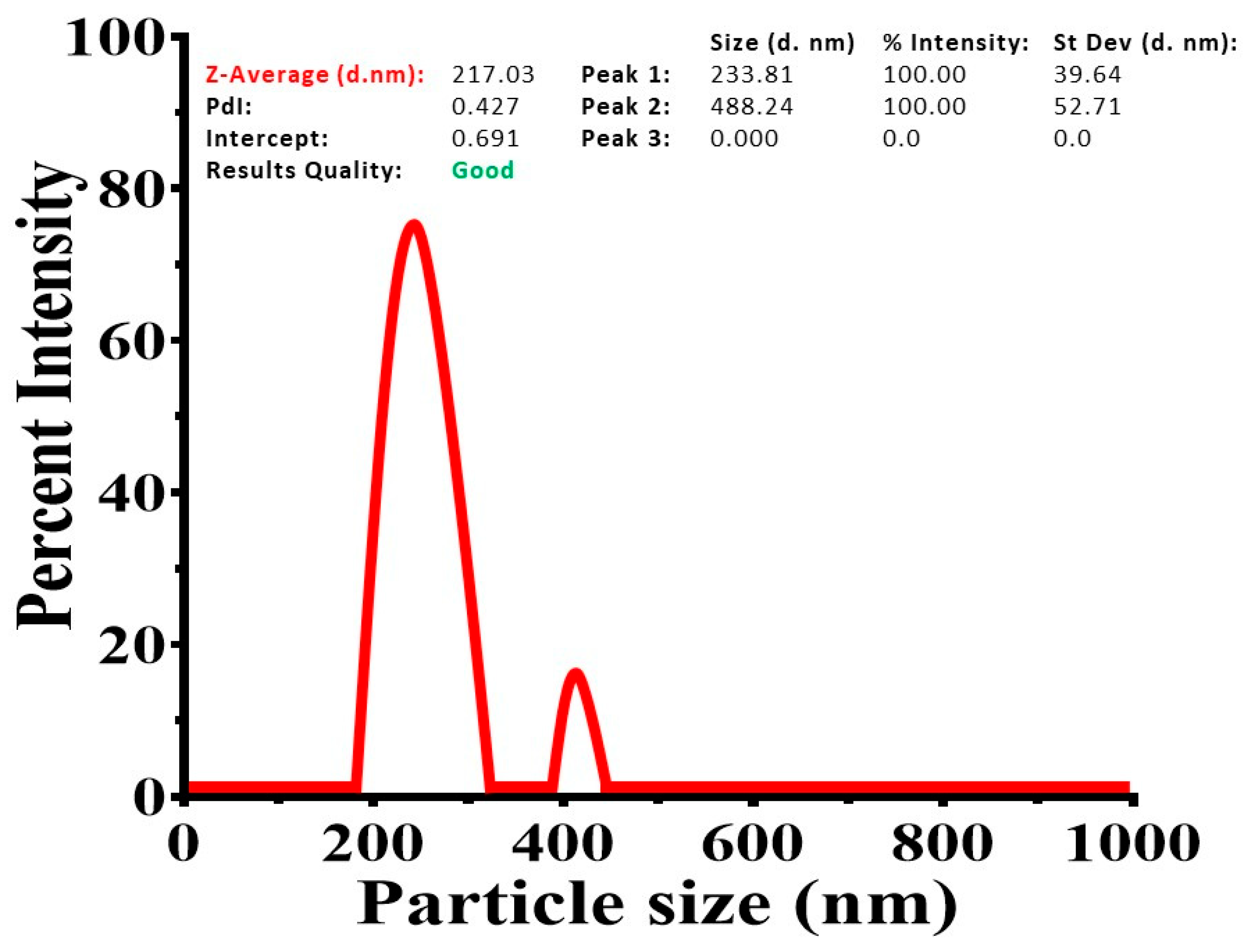
Figure 2.
The physiochemical characterization of HPSL was examined SEM analysis.
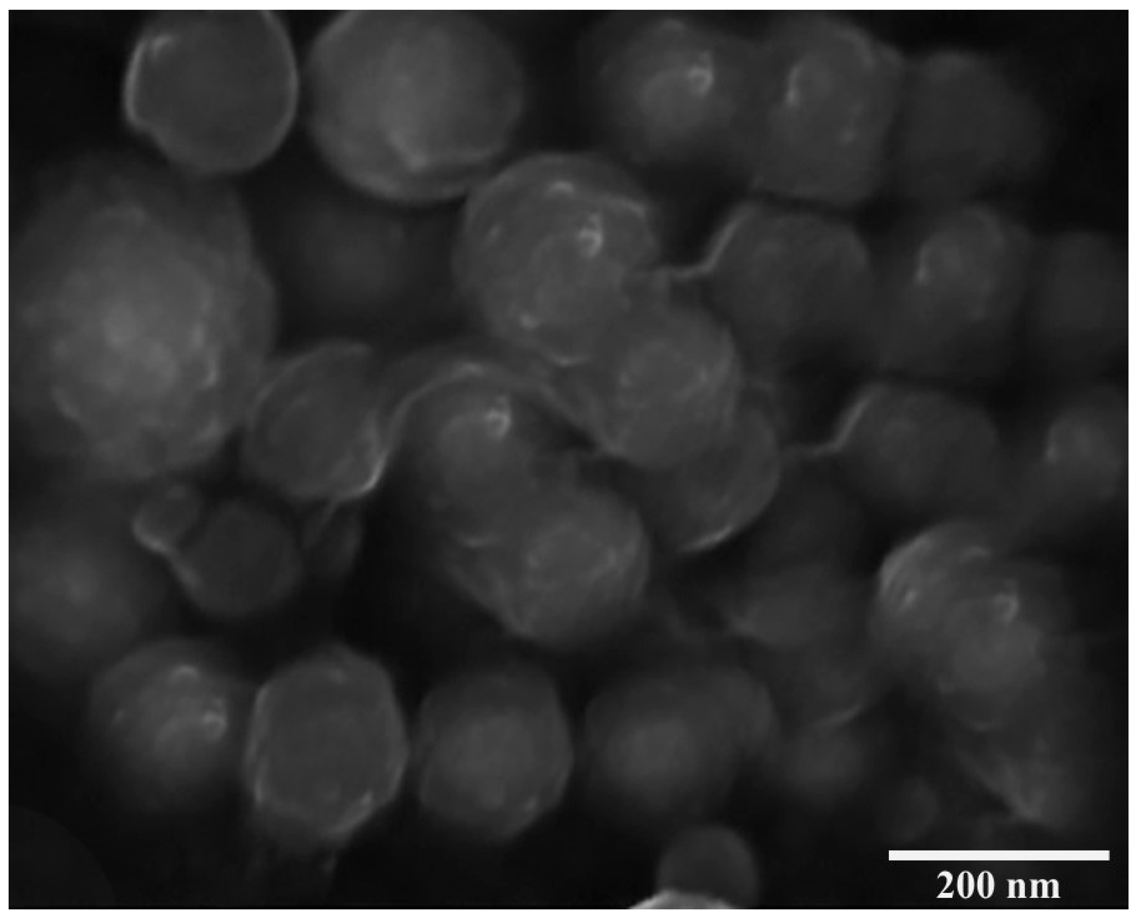
Figure 3.
Assessing its dissolution behavior under pH 7.4 conditions.
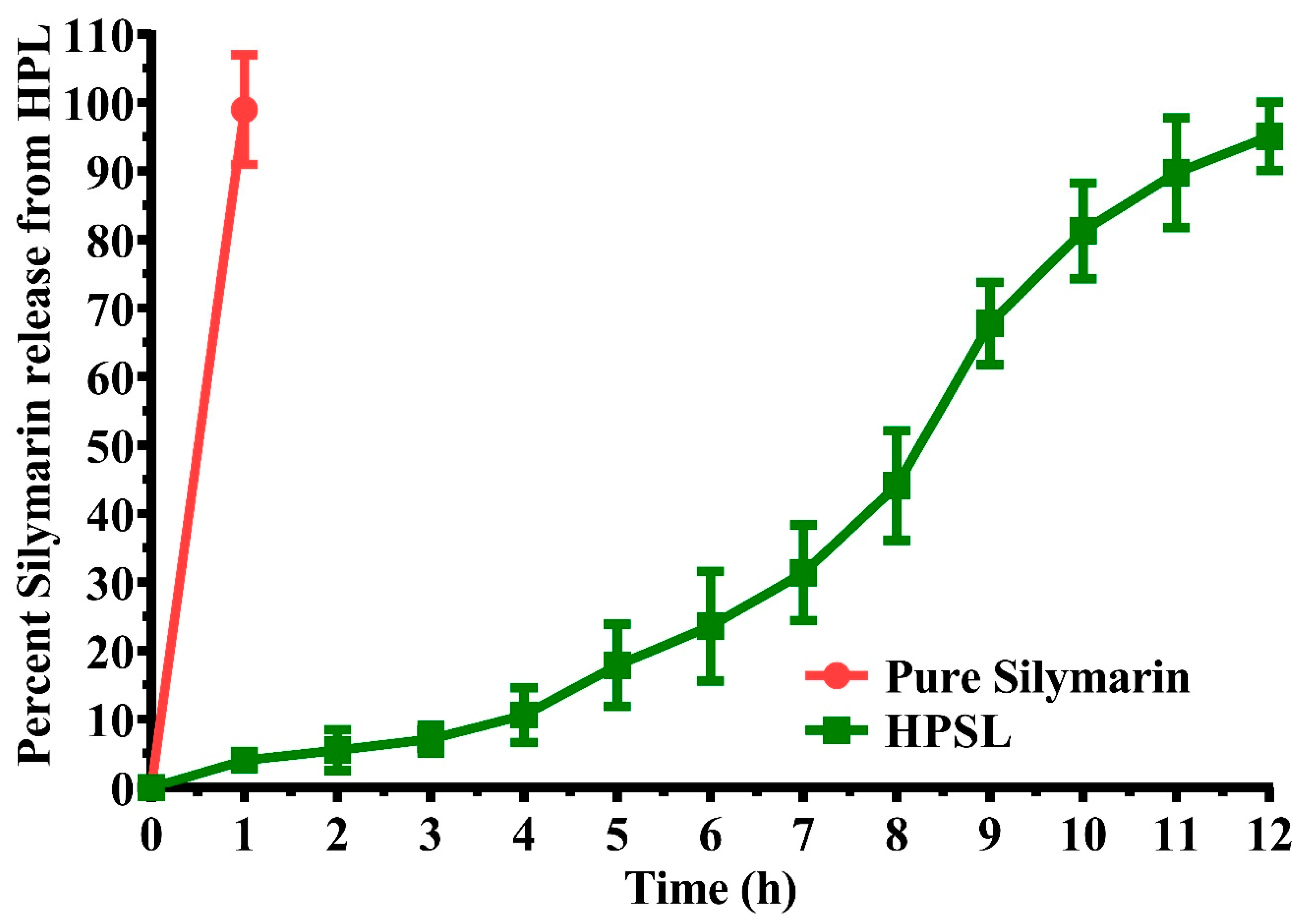
Figure 4.
Photomicrographs of histopathological studies (Hematoxylin & eosin staining, magnification power at 100x). (G-1) Normal Control (G-II) Amnesic Control (G-III) Standard Control, (G-IV) Silymarin Control, (G-V) Experimental Control Low Dose (HPSL nano-gel 20 mg.Kg) and (G-VI) Experimental Control High Dose (HPSL nano-gel 40 mg.Kg).
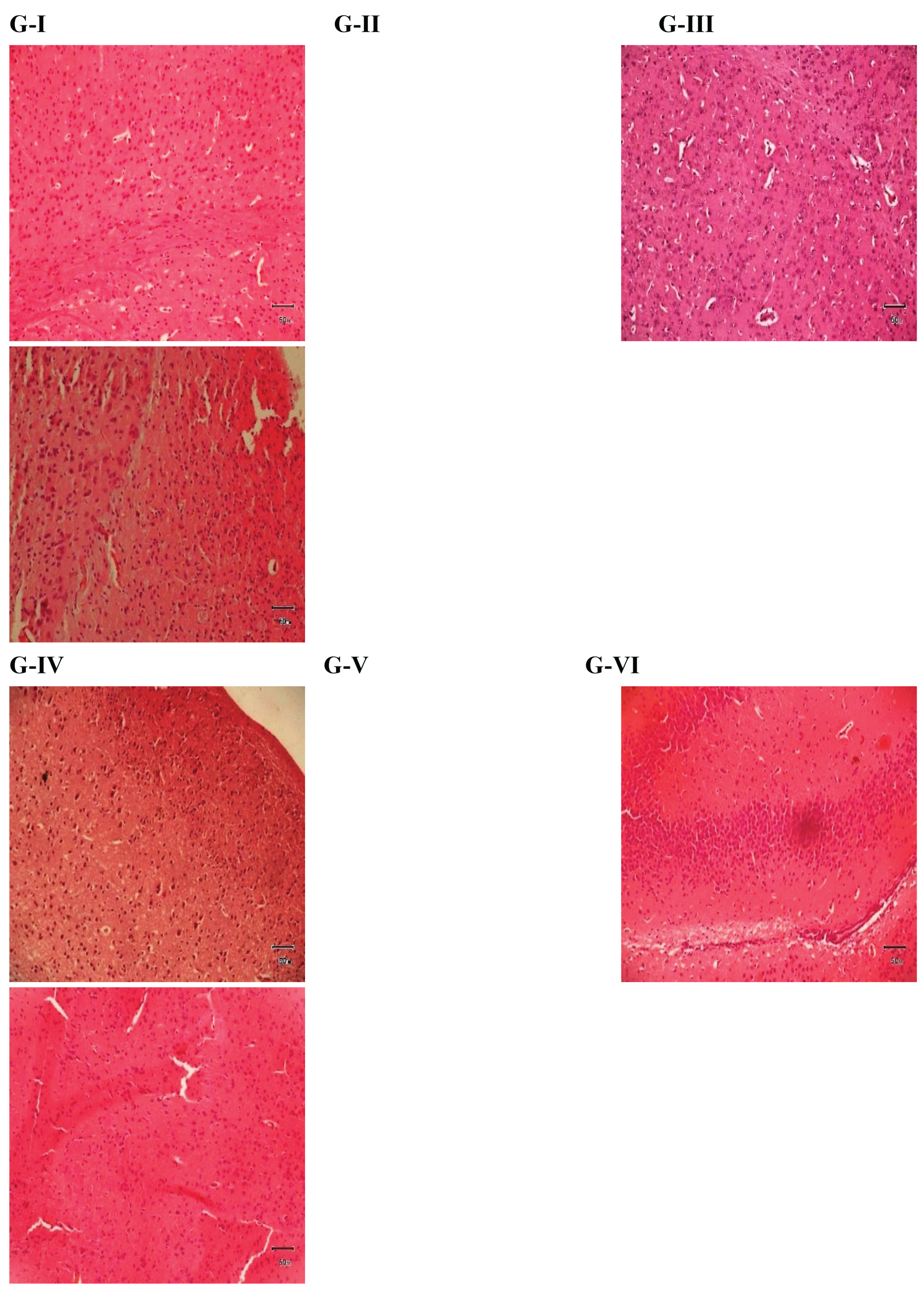

Table 1.
Study design along with treatment protocols for assessment behavioral and biochemical parameters.
Table 1.
Study design along with treatment protocols for assessment behavioral and biochemical parameters.
| Group Number | Group Details | Treatment | |
| Day 1-6 | Day 7th | ||
| G-I | Normal Control | 10 mL/Kg/p.o. Normal saline | 10 mL/Kg/p.o. Normal saline |
| G-II | Amnesic Control | 10 mL/Kg/p.o. 5 % CMC | 10 mg/Kg/p.o. Scopolamine |
| G-III | Standard Control | 200 mg/Kg/p.o. Piracetam | 200 mg/Kg/p.o. Piracetam |
| G-IV | Silymarin Control | 200 mg/Kg/p.o. Silymarin | 10 mg/Kg/p.o. Scopolamine + 200 mg/Kg/p.o. Silymarin |
| G-V | Experiment Control (low dose) | 20 mg/Kg/p.o. HPSL nano-gel | 10 mg/Kg/p.o. Scopolamine + 20 mg/Kg/p.o. HPSL neon-gel |
| G-VI | Experiment Control (high dose) | 40 mg/Kg/p.o. HPSL nano-gel | 10 mg/Kg/p.o. Scopolamine + 40 mg/Kg/p.o. HPSL nano-gel |
Note: HPSL = heparin–pullulan silymarin liposomal. The doses of scopolamine and piracetam were prepared by dissolving them in normal saline while silymarin was suspended in 5 % CMC. The behavioral studies were performed after two hours of dosing the animals on day 7th and also on day 8th. After that the animals were sacrificed and their brains were preserved for biochemical studies.
Table 2.
Calculation of histology activity index (HAI) of photomicrographs of mice brains after histopathology studies.
Table 2.
Calculation of histology activity index (HAI) of photomicrographs of mice brains after histopathology studies.
| Indication | Scoring | Grading | Histology Activity Index (HAI) = Sum of the scores |
| Necrosis |
0 | Absence | |
| 1 | Minor | ||
| 2 | Modest | ||
| 3 | Marked | ||
| Inflammation |
0 | Absence | |
| 1 | Minor | ||
| 2 | Modest | ||
| 3 | Marked | ||
| Fibrosis |
0 | Absence | |
| 1 | Minor | ||
| 2 | Modest | ||
| 3 | Marked | ||
| Edema |
0 | Absence | |
| 1 | Minor | ||
| 2 | Modest | ||
| 3 | Marked | ||
| Degeneration | 0 | Absence | |
| 1 | Minor | ||
| 2 | Modest | ||
| 3 | Marked |
Note: Greater the value of HAI more will be the damage.
Table 3.
Physical characteristics of NS at different concentrations of EC and PVA, with a constant Silymarin percentage of 1% w/v (mean ± SD, n = 3).
Table 3.
Physical characteristics of NS at different concentrations of EC and PVA, with a constant Silymarin percentage of 1% w/v (mean ± SD, n = 3).
| HPSL-1 | HPSL-2 | HPSL-3 | HPSL-4 | |
| Silymarin (% w/v) | 0.5 | 0.5 | 0.5 | 0.5 |
| Heparin (% w/v) | 1 | 1 | 2 | 2 |
| Pullulan (% w/v) | 1 | 2 | 1 | 2 |
| Particle size Mean ± SD (nm) | 189.73 ± 14.29 | 233.81 ± 39.64 | 406.07 ± 19.33 | 319.76 ± 21.57 |
| Loading efficiency | 27.55 ± 6.89 | 39.16 ± 7.45 | 30.91 ± 4.08 | 42.74 ± 6.02 |
| Entrapment efficiency | 67.22 ± 11.37 | 89.31 ± 7.74 | 73.53 ± 12.05 | 93.67 ± 3.21 |
| PDI Mean ± SD | 0.306 ± 0.025 | 0.427 ± 0.014 | 0.504 ± 0.061 | 0.596 ± 0.071 |
| Zeta potential Mean ± SD (mV) | -26.39 ± 4.14 | -29.88 ± 5.62 | -23.72 ± 7.64 | -25.69 ± 7.98 |
Table 4.
Effect of silymarin on transfer latencies and inflexion ratio in albino mice.
| Groups | Initial transfer latency L1 (Sec) | Retention transfer latency L2 (Sec) |
Inflexion Ratio I.R = L1 – L2 / L2 |
| G-I | 21.00 ± 0.73 | 17.83 ± 1.13 | 0.18 ± 0.04 |
| G-II | 70.50 ± 2.86π | 85.16 ± 1.95π | -0.16 ± 0.04µ |
| G-III | 44.83 ± 1.40α | 37.66 ± 1.25α | 0.20 ± 0.05γ |
| G-IV | 19.66 ± 0.88α | 16.50 ± 0.76α | 0.19 ± 0.03γ |
| G-V | 35.50 ± 1.60α | 25.66 ± 1.35α | 0.40 ± 0.11α |
| G-VI | 41.16 ± 1.24α | 24.33 ± 1.47α | 0.71 ± 0.10α |
Note: G-II was compared to G-I and superscripts π and ns present level of significance where, π = P ≤ 0.001, µ = P ≤ 0.05. All other groups (G-III to G-VI) were compared to G-II and superscripts α, β and γ present level of significance where α = P ≤ 0.001, β = P ≤ 0.01, γ = P ≤ 0.05, ns = P > 0.05.
Table 5.
Effect of silymarin on number of hole pokings by albino mice in hole board paradigm.
| Groups | Day-1 | Day-2 |
| No of Poking in 5 min | No of Poking in 5 min | |
| G-I | 45.83 ± 1.35 | 40.33 ± 1.28 |
| G-II | 22.33 ± 1.58π | 24.83 ± 1.55π |
| G-III | 40.66 ± 1.35α | 39.16 ± 1.66α |
| G-IV | 38.66 ± 1.83α | 34.66 ± 2.29β |
| G-V | 41.00 ± 1.93α | 39.16 ± 2.27α |
| G-VI | 50.16 ± 2.34α | 43.66 ± 1.76α |
Note: G-II was compared to G-I and superscripts π and ns present level of significance where, π = P ≤ 0.001, ns = P > 0.05. All other groups (G-III to G-VI) were compared to G-II and superscripts α, β and γ present level of significance where α = P ≤ 0.001, β = P ≤ 0.01, γ = P ≤ 0.05, ns = P > 0.05.
Table 6.
Effect of silymarin on time spent by albino mice in light and dark compartment models.
| Groups | Day-1 | Day-2 | ||
| Time Spent in Dark Compartment (Sec) | Time Spent in Light Compartment (Sec) | Time Spent in Dark Compartment (Sec) | Time Spent in Light Compartment (Sec) | |
| G-I | 247.66 ± 1.97 | 52.33 ± 1.97 | 248.50 ± 4.10 | 51.50 ± 4.10 |
| G-II | 112.33 ± 5.82π | 187.66 ± 5.82π | 089.83 ± 4.52π | 210.16 ± 4.51π |
| G-III | 233.66 ± 2.57α | 66.33 ± 2.57α | 239.66 ± 4.82α | 57.00 ± 3.67α |
| G-IV | 201.66 ± 3.06α | 98.33 ± 3.06α | 241.16 ± 3.60α | 58.83 ± 3.60α |
| G-V | 240.50 ± 3.08α | 59.50 ± 3.08α | 247.66 ± 2.57α | 52.33 ± 2.57α |
| G-VI | 254.66 ± 5.42α | 45.33 ± 5.42α | 258.66 ± 3.98α | 41.33 ± 3.98α |
Note: G-II was compared to G-I and superscripts π and ns present level of significance where, π = P ≤ 0.001, ns = P > 0.05. All other groups (G-III to G-VI) were compared to G-II and superscripts α, β and γ present level of significance where α = P ≤ 0.001, β = P ≤ 0.01, γ = P ≤ 0.05, ns = P > 0.05.
Table 7.
Biochemical estimation of cholinergic enzymes along with antioxidant markers in mice brain.
Table 7.
Biochemical estimation of cholinergic enzymes along with antioxidant markers in mice brain.
| Groups |
AChE (µmol/min/mg) |
ChAT (µmol/min/mg) |
CAT (U/mg of homogenate) |
GSH (U/mg of homogenate) |
MDA (nmol/h/g) |
SOD (U/mg of homogenate) |
| G-I | 04.52 ± 0.26 | 12.29 ± 1.01 | 01.48 ± 0.16 | 40.75 ± 0.89 | 01.62 ± 0.12 | 25.88 ± 0.93 |
| G-II | 09.28 ± 0.37π | 08.08 ± 0.82ns | 00.62 ± 0.03π | 18.12 ± 0.34π | 07.05 ± 0.35π | 08.60 ± 0.26π |
| G-III | 05.02 ± 0.27α | 15.85 ± 1.21α | 01.34 ± 0.07α | 41.34 ± 1.09α | 02.80 ± 0.20α | 23.52 ± 0.81α |
| G-IV | 06.90 ± 0.33α | 12.64 ± 0.73γ | 01.45 ± 0.14α | 42.02 ± 1.06α | 03.16 ± 0.19α | 22.69 ± 0.72α |
| G-V | 05.60 ± 0.33α | 13.68 ± 1.08β | 01.81 ± 0.06α | 47.92 ± 0.78α | 02.88 ± 0.22α | 24.67 ± 0.79α |
| G-VI | 04.09 ± 0.26α | 17.93 ± 1.14α | 01.84 ± 0.07α | 48.22 ± 0.96α | 02.71 ± 0.15α | 25.06 ± 0.97α |
Note: G-II was compared to G-I and superscripts π and ns present level of significance where, π = P ≤ 0.001, ns = P > 0.05. All other groups (G-III to G-VI) were compared to G-II and superscripts α, β and γ present level of significance where α = P ≤ 0.001, β = P ≤ 0.01, γ = P ≤ 0.05, ns = P > 0.05.
Table 8.
Histology activity index (HAI) calculated from histopathological slides.
| Groups | Relevant score of histopathological lesions | |||||
|---|---|---|---|---|---|---|
| Necrosis | Inflammation | Fibrosis | Edema | Degeneration | HAI | |
| G-I | 0 | 0 | 0 | 0 | 0 | 0 |
| G-II | 1 | 3 | 2 | 3 | 2 | 11 |
| G-III | 0 | 1 | 1 | 2 | 0 | 4 |
| G-IV | 1 | 2 | 1 | 2 | 1 | 7 |
| G-V | 0 | 2 | 0 | 2 | 1 | 5 |
| G-VI | 1 | 1 | 0 | 1 | 1 | 4 |
Disclaimer/Publisher’s Note: The statements, opinions and data contained in all publications are solely those of the individual author(s) and contributor(s) and not of MDPI and/or the editor(s). MDPI and/or the editor(s) disclaim responsibility for any injury to people or property resulting from any ideas, methods, instructions or products referred to in the content. |
© 2026 by the authors. Licensee MDPI, Basel, Switzerland. This article is an open access article distributed under the terms and conditions of the Creative Commons Attribution (CC BY) license.
Copyright: This open access article is published under a Creative Commons CC BY 4.0 license, which permit the free download, distribution, and reuse, provided that the author and preprint are cited in any reuse.


